


Abstract
This hypothesis originated from the first author’s personal experience in adult neurology, in which selected refractory cranial or postherpetic neuralgias occasionally appeared to improve rapidly after off-label misoprostol. The original records are unavailable; these physician recollections are not efficacy data and serve only as the hypothesis generator. Independent published precedents are limited to small uncontrolled reports in multiple-sclerosis-associated trigeminal neuralgia. A further recollection was that perceived benefit often waned after several weeks at conventional daily exposure, whereas lower or intermittent exposure sometimes appeared to remain useful for longer; this observation is likewise unverifiable and does not define a therapeutic dose. Misoprostol is rapidly converted to misoprostol acid (plasma half-life of 20–40 min). We ask whether a brief, reversible exposure could produce pharmacodynamic persistence, defined here as a reproducible pain-relevant effect that outlasts measurable drug exposure, through EP-receptor trafficking, compartmentalized GPCR signaling, or downstream cellular changes. The model also predicts that the concentration–response relationship may be non-monotonic: increasing or repeated exposure could attenuate or reverse a favorable effect by recruiting opposing EP pathways or adaptive receptor/cellular responses. No direct evidence shows that misoprostol produces such persistence in neural tissue, and PGE2/EP signaling can also be pronociceptive. Receptor-resolved concentration–response and pulse-washout experiments must therefore separately demonstrate receptor signaling, persistent cellular change, altered neuronal excitability, and ultimately blinded analgesic benefit. Neuropathic corneal pain is considered only a secondary mechanistic model. No evidence-based dose can currently be recommended.
1. Clinical Origin and Rationale
Trigeminal neuralgia (TN) can be profoundly disabling, and carbamazepine or oxcarbazepine remains first-line therapy; intolerance, incomplete response, secondary disease, and recurrence after intervention leave a clinically important refractory group [1]. Although TN is uncommon, its epidemiological burden is not trivial: a recent global synthesis estimated a pooled incidence of 25.33 cases per 100,000 person-years while emphasizing wide between-study heterogeneity related in part to case definition and ascertainment [2]. At the molecular level, chronic neuropathic pain is not simply prolonged acute nociception: peripheral excitability, synaptic plasticity, glial activation, immune-cell recruitment, and altered signaling at the injured nerve can sustain a pathological state after the initiating lesion has changed or resolved [3,4,5].
The present hypothesis did not begin with a literature search. It arose from the personal clinical experience of the first author, Ronald B. Bukowy (R.B.B.), during several decades of adult neurological practice. That origin is retained because it explains the question being asked, but the recollections are deliberately separated from the evidentiary basis of the article. The verifiable published literature is therefore summarized first, before the historical observation is described in detail.
Independent of R.B.B.’s recollections, a small published clinical literature provides the only direct human precedent. In 1995, Reder and Arnason described seven patients with multiple-sclerosis-associated trigeminal neuralgia refractory to conventional treatment; six were reported to improve with misoprostol [6]. An open prospective study subsequently enrolled patients with the same secondary neuralgia: 18 completed treatment with 600 μg/day and 14 showed reductions greater than 50% in both attack frequency and pain intensity, beginning approximately five days after treatment onset [7]. A 2012 letter reported three additional therapy-resistant cases [8]. These reports are summarized structurally in Section 6. None was a modern randomized, placebo-controlled efficacy trial, and none provides evidence for a 60–72 h post-dose analgesic state.
R.B.B. recalls prescribing off-label misoprostol to patients he personally treated for refractory trigeminal, glossopharyngeal, lesion-associated cranial, or postherpetic neuralgic pain. Some appeared to improve rapidly; others did not respond, developed gastrointestinal intolerance, or experienced waning benefit. In his recollection, sustained treatment at approximately conventional daily doses, often around 600 μg/day, frequently lost perceived efficacy after roughly 4–6 weeks, whereas lower or intermittent exposure sometimes appeared to retain benefit for substantially longer. One memorable patient reportedly used 100 μg only when the first pain ‘twinge’ returned, commonly after about 60–72 h, and this pattern was recalled as remaining useful over a prolonged period of follow-up. The original records are no longer available, the observations arose in routine care rather than a research protocol, and no denominator, standardized endpoint, or patient-level reconstruction is possible. Because redosing was triggered by pain recurrence, the interval is intrinsically coupled to the dosing decision and is not an independent measure of drug effect. Likewise, the remembered contrast between conventional and lower exposure cannot establish a dose–response relationship, dose optimum, or duration of efficacy. These physician recollections are therefore not efficacy data, a case report, or a retrospective case series; they function solely as the clinical heuristic that generated two falsifiable questions: whether a brief exposure can produce a post-exposure effect, and whether the relevant exposure–response relationship is non-monotonic rather than simply dose-proportional.
This hypothesis article tests that clinical question against current molecular pharmacology. We distinguish throughout among (i) evidence directly involving misoprostol or misoprostol acid, (ii) indirect evidence from PGE2 or other prostanoid ligands, and (iii) general GPCR mechanisms derived from unrelated receptors. The central question is not whether the historical observation was ‘true’, but whether a reproducible post-exposure effect can be demonstrated prospectively and linked, step by step, to EP-receptor engagement, a persistent cellular alteration, altered neuronal excitability, and finally analgesia.
2. Hypothesis Development and Literature Approach
To develop and challenge the hypothesis, PubMed/MEDLINE and the reference lists of retrieved publications were examined from database inception through 9 September 2026. Search combinations paired misoprostol or misoprostol acid with trigeminal neuralgia, postherpetic neuralgia, neuropathic pain, EP1, EP2, EP3, EP4, receptor binding, receptor affinity, receptor trafficking, internalization, GPCR endosomal signaling, mast cells, macrophages, microglia, astrocytes, pharmacokinetics, tissue exposure, corneal pain, ocular delivery, safety, and drug availability. Searches were iteratively refined when a retrieved study identified a receptor subtype, pharmacokinetic issue, or competing explanation requiring targeted follow-up.
Priority was given to primary clinical reports, official prescribing information, receptor-binding and structural studies, mechanistic human-cell work, and in vivo models with interpretable receptor biology. English-language publications were prioritized; human, animal, and in vitro studies were retained when they directly informed the clinical signal, receptor mechanism, temporal hypothesis, competing explanations, ocular translation, or safety. Titles and abstracts were screened for mechanistic relevance, and full text was examined when needed to verify a specific claim. Tangential prostanoid literature without a clear bearing on these questions was excluded. No formal Embase, Scopus, or Cochrane search was performed, which is an explicit limitation. This targeted hypothesis-development process was not designed to estimate treatment effect and should not be interpreted as a systematic or comprehensive review.
Generative artificial intelligence (ChatGPT, GPT-5.6 Sol, OpenAI, San Francisco, CA, USA) was used to support literature organization, manuscript restructuring, language editing, and preparation of conceptual schematic figures. It was not treated as an evidentiary source and did not determine scientific inclusion or exclusion decisions, interpretation of individual studies, or the conclusions of the article. The cited literature and the claims attributed to it were independently checked by the authors. Additional disclosure is provided in the Acknowledgments.
3. Molecular Pharmacology of Misoprostol and EP Receptors
Misoprostol is the orally administered methyl-ester parent compound and is rapidly de-esterified to misoprostol acid, the principal circulating pharmacologically active species [9,10,11]. Misoprostol/misoprostol acid should not be used interchangeably with PGE1 (including alprostadil) or with endogenous PGE2; these molecules differ in receptor pharmacology, exposure, and biological context. In healthy adults, plasma misoprostol acid peaks within minutes and has a terminal half-life of approximately 20–40 min [10,11]. The current label reports marked inter- and intra-study variability, dose-proportional mean exposure over 200–400 μg, and no accumulation during repeated dosing [10]. Importantly, plasma half-life is not equivalent to tissue residence time. Neural, ganglionic, or corneal tissue concentrations after oral dosing have not been established, and the label describes further metabolism to prostaglandin-F analogs without evidence that a long-lived metabolite explains neuralgic pain. Thus, prolonged tissue exposure or an unrecognized active compartment cannot be excluded merely from plasma clearance; they must be measured directly.
3.1. EP1–EP4 Signaling Is Not a Single Pathway
Prostaglandin E2 acts through four E-type prostanoid receptors (EP1–EP4), all members of the GPCR superfamily [12,13]. EP1 is mainly linked to calcium mobilization; EP2 and EP4 usually stimulate adenylyl cyclase and cyclic adenosine monophosphate (cAMP); common EP3 isoforms inhibit adenylyl cyclase through Gi, although EP3 splice variants diversify coupling [12,13]. These canonical assignments are useful but incomplete. Receptor abundance, ligand concentration, cell lineage, inflammatory state, receptor heteromers or splice variants, and downstream signaling competence can change the net biological output.
Misoprostol acid is not a clean subtype-selective ligand. In recombinant human receptors, reported binding affinities are approximately 7.9 nM at EP3, 23 nM at EP4, 34 nM at EP2, and greater than 10 μM at EP1 [14]. This profile makes substantial direct EP1 engagement at low exposure less plausible than EP2/EP3/EP4 engagement, although binding affinity is not a functional EC50 and cannot be translated directly into tissue occupancy. Structural analysis of misoprostol acid bound to EP3 shows conventional non-covalent ligand recognition [15], while a ligand-bound EP4-Gs cryo-EM structure provides subtype-specific context for orthosteric prostanoid recognition and coupling [16]. These data are direct receptor-level evidence; they do not establish prolonged EP signaling after misoprostol clearance.
Whether the recalled 100 μg oral dose could reach a pain-relevant receptor concentration is unknown. The prescribing information characterizes pharmacokinetics primarily over the 200–400 μg range and reports low pg/mL plasma concentrations with substantial variability [10]; therefore, exposure at 100 μg should not be inferred by simple proportional extrapolation. Conversely, recombinant Ki values [14] cannot be equated with free drug concentration in a nerve, ganglion, or ocular compartment. A pharmacologically credible experimental program should first measure unbound misoprostol-acid exposure and then bracket the EP2/EP3/EP4 affinity range (roughly low-nanomolar to low-hundreds-of-nanomolar concentrations) while clearly separating clinically plausible from supraphysiological conditions.
3.2. Desensitization, Trafficking, and Signaling Location
EP2 and EP4 both couple to Gs, yet they differ in regulation. EP4 undergoes rapid agonist-induced desensitization and internalization, whereas EP2 is relatively resistant to short-term desensitization under comparable experimental conditions [17,18]. Human EP4 internalization involves beta-arrestin- and dynamin-dependent mechanisms [18]. In sensory neurons, however, prostaglandin E2 can increase EP4 availability at the cell surface and enhance subsequent responsiveness [19]. The same receptor family can therefore support attenuation or sensitization depending on cell type and sequence of stimulation.
More broadly, GPCR signaling is no longer understood as a plasma-membrane-only event. Internalized receptors can continue or reshape signaling from endosomes and other intracellular compartments [20,21]. Recent biosensor work further supports temporally structured G-protein regulation from endosomal GPCRs [22]. These studies do not demonstrate persistent EP signaling after misoprostol in neuralgia; they provide a molecular vocabulary for testing whether receptor location and trafficking could separate a brief ligand pulse from a longer downstream response.
3.3. Pain Biology Predicts Opposing EP Effects
The pain literature warns against assigning fixed analgesic or pronociceptive labels to individual EP pathways. Selective EP3 activation was antinociceptive in one inflammatory model [23]. By contrast, Minami et al. found that PGE2-induced allodynia was abolished in EP1-deficient mice but preserved in EP3-deficient mice, identifying EP1 rather than EP3 as necessary in that experimental paradigm [24]. Misoprostol itself has shown a non-monotonic effect on mast-cell histamine release [25]. EP2 and EP4 signal through Gs/cAMP; EP4 can support sensory-neuron sensitization, while EP2/EP4 signaling can suppress activation in some immune-cell contexts [12,13,19,26,27,28]. The hypothesis therefore does not assume that simultaneous activation of several EP subtypes is inherently analgesic. It predicts a favorable net effect only in a biological state in which immune/stromal inhibitory effects and/or EP3-linked effects outweigh sensory-neuron sensitization; the opposite balance predicts nonresponse or worsening. No direct experiment has yet shown that misoprostol creates such a favorable balance in human neuralgia.
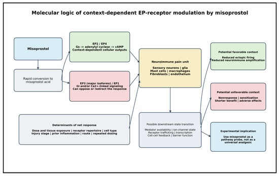
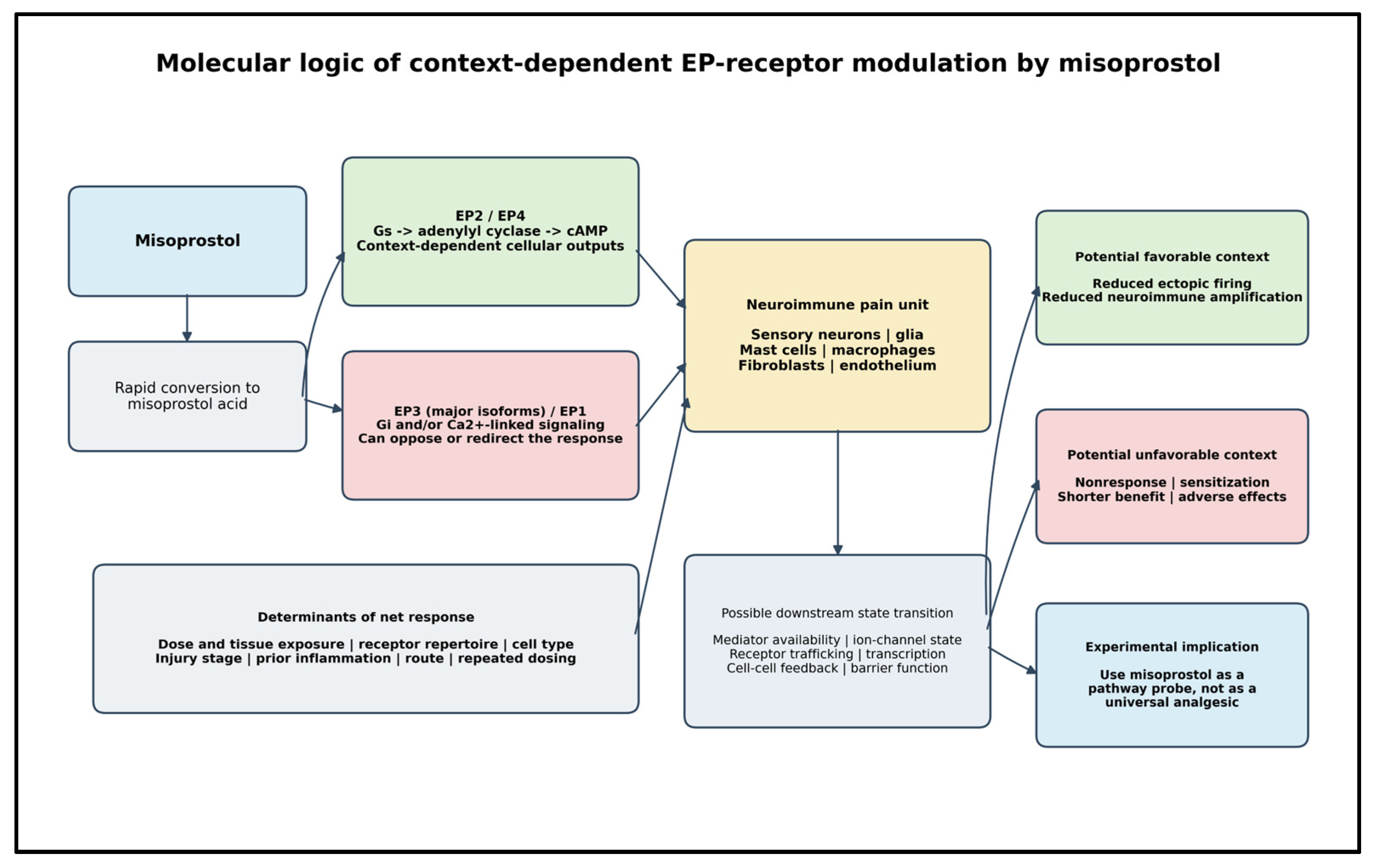
For the present hypothesis, it is therefore preferable not to reduce prostanoid biology to a simple PGE1 ‘anti-inflammatory’ versus PGE2 ‘pro-inflammatory’ dichotomy. PGE2 is a well-established inflammatory and pronociceptive mediator in several settings, yet EP2/EP4 signaling can also restrain activation in selected immune-cell contexts. Misoprostol is a PGE1 analog, but its biological effect is determined by the EP subtypes actually engaged, ligand concentration, exposure pattern, and cellular state. This context dependence provides a mechanistic basis for asking whether low, repeated, and high exposure could produce qualitatively different outcomes. The receptor-specific features relevant to this hypothesis are summarized in Table 1, while the proposed context-dependent balance among receptor, immune-cell, and nociceptor effects is shown schematically in Figure 1.
Table 1.
Molecular features of EP receptors relevant to the misoprostol hypothesis.
Figure 1.
Molecular logic of context-dependent EP-receptor modulation by misoprostol. Rapid conversion to misoprostol acid and direct misoprostol receptor-binding/structural evidence are supported by [9,10,11,14,15]; general EP-receptor pharmacology and EP4 structural context are provided by [12,13,16]. The downstream receptor-regulation, immune-cell, and nociceptor arrows synthesize mainly indirect evidence from PGE2, selective prostanoid ligands, or broader GPCR biology [17,18,19,20,21,22,23,24,26,27,28] and should be interpreted as hypotheses rather than demonstrated consequences of misoprostol in neuralgia. The figure therefore depicts possible directions of effect, not established causality or relative effect size. In the schematic, blue/gray boxes denote exposure, determinants, downstream-state, or experimental elements; green and red boxes denote potentially favorable and unfavorable pathways/outcomes, respectively; yellow denotes the integrated neuroimmune pain unit. Arrows indicate proposed directions of influence, not established causal links.
4. A Neuroimmune Pain Unit as the Candidate Substrate
Neuropathic pain emerges from interacting compartments rather than from a single cell type. Peripheral sensory neurons undergo changes in ion-channel expression, membrane excitability, and intracellular signaling; spinal microglia and astrocytes participate in central sensitization; immune cells accumulate at injured nerves; and tissue-resident stromal and vascular cells shape mediator gradients, barrier properties, and repair [3,4,5]. This cellular diversity is central to the hypothesis because the net effect of a nonselective prostanoid agonist may differ across compartments.
4.1. Mast Cells and Macrophages
Mast cells are attractive candidates because Shah and Pearce found that misoprostol itself produced a strongly non-monotonic response in isolated human mast cells [25]. Immunologically induced histamine release from human colonic mast cells was inhibited at very low concentrations, with bell-shaped dose–inhibition curves, whereas very high concentrations (≥5 μM) induced histamine release. Human mast cells express several EP subtypes with different effects on activation [26]; EP2 stimulation inhibits human lung mast-cell degranulation [27], and EP2/EP4 signaling suppresses hyperosmolar activation in several human mast-cell preparations [28]. Mast cells also lie close to sensory nerves and vessels and can release histamine, proteases, cytokines, growth factors, and lipid mediators that participate in experimental neuropathic pain [29]. The Shah–Pearce result does not prove that the remembered loss of clinical efficacy at sustained higher doses shares the same mechanism. It does, however, establish in a human inflammatory effector cell that more misoprostol need not produce more inhibition and that sufficiently high exposure can reverse the direction of the response. This finding makes a non-monotonic, concentration- and context-dependent exposure–response relationship a testable component of the present hypothesis rather than a purely clinical speculation.
Microglia provide a central nervous system example of immune cells that can alter synaptic transmission after nerve injury [30]. In the periphery, macrophages are equally plausible contributors: nerve injury recruits and reprograms macrophage populations that communicate with nociceptors, Schwann cells, and vascular elements [31]. A recent human study of painful Morton’s neuroma demonstrated persistent intraneural inflammation, including macrophage-associated molecular signatures that correlated with pain phenotypes [32]. The hypothesis therefore does not require a single ‘misoprostol-sensitive immune cell’. It requires only that transient EP-receptor engagement in one or more interacting compartments can weaken a reinforcing loop.
4.2. What Is Meant by a Self-Sustaining Neuroimmune Loop?
Astrocytes deserve explicit consideration in an EP-sensitive network because EP2 stimulation can evoke cAMP-dependent calcium responses in cultured astrocytes [33]. The phrase ‘self-sustaining neuroimmune loop’ is used here as an operational model, not as proof of an autonomous inflammatory circuit. Injured or sensitized sensory fibers can activate immune and stromal cells; their mediators can lower neuronal firing thresholds; sensory fibers can release signals that feedback on surrounding cells; and glia can alter excitatory and inhibitory processing [3,4,5]. These are established elements of neuropathic-pain biology, but their interruption by misoprostol has not been demonstrated.
A prostanoid perturbation can nevertheless outlast the initiating exposure in experimental pain biology. In the study by St-Jacques and Ma, a stabilized PGE2 analog produced nociceptor sensitization/allodynia lasting up to approximately 24 h, associated with increased EP4 synthesis and axonal trafficking [34]. This is indirect and directionally cautionary evidence: the persistent state was pronociceptive, not analgesic, and it still falls short of the recalled 60–72 h interval. No cited experiment demonstrates a 60–72 h analgesic state after misoprostol. Any such duration would require a longer-lived downstream cellular or network alteration and remains a prediction to be tested rather than an explanation already supported.
5. Pharmacodynamic Persistence: Distinguishing Receptor, Cellular, and Clinical Time Scales
5.1. Definition and Temporal Prediction
In this article, pharmacodynamic persistence means a reproducible pain-relevant functional effect that continues after misoprostol-acid exposure has fallen below measurable or pharmacologically relevant levels. The recurrence-triggered pattern described in Section 1 suggests a time scale to test, not a demonstrated drug effect and not a recommended regimen. It should not be conflated with the historical 600 μg/day regimen used in the prospective neuralgia study [7] or with the approved 800 μg/day gastroprotective regimen (200 μg four times daily) [10]. No evidence-based dose or dosing interval for neuralgic pain can currently be recommended; future dosing must be established by formal pharmacokinetic, pharmacodynamic, safety, and dose-ranging studies.
5.2. Candidate Mechanisms and Their Known Time Scales
The proposed mechanisms occupy different time scales and should not be treated as equivalent. EP4 internalization/desensitization is rapid, occurring on a minute’s scale in experimental systems [17,18]. In an unrelated GPCR system, parathyroid-hormone-receptor endocytosis sustained cAMP production for tens of minutes after ligand washout [35], establishing a principle of post-endocytic signaling but not a days-long effect. Contemporary biosensor studies likewise demonstrate endosomal G-protein signaling, but do not show multi-day persistence [20,21,22]. These observations make compartmentalized signaling experimentally plausible while leaving a large temporal gap to 60–72 h.
Longer persistence would therefore have to occur downstream. Transcriptional regulation, receptor synthesis/trafficking, ion-channel phosphorylation or abundance, mediator replenishment, immune-cell phenotype, and multicellular feedback can outlast an initiating receptor pulse. The pain-relevant PGE2-analog model cited above reached approximately 24 h and was pronociceptive [34]. A cellular alteration lasting one day is not evidence for 60–72 h of analgesia, but it identifies a measurable intermediate level. The hypothesis should be evaluated sequentially: persistent receptor signaling (minutes to perhaps hours), persistent cellular alteration (hours to days), persistent change in neuronal excitability or pain behavior, and finally sustained clinical analgesia. Demonstrating an upstream level does not establish any downstream level.
5.3. Current Evidence Does Not Support Irreversible EP-Receptor Binding
The hypothesis initially arose because a long symptom-free interval seemed difficult to reconcile with a short-acting drug. Irreversible EP-receptor binding was one intuitive possibility. Current evidence does not support that explanation: misoprostol-acid affinity has been measured under equilibrium binding conditions, the product label describes receptor binding as saturable and reversible, and the EP3 structure shows ordinary non-covalent ligand recognition [10,14,15]. These observations argue against invoking days-long irreversible receptor occupancy, but they do not exclude unmeasured tissue retention, compartment-specific exposure, or an unrecognized active metabolite. Those alternatives require direct analytical pharmacokinetic testing.
5.4. Competing Explanations Are Part of the Model
Several non-pharmacological explanations remain at least as plausible as a persistent molecular state. Neuralgic pain is episodic and may vary substantially over hours or days; spontaneous fluctuation and regression to the mean are especially relevant when treatment is started during a severe exacerbation. Expectancy/placebo effects, changes in concomitant treatment or behavior, selective recall of apparent responders, and retrospective timing error can also create an apparently treatment-linked pattern. The recalled 60–72 h interval is particularly vulnerable to circularity because redosing was triggered by pain recurrence: the decision rule itself ensures that recurrence precedes each dose and that any subsequent improvement is temporally associated with dosing. The interval therefore cannot distinguish pharmacodynamic persistence from the natural course of the disorder.
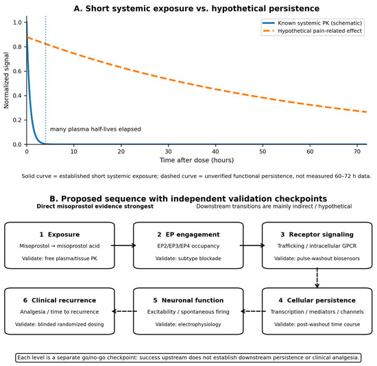
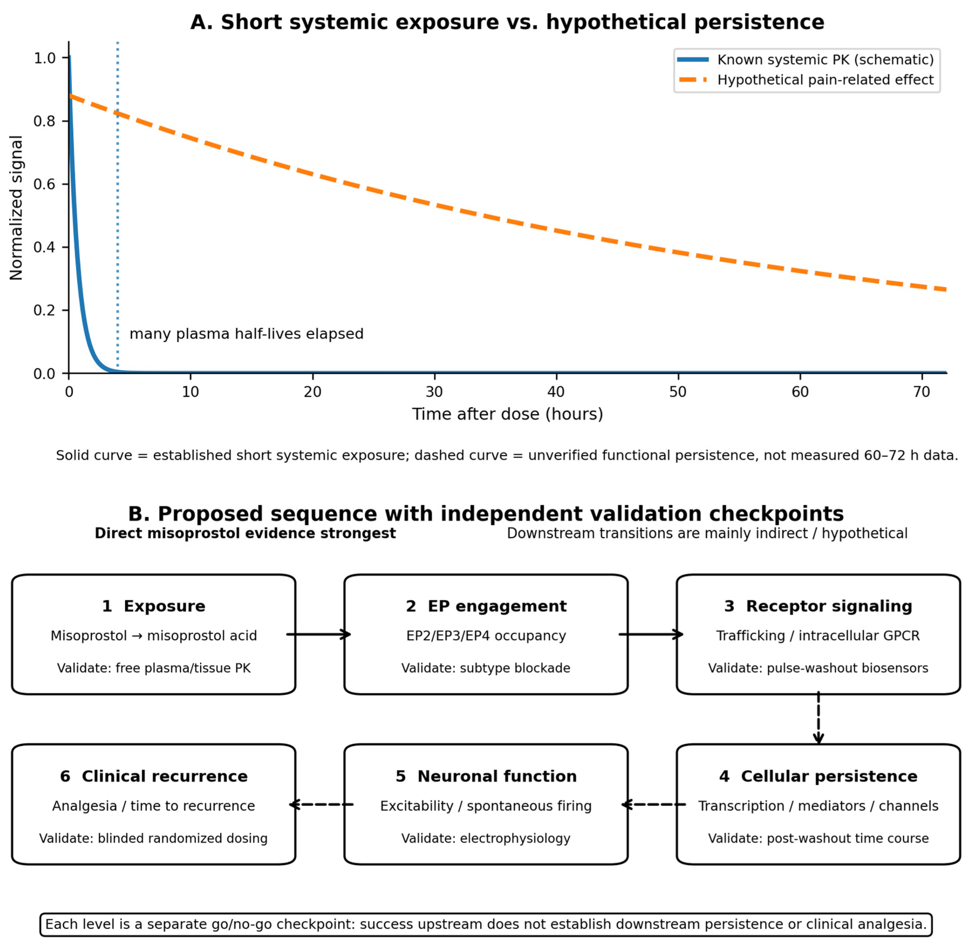
Substantial improvement under blinded placebo conditions further illustrates the problem. In a 2026 randomized, double-blind trial of intravenous fosphenytoin for acute trigeminal neuralgia, 40% of placebo-assigned participants achieved at least 50% improvement in pain intensity at 120 min [36]. That trial does not model multi-day spontaneous remission or the historical misoprostol context, but it shows that marked short-term change can occur without the active intervention. A prospective test must therefore establish baseline attack variability, blind treatment allocation, record attacks continuously, prespecify redosing and rescue criteria, measure exposure and pain on the same time axis, and demonstrate that any post-washout signal exceeds placebo and natural fluctuation. Until then, the 60–72 h recollection is a hypothesis-generating time scale, not evidence of pharmacodynamic persistence. These distinct temporal levels and validation steps are summarized in Figure 2.
Figure 2.
Separating pharmacokinetic exposure, mechanistic persistence, and clinical analgesia. Panel (A) distinguishes the established short systemic exposure of misoprostol acid (solid schematic curve) from a hypothetical pain-related effect (dashed curve). The dashed curve is conceptual and is not a measured 60–72 h response. Panel (B) separates six validation levels: exposure, EP-receptor engagement, receptor trafficking/compartmentalized signaling, persistent cellular alteration, altered neuronal excitability, and clinical analgesia/time to recurrence. Exposure and direct misoprostol receptor-binding/structural evidence is supported by [9,10,11,14,15]. Receptor regulation and downstream persistence hypotheses rely mainly on general EP-receptor pharmacology, PGE2/other prostanoid data, or broader GPCR evidence [12,13,16,17,18,19,20,21,22,23,24,26,27,28,29,30,31,32,33,34,35], with direct misoprostol cellular evidence limited to the context-dependent mast-cell effects in [25]. Each level must be tested independently; a positive result at one level does not establish the next.
6. Evidence for and Against the Hypothesis in Pain
6.1. Published Neuralgia Signal
The only direct clinical evidence for misoprostol in neuralgia concerns multiple-sclerosis-associated trigeminal neuralgia [6,7,8]. The three reports differ in design, sample size, regimen, outcomes, and major limitations. Together they comprise 28 reported patients, but they do not form an efficacy dataset: the initial report was a small case series, the 2003 study was open and uncontrolled, and the 2012 publication was a three-case letter. No contemporary randomized trial has confirmed the signal, no study linked response to plasma or tissue exposure, and none tested multi-day pharmacodynamic persistence after a single dose.
6.2. R.B.B.’s Clinical Experience as the Hypothesis Generator
As detailed once in Section 1, R.B.B.’s own clinical experience generated the question addressed here. It is kept separate from the published evidence and is not used to increase the apparent weight of clinical evidence. The remembered heterogeneity—apparent responses, nonresponses, intolerance, waning benefit, and an apparent tendency for benefit to disappear after several weeks at conventional daily exposure while persisting longer in some lower or intermittently exposed patients—supports only the need for prospective phenotyping and explicit testing of exposure pattern. No response rate, dose optimum, causal inference, or biomarker can be derived from these recollections. Their value is to generate a prespecified prediction: if the clinical impression had a pharmacological basis, concentration–response and repeated-exposure experiments might reveal an intermediate favorable window rather than a monotonic increase in effect with dose.
6.3. In Vivo Neural and Vascular Support and Its Limits
In a murine cerebral ischemia model, misoprostol reduced infarct injury, with the reported mechanism consistent with EP2 and/or EP4 rather than EP3 signaling [37]. The result demonstrates that systemically administered misoprostol can influence neural and endothelial biology in vivo. It does not establish analgesia, and the protection occurred without a detectable regional cerebral blood-flow difference. A vascular contribution to pain modulation remains possible, but misoprostol should not be described as a general vasodilator. PGE2 can generate biphasic vascular responses through competing EP pathways [38]; misoprostol altered mucosal blood flow in an experimental gastrointestinal model [39], whereas a controlled human study found no acute improvement in digital circulation after a 400 μg oral dose [40].
These distinctions matter when discussing the eye or cranial pain. Prostaglandin E1, including alprostadil, has vascular, endothelial, antiplatelet, and rheological properties relevant to ischemic retinal disease [41]. Misoprostol is chemically related but differs in receptor engagement, pharmacokinetics, route, and tissue exposure. Misoprostol and PGE1/alprostadil should therefore not be treated as pharmacologically interchangeable. The PGE1 literature supports interest in prostanoid biology; it cannot be transferred directly to misoprostol.
6.4. Contrary Prostanoid Pain Evidence
Several findings directly challenge a simple analgesic interpretation and are central to the hypothesis. PGE2 is a well-established pain mediator, and cyclooxygenase inhibition is clinically analgesic in many inflammatory states. Experimentally, PGE2/EP4 signaling can increase EP4 surface availability and sensitize primary sensory neurons [19], PGE2-induced allodynia can depend on EP1 [24], and intravenous PGE2 provokes migraine-like attacks in susceptible patients [42]. Conversely, selective EP3 activation can be antinociceptive in inflamed tissue [23], while EP2/EP4 can suppress activation in some immune-cell contexts [26,27,28]. Misoprostol therefore cannot be expected to relieve pain simply because it is a prostaglandin analog. A favorable response, if real, should be restricted to phenotypes in which the net cellular balance favors inhibitory immune/stromal or EP3-linked effects over nociceptor sensitization. Multiple-sclerosis-associated trigeminal neuralgia is the only phenotype with direct published precedent [6,7,8]; chronic post-injury phenotypes with demonstrable neuroimmune activation are plausible research candidates, whereas migraine and other PGE2-sensitive states are cautionary phenotypes in which exacerbation is biologically plausible. The published clinical evidence is summarized in Table 2.
Table 2.
Structured assessment of the published clinical evidence for misoprostol in multiple-sclerosis-associated trigeminal neuralgia.
7. Neuropathic Corneal Pain as a Secondary Mechanistic Model
Neuropathic corneal pain (NCP) is clinically distinct from paroxysmal trigeminal neuralgia and has no published analgesic signal for misoprostol. It is retained only as a secondary mechanistic model because corneal afferents belong to the ophthalmic division of the trigeminal system and the peripheral tissue can be examined directly. The TFOS DEWS II report provides a physiological framework [43], while clinical and in vivo confocal microscopy studies show heterogeneous peripheral and central phenotypes, including pain disproportionate to visible surface disease and candidate nerve biomarkers [44,45,46].
The translational advantage is access, not presumed efficacy. Corneal nerves, epithelial cells, immune cells, stromal fibroblasts, tear mediators, and limbal vascular structures can be sampled or imaged in parallel; IVCM and esthesiometry can provide objective readouts. Current management remains heterogeneous [47,48], and contact-lens discomfort can overlap symptomatically without being equivalent to NCP [49]. These features make the ocular surface suitable for receptor-resolved mechanistic experiments before any therapeutic proposal.
Human ocular tissues express EP receptor subtypes [50], and topical misoprostol reduced intraocular pressure in a murine steroid-induced ocular-hypertension model [51]. This establishes local ocular pharmacological activity, not analgesia. Accordingly, topical misoprostol should be considered only a future development route if receptor-dependent antinociceptive effects are first demonstrated in corneal neuron-immune models.
The ocular model is therefore secondary to the systemic neuralgia hypothesis. Its role is to provide experimentally accessible tissue in which exposure, EP-subtype dependence, nerve morphology, inflammatory-cell behavior, and pain-related function can be linked. Failure to demonstrate such a chain would end the ocular branch without weakening or supporting the systemic clinical hypothesis.
Safety precedent also requires caution. Chronic topical prostaglandin analogs used for glaucoma can adversely affect the ocular surface [52], although those agents are mainly FP-receptor drugs rather than EP agonists. Misoprostol is water-sensitive [53], and a topical formulation would require stability, sterility, local-toxicity, retention, and tissue-exposure testing. Nasolacrimal and conjunctival absorption can produce systemic exposure [54]; topical delivery therefore does not remove the reproductive, gastrointestinal, or cardiovascular precautions discussed in Section 9.
Other inflammatory or fibrotic ocular-surface diseases are not proposed as pain indications. Prior observations of altered EP3/EP4 expression [55,56], prostanoid-responsive or disease-associated conjunctival fibroblast behavior [57,58], and inflammatory ocular-surface pathways that affect conjunctival fibroblasts [59] are relevant only as examples of accessible patient-derived systems in which receptor-dependent cellular effects could be tested. The candidate phenotypes and their interpretive value are summarized in Table 3.
Table 3.
Candidate pain phenotypes and their interpretive value.
8. Experimental Strategy: From Receptor Mechanism to Clinical Signal
8.1. Receptor-Resolved Concentration–Response Studies
The first experiments should determine whether measured, clinically plausible concentrations of misoprostol acid alter pain-relevant functions. Recombinant human binding data place EP3, EP4, and EP2 affinities in the approximate 8–34 nM range, whereas EP1 affinity is greater than 10 μM [14]. Because the pharmacokinetics of a 100 μg dose in the relevant neural compartment are unknown, in vitro work should not begin with the historical dose as though it defined exposure. A core concentration range spanning the low-nanomolar through low-hundreds-of-nanomolar range should be linked to measured free drug concentration, while higher micromolar conditions are reported separately as supraphysiological mechanistic controls. Crucially, the concentration series should be sufficiently broad and densely sampled to detect bell-shaped, biphasic, or direction-reversing responses rather than assuming monotonicity. Acute low-concentration pulses, sustained exposure, and repeated pulses should be compared because the historical observation concerns both dose and exposure pattern. EP-selective antagonists/agonists, receptor knockdown, or CRISPR perturbation should then resolve subtype contributions.
Expected receptor-specific findings should be prespecified. A favorable effect that is lost after EP3 blockade would support an EP3 contribution; loss after immune-cell EP2 or EP4 disruption would support inhibitory immune signaling; and improvement after neuronal EP2/EP4 blockade would indicate that these receptors were counteracting rather than mediating the favorable response. Pronounced EP1 dependence at low measured concentrations would conflict with the very low EP1 affinity of misoprostol acid [14]. A bell-shaped response, if observed, should be dissected experimentally rather than interpreted phenomenologically: receptor subtype recruitment, desensitization/internalization, changes in receptor surface availability, mediator release, and cell-type-specific responses should be measured across the full concentration range and after repeated exposure. Functional endpoints should include neuronal firing/threshold, calcium or cAMP signals, mediator release, macrophage or mast-cell phenotype, barrier function, and cytotoxicity. The key question is not whether ‘low dose is better’ in the abstract, but whether a reproducible exposure window produces a favorable receptor- and cell-specific state that disappears or reverses outside that window.
8.2. Pulse-Washout Experiments for Persistence
Pulse-washout experiments should treat persistent receptor signaling, persistent cellular alteration, and persistent pain-related function as separate endpoints. After a brief exposure and verified drug removal, receptor localization and compartmentalized cAMP/G-protein activity should be measured first. Transcription, mediator recovery, receptor/ion-channel abundance, and immune-cell phenotype should then be followed on longer time scales; only thereafter should neuronal excitability or pain behavior be assessed. Antagonists applied during the pulse, after washout, or during a later phase can distinguish initiation from ongoing receptor dependence. Failure at any level is a go/no-go stop rather than a reason to infer the next level from an upstream signal.
General GPCR biology justifies measuring subcellular signaling but does not define its duration in this system [20,21,22,35]. The available precedents span minutes for post-endocytic cAMP signaling [35] and up to roughly 24 h for a PGE2-analog-associated nociceptor state [34]; neither demonstrates 60–72 h misoprostol analgesia. Experiments should therefore report the actual decay constant or time to baseline for each molecular and functional readout rather than using the generic term ‘sustained’.
8.3. Pharmacokinetic–Pharmacodynamic Coupling
In animal and human studies, plasma misoprostol acid should be sampled densely around dosing while tissue concentrations are measured whenever feasible. The key comparison is between free exposure and the receptor-affinity/functional-response range, not plasma half-life alone. Target-compartment PK is essential because a short plasma half-life does not exclude slower tissue elimination, local partitioning, or metabolite formation. Analytical methods should therefore quantify parent misoprostol where relevant, misoprostol acid, and identifiable metabolites in plasma and target tissue, while pain-related readouts continue after systemic clearance.
8.4. Adult Clinical Signal Finding
Clinical testing should follow, not precede, reproducible receptor-dependent and pain-relevant effects at plausible exposure. Multiple-sclerosis-associated trigeminal neuralgia is the logical first human phenotype because it alone has a published misoprostol signal [6,7,8]. No dose is recommended by the present article. A dose-ranging stage should establish exposure, tolerability, receptor-informed biological plausibility, and stopping rules before any efficacy-oriented comparison. Because both the historical recollection and the mast-cell data [25] raise the possibility of non-monotonicity, dose-ranging should not be designed solely to identify a maximum tolerated or progressively more active dose. It should be capable of identifying an intermediate exposure window, loss of response during repeated conventional exposure, and any reversal toward pronociceptive or pro-inflammatory effects at higher exposure.
To avoid the circularity of recurrence-triggered historical dosing, the primary clinical design should make time-to-recurrence independent of the patient’s knowledge of treatment. Options include fixed randomized active/placebo dosing with continuous electronic attack recording, or a blinded event-driven design in which identical active/placebo doses are assigned by a prespecified randomization rule after a predefined recurrence threshold. Baseline run-in, rescue medication, washout, adherence verification, and prespecified redosing criteria are essential. Pregnancy-related eligibility, cardiovascular assessment, gastrointestinal monitoring, and systemic exposure measurement must be built into the protocol from the outset.
The staged program in Table 4 now separates three distinct persistence claims: continued receptor signaling after washout, a longer-lived cellular alteration after receptor signaling has ended, and a sustained pain-related functional/clinical effect. Each level has an independent go/no-go criterion. A positive molecular result cannot be used as a surrogate for analgesia, and a clinical signal without exposure and mechanistic plausibility would remain uninterpretable.
Table 4.
Staged program separating exposure, receptor signaling, cellular persistence, pain-related function, safety/feasibility, and clinical analgesia.
9. Safety, Developmental Boundaries, and Risks of Overgeneralization
Misoprostol has major reproductive risks, including uterine contractions, pregnancy loss, premature birth, fetal harm, and uterine rupture [10]. Gastrointestinal adverse effects, particularly diarrhea and abdominal cramping, can limit exposure, and the prescribing information advises caution in patients with pre-existing cardiovascular disease [10]. Any future clinical investigation must therefore include explicit reproductive eligibility criteria, confirmation that participants who could become pregnant are not pregnant at enrollment and during treatment as appropriate, effective contraception consistent with the label, predefined gastrointestinal monitoring, cardiovascular history/risk assessment, adverse-event stopping rules, and measurement of systemic exposure. These precautions apply even to topical ocular development because conjunctival and nasolacrimal absorption can be systemic [54].
Developmental age deserves separate caution. Current United States prescribing information states that safety and effectiveness in pediatric patients have not been established [10]. Synaptic refinement continues through adolescence and early adulthood in humans [60], and microglia participate in adolescent synapse elimination in experimental models [61]. No evidence shows that misoprostol disrupts synaptic pruning in children or adolescents. Nevertheless, because the hypothesis explicitly invokes neuroimmune signaling and possible post-exposure cellular persistence, initial clinical investigation should be restricted to adults.
Clinical feasibility also depends on access. Availability of misoprostol-only products varies substantially among countries and has changed over time; a 2026 analysis of 66 countries documented marked regional differences and declining misoprostol-only or misoprostol-NSAID sales in several markets [62]. A trial site would therefore need to confirm lawful prescribing, pharmacy supply, formulation, storage, and reproductive-risk controls before enrollment. If misoprostol itself proves impractical or pharmacologically unsuitable, the mechanistic program remains informative: once a receptor/cell compartment is identified, development could move to a more selective EP ligand rather than forcing clinical development of misoprostol.
The same restraint should apply to broader disease claims. R.B.B. recalls that the clinical observations that originally drew his attention to misoprostol were not confined to one neuralgic phenotype and included other neurological and non-neurological conditions in which chronic inflammatory or neuroimmune mechanisms might be relevant. Because these additional observations are undocumented and often involved isolated individuals, they are not presented here as evidence for additional indications. Cerebral ischemia shows that misoprostol can influence neural tissue in vivo [37], but it does not justify extrapolation to Alzheimer’s disease, Parkinson’s disease, epilepsy, chronic traumatic encephalopathy, psychiatric disorders, systemic autoimmune disease, or other chronic inflammatory disorders. In several of those fields, the desired direction of EP signaling may differ by disease stage and cell type. The broader recollections therefore support only a future question: whether a non-monotonic, exposure-dependent prostanoid effect might be relevant to selected chronic neuroimmune or inflammatory states. The present article remains a molecular pain hypothesis with one accessible ocular extension, not a general repurposing manifesto.
10. Limitations and Criteria for Rejection of the Hypothesis
The strongest limitation is the evidentiary gap between the hypothesis generator and the proposed mechanism. The published clinical signal is sparse, old, and uncontrolled; R.B.B.’s broader physician recollections—including the apparent contrast between waning benefit during conventional daily exposure and longer perceived benefit with lower or intermittent exposure—are unverifiable and are not clinical evidence. No direct study has shown that misoprostol causes post-washout EP signaling, a persistent cellular alteration, sustained analgesia in neural tissue, or a clinically meaningful bell-shaped analgesic dose–response. Plasma clearance does not define tissue clearance, clinically relevant receptor occupancy is unknown, and a nonselective EP ligand may be pronociceptive in some phenotypes. The ocular branch is secondary and similarly lacks a direct analgesic signal.
The hypothesis should therefore be rejected or substantially revised if measured clinically plausible exposure does not engage a relevant EP pathway; if receptor-proximal signaling does not persist after verified washout; if a persistent cellular alteration is absent after receptor signaling subsides; if such a cellular alteration fails to improve neuronal excitability or pain behavior; if selective EP manipulation does not reproduce or block the relevant effect; if activity requires supraphysiological or cytotoxic concentrations; if repeated exposure produces sensitization; or if blinded adult studies show no analgesic/time-to-recurrence signal beyond placebo and natural fluctuation. These are independent failure points: success at one level does not rescue failure at the next.
11. Conclusions
The present hypothesis originates from a clinical puzzle encountered by R.B.B. during decades of adult neurological practice, but it is not an attempt to convert recollection into evidence. Published misoprostol data in multiple-sclerosis-associated trigeminal neuralgia provide only a small independent precedent. Current evidence supports rapid conversion to misoprostol acid, reversible EP-receptor binding, context-dependent EP biology, and at least one human mast-cell response in which inhibition is bell-shaped and reverses at very high concentrations [25]; it does not demonstrate multi-day post-washout EP signaling or analgesia. The remembered tendency for benefit to wane during sustained conventional exposure while appearing more durable with lower or intermittent exposure therefore becomes a testable prediction, not a therapeutic claim. The productive questions are sequential and exposure-aware: does clinically plausible exposure engage a relevant EP subtype; is there a reproducible favorable exposure window rather than a monotonic dose–response; does receptor-proximal signaling outlast washout; does a longer-lived cellular change follow; does neuronal excitability improve rather than worsen; and does blinded clinical analgesia persist independently of dosing decisions? Failure at any step should stop or redirect the program. In this form, R.B.B.’s experience remains the origin of the hypothesis while the evidentiary burden is placed entirely on prospective molecular and clinical testing.
Author Contributions
Conceptualization, R.B.B. and D.R.; clinical observation and origination of the hypothesis, R.B.B.; neurological interpretation, R.B.B.; methodology and literature analysis, D.R.; ophthalmic clinical interpretation, C.G. and M.D.T.; validation, C.G. and M.D.T.; visualization, D.R.; project administration, D.R.; supervision, C.G. and M.D.T.; writing—original draft preparation, D.R.; writing—review and editing, R.B.B., D.R., C.G. and M.D.T.; C.G. and M.D.T. contributed equally as senior authors. All authors have read and agreed to the published version of the manuscript.
Funding
This research received no external funding.
Institutional Review Board Statement
Not applicable. The historical physician recollections that motivated this hypothesis arose during routine clinical care and were not collected under a research protocol. The manuscript includes only non-identifiable remembered features, including an approximate dose/redosing pattern and follow-up duration, without records, identifiers, dates, images, biological samples, or sufficient information to reconstruct a clinical case for research analysis. These recollections are presented solely as the origin of a hypothesis and not as patient-level data or a case report.
Informed Consent Statement
Not applicable. No identifiable patient information or research dataset is reported; the historical recollections are non-identifiable and are not presented as clinical evidence.
Data Availability Statement
No new datasets were created or analyzed in this study. Data sharing is not applicable to this article.
Acknowledgments
During the preparation of this manuscript, the authors used ChatGPT, GPT-5.6 Sol (OpenAI; accessed August 2026) for literature organization, manuscript restructuring, language editing, and preparation of conceptual schematic figures. The tool was not used as a source of scientific evidence. The authors independently checked the cited literature and the scientific claims attributed to it, reviewed and edited all AI-assisted output, and take full responsibility for the content of the publication.
Conflicts of Interest
The authors declare no conflicts of interest.
References
- Bendtsen, L.; Zakrzewska, J.M.; Abbott, J.; Braschinsky, M.; Di Stefano, G.; Donnet, A.; Eide, P.K.; Leal, P.R.L.; Maarbjerg, S.; May, A.; et al. European Academy of Neurology guideline on trigeminal neuralgia. Eur. J. Neurol. 2019, 26, 831–849. [Google Scholar] [CrossRef] [Scilit] [PubMed]
- Jeong, Y.D.; Jo, Y.; Son, Y.; Kim, D.K.; Kim, T.H.; Cho, J.; Kim, S.; Kang, J.; Smith, L.; Woo, H.G.; et al. Global Incidence and Prevalence of Trigeminal Neuralgia, 1945-2024: A Systematic Review and Meta-Regression Analysis. J. Clin. Neurol. 2026, 22, 102–112. [Google Scholar] [CrossRef] [Scilit] [PubMed]
- Basbaum, A.I.; Bautista, D.M.; Scherrer, G.; Julius, D. Cellular and molecular mechanisms of pain. Cell 2009, 139, 267–284. [Google Scholar] [CrossRef] [Scilit] [PubMed]
- Ji, R.R.; Nackley, A.; Huh, Y.; Terrando, N.; Maixner, W. Neuroinflammation and central sensitization in chronic and widespread pain. Anesthesiology 2018, 129, 343–366. [Google Scholar] [CrossRef] [Scilit] [PubMed]
- Yong, H.J.; Renthal, W. The neuroimmune circuitry of peripheral sensory neuron subtypes in chronic pain. Annu. Rev. Immunol. 2026, 44, 583–610. [Google Scholar] [CrossRef] [Scilit] [PubMed]
- Reder, A.T.; Arnason, B.G.W. Trigeminal neuralgia in multiple sclerosis relieved by a prostaglandin E analogue. Neurology 1995, 45, 1097–1100. [Google Scholar] [CrossRef] [Scilit] [PubMed]
- DMKG Study Group. Misoprostol in the treatment of trigeminal neuralgia associated with multiple sclerosis. J. Neurol. 2003, 250, 542–545. [Google Scholar] [CrossRef] [Scilit] [PubMed]
- Pfau, G.; Brinkers, M.; Treuheit, T.; Kretzschmar, M.; Senturk, M.; Hachenberg, T. Misoprostol as a therapeutic option for trigeminal neuralgia in patients with multiple sclerosis. Pain Med. 2012, 13, 1377–1378. [Google Scholar] [CrossRef] [Scilit] [PubMed]
- Davies, N.M.; Longstreth, J.; Jamali, F. Misoprostol therapeutics revisited. Pharmacotherapy 2001, 21, 60–73. [Google Scholar] [CrossRef] [Scilit] [PubMed]
- DailyMed. CYTOTEC (Misoprostol) Tablets: U.S. Prescribing Information. June 2026. Available online: https://www.dailymed.nlm.nih.gov/dailymed/drugInfo.cfm?setid=4ab12da7-5731-4e06-bf1c-bc3f2e711f12 (accessed on 9 September 2026).
- Schoenhard, G.; Oppermann, J.; Kohn, F.E. Metabolism and pharmacokinetic studies of misoprostol. Dig. Dis. Sci. 1985, 30, 126S–128S. [Google Scholar] [CrossRef] [Scilit] [PubMed]
- Sugimoto, Y.; Narumiya, S. Prostaglandin E receptors. J. Biol. Chem. 2007, 282, 11613–11617. [Google Scholar] [CrossRef] [Scilit] [PubMed]
- Norel, X.; Sugimoto, Y.; Ozen, G.; Abdelazeem, H.; Amgoud, Y.; Bouhadfane, M.; Bassiouni, W.; Goepp, M.; Mani, S.; Manikpurage, H.D.; et al. International Union of Basic and Clinical Pharmacology. CIX. Differences and similarities between human and rodent prostaglandin E2 receptors (EP1-4) and prostacyclin receptor (IP): Specific roles in pathophysiologic conditions. Pharmacol. Rev. 2020, 72, 910–968. [Google Scholar] [CrossRef] [Scilit] [PubMed]
- Abramovitz, M.; Adam, M.; Boie, Y.; Carrière, M.C.; Denis, D.; Godbout, C.; Lamontagne, S.; Rochette, C.; Sawyer, N.; Tremblay, N.M.; et al. The utilization of recombinant prostanoid receptors to determine the affinities and selectivities of prostaglandins and related analogs. Biochim. Biophys. Acta 2000, 1483, 285–293. [Google Scholar] [CrossRef] [Scilit] [PubMed]
- Audet, M.; White, K.L.; Breton, B.; Zarzycka, B.; Han, G.W.; Lu, Y.; Gati, C.; Batyuk, A.; Popov, P.; Velasquez, J.; et al. Crystal structure of misoprostol bound to the labor inducer prostaglandin E2 receptor. Nat. Chem. Biol. 2019, 15, 11–17, Erratum in Nat. Chem. Biol. 2019, 15, 206. https://doi.org/10.1038/s41589-018-0214-1. [Google Scholar] [CrossRef] [Scilit] [PubMed]
- Nojima, S.; Fujita, Y.; Terakado Kimura, K.; Nomura, N.; Suno, R.; Morimoto, K.; Yamamoto, M.; Noda, T.; Iwata, S.; Shigematsu, H.; et al. Cryo-EM Structure of the Prostaglandin E Receptor EP4 Coupled to G Protein. Structure 2021, 29, 252–260.e6. [Google Scholar] [CrossRef] [Scilit] [PubMed]
- Nishigaki, N.; Negishi, M.; Ichikawa, A. Two Gs-coupled prostaglandin E receptor subtypes, EP2 and EP4, differ in desensitization and sensitivity to the metabolic inactivation of the agonist. Mol. Pharmacol. 1996, 50, 1031–1037. [Google Scholar] [CrossRef] [Scilit]
- Desai, S.; Ashby, B. Agonist-induced internalization and mitogen-activated protein kinase activation of the human prostaglandin EP4 receptor. FEBS Lett. 2001, 501, 156–160. [Google Scholar] [CrossRef] [Scilit] [PubMed]
- St-Jacques, B.; Ma, W. Prostaglandin E2/EP4 signalling facilitates EP4 receptor externalization in primary sensory neurons in vitro and in vivo. Pain 2013, 154, 313–323. [Google Scholar] [CrossRef] [Scilit] [PubMed]
- Eichel, K.; von Zastrow, M. Subcellular organization of GPCR signaling. Trends Pharmacol. Sci. 2018, 39, 200–208. [Google Scholar] [CrossRef] [Scilit] [PubMed]
- Irannejad, R.; Tomshine, J.C.; Tomshine, J.R.; Chevalier, M.; Mahoney, J.P.; Steyaert, J.; Rasmussen, S.G.; Sunahara, R.K.; El-Samad, H.; Huang, B.; et al. Conformational biosensors reveal GPCR signalling from endosomes. Nature 2013, 495, 534–538. [Google Scholar] [CrossRef] [Scilit] [PubMed]
- Wysolmerski, B.; Fisher, N.M.; Dates, A.N.; Inoue, A.; Blythe, E.E.; von Zastrow, M. Conformational biosensors delineate endosomal G protein regulation by GPCRs. Nat. Commun. 2026, 17, 2911. [Google Scholar] [CrossRef] [Scilit] [PubMed]
- Natura, G.; Bär, K.J.; Eitner, A.; Boettger, M.K.; Richter, F.; Hensellek, S.; Ebersberger, A.; Leuchtweis, J.; Maruyama, T.; Hofmann, G.O.; et al. Neuronal prostaglandin E2 receptor subtype EP3 mediates antinociception during inflammation. Proc. Natl. Acad. Sci. USA 2013, 110, 13648–13653. [Google Scholar] [CrossRef] [Scilit] [PubMed]
- Minami, T.; Nakano, H.; Kobayashi, T.; Sugimoto, Y.; Ushikubi, F.; Ichikawa, A.; Narumiya, S.; Ito, S. Characterization of EP receptor subtypes responsible for prostaglandin E2-induced pain responses by use of EP1 and EP3 receptor knockout mice. Br. J. Pharmacol. 2001, 133, 438–444. [Google Scholar] [CrossRef] [Scilit] [PubMed]
- Shah, P.M.; Pearce, F.L. Effect of misoprostol on histamine secretion from isolated rat and human mast cells and basophils. Am. J. Ther. 1995, 2, 768–771. [Google Scholar] [CrossRef] [Scilit] [PubMed]
- Feng, C.; Beller, E.M.; Bagga, S.; Boyce, J.A. Human mast cells express multiple EP receptors for prostaglandin E2 that differentially modulate activation responses. Blood 2006, 107, 3243–3250. [Google Scholar] [CrossRef] [Scilit] [PubMed]
- Kay, L.J.; Yeo, W.W.; Peachell, P.T. Prostaglandin E2 activates EP2 receptors to inhibit human lung mast cell degranulation. Br. J. Pharmacol. 2006, 147, 707–713. [Google Scholar] [CrossRef] [Scilit] [PubMed]
- Torres-Atencio, I.; Ainsua-Enrich, E.; de Mora, F.; Picado, C.; Martin, M. Prostaglandin E2 prevents hyperosmolar-induced human mast cell activation through prostanoid receptors EP2 and EP4. PLoS ONE 2014, 9, e110870. [Google Scholar] [CrossRef] [Scilit] [PubMed]
- Kaur, G.; Singh, N.; Jaggi, A.S. Mast cells in neuropathic pain: An increasing spectrum of their involvement in pathophysiology. Rev. Neurosci. 2017, 28, 759–766. [Google Scholar] [CrossRef] [Scilit] [PubMed]
- Inoue, K.; Tsuda, M. Microglia in neuropathic pain: Cellular and molecular mechanisms and therapeutic potential. Nat. Rev. Neurosci. 2018, 19, 138–152. [Google Scholar] [CrossRef] [Scilit] [PubMed]
- Ye, Y.; Cheng, H.; Wang, Y.; Sun, Y.; Zhang, L.D.; Tang, J. Macrophage: A key player in neuropathic pain. Int. Rev. Immunol. 2024, 43, 326–339. [Google Scholar] [CrossRef] [Scilit] [PubMed]
- Sandy-Hindmarch, O.P.; Chang, P.S.; Scheuren, P.S.; De Schoenmacker, I.; Hubli, M.; Loizou, C.; Wirth, S.; Mahadevan, D.; Wiberg, A.; Furniss, D.; et al. The local molecular signature of human peripheral neuropathic pain. Pain 2025, 166, 1143–1156. [Google Scholar] [CrossRef] [Scilit] [PubMed]
- Di Cesare, A.; Del Piccolo, P.; Zacchetti, D.; Grohovaz, F. EP2 receptor stimulation promotes calcium responses in astrocytes via activation of the adenylyl cyclase pathway. Cell Mol. Life Sci. 2006, 63, 2546–2553. [Google Scholar] [CrossRef] [Scilit] [PubMed]
- St-Jacques, B.; Ma, W. Peripheral prostaglandin E2 prolongs the sensitization of nociceptive dorsal root ganglion neurons possibly by facilitating the synthesis and anterograde axonal trafficking of EP4 receptors. Exp. Neurol. 2014, 261, 354–366. [Google Scholar] [CrossRef] [Scilit] [PubMed]
- Ferrandon, S.; Feinstein, T.N.; Castro, M.; Wang, B.; Bouley, R.; Potts, J.T.; Gardella, T.J.; Vilardaga, J.P. Sustained cyclic AMP production by parathyroid hormone receptor endocytosis. Nat. Chem. Biol. 2009, 5, 734–742. [Google Scholar] [CrossRef] [Scilit] [PubMed]
- Noro, S.; Hatayama, T.; Iwai, Y.; Toda, H.; Moroi, J.; Abe, Y.; Asayama, B.; Nakamura, H. IV Fosphenytoin for Acute Trigeminal Neuralgia: A Multicenter, Randomized, Double-Blind, Placebo-Controlled Trial. Neurology 2026, 106, e218037. [Google Scholar] [CrossRef] [Scilit] [PubMed]
- Li, J.; Liang, X.; Wang, Q.; Breyer, R.M.; McCullough, L.; Andreasson, K. Misoprostol, an anti-ulcer agent and PGE2 receptor agonist, protects against cerebral ischemia. Neurosci. Lett. 2008, 438, 210–215. [Google Scholar] [CrossRef] [Scilit] [PubMed]
- Tang, L.; Loutzenhiser, K.; Loutzenhiser, R. Biphasic actions of prostaglandin E2 on the renal afferent arteriole: Role of EP3 and EP4 receptors. Circ. Res. 2000, 86, 663–670. [Google Scholar] [CrossRef] [Scilit] [PubMed]
- Yamada, T.; Specian, R.D.; Granger, D.N.; Gaginella, T.S.; Grisham, M.B. Misoprostol attenuates acetic acid-induced increases in mucosal permeability and inflammation: Role of blood flow. Am. J. Physiol. 1991, 261, G332–G339. [Google Scholar] [CrossRef] [Scilit] [PubMed]
- Wise, R.A.; Wigley, F.M. Acute effects of misoprostol on digital circulation in patients with Raynaud’s phenomenon. J. Rheumatol. 1994, 21, 80–83. [Google Scholar] [PubMed]
- Rusciano, D.; Gagliano, C.; Avitabile, A.; Maya-Vetencourt, J.F. Prostaglandin E1 in ischemic retinal diseases: Mechanisms, evidence, and clinical perspectives. Front. Med. 2026, 13, 1749690. [Google Scholar] [CrossRef] [Scilit] [PubMed]
- Antonova, M.; Wienecke, T.; Olesen, J.; Ashina, M. Prostaglandin E2 induces immediate migraine-like attack in migraine patients without aura. Cephalalgia 2012, 32, 822–833. [Google Scholar] [CrossRef] [Scilit] [PubMed]
- Belmonte, C.; Nichols, J.J.; Cox, S.M.; Brock, J.A.; Begley, C.G.; Bereiter, D.A.; Dartt, D.A.; Galor, A.; Hamrah, P.; Ivanusic, J.J.; et al. TFOS DEWS II Pain and Sensation Report. Ocul. Surf. 2017, 15, 404–437. [Google Scholar] [CrossRef] [Scilit] [PubMed]
- Ross, A.R.; Al-Aqaba, M.A.; Almaazmi, A.; Messina, M.; Nubile, M.; Mastropasqua, L.; Dua, H.S.; Said, D.G. Clinical and in vivo confocal microscopic features of neuropathic corneal pain. Br. J. Ophthalmol. 2020, 104, 768–775. [Google Scholar] [CrossRef] [Scilit] [PubMed]
- Galor, A.; Batawi, H.; Felix, E.R.; Margolis, T.P.; Sarantopoulos, K.D.; Martin, E.R.; Levitt, R.C. Incomplete response to artificial tears is associated with features of neuropathic ocular pain. Br. J. Ophthalmol. 2016, 100, 745–749. [Google Scholar] [CrossRef] [Scilit] [PubMed]
- Moein, H.R.; Akhlaq, A.; Dieckmann, G.; Abbouda, A.; Pondelis, N.; Salem, Z.; Müller, R.T.; Cruzat, A.; Cavalcanti, B.M.; Jamali, A.; et al. Visualization of microneuromas by using in vivo confocal microscopy: An objective biomarker for the diagnosis of neuropathic corneal pain? Ocul. Surf. 2020, 18, 651–656. [Google Scholar] [CrossRef] [Scilit] [PubMed]
- Patel, S.; Mittal, R.; Sarantopoulos, K.D.; Galor, A. Neuropathic ocular surface pain: Emerging drug targets and therapeutic implications. Expert Opin. Ther. Targets 2022, 26, 681–695. [Google Scholar] [CrossRef] [Scilit] [PubMed]
- El Omda, S.; Tzoumas, N.; Calonge, M.; Figueiredo, F. International Survey of Current Approaches to the Management of Neuropathic Corneal Pain by Experts. Ophthalmol. Ther. 2025, 14, 3035–3046. [Google Scholar] [CrossRef] [Scilit] [PubMed]
- Valencia-Nieto, L.; González García, M.J.; López-Miguel, A. A review of contact lens discomfort: From the clinic to the laboratory. J. Optom. 2026, 19, 100581. [Google Scholar] [CrossRef] [Scilit] [PubMed]
- Schlötzer-Schrehardt, U.; Zenkel, M.; Nüsing, R.M. Expression and localization of FP and EP prostanoid receptor subtypes in human ocular tissues. Investig. Ophthalmol. Vis. Sci. 2002, 43, 1475–1487. [Google Scholar]
- Sharif, N.A.; Millar, J.C.; Zode, G.; Ota, T. Steroid-induced ocular hypertension in mice is differentially reduced by selective EP2, EP3, EP4, and IP prostanoid receptor agonists. Int. J. Mol. Sci. 2024, 25, 3328. [Google Scholar] [CrossRef] [Scilit] [PubMed]
- Monge-Carmona, R.; Caro-Magdaleno, M.; Sánchez-González, M.C. Association between the use of prostaglandin analogues and ocular surface disease: A systematic review. Eye 2025, 39, 28–39. [Google Scholar] [CrossRef] [Scilit] [PubMed]
- Kararli, T.T.; Catalano, T. Stabilization of misoprostol with hydroxypropyl methylcellulose against degradation by water. Pharm. Res. 1990, 7, 1186–1189. [Google Scholar] [CrossRef] [Scilit] [PubMed]
- Salminen, L. Review: Systemic absorption of topically applied ocular drugs in humans. J. Ocul. Pharmacol. 1990, 6, 243–249. [Google Scholar] [CrossRef] [Scilit] [PubMed]
- Ueta, M.; Sotozono, C.; Yokoi, N.; Inatomi, T.; Kinoshita, S. Prostaglandin E receptor subtype EP3 expression in human conjunctival epithelium and its changes in various ocular surface disorders. PLoS ONE 2011, 6, e25209. [Google Scholar] [CrossRef] [Scilit] [PubMed]
- Ueta, M.; Sotozono, C.; Yamada, K.; Yokoi, N.; Inatomi, T.; Kinoshita, S. Expression of prostaglandin E receptor subtype EP4 in conjunctival epithelium of patients with ocular surface disorders: Case-control study. BMJ Open 2012, 2, e001330. [Google Scholar] [CrossRef] [Scilit] [PubMed][Green Version]
- Shin, J.H.; Seo, J.H.; Jung, J.H.; Kim, T.W. Anti-scarring effects of butaprost on human subconjunctival Tenon’s fibroblasts. Int. J. Ophthalmol. 2017, 10, 1028–1033. [Google Scholar] [CrossRef] [Scilit] [PubMed]
- Saw, V.P.J.; Schmidt, E.; Offiah, I.; Galatowicz, G.; Zillikens, D.; Dart, J.K.; Calder, V.L.; Daniels, J.T. Profibrotic phenotype of conjunctival fibroblasts from mucous membrane pemphigoid. Am. J. Pathol. 2011, 178, 187–197. [Google Scholar] [CrossRef] [Scilit] [PubMed]
- An, S.; Raju, I.; Surenkhuu, B.; Kwon, J.E.; Gulati, S.; Karaman, M.; Pradeep, A.; Sinha, S.; Mun, C.; Jain, S. Neutrophil extracellular traps contribute to pathological changes of ocular graft-vs.-host disease dry eye: Implications for novel biomarkers and therapeutic strategies. Ocul. Surf. 2019, 17, 589–614. [Google Scholar] [CrossRef] [Scilit] [PubMed]
- Petanjek, Z.; Judas, M.; Simic, G.; Rasin, M.R.; Uylings, H.B.M.; Rakic, P.; Kostovic, I. Extraordinary neoteny of synaptic spines in the human prefrontal cortex. Proc. Natl. Acad. Sci. USA 2011, 108, 13281–13286. [Google Scholar] [CrossRef] [Scilit] [PubMed]
- Mallya, A.P.; Wang, H.D.; Lee, H.N.R.; Deutch, A.Y. Microglial pruning of synapses in the prefrontal cortex during adolescence. Cereb. Cortex 2019, 29, 1634–1643. [Google Scholar] [CrossRef] [Scilit] [PubMed]
- Fernandez, M.M.; Kapp, N.; Ramarao, S.; Harrison, D.L. Changes in the global availability of misoprostol from 2007 to 2020. J. Pharm. Health Serv. Res. 2026, 17, rmag013. [Google Scholar] [CrossRef]

Disclaimer/Publisher’s Note: The statements, opinions and data contained in all publications are solely those of the individual author(s) and contributor(s) and not of MDPI and/or the editor(s). MDPI and/or the editor(s) disclaim responsibility for any injury to people or property resulting from any ideas, methods, instructions or products referred to in the content. |
© 2026 by the authors. Licensee MDPI, Basel, Switzerland. This article is an open access article distributed under the terms and conditions of the Creative Commons Attribution (CC BY) license.