


Abstract
Indoles are chemical compounds that naturally occur in Prokaryotes and Eukaryotes. For example, indole alkaloids mainly occur in plant families such as Nyssaceae, Rubiaceae, Apocynaceae, and Loganiaceae. The popularity of indoles has led to further research into their function and synthetic modification. Meanwhile, the Wnt (Wingless and Int-1) signaling pathway is responsible for cell regulatory processes, while its dysfunction often constitutes the molecular basis for the pathogenesis of neoplastic processes. Databases such as PubMed, Scopus, Google Scholar, and Science Direct were used to perform a literature search with keywords including “indoles” and those related to their biological activity, as well as “Wnt pathway” and “thyroid cancer (TC)”. We used ClinicalTrials.gov to search current clinical trials for selected indoles, such as melatonin, indole-3-propionic acid, and indole-3-carbinol. While indoles possess many therapeutic properties, we focused here on the following: antitumor, antiviral, anti-inflammatory, antidepressant, antimigraine, antiemetic, and antihypertensive activities. We analyzed the Wnt pathway and its disruptions in thyroid cancer. In this manuscript, seventy-six (76) indoles, which have natural and synthetic origins, are presented. In addition to all activities mentioned, we discuss the therapeutic goals of indole applications in anticancer research. The Wnt signaling pathway can be changed in tumor cells, and such disruptions can occur in thyroid cancers.
1. Introduction
The Wnt (Wingless and Int-1) signaling pathway is responsible for cell regulatory processes, while its dysfunction often constitutes the molecular basis for carcinogenesis [1]. Cancer is a complex disease process that undoubtedly threatens human life, regardless of where it occurs in the body. Although cancer cells are characterized by uncontrolled growth, many other different mechanisms may be responsible for the development of this disease [2,3]. Epidemiological data from Eurostat suggest that cancer was the most common cause of death for people under 65 years old in the European Union in 2021 [4]. The World Health Organization (WHO) has revealed that cancer is the most common cause of death, ranking above ischemic disease and stroke [5]. Despite progress in cancer diagnostics and treatment, the success of anticancer therapy remains a challenge. There is a constant need to improve and develop new and safe anticancer medications [2,6,7]. This is due to the lack of selectivity of conventional chemotherapeutic agents and to multidrug resistance [2,8].
Apart from cancer, disturbances to the Wnt pathway have been observed in neurodegenerative and metabolic diseases [2,9,10]. There are two main pathway types: canonical (i.e., the Wnt/β-catenin pathway) and non-canonical (i.e., without the involvement of β-catenin). In addition, there is a non-canonical Wnt pathway that depends on the concentration of calcium ions. The Wnt/β-catenin pathway is a major signaling pathway that plays an important role in the epithelial–mesenchymal transition (EMT) because the target genes of this pathway are involved in cellular proliferation and differentiation. Disturbances of the Wnt/β-catenin pathway may lead to the development of thyroid cancer (TC). Therefore, drugs are being sought that affect proteins influencing this pathway.
Various histological types of TC can develop from thyroid follicular cells. The most frequently diagnosed are differentiated thyroid cancers (DTCs), of which approximately 80% are papillary thyroid cancers (PTCs), while follicular thyroid cancer (FTC) is less frequently diagnosed. Disturbances in the transmission of Wnt pathway signals may be the basis for oncogenesis in thyroid cells.
Indole (1) (Figure 1) and its derivatives show a wide spectrum of biological activity, which indicates the possibility of their use in medicine. In addition to their anticancer effects, they also have antiviral (including anti-HIV), anti-inflammatory, antioxidant, and antidiabetic properties. Confirmation of their medicinal properties lies in the fact that the Food and Drug Administration (FDA) has approved compounds for the treatment of cancer that contain an indole ring in their chemical structure, including anlotinib (2), panobinostat (3), and sunitinib (4) (Figure 1). The results of previously published experimental work indicate various biological properties of indole and its derivatives, which has led to great interest regarding this group of compounds. Due to the constant need to search for anticancer drugs, much attention is focused on chemical compounds that occur naturally, and the introduced chemical modifications of substituents often result in an increase in the cytotoxic activity of these compounds [2].
Figure 1.
Structures 1–4.
2. Indoles—A Short Description of Their Characteristics and Medicinal Properties
The indole ring is one of the most common heterocyclic systems in bioactive organic compounds of natural and synthetic origin. It is an element of the structure of drugs used in cancer chemotherapy. Indole and its derivatives occur in natural products in the form of alkaloids, as well as in plant and animal hormones [2,11,12]. They have many pharmacological properties, such as antibacterial, antifungal [13,14,15,16,17,18], anti-inflammatory [13,19,20,21,22,23], antihistamine [13,24,25], antioxidant [13,26,27,28,29], antidiabetic [13,30], antiviral [13,31], anticholinergic [13,32,33], and anticancer [13,34,35,36,37,38,39]. Among naturally occurring indoles, the antimitotic effects of vincristine (5) and vinblastine (6) (Figure 2), which were isolated from Catharanthus roseus, have been used in the treatment of Hodgkin’s lymphoma, non-Hodgkin’s lymphoma, Kaposi’s sarcoma, and breast cancer [2,13,40].
Figure 2.
Structures of vincristine (5) and vinblastine (6).
Another example of an anticancer indole derivative is mitomycin C, which is an antibiotic. It is activated by reduction, and then its neutral form is created, which forms extra- and intrastrand crosslinks with DNA in vivo [2,41]. Moreover, alkaloids composed of two indole rings, such as dragmacidin (7), hyrtinadine A (8), nortopsentin D (9), topsentin (10), and hyrtiosin B (11) (Figure 3), have shown cytotoxic activity against various cancer cell lines [13,42]. Eudistomin K (12) (Figure 3), which is a marine alkaloid, was shown to have antiproliferative activity against the P-388 cancer cell line. This indole derivative is considered a leading compound in the design of anticancer medications [13,43,44,45].
Figure 3.
Structures 7–10.
Melatonin (N-acetyl-5-methoxytryptamine) (13) (Figure 4) is an indole derivative that is secreted mainly by the pineal gland in animals. It is also present in smaller amounts in the eyes, lymphocytes, bone marrow, gastrointestinal tract, and the skin of humans and rodents [46]. Melatonin regulates circadian rhythms and influences sleep. It also occurs naturally in various fruits, including grapes, cherries, strawberries, apples, pineapples, pomegranates, and kiwifruit [47]. Among vegetables, the highest levels have been reported in tomatoes and peppers. Among legume seeds, the highest melatonin content has been found in white mustard (189 ng/g) and black mustard [47]. For many years, considerable attention has been devoted to 13 because of its protective and antioxidant properties, particularly in experimental studies of lipid peroxidation in the thyroid gland. Various in vitro experimental models have been used to investigate the protective effects of melatonin in porcine thyroid tissue. The results indicate that 13 does not increase oxidative damage to membrane lipids, as assessed by lipid peroxidation (LPO), in the thyroid beyond the physiological threshold. Studies using porcine thyroid tissue have demonstrated that melatonin exerts preventive effects by protecting membrane lipids against pro-oxidants, including Fe2+ ions [48], and sodium/iodide symporter inhibitors, such as ammonium thiocyanate (NH4SCN), potassium selenocyanate (KSeCN), and sodium fluoride (NaF) [49], which induce membrane lipid peroxidation in thyroid homogenates. It is worth noting that thyroid follicular cells and C cells possess genes encoding enzymes involved in melatonin biosynthesis, including acetylserotonin O-methyltransferase (ASMT) and aralkylamine N-acetyltransferase (AANAT) [50]. In addition, the melatonin receptor MT1 is also expressed in these cells [50]. Melatonin influences thyroid function and thyroid hormone concentrations in the blood. As an antioxidant, melatonin (13) scavenges free radicals and reactive species, including hydroxyl (•OH), alkoxy (RO•), peroxy (ROO•), and nitric oxide (•NO) radicals. Melatonin can also stimulate antioxidant enzymes, such as superoxide dismutase (SOD), catalase (CAT), glutathione reductase (GR), and glutathione peroxidase (GPx) [51,52,53]. In contrast, indole-3-propionic acid (14), which is structurally similar to melatonin (13), inhibits xanthine oxidase and nitric oxide synthase, which are pro-oxidant enzymes [46]. Melatonin (13) was the first documented antioxidant shown to protect against oxidative damage to membrane lipids caused by the Fenton reaction in the thyroid gland [48]. An experiment conducted in albino Wistar rats administered 2–4 mg/kg body weight of mercury chloride demonstrated that co-administration with 13 provided partial protection against oxidative damage induced by this inorganic salt [54]. In turn, treatment with nitrobenzene increased LPO levels in porcine thyroid homogenates, whereas melatonin prevented the damage induced by this synthetic chemical [55]. Another study using potassium bromate (KBrO3) showed that compounds 13 and 14 decreased LPO levels in the thyroid glands of rats injected with this potassium salt. However, this effect was not observed in vitro in experiments using porcine thyroid homogenates [56]. Numerous clinical trials involving compound 13 have been conducted. According to the ClinicalTrials.gov database, examples of such studies include investigations of uveal melanoma (melatonin as an adjuvant), childhood pneumonia (the role of melatonin as an adjuvant therapy), the effect of preoperative melatonin administration on recovery quality after elective laparoscopic cholecystectomy, and the association between melatonin and the risk of cardiovascular events and malignant tumors in older adults [57]. Compound 14 is also being investigated in clinical trials, including studies on multiple sclerosis and a pilot study evaluating its effects as a dietary supplement on the regulation of T cells and brain-derived neurotrophic factor (BDNF), as well as its potential effects on blood biomarkers used to assess the risk of metabolic disorders such as type 2 diabetes and cardiovascular disease [57].
Figure 4.
Melatonin (13), indole-3-propionic acid (14) and indole-3-butyric acid (15).
In an experiment described by Cheng et al. [58], compound 13 affected the Wnt signaling pathway by downregulating the expression of Wnt5a and β-catenin in human gastric cancer cells (SGC-7901). In another study conducted on nasopharyngeal carcinoma cells, melatonin inhibited the Wnt/β-catenin signaling pathway by suppressing the nuclear translocation of β-catenin [59]. The antitumor activity of 13 was also investigated in papillary and anaplastic thyroid cancer cell lines. The results demonstrated that 13 inhibited the proliferation of the thyroid cancer cell lines tested; however, these effects were not observed at physiological concentrations (0.01–3 nM). Moreover, simultaneous irradiation and treatment with 13 increased the radiosensitivity of these thyroid cancer cells through inhibition of NF-κB/p65 phosphorylation [60]. Figure 5 presents a proposed scheme illustrating the interaction of an indole derivative with the Wnt signaling pathway in thyroid cancer cells, using melatonin as an example. This process represents a multistep inhibitory cascade in which melatonin (13) acts as a natural antagonist of the Wnt/β-catenin pathway. The process can be divided into three cellular compartments: the extracellular space, the cytoplasm, and the nucleus. Binding of melatonin (13) to its receptors, MT1/MT2, initiates intracellular signaling that promotes activation of the β-catenin destruction complex, in which GSK-3β plays a key role. Subsequent degradation of β-catenin prevents the proliferation and dissemination of cancer cells.
Figure 5.
Proposed scheme illustrating the interaction of indole derivative with the Wnt signaling pathway in TC cells, using melatonin (13) as an example. (MT1/MT2 receptors—melatonin receptors).
In turn, indole-3-butyric acid (15) (Figure 4) is a well-known plant growth hormone (auxin) that occurs naturally in Arabidopsis thaliana. This indole derivative was also experimentally investigated as a potential antioxidant in porcine thyroid homogenates. It was shown that 15 exhibited promising protective activity against oxidative damage induced by the Fenton reaction (Fe2+ + H2O2 → Fe3+ + •OH + OH−), as it decreased experimentally induced lipid peroxidation (LPO). Therefore, 15 may be considered a potential dietary supplement because of its protective effects on thyroid membrane lipids [61]. Interesting results were obtained when KIO3 was used as a pro-oxidant in homogenates of different porcine tissues (thyroid, ovary, spleen, liver, brain, intestine, and kidney), with melatonin used as a potential antioxidant. Compound 13 was more effective at higher KIO3 concentrations, and the strongest antioxidant effect of melatonin was observed in thyroid homogenates [62]. Of particular importance was the observation that compounds 13 and 14 exhibited a cumulative protective effect against KIO3-induced damage in porcine thyroid homogenates, as assessed by the lipid peroxidation assay [63]. Another study conducted on porcine skin homogenates revealed that compounds 13 and 14 (Figure 4) exerted protective effects against oxidative damage induced by high Fe2+ concentrations [64]. Further studies of lipid peroxidation in porcine thyroid and liver homogenates demonstrated that caffeic acid phenethyl ester (CAPE) decreased the level of LPO products induced by the Fenton reaction. Compound 13 exhibited a similar protective effect when used at the same concentrations [65]. Based on the results of an experiment conducted on hamster kidney tissue, compounds 13 and 14, as well as ascorbic acid, were identified as promising agents for combination therapy with estrogens and may have potential pharmaceutical applications. A comparison of the antioxidant activities of 13 and 17β-estradiol against KIO3-induced lipid peroxidation in porcine thyroid homogenates revealed that melatonin was a more effective protective agent than 17β-estradiol [66]. Another interesting biological activity of 13 is its potential to accelerate tissue repair. Melatonin was shown to exert a regulatory effect on collagen accumulation in the scar tissue of infarcted hearts [67].
In addition to their anticancer properties, indole compounds exhibit a broad range of pharmacological activities with potential applications in drug development, including antiviral, anti-inflammatory, antidepressant, antimigraine, antiemetic, and antihypertensive activities. These represent only a selection of the numerous biological properties of indoles discussed in this paper.
Viral diseases are widespread globally and include infections caused by influenza viruses, human immunodeficiency virus (HIV), hepatitis viruses, and Ebola virus. The indole derivative sattazolin (16) (Figure 6) is an indole acyloin that has demonstrated potential antiviral activity against HSV-1 and HSV-2 (Herpes simplex virus) [13,68]. Chen and co-workers reported the isolation of 17 new indole alkaloids and 14 known analogs from an aqueous extract of Isatis indigotica roots, which exhibited antiviral activity against influenza A virus A/Hanfang/359/95 (H3N2) [13,69]. Dragmacidins, isolated from marine sponges of the genus Halicortex, have demonstrated moderate antiviral activity. In particular, dragmacidin F (17) (Figure 6) exhibited activity against HSV-1 and HIV-1 [13,70]. Delavirdine (18) (Figure 6) is an antiretroviral agent approved by the FDA in 1997. It is a non-nucleoside reverse transcriptase inhibitor (NNRTI) used in the treatment of human immunodeficiency virus (HIV) infection. Delavirdine also inhibits the cytochrome P450 enzyme CYP3A4 and therefore has the potential to interact with numerous drugs. However, its clinical use is limited by its hepatotoxicity [13,71,72]. Based on the crystal structure of the delavirdine–HIV-1 reverse transcriptase complex, it has been proposed that the carbonyl and amino groups of the indole moiety form hydrogen bonds with lysine residues [13,73].
Figure 6.
Structures 16–18.
Several synthetic indole derivatives have been evaluated for their anti-inflammatory properties. Rani et al. synthesized and evaluated a series of indole derivatives based on chalcone and pyrazoline scaffolds. Among the compounds investigated, 3-[1-acetyl-5-(p-hydroxyphenyl)-2-pyrazolin-3-yl]indole (19) (Figure 7) exhibited the most promising anti-inflammatory activity, showing the greatest inhibition of edema, lower ulcerogenicity, and lower acute toxicity than the reference drug phenylbutazone (20) (Figure 7). However, its protective effect was weaker than that of indomethacin (21) (Figure 7) [13,74].
Figure 7.
Structures 19–21.
Singh et al. synthesized a series of compounds containing a bis-indole moiety and evaluated their anti-inflammatory activity. The most active compound in the series exhibited greater edema-inhibitory activity, as well as lower ulcerogenic potential and acute toxicity, than 20 (Figure 7) [13,75]. Antidepressants are a group of drugs used in the treatment of depressive disorders. Tabernanthe iboga, a tropical shrub native to Central Africa, exhibits central nervous system stimulant properties, which are primarily attributed to the presence of indole alkaloids. One representative compound is ibogaine (22) (Figure 8) [13,76]. Ibogaine has been shown to inhibit serotonin and dopamine transporters, providing a potential mechanistic basis for its psychoactive effects. Several marine-derived indole compounds have also demonstrated antidepressant-like or anxiolytic activity. Methylaplysinopsin (23) (Figure 8), isolated from Aplysinopsis reticulata, has been reported to interact with serotonergic mechanisms and to inhibit monoamine oxidase [13,77]. 6- 6-Bromoaplysinopsin (24) (Figure 8), isolated from Smenospongia aurea, exhibits affinity for the 5-HT2A and 5-HT2C receptors [13,78]. 5,6-Dibromo-N,N-dimethyltryptamine (25) (Figure 8) exhibited significant antidepressant-like activity in behavioral studies conducted in mice [13,77].
Figure 8.
Structures 22–25.
Some synthetic antidepressants developed in the 1990s act as reversible inhibitors of monoamine oxidase A (MAO-A). MAO-A catalyzes the oxidative deamination of monoamine neurotransmitters, including serotonin, dopamine, and norepinephrine. Inhibition of this enzyme prevents the degradation of these neurotransmitters and consequently increases their availability in the synaptic cleft. Three drugs, namely metralindole (26), pirlindole (27), and terindole (28) (Figure 9), are reversible inhibitors of MAO-A [13,79,80]. These compounds belong to the class of tetracyclic antidepressants and act as reversible monoamine oxidase inhibitors (RIMAs). Metralindole (26) is a triazolo-containing tetracyclic compound with a methoxy substituent, whereas pirlindole (27) and terindole (28) are structurally related tetracyclic derivatives bearing different substituents on the aromatic ring.
Figure 9.
Structures 26–28.
Physostigmine (29) (Figure 10) is an important reversible acetylcholinesterase inhibitor derived from the Calabar bean (Physostigma venenosum). By inhibiting acetylcholinesterase, physostigmine increases the concentration of acetylcholine at cholinergic synapses and enhances cholinergic neurotransmission. Acetylcholinesterase inhibitors are used in the treatment of several disorders, including glaucoma, Alzheimer’s disease, delayed gastric emptying, and myasthenia gravis. Physostigmine has also been reported to improve memory and has been used in the treatment of anticholinergic, myasthenia gravis and orthostatic hypotension [13,81].
Figure 10.
Structure of 29.
Migraine is a common neurological disorder characterized by recurrent, often unilateral, throbbing headache. It is estimated that migraine affects approximately 12–15% of the population [82]. Triptans are a class of tryptamine-based drugs commonly used for the acute treatment of migraine and include sumatriptan (30), rizatriptan (31), naratriptan (32), eletriptan (33), almotriptan (34), frovatriptan (35), avitriptan (36), and zolmitriptan (37) (Figure 11). These drugs exert their therapeutic effects primarily by acting as agonists at the 5-HT1B and 5-HT1D receptors, which are G protein-coupled receptors [13].
Figure 11.
Structures 30–37.
Indole-containing antihypertensive drugs exhibit diverse mechanisms of action and include α/β-blockers, angiotensin-converting enzyme (ACE) inhibitors, thiazide-like diuretics, and angiotensin II receptor type 1 (AT1) antagonists. Perindopril (38) and trandolapril (39) (Figure 12) are commercially available ACE inhibitors containing an octahydroindole moiety in their structures. They exert their antihypertensive effects by inhibiting the conversion of angiotensin I to angiotensin II, a key component of the renin–angiotensin–aldosterone system [13,83,84,85]. Adverse effects associated with these drugs include hypotension, dry cough, headache, dizziness, fatigue, nausea, and renal dysfunction. Indapamide (40) (Figure 12) is a dihydroindole-based drug belonging to the class of thiazide-like diuretics and is used in the treatment of hypertension and heart failure [13,84,86]. Common adverse effects include fatigue, orthostatic hypotension, allergic reactions, and hypokalemia. Carvedilol (41) (Figure 12) is a well-known β-adrenergic receptor blocker used in the treatment of congestive heart failure and hypertension. Pindolol (42) (Figure 12) is another β-adrenergic receptor blocker that received FDA approval in 1982 for the treatment of hypertension [13,86,87].
Figure 12.
Structures 38–42.
Reserpine (43) (Figure 13) is an indole alkaloid with antihypertensive and antipsychotic properties. Its pharmacological effects result from the depletion of catecholamines in peripheral sympathetic nerve endings [13,85]. Hirsutine (44) (Figure 13) is an indole alkaloid isolated from Uncaria rhynchophylla that has been reported to alleviate headache and dizziness associated with hypertension. It has also been reported to exhibit sedative and antiarrhythmic effects [13,88].
Figure 13.
Structures 43 and 44.
In addition to their therapeutic effects, including anticancer, antimicrobial, antidepressant, anti-inflammatory, anticholinergic, antimigraine, antiemetic, and antihypertensive activities, indole derivatives exhibit a broad range of antimicrobial properties. Alstonia scholaris is a plant used in traditional medicine for the treatment of malaria, gastrointestinal disorders, cancer, and jaundice. It contains numerous indole derivatives, particularly alkaloids, among which normavacurine-21-one (45) (Figure 14) and strictamine (46) (Figure 14) have been shown to inhibit the growth of Enterococcus faecalis [69]. 6-Bromoaplysinopsin (23) (Figure 14), isolated from Smenospongia aurea, exhibited antimalarial activity against Plasmodium falciparum [13,78]. Indolmycin (47) (Figure 14) has demonstrated antibacterial activity against Staphylococcus aureus and Helicobacter pylori [13,89,90]. Cryptolepine (48) (Figure 14) is an indoloquinoline derivative isolated from Cryptolepis sanguinolenta that has demonstrated antiplasmodial activity against chloroquine-resistant Plasmodium falciparum [13,44,91]. Canthin-6-one (49) (Figure 14), isolated from Allium neapolitanum, exhibited antibacterial activity against Staphylococcus aureus and Mycobacterium species [13,92]. Uleine (50) (Figure 14) is the major alkaloid isolated from the bark of Aspidosperma parvifolium and exhibits high in vitro antimalarial activity against a chloroquine-resistant strain of Plasmodium falciparum [13,93].
Figure 14.
Structures 45–50.
Effective pharmacological agents used for the treatment and prevention of nausea and vomiting include antiemetic drugs, such as ondansetron (51), dolasetron (52), tropisetron (53), ramosetron (54), and alosetron (55) (Figure 15). Ondansetron (51) is a 5-HT3 receptor antagonist commonly used to prevent nausea and vomiting associated with cancer chemotherapy, radiotherapy, and surgery. It was approved by the U.S. Food and Drug Administration (FDA) in 1991 [13,94]. The most common adverse effects associated with ondansetron include headache, allergies, and diarrhea [13,95]. Dolasetron (52) is used to prevent nausea and vomiting associated with chemotherapy and surgery [13,96]. It is a prodrug that is metabolized to hydrodolasetron, its pharmacologically active metabolite, primarily by carbonyl reductase. Tropisetron (53) is a 5-HT3 receptor antagonist used as an antiemetic and has also been investigated for analgesic effects in patients with fibromyalgia. Common adverse effects associated with tropisetron include headache, constipation, and dizziness [13,97]. Alosetron (55) is a selective 5-HT3 receptor antagonist that was approved by the FDA in 2000 for the treatment of women with diarrhea-predominant irritable bowel syndrome [13,98].
Figure 15.
Structures 51–55.
3. Therapeutic Goals of Indoles in Cancer Transformation
Biological evaluation and studies investigating the anticancer mechanisms of indoles have revealed that these compounds exert their effects through various molecular pathways in cancer cells. Numerous indole derivatives have been described as inhibitors of tubulin polymerization, with the potential to interact with the colchicine-binding site [99,100]. Such compounds are referred to as colchicine-binding site inhibitors (CBSIs) because they interfere with microtubule formation and consequently induce cell-cycle arrest [101]. Other important therapeutic targets of anticancer indoles include histone acetyltransferases and deacetylases, sirtuins (SIRT proteins; silent information regulator 2 proteins), PIM kinases, DNA topoisomerases, and sigma (σ) receptors (Figure 16). For example, dysregulation of the balance between histone acetylation and deacetylation may contribute to carcinogenesis, as alterations in the activity of these enzymes can affect gene expression, DNA repair, and cell survival. SIRT proteins exhibit several enzymatic activities, including deacetylase and ADP-ribosyltransferase (ART) activities, and are dysregulated in various types of malignancies [102]. PIM kinases are involved in signaling pathways that regulate tumor cell proliferation and are frequently overexpressed in different types of cancer. Inhibition of DNA topoisomerases in tumor cells can interfere with DNA replication and transcription, thereby contributing to cell cycle arrest and cell death. Sigma-1 and sigma-2 receptors are considered potential targets for multitarget-directed ligands (MTDLs), allowing the simultaneous modulation of multiple molecular targets and potentially producing synergistic anticancer effects [13,103].
Figure 16.
Molecular goals of indoles in cancer transformation (-) indicates inhibitory ability of indoles towards target molecules such as: SIRT (silent information regulator 2 proteins); histone acetylases; DNA topoisomerases; PIM (a serine/threonine kinase). Sigma (σ) receptors: affinity and selectivity of indoles).
3.1. Histone Acetyltransferases and Deacetylases
Histone acetyltransferases (HATs) and histone deacetylases (HDACs) are two families of key enzymes that regulate gene expression and the cell cycle [2,13,104]. Acetylation of lysine residues in histone tails leads to chromatin relaxation and promotes gene transcription. Conversely, removal of acetyl groups from histone tails results in increased chromatin condensation and promotes gene silencing [2,13,105,106,107]. The balance between histone acetylation and deacetylation is essential for various cellular processes, including cell proliferation, cell cycle regulation, and apoptosis [2,13,108]. The human genome encodes 18 histone deacetylases, which are classified into four classes based on their structural characteristics, functions, subcellular localization, and expression patterns [2,109,110]. Increased HDAC expression has been observed in several types of cancer, including myeloid neoplasms and solid tumors. Therefore, HDACs represent promising molecular targets for anticancer therapy [2,104].
3.2. Kinases
This subsection briefly describes selected kinases that are important molecular targets in anticancer therapy. PIM kinases are serine/threonine kinases that occur as three isoforms, PIM1, PIM2, and PIM3, which constitute a family of short-lived, homologous serine/threonine kinases [2,111,112,113]. PIM kinases respond to cytokines and influence the JAK/STAT signaling pathway. They play key roles in regulating cell survival, proliferation, differentiation, and apoptosis. PIM kinases respond to cytokines and influence the Jak/Stat pathway. They play a key role in controlling cell survival, proliferation, differentiation and apoptosis [2,114,115,116]. In various human cancers, including hematological malignancies and adenocarcinomas, dysregulated expression of PIM kinases has been identified, making these enzymes potential targets for anticancer drug development [2,117,118]. Several PIM kinase inhibitors have been investigated, including compounds 56 [2,119,120], 57 [2,121], 58 [2,122,123], 59 [2,124] (Figure 17). Compounds 58 and 59 are 3- and 5-substituted derivatives of compound 60 and exhibited anti-FLT3, anti-c-KIT, anti-PDGFR, and anti-KDR activities. Structure–activity relationship (SAR) studies demonstrated that compound 61 (Figure 17) is active against PIM kinases (serine/threonine kinases) while showing no significant activity against other kinases. The 2-pyrazine and 3-pyridine analogs at the C-5 position exhibited improved activity. Replacement of the phenylpiperazine moiety with 2-fluoroaminopyridine resulted in increased potency [2].
Figure 17.
Structures 56–61.
Compound 62 (Figure 18), which contains a dimethylaminoethylamine group, showed higher affinity for PIM1 and PIM3 kinases. Moreover, this compound exhibited good selectivity across a panel of 13 protein kinases, including serine/threonine kinases and receptor and non-receptor tyrosine kinases, with the exception of CDK2/cyclin A. Molecular modeling of the interaction between compound 62 and the ATP-binding site of PIM1 kinase suggests the formation of hydrogen bonds involving Pro123, Lys67, Glu121, Glu171, and Asn172 [2,125].
Figure 18.
Structure 62.
3.3. Other Proteins
In this subsection, we characterize the roles of sirtuins, sigma receptors, and DNA topoisomerases as potential targets in anticancer therapy.
Sirtuins are a family of seven proteins (SIRT1–SIRT7) classified as class III histone deacetylases. Compounds 63 and 64 (Figure 19) exhibited high cytotoxicity against cancer cells of the MDA-MB-231, HT-29, and MCF-7 cell lines. Evaluation of the activity of compounds 63 and 64 against SIRT1 demonstrated promising inhibitory effects. Molecular docking of compound 63 at the catalytic site of SIRT1 revealed interactions with Phe297, Phe271, Asp348, and Ile347, as well as with a glutamine residue [2,126]. Elevated SIRT1 expression or activity has been reported in several types of cancer, including leukemia, prostate cancer, lung cancer, colorectal cancer, lymphoma, and soft tissue sarcomas, making SIRT1 a potential target for anticancer therapy [2,127,128,129]. It should also be noted that SIRT1 is involved in growth hormone signaling, which may contribute to the regulation of growth-related processes in humans [130].
Figure 19.
Structures 63 and 64.
Sigma receptors, which are expressed in the central nervous system, are classified into two main subtypes, σ1 and σ2 receptors. Sigma receptors are involved in various biological and pathological processes, including depression [2,131,132], inflammation [2,133], anxiety [2,134], Alzheimer’s disease [2,135], cancer and epilepsy [2,135,136]. Studies assessing the affinity of selected compounds for sigma receptors have been conducted. Compound 65 (Figure 20) exhibited moderate affinity, whereas compound 66 (Figure 20), containing a phenylethyl substituent, showed high affinity for both σ1 and σ2 receptors. Compound 67 (Figure 20), bearing a phenylpiperazin-1-ylpropyl substituent, demonstrated high selectivity and affinity for the σ2 receptor [2].
Figure 20.
Structures 65–67.
Siramesine (68) (Figure 21), a σ2 receptor agonist, has been investigated as a potential therapeutic agent for the treatment of depression and anxiety [2,137]. Its selectivity for the σ2 receptor has been attributed to the presence of an indole ring and a butylene linker connecting the indole moiety to the spirocyclic piperidine ring [2,138].
Figure 21.
Structure of siramesine (68).
DNA topoisomerases introduce transient breaks in DNA strands to regulate DNA topology [2,139]. Cytotoxicity was evaluated against seven cell lines: HepG2 (human hepatocellular carcinoma), MCF-7 (human breast cancer), A375 (human malignant melanoma), HT1080 (human fibrosarcoma), LS174T (human colon cancer), BGC-823 (human gastric cancer), and A549 (human lung cancer), using the MTT assay. In particular, compound 69 (Figure 22) demonstrated good activity in human colon and lung cancer xenograft models. Moreover, evaluation of the therapeutic efficacy of compounds 70 and 71 (Figure 22) in the MX-1 mouse model of human breast cancer resulted in tumor inhibition of more than 99%. Cytotoxicity was evaluated against seven cell lines: HepG2 (human hepatocellular carcinoma), MCF-7 (human breast cancer), A375 (human malignant melanoma), HT1080 (human fibrosarcoma), LS174T (human colon cancer), BGC-823 (human gastric cancer), and A549 (human lung cancer), using the MTT assay. In particular, compound 69 (Figure 22) demonstrated good activity in human colon and lung cancer xenograft models. Moreover, evaluation of the therapeutic efficacy of compounds 70 and 71 (Figure 22) in the MX-1 mouse model of human breast cancer resulted in tumor inhibition of more than 99%.
Figure 22.
Structures 69–73.
Flow cytometric analysis of the effects of compounds 69–71 (Figure 22) on the cell cycle in CL141T human non-small cell lung cancer cells revealed an initial delay in S-phase progression, preceded by cell cycle arrest in the G2 phase. Experiments using supercoiled plasmid DNA followed by a relaxation assay with ATP-dependent topoisomerase II demonstrated that compound 69 inhibited both topoisomerase I and topoisomerase II, although its activity was lower than that of the respective positive controls: irinotecan for topoisomerase I and etoposide for topoisomerase II [2,140]. Compounds 72 and 73 (Figure 22) exhibited the highest cytotoxic activity. Evaluation of their effects on H460 lung cancer cells demonstrated induction of cell cycle arrest at the G2/M phase [2,141].
4. Thyroid Tumors
Thyroid cancer is the most common endocrine malignancy. Thyroid tumors occur more frequently in women and are often asymptomatic, with the highest incidence observed between 35 and 65 years of age. According to the International Agency for Research on Cancer (IARC), more than 237,000 new cases of thyroid cancer and approximately 2100 deaths attributable to the disease were reported worldwide in 2022. Thyroid cancer was among the three most frequently diagnosed cancers in individuals aged 15–39 years in 100 countries for women and 26 countries for men. Despite its relatively high incidence, thyroid cancer is associated with a low mortality rate [142].
The main types of TC include papillary, follicular, medullary, and anaplastic thyroid carcinoma. Papillary and follicular thyroid cancers, which are the most common types, are classified as DTCs and generally have the most favorable prognosis and the highest rates of curability. The initial treatment of thyroid cancer is primarily surgical and generally involves complete tumor resection, although the extent of surgery may vary depending on the tumor type and clinical characteristics, particularly in patients with papillary thyroid cancer (PTC) [143]. PTC accounts for approximately 80% of all DTCs [144,145]. Thyroid cancers are characterized by variable degrees of cellular proliferation, which may result from the accumulation of genetic alterations [144,146]. PTC is commonly associated with activating alterations in the BRAF and RAS genes and RET/PTC rearrangements, which can promote abnormal proliferation and survival of thyroid follicular cells.
Follicular thyroid cancer (FTC) is the second most common type of thyroid cancer after PTC. FTC accounts for approximately one-third of all DTCs [147,148]. The major oncogenic alterations in FTC include activating mutations in the RAS family of genes (NRAS, HRAS, and KRAS) and PAX8–PPARγ rearrangements. RAS mutations are detected in a substantial proportion of FTC cases. In addition, molecular alterations, including mutations and copy-number changes affecting genes involved in the PI3K/AKT pathway, have been identified in FTC. Alterations in PIK3CA are relatively common in FTC. In some cases, PTEN function is also impaired through inactivating mutations or epigenetic mechanisms [147,148,149,150]. The treatment of DTCs generally includes surgical resection, followed, when indicated, by radioactive iodine (RAI) therapy and thyroid hormone replacement with levothyroxine.
Poorly differentiated thyroid cancer (PDTC), including oncocytic cancer, is characterized by the absence of the characteristic nuclear features of differentiated thyroid cancer, increased mitotic activity, and tumor necrosis. Patients with PDTC often present with vascular invasion, lymph node metastases, extrathyroidal extension, and, in some cases, distant metastases [147,151,152]. RAS and BRAF mutations have been reported in approximately one-third of PDTC cases [147,153]. Mutations in the TERT promoter and TP53 are characteristic molecular alterations in PDTC and are associated with tumor aggressiveness; these mutations may co-occur with RAS and BRAF mutations [154,155]. Other genetic alterations, including CTNNB1 and EIF1AX mutations and ALK rearrangements, have also been reported in PDTC [154,156]. Somatic copy-number alterations are common in PDTC and include gains of chromosome 1q and losses of chromosomes 1p, 13q, 15q, and 22q [147,154]. Anaplastic thyroid cancer (ATC) is an undifferentiated thyroid cancer characterized by extremely rapid and aggressive growth. The disease progresses rapidly and is associated with a poor prognosis and a high mortality rate. ATC predominantly affects older individuals, with the majority of cases occurring in patients over 50 years of age [157].
5. Description of the Wnt Signaling Pathway and Its Involvement in Thyroid Physiology/Pathology
The Wnt pathway is an important signaling pathway in cell biology and is essential for proper morphogenesis, cellular differentiation, survival, and proliferation of cells (Figure 23). The pathway was first described in the 1970s and 1980s [158]. Dysregulation of this pathway is associated with the pathogenesis of numerous diseases, including cancers, neurodegenerative disorders and bone diseases [159,160]. Wnt/β-catenin signaling plays a crucial role in carcinogenesis by regulating the expression of genes involved in proliferation, cell survival, invasion, and metastasis [161]. Mutations in genes encoding components of the Wnt pathway, such as APC (Adenomatous Polyposis Coli) or β-catenin (CTNNB1), lead to the stabilization of β-catenin in the cytoplasm and its translocation to the nucleus, where it activates the transcription of target genes [161]. Aberrant activation of this pathway has been observed in many types of cancers, including colorectal cancer, hepatocellular carcinoma (HCC), and pediatric tumors [161,162,163]. It should be emphasized that the Wnt pathway is essential for the proper functioning of the nervous system, regulating neurogenesis, synaptogenesis, and synaptic plasticity [164]. Dysfunction of this pathway is involved in the pathogenesis of neurodegenerative diseases such as Parkinson’s disease (PD), Alzheimer’s disease (AD) and Huntington’s disease (HD) [164,165]. Potential mechanisms underlying this dysregulation include accumulation of toxic proteins, mitochondrial dysfunction, and increased oxidative stress, contributing to neuronal degeneration. For example, in AD, the Wnt/β-catenin pathway is often inhibited, which exacerbates amyloid and tau pathology [164]. Other important roles of this pathway in human physiology include participation in bone metabolism, regulation of osteogenesis, bone homeostasis, and osteoblast differentiation [166]. Mutations in the LRP5 gene (low-density lipoprotein receptor-related protein 5) (Wnt-related gene), can lead to increased or decreased bone mass [166]. Dysregulation of the Wnt pathway is associated with bone diseases, such as osteoporosis, sclerosteosis, van Buchem disease [167,168].
Figure 23.
Role of the Wnt signaling pathway in cell physiology.
Generally, the Wnt pathway is divided into two types: canonical (β-catenin-dependent pathway) and non-canonical (β-catenin-independent pathway). Additionally, cytogenetic studies confirmed the presence of two non-canonical Wnt pathways, which are independent of β-catenin and involved in the regulation of cell movement. The non-canonical Wnt pathways are further divided into two main branches: the Wnt/Ca2+ pathway and the Wnt/planar cell polarity (PCP) pathway. Both are independent of β-catenin and involved in the regulation of cell movement and polarity. The Wnt/Ca2+ pathway regulates intracellular calcium signaling, inflammation, and apoptosis, whereas the Wnt/PCP pathway modulates cell migration and plays a key role in the regulation of cell polarity. In contrast, the canonical Wnt pathway depends on β-catenin and regulates the transcriptional activity of T-cell factor/lymphoid enhancer factor (TCF/LEF). Non-canonical Wnt pathways are independent of β-catenin and TCF/LEF and are thought to play important roles in cytoskeletal rearrangement and neuronal migration [169,170]. Numerous studies have supported the hypothesis that Wnt family proteins play a crucial role in cancer transformation, primarily by promoting the self-renewal capacity of cells and inhibiting their differentiation. It has been demonstrated that cancer cells with increased Wnt-1 expression are resistant to apoptosis, as Wnt-1 signaling inhibits the release of cytochrome c into the cytosol and suppresses the proteolytic activity of caspase-9 [171]. Therefore, the use of Wnt pathway inhibitors as targeted therapeutic agents may significantly reduce cancer cell proliferation, growth, and migration. The use of Wnt pathway inhibitors as activators of targeted therapy could significantly reduce the growth of cancer cells, their division and even migration. The canonical Wnt pathway requires β-catenin and mediates the activation of TCF/LEF-dependent transcription [171]. B-Catenin is also a component of adherens junctions, where, together with E-cadherin, it contributes to the stabilization and organization of the cytoskeleton. The JNK (c-Jun N-terminal kinase) pathway is also involved in non-canonical Wnt signaling. The Jnk (c-Jun amino-terminal kinase) pathway is also involved in the non-canonical type of Wnt pathway [172]. It is worth mentioning that the bisindole alkaloid bisleuconothine A (74) (Figure 24), isolated from the bark of Leuconotis griffithii, has been shown to inhibit Wnt signaling in human colorectal cancer cell lines (HCT116 and SW480) both in vitro and in vivo [173].
Figure 24.
Bisleuconothine A (74).
The thyroid gland is essential for maintaining systemic homeostasis through the production and secretion of thyroid hormones. The differentiated epithelial phenotype of the thyroid gland is characterized by structural and functional polarization of the cell surface into apical and basolateral domains, as well as by the formation of intercellular junctions that mediate cell–cell adhesion. Cell polarity plays a key role in maintaining the structural organization of thyroid follicles, and alterations in cell polarity may occur during cellular transformation. The presence of tight junctions between thyroid epithelial cells is important for maintaining epithelial integrity and the structural organization of thyroid follicles. Claudins and occludin play important roles in maintaining epithelial cell integrity and phenotype [174]. These proteins are integral components of tight junctions, although they belong to distinct protein families. Claudins are the major structural components of tight junctions and are essential for the formation and regulation of the paracellular barrier, whereas occludin contributes to the organization and regulation of tight junction structure and function. Under pathological conditions, including inflammation, cancer, and infection, tight junction integrity may be disrupted [175]. Thyrotropin (TSH) secretion is primarily regulated by the hypothalamic–pituitary–thyroid axis through a negative feedback mechanism involving circulating thyroid hormones. The role of Wnt signaling in thyroid carcinogenesis is supported by evidence from familial cancer syndromes, particularly familial adenomatous polyposis (FAP), which is associated with an increased risk of TC. FAP is caused by germline mutations in the APC gene, a key negative regulator of the canonical Wnt/β-catenin signaling pathway. Patients with FAP, as well as those with Gardner syndrome and Turcot syndrome, may have an increased risk of TC, particularly papillary thyroid carcinoma (PTC), suggesting a possible association between APC mutations, aberrant Wnt signaling, and thyroid tumorigenesis [176,177].
However, the genetic alterations characteristic of TC are complex, and dysregulation of the Wnt pathway has been reported across different TC subtypes. It has been shown that the metastatic potential of papillary thyroid carcinoma (PTC) is associated with decreased E-cadherin expression and increased β-catenin levels [178,179]. There are three main genetic alterations in PTC: the BRAFV600E mutation, RET/PTC fusion, and RAS mutations. Recent studies have shown that all three genetic subtypes ultimately stimulate the Wnt signaling pathway through distinct tumor-promoting mechanisms, leading to dysregulation of the physiological functions of this pathway. The Wnt signaling pathway frequently interacts with other signaling pathways, such as MAPK, RET/PTC, and Akt/MAPK, which are involved in the initiation and progression of cancer. Crosstalk between the Wnt and MAPK/ERK pathways has been demonstrated mainly in melanoma and lung cancer. Additionally, transcriptomic analyses have revealed that BRAFV600E mutations can activate β-catenin, which may contribute to resistance to immunotherapy in PTC. Moreover, β-catenin can be stabilized through phosphorylation at sites other than those targeted by GSK3β, a process that may be mediated by RET/PTC rearrangements. Inhibition of GSK3β by Akt/MAPK signaling promotes the accumulation of β-catenin in the nucleus and activation of cyclin D1 (CCND1), thereby contributing to the proliferation of PTC cells. RET/PTC fusion stimulates Wnt signaling by promoting β-catenin stabilization [178,179,180,181,182]. High WNT10A expression in PTCs has been reported to enhance β-catenin signaling [178]. Data obtained from the database showed increased expression of WNT10A, CTNNB1, and CCND1, whereas AXIN2 expression was decreased, suggesting activation of Wnt signaling in PTC. As mentioned above, the canonical Wnt pathway is dependent on the levels of β-catenin. In the absence of Wnt signaling, free cytoplasmic β-catenin that is not bound to E-cadherin is targeted for degradation by the β-catenin destruction complex, which consists primarily of APC, Axin, CK1, and GSK3β. CK1 and GSK3β phosphorylate β-catenin, thereby promoting its recognition by β-TrCP (beta-transducin repeat-containing E3 ubiquitin protein ligase), an E3 ubiquitin ligase that mediates the ubiquitination of β-catenin and targets it for proteasomal degradation.
The Wnt signaling cascade is activated when Wnt ligands bind to the Frizzled (FZD) receptors and the LRP5/6 coreceptors, leading to the recruitment and activation of Dishevelled (DVL) and inhibition of the β-catenin destruction complex. Consequently, β-catenin accumulates in the cytoplasm, translocates to the nucleus, and forms a complex with TCF/LEF transcription factors, thereby promoting the expression of target genes such as CCND1 and MYC. Dickkopf-1 (Dkk1) is a negative regulator of Wnt signaling that binds to LRP5/6 and inhibits the formation of the Wnt–FZD–LRP5/6 receptor complex.
The literature provides evidence for the role of genetic alterations leading to activation of the MAPK pathway in PTC. These include predominantly BRAF V600E mutations, BRAF rearrangements, RET/PTC rearrangements, and RAS oncogene mutations. Increasing evidence also indicates that oncogenic alterations can influence the PI3K pathway. A PAX8/PPARγ-induced transformation mechanism has been identified in a significant proportion of these tumors [180].
APC and β-catenin mutations, as well as RET/PTC rearrangements, occur in the cribriform-morular variant of TC [178,183,184,185]. These rearrangements lead to constitutive activation of the RET oncogene and stimulation of the MAPK and PI3K/AKT pathways [178,186,187]. Recent studies indicate that RET/PTC fusions also activate the Wnt pathway by phosphorylating β-catenin at the tyrosine residue Y654, thereby inhibiting its turnover [178,180,182]. It is worth mentioning that the molecular pathogenesis of anaplastic thyroid carcinoma (ATC), a highly aggressive form of thyroid carcinoma, is mainly associated with dysfunction of the p53 pathway and mutations affecting β-catenin [156]. Moreover, ATC is characterized by enhanced Wnt/β-catenin signaling, as demonstrated by analyses of thyroid resection specimens from patients. In addition, increasing evidence supports the involvement of other signaling pathways, including MAPK, in PTC [188].
Although PTC generally has an excellent prognosis, several challenges remain unresolved by clinicopathological studies. Differentiated thyroid cells form follicular structures surrounding the lumen, within which thyroid-specific transcription factors and other thyroid-specific proteins are expressed [174,189,190]. The proliferation and differentiation of thyroid cells are regulated by TSH, whose binding to its receptor leads to activation of the cAMP/PKA signaling cascade. TSH has also been shown to participate in the regulation of the PI3K/AKT pathway [191].
Vegetables from the Brassicaceae family, including cabbage, brussels sprouts, broccoli, and cauliflower, contain substantial amounts of glucosinolates, including glucobrassicin and neoglucobrassicin [192,193]. Glucobrassicin is hydrolyzed by the enzyme myrosinase, which is physically separated from glucosinolates in intact plant cells. When plant tissues are disrupted, for example, during chewing or food preparation, myrosinase is released and comes into contact with glucosinolates. Hydrolysis of glucobrassicin results in the formation of indole-3-carbinol (I3C) (75) (Figure 25) [194]. Compound 75 is unstable under the acidic conditions of the human stomach, where it undergoes condensation reactions leading to the formation of various oligomeric and polycyclic derivatives [195]. 3,3′-Diindolylmethane (DIM) (76) (Figure 25), a major product of I3C metabolism, has been reported to induce apoptosis in ML-1 and CGTH-W-1 cells, which are FTC cell lines. The level of apoptosis induced by DIM was 4–5-fold higher than that observed in untreated cells. Cell cycle arrest at the G1 phase was suggested to be one of the mechanisms contributing to DIM-induced cell death. In the 8505-C cell line, derived from human anaplastic thyroid cancer (ATC), neither 75 nor 76 produced a significant effect on apoptosis. The most pronounced effects of these compounds were observed in primary cultures of goiter-derived thyroid cells. These findings suggest that DIM may exert antiproliferative effects in FTC cells. Its potential advantages include relatively high stability, low toxicity, and favorable cellular uptake under experimental conditions [192]. At lower concentrations, compound 75 prevented Fenton reaction-induced lipid peroxidation in porcine ovary and kidney homogenates. In contrast, higher concentrations of 75 exhibited pro-oxidative effects [196]. Clinical trials investigating compound 75 have reported that its recommended dose for a 70 kg person should be from 2.9 to 5.7 mg/kg. Moreover, compound 75 has been approved by the FDA as a dietary supplement [197], but it has not been approved for use as a nutritional supplement in Europe. A safety assessment is required in accordance with the new regulations governing dietary supplements for approval by the European Commission [197,198].
Figure 25.
Structures 75 and 76.
6. Conclusions
Despite the limited number of studies investigating the effects of indole compounds on TC cells, this review provides an overview of indole compounds and their potential interactions with the Wnt signaling pathway. Indoles represent a diverse and widely studied class of naturally occurring compounds and their synthetically modified derivatives. Structural modification of these compounds has enabled the development of derivatives that have been used in the treatment of various diseases, including cancer. However, there is a lack of experimental studies directly evaluating the effects of indole compounds on the Wnt pathway in TC cells. The Wnt signaling pathway is involved in numerous developmental and physiological processes; however, its dysregulation may lead to pathological conditions, including cancer. This signaling pathway is altered in several types of cancer and is associated with tumor progression and metastasis.
Many of the indole compounds described in this review have been investigated in vitro. However, the results obtained from such studies cannot necessarily be extrapolated to in vivo conditions. Moreover, chemical compounds exhibiting therapeutic activity may also exert toxic effects at higher concentrations. The therapeutic application of naturally occurring compounds may be limited by their physicochemical properties, including poor solubility, which can reduce their bioavailability. Consequently, insufficient bioavailability may decrease the therapeutic efficacy of these compounds. Furthermore, interactions between different biochemical signaling pathways may significantly influence the response to a given therapy and contribute to the development of drug resistance.
In summary, this review characterizes indole derivatives in terms of their potential medicinal properties and provides examples of their applications in this field, with particular emphasis on their potential effects on the Wnt signaling pathway in TC. Special attention is given to PTC, as it is the most common histological type of thyroid cancer worldwide. Targeting the Wnt/β-catenin pathway with indole derivatives represents a promising therapeutic strategy. Given the critical role of this pathway in the development and progression of TC, its modulation by naturally occurring compounds such as indoles may provide opportunities for the development of novel therapeutic approaches. The ability of certain indole derivatives to inhibit cellular proliferation and invasion while promoting apoptosis further supports their potential as candidates for targeted therapy. Further research into the precise mechanisms of action of individual indole compounds, as well as their potential synergistic effects with existing anticancer treatments, may contribute to the development of more effective and less toxic therapeutic strategies for TC and other malignancies driven by aberrant Wnt signaling.
Author Contributions
Conceptualization, A.K.S., A.L. and M.K.-L.; Data curation, A.K.S.; Writing—original draft, A.K.S.; Visualization, A.K.S.; Writing—review and editing, M.K.-L.; Supervision, M.K.-L. All authors have read and agreed to the published version of the manuscript.
Funding
This review was funded by statutory funds of the Medical University of Lodz, Poland (No. 503/1–107-03/503–11-001).
Institutional Review Board Statement
Not applicable.
Informed Consent Statement
Not applicable.
Data Availability Statement
No new data were created or analyzed in this study. Data sharing is not applicable.
Conflicts of Interest
The authors declare that they have no conflicts of interest.
Abbreviations
The following abbreviations are used in this manuscript:
| TC | Thyroid cancer |
| WNT10A | Wnt Family Member 10A |
| WHO | World Health Organization |
| EU | European Union |
| EMT | Epithelial–mesenchymal transition |
| DTCs | Differentiated thyroid cancers |
| PTCs | Papillary thyroid cancers |
| FTC | Follicular thyroid cancer |
| FDA | Food and Drug Administration |
| LPO | Lipid peroxidation |
| ASMT | Acetyl serotonin methyltransferase |
| AANAT | Aralkylamine N-acetyltransferase |
| MT1 | Melatonin receptor |
| SGC-7901 | Human gastric cancer cell line |
| CAPE | Caffeic acid phenethyl ester |
| HSV1, HSV2 | Human herpesvirus |
| H3N2 | Virus A/Hanfang/359/95 |
| HIV-1 | Human immunodeficiency virus 1 |
| NNRT1 | Non-nucleoside reverse transcriptase inhibitor |
| P-388 | Murine (mouse) lymphoblastic leukemia model |
| P450 | Cytochrome enzyme |
| CYP3A4 | Cytochrome enzyme |
| 5HT2A | Serotonin receptor 2A |
| 5HT2C | Serotonin receptor 2C |
| MOA-A | Monoamine oxidase A |
| RIMAs | Reversible Monoamine Oxidase Inhibitors |
| 5-HT1B/1D | Serotonin receptor 1B/1D |
| ACE | Angiotensin-converting enzyme |
| AT1 | Angiotensin II type 1 receptor |
| 5HT3 | Serotonin receptor 3 |
| SIRT | Sirtuin proteins |
| ART | ADP-ribosyltransferase |
| ADP | Adenosine diphosphate |
| PIM | Serine/threonine kinases |
| MTDL | Multi-target directed ligand |
| HATs | Histone acetyltransferases |
| HDACs | Histone deacetylases |
| PIM1, PIM2, PIM3 | Serine/threonine kinases |
| Jak/Stat | Janus kinase (JAK)-signal transducer of activators of transcription (STAT) pathway |
| anti-Flt-3 | FMS-like tyrosine kinase-3 |
| anti-c-Kit | Tyrosine-protein kinase KIT |
| anti-PDGFR | Platelet-Derived Growth Factor Receptor |
| anti-Kdr | Protein kinase insert domain receptor |
| SAR | Structure–activity relationship |
| CDK2/cyclin A | Protein complex that activates cyclin-dependent kinase 2 |
| Pro123 | Proline in position 123 |
| Lys67 | Lysine in position 67 |
| Glu121 | Glutamic acid in position 121 |
| Glu171 | Glutamic acid in position 171 |
| Asn172 | Asparagine in position 172 |
| MDA-MB-231 | Human breast cancer cell line |
| HT-29 | Human colon cancer cell line |
| MCF7 | Human breast cancer cell line |
| SIRT1 | Sirtuin 1 |
| Phe271 | Phenylalanine in position 271 |
| Phe297 | Phenylalanine in position 297 |
| Asp348 | Aspartic acid in position 348 |
| HepG2 | Human liver cancer cell line |
| A375 | Human malignant melanoma cell line |
| HT1080 | Human fibrosarcoma cell line |
| LS174T | Human colonic cancer cells |
| BGC-823 | Human gastric cancer cell line |
| A549 | Human lung cancer cell line |
| MTT | A colorimetric assay for assessing cell metabolic activity |
| IARC | International Agency for Research on Cancer |
| SOD | Superoxide dismutase |
| CAT | Catalase |
| MT1/MT2 | Melatonin receptors |
| GSK3β | Glycogen synthase kinase-3 β |
| GR | Glucocorticoid receptor |
| GPx | Glutathione peroxidase |
| MX-1 | Human breast carcinoma cell line |
| CL141T | Human non-small cell lung cancer cell line |
| ATP | Adenosine triphosphate |
| TopI | Topoisomerase I |
| TopII | Topoisomerase II |
| BRAF | Human gene that encodes a protein called B-Raf |
| RAS | Oncogene |
| RET/PTC | Rearrangement of the RET gene |
| RAI | Radioactive iodine |
| cPTC | Cystic papillary thyroid carcinoma |
| PAX8/PPARy | A fusion gene, encoding the PPFP fusion protein |
| PI3K/Akt | An intracellular signaling pathway important in regulating the cell cycle |
| PIK3CA | Phosphatidylinositol-4,5-bisphosphate 3-kinase catalytic subunit alpha |
| PTEN | Phosphatase and tensin homolog deleted on chromosome ten |
| PDTC | Poorly differentiated thyroid carcinoma |
| CTNNB1 | Catenin β 1 |
| Wnt Wingless and Int-1 | Wnt signaling pathway |
| PD | Parkinson’s disease |
| AD | Alzheimer’s disease |
| HD | Huntington’s disease |
| Wnt/β-catenin | Canonical Wnt pathway; β-catenin-dependent pathway |
| Non-canonical Wnt pathway | β-catenin-independent pathway |
| Wnt/Ca2+ pathway | Non-canonical Wnt pathway |
| Wnt/PCP pathway | Wnt/planar cell polarity pathway (non-canonical Wnt pathway) |
| TERT | Telomerase reverse transcriptase |
| TP53 | Tumor protein p53 |
| EIF1AX | Eukaryotic translation initiation factor |
| ALK | Anaplastic lymphoma kinase |
| ATC | Anaplastic thyroid carcinoma |
| TCF | T cell factor |
| Wnt-1 | Wnt family member 1 |
| Tcf/Lef | T cell factor/lymphoid enhancer factor family |
| Jnk | c-Jun N-terminal kinase |
| HCT116 | Human cancer colorectal cell line |
| SW480 | Human cancer colorectal cell line |
| JAM | Junctional adhesion molecule |
| TSH | Thyroid stimulating hormone |
| FAP | Familial adenomatous polyposis |
| BRAFV600E | mutation of gene BRAF |
| APC | Adenomatous polyposis coli |
| CCND1 | Cyclin D1 |
| AXIN2 | Axis inhibition protein 2 |
| CKI | Cyclin-dependent kinase inhibitor protein |
| β-TrCP | Beta-transducin repeat-containing protein |
| E3 | Ubiquitin ligase |
| FZD | Frizzled gene |
| LRP5/6 | Low-density lipoprotein receptor-related proteins 5 and 6 |
| Myc | A transcription factor belonging to the basic helix–loop–helix leucine zipper (BHLH-LZ) family |
| Dkk1 | A member of the DICKKOPF gene family |
| MAPK | Mitogen-activated protein kinase |
| PI3K | Phosphatidylinositol 3-kinases |
| DIM | 3,3′-Diindolylmethane |
| I3C | Indole-3-carbinol |
| ML-1 | Follicular thyroid cancer cells |
| CGTH-W-1 | Follicular thyroid cancer cells |
| 8505-C | Human papillary thyroid cancer cells |
References
- Yu, M.; Qin, K.; Fan, J.; Zhao, G.; Zhao, P.; Zeng, W.; Chen, C.; Wang, A.; Wang, Y.; Zhong, J.; et al. The evolving roles of Wnt signaling in stem cell proliferation and differentiation, the development of human diseases, and therapeutic opportunities. Genes Dis. 2023, 11, 101026. [Google Scholar] [CrossRef] [Scilit]
- Dadashpour, S.; Emami, S. Indole in the Target-Based Design of Anticancer Agents: A Versatile Scaffold with Diverse Mechanisms. Eur. J. Med. Chem. 2018, 150, 9–29. [Google Scholar] [CrossRef] [Scilit]
- Hanahan, D.; Weinberg, R.A. Hallmarks of Cancer: The next Generation. Cell 2011, 144, 646–674. [Google Scholar] [CrossRef] [Scilit] [PubMed]
- Eurostat. Main Cause of Death for People Under 65 Years: Cancer. Available online: https://ec.europa.eu/eurostat/web/products-eurostat-news/w/ddn-20240507-1 (accessed on 5 May 2026).
- Mattiuzzi, C.; Lippi, G. Current Cancer Epidemiology. J. Epidemiol. Glob. Health 2019, 9, 217–222. [Google Scholar] [CrossRef] [Scilit] [PubMed]
- Jayashree, B.; Nigam, S.; Pai, A.; Patel, H.K.; Reddy, N.; Kumar, N.; Rao, C. Targets in Anticancer. Indian J. Exp. Biol. 2015, 53, 489–507. [Google Scholar] [PubMed]
- Emami, S.; Dadashpour, S. Current Developments of Coumarin-Based Anti-Cancer Agents in Medicinal Chemistry. Eur. J. Med. Chem. 2015, 102, 611–630. [Google Scholar] [CrossRef] [Scilit] [PubMed]
- Zwick, E.; Bange, J.; Ullrich, A. Receptor Tyrosine Kinase Signalling as a Target for Cancer Intervention Strategie. Endocr. Relat. Cancer 2001, 8, 161–173. [Google Scholar] [CrossRef] [Scilit] [PubMed]
- Mahboobi, S.; Sellmer, A.; Heocher, H.; Garhammer, C.; Pongratz, H.; Maier, T.; Ciossek, T.; Beckers, T. 2-Aroylindoles and 2-aroylbenzofurans with Nhydroxyacrylamide substructures as a novel series of rationally designed histone deacetylase inhibitors. J. Med. Chem. 2007, 50, 4405–4418. [Google Scholar] [CrossRef] [Scilit] [PubMed]
- Napper, A.D.; Hixon, J.; McDonagh, T.; Keavey, K.; Pons, J.-F.; Barker, J.; Yau, W.T.; Amouzegh, P.; Flegg, A.; Hamelin, E. Discovery of indoles as potent and selective inhibitors of the deacetylase SIRT1. J. Med. Chem. 2005, 48, 8045–8054, Correction in J. Med. Chem. 2007, 50, 1086. [Google Scholar] [CrossRef] [Scilit] [PubMed]
- Lakhdar, S.; Westermaier, M.; Terrier, F.; Goumont, R.; Boubaker, T.; Ofial, A.R.; Mayr, H. Nucleophilic Reactivities of Indoles. J. Org. Chem. 2006, 71, 9088–9095. [Google Scholar] [CrossRef] [Scilit] [PubMed]
- Diss, L.B.; Robinson, S.D.; Wu, Y.; Fidalgo, S.; Yeoman, M.S.; Patel, B.A. Age-Related Changes in Melatonin Release in the Murine Distal Colon. ACS Chem. Neurosci. 2013, 4, 879–887. [Google Scholar] [CrossRef] [Scilit] [PubMed]
- Chadha, N.; Silakari, O. Indoles as Therapeutics of Interest in Medicinal Chemistry: Bird’s Eye View. Eur. J. Med. Chem. 2017, 134, 159–184. [Google Scholar] [CrossRef] [Scilit] [PubMed]
- Sivaprasad, G.; Perumal, P.T.; Prabavathy, V.R.; Mathivanan, N. Synthesis and Anti-Microbial Activity of Pyrazolylbisindoles-Promising Anti-Fungal Compounds. Bioorg. Med. Chem. Lett. 2006, 16, 6302–6305. [Google Scholar] [CrossRef] [Scilit] [PubMed]
- Donawade, D.S.; Raghu, A.V.; Gadaginamath, G.S. Synthesis and Antimicrobial Activity of Some New 1-Substituted-3-Pyrrolyl Aminocarbonyl/Oxadiazolyl/Triazolyl/5-Methoxy-2-Methylindoles and Benz[g]Indoles. Indian J. Chem. Sect. B 2006, 45, 689–696. [Google Scholar] [CrossRef] [Scilit]
- Samosorn, S.; Bremner, J.B.; Ball, A.; Lewis, K. Synthesis of Functionalised 2-Aryl-5-Nitro-1H-Indoles and Their Activity as Bacterial NorA Efflux Pump Inhibitors. Bioorg. Med. Chem. 2006, 14, 857–865. [Google Scholar] [CrossRef] [Scilit] [PubMed]
- Al-Hiari, Y.M.; Qaisi, A.M.; El-Abadelah, M.M.; Voelter, W. Synthesis and Antibacterial Activity of Some Substituted 3-(Aryl)- and 3-(Heteroaryl)Indoles. Monatshefte Für Chem. 2006, 137, 243–248. [Google Scholar] [CrossRef] [Scilit]
- Leboho, T.C.; Michael, J.P.; van Otterlo, W.A.L.; van Vuuren, S.F.; de Koning, C.B. The Synthesis of 2- and 3-Aryl Indoles and 1,3,4,5-Tetrahydropyrano[4,3-b]Indoles and Their Antibacterial and Antifungal Activity. Bioorg. Med. Chem. Lett. 2009, 19, 4948–4951. [Google Scholar] [CrossRef] [Scilit] [PubMed]
- Hu, W.; Guo, Z.; Yi, X.; Guo, C.; Chu, F.; Cheng, G. Discovery of 2-Phenyl-3-Sulfonylphenyl-Indole Derivatives as a New Class of Selective COX-2 Inhibitors. Bioorg. Med. Chem. 2003, 11, 5539–5544. [Google Scholar] [CrossRef] [Scilit] [PubMed]
- Narayana, B.; Ashalatha, B.V.; Vijaya Raj, K.K.; Fernandes, J.; Sarojini, B.K. Synthesis of Some New Biologically Active 1,3,4-Oxadiazolyl Nitroindoles and a Modified Fischer Indole Synthesis of Ethyl Nitro Indole-2-Carboxylates. Bioorg. Med. Chem. 2005, 13, 4638–4644. [Google Scholar] [CrossRef] [Scilit] [PubMed]
- Radwan, M.A.A.; Ragab, E.A.; Sabry, N.M.; El-Shenawy, S.M. Synthesis and Biological Evaluation of New 3-Substituted Indole Derivatives as Potential Anti-Inflammatory and Analgesic Agents. Bioorg. Med. Chem. 2007, 15, 3832–3841. [Google Scholar] [CrossRef] [Scilit] [PubMed]
- Kuduk, S.D.; Chang, R.K.; Wai, J.M.C.; Di Marco, C.N.; Cofre, V.; DiPardo, R.M.; Cook, S.P.; Cato, M.J.; Jovanovska, A.; Urban, M.O.; et al. Amidine Derived Inhibitors of Acid-Sensing Ion Channel-3 (ASIC3). Bioorg. Med. Chem. Lett. 2009, 19, 4059–4063. [Google Scholar] [CrossRef] [Scilit] [PubMed]
- Kumar, D.; Kumar, N.; Kumar, S.; Singh, T.; Singh, C. Synthesis of pharmacologically active 2-phenyl sulpha/substituted indoles. Int. J. Eng. Sci. Technol. 2010, 2, 2553–2557. [Google Scholar]
- Rahaman, S.A.; Prasad, Y.R.; Bhuvaneswari, K.; Kumar, P. Synthesis and Antihistaminic Activity of Novel Pyrazoline Derivatives. Saudi Pharm. J. 2010, 1, 16–20. [Google Scholar] [CrossRef] [Scilit] [PubMed]
- Battaglia, S.; Boldrini, E.; Da Settimo, F.; Dondio, G.; La Motta, C.; Marini, A.M.; Primofiore, G. Indole Amide Derivatives: Synthesis, Structure-Activity Relationships and Molecular Modelling Studies of a New Series of Histamine H 1-Receptor Antagonists. Eur. J. Med. Chem. 1999, 34, 93–105. [Google Scholar] [CrossRef] [Scilit]
- Sharma, V.; Kumar, P.; Pathaka, D. Biological Importance of the Indole Nucleus in Recent Years: A Comprehensive Review. J. Heterocycl. Chem. 2010, 47, 491–502. [Google Scholar] [CrossRef] [Scilit]
- Estevao, M.S.; Carvalho, L.C.; Ribeiro, D.; Couto, D.; Freitas, M.; Gomes, A.; Ferreira, L.; Fernandes, E.; Marques, M.M.B. Antioxidant activity of unexplored indole derivatives: Synthesis and screening. Eur. J. Med. Chem. 2010, 45, 4869–4878. [Google Scholar] [CrossRef] [Scilit] [PubMed]
- Mohamed, M.S.; Youns, M.M.; Ahmed, N.M. Novel Indolyl-Pyrimidine Derivatives: Synthesis, Antimicrobial, and Antioxidant Evaluations. Med. Chem. Res. 2014, 23, 3374–3388. [Google Scholar] [CrossRef] [Scilit]
- Karaaslan, C.; Kadri, H.; Coban, T.; Suzen, S.; Westwell, A.D. Synthesis and Antioxidant Properties of Substituted 2-Phenyl-1H-Indoles. Bioorg. Med. Chem. Lett. 2013, 23, 2671–2674. [Google Scholar] [CrossRef] [Scilit] [PubMed]
- Li, Y.-Y.; Wu, H.-S.; Tang, L.; Feng, C.-R.; Yu, J.-H.; Li, Y.; Yang, Y.-S.; Yang, B.; He, Q.-J. The potential insulin sensitizing and glucose lowering effects of a novel indole derivative in vitro and in vivo. Pharmacol. Res. 2007, 56, 335–343. [Google Scholar] [CrossRef] [Scilit] [PubMed]
- Abdel-Gawad, H.; Mohamed, H.A.; Dawood, F.A.-R.; Badria, K.M. Synthesis and antiviral activity of new indole-based heterocycles. Chem. Pharm. Bull. 2010, 58, 1529–1531. [Google Scholar] [CrossRef] [Scilit] [PubMed]
- Ghanei-Nasab, S.; Khoobi, M.; Hadizadeh, F.; Marjani, A.; Moradi, A.; Nadri, H.; Emami, S.; Foroumadi, A.; Shafiee, A. Synthesis and anticholinesterase activity of coumarin-3 carboxamides bearing tryptamine moiety. Eur. J. Med. Chem. 2016, 121, 40–46. [Google Scholar] [CrossRef] [Scilit] [PubMed]
- Akrami, H.; Mirjalili, B.F.; Khoobi, M.; Nadri, H.; Moradi, A.; Sakhteman, A.; Emami, S.; Foroumadi, A.; Shafiee, A. Indolinone-based acetylcholinesterase inhibitors: Synthesis, biological activity and molecular modelling. Eur. J. Med. Chem. 2014, 84, 375–381. [Google Scholar] [CrossRef] [Scilit] [PubMed]
- MacDonough, M.T.; Strecker, T.E.; Hamel, E.; Hall, J.J.; Chaplin, D.J.; Trawick, M.L.; Pinney, K.G. Synthesis and biological evaluation of indole-based, anti-cancer agents inspired by the vascular disrupting agent 2-(30-hydroxy-40-methoxyphenyl)- 3-(300,400,500-trimethoxybenzoyl)-6-methoxyindole (OXi8006). Bioorg. Med. Chem. 2013, 21, 6831–6843. [Google Scholar] [CrossRef] [Scilit] [PubMed]
- Akkoç, M.K.; Yüksel, M.Y.; Durmaz, I.; Atalay, R.Ç. Design, synthesis, and biological evaluation of indole-based 1, 4-disubstituted piperazines as cytotoxic agents. Turk. J. Chem. 2012, 36, 515–525. [Google Scholar] [CrossRef] [Scilit]
- Kumar, D.; Kumar, N.M.; Noel, B.; Shah, K. A series of 2-arylamino-5-(indolyl)-1, 3, 4-thiadiazoles as potent cytotoxic agents. Eur. J. Med. Chem. 2012, 55, 432–438. [Google Scholar] [CrossRef] [Scilit] [PubMed]
- Queiroz, M.-J.R.; Abreu, A.S.; Carvalho, M.S.D.; Ferreira, P.M.; Nazareth, N.; Nascimento, M.S.-J. Synthesis of new heteroaryl and heteroannulated indoles from dehydrophenylalanines: Antitumor evaluation. Bioorg. Med. Chem. 2008, 16, 5584–5589. [Google Scholar] [CrossRef] [Scilit] [PubMed]
- Zhang, F.; Zhao, Y.; Sun, L.; Ding, L.; Gu, Y.; Gong, P. Synthesis and anti-tumor activity of 2-amino-3-cyano-6-(1H-indol-3-yl)-4-phenylpyridine derivatives in vitro. Eur. J. Med. Chem. 2011, 46, 3149–3157. [Google Scholar] [CrossRef] [Scilit] [PubMed]
- Ahmad, A.; Sakr, W.A.; Wahidur Rahman, K.M. Mechanisms and Therapeutic Implications of Cell Death Induction by Indole Compounds. Cancers 2011, 3, 2955–2974. [Google Scholar] [CrossRef] [Scilit] [PubMed]
- Almagro, L.; Fernández-Pérez, F.; Pedreño, M.A. Indole Alkaloids from Catharanthus Roseus: Bioproduction and Their Effect on Human Health. Molecules 2015, 20, 2973–3000. [Google Scholar] [CrossRef] [Scilit] [PubMed]
- Bradner, W.T. Antitumour Treatment: Mitomycin C: A Clinical Update. Cancer Treat. Rev. 2001, 27, 35–50. [Google Scholar] [CrossRef] [Scilit] [PubMed]
- Kumar, D.; Rawat, D.S. Marine natural Alkaloids as Anticancer Agents. In Opportunity, Challenge and Scope of Natural Products in Medicinal Chemistry; Research Signpost: Kerala, India, 2011; pp. 213–268. [Google Scholar]
- Lake, R.; Blunt, J.; Munro, M. Eudistomins from the New Zealand ascidian Ritterella sigillinoides. Aust. J. Chem. 1989, 42, 1201–1206. [Google Scholar] [CrossRef] [Scilit]
- Grellier, P.; Ramiaramanana, L.; Millerioux, V.; Deharo, E.; Schrével, J.; Frappier, F.; Trigalo, F.; Bodo, B.; Pousset, J.L. Antimalarial Activity of Cryptolepine and Isocryptolepine, Alkaloids Isolated from Cryptolepis sanguinolenta. Phytother. Res. 1996, 10, 317–321. [Google Scholar] [CrossRef]
- Kanamaru, T.; Nakano, Y.; Toyoda, Y.; Miyagawa, K.I.; Tada, M.; Kaisho, T.; Nakao, M. In Vitro and in Vivo Antibacterial Activities of TAK-083, an Agent for Treatment of Helicobacter Pylori Infection. Antimicrob. Agents Chemother. 2001, 45, 2455–2459. [Google Scholar] [CrossRef] [Scilit] [PubMed]
- Stępniak, J.; Karbownik-Lewińska, M. Protective Effects of Melatonin against Carcinogen-Induced Oxidative Damage in the Thyroid. Cancers 2024, 16, 1646. [Google Scholar] [CrossRef] [Scilit] [PubMed]
- Xu, T.; Chen, Y.; Kang, H. Melatonin Is a Potential Target for Improving Post-Harvest Preservation of Fruits and Vegetables. Front. Plant. Sci. 2019, 10, 1388. [Google Scholar] [CrossRef] [Scilit] [PubMed]
- Karbownik, M.; Lewiński, A. Melatonin Reduces Fenton Reaction-Induced Lipid Peroxidation in Porcine Thyroid Tissue. J. Cell. Biochem. 2003, 90, 806–811. [Google Scholar] [CrossRef] [Scilit] [PubMed]
- Gładysz, A.K.; Stępniak, J.; Karbownik-Lewińska, M. Exogenous Melatonin Protects against Oxidative Damage to Membrane Lipids Caused by Some Sodium/Iodide Symporter Inhibitors in the Thyroid. Antioxidants 2023, 12, 1688. [Google Scholar] [CrossRef] [Scilit] [PubMed]
- Garcia-Marin, R.; de Miguel, M.; Fernández-Santos, J.M.; Carrillo-Vico, A.; Utrilla, J.C.; Morillo-Bernal, J.; Díaz-Parrado, E.; Rodríguez-Prieto, I.; Guerrero, J.M.; Martín-Lacave, I. Melatonin-synthesizing enzymes and melatonin receptor in rat thyroid cells. Histol. Histopathol. 2012, 27, 1429–1438. [Google Scholar] [CrossRef] [Scilit] [PubMed]
- Barlow-Walden, L.R.; Reiter, R.J.; Abe, M.; Pablos, M.; Menendez-Pelaez, A.; Chen, L.D.; Poeggeler, B. Melatonin stimulates brain glutathione peroxidase activity. Neurochem. Int. 1995, 26, 497–502. [Google Scholar] [CrossRef] [Scilit] [PubMed]
- Abo Taleb, H.A.; Alghamdi, B.S. Neuroprotective Effects of Melatonin during Demyelination and Remyelination Stages in a Mouse Model of Multiple Sclerosis. J. Mol. Neurosci. 2020, 70, 386–402. [Google Scholar] [CrossRef] [Scilit] [PubMed]
- Fischer, T.W.; Kleszczyński, K.; Hardkop, L.H.; Kruse, N.; Zillikens, D. Melatonin enhances antioxidative enzyme gene expression (CAT, GPx, SOD), prevents their UVR-induced depletion, and protects against the formation of DNA damage (8-hydroxy-2′-deoxyguanosine) in ex vivo human skin. J. Pineal Res. 2013, 54, 303–312. [Google Scholar] [CrossRef] [Scilit] [PubMed]
- Rao, M.V.; Chhunchha, B. Protective Role of Melatonin against the Mercury Induced Oxidative Stress in the Rat Thyroid. Food Chem. Toxicol. 2010, 48, 7–10. [Google Scholar] [CrossRef] [Scilit] [PubMed]
- Zasada, K.; Karbownik-Lewinska, M. Comparison of Potential Protective Effects of Melatonin and Propylthiouracil against Lipid Peroxidation Caused by Nitrobenzene in the Thyroid Gland. Toxicol. Ind. Health 2015, 31, 1195–1201. [Google Scholar] [CrossRef] [Scilit] [PubMed]
- Karbownik, M.; Stasiak, M.; Zasada, K.; Zygmunt, A.; Lewinski, A. Comparison of Potential Protective Effects of Melatonin, Indole-3-Propionic Acid, and Propylthiouracil against Lipid Peroxidation Caused by Potassium Bromate in the Thyroid Gland. J. Cell. Biochem. 2005, 95, 131–138. [Google Scholar] [CrossRef] [Scilit] [PubMed]
- Available online: www.clinicaltrials.gov (accessed on 28 July 2026).
- Cheng, L.; Li, S.; He, K.; Kang, Y.; Li, T.; Li, C.; Zhang, Y.; Zhang, W.; Huang, Y. Melatonin Regulates Cancer Migration and Stemness and Enhances the Anti-Tumour Effect of Cisplatin. J. Cell. Mol. Med. 2023, 27, 2215–2227. [Google Scholar] [CrossRef] [Scilit] [PubMed]
- Zhang, J.; Xie, T.; Zhong, X.; Jiang, H.L.; Li, R.; Wang, B.Y.; Huang, X.T.; Cen, B.H.; Yuan, Y.W. Melatonin Reverses Nasopharyngeal Carcinoma Cisplatin Chemoresistance by Inhibiting the Wnt/β-Catenin Signaling Pathway. Aging 2020, 12, 5423–5438. [Google Scholar] [CrossRef] [Scilit] [PubMed]
- Zou, Z.W.; Liu, T.; Li, Y.; Chen, P.; Peng, X.; Ma, C.; Zhang, W.J.; Li, P.D. Melatonin suppresses thyroid cancer growth and overcomes radioresistance via inhibition of p65 phosphorylation and induction of ROS. Redox Biol. 2018, 16, 226–236. [Google Scholar] [CrossRef] [Scilit] [PubMed]
- Skoczyńska, A.K.; Gładysz, A.K.; Stępniak, J.; Karbownik-Lewińska, M. Indole-3-Butyric Acid, a Natural Auxin, Protects against Fenton Reaction-Induced Oxidative Damage in Porcine Thyroid. Nutrients 2024, 16, 3010. [Google Scholar] [CrossRef] [Scilit] [PubMed]
- Iwan, P.; Stepniak, J.; Karbownik-Lewinska, M. Pro-Oxidative Effect of KIO3 and Protective Effect of Melatonin in the Thyroid—Comparison to Other Tissues. Life 2021, 11, 592, Correction in Life 2022, 12, 1007. [Google Scholar] [CrossRef] [Scilit] [PubMed]
- Iwan, P.; Stepniak, J.; Karbownik-Lewinska, M. Cumulative Protective Effect of Melatonin and Indole-3-Propionic Acid against Kio3 —Induced Lipid Peroxidation in Porcine Thyroid. Toxics 2021, 9, 89. [Google Scholar] [CrossRef] [Scilit] [PubMed]
- Rynkowska, A.; Stępniak, J.; Karbownik-Lewińska, M. Melatonin and Indole-3-Propionic Acid Reduce Oxidative Damage to Membrane Lipids Induced by High Iron Concentrations in Porcine Skin. Membranes 2021, 11, 571. [Google Scholar] [CrossRef] [Scilit] [PubMed]
- Kokoszko-Bilska, A.; Stepniak, J.; Lewinski, A.; Karbownik-Lewinska, M. Protective Antioxidative Effects of Caffeic Acid Phenethyl Ester (CAPE) in the Thyroid and the Liver Are Similar to Those Caused by Melatonin. Thyroid Res. 2014, 7, 5. [Google Scholar] [CrossRef] [Scilit] [PubMed]
- Iwan, P.; Stepniak, J.; Karbownik-Lewinska, M. Melatonin Reduces High Levels of Lipid Peroxidation Induced by Potassium Iodate in Porcine Thyroid. Int. J. Vitam. Nutr. Res. 2021, 91, 271–277. [Google Scholar] [CrossRef] [Scilit] [PubMed]
- Drobnik, J.; Karbownik-Lewińska, M.; Szczepanowska, A.; Słotwińska, D.; Olczak, S.; Jakubowski, L.; Dabrowski, R. Regulatory influence of melatonin on collagen accumulation in the infarcted heart scar. J. Pineal Res. 2008, 3, 285–290. [Google Scholar] [CrossRef] [Scilit] [PubMed]
- Lampis, G.; Deidda, D.; Maullu, C.; Madeddu, M.A.; Pompei, R.; Monache, F.D.; Satta, G. Sattabacins and Sattazolins: NewBiologically Active Compounds with Antiviral Properties Extracted from a Bacillus sp. J. Antibiot. 1995, 48, 967–972. [Google Scholar] [CrossRef] [Scilit] [PubMed]
- Liu, L.; Chen, Y.Y.; Qin, X.J.; Wang, B.; Jin, Q.; Liu, Y.P.; Luo, X.D. Antibacterial Monoterpenoid Indole Alkaloids from Alstonia Scholaris Cultivated in Temperate Zone. Fitoterapia 2015, 105, 160–164. [Google Scholar] [CrossRef] [Scilit] [PubMed]
- Cutignano, A.; Bifulco, G.; Bruno, I.; Casapullo, A.; Gomez-Paloma, L.; Riccio, R. Dragmacidin F: A New Antiviral Bromoindole Alkaloid from the Mediterranean Sponge Halicortex sp. Tetrahedron 2000, 56, 3743–3748. [Google Scholar] [CrossRef] [Scilit]
- Palmer, J.R.; Romero, D.L.; Aristoff, P.A.; Thomas, R.C.; Smith, H.W. Diaromatic Substituted Compounds as Anti-HIV-1 Agents. U.S. Patent 5563142A, 8 October 1996. [Google Scholar]
- Romero, D.L.; Morge, R.A.; Genin, M.J.; Biles, C.; Busso, M.; Resnick, L.; Althaus, I.W.; Reusser, F.; Thomas, R.C.; Tarpleyt, W.G. Bis(Heteroary1)Piperazine (BHAP) Reverse Transcriptase Inhibitors: Structure-Activity Relationships of Novel Substituted Indole Analogues and the Identification of 1-[(5-Met Hanesulfonamido-1H-Indol-2-Yl)-Carbonyl]-4-[3-[(l-Methylet Hyl)Amino]-Pyridinyl] Piperazine Monomethanesulfonate (U-90152S), a Second-Generation Clinical Candidate. J. Med. Chem. 1993, 36, 1505–1508. [Google Scholar] [CrossRef] [Scilit] [PubMed]
- Esnouf, R.M.; Ren, J.; Hopkins, A.L.; Ross, C.K.; Jones, E.Y.; Stammers, D.K.; Stuart, D.I. Unique Features in the Structure of the Complex between HIV-1 Reverse Transcriptase and the Bis(Heteroaryl)Piperazine (BHAP) U-90152 Explain Resistance Mutations for This Nonnucleoside Inhibitor. Proc. Natl. Acad. Sci. USA. 1997, 94, 3984–3989. [Google Scholar] [CrossRef] [Scilit] [PubMed]
- Rani, P.; Srivastava, V.K.; Kumar, A. Synthesis and Antiinflammatory Activity of Heterocyclic Indole Derivatives. Eur. J. Med. Chem. 2004, 39, 449–452. [Google Scholar] [CrossRef] [Scilit] [PubMed]
- Singh, N.; Bhati, S.K.; Kumar, A. Thiazolyl/Oxazolyl Formazanyl Indoles as Potent Anti-Inflammatory Agents. Eur. J. Med. Chem. 2008, 43, 2597–2609. [Google Scholar] [CrossRef] [Scilit] [PubMed]
- Bulling, S.; Schicker, K.; Zhang, Y.-W.; Steinkellner, T.; Stockner, T.; Gruber, C.W.; Boehm, S.; Freissmuth, M.; Rudnick, G.; Sitte, H.H. The mechanistic basis for noncompetitive ibogaine inhibition of serotonin and dopamine transporters. J. Bio. Chem. 2012, 287, 18524–18534. [Google Scholar] [CrossRef] [Scilit] [PubMed]
- Diers, J.A.; Ivey, K.D.; El-Alfy, A.; Shaikh, J.; Wang, J.; Kochanowska, A.J.; Stoker, J.F.; Hamann, M.T.; Matsumoto, R.R. Identification of Antidepressant Drug Leads through the Evaluation of Marine Natural Products with Neuropsychiatric Pharmacophores. Pharmacol. Biochem. Behav. 2008, 89, 46–53. [Google Scholar] [CrossRef] [Scilit] [PubMed]
- Hu, J.F.; Schetz, J.A.; Kelly, M.; Peng, J.N.; Ang, K.K.H.; Flotow, H.; Leong, C.Y.; Ng, S.B.; Buss, A.D.; Wilkins, S.P.; et al. New Antiinfective and Human 5-HT2 Receptor Binding Natural and Semisynthetic Compounds from the Jamaican Sponge Smenospongia Aurea. J. Nat. Prod. 2002, 65, 476–480. [Google Scholar] [CrossRef] [Scilit] [PubMed]
- Medvedev, A.; Shvedov, V.; Fedotova, O.; Saederup, E.; Squires, R. The Inftuence of the Antidepressant Pirlindole and Its Dehydro-Derivative on the Activity of Monoamine Oxidase A and GABA A Receptor Binding. J. Neural. Transm. Suppl. 1998, 52, 337–342. [Google Scholar] [CrossRef] [Scilit] [PubMed]
- Andreeva, N.; Golovina, S.; Faermark, M.; Shvarts, G.; Mashkovski, M. The Comparative Influence of Pyrazidol, Inkazan and Other Antidepressant Monoamine Oxidase Inhibitors on the Pressor Effect of Tyramine. Farmakol. Toksikol. 1990, 54, 38–40. [Google Scholar] [PubMed]
- Rumack, B.H. Anticholinergic Poisoning: Treatment with Physostigmine. Pediatrics 1973, 52, 449–451. [Google Scholar] [CrossRef] [Scilit] [PubMed]
- Burch, R.; Rizzoli, P.; Loder, E. The Prevalence and Impact of Migraine and Severe Headache in the United States: Figures and Trends From Government Health Studies. Headache 2018, 58, 496–505. [Google Scholar] [CrossRef] [Scilit] [PubMed]
- Køber, L.; Torp-Pedersen, C.; Carlsen, J.E.; Bagger, H.; Eliasen, P.; Lyngborg, K.; Videbæk, J.; Cole, D.S.; Auclert, L.; Pauly, N.C. A Clinical Trial of the Angiotensin converting enzyme Trandolapril in Patients with Left Ventricular Dysfunction after Myocardial Infarction. N. Engl. J. Med. 1995, 333, 1670–1676. [Google Scholar] [CrossRef] [Scilit] [PubMed]
- London, G.M.; Asmar, R.G.; O’Rourke, M.F.; Safar, M.E. Mechanism(s) of Selective Systolic Blood Pressure Reduction after a Low-Dose Combination of Perindopril/Indapamide in Hypertensive Subjects: Comparison with Atenolol. J. Am. Coll. Cardiol. 2004, 43, 92–99. [Google Scholar] [CrossRef] [Scilit] [PubMed]
- PROGRESS Collaborative Group. Randomised Trial of a Perindopril-Based Blood-Pressure-Lowering Regimen among 6105 Individuals with Previous Stroke or Transient Ischaemic Attack. Lancet 2001, 358, 1033–1041. [Google Scholar] [CrossRef] [Scilit] [PubMed]
- Atterhog, J.-H.; Duner, H.; Pernow, B. Experience with Pindolol, a Betareceptor, in the Treatment of Hypertension. Am. J. Med. 1976, 60, 872–876. [Google Scholar] [CrossRef] [Scilit] [PubMed]
- Shamon, S.D.; Perez, M.I. Blood Pressure-Lowering Efficacy of Reserpine for Primary Hypertension. Cochrane Database Syst. Rev. 2016, 21, 1–34. [Google Scholar] [CrossRef] [Scilit] [PubMed]
- Zhu, K.; Yang, S.N.; Ma, F.F.; Gu, X.F.; Zhu, Y.C.; Zhu, Y.Z. The Novel Analogue of Hirsutine as an Anti-Hypertension and Vasodilatary Agent Both in Vitro and in Vivo. PLoS ONE 2015, 10, e0119477. [Google Scholar] [CrossRef] [Scilit] [PubMed]
- Hurdle, J.G.; O’Neill, A.J.; Chopra, I. Anti-Staphylococcal Activity of Indolmycin, a Potential Topical Agent for Control of Staphylococcal Infections. J. Antimicrob. Chemother. 2004, 54, 549–552. [Google Scholar] [CrossRef] [Scilit] [PubMed]
- Hoffarth, E.R.; Kong, S.; He, H.Y.; Ryan, K.S. An engineered biosynthetic-synthetic platform for production of halogenat-ed indolmycin antibiotics. Chem. Sci. 2021, 12, 8817–8821. [Google Scholar] [CrossRef] [Scilit] [PubMed]
- Cimanga, K.; De Bruyne, T.; Pieters, L.; Vlietinck, A.J.; Turger, C.A. In Vitro and in Vivo Antiplasmodial Activity of Cryptolepine and Related Alkaloids from Cryptolepis sanguinolenta. J. Nat. Prod. 1997, 60, 688–691. [Google Scholar] [CrossRef] [Scilit] [PubMed]
- O’Donnell, G.; Gibbons, S. Antibacterial Activity of Two Canthin-6-One Alkaloids from Allium neapolitanum. Phytother. Res. 2007, 21, 653–657. [Google Scholar] [CrossRef] [Scilit] [PubMed]
- Dolabela, M.F.; Póvoa, M.M.; Brandão, G.C.; Rocha, F.D.; Soares, L.F.; De Paula, R.C.; De Oliveira, A.B. Aspidosperma Species as Sources of Anti-Malarials: Uleine Is the Major Anti-Malarial Indole Alkaloid from Aspidosperma parvifolium (Apocynaceae). Malar. J. 2015, 14, 498. [Google Scholar] [CrossRef] [Scilit] [PubMed][Green Version]
- FDA Zofran Information (Revised). Available online: www.fda.gov/medwatch (accessed on 4 November 2025).
- Scuderi, P.; Wetchler, B.; Sung, Y.-F.; Mingus, M.; DuPen, S.; Claybon, L.; Leslie, J.; Talke, P.; Apfelbaum, J.; Sharifi-Azad, S. Treatment of Postoperative Nausea and Vomiting after Outpatient Surgery with the 5-HT3 Antagonist Ondansetron. Anesthesiology 1993, 78, 15–20. [Google Scholar] [CrossRef] [Scilit] [PubMed]
- Balfour, J.A.; Goa, K.L. Dolasetron: A Review of Its Pharmacology and Therapeutic Potential in the Management of Nausea and Vomiting Induced by Chemotherapy, Radiotherapy or Surgery. Drugs 1997, 54, 273–298. [Google Scholar] [CrossRef] [Scilit] [PubMed]
- Macor, J.E.; Gurley, D.; Lanthorn, T.; Loch, J.; Mack, R.A.; Mullen, G.; Tran, O.; Wright, N.; Gordon, J.C. The 5-HT 3 Antagonist Tropisetron (ICS 205-930) Is a Potent and Selective 7 Nicotinic Receptor Partial Agonist. Bioorg. Med. Chem. Lett. 2001, 11, 319–321. [Google Scholar] [CrossRef] [Scilit] [PubMed]
- Lembo, T.; Wright, R.A.; Team, L.I.; Bagby, B.; Decker, C.; Gordon, S.; Jhingran, P.; Carter, E. Alosetron Controls Bowel Urgency and Provides Global Symptom Improvement in Women with Diarrhea-Predominant Irritable Bowel Syndrome. Am. J. Gastroenterol. 2001, 96, 2662–2670. [Google Scholar] [CrossRef] [Scilit]
- Mirzaei, H.; Shokrzadeh, M.; Modanloo, M.; Ziar, A.; Riazi, G.H.; Emami, S. New Indole-Based Chalconoids as Tubulin-Targeting Antiproliferative Agents. Bioorg. Chem. 2017, 75, 86–98. [Google Scholar] [CrossRef] [Scilit] [PubMed]
- Mirzaei, H.; Emami, S. Recent Advances of Cytotoxic Chalconoids Targeting Tubulin Polymerization: Synthesis and Biological Activity. Eur. J. Med. Chem. 2016, 121, 610–639. [Google Scholar] [CrossRef] [Scilit] [PubMed]
- Lu, Y.; Chen, J.; Xiao, M.; Li, W.; Miller, D.D. An overview of tubulin inhibitors that interact with the colchicine binding site. Pharm. Res. 2012, 11, 2943–2971. [Google Scholar] [CrossRef] [Scilit] [PubMed]
- Cantó, C.; Sauve, A.A.; Bai, P. Crosstalk between Poly(ADP-Ribose) Polymerase and Sirtuin Enzymes. Mol. Asp. Med. 2013, 34, 1168–1201. [Google Scholar] [CrossRef] [Scilit] [PubMed]
- Abatematteo, F.S.; Niso, M.; Lacivita, E.; Abate, C. Σ2 Receptor and Its Role in Cancer with Focus on a Multitarget Directed Ligand (Mtdl) Approach. Molecules 2021, 26, 3743. [Google Scholar] [CrossRef] [Scilit] [PubMed]
- Gallinari, P.; Di Marco, S.; Jones, P.; Pallaoro, M.; Steinkühler, C. HDACs, Histone Deacetylation and Gene Transcription: From Molecular Biology to Cancer Therapeutics. Cell Res. 2007, 17, 195–211. [Google Scholar] [CrossRef] [Scilit] [PubMed]
- Thiagalingam, S.; Cheng, K.H.; Lee, H.J.; Mineva, N.; Thiagalingam, A.; Ponte, J.F. Histone Deacetylases: Unique Players in Shaping the Epigenetic Histone Code. Ann. N. Y. Acad. Sci. 2003, 983, 84–100. [Google Scholar] [CrossRef] [Scilit] [PubMed]
- Lehrmann, H.; Pritchard, L.L.; Harel-Bellan, A. Histone Acetyltransferases and Deacetylases in the Control of Cell Proliferation and Differentiation. Adv. Cancer Res. 2002, 97, 13708–13713. [Google Scholar] [CrossRef] [Scilit] [PubMed]
- Bernstein, B.E.; Tong, J.K.; Schreiber, S.L. Genomewide Studies of Histone Deacetylase Function in Yeast. Proc. Natl. Acad. Sci. USA 2000, 97, 13708–13713, Correction in Proc. Natl. Acad. Sci. USA 2001, 98, 5368. [Google Scholar] [CrossRef] [Scilit] [PubMed]
- Witt, O.; Deubzer, H.E.; Milde, T.; Oehme, I. HDAC Family: What Are the Cancer Relevant Targets? Cancer Lett. 2009, 277, 8–21. [Google Scholar] [CrossRef] [Scilit] [PubMed]
- Dokmanovic, M.; Clarke, C.; Marks, P.A. Histone Deacetylase Inhibitors: Overview and Perspectives. Mol. Cancer Res. 2007, 5, 981–989. [Google Scholar] [CrossRef] [Scilit] [PubMed]
- Haberland, M.; Montgomery, R.L.; Olson, E.N. The Many Roles of Histone Deacetylases in Development and Physiology: Implications for Disease and Therapy. Nat. Rev. Genet. 2009, 10, 32–42. [Google Scholar] [CrossRef] [Scilit] [PubMed]
- Cuypers, H.T.; Selten, G.; Quint, W.; Zijlstra, M.; Maandag, E.R.; Boelens, W.; van Wezenbeek, P.; Melief, C.; Berns, A. Murine Leukemia Virus-Induced T-Cell Lymphomagenesis: Integration of Proviruses in a Distinct Chromosomal Region. Cell 1984, 37, 141–150. [Google Scholar] [CrossRef] [Scilit] [PubMed]
- Zhukova, Y.N.; Alekseeva, M.; Zakharevich, N.; Shtil, A.; Danilenko, V. Pim Family of Protein Kinases: Structure, Functions, and Roles in Hematopoietic Malignancies. Mol. Biol. 2011, 45, 755–764. [Google Scholar] [CrossRef] [Scilit] [PubMed]
- Brault, L.; Gasser, C.; Bracher, F.; Huber, K.; Knapp, S.; Schwaller, J. Pim Serine/Threonine Kinases in the Pathogenesis and Therapy of Hematologic Malignancies and Solid Cancers. Haematologica 2010, 95, 1004–1015. [Google Scholar] [CrossRef] [Scilit] [PubMed]
- Fox, C.J.; Hammerman, P.S.; Cinalli, R.M.; Master, S.R.; Chodosh, L.A.; Thompson, C.B. The Serine/Threonine Kinase Pim-2 Is a Transcriptionally Regulated Apoptotic Inhibitor. Genes Dev. 2003, 17, 1841–1854. [Google Scholar] [CrossRef] [Scilit] [PubMed]
- Amaravadi, R.; Thompson, C.B. The Survival Kinases Akt and Pim as Potential Pharmacological Targets. J. Clin. Investig. 2005, 115, 2618–2624. [Google Scholar] [CrossRef] [Scilit] [PubMed]
- Wang, Z.P.; Bhattacharya, N.; Weaver, M.; Petersen, K.; Meyer, M.; Gapter, L.; Magnuson, N.S. PIM-1 a Serine/Threonine Kinase with a Role in Cell Survival, Proliferation, Differentiation and Tumorigenesis. J. Vet. Sci. 2001, 2, 167–179. [Google Scholar] [CrossRef] [Scilit] [PubMed]
- Nawijn, M.C.; Alendar, A.; Berns, A. For Better or for Worse: The Role of Pim Oncogenes in Tumorigenesis. Nat. Rev. Cancer 2011, 11, 23–34. [Google Scholar] [CrossRef] [Scilit] [PubMed]
- Swords, K.; Kelly, R.; Carew, J.; Nawrocki, A.; Mahalingam, D.; Sarantopoulos, J.; Bearss, D.; Giles, F. The Pim Kinases: New Targets for Drug Development. Curr. Drug Targets 2011, 12, 2059–2066. [Google Scholar] [CrossRef] [Scilit] [PubMed]
- Grundler, R.; Brault, L.; Gasser, C.; Bullock, A.N.; Dechow, T.; Woetzel, S.; Pogacic, V.; Villa, A.; Ehret, S.; Berridge, G.; et al. Dissection of PIM Serine/Threonine Kinases in FLT3-ITD-Induced Leukemogenesis Reveals PIM1 as Regulator of CXCL12-CXCR4-Mediated Homing and Migration. J. Exp. Med. 2009, 206, 1957–1970. [Google Scholar] [CrossRef] [Scilit] [PubMed]
- Pogacic, V.; Bullock, A.N.; Fedorov, O.; Filippakopoulos, P.; Gasser, C.; Biondi, A.; Meyer-Monard, S.; Knapp, S.; Schwaller, J. Structural Analysis Identifies Imidazo[1,2-b]Pyridazines as PIM Kinase Inhibitors with in Vitro Antileukemic Activity. Cancer Res. 2007, 67, 6916–6924. [Google Scholar] [CrossRef] [Scilit] [PubMed]
- Mumenthaler, S.M.; Ng, P.Y.B.; Hodge, A.; Bearss, D.; Berk, G.; Kanekal, S.; Redkar, S.; Taverna, P.; Agus, D.B.; Jain, A. Pharmacologic Inhibition of Pim Kinases Alters Prostate Cancer Cell Growth and Resensitizes Chemoresistant Cells to Taxanes. Mol. Cancer Ther. 2009, 8, 2882–2893. [Google Scholar] [CrossRef] [Scilit] [PubMed]
- Akué-Gédu, R.; Rossignol, E.; Azzaro, S.; Knapp, S.; Filippakopoulos, P.; Bullock, A.N.; Bain, J.; Cohen, P.; Prudhomme, M.; Anizon, F.; et al. Synthesis, Kinase Inhibitory Potencies, and in Vitro Antiproliferative Evaluation of New Pim Kinase Inhibitors. J. Med. Chem. 2009, 52, 6369–6381. [Google Scholar] [CrossRef] [Scilit] [PubMed]
- Santio, N.M.; Vahakoski, R.L.; Rainio, E.M.; Sandholm, J.A.; Virtanen, S.S.; Prudhomme, M.; Anizon, F.; Moreau, P.; Koskinen, P.J. Pim-Selective Inhibitor DHPCC-9 Reveals Pim Kinases as Potent Stimulators of Cancer Cell Migration and Invasion. Mol. Cancer 2010, 9, 279. [Google Scholar] [CrossRef] [Scilit] [PubMed]
- Haddach, M.; Michaux, J.; Schwaebe, M.K.; Pierre, F.; O’Brien, S.E.; Borsan, C.; Tran, J.; Raffaele, N.; Ravula, S.; Drygin, D. Discovery of CX-6258. A Potent, Selective, and Orally Efficacious Pan-Pim Kinases Inhibitor. ACS Med. Chem. Lett. 2011, 3, 135–139. [Google Scholar] [CrossRef] [Scilit] [PubMed]
- More, K.N.; Jang, H.W.; Hong, V.S.; Lee, J. Pim Kinase Inhibitory and Antiproliferative Activity of a Novel Series of Meridianin C Derivatives. Bioorg. Med. Chem. Lett. 2014, 24, 2424–2428. [Google Scholar] [CrossRef] [Scilit] [PubMed]
- Panathur, N.; Gokhale, N.; Dalimba, U.; Koushik, P.V.; Yogeeswari, P.; Sriram, D. New Indoleeisoxazolone Derivatives: Synthesis, Characterisation and in Vitro SIRT1 Inhibition Studies. Bioorg. Med. Chem. Lett. 2015, 25, 2768–2772. [Google Scholar] [CrossRef] [Scilit] [PubMed]
- Fraga, M.F.; Esteller, M. Epigenetics and Aging: The Targets and the Marks. Trends Genet. 2007, 23, 413–418. [Google Scholar] [CrossRef] [Scilit] [PubMed]
- Fraga, M.F.; Agrelo, R.; Esteller, M. Cross-Talk between Aging and Cancer: The Epigenetic Language. Ann. N. Y. Acad. Sci. 2007, 1100, 60–74. [Google Scholar] [CrossRef] [Scilit] [PubMed]
- Lim, C.S. Human SIRT1: A Potential Biomarker for Tumorigenesis? Cell Biol. Int. 2007, 31, 636–637. [Google Scholar] [CrossRef] [Scilit] [PubMed]
- Fedorczak, A.; Lewiński, A.; Stawerska, R. Involvement of Sirtuin 1 in the Growth Hormone/Insulin-like Growth Factor 1 Signal Transduction and Its Impact on Growth Processes in Children. Int. J. Mol. Sci. 2023, 24, 15406. [Google Scholar] [CrossRef] [Scilit] [PubMed]
- Volz, H.-P.; Stoll, K. Clinical Trials with Sigma Ligands. Pharmacopsychiatry 2004, 37, 214–220. [Google Scholar] [CrossRef] [Scilit] [PubMed]
- Maurice, T.; Su, T.P. The Pharmacology of Sigma-1 Receptors. Pharmacol. Ther. 2009, 124, 195–206. [Google Scholar] [CrossRef] [Scilit] [PubMed]
- Bourrie, B.; Bribes, E.; Derocq, J.; Vidal, H.; Casellas, P. Sigma Receptor Ligands: Applications in Inflammation and Oncology. Curr. Opin. Investig. Drugs 2004, 5, 1158–1163. [Google Scholar] [PubMed]
- Sanchez, C.; Arnt, J.; Costall, B.; Kelly, M.; Meier, E.; Naylor, R.; Perregaard, J. The Selective S2-Ligand Lu28-179 Has Potent Anxiolytic-like Effects in Rodents. J. Pharmacol. Exp. Ther. 1997, 283, 1323–1332. [Google Scholar] [CrossRef] [Scilit] [PubMed]
- Villard, V.; Espallergues, J.; Keller, E.; Alkam, T.; Nitta, A.; Yamada, K.; Nabeshima, T.; Vamvakides, A.; Maurice, T. Antiamnesic and Neuroprotective Effects of the Aminotetrahydrofuran Derivative ANAVEX1-41 against Amyloid β25–35-Induced Toxicity in Mice. Neuropsychopharmacology 2009, 34, 1552–1566. [Google Scholar] [CrossRef] [Scilit] [PubMed]
- Van Waarde, A.; Rybczynska, A.A.; Ramakrishnan, N.K.; Ishiwata, K.; Elsinga, P.H.; Dierckx, R.A.O. Sigma Receptors in Oncology: Therapeutic and Diagnostic Applications of Sigma Ligands. Curr. Pharm. Des. 2010, 16, 3519–3537. [Google Scholar] [CrossRef] [Scilit] [PubMed]
- Heading, C. Siramesine H Lundbeck. Curr. Opin. Investig. Drugs 2001, 2, 266–270. [Google Scholar] [PubMed]
- Abate, C.; Perrone, R.; Berardi, F. Classes of Sigma2 (S2) Receptor Ligands: Structure Affinity Relationship (SAfiR) Studies and Antiproliferative Activity. Curr. Pharm. Des. 2012, 18, 938–949. [Google Scholar] [CrossRef] [Scilit] [PubMed]
- Wang, J.C. Moving One DNA Double Helix through Another by a Type II DNA Topoisomerase: The Story of a Simple Molecular Machine. Q. Rev. Biophys. 1998, 31, 107–144. [Google Scholar] [CrossRef] [Scilit] [PubMed]
- Chaniyara, R.; Tala, S.; Chen, C.-W.; Zang, X.; Kakadiya, R.; Lin, L.-F.; Chen, C.-H.; Chien, S.-I.; Chou, T.-C.; Tsai, T.-H. Novel Antitumor Indolizino[6, 7-b] Indoles with Multiple Modes of Action: DNA Cross-Linking and Topoisomerase I and II Inhibition. J. Med. Chem. 2013, 56, 1544–1563. [Google Scholar] [CrossRef] [Scilit] [PubMed]
- Chang, S.-M.; Christian, W.; Wu, M.H.; Chen, T.L.; Lin, Y.W.; Suen, C.S.; Pidugu, H.B.; Detroja, D.; Shah, A.; Hwang, M.J.; et al. Novel Indolizino[8,7-b]Indole Hybrids as Anti-Small Cell Lung Cancer Agents: Regioselective Modulation of Topoisomerase II Inhibitory and DNA Crosslinking Activities. Eur. J. Med. Chem. 2017, 127, 235–249. [Google Scholar] [CrossRef] [Scilit] [PubMed]
- Available online: www.iarc.who.int (accessed on 29 July 2026).
- Jarząb, B.; Dedecjus, M.; Lewiński, A.; Adamczewski, Z.; Bakuła-Zalewska, E.; Bałdys-Waligórska, A.; Barczyński, M.; Biskup-Frużyńska, M.; Bobek-Billewicz, B.; Bossowski, A.; et al. Diagnosis and treatment of thyroid cancer in adult patients—Recommendations of Polish Scientific Societies and the National Oncological Strategy. 2022 Update. Endokrynol. Pol. 2022, 73, 173–300. [Google Scholar] [CrossRef] [Scilit] [PubMed]
- Wen, D.; Liao, T.; Ma, B.; Qu, N.; Shi, R.L.; Lu, Z.W.; Wang, Y.L.; Wei, W.J.; Ji, Q.H. Downregulation of CSN6 Attenuates Papillary Thyroid Carcinoma Progression by Reducing Wnt/β-Catenin Signaling and Sensitizes Cancer Cells to FH535 Therapy. Cancer Med. 2018, 7, 285–296. [Google Scholar] [CrossRef] [Scilit] [PubMed]
- Lubitz, C.C.; Sosa, J.A. The Changing Landscape of Papillary Thyroid Cancer: Epidemiology, Management, and the Implications for Patients. Cancer 2016, 122, 3754–3759. [Google Scholar] [CrossRef] [Scilit] [PubMed]
- Rebaï, M.; Rebaï, A. Molecular Genetics of Thyroid Cancer. Genet. Res. 2016, 98, e7. [Google Scholar] [CrossRef] [Scilit] [PubMed]
- Laha, D.; Nilubol, N.; Boufraqech, M. New Therapies for Advanced Thyroid Cancer. Front. Endocrinol. 2020, 11, 82. [Google Scholar] [CrossRef] [Scilit] [PubMed]
- McHenry, C.R.; Phitayakorn, R. Follicular Adenoma and Carcinoma of the Thyroid Gland. Oncologist 2011, 16, 585–593. [Google Scholar] [CrossRef] [Scilit] [PubMed]
- Liu, Z.; Hou, P.; Ji, M.; Guan, H.; Studeman, K.; Jensen, K.; Vasko, V.; El-Naggar, A.K.; Xing, M.M. Highly Prevalent Genetic Alterations in Receptor Tyrosine Kinases and Phosphatidylinositol 3-Kinase/Akt and Mitogen-Activated Protein Kinase Pathways in Anaplastic and Follicular Thyroid Cancers. J. Clin. Endocrinol. Metab. 2008, 93, 3106–3116. [Google Scholar] [CrossRef] [Scilit] [PubMed]
- Hou, P.; Ji, M.; Xing, M. Association of PTEN Gene Methylation with Genetic Alterations in the Phosphatidylinositol 3-Kinase/AKT Signaling Pathway in Thyroid Tumors. Cancer 2008, 113, 2440–2447. [Google Scholar] [CrossRef] [Scilit] [PubMed]
- Volante, M.; Landolfi, S.; Chiusa, L.; Palestini, N.; Motta, M.; Codegone, A.; Torchio, B.; Papotti, M.G. Poorly Differentiated Carcinomas of the Thyroid with Trabecular, Insular, and Solid Patterns: A Clinicopathologic Study of 183 Patients. Cancer 2004, 100, 950–957. [Google Scholar] [CrossRef] [Scilit] [PubMed]
- Hiltzik, D.; Carlson, D.L.; Tuttle, R.M.; Chuai, S.; Ishill, N.; Shaha, A.; Shah, J.P.; Singh, B.; Ghossein, R.A. Poorly Differentiated Thyroid Carcinomas Defined on the Basis of Mitosis and Necrosis: A Clinicopathologic Study of 58 Patients. Cancer 2006, 106, 1286–1295. [Google Scholar] [CrossRef] [Scilit] [PubMed]
- Jung, C.K.; Little, M.P.; Lubin, J.H.; Brenner, A.V.; Wells, S.A.; Sigurdson, A.J.; Nikiforov, Y.E. The Increase in Thyroid Cancer Incidence during the Last Four Decades Is Accompanied by a High Frequency of BRAF Mutations and a Sharp Increase in RAS Mutations. J. Clin. Endocrinol. Metab. 2014, 99, 276–285. [Google Scholar] [CrossRef] [Scilit] [PubMed]
- Landa, I.; Ibrahimpasic, T.; Boucai, L.; Sinha, R.; Knauf, J.A.; Shah, R.H.; Dogan, S.; Ricarte-Filho, J.C.; Krishnamoorthy, G.P.; Xu, B.; et al. Genomic and Transcriptomic Hallmarks of Poorly Differentiated and Anaplastic Thyroid Cancers. J. Clin. Investig. 2016, 126, 1052–1066. [Google Scholar] [CrossRef] [Scilit] [PubMed]
- Donghi, R.; Longoni, A.; Pilotti, S.; Michieli, P.; Porta, G.D.; Divisione, M.A.P.; Sperimentalea, O. Gene P53 Mutations Are Restricted to Poorly Differentiated and Undifferentiated Carcinomas of the Thyroid Gland. J. Clin. Investig. 1993, 91, 1753–1760. [Google Scholar] [CrossRef] [Scilit] [PubMed]
- Garcia-Rostan, G.; Camp, R.L.; Herrero, A.; Carcangiu, M.L.; Rimm, D.L.; Tallini, G. Beta-catenin Dysregulation in Thyroid Neoplasms Down-Regulation, Aberrant Nuclear Expression, and CTNNB1 Exon 3 Mutations Are Markers for Aggressive Tumor Phenotypes and Poor Prognosis. Am. J. Pathol. 2001, 158, 987–996. [Google Scholar] [CrossRef] [Scilit] [PubMed]
- Bahari, F.; Mossahebi-Mohammadi, M.; Samimi, H.; Sharifi, F.; Rakhsh-Khorshid, H.; Naderi, M.; Tavangar, S.M.; Mahmoodzadeh, H.; Nili, F.; Haghpanah, V. Immunohistochemical Analysis Reveals Increased Active GSK3 and Inactive PP2A Levels in Anaplastic Thyroid Cancer. J. Diabetes Metab. Disord. 2025, 24, 213. [Google Scholar] [CrossRef] [Scilit] [PubMed]
- Xue, C.; Chu, Q.; Shi, Q.; Zeng, Y.; Lu, J.; Li, L. Wnt signaling pathways in biology and disease: Mechanisms and therapeutic advances. Signal Transduct. Target. Ther. 2025, 10, 106. [Google Scholar] [CrossRef] [Scilit] [PubMed]
- Fang, W.; Luo, W.; Hu, J.; He, Z.; Liu, X.; Tang, X.; Li, Y.; Ming, T.; Li, Y.; He, Y.; et al. Wnt signaling pathway: Therapeutic target of natural products for cancer therapy. Pharmacol. Ther. 2026, 281, 109000. [Google Scholar] [CrossRef] [Scilit] [PubMed]
- Maiese, K. Wnt Signaling, Musculoskeletal Disease, and the Onset of Dementia and Alzheimer’s Disease. Curr. Neurovasc. Res. 2024, 21, 523–528. [Google Scholar] [CrossRef] [Scilit] [PubMed]
- Liang, J.; Pan, Y.; Yang, J.; Zeng, D.; Li, J. WNT signaling in cancer: Molecular mechanisms and potential therapies. Mol. Biomed. 2025, 6, 83. [Google Scholar] [CrossRef] [Scilit] [PubMed]
- Liang, Z.; Li, S.; Wang, Z.; Zhou, J.; Huang, Z.; Li, J.; Bao, H.; Yam, J.W.P.; Xu, Y. Unraveling the Role of the Wnt Pathway in Hepatocellular Carcinoma: From Molecular Mechanisms to Therapeutic Implications. J. Clin. Transl. Hepatol. 2025, 28, 315–326. [Google Scholar] [CrossRef] [Scilit] [PubMed]
- Choudhary, S.; Singh, M.K.; Kashyap, S.; Seth, R.; Singh, L. Wnt/β-Catenin Signaling Pathway in Pediatric Tumors: Implications for Diagnosis and Treatment. Children 2024, 11, 700. [Google Scholar] [CrossRef] [Scilit] [PubMed]
- Faraji, N.; Ebadpour, N.; Abavisani, M.; Gorji, A. Unlocking Hope: Therapeutic Advances and Approaches in Modulating the Wnt Pathway for Neurodegenerative Diseases. Mol. Neurobiol. 2025, 62, 3630–3652. [Google Scholar] [CrossRef] [Scilit] [PubMed]
- Anand, S.; Kumari, N.; Amruthavarshini, S.N. Mechanistic and therapeutic perspectives on the role of the Wnt/β-catenin axis in aging and neurodegeneration. Discov. Neurosci. 2026, 21, 42. [Google Scholar] [CrossRef] [Scilit]
- Hu, L.; Chen, W.; Qian, A.; Li, Y.-P. Wnt/β-catenin signaling components and mechanisms in bone formation, homeostasis, and disease. Bone Res. 2024, 12, 39. [Google Scholar] [CrossRef] [Scilit] [PubMed]
- Wang, X.; Qu, Z.; Zhao, S.; Luo, L.; Yan, L. Wnt/β-catenin signaling pathway: Proteins’ roles in osteoporosis and cancer diseases and the regulatory effects of natural compounds on osteoporosis. Mol. Med. 2024, 30, 193. [Google Scholar] [CrossRef] [Scilit] [PubMed]
- Wang, Y.; Li, Y.P.; Paulson, C.; Shao, J.Z.; Zhang, X.; Wu, M.; Chen, W. Wnt and the Wnt signaling pathway in bone development and disease. Front. Biosci. 2014, 19, 379–407. [Google Scholar] [CrossRef] [Scilit] [PubMed]
- Kohn, A.D.; Moon, R.T. Wnt and Calcium Signaling: β-Catenin-Independent Pathways. Cell Calcium 2005, 38, 439–446. [Google Scholar] [CrossRef] [Scilit] [PubMed]
- Seifert, J.R.K.; Mlodzik, M. Frizzled/PCP Signalling: A Conserved Mechanism Regulating Cell Polarity and Directed Motility. Nat. Rev. Genet. 2007, 8, 126–138. [Google Scholar] [CrossRef] [Scilit] [PubMed]
- Chen, S.; Guttridge, D.C.; You, Z.; Zhang, Z.; Fribley, A.; Mayo, M.W.; Kitajewski, J.; Wang, C.Y. Wnt-1 signaling inhibits apoptosis by activating beta-catenin/T cell factor-mediated transcription. J. Cell Biol. 2001, 152, 87–96. [Google Scholar] [CrossRef] [Scilit] [PubMed]
- Sugimura, R.; Li, L. Noncanonical Wnt Signaling in Vertebrate Development, Stem Cells, and Diseases. Birth Defects Res. Part C Embryo Today Rev. 2010, 90, 243–256. [Google Scholar] [CrossRef] [Scilit] [PubMed]
- Blagodatski, A.; Klimenko, A.; Jia, L.; Katanaev, V.L. Small Molecule Wnt Pathway Modulators from Natural Sources: History, State of the Art and Perspectives. Cells 2020, 9, 589. [Google Scholar] [CrossRef] [Scilit] [PubMed]
- Sastre-Perona, A.; Santisteban, P. Role of the Wnt Pathway in Thyroid Cancer. Front. Endocrinol. 2012, 3, 31. [Google Scholar] [CrossRef] [Scilit] [PubMed]
- Morrow, C.M.; Mruk, D.; Cheng, C.Y.; Hess, R.A. Claudin and occludin expression and function in the seminiferous epithelium. Philos. Trans. R. Soc. Lond. B. Biol. Sci. 2010, 365, 1679–1696. [Google Scholar] [CrossRef] [Scilit] [PubMed]
- Soravia, C.; Sugg, S.L.; Berk, T.; Mitri, A.; Cheng, H.; Gallinger, S.; Cohen, Z.; Asa, S.L.; Bapat, B.V. Familial adenomatous polyposis-associated thyroid cancer: A clinical, pathological, and molecular genetics study. Am. J. Pathol. 1999, 154, 127–135. [Google Scholar] [PubMed]
- Kurihara, K.; Shimizu, S.; Chong, J.; Hishima, T.; Funata, N.; Kashiwagi, H.; Nagai, H.; Miyaki, M.; Fukayama, M. Nuclear localization of immunoreactive _-catenin is specific to familial adenomatous polyposis in papillary thyroid carcinoma. Jpn. J. Cancer Res. 2000, 91, 1100–1102. [Google Scholar] [CrossRef] [Scilit] [PubMed]
- Ely, K.A.; Bischoff, L.A.; Weiss, V.L. Wnt Signaling in Thyroid Homeostasis and Carcinogenesis. Genes 2018, 9, 204. [Google Scholar] [CrossRef] [Scilit] [PubMed]
- Dong, W.; Zhang, H.; Li, J.; Guan, H.; He, L.; Wang, Z.; Shan, Z.; Teng, W. Estrogen Induces Metastatic Potential of Papillary Thyroid Cancer Cells through Estrogen Receptor α and β. Int. J. Endocrinol. 2013, 2013, 941568. [Google Scholar] [CrossRef] [Scilit] [PubMed]
- Cassinelli, G.; Favini, E.; Degl’Innocenti, D.; Salvi, A.; De Petro, G.; Pierotti, M.A.; Zunino, F.; Borrello, M.G.; Lanzi, C. RET/PTC1-Driven Neoplastic Transformation and Proinvasive Phenotype of Human Thyrocytes Involve Met Induction and β-Catenin Nuclear Translocation. Neoplasia 2009, 11, 10–21. [Google Scholar] [CrossRef] [Scilit] [PubMed]
- Castellone, M.D.; De Falco, V.; Rao, D.M.; Bellelli, R.; Muthu, M.; Basolo, F.; Fusco, A.; Gutkind, J.S.; Santoro, M. The β-Catenin Axis Integrates Multiple Signals Downstream from RET/Papillary Thyroid Carcinoma Leading to Cell Proliferation. Cancer Res. 2009, 69, 1867–1876. [Google Scholar] [CrossRef] [Scilit]
- Tartari, C.J.; Donadoni, C.; Manieri, E.; Mologni, L.; Mina, P.D.; Villa, A.; Gambacorti-Passerini, C. Dissection of the RET/β-Catenin Interaction in the TPC1 Thyroid Cancer Cell Line. Am. J. Cancer Res. 2011, 1, 716–725. [Google Scholar] [PubMed]
- Xu, B.; Yoshimoto, K.; Miyauchi, A.; Kuma, S.; Mizusawa, N.; Hirokawa, M.; Sano, T. Cribriform-morular variant of papillary thyroid carcinoma: A pathological and molecular genetic study with evidence of frequent somatic mutations in exon 3 of the β-catenin gene. J. Pathol. 2003, 199, 58–67. [Google Scholar] [CrossRef] [Scilit] [PubMed]
- Cetta, F.; Chiappetta, G.; Melillo, R.M.; Petracci, M.; Montalto, G.; Santoro, M.; Fusco, A. The ret/ptc1 oncogene is activated in familial adenomatous polyposis-associated thyroid papillary carcinomas. J. Clin. Endocrinol. Metab. 1998, 83, 1003–1006. [Google Scholar] [CrossRef] [Scilit]
- Cetta, F.; Montalto, G.; Gori, M.; Curia, M.C.; Cama, A.; Olschwang, S. Germline mutations of the APC gene in patients with familial adenomatous polyposis-associated thyroid carcinoma: Results from a European cooperative study. J. Clin. Endocrinol. Metab. 2000, 85, 286–292. [Google Scholar] [CrossRef] [Scilit] [PubMed]
- Xing, M. Molecular Pathogenesis and Mechanisms of Thyroid Cancer. Nat. Rev. Cancer 2013, 13, 184–199. [Google Scholar] [CrossRef] [Scilit] [PubMed]
- Nikiforov, Y.E. RET/PTC Rearrangement in Thyroid Tumors. Endocr. Pathol. 2002, 13, 3–16. [Google Scholar] [CrossRef] [Scilit] [PubMed]
- Dumaz, N.; Marais, R. Integrating Signals between CAMP and the RAS/RAF/MEK/ERK Signalling Pathways: Based on the Anniversary Prize of the Gesellschaft Für Biochemie Und Molekularbiologie Lecture Delivered on 5 July 2003 at the Special FEBS Meeting in Brussels. FEBS J. 2005, 272, 3491–3504. [Google Scholar] [CrossRef] [Scilit] [PubMed]
- De Felice, M.; Di Lauro, R. Thyroid Development and Its Disorders: Genetics and Molecular Mechanisms. Endocr. Rev. 2004, 25, 722–746. [Google Scholar] [CrossRef] [Scilit] [PubMed]
- Santisteban, P.; Bernal, J. Thyroid Development and Effect on the Nervous System. Rev. Endocr. Metab. Disord. 2005, 6, 217–228. [Google Scholar] [CrossRef] [Scilit] [PubMed]
- Vassart, G.; Dumont, J.E. The thyrotropin receptor and the regulation of thyrocyte function and growth. Endocr. Rev. 1992, 13, 596–611. [Google Scholar] [CrossRef] [Scilit] [PubMed]
- Tadi, K.; Chang, Y.; Ashok, B.T.; Chen, Y.; Moscatello, A.; Schaefer, S.D.; Schantz, S.P.; Policastro, A.J.; Geliebter, J.; Tiwari, R.K. 3,3′-Diindolylmethane, a Cruciferous Vegetable Derived Synthetic Anti-Proliferative Compound in Thyroid Disease. Biochem. Biophys. Res. Commun. 2005, 337, 1019–1025. [Google Scholar] [CrossRef] [Scilit] [PubMed]
- Keck, A.S.; Finley, J.W. Cruciferous Vegetables: Cancer Protective Mechanisms of Glucosinolate Hydrolysis Products and Selenium. Integr. Cancer Ther. 2004, 3, 5–12. [Google Scholar] [CrossRef] [Scilit] [PubMed]
- Angelino, D.; Jeffery, E. Glucosinolate Hydrolysis and Bioavailability of Resulting Isothiocyanates: Focus on Glucoraphanin. J. Funct. Foods 2014, 7, 67–76. [Google Scholar] [CrossRef] [Scilit]
- Grose, K.R.; Bjeldanes, L.F. Oligomerization of Indole-3-Carbinol in Aqueous Acid. Chem. Res. Toxicol. 1992, 5, 188–193. [Google Scholar] [CrossRef] [Scilit] [PubMed]
- Skoczyńska, A.K.; Stępniak, J.; Karbownik-Lewińska, M. Two Faces of Indole-3-Carbinol-Analysis of Lipid Peroxidation Induced by Fenton Reaction Substrates in Porcine Ovary and Kidney Homogenates. Nutrients 2025, 17, 3032. [Google Scholar] [CrossRef] [Scilit] [PubMed]
- Reyes-Hernández, O.D.; Figueroa-González, G.; Quintas-Granados, L.I.; Gutiérrez-Ruíz, S.C.; Hernández-Parra, H.; Romero-Montero, A.; Del Prado-Audelo, M.L.; Bernal-Chavez, S.A.; Cortés, H.; Peña-Corona, S.I.; et al. 3,3′-Diindolylmethane and Indole-3-Carbinol: Potential Therapeutic Molecules for Cancer Chemoprevention and Treatment via Regulating Cellular Signaling Pathways. Cancer Cell Int. 2023, 23, 180. [Google Scholar] [CrossRef] [Scilit] [PubMed]
- European Commission. EU Novel Food Catalogue. 2023. Available online: https://ec.europa.eu/food/food-feed-portal/screen/novel-food-catalogue/search (accessed on 28 July 2026).
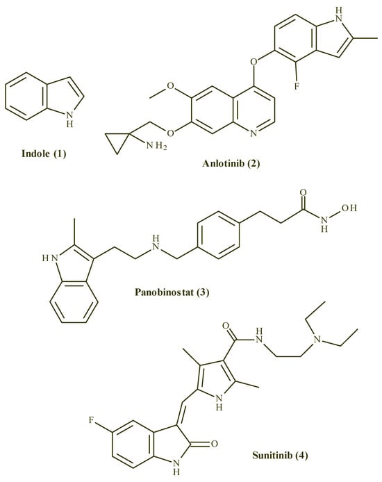
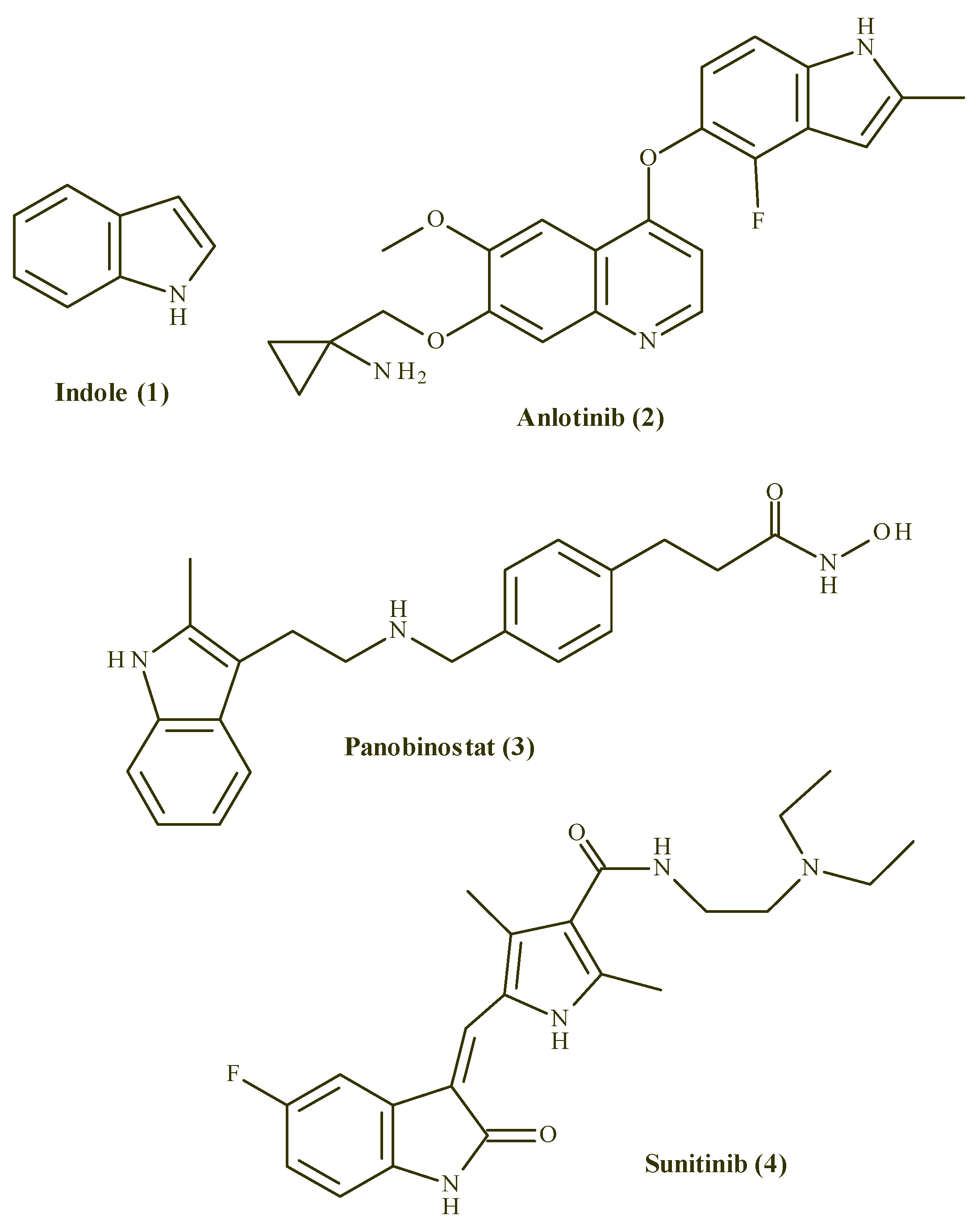
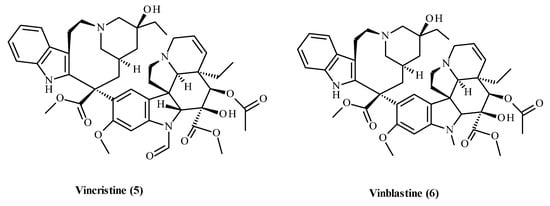
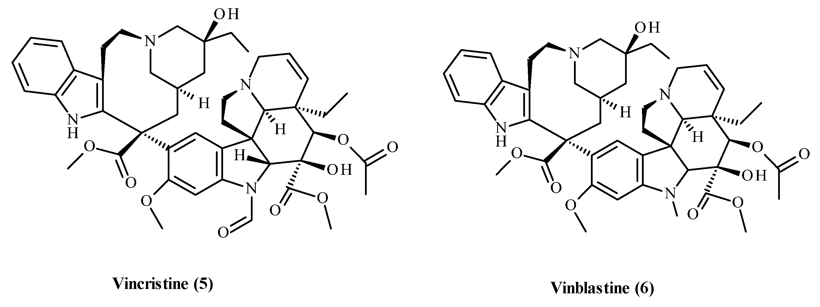
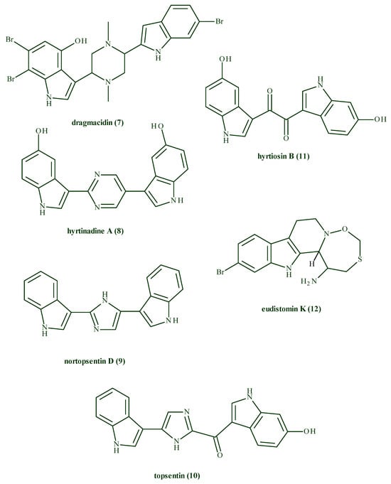
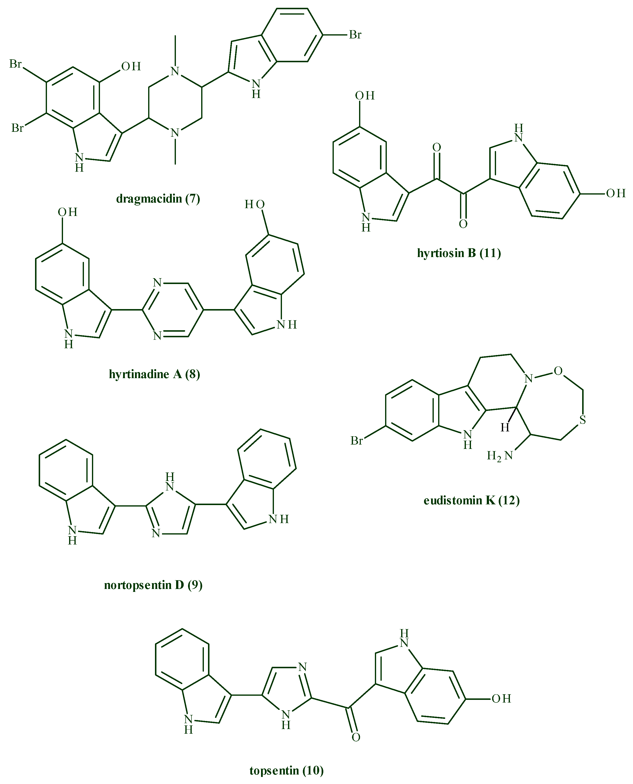
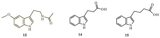
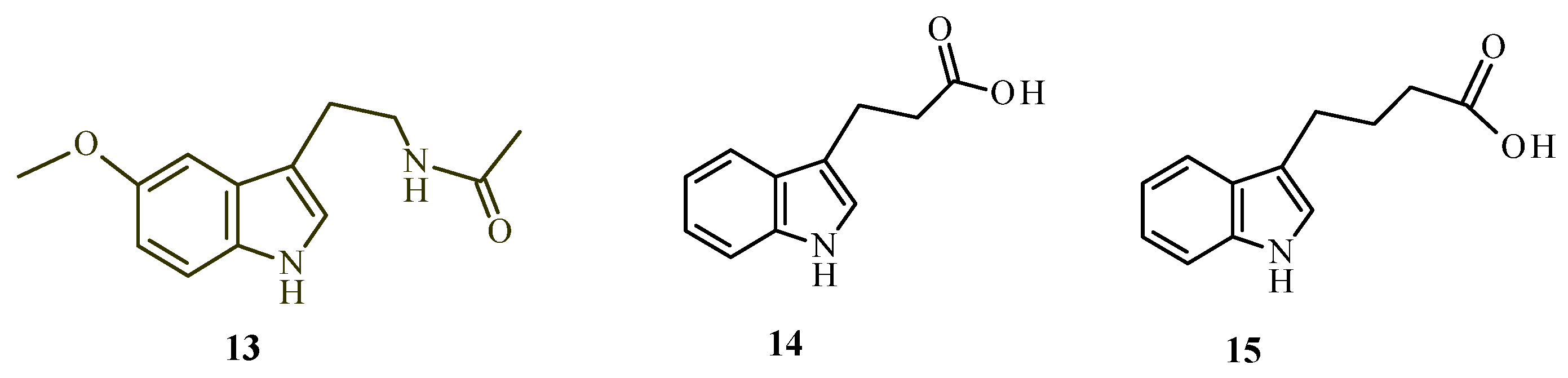
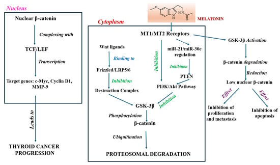
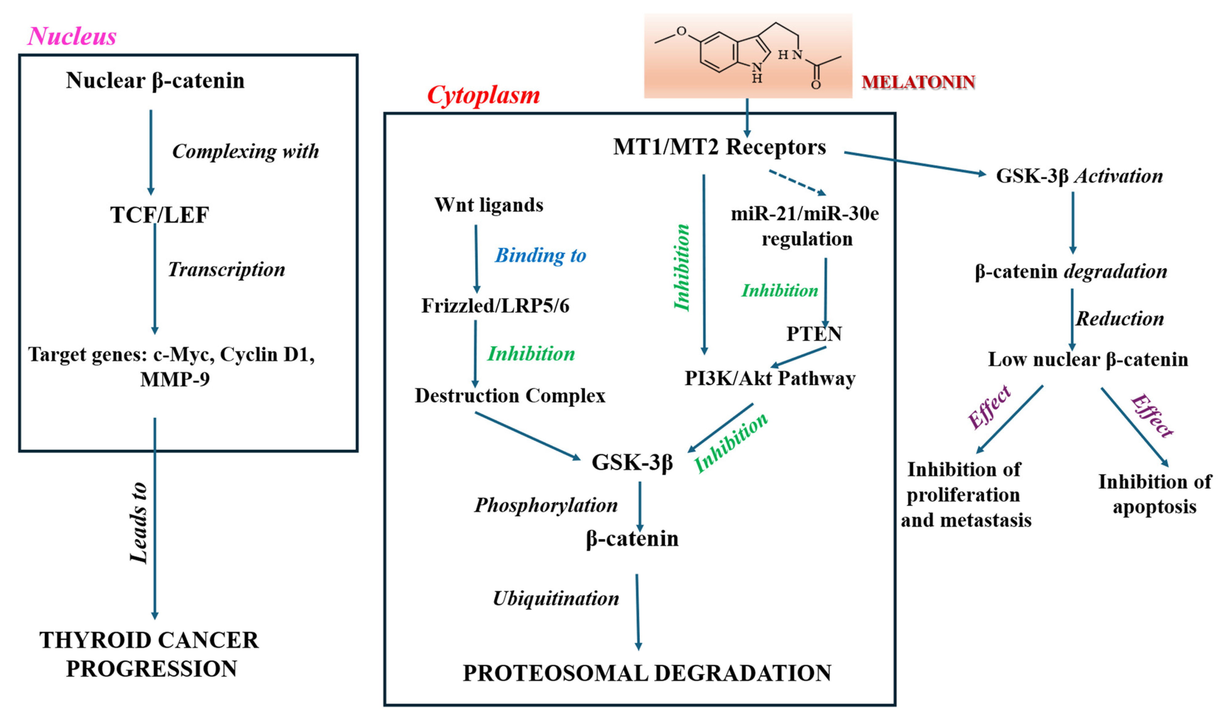
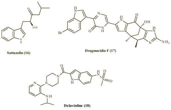
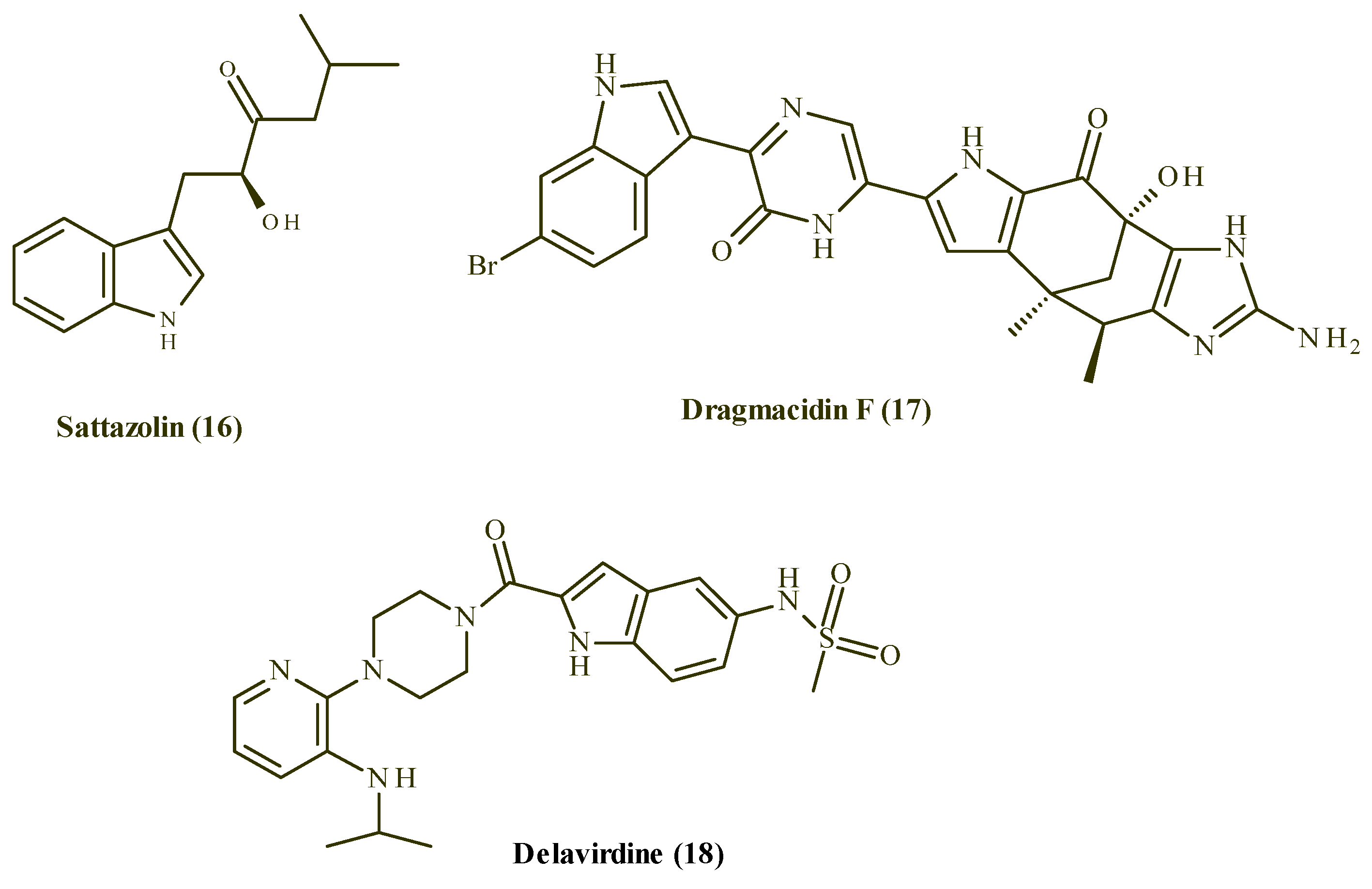
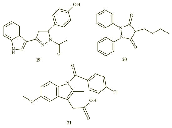
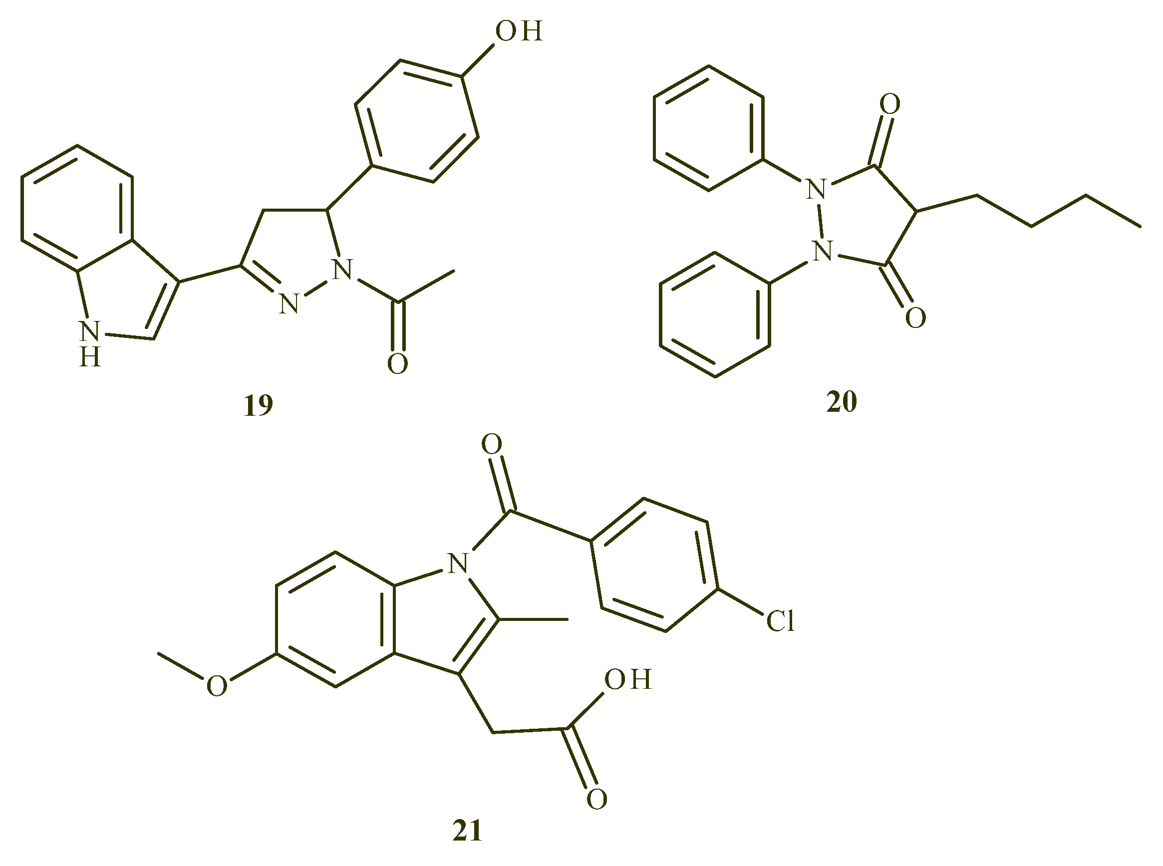
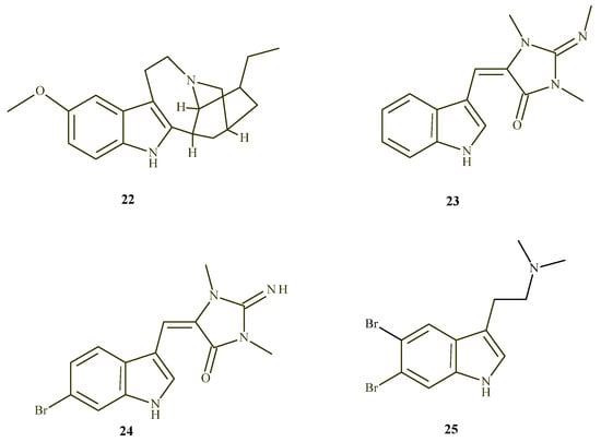
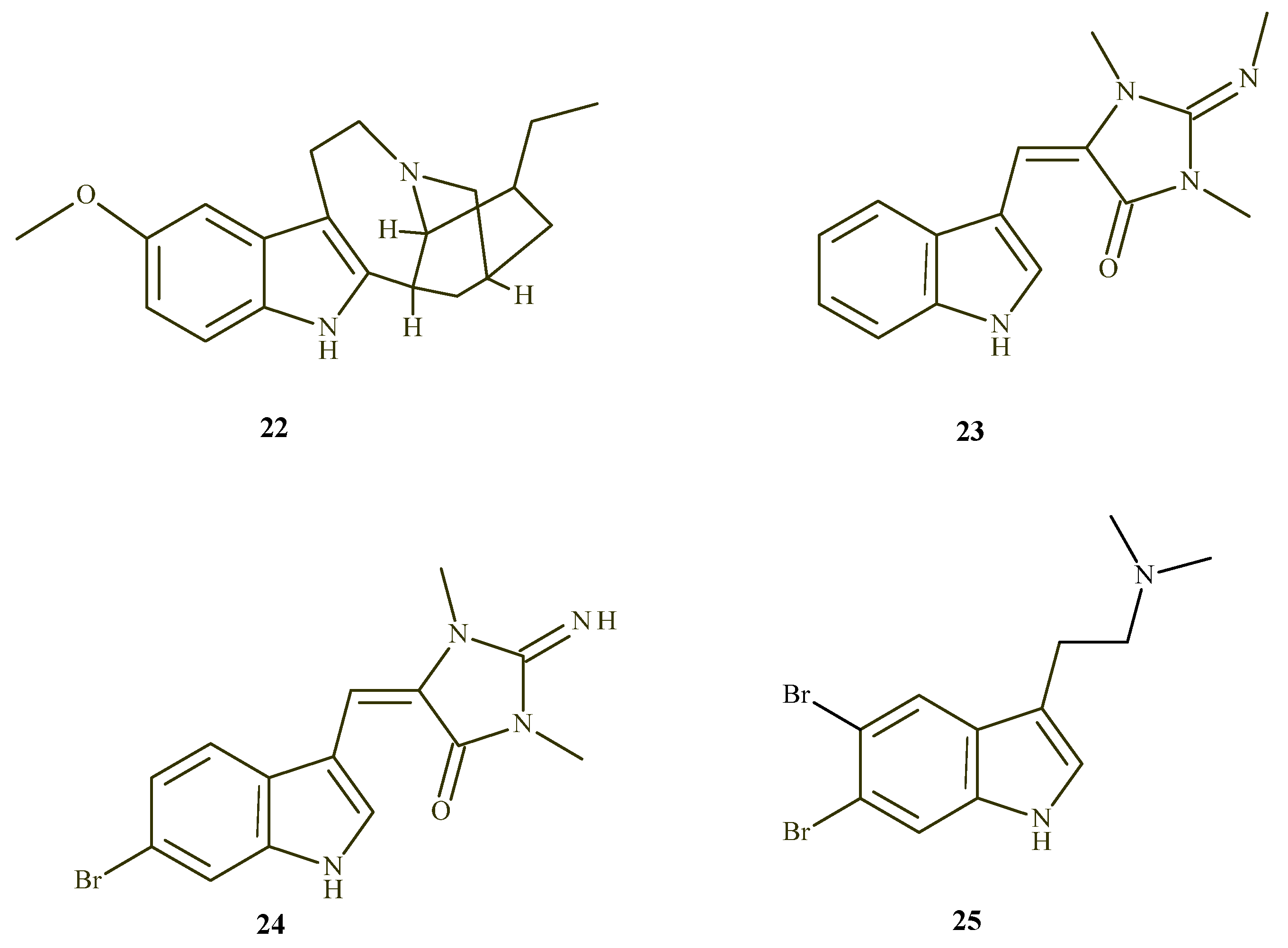
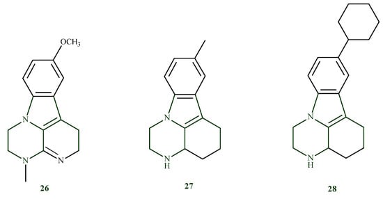
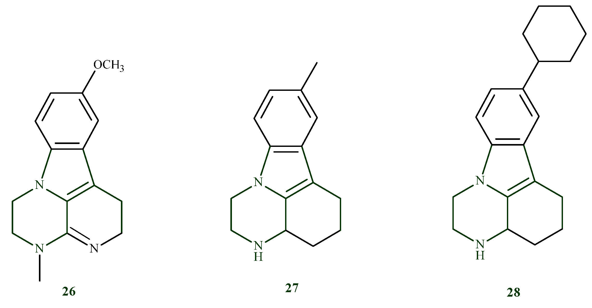
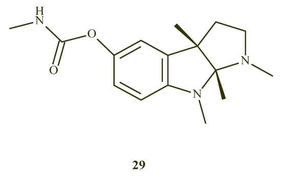
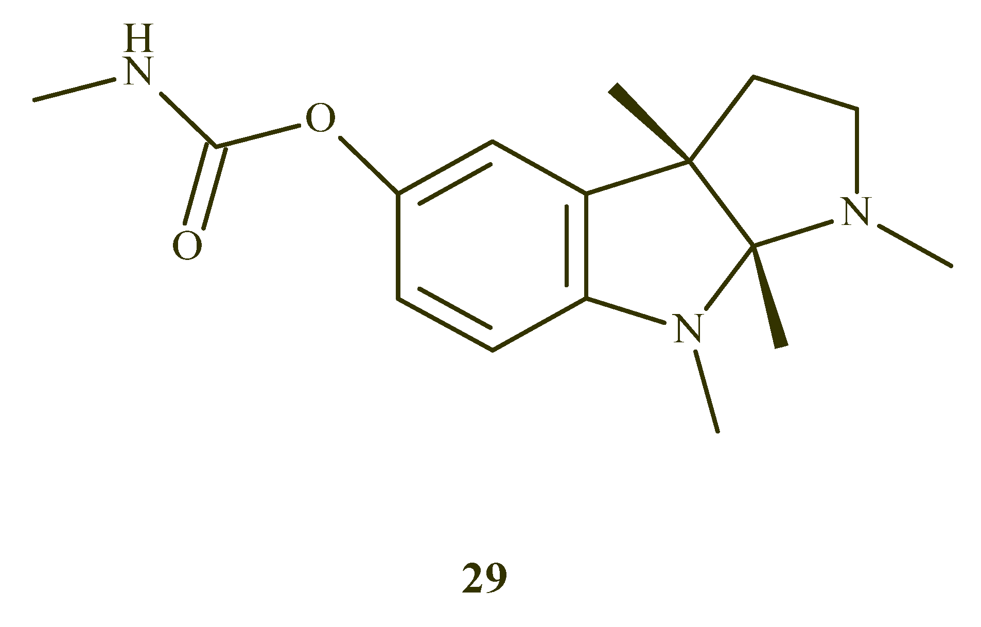
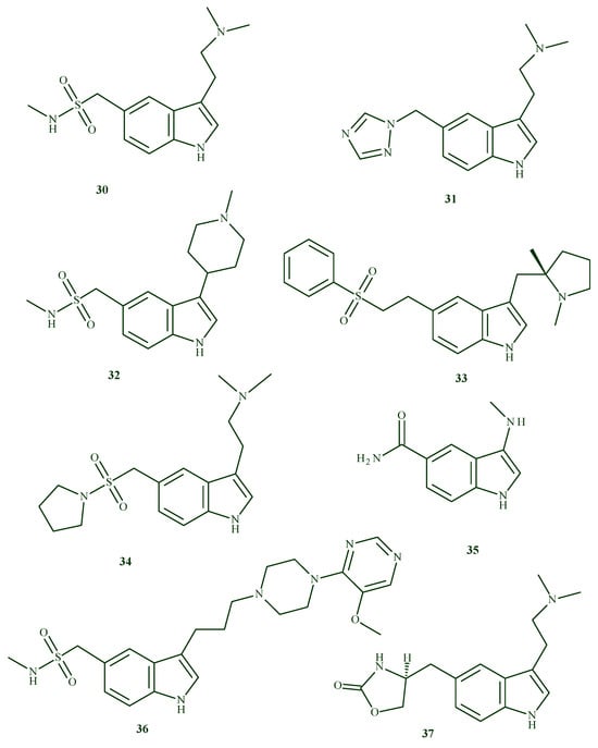
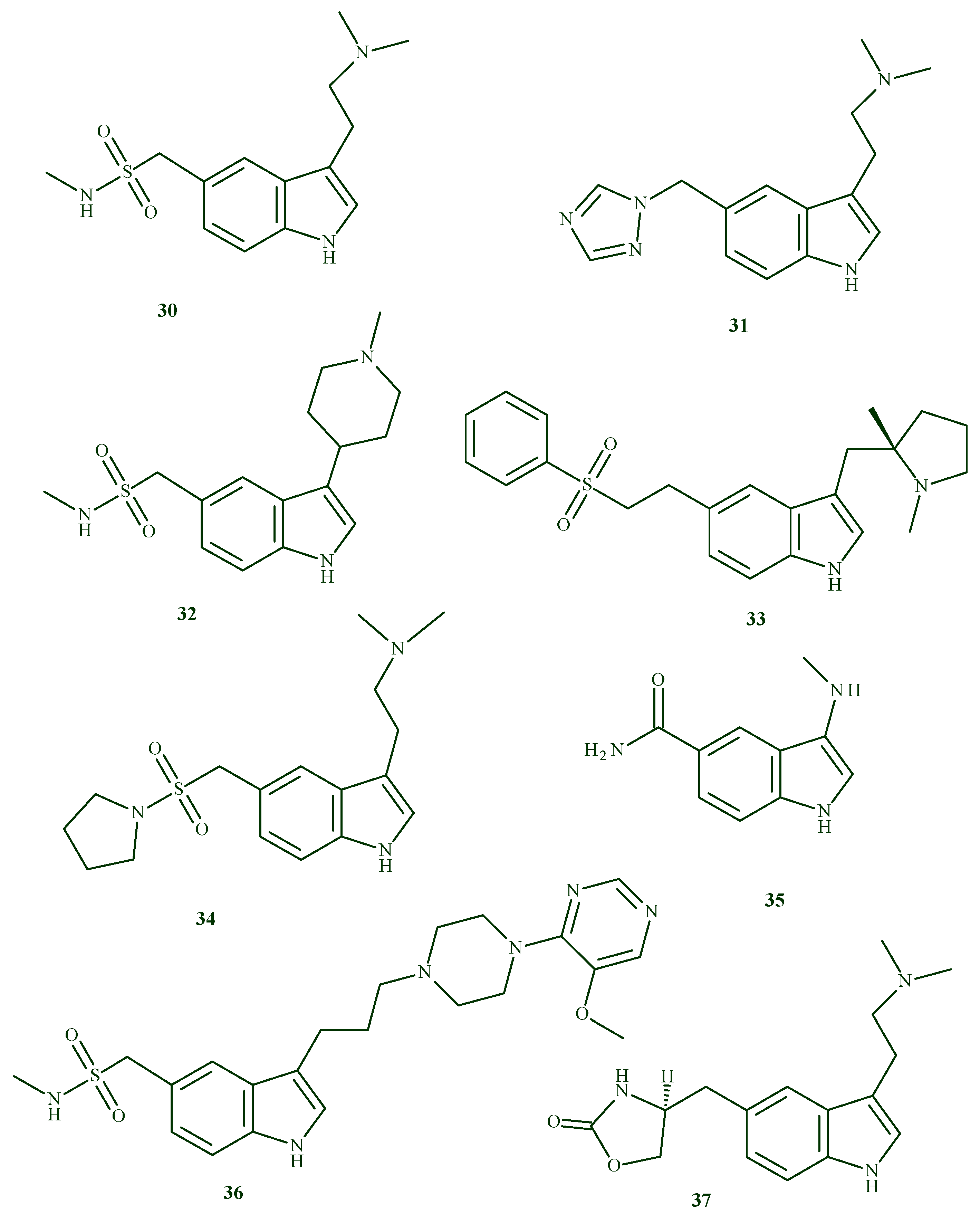
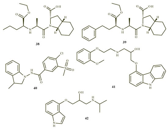
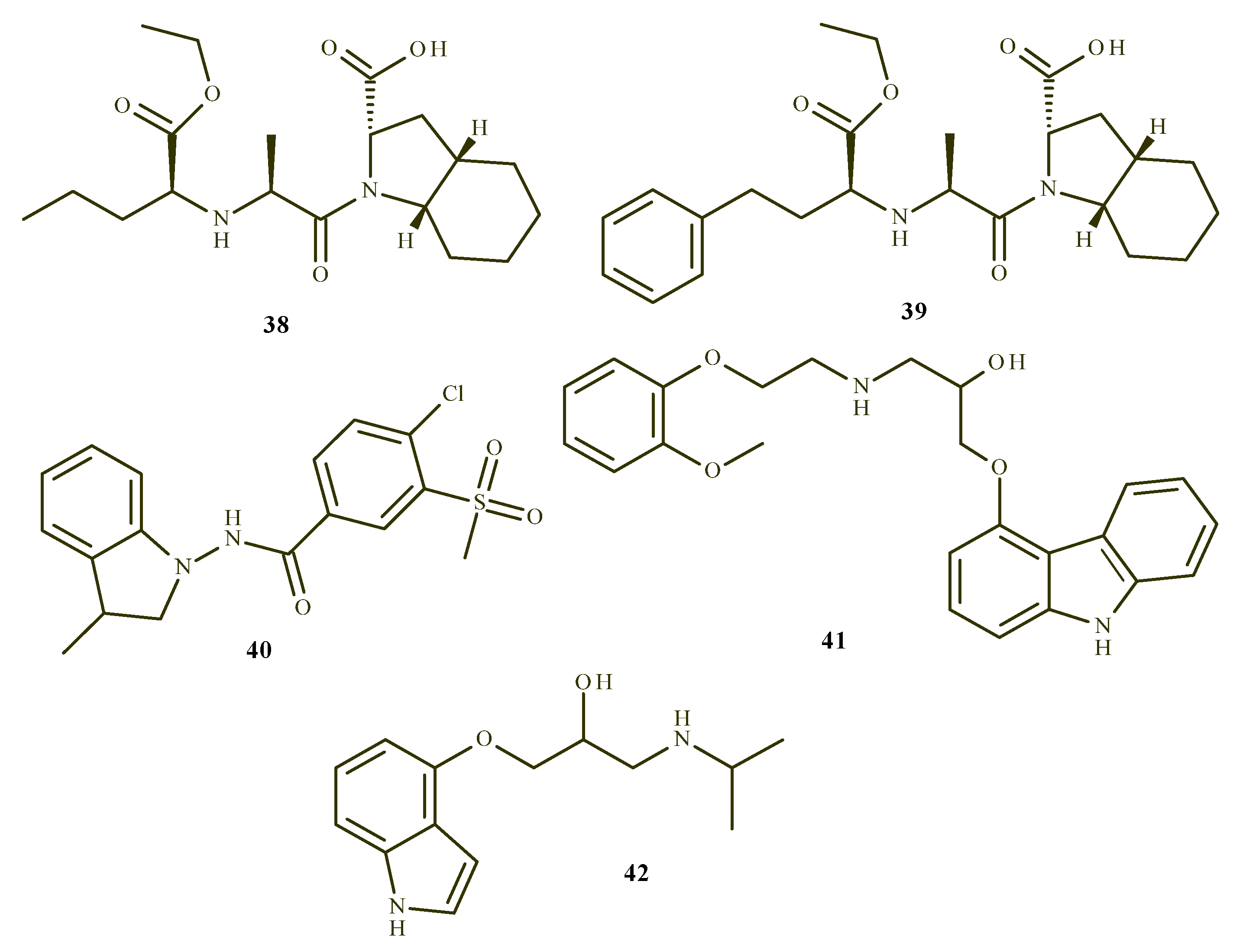
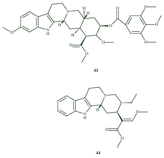
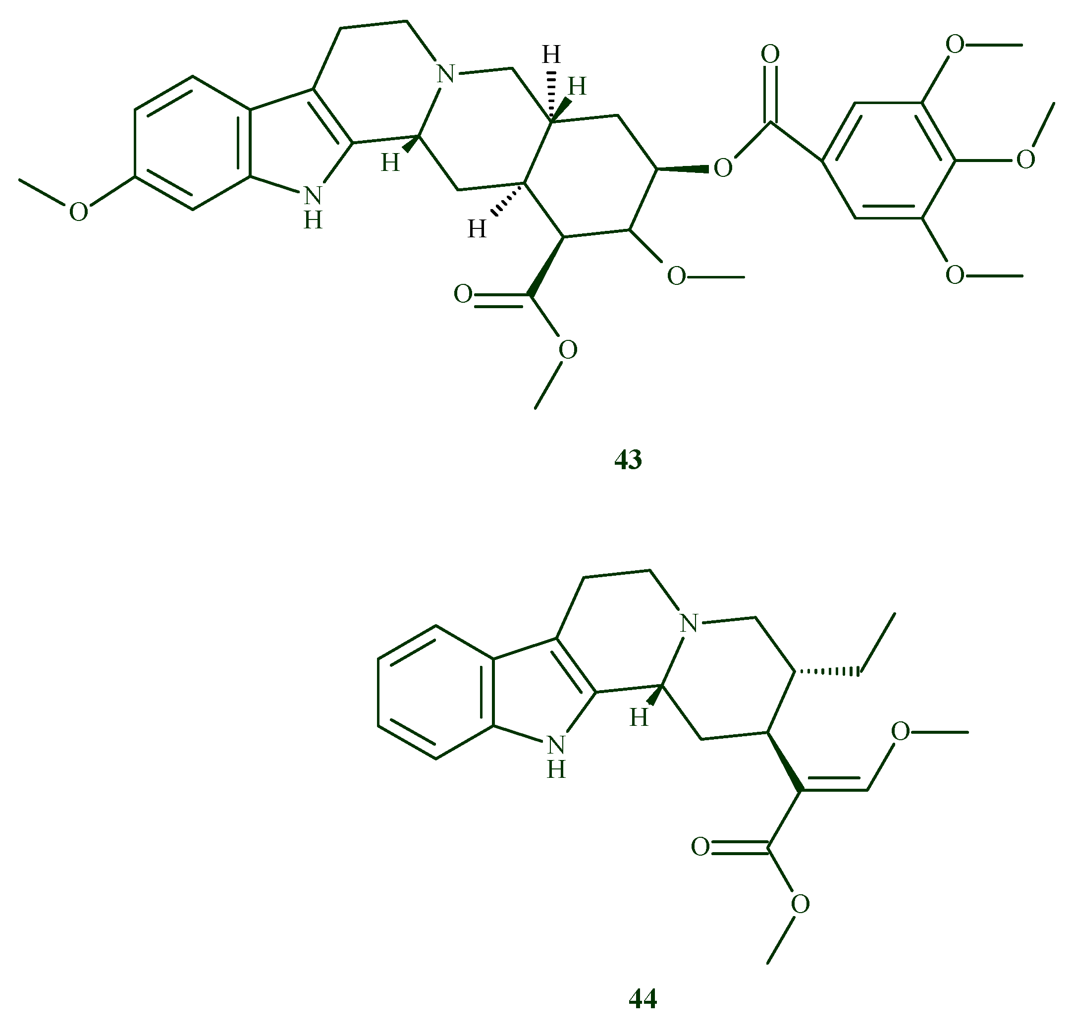
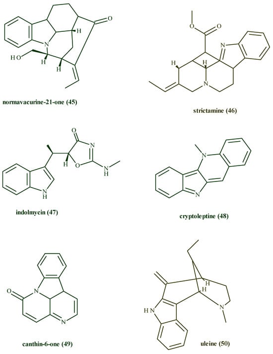
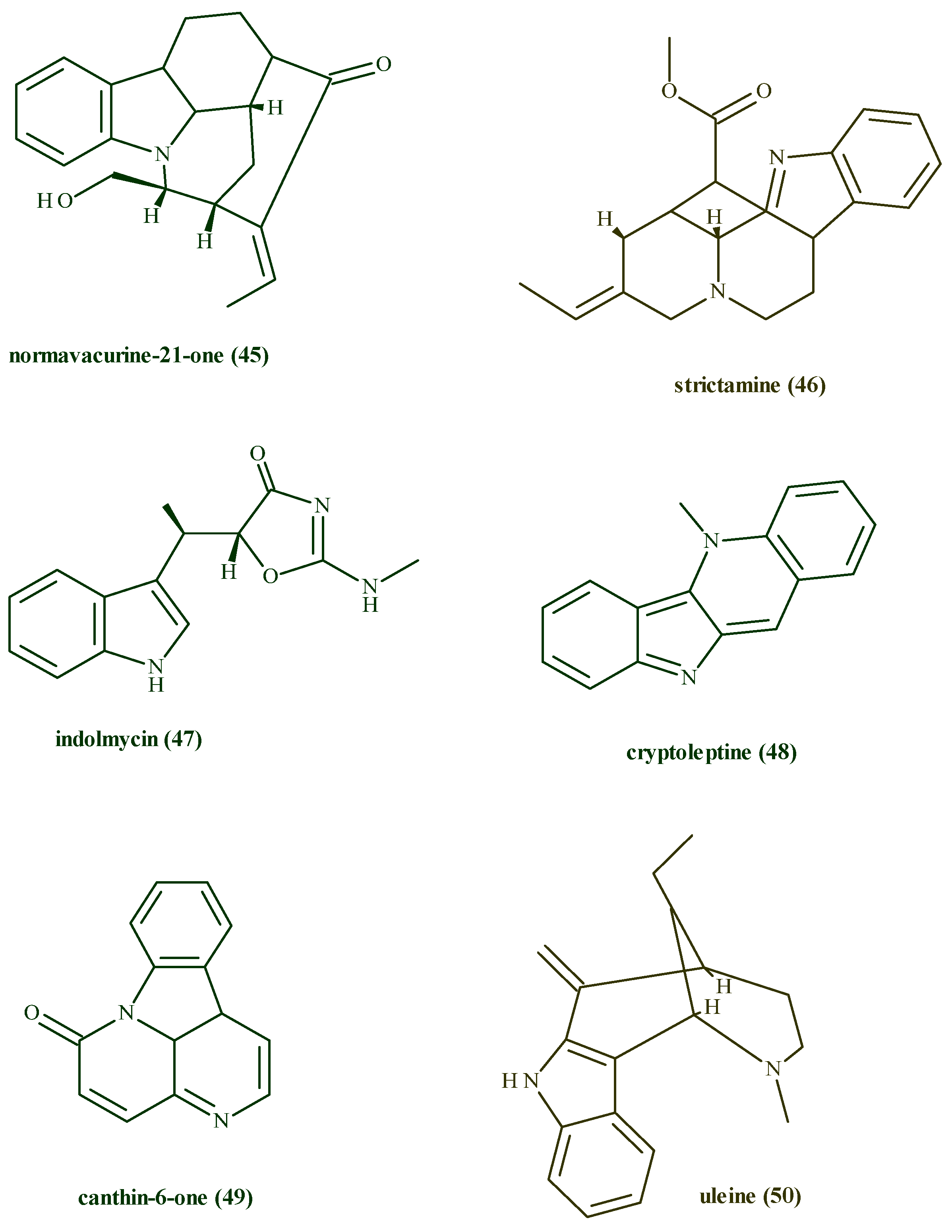
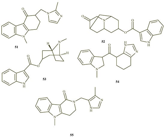
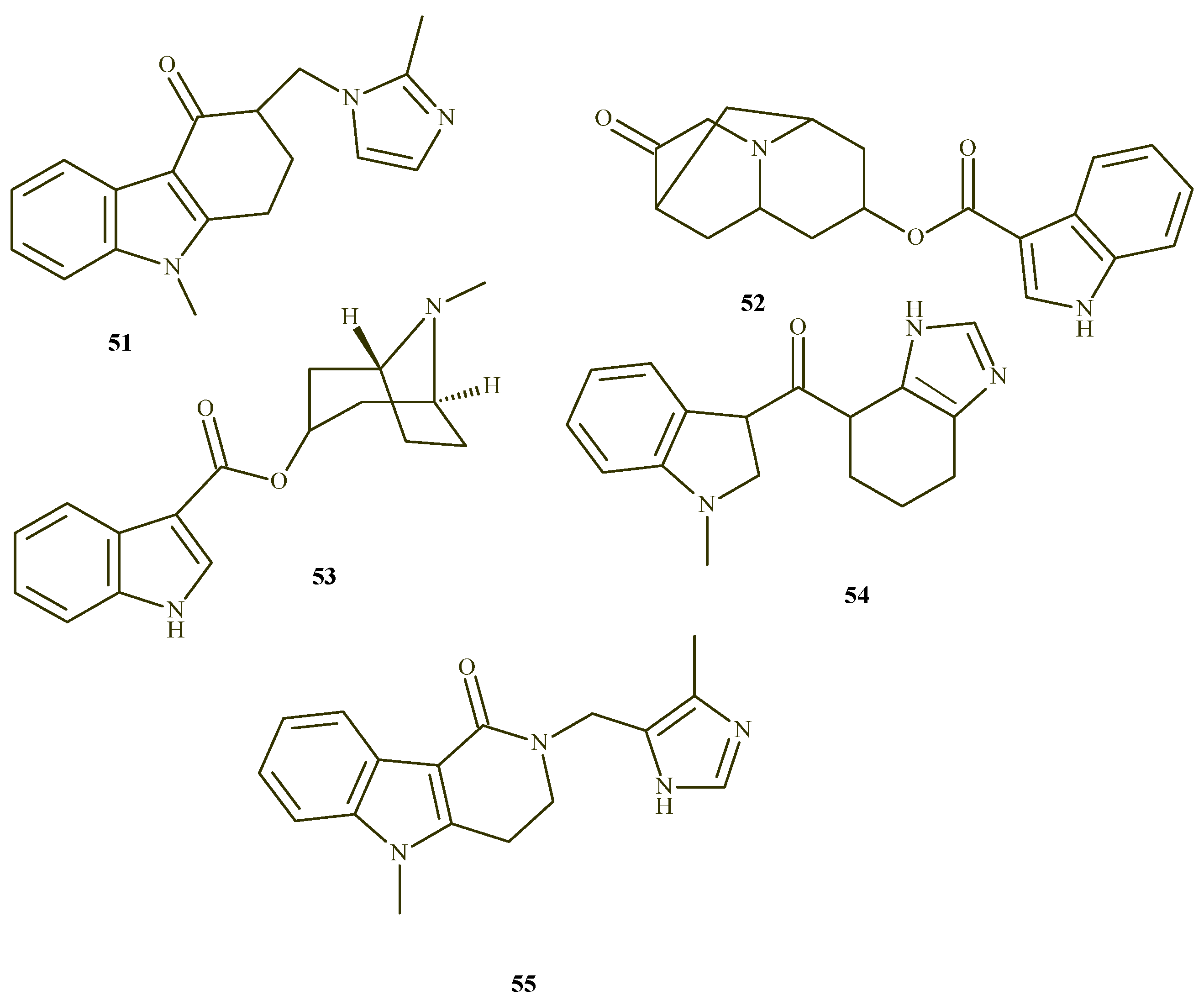
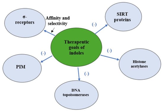
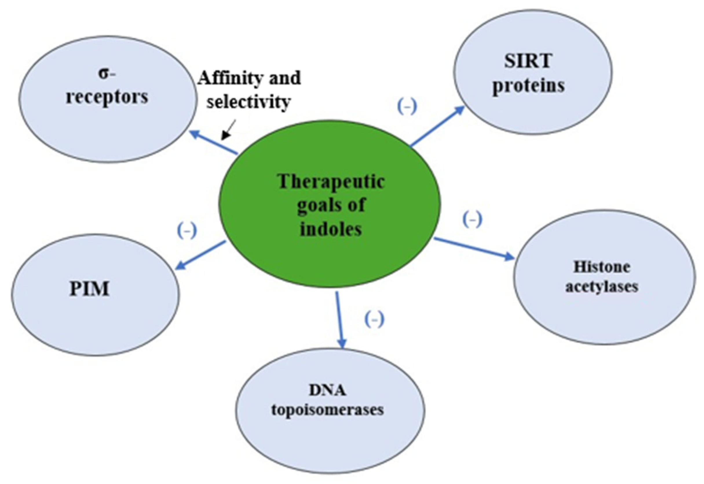
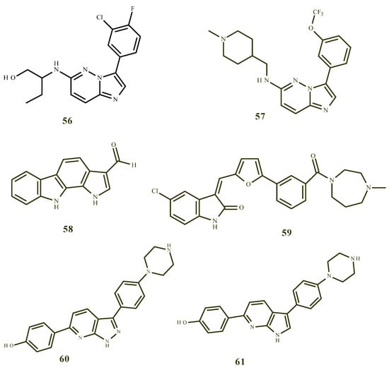
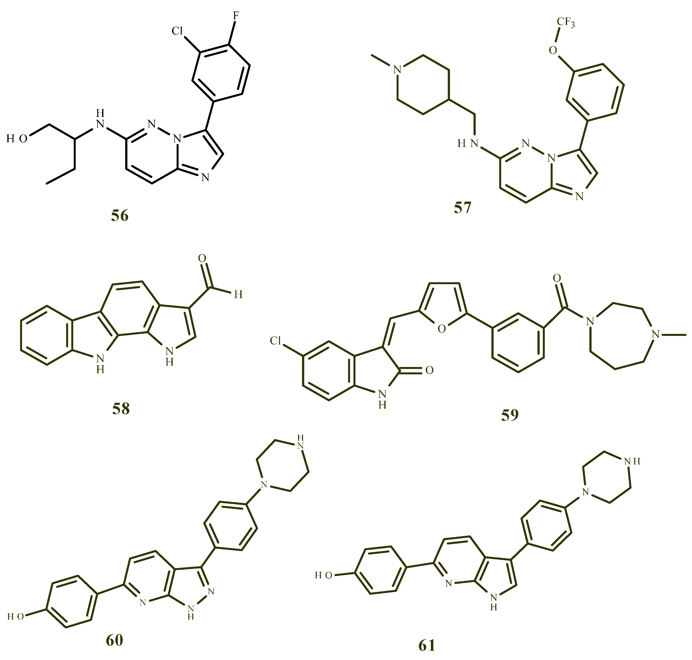
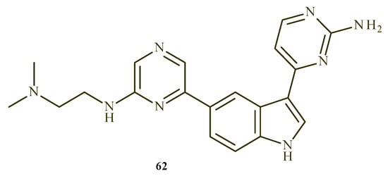
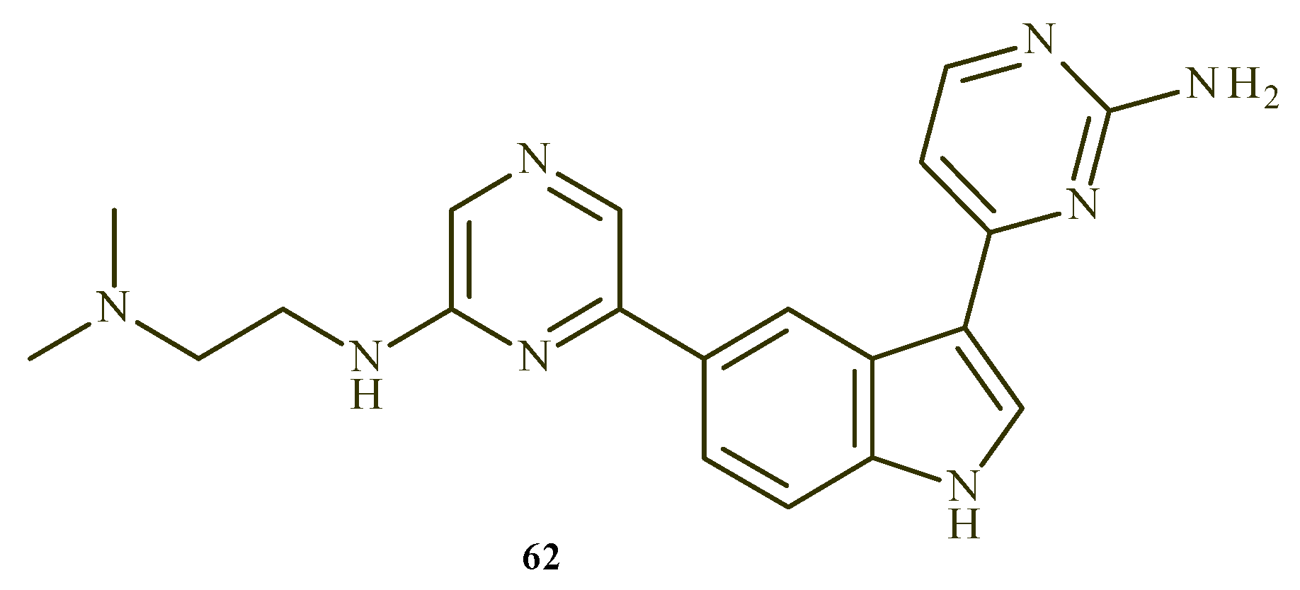
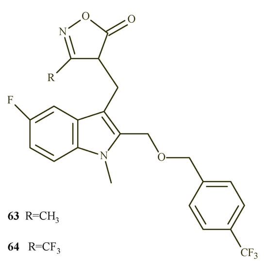
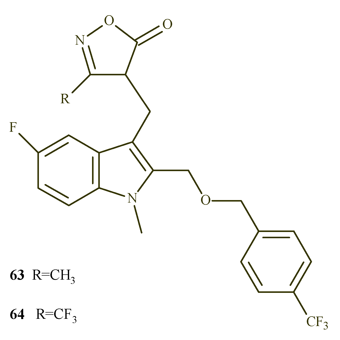
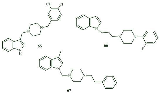
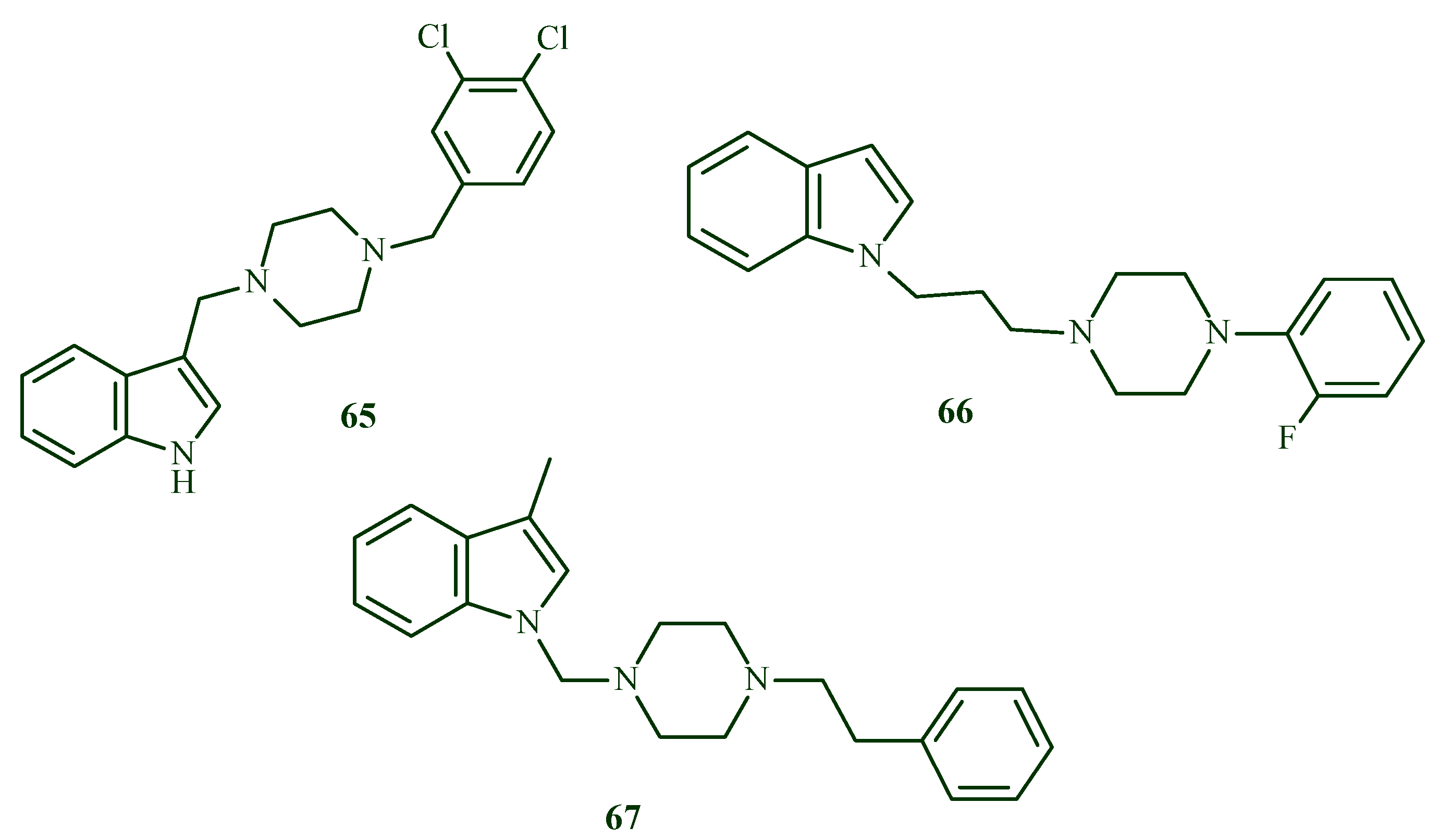
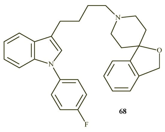
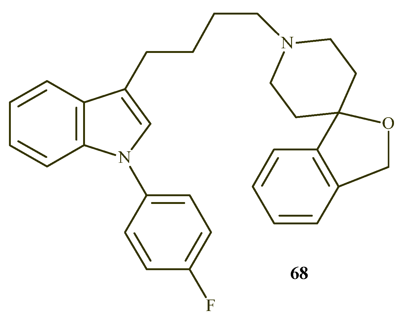
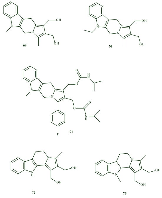
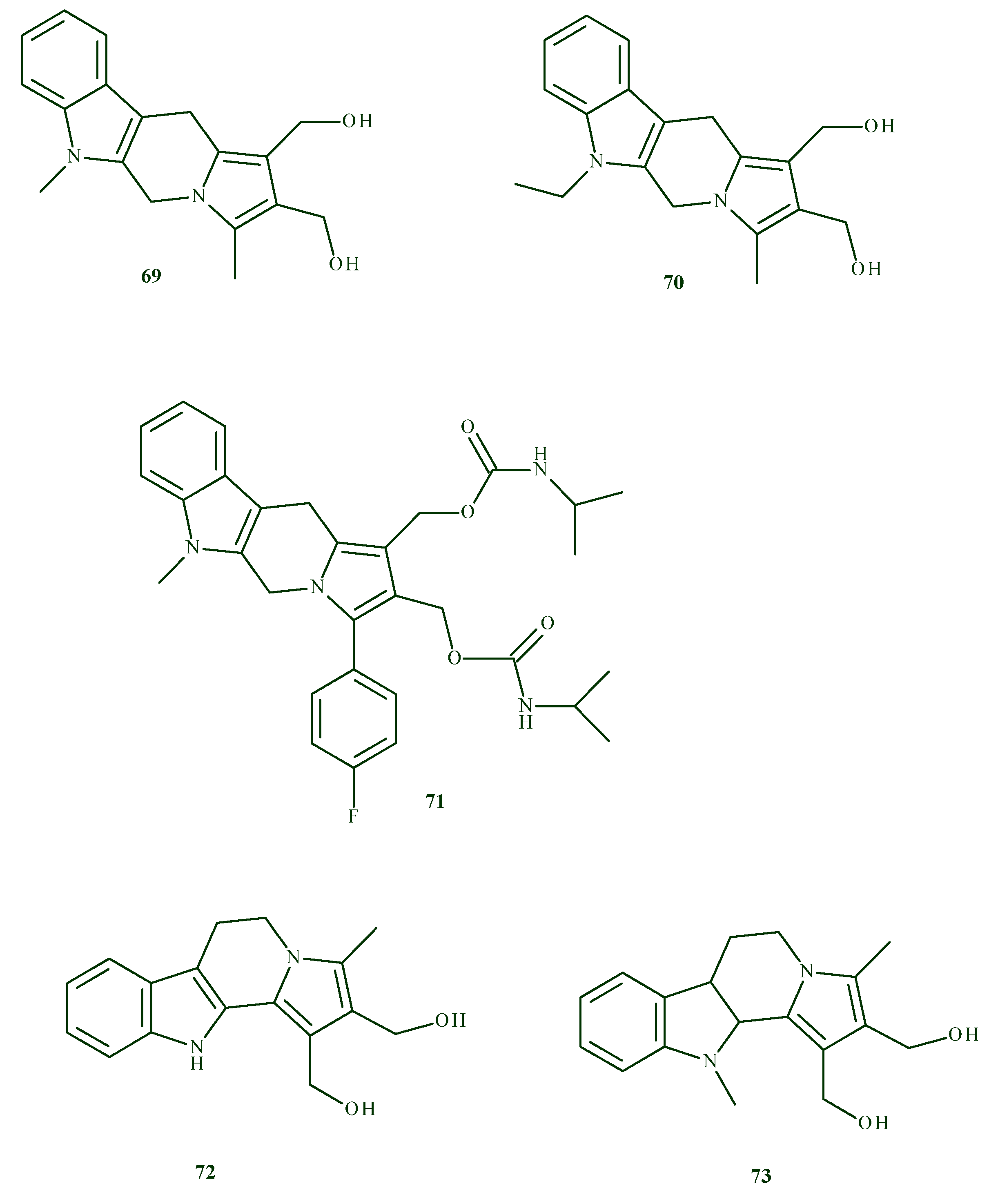
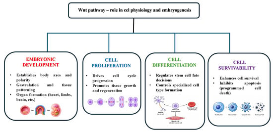
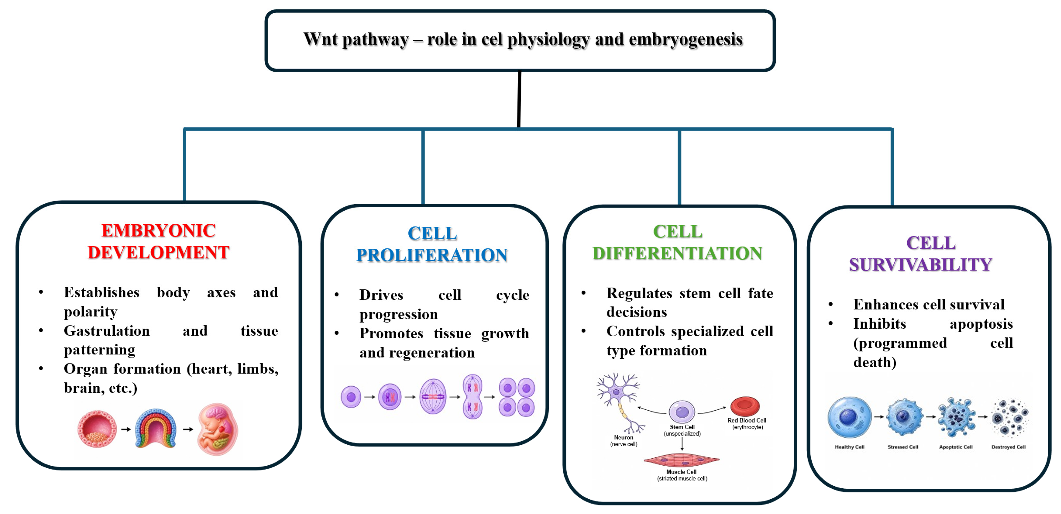
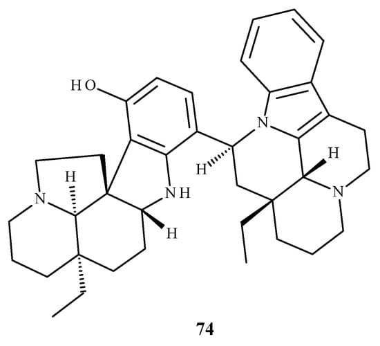
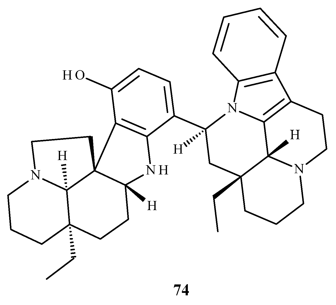
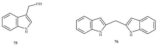
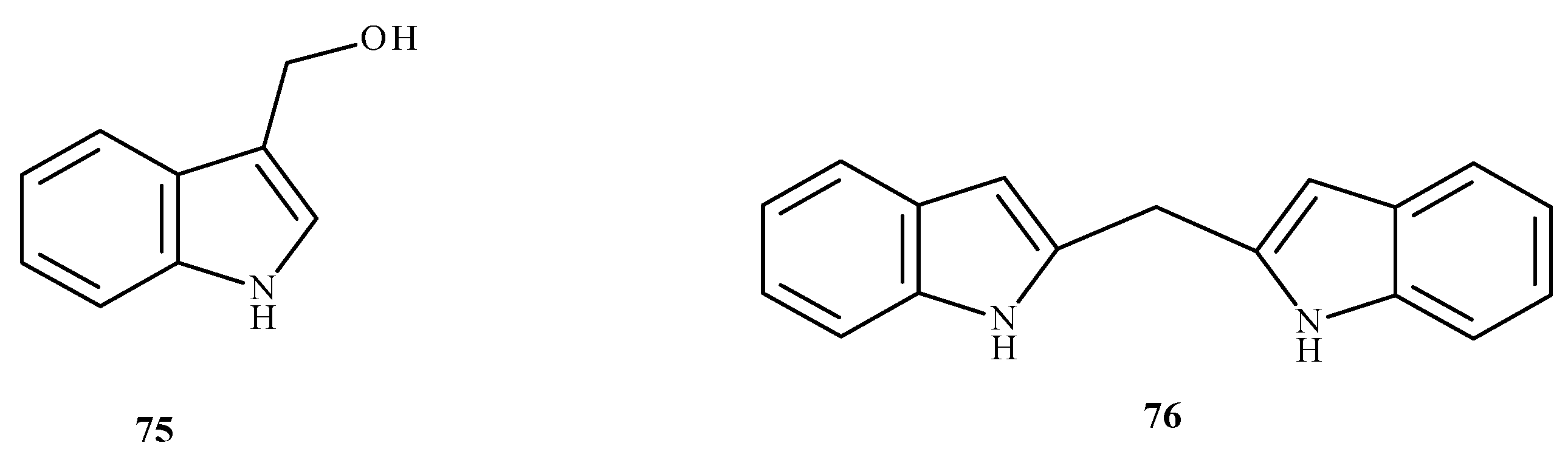
Disclaimer/Publisher’s Note: The statements, opinions and data contained in all publications are solely those of the individual author(s) and contributor(s) and not of MDPI and/or the editor(s). MDPI and/or the editor(s) disclaim responsibility for any injury to people or property resulting from any ideas, methods, instructions or products referred to in the content. |
© 2026 by the authors. Licensee MDPI, Basel, Switzerland. This article is an open access article distributed under the terms and conditions of the Creative Commons Attribution (CC BY) license.