


Abstract
Glioblastoma multiforme (GBM) is the most common brain tumor in the adult population, and it is characterized by poor prognosis; thus, new treatment strategies are strongly needed. Precision medicine is a rapidly growing field in oncology that identifies specific molecular features of tumor cells and allows theranostic strategies (combining diagnosis and therapy). In nuclear medicine, prostate-specific membrane antigen (PSMA) represents an interesting molecular target in theranostics, considering its involvement in the neoangiogenesis of many kinds of tumors, including GBM. In this narrative review, we performed a web-based literature search to explore the current evidence regarding PSMA-targeted nuclear medicine theranostics in GBM, highlighting diagnostic applications, therapeutic potential, and future clinical perspectives.
Keywords:
PSMA; theragnostics; glioblastoma multiforme; nuclear medicine; radioligand therapy; PET; SPECT 1. Introduction
1.1. Glioblastoma Multiforme: Background and Potential Theranostic Application Targeting PSMA
Glioblastoma multiforme (GBM) is the most common brain tumor in adults. It is a World Health Organization (WHO) grade IV glioma, and it is characterized by rapid growth and poor prognosis (median survival 12–15 months) [1]. Moreover, it is well known that the high cellular heterogeneity and the blood–brain barrier (BBB) structure give survival advantages to GBM cells [2]. Current treatment management of GBM includes a combined approach of surgery, radiotherapy and chemotherapy with temozolomide that remains insufficient to overcome the high aggressiveness of GBM cells; therefore, new weapons and treatment strategies are strongly needed.
Precision medicine is a rapidly growing field in oncology, identifying specific tumor molecular features and tailoring individual treatment to patients [3]. The importance of effective targeted therapies has been underlined in the literature [3,4], and several nuclear medicine techniques may assess specific molecular targets for diagnostic and therapeutic purposes with the use of radiopharmaceuticals, allowing theranostic strategies. Nuclear medicine imaging techniques, such as single-photon emission computed tomography (SPECT) or Positron Emission Tomography (PET)—which are commonly integrated with computed tomography (CT) or magnetic resonance imaging (MRI) in hybrid imaging platforms—enable the non-invasive assessment of specific biological targets, thereby supporting patient selection for targeted therapies and facilitating theranostic approaches [3]. In this context, a theranostic approach is based on the use of radiopharmaceuticals targeting the same molecular structure for both diagnostic imaging and radioligand therapy (RLT), allowing a personalized approach to cancer management.
It is well known that GBM is a highly vascularized tumor, and its aggressive behavior is closely associated with tumor angiogenesis [5]. In recent years, GBM endothelium has attracted considerable attention because, unlike the vasculature of many other brain tumors, it is not fully protected by the BBB and is therefore more exposed to circulating factors. Moreover, alterations in endothelial protein expression play a pivotal role in tumor neoangiogenesis, providing potential targets for the development of novel therapeutic strategies.
In this scenario, prostate-specific membrane antigen (PSMA) represents a potential theranostic target in consideration of its involvement in tumoral neoangiogenesis, and it has gained attention in the field of nuclear medicine theranostics [4]. PSMA is a type II transmembrane glycoprotein encoded by the gene FOLH1, and it plays an important role in neoangiogenesis of many kinds of tumors, including GBM [6]. Despite its name, this protein is not specific for prostate cancer, and it has been shown to be expressed in endothelial cells of neovasculature from benign and malignant conditions [7,8], but not in normal endothelium [9,10].
Regarding applications in oncology, the usefulness of theragnostic approaches targeting PSMA in metastatic prostate cancer is well-documented. Many PSMA-targeted radiopharmaceuticals are used in clinical practice for detection, staging, recurrence evaluation, and RLT in prostate cancer [11]. Moreover, potential and innovative theragnostic strategies have been described in other tumors [12]. In this context, evidence highlights the potential theranostic application targeting PSMA in GBM as well, paving the way for new imaging and treatment strategies [4,12].
1.2. Molecular and Biological Basis of PSMA in Glioblastoma Multiforme
As outlined above, PSMA has emerged as a promising target in GBM owing to its selective expression within tumor-associated neovasculature and its potential involvement in angiogenic processes. Understanding the biological role of PSMA in GBM is essential for evaluating its suitability as a target for nuclear medicine imaging and RLT. Recent studies showed that normal cerebral parenchyma expresses little or no PSMA either in brain cells or vessels, so its expression is exclusive to neovasculature in patients affected with GBM [10,13] and correlates negatively with survival [14].
Although the precise mechanisms underlying the role of PSMA in tumor angiogenesis have not yet been fully elucidated, evidence suggests that PSMA contributes to angiogenesis through several interconnected molecular pathways, including integrin-mediated signaling, extracellular matrix remodeling, and modulation of local folic acid metabolism [15,16]. In GBM, tumor neovasculature plays a key role in disease progression, and many studies demonstrated PSMA expression in the vessels of this malignancy to varying degrees [6,13], with moderate-to-high intensity, also with differences in initial diagnosis and recurrences. Several hypotheses have been proposed for the explanation of heterogeneity in PSMA expression, including differences in transcriptomic profile of FOLH1, but the mechanism remains unknown [17].
Several studies have demonstrated increased PSMA expression in the endothelial cells of high-grade gliomas, with higher levels associated with tumor aggressiveness and poorer prognosis [6,18,19,20]. Experimental evidence further suggests that PSMA may actively participate in GBM angiogenesis through interaction with integrin β4 (ITGB4), leading to activation of the nuclear factor kappa-light-chain-enhancer of activated B cells (NF-kB) signaling pathway [18,21]; in turn, activation of the ITGB4/NF-kb axis has been shown to upregulate PSMA expression in endothelial cells, suggesting the existence of a positive regulatory feedback loop that may promote endothelial tube formation and tumor neoangiogenesis [17,22,23]. Moreover, many studies have shown that abnormal activation of the NF-kB can cause an increase in the concentration of vascular endothelial growth factor (VEGF), another important target in tumor microvasculature, causing invasion of cancer cells [24].
PSMA seems to be independent of VEGF [25]. The level of VEGF expression correlates with glioma grade, and it is thought to be the main promoter of angiogenesis in glioblastoma [26], so potential interaction between PSMA and VEGF warrants further investigation, given that they are both possible targets in these malignancies. However, the relationship between PSMA and VEGF expression in GBM remains under investigation, and further evidence is expected from ongoing clinical trials (https://clinicaltrials.gov/study/NCT07052877?cond=Glioblastoma%20Multiforme&term=PSMA&viewType=Card&page=1&rank=1, accessed on 26 July 2026).
1.3. Scope of the Review
The aim of this narrative review is to provide a comprehensive overview of the current evidence on PSMA-targeted theranostic approaches in GBM, with particular emphasis on molecular imaging in nuclear medicine, radioligand therapy, current limitations, and future research directions. Furthermore, it aims to identify current knowledge gaps and highlight the potential of PSMA-targeted strategies as a promising field for future investigation.
2. Methods
This narrative review was conducted through a literature search aimed at identifying published evidence on PSMA-targeted imaging and RLT in GBM.
A comprehensive literature search was performed in PubMed/MEDLINE and Scopus. The final search was conducted on 30 June 2026, and the following keywords were used:
(“glioblastoma” OR “glioblastoma multiforme” OR “high-grade glioma”) AND (“PSMA” OR “prostate-specific membrane antigen”) AND (“PET” OR “SPECT” OR “molecular imaging” OR “radioligand therapy” OR “theranostics”)
Original articles, clinical studies, case reports, and relevant preclinical investigations published in English were considered for inclusion. Additional publications providing background information on GBM, molecular imaging in nuclear medicine, RLT, or theranostic approaches were also included when considered relevant to the aims of this review. Editorials, conference abstracts without sufficient data, and duplicate publications were excluded.
Two authors (F.R. and A.C.) screened the titles and abstracts of the retrieved records, followed by full-text evaluation of potentially eligible publications.
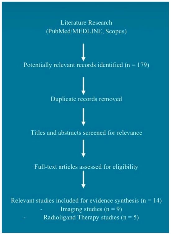
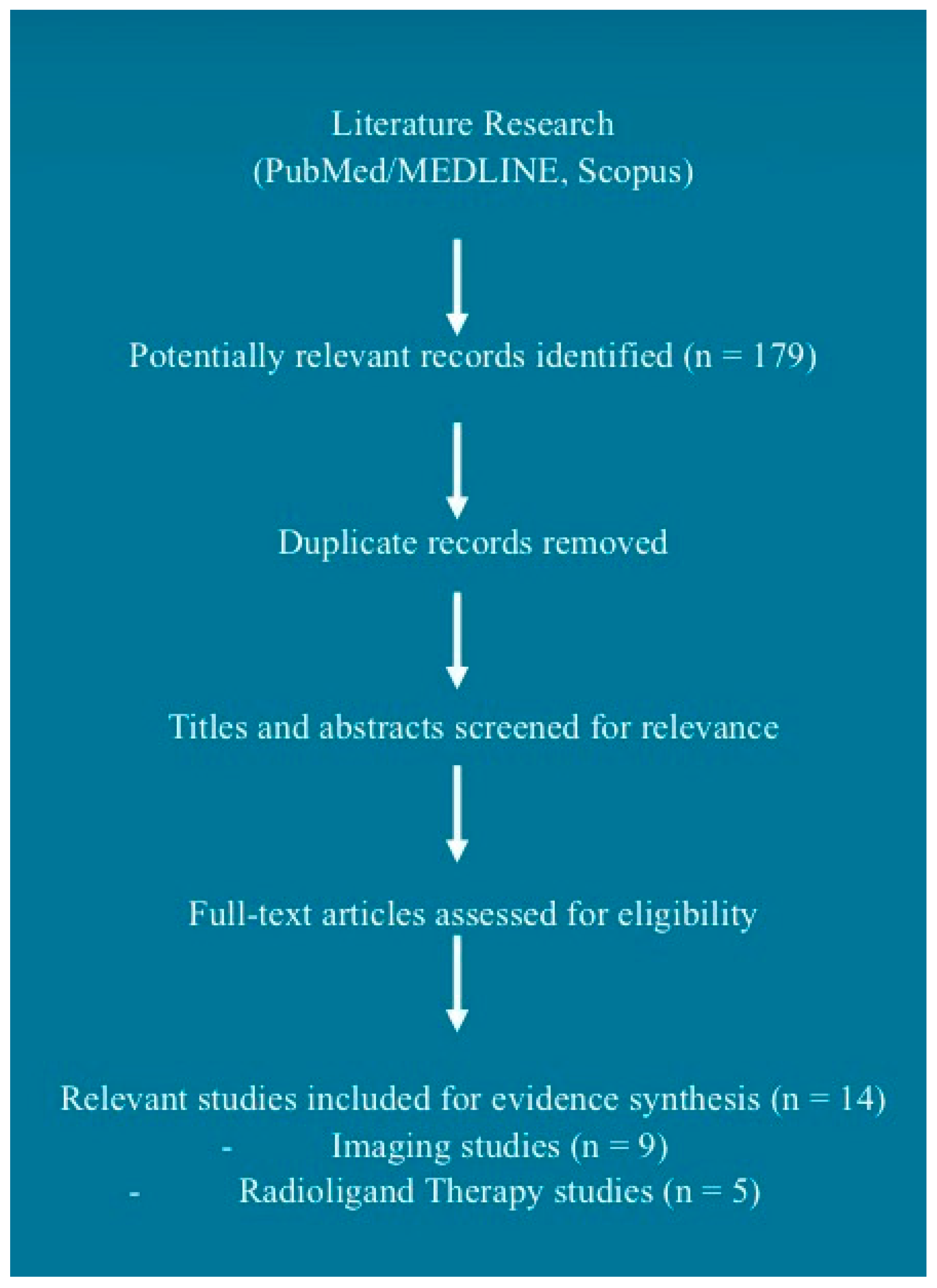
Given the narrative nature of the review and the limited number of available publications, no formal quality assessment or meta-analysis was performed. The selected publications were qualitatively analyzed to summarize current evidence regarding diagnostic imaging, RLT, and future perspectives of PSMA-targeted theranostics in GBM. A schematic overview of the literature identification and study selection process adopted for this narrative review is provided in Figure 1.
Figure 1.
Schematic overview of the literature identification and study selection process adopted for this narrative review.
3. PSMA and Nuclear Medicine Imaging in Glioblastoma Multiforme
Although MRI remains the gold standard for the diagnosis and follow-up of glioblastoma, PET imaging provides complementary functional information and has emerged as a promising tool for the characterization of brain tumors. In this context, PET imaging with amino acid tracers such as [11C-methyl]-methionine ([11C]MET), O-(2-[18F]fluoroethyl)-L-tyrosine ([18F]FET) and 3,4-dihydroxy-6-[18F]fluoro-L-phenylalanine ([18F]DOPA) may contribute to glioma grading, detection of multifocal disease, and differentiation between tumor recurrence and treatment-related changes [27].
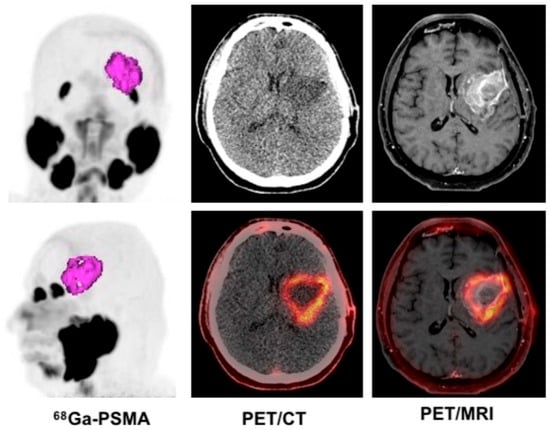
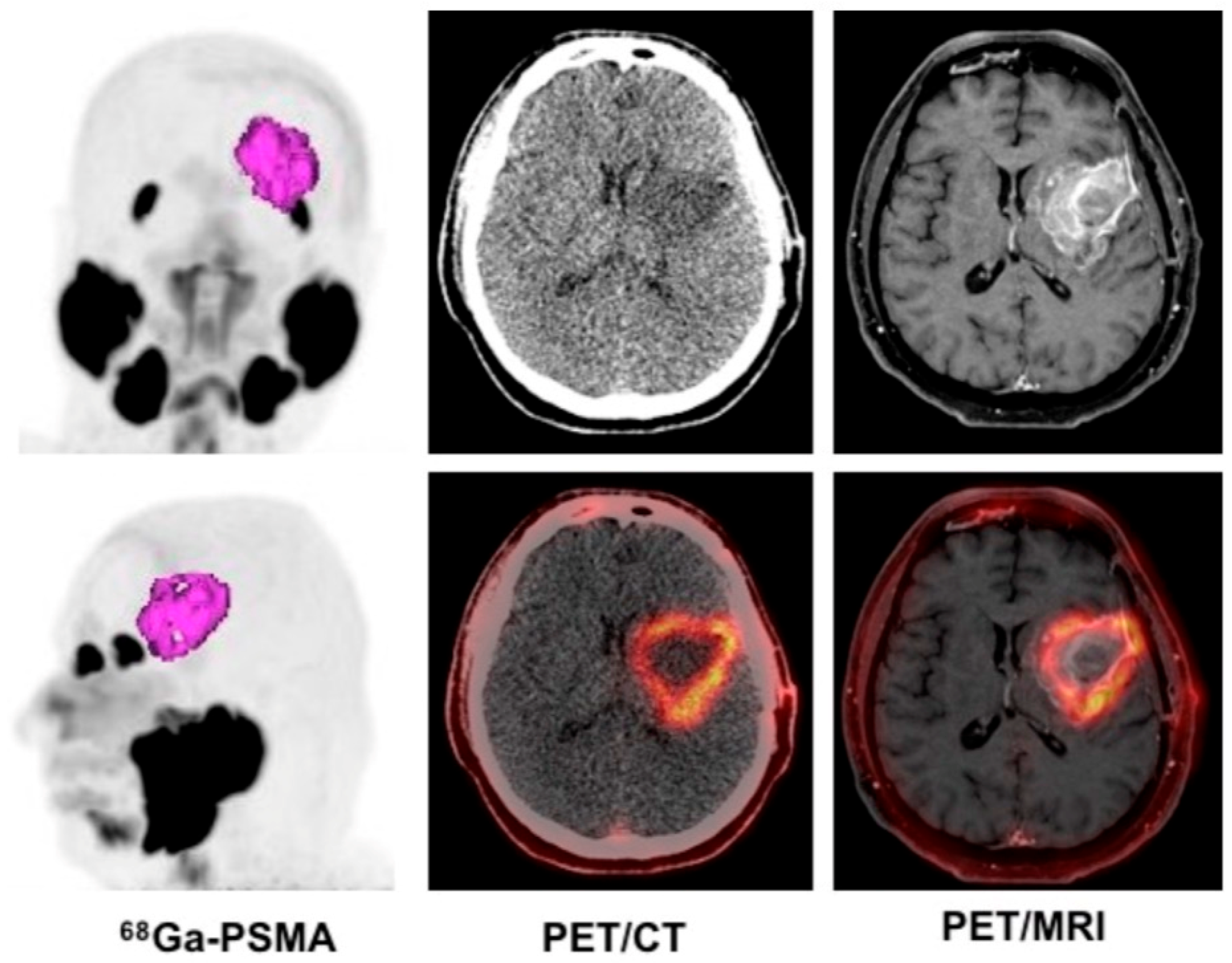
As we highlighted before, the rationale for PSMA-targeted imaging in GBM derives from the expression of PSMA within tumor-associated neovasculature (Figure 2). In this regard, several studies have demonstrated the feasibility and the promising role of non-invasive assessment of PSMA expression in both newly diagnosed and recurrent GBM using PSMA-targeted PET radiopharmaceuticals [8,11,19,28,29,30]. Furthermore, interindividual variability and temporal changes in PSMA expression between primary and recurrent lesions have been reported, reflecting the biological heterogeneity of the disease [8,18].
Figure 2.
A 44-year-old male patient with glioblastoma multiforme (IDH-wildtype), treated with craniotomy: 68Ga-PSMA positron emission tomography (PET)/computed tomography (CT) and PET/magnetic resonance imaging (MRI) axial images show a lesion with abnormal PSMA expression in the left fronto-temporal region, with maximum standardized uptake value (SUVmax) 4.6 and metabolic tumor volume (MTV) 27.9 cm3 at PET/CT images. Findings are consistent with a significant active residual tumor characterized by neovascularization. Written informed consent has been obtained from the patient to publish this figure.
An important advantage of PSMA PET imaging in brain tumors is the very low physiological uptake of PSMA-targeted tracers in normal cerebral tissues, resulting in excellent tumor-to-background ratios (TBRs) and facilitating image interpretation [29]. In this regard, Kunikowska et al. reported a median TBR value of 96.7 in a study involving GBM patients who underwent [68Ga] Ga-PSMA-11 PET/CT [31]. In addition, tracer uptake has been shown to correlate with tumor grade and proliferation indices such as Ki-67, supporting the association between PSMA expression and tumor aggressiveness [32]; these characteristics have generated interest in the potential role of PSMA PET for differentiating high-grade from low-grade gliomas [32].
Most available evidence has been obtained using radiotracers labeled with Gallium-68, particularly [68Ga] Ga-PSMA-11, which remains the most extensively investigated compound in neuro-oncology [31]. Nevertheless, fluorinated tracers such as 18F-Piflufolastat (also known as [18F] F-DCFPYL) have also shown promising results [30]. Biodistribution and time–activity curves appear largely comparable between gallium- and fluorine-labeled compounds, especially when comparing 18F-Piflufolastat and [68Ga]Ga-PSMA-11 [30], but the use of 18F-labeled tracers may offer practical advantages related to production and distribution [33]; however, comparative studies specifically addressing GBM are currently lacking.
An important application of PSMA PET concerns the evaluation of recurrent disease. In fact, preliminary evidence suggests that PSMA-targeted imaging may help distinguish tumor recurrence from radionecrosis, a clinically relevant challenge in the follow-up of GBM patients. In this setting, PSMA PET has demonstrated favorable TBR values and, in some reports, has shown potential advantages over amino acid tracers such as 18F-FET [19,28,34]. However, larger prospective studies are required to validate these findings. In this regard, PET imaging with radiolabeled PSMA minibody such as 89Zr-Df-IAB2M may have a potential value in distinguishing recurrence from radionecrosis, as demonstrated in a previous study by Matsuda et al. evaluating 83 brain tumors (including 41 GBM) [35].
PSMA expression in PET imaging may also provide prognostic information. In 2021, Holzgreve et al. demonstrated, in a group of 16 patients with GBM, that high vascular PSMA expression, particularly at recurrence, is associated with shorter survival and poorer clinical outcomes [14]. Moreover, increasing PSMA expression over time appears to correlate with disease progression, suggesting a possible role as a prognostic biomarker [14].
Beyond diagnosis, PSMA PET may play a role in treatment planning. Several investigations have explored the integration of PSMA PET with MRI for target volume delineation prior to radiotherapy, reporting improved visualization of tumor margins, particularly in non-enhancing lesions [29,36]. Nevertheless, discrepancies between biological tumor volumes defined by PSMA PET and gross tumor volumes identified on MRI have been observed, suggesting that PSMA PET should currently be considered complementary rather than alternative to conventional imaging modalities.
It is important to underline that PET imaging may also represent a crucial step in patient selection for PSMA-targeted RLT. A correlation between PET uptake and endothelial PSMA expression assessed by immunohistochemistry has been demonstrated, supporting the use of PSMA PET as a biomarker for identifying patients who may benefit from therapeutic approaches using radiolabeled PSMA ligands [18]. However, compared with prostate cancer, GBM generally exhibits lower standardized uptake value (SUV) and TBR values, limiting the proportion of patients who may be considered potential candidates for PSMA-directed RLT [13,16,37,38].
In comparison to PET, SPECT presents lower spatial resolution and lower image quality [39,40]; however, it remains a clinically useful imaging modality and may serve as a practical alternative when PET is not available. In this context, Ghaedian et al. evaluated 31 GBM lesions using SPECT/CT with the PSMA-targeted radiopharmaceutical [99mTc]-Tc-HYNIC-PSMA-11. The authors demonstrated the promising reliability of this imaging technique for the detection of GBM lesions, especially using quantitative TBR analysis [41].
Overall, despite the heterogeneity of the available studies, the current evidence supports the potential clinical value of PSMA-targeted imaging in GBM. However, larger prospective studies with standardized imaging protocols and semiquantitative assessment are still required before its clinical role can be fully established.
4. PSMA and Radioligand Therapy in Glioblastoma Multiforme
The encouraging preliminary imaging findings obtained with PSMA-targeted PET have naturally stimulated interest in the development of PSMA-RLT for GBM; however, biological and technical challenges currently limit the clinical application of this strategy.
A major limitation is represented by the predominantly vascular expression of PSMA in GBM. Unlike prostate cancer cells, GBM cells generally do not exhibit significant internalization of PSMA-targeted radioligands, resulting in relatively short tracer retention times within tumor tissue [42]. This aspect may reduce the therapeutic efficacy of RLT, particularly when using α-emitting radionuclides, whose short path length (40–100 μm) requires prolonged retention at the target site to maximize radiation-induced cellular damage [42]. Despite these limitations, the available preliminary clinical experience has stimulated further interest in the development of PSMA-targeted radioligand therapy for selected patients with recurrent high-grade gliomas [43]. Among the available radionuclides, β-emitting agents such as lutetium-177 are generally considered more suitable because of their longer tissue penetration range (0.05–12 mm), which may allow a crossfire effect capable of irradiating adjacent tumor cells even in the absence of direct tracer uptake [44]. Kumar et al. highlighted the potential feasibility of PSMA-RLT in a 37-year-old patient with GBM, with a significant reduction in lesion size (demonstrated on MRI) after 3 cycles of [177Lu] Lu-PSMA-617 (and a significant clinical improvement in the patient’s symptoms reported) [45]. A recent case report showed the efficacy of 177Lu-PSMA RLT in a 42-year-old patient with GBM, with excellent response to treatment (assessed both with MRI and [99mTc]-Tc-HYNIC-PSMA-11 scintigraphy) after 6 doses of [177Lu] Lu-PSMA-617 [46]. Furthermore, a recent study by Ghaedian et al. evaluated the feasibility of 177Lu-PSMA therapy in 10 patients with non-responsive or progressive GBM receiving standard therapies: the authors demonstrated safety (no significant hematologic or renal toxicity was reported) and effectiveness of RLT in these patients, with 6-month and 1-year survival rates of 100% and 80%, respectively, after the first dose of 177Lu-PSMA therapy [47].
In contrast, Graef et al. evaluated intratherapeutic dosimetry following [177Lu] Lu-PSMA-RLT and reported a median absorbed tumor dose of only 0.56 Gy, concluding that the therapeutic efficacy of this approach in high-grade gliomas remains questionable despite measurable tracer uptake [48]: these findings appear to contrast with the aforementioned case reports and small case series [45,46,47]. This apparent discrepancy may reflect the marked biological heterogeneity of high-grade gliomas, particularly GBM, including variability in PSMA expression and radioligand uptake and retention, although the relative contribution of these factors remains to be established. Moreover, the currently available clinical evidence remains limited and is based almost exclusively on individual cases and small patient cohorts as described before [45,46,47,48].
Alternative administration strategies are also being investigated to improve radioligand delivery. Intra-arterial delivery has attracted attention because it may increase local radioligand concentration within the tumor while maintaining an acceptable safety profile [49,50,51,52]. Preliminary studies suggest that this approach could improve tumor dosimetry and optimize therapeutic efficacy, especially when combined with pre-treatment PET imaging and individualized dosimetric assessment. In this regard, Pruis et al. evaluated 10 brain tumor patients (including 4 patients with GBM) with [68Ga]Ga-PSMA-11 PET/MRI, administering the radiopharmaceutical intravenously and intra-arterially; intra-arterial administration led to a fifteen-fold higher radioligand uptake at the lesion site (with no treatment-related toxicities reported), suggesting that this approach may also have potential implications for PSMA-RLT by enhancing tumor delivery and potentially improving therapeutic efficacy [51].
Another important consideration concerns treatment safety. As PSMA is physiologically expressed in several normal organs, including salivary and lacrimal glands, kidneys, liver, spleen, intestines, and urinary bladder, careful dosimetric evaluation is required to maximize tumor irradiation while limiting radiation exposure to healthy tissues [35]; consequently, investigations should also be focused on defining optimal eligibility criteria, including minimum TBR thresholds for treatment selection.
Collectively, the available therapeutic studies support the feasibility of PSMA-RLT in selected GBM patients. However, the current evidence remains limited to case reports and small series, preventing robust conclusions regarding this approach.
An overview of the most relevant research articles concerning PSMA-targeted nuclear medicine imaging in GBM cited in the manuscript is reported in Table 1. An overview of the publications regarding PSMA-targeted nuclear medicine therapy in GBM cited in the manuscript is reported in Table 2. Given the limited number of available studies and the possibility of multiple publications originating from the same institution, potential overlap between patient cohorts cannot be excluded. The available information reported in the original publications was carefully evaluated; however, it was not possible to determine with certainty whether overlapping patient populations were present.
Table 1.
Summary of the most relevant studies on PSMA-targeted nuclear medicine imaging in GBM cited in the paper.
Table 2.
Summary of the studies on PSMA-targeted nuclear medicine therapy in GBM cited in the paper.
5. Discussion
The available evidence suggests that PSMA-targeted theranostics may represent a promising strategy for the management of GBM. Although PSMA represents one of the most investigated vascular targets in GBM, several alternative molecular targets are currently under evaluation for theranostic applications, such as C-X-C chemokine receptor type 4 (CXCR-4) [53,54], fibroblast activation protein (FAP) [55,56], and epidermal growth factor receptor (EGFR) [56]. Compared to these molecular targets, PSMA benefits from the availability of clinically established radiopharmaceuticals and extensive experience gained in oncology (especially in prostate cancer).
Overall, PSMA-targeted RLT represents a promising therapeutic approach for GBM, although several challenges currently limit its clinical implementation, and while preliminary evidence has demonstrated its feasibility in selected patients, further studies are needed before this approach can be translated into routine clinical practice. As we described before, PSMA-targeted nuclear medicine imaging in GBM (with PET or SPECT) may have potential applications in tumor characterization, recurrence assessment, radiotherapy planning, and patient selection for RLT; as regards the last point, patient selection remains challenging, and the integrity of the BBB and the heterogeneous permeability of the blood–tumor barrier may critically influence the delivery of PSMA-targeted radiopharmaceuticals. In this context, intra-arterial radiopharmaceutical administration may overcome limitations, enhancing tumor delivery and optimizing RLT efficacy as well [57,58]. Future research in this context is needed, but the role of intra-arterial radiopharmaceutical administration in patient selection for PSMA-RLT in GBM seems to be promising. In addition, although β-emitting radionuclides currently appear more suitable for PSMA-RLT in GBM, α-emitting radionuclides should not be completely excluded. In this regard, intra-arterial administration may increase the potential efficacy of α-particle therapy in selected patients, improving radioligand retention [59]. More broadly, the potential role of α-emitting radionuclides and other emerging theranostic strategies in GBM has recently been comprehensively reviewed by Roncali et al. [60].
Dosimetry may become a key determinant of treatment success in GBM. Because PSMA expression is confined mainly to tumor neovasculature, absorbed tumor doses may differ substantially from those achieved in metastatic prostate cancer. Individualized dosimetry is increasingly recognized as a cornerstone of RLT, allowing optimization of tumor absorbed dose while minimizing radiation exposure to organs at risk and supporting a more personalized treatment approach [61,62]. Therefore, future studies should investigate individualized dosimetric approaches in PSMA-RLT for GBM patients, establishing dose thresholds associated with clinical response.
An additional aspect deserving consideration is the choice of the PSMA-targeting ligand. Brighi et al. reported that [68Ga] Ga-PSMA-617 shows lower nonspecific renal uptake than [68Ga] Ga-PSMA-11 [63], the radiopharmaceutical employed in the study by Pruis et al. [51]. This observation may be clinically relevant in this context, as radiation exposure to the kidneys represents one of the principal limitations of PSMA-targeted RLT [64]. In addition, [68Ga] Ga-PSMA-617 has been shown to accumulate not only within lesions but also in peritumoral regions characterized by early neoangiogenic activity, where BBB disruption is not yet evident, suggesting a potential advantage for theranostic applications. By contrast, uptake of [68Ga] Ga-PSMA-11 appears to be more closely associated with regions of BBB impairment, which may reduce its ability to target tumor cells [64]. Overall, optimization of ligand selection may represent a key factor for PSMA-targeted theranostic approaches in GBM.
It is well-known that artificial intelligence (AI) methods may provide reliable assessments of tumor burden on PSMA PET in prostate cancer, allowing survival prediction after RLT as well [65]. As a future perspective, similar approaches may be applied in GBM, where AI-based analysis of PSMA PET images may improve lesion characterization, facilitate automated quantification of tracer uptake, and support patient selection for PSMA-targeted therapies. Furthermore, the integration of PET-derived radiomic features with clinical, histopathological, and molecular data may enable the development of predictive models for treatment response and prognosis, contributing to a more personalized theranostic approach. Collectively, these emerging technologies may contribute to a more personalized theranostic approach once validated in prospective clinical studies.
Despite the encouraging results reported, the current evidence supporting PSMA-targeted imaging and RLT in GBM remains limited. Most available data derive from case reports and small single-center cohorts, resulting in substantial heterogeneity in patient populations, imaging protocols, and radiopharmaceuticals. Larger prospective multicenter studies are required to validate current findings. Moreover, important insights may be gained from the results of ongoing clinical trials (https://clinicaltrials.gov/study/NCT07223034?cond=Glioblastoma%20Multiforme&term=PSMA&viewType=Card&page=1&rank=8, accessed on 26 July 2026) to define the precise role of PSMA-targeted theranostics in clinical practice.
Current Limitations and Future Perspectives
Overall, the currently available evidence indicates that PSMA-targeted theranostics in GBM has reached a proof-of-concept stage; nevertheless, several challenges still prevent its routine implementation. Based on the available literature, three major priorities emerge for the future development of this field. First, larger prospective multicenter studies are required to validate the promising diagnostic performance of PSMA-targeted imaging and to establish standardized quantitative criteria for patient selection. Second, optimization of therapeutic strategies should focus on individualized dosimetry, improved radioligand delivery, and identification of the most appropriate PSMA-targeting ligand. Finally, emerging approaches, including intra-arterial administration, α-emitting radionuclides, and AI-based quantitative image analysis, may further improve treatment personalization, although their clinical value remains to be demonstrated in prospective clinical studies.
6. Conclusions
PSMA-targeted theranostics in GBM represents an emerging and promising field of research, with encouraging results reported for PSMA-targeted imaging, whereas therapeutic applications have so far been limited to a small number of patients. However, current evidence remains preliminary, as it is based predominantly on case reports and small retrospective single-center studies, without prospective controlled trials. Larger prospective studies are needed to clarify patient selection, optimize dosimetry, and determine whether PSMA-RLT can provide meaningful survival benefits in this highly aggressive disease. The evolution of PSMA-targeted theranostics in GBM will likely depend on future research, paving the way toward a truly personalized theranostic approach.
Author Contributions
Conceptualization, F.R. and A.C.; methodology, A.C.; data curation, F.R., S.U., H.D. and A.C.; writing—original draft preparation, F.R. and A.C.; writing—review and editing, F.R., A.C., M.R., L.T., M.S.D.F. and G.B.; supervision, H.A., F.M.A., A.N.K., L.F. and A.C. All authors have read and agreed to the published version of the manuscript.
Funding
This research received no external funding.
Data Availability Statement
No new data were created or analyzed in this study. Data sharing is not applicable to this article.
Conflicts of Interest
The authors declare no conflicts of interest.
References
- Lan, Z.; Li, X.; Zhang, X. Glioblastoma: An Update in Pathology, Molecular Mechanisms and Biomarkers. Int. J. Mol. Sci. 2024, 25, 3040. [Google Scholar] [CrossRef] [Scilit] [PubMed]
- Pouyan, A.; Ghorbanlo, M.; Eslami, M.; Jahanshahi, M.; Ziaei, E.; Salami, A.; Mokhtari, K.; Shahpasand, K.; Farahani, N.; Meybodi, T.E.; et al. Glioblastoma multiforme: Insights into pathogenesis, key signaling pathways, and therapeutic strategies. Mol. Cancer 2025, 24, 58, Correction in Mol. Cancer 2026, 25, 156. [Google Scholar] [CrossRef] [Scilit] [PubMed]
- Cimini, A.; Ricci, M.; Chiaravalloti, A.; Filippi, L.; Schillaci, O. Theragnostic aspects and radioimmunotherapy in pediatric tumors. Int. J. Mol. Sci. 2020, 21, 3849. [Google Scholar] [CrossRef] [Scilit] [PubMed]
- Cimini, A.; Ricci, M.; Russo, F.; Egidi, M.; Calabria, F.; Bagnato, A.; Schillaci, O.; Chiaravalloti, A. Peptide receptor radionuclide therapy and primary brain tumors: An overview. Pharmaceuticals 2021, 14, 872. [Google Scholar] [CrossRef] [Scilit] [PubMed]
- Touat, M.; Idbaih, A.; Sanson, M.; Ligon, K.L. Glioblastoma targeted therapy: Updated approaches from recent biological insights. Ann. Oncol. 2017, 28, 1457–1472. [Google Scholar] [CrossRef] [Scilit] [PubMed]
- Wernicke, A.G.; Edgar, M.A.; Lavi, E.; Liu, H.; Salerno, P.; Bander, N.H.; Gutin, P.H. Prostate-specific membrane antigen as a potential novel vascular target for treatment of glioblastoma multiforme. Arch. Pathol. Lab. Med. 2011, 135, 1486–1489. [Google Scholar] [CrossRef] [Scilit] [PubMed]
- Derlin, T.; Kreipe, H.H.; Schumacher, U.; Soudah, B. PSMA expression in tumor neovasculature endothelial cells of follicular thyroid adenoma as identified by molecular imaging using 68Ga-PSMA ligand PET/CT. Clin. Nucl. Med. 2017, 42, e173–e174. [Google Scholar] [CrossRef] [Scilit] [PubMed]
- Wernicke, A.G.; Varma, S.; Greenwood, E.A.; Christos, P.J.; Chao, K.S.C.; Liu, H.; Bander, N.H.; Shin, S.J. Prostate-specific membrane antigen expression in tumor-associated vasculature of breast cancers. J. Pathol. Microbiol. Immunol. 2014, 122, 482–489. [Google Scholar] [PubMed]
- Horoszewicz, J.S.; Kawinski, E.; Murphy, G.P. Monoclonal antibodies to a new antigenic marker in epithelial prostatic cells and serum of prostatic cancer patients. Anticancer Res. 1987, 7, 927–935. [Google Scholar] [PubMed]
- Chang, S.S.; Reuter, V.E.; Heston, W.D.W.; Bander, N.H.; Grauer, L.S.; Gaudin, P.B. Five different anti-prostate-specific membrane antigen (PSMA) antibodies confirm PSMA expression in tumor-associated neovasculature. Cancer Res. 1999, 59, 3192–3198. [Google Scholar] [PubMed]
- Haberkorn, U.; Eder, M.; Kopka, K.; Babich, J.W.; Eisenhut, M. New Strategies in Prostate Cancer: Prostate-Specific Membrane Antigen (PSMA) Ligands for Diagnosis and Therapy. Clin. Cancer Res. 2016, 22, 9–15. [Google Scholar] [CrossRef] [Scilit] [PubMed]
- Lauri, C.; Chiurchioni, L.; Russo, V.M.; Zannini, L.; Signore, A. PSMA Expression in Solid Tumors beyond the Prostate Gland: Ready for Theranostic Applications? J. Clin. Med. 2022, 11, 6590. [Google Scholar] [CrossRef] [Scilit] [PubMed]
- Shavakhi, M.; Mahzouni, P. Prostate-specific membrane antigenexpression in neovasculature of glioblastoma multiforme. Adv. Biomed. Res. 2019, 8, 18. [Google Scholar] [CrossRef] [Scilit] [PubMed]
- Holzgreve, A.; Biczok, A.; Ruf, V.C.; Liesche-Starnecker, F.; Steiger, K.; Kirchner, M.A.; Unterrainer, M.; Mittlmeier, L.; Herms, J.; Schlegel, J.; et al. PSMA expression in glioblastoma as a basis for theranostic approaches: A retrospective, correlational panel study including immunohistochemistry, clinical parameters and PET imaging. Front. Oncol. 2021, 11, 646387. [Google Scholar] [CrossRef] [Scilit] [PubMed]
- Conway, R.E.; Petrovic, N.; Li, Z.; Heston, W.; Wu, D.; Shapiro, L.H. Prostate-specific membrane antigen regulates angiogenesis by modulating integrin signal transduction. Mol. Cell. Biol. 2006, 26, 5310–5324. [Google Scholar] [CrossRef] [Scilit] [PubMed]
- Moreau, A.; Febvey, O.; Mognetti, T.; Frappaz, D.; Kryza, D. Contribution of different positron emission tomography tracers in glioma management: Focus on glioblastoma. Front. Oncol. 2019, 9, 1134. [Google Scholar] [CrossRef] [Scilit] [PubMed]
- Gao, Y.; Zheng, H.; Li, L.; Feng, M.; Chen, X.; Hao, B.; Lv, Z.; Zhou, X.; Cao, Y. Prostate-specific membrane antigen (PSMA) promotes angiogenesis of glioblastoma through interacting with ITGB4 and regulating NF-κB signaling pathway. Front. Cell Dev. Biol. 2021, 9, 598377. [Google Scholar] [CrossRef] [Scilit] [PubMed]
- Truckenmueller, P.; Graef, J.; Scheel, M.; Vajkoczy, P.; Capper, D.; Kaul, D.; Furth, C.; Amthauer, H.; Brenner, W.; Onken, J.S. [68Ga] Ga-PSMA PET/MRI, histological PSMA expression and preliminary experience with [177Lu]Lu-PSMA therapy in relapsing high-grade glioma. Front. Oncol. 2022, 12, 980058. [Google Scholar] [CrossRef] [Scilit] [PubMed]
- Nomura, N.; Pastorino, S.; Jiang, P.; Lambert, G.; Crawford, J.R.; Gymnopoulos, M.; Piccioni, D.; Juarez, T.; Pingle, S.C.; Makale, M.; et al. Prostate-specific membrane antigen (PSMA) expression in primary gliomas and breast cancer brain metastases. Cancer Cell Int. 2014, 14, 26. [Google Scholar] [CrossRef] [Scilit] [PubMed]
- Saffar, H.; Noohi, M.; Tavangar, S.M.; Saffar, H.; Azimi, S. Expression of prostate-specific membrane antigen (PSMA) in brain glioma and its correlation with tumor grade. Iran. J. Pathol. 2018, 13, 45–53. [Google Scholar] [CrossRef] [Scilit]
- Anilkumar, G.; Barwe, S.P.; Christiansen, J.J.; Rajasekaran, S.A.; Kohn, D.B.; Rajasekaran, A.K. Association of prostate-specific membrane antigen with caveolin-1 and its caveolae-dependent internalization in microvascular endothelial cells: Implications for targeting to tumor vasculature. Microvasc. Res. 2006, 72, 54–61. [Google Scholar] [CrossRef] [Scilit] [PubMed]
- Liu, T.; Jabbes, M.; Nedrow-Byers, J.R.; Wu, L.Y.; Bryan, J.N.; Berkman, C.E. Detection of prostate-specific membrane antigen on HUVECs in response to breast tumor-conditioned medium. Int. J. Oncol. 2011, 38, 1349–1355. [Google Scholar] [CrossRef] [Scilit] [PubMed]
- Nguyen, D.P.; Xiong, P.L.; Liu, H.; Pan, S.; Leconet, W.; Navarro, V.; Guo, M.; Moy, J.; Kim, S.; Ramirez-Fort, M.K.; et al. Induction of PSMA and Internalization of an Anti-PSMA mAb in the Vascular Compartment. Mol. Cancer Res. 2016, 14, 1045–1053. [Google Scholar] [CrossRef] [Scilit] [PubMed]
- Zanca, C.; Villa, G.R.; Benitez, J.A.; Thorne, A.H.; Koga, T.; D’Antonio, M.; Ikegami, S.; Ma, J.; Boyer, A.D.; Banisadr, A.; et al. Glioblastoma cellular cross-talk converges on NF-κB to attenuate EGFR inhibitor sensitivity. Genes Dev. 2017, 31, 1212–1227. [Google Scholar] [CrossRef] [Scilit] [PubMed]
- Grant, C.L.; Caromile, L.A.; Ho, V.; Durrani, K.; Rahman, M.M.; Claffey, K.P.; Fong, G.H.; Shapiro, L.H. Prostate specific membrane antigen (PSMA) regulates angiogenesis independently of VEGF during ocular neovascularization. PLoS ONE 2012, 7, e41285, Correction in PLoS ONE 2012, 7. [Google Scholar] [CrossRef] [Scilit] [PubMed]
- Stefanik, D. Vascular endothelial growth factor in malignant disease of the central nervous system. In Madame Curie Bioscience Database; Landes Bioscience: Austin, TX, USA, 2013. [Google Scholar]
- Law, I.; Albert, N.L.; Arbizu, J.; Boellaard, R.; Drzezga, A.; Galldiks, N.; la Fougère, C.; Langen, K.J.; Lopci, E.; Lowe, V.; et al. Joint ANM/EANO/RANO practice guidelines/SNMMI procedure standards for imaging of gliomas using PET with radiolabelled amino acids and [18F]FDG: Version 1.0. Eur. J. Nucl. Med. Mol. Imaging 2019, 46, 540–557. [Google Scholar] [PubMed]
- Sasikumar, A.; Joy, A.; Pillai, M.R.A.; Nanabala, R.; Anees, M.K.; Jayaprakash, P.G.; Madhavan, J.; Nair, S. Diagnostic value of 68Ga-PSMA-11 PET/CT imaging of brain tumors: Preliminary analysis. Clin. Nucl. Med. 2017, 42, e41–e48. [Google Scholar] [CrossRef] [Scilit] [PubMed]
- Şahin, M.; Akgun, E.; Sirolu, S.; Can, G.; Sayman, H.B.; Öner Dincbas, F. Is there any additional benefit of ^68Ga-PSMA PET on radiotherapy target volume definition in patients with glioblastoma? Br. J. Radiol. 2022, 95, 20220049. [Google Scholar] [CrossRef] [Scilit] [PubMed]
- Ferreira, G.; Iravani, A.; Hofman, M.S.; Hicks, R.J. Intra-individual comparison of 68Ga-PSMA-11 and 18F-DCFPyL normal-organ biodistribution. Cancer Imaging 2019, 19, 23. [Google Scholar] [CrossRef] [Scilit] [PubMed]
- Kunikowska, J.; Kuliński, R.; Muylle, K.; Koziara, H.; Królicki, L. 68Ga-Prostate-Specific Membrane Antigen-11 PET/CT: A new imaging option for recurrent glioblastoma multiforme? Clin. Nucl. Med. 2020, 45, 11–18. [Google Scholar] [CrossRef] [Scilit] [PubMed]
- Akgun, E.; Akgun, M.Y.; Selçuk, H.H.; Uzan, M.; Sayman, H.B. 68Ga PSMA PET/MR in the differentiation of low and high grade gliomas: Is 68Ga PSMA PET/MRI useful to detect brain gliomas? Eur. J. Radiol. 2020, 130, 109199. [Google Scholar] [CrossRef] [Scilit] [PubMed]
- Shen, Z.; Li, Z.; Li, Y.; Tang, X.; Lu, J.; Chen, L.; Cheng, Z.Z.; Liao, H.; Zhou, S. PSMA PET/CT for prostate cancer diagnosis: Current applications and future directions. J. Cancer Res. Clin. Oncol. 2025, 151, 155. [Google Scholar] [CrossRef] [Scilit] [PubMed]
- Vettermann, F.; Suchorska, B.; Unterrainer, M.; Nelwan, D.; Forbrig, R.; Ruf, V.; Wenter, V.; Kreth, F.-W.; Herms, J.; Bartenstein, P.; et al. Non-invasive prediction of IDH-wildtype genotype in gliomas using dynamic 18F-FET PET. Eur. J. Nucl. Med. Mol. Imaging 2019, 46, 1397–1406. [Google Scholar] [CrossRef] [Scilit] [PubMed]
- Matsuda, M.; Ishikawa, E.; Yamamoto, T.; Hatano, K.; Joraku, A.; Iizumi, Y.; Masuda, Y.; Nishiyama, H.; Matsumura, A. Potential use of prostate-specific membrane antigen (PSMA) for detecting the tumor neovasculature of brain tumors by PET imaging with 89Zr-Df-IAB2M anti-PSMA minibody. J. Neuro-Oncol. 2018, 138, 581–589. [Google Scholar] [CrossRef] [Scilit] [PubMed]
- Ferini, G.; Scalia, G.; Harikar, M.; Zagardo, V.; Castorina, L.; Comis, A.; Boncoraglio, A.; Chaurasia, B.; Palmisciano, P.; Umana, G.E. Comparative analysis of recurrent glioblastoma target contours via 11C-methionine, 68Ga-prostate-specific membrane antigen positron emission tomography, and magnetic resonance imaging: Implications for precision radiotherapy planning. Adv. Radiat. Oncol. 2024, 9, 101548. [Google Scholar] [CrossRef] [Scilit] [PubMed]
- Zhang, H.; Koumna, S.; Pouliot, F.; Beauregard, J.M.; Kolinsky, M. PSMA theranostics: Current landscape and future outlook. Cancers 2021, 13, 4023. [Google Scholar] [CrossRef] [Scilit] [PubMed]
- Tu, W.; Zheng, H.; Li, L.; Zhou, C.; Feng, M.; Chen, L.; Li, D.; Chen, X.; Hao, B.; Sun, H.; et al. Secreted phosphoprotein 1 promotes angiogenesis of glioblastoma through upregulating PSMA expression via transcription factor HIF1α. Acta Biochim. Biophys. Sin. 2023, 55, 417–425. [Google Scholar] [CrossRef] [Scilit] [PubMed]
- Salerno, I.; Dunn, N.; White, H.; Miyazaki, W.; Jeziorski, K.; Silvestros, D.; Jha, A.K.; Laforest, R.; Akerele, M.I.; Thomas, M.A.; et al. Quantitative comparison of SPECT and PET performance for clinical theranostic applications. J. Nucl. Med. 2026, 67, 423–428. [Google Scholar] [CrossRef] [Scilit] [PubMed]
- Filippi, L.; Schillaci, O.; Santoni, R.; Manni, C.; Danieli, R.; Simonetti, G. Usefulness of SPECT/CT with a hybrid camera for the functional anatomical mapping of primary brain tumors by [Tc99m] tetrofosmin. Cancer Biother. Radiopharm. 2006, 21, 41–48. [Google Scholar] [CrossRef] [Scilit] [PubMed]
- Ghaedian, T.; Aghaei, H.; Rakhsha, A.; Ghaedian, M.; Alipour, A.; Shariat, S.; Shabani, Z.; Eghbal, K.; Jamali, M.; Rahmanian, A.; et al. Diagnostic accuracy of [99mTc]-Tc-HYNIC-PSMA-11 SPECT/CT in differentiating tumor recurrence from post-treatment changes in glioblastoma. Neuroradiology 2026, 68, 1699–1706. [Google Scholar] [CrossRef] [Scilit] [PubMed]
- van Lith, S.A.M.; Pruis, I.J.; Tolboom, N.; Snijders, T.J.; Henssen, D.; ter Laan, M.; te Dorsthorst, M.; Leenders, W.P.J.; Gotthardt, M.; Nagarajah, J.; et al. PET imaging and protein expression of prostate-specific membrane antigen in glioblastoma: A multicenter inventory study. J. Nucl. Med. 2023, 64, 1526–1531. [Google Scholar] [CrossRef] [Scilit] [PubMed]
- Kunikowska, J.; Charzyńska, I.; Kuliński, R.; Pawlak, D.; Maurin, M.; Królicki, L. Tumor uptake in glioblastoma multiforme after IV injection of [177Lu]Lu-PSMA-617. Eur. J. Nucl. Med. Mol. Imaging 2020, 47, 1603–1604. [Google Scholar] [CrossRef] [Scilit] [PubMed]
- van Solinge, T.S.; Nieland, L.; Chiocca, E.A.; Broekman, M.L.D. Advances in local therapy for glioblastoma: Taking the fight to the tumour. Nat. Rev. Neurol. 2022, 18, 221–236. [Google Scholar] [CrossRef] [Scilit] [PubMed]
- Kumar, A.; Ballal, S.; Yadav, M.P.; ArunRaj, S.T.; Haresh, K.P.; Gupta, S.; Damle, N.A.; Garg, A.; Tripathi, M.; Bal, C. 177Lu-/68Ga-PSMA theranostics in recurrent glioblastoma multiforme: Proof of concept. Clin. Nucl. Med. 2020, 45, e512–e513. [Google Scholar] [CrossRef] [Scilit] [PubMed]
- Ghaedian, T.; Alipour, A.; Rakhsha, A.; Nasrollahi, H.; Ghaedian, M.; Andalibi, S.; Saffarian, A. Excellent response of glioblastoma multiforme to [177Lu] Lu-PSMA therapy. Clin. Nucl. Med. 2026, 51, e23–e24. [Google Scholar] [CrossRef] [Scilit] [PubMed]
- Ghaedian, T.; Alipour, A.; Rakhsha, A.; Nasrollahi, H.; Ghaedian, M.; Yazdani, B.; Atefi, M.; Shabani, Z.; Masoudi, N. Safety and feasibility of [177Lu] Lu-PSMA therapy in glioblastoma. Clin. Nucl. Med. 2026, 51, e147–e155. [Google Scholar] [CrossRef] [Scilit] [PubMed]
- Graef, J.; Bluemel, S.; Brenner, W.; Amthauer, H.; Truckenmueller, P.; Kaul, D.; Vajkoczy, P.; Onken, J.S.; Furth, C. [177Lu] Lu-PSMA therapy as an individual treatment approach for patients with high-grade glioma: Dosimetry results and critical statement. J. Nucl. Med. 2023, 64, 892–895. [Google Scholar] [CrossRef] [Scilit] [PubMed]
- Pinkiewicz, M.; Pinkiewicz, M.; Walecki, J.; Zawadzki, M. A systematic review on intra-arterial cerebral infusions of chemotherapeutics in the treatment of glioblastoma multiforme: The state-of-the-art. Front. Oncol. 2022, 12, 950167. [Google Scholar] [CrossRef] [Scilit] [PubMed]
- Salas Fragomeni, R.A.; Pienta, K.J.; Pomper, M.G.; Gorin, M.A.; Rowe, S.P. Uptake of prostate-specific membrane antigen-targeted 18F-DCFPyL in cerebral radionecrosis: Implications for diagnostic imaging of high-grade gliomas. Clin. Nucl. Med. 2018, 43, e419–e421. [Google Scholar]
- Pruis, I.J.; van Doormaal, P.J.; Balvers, R.K.; van den Bent, M.J.; Harteveld, A.A.; de Jong, L.C.; Konijnenberg, M.W.; Segbers, M.; Valkema, R.; Verburg, F.A.; et al. Potential of PSMA-targeting radioligand therapy for malignant primary and secondary brain tumours using super-selective intra-arterial administration: A single-centre, open-label, non-randomised prospective imaging study. EBioMedicine 2024, 102, 105068. [Google Scholar] [CrossRef] [Scilit] [PubMed]
- Kaufmann, T.J.; Huston, J.; Mandrekar, J.N.; Schleck, C.D.; Thielen, K.R.; Kallmes, D.F. Complications of diagnostic cerebral angiography: Evaluation of 19,826 consecutive patients. Radiology 2007, 243, 812–819. [Google Scholar] [CrossRef] [Scilit] [PubMed]
- Jacobs, S.M.; Wesseling, P.; de Keizer, B.; Tolboom, N.; Ververs, F.F.T.; Krijger, G.C.; Westerman, B.A.; Snijders, T.J.; Robe, P.A.; van der Kolk, A.G. CXCR4 expression in glioblastoma tissue and the potential for PET imaging and treatment with [68Ga]Ga-Pentixafor/[177Lu]Lu-Pentixather. Eur. J. Nucl. Med. Mol. Imaging 2022, 49, 481–491. [Google Scholar] [CrossRef] [Scilit] [PubMed]
- Dadgar, H.; Jokar, N.; Nemati, R.; Larvie, M.; Assadi, M. PET tracers in glioblastoma: Toward neurotheranostics as an individualized medicine approach. Front. Nucl. Med. 2023, 3, 1103262. [Google Scholar] [CrossRef] [Scilit]
- Serumula, W.; Pillay, V.; Hadebe, B.; Vorster, M. Fibroblast activation protein inhibitor (FAPI)-based theranostics. Pharmaceuticals 2025, 18, 522. [Google Scholar] [CrossRef] [Scilit]
- Bailly, C.; Vidal, A.; Bonnemaire, C.; Kraeber-Bodéré, F.; Chérel, M.; Pallardy, A.; Rousseau, C.; Garcion, E.; Lacoeuille, F.; Hindré, F.; et al. Potential for nuclear medicine therapy for glioblastoma treatment. Front. Pharmacol. 2019, 10, 772. [Google Scholar] [CrossRef] [Scilit] [PubMed]
- Kawauchi, D.; Narita, Y. The curse of blood-brain barrier and blood-tumor barrier in malignant brain tumor treatment. Int. J. Clin. Oncol. 2025, 30, 1276–1286. [Google Scholar] [CrossRef] [Scilit] [PubMed]
- Da Ros, V.; Filippi, L.; Garaci, F. Intra-arterial administration of PSMA-targeted radiopharmaceuticals for brain tumors: Is the era of interventional theranostics next? Expert Rev. Anticancer Ther. 2024, 24, 925–929. [Google Scholar] [CrossRef] [Scilit] [PubMed]
- Kauffman, N.; Morrison, J.; O’Brien, K.; Fan, J.; Zinn, K.R. Intra-arterial delivery of radiopharmaceuticals in oncology: Current trends and the future of alpha-particle therapeutics. Pharmaceutics 2023, 15, 1138. [Google Scholar] [CrossRef] [Scilit] [PubMed]
- Roncali, L.; Hindré, F.; Samarut, E.; Lacoeuille, F.; Rousseau, A.; Lemée, J.M.; Garcion, E.; Chérel, M. Current Landscape and Future Directions of Targeted-Alpha-Therapy for Glioblastoma Treatment. Theranostics 2025, 15, 4861–4889. [Google Scholar] [CrossRef] [Scilit] [PubMed]
- Kratochwil, C.; Fendler, W.P.; Eiber, M.; Hofman, M.S.; Emmett, L.; Calais, J.; Osborne, J.R.; Iravani, A.; Koo, P.; Lindenberg, L.; et al. Joint EANM/SNMMI Procedure Guideline for the Use of 177Lu-Labeled PSMA-Targeted Radioligand Therapy (177Lu-PSMA-RLT). Eur. J. Nucl. Med. Mol. Imaging 2023, 50, 2830–2845. [Google Scholar] [CrossRef] [Scilit] [PubMed]
- Dadgar, H.; Pashazadeh, A.; Norouzbeigi, N.; Assadi, M.; Al-balooshi, B.; Baum, R.P.; Al-Ibraheem, A.; Haidar, M.; Beheshti, M.; Geramifar, P.; et al. Targeted Radioligand Therapy: Physics and Biology, Internal Dosimetry and Other Practical Aspects during 177Lu/225Ac Treatment in Neuroendocrine Tumors and Metastatic Prostate Cancer. Theranostics 2025, 15, 4368–4397. [Google Scholar] [CrossRef] [Scilit] [PubMed]
- Brighi, C.; Puttick, S.; Woods, A.; Keall, P.; Tooney, P.A.; Waddington, D.E.J.; Sproule, V.; Rose, S.; Fay, M. Comparison between [68Ga]Ga-PSMA-617 and [18F]FET PET as imaging biomarkers in adult recurrent glioblastoma. Int. J. Mol. Sci. 2023, 24, 16208. [Google Scholar] [CrossRef] [Scilit] [PubMed]
- Sabuzi, F.; Filippi, L.; Trulli, M.; Domenici, F.; Garaci, F.; Da Ros, V. Intra-arterial radioligand therapy in brain cancer: Bridging nuclear medicine and interventional neuroradiology. Diagnostics 2026, 16, 341. [Google Scholar] [CrossRef] [Scilit] [PubMed]
- Civelek, A.C. PSMA-based radiopharmaceuticals in prostate cancer theranostics: Imaging, clinical advances, and future directions. Cancers 2026, 18, 234. [Google Scholar] [CrossRef] [Scilit] [PubMed]

Disclaimer/Publisher’s Note: The statements, opinions and data contained in all publications are solely those of the individual author(s) and contributor(s) and not of MDPI and/or the editor(s). MDPI and/or the editor(s) disclaim responsibility for any injury to people or property resulting from any ideas, methods, instructions or products referred to in the content. |
© 2026 by the authors. Licensee MDPI, Basel, Switzerland. This article is an open access article distributed under the terms and conditions of the Creative Commons Attribution (CC BY) license.