


Abstract
Metabolic Syndrome (MetS) is a complex and multifactorial condition characterized by insulin resistance, visceral obesity, dyslipidemia, hypertension, and chronic low-grade inflammation, all of which contribute to increased cardiovascular risk. Central to its pathophysiology is metainflammation, a persistent inflammatory state closely linked to oxidative stress and mitochondrial dysfunction. This review aims to provide an integrated and updated overview of the interplay between metainflammation, oxidative stress, mitochondrial dysfunction, and organokine signaling in the development and progression of MetS and its cardiovascular complications. Current evidence indicates that mitochondrial dysfunction plays a pivotal role by promoting excessive production of reactive oxygen species (ROS), impairing ATP synthesis, and disrupting redox balance, thereby exacerbating insulin resistance and endothelial dysfunction. In parallel, dysregulated secretion of organokines—including adipokines, myokines, hepatokines, cardiokines, osteokines, and renokines—alters interorgan communication and amplifies pro-inflammatory and atherogenic pathways. Additionally, gut microbiota contributes to metabolic homeostasis through the production of short-chain fatty acids, whereas dysbiosis is associated with worsening metabolic parameters. Collectively, these interconnected mechanisms establish a self-perpetuating cycle that drives metabolic dysfunction and cardiovascular disease progression. This review highlights the central role of the metainflammation–mitochondrial dysfunction axis and emphasizes the importance of organokine-mediated crosstalk as a key regulator of systemic metabolism. Targeting these pathways may represent a promising strategy for the prevention and management of MetS and its associated complications.
1. Introduction
Metabolic Syndrome (MetS) is a global health concern that affects more than one in four adults worldwide [1]. It is often associated with industrialized countries, where people are sedentary and constantly eat fast food that is characterized by being composed mainly of high levels of saturated fat, carbohydrates, sugar, and lipids, affecting the health and quality of life [2,3,4,5].
MetS is characterized by metainflammation, a result of genetic predisposition combined with changes in the body, such as obesity, insulin resistance, elevated abdominal circumference, dysglycemia, and cardiovascular disease (CVD) which also affect the liver, brain, and intestine [6,7,8]. These processes, when combined, affected the white adipose tissue (WAT). This endocrine tissue is responsible for the secretion of adipokines (such as leptin, visfatin, and resistin), cytokines, and cells, including endothelial cells, macrophages, fibroblasts, and leukocytes. Chronic energy surplus promotes adipocyte hypertrophy and expansion of white adipose tissue (WAT), leading to profound alterations in its endocrine and secretory profile [9,10]. An example of this is the increase in leptin secretion, leading to hyperleptinemia, which can affect satiety. The lack of sensitivity to the hormone in the brain does not stop the hunger stimulus, even when people have already eaten enough [11,12].
Once the syndrome is established in the human body, there is an increase in pro-inflammatory adipokines produced by WAT, leading to chronic inflammation, increased secretion of pro-inflammatory cells, and decreased secretion of adiponectins. In this scenario, there is also an elevated production of reactive oxygen species (ROS), due to mitochondrial dysfunction, resulting in oxidative stress that can lead to irreversible systemic tissue damage [13,14]. The mitochondria are cellular organelles responsible for producing energy by generating adenosine triphosphate (ATP) through oxidative phosphorylation (OXPHOS). In normal conditions, this organelle produces ROS, but enzymes neutralize them before they can damage mitochondria and other organelles. However, due to insulin resistance, the organism in the MetS works differently, altering normal metabolic functions, causing mitochondrial dysfunction and, consequently, altering the electron transport chain, producing ROS that are no longer fully neutralized. In addition to this factor, mitochondrial dysfunction results in ATP deficiency, disrupting all cellular processes that require energy, and can lead to apoptosis [15,16]. Figure 1 summarizes the central axis of MetS: metainflammation, oxidative stress, and mitochondrial dysfunction.
Figure 1.
Central axis of MetS: metainflammation, oxidative stress, and mitochondrial dysfunction. Schematic representation of the central pathophysiological triad underlying Metabolic Syndrome. Metainflammation, oxidative stress, and mitochondrial dysfunction interact bidirectionally, forming a vicious cycle that perpetuates insulin resistance, endothelial dysfunction, and tissue injury. These interconnected processes contribute to the development and progression of cardiovascular disease and other metabolic complications. Each element of the triad amplifies the others, exacerbating systemic inflammation and impairing cellular energy homeostasis; ROS, Reactive Oxygen Species. Figure prepared with Canva.com.
The consequences of these changes in the body can lead to lower cognitive performance, vascular dementia, tissue fibrosis, atherogenesis, and other cardiovascular problems. For example, studies have shown that oxidative stress is linked to atrial fibrillation and can also contribute to neuroinflammation. Others have demonstrated a relationship between hepatic and pancreatic steatosis [17,18]. Regardless of location, mitochondrial dysfunction and oxidative stress induced by MetS result in systemic changes, altering the function of several organs and causing various diseases [19,20,21,22].
Although metainflammation, oxidative stress, mitochondrial dysfunction, organokine imbalance, and gut microbiota dysbiosis have each been implicated in MetS, these processes are often discussed as separate mechanisms. This fragmented view limits understanding of how local metabolic stress in adipose tissue, skeletal muscle, liver, heart, bone, kidney, and intestine becomes translated into systemic cardiometabolic injury. In particular, the integration of mitochondrial dysfunction with organokine-mediated interorgan crosstalk remains insufficiently synthesized, despite its potential relevance for biomarker discovery and multi-target therapeutic strategies.
Therefore, this review aims to synthesize current evidence on the metainflammation–mitochondrial dysfunction axis in MetS, with particular emphasis on organokine-mediated interorgan crosstalk, gut microbiota-derived metabolites, and their contribution to cardiovascular disease progression. By integrating these mechanisms, this review seeks to provide a systems-level framework for identifying convergent biomarkers and therapeutic targets in cardiometabolic disease.
Literature Search Strategy
This narrative review was based on a comprehensive literature search conducted in PubMed/MEDLINE, EMBASE, and Google Scholar. The search included relevant articles published up to May 2026, with particular emphasis on studies published during the last 10 years.
The search strategy combined Medical Subject Headings (MeSH) terms and free-text keywords, including “metabolic syndrome,” “organokines,” “adipokines,” “myokines,” “hepatokines,” “cardiokines,” “osteokines,” “renokines,” “gut microbiota,” “dysbiosis,” “short-chain fatty acids,” “mitochondrial dysfunction,” “oxidative stress,” “metainflammation,” “insulin resistance,” “endothelial dysfunction,” “cardiovascular disease,” and “interorgan crosstalk.” Boolean operators (AND/OR) were applied to refine the search and retrieve the most relevant publications.
Studies were selected based on their relevance to the role of organokine-mediated interorgan communication, mitochondrial dysfunction, oxidative stress, inflammation, gut microbiota, and cardiometabolic complications in metabolic syndrome. Conference abstracts, editorials, and publications with limited relevance to the scope of this review were excluded.
As this is a narrative review, the objective of the literature search was not to perform a systematic assessment of the available evidence, but rather to identify, critically evaluate, and integrate the most relevant and up-to-date studies addressing organokine-mediated interorgan crosstalk and its contribution to the pathophysiology of metabolic syndrome and its cardiometabolic complications.
2. MetS and Metainflammation
MetS, also known as Syndrome X and VIRAS (visceral insulin resistance adiposity syndrome), is associated with obesity (primarily abdominal fat), insulin resistance, hypertension, elevated triglycerides, and reduced high-density lipoprotein (HDL-c) [23,24]. This scenario is more prevalent in adults, it is closely related to the risk for CVD, and it is a warning sign, since CVD are the leading cause of death in the world [2]. Studies have shown that obese individuals experience a significant increase in white adipose tissue throughout the body [25]. This change alters the integrity and sensitivity of cells to hormones such as insulin and adiponectin, contributing to metabolic dysfunction [26,27]. Besides WAT, there is also the brown adipose tissue (BAT) which plays distinct functions. The first type is characterized by cells containing a large unilocular lipid droplet that is associated with metabolism and fat storage and also serves an endocrine function [4]. These cells can be found in the viscera or subcutaneous tissue [28]. At the same time, the second is associated with the body’s thermal regulation, because its cells have a high mitochondrial density, enabling energy dissipation through uncoupled mitochondrial respiration that produces heat and maintains body temperature [29,30,31]. In patients with MetS, excess calorie consumption leads to an increase in the amount of visceral WAT. As a result, the WAT will be responsible for the release of hormones and pro-inflammatory cells that will cause imbalance in glucose metabolism, insulin resistance, liver complications, and increased release of free fatty acids (FFAs) and cholesterol, establishing a condition known as metainflammation [32,33,34,35].
Once MetS is established, profound metabolic disturbances develop across multiple organs. WAT undergoes marked endocrine remodeling characterized by adipocyte hypertrophy and increased infiltration of pro-inflammatory macrophages. This inflammatory microenvironment promotes the production of cytokines such as tumor necrosis factor-α (TNF-α) and interleukin-1β (IL-1β), while activating intracellular signaling pathways, including IκB kinase (IKK) and c-Jun N-terminal kinase (JNK), within adipocytes. Collectively, these alterations contribute to chronic metainflammation, insulin resistance, and the progression of metabolic dysfunction [36,37,38,39]. Macrophages (classified as M1 in this case) derived from monocytes leave the bloodstream and are responsible for triggering a series of inflammatory responses in adipose tissue. They release excess free FFA through changes in lipolysis which can activate JNK and IKKα. The IkB kinase mediators (IKKα and JNK), when activated, are responsible for the cascade release of pro-inflammatory mediators, in addition to causing insulin resistance [40]. Furthermore, interleukin-6 (IL-6) that is released by adipocytes and macrophages has been associated with endothelial dysfunction. After release, it stimulates the renin–angiotensin system to express more angiotensin II type 1 receptors (AT1) which are related to the development of atherosclerosis [41,42,43,44]. Furthermore, TNF-α release contributes to adipocyte hypertrophy and stimulates hepatic lipolysis leading to increased circulating FFA [45,46,47,48]. With all these changes, several vicious cycles are established; for example, insulin resistance is linked to the development of obesity, and obesity also leads to insulin resistance [49,50,51].
Normally, the binding of insulin to the receptor (IRS) leads to the phosphorylation of tyrosine kinase residues in the beta subunit, triggering a signaling cascade within the cell that culminates in the release of GLUT-4 (an insulin-sensitive glucose transporter) to the cell plasma membrane. During metainflammation, activation of IκB kinase α/β (IKKα/β) promotes serine/threonine phosphorylation of insulin receptor substrate (IRS) proteins, disrupting insulin receptor signaling and contributing to insulin resistance [52]. Simultaneously, adipocyte hypertrophy induces a marked shift in the adipose tissue secretome, characterized by increased production of pro-inflammatory adipokines and cytokines together with reduced secretion of metabolically protective mediators. These alterations are accompanied by dysregulated lipid and cholesterol metabolism, reinforcing chronic inflammation, mitochondrial dysfunction, and systemic metabolic impairment. This inflammatory response leads to an excessive increase in the release of pro-inflammatory adipokines [53,54,55]. In obese individuals, adipose tissue undergoes endocrine remodeling characterized by increased secretion of leptin (despite the development of leptin resistance), TNF-α, IL-6, and monocyte chemoattractant protein-1 (MCP-1), together with reduced production of adiponectin and omentin. Rather than isolated changes in individual adipokines, obesity is characterized by a disruption of the physiological balance between metabolically protective and pathological adipokines. This adipokine imbalance amplifies chronic low-grade inflammation, promotes insulin resistance, and perpetuates metainflammation [56,57]. Thus, mediators such as leptin acts on the hypothalamus to reduce hunger and are elevated in overweight individuals. However, due to the organism’s inflammatory state, this adipokine cannot efficiently perform its role, characterizing leptin resistance in individuals with MetS [58,59]. In contrast, adiponectin which facilitates greater glucose entry into cells by increasing insulin sensitivity is present in lower percentages in metainflammation. This factor further contributes to the development of type 2 diabetes mellitus (T2DM) in individuals with MetS [60].
Chronic low-grade inflammation disrupts interorgan communication, particularly along the gut–liver–adipose tissue axis, thereby contributing to metabolic dysfunction. In addition to the reciprocal interactions between adipocytes and hepatocytes described above, MetS is associated with impaired FGF21 signaling in adipose tissue. Although FGF21 is primarily secreted by the liver as an endocrine regulator of energy metabolism, obesity is frequently accompanied by FGF21 resistance, which reduces its ability to stimulate energy expenditure, fatty acid oxidation, and insulin sensitivity in adipose tissue. Consequently, these alterations further exacerbate adipose tissue dysfunction and systemic metabolic disturbances [61,62,63]. The mechanism of action of FGF21 involves binding to its receptor (FGFR1c), as well as its coreceptor, β-klotho, which is expressed only in metabolic tissues [64]. Studies indicate that, once bound to its receptor, FGF21 increases glucose uptake in WAT, thereby improving adipocyte thermogenesis and contributing to reduced insulin resistance and increased adiponectin secretion [65,66]. The reduction in its action during obesity, even with high liver production of this hormone, is due to receptor resistance to FGF21, which is caused by the individual’s inflammatory state. Consequently, changes in adipose cell metabolism lead to increased fat storage and, as a domino effect, resulting in weight gain [67,68].
All of these changes cause an imbalance in metabolism, leading to the release of mediators that, rather than reducing the inflammatory state, further increase it, impairing the organism’s overall functioning and creating a vicious cycle of inflammation and oxidative stress [69,70,71].
3. Oxidative Stress as an Amplifier of Metainflammation and Vascular Injury in MetS
As previously shown, in MetS, the sustained intake of calories contributes to the inflammatory process in WAT, leading to alterations in lipid metabolism. A series of studies has shown that, at the molecular level, mitochondria in WAT produce ROS, leading to mitochondrial dysfunction and lipid oxidation [24,72,73,74]. The mechanism is composed of a series of altered reactions due to a large amount of electrons being transferred to the respiratory chain (during energy production by mitochondria), resulting in an imbalance between antioxidant and oxidant species. The most serious consequence of this imbalance is that reactive oxygen species can interact with the cell’s DNA, altering protein function or disrupting signaling pathways, ultimately leading to programmed cell death [75,76,77].
In the inner mitochondrial membrane, during the mitochondrial electron transport chain (ETC), the NADH (nicotinamide adenine dinucleotide) molecule donates an electron to ubiquinone (CoQ), generating the reduced molecule ubiquinol (QH2) [78]. QH2 is oxidized by complex III, removing electrons from the molecule one at a time, which are transferred to cytochrome C. Then, this cytochrome carries these electrons to complex IV, and oxygen serves as the final electron acceptor, generating water. All these oxidation and reduction reactions create an electrochemical gradient across the membrane spaces. Ultimately, during the entry of H+ into the mitochondrial matrix, ADP is phosphorylated to ATP [79]. In metabolism, reactive oxygen species are produced in the mitochondria during the reduction of NADH to NAD+ in oxidative phosphorylation [80]. The NADPH (nicotine adenine dinucleotide phosphate) oxidase family of enzymes is extremely important in understanding the mechanism of oxidative stress, since the enzyme NADPH oxidase 2 (NOX2) (catalytic subunit of NADPH) is responsible for the overproduction of ROS [81,82,83].
When an individual consumes high-calorie foods or expends more energy than they can metabolize during the day, nutrients accumulate in the body. This excess of ingested nutrients causes an overload of oxidative phosphorylation [84]. This excess of ingested inputs for metabolism leads to an overload of oxidative phosphorylation [85]. Associated with this overload, individuals with MetS are constantly under metabolic stress, as a significant amount of NADH is released, and the body requires little ATP [86]. Since the caloric expenditure is small compared to the food intake, there is a disproportion between the amounts of NAPH and NAD+ [87,88,89]. All of this, when combined, generates an escape of electrons during their transfer between the complexes, reacting with oxygen and forming −. Together, these changes promote electron leakage from the mitochondrial respiratory chain, allowing partially reduced oxygen species, particularly superoxide anion, to accumulate and initiate downstream oxidative damage [76,90,91].
Once produced, ROS directly promote an inflammatory environment, as they activate NF-κB (nuclear factor-κB) transcription, a complex involved in the immune response during inflammation [92,93,94]. This factor is responsible for activating signaling pathways that produce pro-inflammatory cytokines, including IL-6, TNF-α, and MCP-1, as well as IL-1β. These cytokines, in turn, attract additional pro-inflammatory cells, leading to angiogenesis and fibrosis. Furthermore, they can act on the cell nucleus, resulting in cellular dysfunction [95,96]. In addition, TNF-α inhibits insulin receptor signaling in cells, leading to increased insulin resistance and, in turn, enhanced lipolysis, the release of FFA into the circulation, and decreased glycogen synthesis [97,98,99]. All these processes are closely linked to the establishment of an environment characterized by chronic inflammation and oxidative stress, which, consequently, leads to MetS. The consequence is that normal metabolic reactions are affected and adipose tissue function is altered, leading to changes in systemic metabolism [96,100]. One of the major problems with this syndrome is that, once established, the release of pro-inflammatory factors attracts additional inflammatory cytokines, forming a vicious cycle [101,102].
High levels of ROS cause vascular endothelial dysfunction, as these molecules induce lipid peroxidation and protein oxidation and can even lead to changes in cellular DNA [103,104]. In addition, ROS can alter the expression of adhesion molecules, such as P- and E-selectins, on endothelial cells, leading to endothelial dysfunction [105,106]. These cellular changes induced by ROS damage the cell membrane, releasing damage-associated molecular patterns (DAMPs) and pathogen-associated molecular patterns (PAMPs), which activate inflammatory pathways [107]. As noted above, cells such as macrophages and neutrophils are recruited to the tissue, where they attempt to eliminate the aggressive agent by releasing cytokines such as TNF-α and IL-6, which, in turn, attract additional inflammatory cells, creating a constant inflammatory environment [108,109]. Still in the vascular bed, the homeostatic imbalance of ROS leads to the oxidation of low-density lipoprotein (LDL). Macrophages recognize the oxidized LDL (ox-LDL) through scavenger receptors as a foreign cell, and the macrophages phagocytose the ox-LDL, generating foam cells [110,111]. After that, with a dysfunctional endothelium, these foam cells adhere to the intimal layer of the vessel, initiating atherosclerotic plaque formation [112,113,114]. Over time, ROS excess causes the rupture of endothelial cell junctions. It increases vascular permeability, allowing foam cells to accumulate in the vessel and form an atherosclerotic plaque, which narrows the blood vessel lumen and hinders blood flow. If this occurs in the coronary artery, the obstruction of the vessel leads to infarction [115,116,117,118]. Oxidized low-density lipoprotein receptor-1 (LOX-1) binds ox-LDL, triggering endothelial activation and platelet aggregation. The interaction between activated platelets and the dysfunctional endothelium, together with ROS-induced activation of matrix metalloproteinases (MMPs), promotes degradation of the extracellular matrix and thinning of the fibrous cap of atherosclerotic plaques. As plate stability declines, the risk of plate rupture and subsequent thrombus formation markedly increases [119,120,121]. Once formed, this thrombus can travel through blood vessels, reaching the heart, where it can obstruct the coronary arteries and cause a heart attack [122,123].
Beyond metabolic regulation, organokine imbalance is increasingly recognized as a major contributor to vascular dysfunction. Altered secretion of adipokines, hepatokines, cardiokines, osteokines, and renokines promotes endothelial dysfunction by reducing nitric oxide (NO) bioavailability, enhancing oxidative stress, increasing endothelial permeability, and upregulating adhesion molecules such as VCAM-1 and ICAM-1. These changes facilitate leukocyte recruitment, vascular inflammation, and endothelial activation, representing early events in the development of cardiovascular complications associated with MetS.
4. Mitochondrial Dysfunction as a Bioenergetic Driver of MetS Progression
As pointed out above, mitochondria are cellular organelles present in the cytoplasm of eukaryotic cells and are composed of two membranes, one internal and one external [124,125]. In their normal functioning, the primary function of these organelles is to generate energy, that is, to produce ATP through compounds such as sugars, amino acids, and fatty acids [126,127]. When these nutrients are transported into the mitochondrial membrane they undergo oxidation through the tricarboxylic acid cycle and beta oxidation [128]. During the degradation of these compounds, electrons are released and captured by the molecules adenine dinucleotide (NAD) and flavin adenine dinucleotide (FAD), which are then reduced to NADH and FADH2 [129,130]. After that, these molecules donate their electrons to the electron transport chain, which, in turn, generates ATP through ATP synthase after a series of events, thereby completing the process known as oxidative phosphorylation [131]. In MetS, the main mitochondrial alterations are ROS generation, membrane and mitochondrial DNA (mtDNA) damage, and decreased ATP production [132,133,134]. Beyond classical bioenergetic pathways, tryptophan–kynurenine metabolism may also connect inflammatory signaling to mitochondrial ATP maintenance, redox regulation, NAD+ metabolism, mitophagy, and inflammasome control [135,136]. Figure 2 shows an overview of mitochondrial alterations observed in MetS.
Figure 2.
Overview of mitochondrial alterations observed in MetS. Electron leakage from the respiratory chain leads to excessive production of ROS, reduced ATP synthesis, and mtDNA release. These events contribute to redox imbalance, activation of apoptotic pathways, and stimulation of the NLRP3 inflammasome, triggering pro-inflammatory responses. Mitochondrial dysfunction is a central factor in insulin resistance, oxidative stress, and cellular damage in metabolic tissues, further contributing to the development of cardiovascular and metabolic diseases. ROS: Reactive Oxygen Species. Figure prepared with Canva.com.
In individuals with this syndrome, adipocyte hypertrophy causes an overload during energy-generating processes in the organelle, especially during oxidative phosphorylation. This disruption of mitochondrial homeostasis results in electron leakage during NADH- and FADH2-mediated electron transfer, generating ROS. These reactive species remain unneutralized due to overloaded antioxidant defenses, ultimately leading to mitochondrial dysfunction [137,138]. In skeletal muscle, mitochondrial dysfunction induced by oxidative stress reduces lipid utilization as an energy source [139]. The normal and functional state of beta-oxidation is disturbed by ROS generation and, as a result, non-oxidized FFA, such as diacylglycerides and ceramides, accumulate in other tissues, modulating tissue cell signaling and generating insulin resistance [131,140,141,142]. In addition, AMP-activated kinase (AMPK) plays a crucial role in the generation of ATP within mitochondria [143,144]. This enzyme is activated when the available energy in the cell drops, that is, when ATP levels are low, and its primary function is to interrupt anabolic pathways and activate catabolic pathways [145,146]. In dysfunctional mitochondria, there is decreased AMPK synthesis, leading to increased lipogenesis and reduced FFA oxidation, factors that further increase insulin resistance in individuals with MetS [147,148,149].
A decrease in mtDNA is important for carrying out functions such as the production of constituents necessary for mitochondrial function, which is also associated with MetS [150,151]. Several copies of mtDNA are present within the mitochondrial matrix. Its alteration is susceptible in individuals with the syndrome because it is located near the electron transport chain and is susceptible to changes during oxidative stress [152,153,154,155]. The primary consequence of this alteration in mtDNA is a decrease in its copies, leading to deficiencies in OXPHOS proteins (oxidative phosphorylation) and activating the transcription factor-1 [107,156,157]. This factor is responsible for activating the polarization of M1 macrophages. Then, they will release a series of pro-inflammatory mediators, which, as a final result, lead to an increase in insulin resistance [158,159,160]. In addition to these changes, studies show that MetS is associated with decreased mitochondrial DNA copy number (mtDNA CN) and this change in mtDNA copy number is associated with an increased risk of type 2 diabetes [161,162]. Moreover, studies have proven a relationship between the formation of atherosclerotic plaques and a decrease in mtDNA [163,164,165].
During mitochondrial dysfunction, processes such as mitophagy (elimination of damaged mitochondria) are deregulated, leading to the activation of inflammasomes and further increasing the inflammatory state. As previously demonstrated, this scenario contributes to the formation of atherosclerotic plaques [166,167,168,169]. Furthermore, mtDNA is closely linked to cell death [170]. mtDNA is released into the cytosol under conditions of mitochondrial stress via signaling from the pro-inflammatory cytokines TNF-α and IL-β [154,171,172]. The mechanism by which this occurs is that, once mtDNA is recognized outside the mitochondrial matrix as a DAMP, it triggers a series of intracellular signaling processes that ultimately aim to induce apoptosis [173].
From a systemic perspective, renal cells with accelerated metabolism, which constantly require ATP, are also susceptible to mitochondrial dysfunction. Studies show that mitochondrial ROS production in tubular cells damages these cells, which are sensitive to toxicity, and alters mitochondrial energy production, resulting in cell death [174,175]. This factor, combined with the changes already mentioned in liver and heart tissues, suggests that MetS does not occur solely in adipose tissue, necessitating careful examination of the organs affected [176,177,178].
Beyond their endocrine actions on distant organs, organokines have emerged as critical regulators of mitochondrial homeostasis, directly influencing mitochondrial biogenesis, dynamics, mitophagy, oxidative phosphorylation, and cellular metabolic adaptation. Under physiological conditions, several protective organokines contribute to the maintenance of mitochondrial fitness by activating signaling pathways involved in energy sensing and mitochondrial quality control. Conversely, chronic metabolic stress disrupts the balance between protective and pathological organokines, promoting mitochondrial dysfunction that further amplifies metabolic inflammation and cardiometabolic disease progression [179,180].
One of the major mechanisms involves the regulation of mitochondrial biogenesis through the AMP-activated protein kinase (AMPK)–Sirtuin 1 (SIRT1)–peroxisome proliferator-activated receptor gamma coactivator-1 alpha (PGC-1α) signaling axis. Protective organokines such as adiponectin, irisin, fibroblast growth factor 21 (FGF21), and osteocalcin stimulate AMPK activation, leading to increased PGC-1α activity and subsequent induction of nuclear respiratory factors (NRF1 and TFAN) and mitochondrial transcription factor A (TFAM). These transcriptional regulators coordinate mitochondrial DNA replication, mitochondrial protein synthesis, oxidative phosphorylation, and ATP production, thereby preserving cellular bioenergetic capacity and metabolic flexibility. In contrast, reduced circulating levels of these organokines during MetS impair mitochondrial biogenesis, contributing to defective energy metabolism in adipose tissue, skeletal muscle, liver, and the cardiovascular system [181,182,183,184].
Organokines also influence mitochondrial quality control by regulating mitochondrial dynamics and mitophagy. Mitochondrial integrity depends on a dynamic balance between fusion and fission processes mediated by mitofusins (MFN1 and MFN2), optical atrophy protein 1 (OPA1), and dynamin-related protein 1 (DRP1). Protective organokines promote balanced mitochondrial remodeling and facilitate the selective removal of dysfunctional mitochondria through PINK1/Parkin-mediated mitophagy, preventing the accumulation of damaged organelles and limiting excessive reactive oxygen species (ROS) production. However, chronic exposure to pathological organokines, including resistin, leptin resistance-associated signaling, fetuin-A, c, and certain pro-inflammatory hepatokines, disrupts mitochondrial dynamics, suppresses mitophagy, and favors the accumulation of dysfunctional mitochondria characterized by reduced oxidative phosphorylation efficiency and increased electron leakage [185,186,187].
The resulting mitochondrial dysfunction promotes excessive mitochondrial ROS generation, lipid peroxidation, oxidative damage to mtDNA, and activation of redox-sensitive inflammatory pathways such as NF-κB and the NLRP3 inflammasome. This creates a vicious cycle in which mitochondrial oxidative stress further stimulates the production of pro-inflammatory organokines and cytokines, amplifying systemic metabolic inflammation, insulin resistance, endothelial dysfunction, and cardiovascular remodeling. Furthermore, impaired mitochondrial ATP production compromises insulin signaling, substrate utilization, and metabolic flexibility, limiting the ability of metabolically active tissues to adapt to fluctuations in nutrient availability and energetic demand [188,189].
Importantly, mitochondrial dysfunction is not merely a downstream consequence of altered organokine secretion but also actively contributes to endocrine dysregulation. Mitochondrial stress modifies adipokine, myokine, hepatokine, and cardiokine secretion profiles, reinforcing maladaptive interorgan communication and establishing a feed-forward loop that perpetuates chronic inflammation and progressive cardiometabolic deterioration. Therefore, preservation of mitochondrial fitness represents a central mechanism through which organokines coordinate whole-body metabolic homeostasis and may constitute an attractive therapeutic target for interrupting the reciprocal interactions linking MetS to cardiovascular disease [190,191].
In summary, mitochondrial dysfunction further amplifies the detrimental effects of organokine dysregulation by increasing mitochondrial ROS production, impairing endothelial energy metabolism, and activating redox-sensitive inflammatory pathways, including NF-κB and the NLRP3 inflammasome. Persistent oxidative stress disrupts vascular homeostasis, promotes endothelial senescence, and contributes to arterial stiffness and hypertension. Therefore, mitochondrial dysfunction and altered organokine signaling act synergistically to accelerate cardiovascular injury in MetS.
All together, these findings indicate that organokine imbalance should not be viewed solely as a consequence of MetS but rather as a major upstream regulator of mitochondrial homeostasis. Through coordinated effects on mitochondrial biogenesis, mitophagy, redox balance, and metabolic adaptation, organokines integrate nutrient sensing with interorgan communication, ultimately determining cardiometabolic resilience or disease progression. Thus, restoring the protective organokine network may offer a dual therapeutic opportunity, simultaneously preserving mitochondrial fitness and breaking the vicious cycle of meta-inflammation that fuels cardiometabolic disorders.
5. Gut Microbiota-Derived Metabolites as Modulators of Metainflammation and Mitochondrial Dysfunction
In general, humans and the intestinal microbiota form a symbiotic relationship of commensalism, orchestrated by positive or negative feedback mechanisms [192,193,194]. The microbiome is an intestinal system of microbial mass, which means it is composed of bacteria and their metabolic activities [195,196,197]. The importance of this system lies mainly in its ability to digest non-digestible substrates, such as dietary fiber [198]. Through the fermentation of these carbohydrates, these microorganisms produce short-chain fatty acids (SCFAs). Among these are butyrate, acetate, and propionate, which are part of a group that has 1 to 6 saturated carbons in its composition [199,200]. Through mechanisms that regulate energy expenditure, these SCFAs balance glucose metabolism, influence appetite, modulate the immune system, and directly impact metabolic health [201,202]. Among their primary functions, these SCFAs provide energy to colonocytes, ensuring the system’s sustainability. Additionally, they can be absorbed and enter the peripheral circulation via the portal system, thereby ensuring systemic effects [203]. The high production of these metabolites may be related to the prevention of gastrointestinal dysfunctions and other metabolic disorders, such as obesity. Conversely, the opposite is also observed [204]. In the gastrointestinal tract, SCFAs function as signaling molecules by activating G protein-coupled receptors (GPCRs), mainly FFAR2 (GPR43) and FFAR3 (GPR41), expressed on enteroendocrine L cells. This signaling promotes the release of glucagon-like peptide-1 (GLP-1) and peptide YY (PYY), hormones that regulate satiety, intestinal motility, insulin secretion, and systemic energy homeostasis [205,206]. Furthermore, when activated, these receptors promote the activation of immune system cells, leading to the release of prostaglandin E2 and IL-10 which together mediate an anti-inflammatory action and regulate intestinal cell proliferation [207].
In the case of MetS, several authors have reported an association between decreased SCFA-producing bacteria and increased pathological mechanisms of the disease [208,209]. By binding to GPCRs (e.g., GPR43), these metabolites activate pathways that stimulate GLP-1 secretion. As a multifunctional incretin hormone, GLP-1 enhances insulin sensitivity and promotes its secretion from pancreatic β-cells, directly countering insulin resistance, a key determinant of MetS [210,211,212,213]. In addition, its action on pancreatic beta cells promotes insulin secretion, which also helps regulate serum glucose levels [214]. Evidence for this lies in the high incidence of MetS risk factors in Western populations where dietary patterns are characterized by low fiber intake [215]. Critically, GPR43 activation inhibits insulin-mediated lipid and glucose deposition in adipose tissue. Consequently, a deficiency in SCFAs, which stimulates GPR43, promotes ectopic fat accumulation while reducing muscle utilization of these substrates. This process directly contributes to abdominal obesity, a core determinant of MetS [15,216,217]. Thus, it is evident that the intestinal microbiota and its mechanisms of self-regulation and metabolic regulation influence systemic factors that determine predisposition, the presence of risk factors, and even the severity of MetS. This is why the microbiome is also strongly associated with possible treatments for MetS, seeking to maintain this system as a path to metabolic balance [218,219,220,221]. Figure 3 summarizes the role of gut microbiota, SCFAs, and MetS.
Figure 3.
Gut microbiota, SCFAs, and their impact on metabolic health. The role of the gut microbiota in regulating metabolic health through the production of SCFAs (acetate, propionate, and butyrate). SCFAs improve insulin sensitivity, lipids, and blood pressure; reduce BMI, modulate appetite, and reduce systemic inflammation, oxidative stress, and mitochondrial dysfunction. Dysbiosis and decreased SCFA production contribute to the pathophysiology of MetS. BMI: body mass index; SCFAs: short-chain fatty acids. Figure prepared with Canva.com and improved by GPT.com.
Several studies have found that supplementation with probiotics will increase the amount of intestinal bacteria that produce SCFAs and it is a solution for reducing risk factors for the syndrome. The administration of Bifidobacterium lactis was associated with a reduction in hepatic gluconeogenesis and improved translocation of the glucose transporter-4 (GLUT4), reducing insulin resistance [207,222,223,224]. In addition, the increase in fiber intake creates an environment conducive to the proliferation of these bacteria, thereby positively influencing the stabilization of the fermentation process and the production of SCFAs. Thus, increased fiber intake is positively associated with reduced body adiposity which directly influences MetS [225,226]. For these reasons, studies indicate that factors associated with greater GLP-1 release, such as the high production of SCFAs by the intestinal microbiota through fiber intake, are directly related to MetS [227]. Furthermore, studies indicate that factors associated with greater GLP-1 release, such as the high production of SCFAs by the intestinal microbiota through fiber intake, are directly associated with attenuation of MetS and a reduction in its risk factors for future cardiovascular and metabolic diseases. Collectively, these findings suggest that dietary fiber, SCFA-producing bacteria, and GLP-1-mediated signaling form a microbiota–metabolic axis that may attenuate insulin resistance, adiposity, and cardiovascular risk in MetS [75,228,229,230].
6. Organokine-Mediated Interorgan Crosstalk in MetS
In individuals with MetS, chronic low-grade inflammation, driven by sustained positive energy balance (high caloric intake and reduced expenditure), alters the secretion profiles of hepatokines, myokines, adipokines, and cardiokines [231]. Dysregulation of the release of these proteins strongly influences the development and progression of MetS and, consequently, cardiovascular diseases [231,232,233,234,235] (Figure 4).
Figure 4.
Interorgan crosstalk in metabolic syndrome (MetS). Interorgan communication mediated by organokines, bioactive molecules secreted by metabolically active organs, including skeletal muscle (myokines), liver (hepatokines), adipose tissue (adipokines), heart (cardiokines), bone (osteokines), and kidney (renokines). Through endocrine, paracrine, and autocrine signaling, these organokines regulate inflammation, oxidative stress, mitochondrial function, insulin sensitivity, lipid metabolism, and vascular homeostasis. In MetS, chronic low-grade inflammation and metabolic stress disrupt organokine secretion, leading to impaired interorgan communication that contributes to insulin resistance, dyslipidemia, endothelial dysfunction, and the progression of cardiometabolic complications. Abbreviations: MetS, metabolic syndrome; FGF21, fibroblast growth factor 21; ANGPTL8, angiopoietin-like protein 8; BDNF, brain-derived neurotrophic factor; IL-6, interleukin-6; ANP, atrial natriuretic peptide; BNP, B-type natriuretic peptide; GDF15, growth differentiation factor 15; FGF23, fibroblast growth factor 23; ROS, reactive oxygen species; NAFLD, non-alcoholic fatty liver disease. Figure prepared with Canva.com and improved by GPT.com.
6.1. Adipokines: Adipose Tissue Inflammation and Insulin Resistance
Since its discovery, studies on adiponectin, leptin, adipsin, and other adipose tissue secretions have advanced, particularly regarding the organ’s importance in regulating metabolism. Adipokines are protein hormones released by adipocytes in response to the body’s metabolic conditions. In general, they perform their functions by regulating glucose and lipid metabolism, modulating immune responses, and regulating the body’s response to insulin [236,237,238]. Due to their fundamental role in metabolic homeostasis, several pharmacological and therapeutic interventions targeting adipose tissue are being developed for the treatment of obesity, type 2 diabetes, cardiovascular disease, and MetS.
Leptin is a crucial ally in weight loss, as it suppresses hunger, increases energy expenditure, and influences the immune system. In obese individuals, as noted above, the body may exhibit leptin resistance [239,240]. High circulating levels of resistin are associated with metabolic disorders, indicating the presence of inflammation and insulin resistance [241,242]. Adiponectin has beneficial effects on the body by promoting insulin sensitivity, inhibiting the formation of atherosclerotic plaques, and exerting anti-inflammatory effects. However, its levels are low in people with obesity and MetS [243,244].
6.2. Hepatokines: Liver-Derived Regulators of Lipid and Glucose Homeostasis
In liver tissue, hepatokines influence metabolism and are associated with the pathogenesis of MetS. In general, these molecules are capable of controlling lipid and glucose metabolism, maintaining whole-body homeostasis, increasing insulin resistance, and acting as pro-inflammatory factors [245,246]. These functions highlight the liver’s crucial role in the development of metabolic disorders associated with overnutrition. In MetS, hepatokines are linked to increased oxidative stress, endoplasmic reticulum stress, altered insulin signaling, altered lipid profiles, mitochondrial dysfunction, and lipid accumulation in the liver and skeletal muscle [247,248,249,250].
In addition to being secreted by various tissues such as the liver, pancreas, muscle, and adipose tissue, fibroblast growth factor-21 (FGF-21) plays essential roles in metabolism. FGF-21 increases glucose uptake by adipose tissue, exhibits anti-inflammatory effects, and also participates in adipose tissue browning [251,252]. Growth differentiation factor 15 (GDF-15) is associated with appetite regulation and increased insulin sensitivity, and contributes to weight loss by increasing energy expenditure [253,254]. Fetuin-A is widely used as a marker for metabolic diseases, especially type 2 diabetes mellitus (T2DM), as its action is associated with increased insulin resistance [255,256]. Angiopoietin-like 4 (ANGPTL4) has been associated with stimulating lipolysis and is also responsible for fat redistribution [218,257].
6.3. Myokines: Skeletal Muscle Metabolism and Exercise-Responsive Signaling
Skeletal muscle has essential functions in the body; in addition to producing mechanical energy and influencing basal metabolism, it also exhibits secretory activity, releasing myokines [219,220,221]. These molecules are bioactive peptides released by muscles and exert their functions through autocrine, paracrine, and endocrine effects. Its main functions include increasing insulin sensitivity, regulating lipolysis, inflammation, and metabolic homeostasis. These factors highlight physical exercise and increased muscle mass as important allies in the treatment and prevention of MetS [222,223,224]. For example, irisin is responsible for the browning of brown adipose tissue and also exhibits anti-inflammatory properties [225,226]. IL-6 released after physical exercise has been shown to affect lipid metabolism by increasing lipolysis, and it is also associated with increased glucose uptake in muscle. Myonectin has effects on the oxidation of FFA and also increases glucose uptake by skeletal muscle [227,228].
6.4. Cardiokines: Cardiac Stress Signals and Cardiometabolic Remodeling
Similar to liver and adipose tissue, skeletal muscle and cardiac tissue also have endocrine actions. Cardiokines, proteins secreted by cardiomyocytes, have beneficial effects on regulating metabolism, such as stimulating lipolysis, and play a crucial role in systemic anti-inflammatory responses. Cardiokines influence cardiac stress, reduced muscle mass, atherosclerosis, diabetic cardiomyopathy, and heart failure [258,259,260,261]. For example, atrial natriuretic peptide (ANP) and B-type natriuretic peptide (BNP) are the main cardiokines released by the heart. These hormones are cardioprotective and have recently been identified at reduced levels in obese individuals. They affect lipid and FFA metabolism and are linked to improved insulin sensitivity [262,263].
Myostatin, found in high levels in individuals with MetS, has the ability to alter mitochondrial function, thereby affecting lipid metabolism, increasing insulin resistance, and decreasing muscle mass [264]. Myonectin is responsible for increasing the uptake of FFA by adipose and hepatic tissue and may be associated with insulin resistance when circulating at low levels [265,266]. Therefore, new treatments targeting the adipomyocardiokine axis may help treat cardiovascular disease, obesity, and MetS (MetS), as these tissues communicate and influence one another.
6.5. Osteokines and Renokines: Bone–Kidney Contributions to Systemic Metabolic Dysfunction
In the bones, osteokines exert a significant influence on MetS through multiple mechanisms [267]. Their actions involve regulating insulin sensitivity and glycemic homeostasis. Examples include osteocalcin, which stimulates pancreatic beta-cell proliferation and insulin secretion [268]. Furthermore, it promotes increased glucose uptake in muscle and adipose tissue via activation of the AMPK/FOXO1 pathway. It reduces pro-inflammatory cytokines, such as TNF-α and IL-6, and oxidative stress. It reduces lipid accumulation and activates antioxidant pathways, such as the Nrf2 pathway. It also aggravates hepatic and systemic inflammation [269,270].
Sclerostin inhibits the Wnt/β-catenin pathway, impairing insulin signaling and contributing to insulin resistance [271]. Fibroblast Growth Factor 23 (FGF-23), when elevated, is associated with hepatic steatosis; it induces vascular calcification and endothelial dysfunction, increasing the risk of hypertension and cardiovascular events [15]. Neutrophil gelatinase-associated lipocalin (NGAL), also known as lipocalin-2, improves glucose intolerance via PPARγ, and in excess, it exacerbates inflammation. It also modulates hepatic adipogenesis, influencing abdominal circumference [272]. Therefore, an imbalance in osteokines promotes insulin resistance, dyslipidemia, chronic inflammation, and hepatic steatosis, amplifying the components of MetS.
As previously mentioned, individuals with MetS experience systemic changes, including kidney involvement. When released, renokines primarily affect glucose and lipid metabolism, but they also influence blood pressure through the renin–angiotensin–aldosterone system (RAAS) [273,274]. For example, erythropoietin, in addition to its role in erythrocyte production, has been associated with improved insulin sensitivity and vascular endothelial protection and may even aid in weight loss [275,276]. Renin, through the RAAS, has been strongly associated with hypertension in patients with MetS; it also has effects on oxidative stress and insulin resistance [277,278]. NGAL is known to be a marker of cell injury, and elevated levels of this biomarker are present in people with diabetic nephropathy [279]. Furthermore, NGAL participates in immune responses, and although its involvement in glucose homeostasis and lipogenesis in individuals with MetS is not yet fully understood, this molecule may play a role in these processes [280,281].
In summary, these biomarkers are essential for the establishment and progression of MetS, since, in general, when released, myokines, hepatokines, adipokines, cardiokines, osteokines and renokines are responsible for increased insulin resistance, modulation of adipose tissue with increased lipolysis, changes in glucose metabolism, increased pro-inflammatory cytokines, hypertension, and heart, liver or kidney disease.
Different organokines play a specific role in MetS. Table 1 summarizes their general functions and the effects related to MetS.
Table 1.
Main characteristics of organokines and their role in the pathophysiology of MetS.
6.6. Integrated Organokine Crosstalk in MetS
In a scenario of systemic inflammation and insulin resistance, all organokines (hepatokines, myokines, cardiokines, adipokines, osteokines, and rinokines) are involved in the regulation of inflammation, lipid and glucose metabolism, and insulin sensitivity. MetS is related to an imbalance in these molecules that promote insulin resistance (thanks to the inhibition of signaling pathways, such as Wnt/β-catenin and AMPK), mitochondrial dysfunction, and oxidative stress; accumulation of lipids in tissues such as liver, muscle, and heart; and hypertension and cardiovascular diseases [337,338].
In the muscle–liver–adipose tissue axis, it is possible to mention that myokines (such as irisin and myostatin) and hepatokines (such as FGF-21) can modulate the browning of adipose tissue, increasing energy expenditure and reducing adiposity. Adipokines (such as leptin and adiponectin) regulate fatty acid oxidation in muscle and liver. FGF-21 (secreted by liver, muscle, and adipose tissue) acts as a central metabolic regulator, improving insulin sensitivity and lipid homeostasis [24,25].
As seen above, some molecules can be secreted by multiple tissues and have integrated roles that are summarized in Table 2 and Figure 4 and Figure 5.
Table 2.
Example of the cross-talk of organokines and the role in MetS.
Figure 5.
Illustration of interorgan communication mediated by organokines, bioactive molecules secreted by various organs involved in metabolic regulation. Skeletal muscle (myokines), liver (hepatokines), adipose tissue (adipokines), heart (cardiokines), bone (osteokines), kidney (renokines), and intestine (enterokines) release signaling molecules such as irisin, FGF-21, adiponectin, ANP, osteocalcin, erythropoietin, and GLP-1. These organokines modulate inflammation, insulin sensitivity, lipid metabolism, and vascular function. Dysregulation of this interaction plays a crucial role in the pathogenesis of MetS and its cardiometabolic complications. Abbreviations: ANP: atrial natriuretic peptide; GLP-1: Glucagon-like peptide 1; FGF-21: fibroblast growth factor 21. Figure prepared with Canva.com and improved by GPT.com.
6.7. Organokine Network Imbalance: Beyond Isolated Alterations in Circulating Levels
An emerging concept in MetS is that disease progression is not determined solely by changes in the circulating concentrations of individual organokines but rather by a profound disruption of the physiological balance among protective and pathological endocrine mediators. Under healthy conditions, organokines released by adipose tissue, skeletal muscle, liver, bone, heart, kidney, and the nervous system act in a coordinated manner to maintain metabolic homeostasis through the regulation of energy expenditure, insulin sensitivity, mitochondrial fitness, substrate utilization, redox balance, and inflammatory responses. However, chronic nutrient excess, obesity, physical inactivity, and aging progressively disrupt this finely regulated endocrine network, leading to an imbalance characterized by reduced secretion or impaired responsiveness to metabolically beneficial organokines, together with persistent elevation of pro-inflammatory and metabolically detrimental mediators. This endocrine disequilibrium compromises interorgan communication, exacerbates mitochondrial dysfunction, promotes oxidative stress and chronic low-grade inflammation, and accelerates the development of insulin resistance, endothelial dysfunction, and cardiovascular complications. Therefore, MetS should be viewed as a disorder of organokine network dysregulation, in which the loss of coordinated endocrine signaling is more relevant than isolated alterations in individual organokines [191,339,340,341] (Figure 4 and Figure 5).
6.7.1. Protective Versus Pathological Organokines
Although many organokines exhibit context-dependent biological activities, they can be broadly categorized according to their predominant effects on metabolic homeostasis. Protective organokines—including adiponectin, irisin, FGF21, ANP, osteocalcin, and several cardiokines and batokines—generally enhance insulin sensitivity, stimulate AMPK/SIRT1/PGC-1α signaling, promote mitochondrial biogenesis and oxidative metabolism, improve endothelial function, and suppress chronic inflammation. In contrast, persistent elevation of pathological organokines, such as resistin, fetuin-A, myostatin, NGAL, and conditions characterized by leptin resistance, contributes to insulin resistance, mitochondrial dysfunction, oxidative stress, activation of NF-κB and NLRP3 inflammasome pathways, vascular inflammation, and adverse cardiovascular remodeling. Importantly, several organokines may exert either protective or detrimental effects depending on tissue source, circulating concentration, disease stage, receptor sensitivity, and metabolic context. Thus, the pathological hallmark of MetS is not the action of a single organokine but the disruption of the dynamic equilibrium within the organokine network [342,343].
These observations support the concept that MetS represents a state of organokine network imbalance, in which the progressive loss of beneficial endocrine signaling and the predominance of pathological mediators reinforce metainflammation, impair mitochondrial fitness, and establish a vicious cycle that drives cardiometabolic disease progression.
6.7.2. Cardiovascular Consequences of Organokine Imbalance
To summarize, we can say that the cardiovascular consequences of organokine imbalance extend beyond endothelial dysfunction and involve multiple pathological processes. Dysregulated adipokines, including reduced adiponectin and increased leptin and resistin, favor vascular inflammation, hypertension, and atherosclerosis. Elevated hepatokines such as fetuin-A promote insulin resistance, vascular calcification, and endothelial dysfunction, whereas altered cardiokines may impair myocardial metabolism and contractile function. Likewise, dysregulated osteokines and renokines contribute to vascular calcification, myocardial remodeling, fibrosis, and heart failure. Collectively, these alterations establish a chronic pro-inflammatory and pro-oxidative environment that accelerates cardiovascular disease progression in individuals with MetS.
Importantly, these cardiovascular abnormalities should not be interpreted as the consequence of isolated changes in individual organokines but rather as the result of a complex disruption of interorgan communication. The convergence of mitochondrial dysfunction, oxidative stress, chronic low-grade inflammation, and organokine imbalance establishes a self-perpetuating cycle that promotes endothelial dysfunction, vascular remodeling, myocardial injury, and progression of cardiovascular disease. This integrative perspective reinforces the concept that restoring physiological organokine signaling may represent a promising strategy to reduce cardiovascular risk in MetS.
7. Integrated Model Linking MetS to Cardiovascular Disease
The adipo-myo-cardiac axis may include adipokines (leptin and resistin) and myokines (IL-6 and irisin) that regulate fatty acid oxidation and insulin sensitivity in muscle and the heart. Cardiokines (ANP and BNP) modulate the release of adiponectin by adipose tissue [24,25,71,72,83] (Figure 4).
The bone-kidney-liver axis includes osteokines (osteocalcin and FGF-23) and rinokines (erythropoietin and Klotho) that regulate mineral metabolism, inflammation, and vascular function. FGF-23 (osteokine) and renin are linked to vascular calcification and hypertension in MetS [55,58,112].
The metabolic pattern in patients with MetS leads to systemic dysfunction of organokines. Hepatokines (such as Fetuin-A) and adipokines (such as leptin) exacerbate insulin resistance and hepatic steatosis [19,28,36]. Cardiokines and myokines (such as IL-6) contribute to cardiac dysfunction and muscle loss [41,79]. Osteokines (such as sclerostin) and rinokines (such as renin) promote hypertension and kidney disease [55,61].
As observed above, MetS is a result of a complex cross-talk among hepatokines, myokines, cardiokines, adipokines, rinokines, and osteokines. Dysfunctions in these axes promote an oxidative environment, chronic inflammation, insulin resistance, metabolic imbalance, and cardiometabolic complications.
The evidence summarized in this review supports an integrated model in which nutrient excess and adipose tissue expansion initiate metainflammation, leading to oxidative stress, mitochondrial dysfunction, and altered secretion of organokines. Mitochondrial impairment amplifies ROS production, ATP deficiency, mtDNA release, inflammasome activation, and endothelial dysfunction, while dysregulated adipokines, myokines, hepatokines, cardiokines, osteokines, and renokines propagate metabolic stress across organs. Gut microbiota dysbiosis and reduced SCFA production further weaken immunometabolic homeostasis. Together, these processes create a self-reinforcing network that links MetS to atherosclerosis, hypertension, insulin resistance, renal injury, hepatic steatosis, and cardiovascular disease progression.
8. Conclusions
MetS should be understood not merely as a cluster of cardiometabolic risk factors, but as a self-perpetuating network of metainflammation, mitochondrial dysfunction, oxidative stress, organokine imbalance, and microbial dysregulation. Metainflammation contributes to insulin resistance, vascular damage, and energy imbalance, ultimately promoting the onset and progression of cardiovascular disease. Mitochondrial dysfunction, a central feature of MetS, exacerbates ROS production, impairs ATP synthesis, and activates apoptotic and inflammatory signaling pathways, further exacerbating metabolic and cardiovascular imbalance. In this context, organokines, bioactive molecules secreted by the liver, muscle, adipose tissue, heart, bone, kidneys, and intestine, emerge as key inter-organ modulators for homeostasis or disease progression. Their dysregulated secretion promotes a pro-inflammatory, atherogenic, and insulin-resistant state. Furthermore, alterations in the gut microbiota, especially the reduction of short-chain fatty acids, disrupt immunometabolic homeostasis and contribute to the metabolic dysregulation observed in MetS. This integrated perspective shifts the focus from isolated metabolic abnormalities to interconnected signaling networks, supporting the development of biomarker-guided, multi-target, and organ-specific strategies for preventing cardiovascular complications in MetS.
9. Clinical and Translational Perspectives
The growing understanding of organokine-mediated interorgan communication has opened new opportunities for the prevention and treatment of MetS. Rather than considering insulin resistance, chronic inflammation, mitochondrial dysfunction, and dyslipidemia as isolated pathological processes, current evidence supports the concept that these alterations are interconnected through a complex network of endocrine signaling among metabolic organs. Consequently, therapeutic strategies capable of restoring organokine balance may provide broader cardiometabolic benefits than interventions targeting individual metabolic abnormalities [9,24,32].
Lifestyle modification remains the cornerstone of MetS management. Many studies have demonstrated that structured exercise programs and dietary patterns improve insulin sensitivity, reduce chronic low-grade inflammation, enhance endothelial function, and promote favorable changes in adipokine and myokine secretion. Exercise interventions have also been associated with increased circulating irisin and adiponectin levels, reduced leptin concentrations, improved mitochondrial function, and attenuation of oxidative stress, reinforcing the importance of organokine-mediated adaptations [7,221,324,325].
Pharmacological therapies have further expanded the therapeutic landscape. GLP-1 receptor agonists have demonstrated substantial reductions in body weight and improvements in glycemic control and cardiovascular risk factors. More recently, dual GIP/GLP-1 receptor agonists such as tirzepatide produced even greater improvements in weight reduction and insulin sensitivity. In addition, SGLT2 inhibitors have consistently reduced cardiovascular and renal events in high-risk patients while exerting favorable effects on inflammation, oxidative stress, and mitochondrial function, mechanisms that may partly involve modulation of organokine signaling [16,196,197,198,199,343,344].
Emerging evidence also suggests that therapeutic approaches targeting gut microbiota composition, SCFA production, mitochondrial function, and chronic metainflammation may complement conventional metabolic therapies [14,24]. Furthermore, advances in multi-omics technologies are facilitating the identification of circulating organokines as potential biomarkers for disease progression, cardiovascular risk stratification, and treatment response. Integrating clinical phenotyping with metabolomics, proteomics, transcriptomics, and organokine profiling may enable more precise therapeutic decisions and accelerate the implementation of personalized medicine in patients with MetS. Although further longitudinal and interventional studies are required, targeting organokine-mediated interorgan communication represents a promising translational strategy for reducing the burden of cardiometabolic disease.
10. Future Perspectives
Future research should move beyond single-pathway models of MetS and adopt multidimensional strategies that capture the interaction among metainflammation, mitochondrial dysfunction, organokine signaling, gut microbiota, and cardiovascular injury. First, organokines should be systematically evaluated as early biomarkers, diagnostic tools, and therapeutic targets, particularly in individuals transitioning from metabolically unhealthy obesity to overt cardiovascular disease. Second, mitochondrial-targeted interventions that restore redox balance, improve ATP synthesis, preserve mtDNA integrity, and limit inflammasome activation deserve further translational investigation. Third, modulation of gut microbiota composition and SCFA production may provide an additional strategy to restore immunometabolic homeostasis.
Systems biology, multi-omics profiling, and network-based approaches will be essential for mapping the crosstalk among adipose tissue, skeletal muscle, liver, heart, bone, kidney, and intestine. Clinical trials should test combined interventions, including exercise, dietary modulation, microbiota-directed therapies, and agents targeting FGF-21, myostatin, adiponectin, or related organokine pathways. Ultimately, personalized cardiometabolic medicine may help identify patient subgroups that benefit from systemic versus organ-specific interventions, improving therapeutic efficacy while minimizing adverse effects.
Author Contributions
Conceptualization, S.M.B., V.E.V. and M.T., investigation, A.F.P.S., L.G.R., R.d.A.G., R.C., K.P.S., L.N.T., A.L.D.M.d.S., A.C.A.C. and C.J.R., writing—original draft preparation, A.F.P.S., L.G.R., R.J.T., C.R.P.D., E.L.G. and A.C.A.; writing—review and editing, L.A.S., K.P.S., A.B., M.T. and S.M.B.; supervision, S.M.B. All authors have read and agreed to the published version of the manuscript.
Funding
This research received no external funding.
Institutional Review Board Statement
Not applicable.
Informed Consent Statement
Not applicable.
Data Availability Statement
No new data were created or analyzed in this study.
Acknowledgments
During the preparation of this manuscript, the authors used ChatGPT (OpenAI, GPT-5.5 Thinking), Grammarly-14.1291.0, and OpenAI image-generation tools for editorial and conceptual support, including language refinement, structural organization, and generation/refinement of conceptual scientific figures. The authors reviewed, edited, verified, and approved all AI-assisted outputs and take full responsibility for the accuracy, originality, integrity, and scientific content of this publication. No generative AI tool was listed as an author, and no generative AI tool was used to generate original research data, perform autonomous literature selection, or replace the authors’ scientific judgment, critical interpretation, and responsibility for the conclusions of the review.
Conflicts of Interest
The authors declare no conflicts of interest.
References
- Ho, K.C.; Hui, R.W.; Wu, T.K.; Lee, C.H.; Lui, D.T.; Yuen, M.F.; Seto, W.K.; Mak, L.Y. Adopting the lancet commission definition of clinical obesity in metabolic dysfunction-associated steatotic liver disease (MASLD). Int. J. Obes. 2026, 1–8. [Google Scholar] [CrossRef] [Scilit] [PubMed]
- Bakhshimoghaddam, F.; Chaharlang, R.; Mansoori, A.; Dehghanseresht, N. Dietary inflammatory index and its association with risk of metabolic syndrome and its components: A systematic review and Meta-analysis of Observational studies. J. Health Popul. Nutr. 2024, 43, 87. [Google Scholar] [CrossRef] [Scilit] [PubMed]
- Choe, H.J.; Park, D.; Neeland, I.J.; Després, J.P.; Lim, S. Ten- and thirty-year cardiovascular disease risks using PREVENT equation in Koreans from 2001 to 2021. Int. J. Epidemiol. 2026, 55, dyag104. [Google Scholar] [CrossRef] [Scilit] [PubMed]
- Lingvay, I.; Cohen, R.V.; le Roux, C.W.; Sumithran, P. Obesity in adults. Lancet 2024, 404, 972–987. [Google Scholar] [CrossRef] [Scilit] [PubMed]
- Pigeot, I.; Ahrens, W. Epidemiology of metabolic syndrome. Pflügers Arch. 2025, 477, 669–680. [Google Scholar] [CrossRef] [Scilit] [PubMed]
- Cornali, K.; Di Lauro, M.; Marrone, G.; Masci, C.; Montalto, G.; Giovannelli, A.; Schievano, C.; Tesauro, M.; Pieri, M.; Bernardini, S. The Effects of a Food Supplement, Based on Co-Micronized Palmitoylethanolamide (PEA)–Rutin and Hydroxytyrosol, in Metabolic Syndrome Patients: Preliminary Results. Nutrients 2025, 17, 413. [Google Scholar] [CrossRef] [Scilit] [PubMed]
- Silveira Rossi, J.L.; Barbalho, S.M.; Reverete de Araujo, R.; Bechara, M.D.; Sloan, K.P.; Sloan, L.A. Metabolic syndrome and cardiovascular diseases: Going beyond traditional risk factors. Diabetes/Metab. Res. Rev. 2022, 38, e3502. [Google Scholar] [PubMed]
- Xu, H.; Li, X.; Adams, H.; Kubena, K.; Guo, S. Etiology of Metabolic Syndrome and Dietary Intervention. Int. J. Mol. Sci. 2018, 20, 128. [Google Scholar] [CrossRef] [Scilit] [PubMed]
- Milić, M.; Kazensky, L.; Matovinović, M. The Impact of the Metabolic Syndrome Severity on the Appearance of Primary and Permanent DNA Damage. Medicina 2024, 61, 21. [Google Scholar] [CrossRef] [Scilit] [PubMed]
- Varra, F.-N.; Varras, M.; Varra, V.-K.; Theodosis-Nobelos, P. Molecular and pathophysiological relationship between obesity and chronic inflammation in the manifestation of metabolic dysfunctions and their inflammation-mediating treatment options. Mol. Med. Rep. 2024, 29, 95. [Google Scholar] [CrossRef] [Scilit] [PubMed]
- Han, Y.; Sun, Q.; Chen, W.; Gao, Y.; Ye, J.; Chen, Y.; Wang, T.; Gao, L.; Liu, Y.; Yang, Y. New advances of adiponectin in regulating obesity and related metabolic syndromes. J. Pharm. Anal. 2024, 14, 100913. [Google Scholar] [CrossRef] [Scilit] [PubMed]
- Mocanu, V.; Timofte, D.V.; Zară-Dănceanu, C.-M.; Labusca, L. Obesity, metabolic syndrome, and osteoarthritis require integrative understanding and management. Biomedicines 2024, 12, 1262. [Google Scholar] [CrossRef] [Scilit] [PubMed]
- Byrne, M.E.; Shank, L.M.; Lavender, J.M.; Higgins-Neyland, M.K.; Rice, A.; Sweeney, R.S.; Norton, C.; Haigney, M.; Yanovski, J.A.; Tanofsky-Kraff, M. A systematic review and meta-analysis of loss-of-control eating in relation to cardiometabolic health components and inflammatory markers. Obes. Rev. 2024, 25, e13805. [Google Scholar] [CrossRef] [Scilit] [PubMed]
- Chero-Sandoval, L.; Martínez-Urbistondo, M.; Cuevas-Sierra, A.; Higuera-Gómez, A.; Martin-Domenech, E.; Castejón, R.; Mellor-Pita, S.; Moreno-Torres, V.; Ramos-Lopez, O.; de Luis, D. Comparison of Metabolic Syndrome, Autoimmune and Viral Distinctive Inflammatory Related Conditions as Affected by Body Mass Index. J. Clin. Med. 2024, 13, 6298. [Google Scholar] [CrossRef] [Scilit] [PubMed]
- Jung, Y.H.; Chae, C.W.; Han, H.J. The potential role of gut microbiota-derived metabolites as regulators of metabolic syndrome-associated mitochondrial and endolysosomal dysfunction in Alzheimer’s disease. Exp. Mol. Med. 2024, 56, 1691–1702. [Google Scholar] [CrossRef] [Scilit] [PubMed]
- Zong, Y.; Li, H.; Liao, P.; Chen, L.; Pan, Y.; Zheng, Y.; Zhang, C.; Liu, D.; Zheng, M.; Gao, J. Mitochondrial dysfunction: Mechanisms and advances in therapy. Signal Transduct. Target. Ther. 2024, 9, 124. [Google Scholar] [CrossRef] [Scilit] [PubMed]
- Theodorakis, N.; Nikolaou, M. Integrated management of cardiovascular-renal-hepatic-metabolic syndrome: Expanding roles of SGLT2is, GLP-1RAs, and GIP/GLP-1RAs. Biomedicines 2025, 13, 135. [Google Scholar] [CrossRef] [Scilit] [PubMed]
- Younossi, Z.M.; Kalligeros, M.; Henry, L. Epidemiology of metabolic dysfunction-associated steatotic liver disease. Clin. Mol. Hepatol. 2024, 31, S32. [Google Scholar] [PubMed]
- Balan, A.I.; Halațiu, V.B.; Scridon, A. Oxidative stress, inflammation, and mitochondrial dysfunction: A link between obesity and atrial fibrillation. Antioxidants 2024, 13, 117. [Google Scholar] [CrossRef] [Scilit] [PubMed]
- Lința, A.V.; Lolescu, B.M.; Ilie, C.A.; Vlad, M.; Blidișel, A.; Sturza, A.; Borza, C.; Muntean, D.M.; Crețu, O.M. Liver and pancreatic toxicity of endocrine-disruptive chemicals: Focus on mitochondrial dysfunction and oxidative stress. Int. J. Mol. Sci. 2024, 25, 7420. [Google Scholar] [CrossRef] [Scilit] [PubMed]
- Mao, X.W.; Pecaut, M.J.; Stanbouly, S.; Nelson, G. Oxidative stress, neuroinflammation, and the blood-brain barrier biomarkers on the brain response to spaceflight. Life Sci. Space Res. 2024, 43, 22–28. [Google Scholar] [CrossRef] [Scilit]
- Todosenko, N.; Yurova, K.; Khaziakhmatova, O.; Vulf, M.; Malashchenko, V.; Komar, A.; Kozlov, I.; Khlusov, I.; Litvinova, L. The Role of (Nuclear) Lipid Droplets in the Pathogenesis of Metabolic Syndrome. Front. Biosci. (Landmark Ed.) 2025, 30, 26742. [Google Scholar] [CrossRef] [Scilit] [PubMed]
- Ferriere, A.; Santa, P.; Garreau, A.; Bandopadhyay, P.; Blanco, P.; Ganguly, D.; Sisirak, V. Self-nucleic acid sensing: A novel crucial pathway involved in obesity-mediated metaflammation and metabolic syndrome. Front. Immunol. 2021, 11, 624256. [Google Scholar] [CrossRef] [Scilit] [PubMed]
- Laurindo, L.F.; Rodrigues, V.D.; Laurindo, L.F.; Cherain, L.M.A.; de Lima, E.P.; Boaro, B.L.; da Silva Camarinha Oliveira, J.; Chagas, E.F.B.; Catharin, V.C.S.; Dos Santos Haber, J.F.; et al. Targeting AMPK with Irisin: Implications for metabolic disorders, cardiovascular health, and inflammatory conditions—A systematic review. Life Sci. 2025, 360, 123230. [Google Scholar] [CrossRef] [Scilit] [PubMed]
- de Lima, E.P.; Moretti, R.C., Jr.; Torres Pomini, K.; Laurindo, L.F.; Sloan, K.P.; Sloan, L.A.; Castro, M.V.M.; Baldi, E., Jr.; Ferraz, B.F.R.; de Souza Bastos Mazuqueli Pereira, E.; et al. Glycolipid Metabolic Disorders, Metainflammation, Oxidative Stress, and Cardiovascular Diseases: Unraveling Pathways. Biology 2024, 13, 519. [Google Scholar] [CrossRef] [Scilit] [PubMed]
- Saltiel, A.R.; Olefsky, J.M. Inflammatory mechanisms linking obesity and metabolic disease. J. Clin. Investig. 2017, 127, 1–4. [Google Scholar] [CrossRef] [Scilit] [PubMed]
- Zhao, Y.; Yue, R. White adipose tissue in type 2 diabetes and the effect of antidiabetic drugs. Diabetol. Metab. Syndr. 2025, 17, 116. [Google Scholar] [CrossRef] [Scilit] [PubMed]
- Heinonen, S.; Jokinen, R.; Rissanen, A.; Pietiläinen, K.H. White adipose tissue mitochondrial metabolism in health and in obesity. Obes. Rev. 2020, 21, e12958. [Google Scholar] [CrossRef] [Scilit] [PubMed]
- Cheng, L.; Wang, J.; Dai, H.; Duan, Y.; An, Y.; Shi, L.; Lv, Y.; Li, H.; Wang, C.; Ma, Q.; et al. Brown and beige adipose tissue: A novel therapeutic strategy for obesity and type 2 diabetes mellitus. Adipocyte 2021, 10, 48–65. [Google Scholar] [CrossRef] [Scilit] [PubMed]
- Kahn, C.R.; Wang, G.; Lee, K.Y. Altered adipose tissue and adipocyte function in the pathogenesis of metabolic syndrome. J. Clin. Investig. 2019, 129, 3990–4000. [Google Scholar] [CrossRef] [Scilit] [PubMed]
- Malicka, A.; Ali, A.; MacCannell, A.D.; Roberts, L.D. Brown and beige adipose tissue-derived metabokine and lipokine inter-organ signalling in health and disease. Exp. Physiol. 2025, 110, 918–935. [Google Scholar] [PubMed]
- Qu, L.; Matz, A.J.; Karlinsey, K.; Cao, Z.; Vella, A.T.; Zhou, B. Macrophages at the crossroad of meta-inflammation and inflammaging. Genes 2022, 13, 2074. [Google Scholar] [CrossRef] [Scilit] [PubMed]
- Reyes-Farias, M.; Fos-Domenech, J.; Serra, D.; Herrero, L.; Sanchez-Infantes, D. White adipose tissue dysfunction in obesity and aging. Biochem. Pharmacol. 2021, 192, 114723. [Google Scholar] [CrossRef] [Scilit] [PubMed]
- Silva, I.F.D.; Bragante, W.R.; Junior, R.C.M.; Laurindo, L.F.; Guiguer, E.L.; Araújo, A.C.; Fiorini, A.M.R.; Nicolau, C.C.T.; Oshiiwa, M.; Lima, E.P.; et al. Effects of Smallanthus sonchifolius Flour on Metabolic Parameters: A Systematic Review. Pharmaceuticals 2024, 17, 658. [Google Scholar] [CrossRef] [Scilit] [PubMed]
- Sleiman, L.; Dinescu, S. Role of Non-Coding RNAs in White and Brown Adipose Tissue Differentiation and Development. Non-Coding RNA 2025, 11, 30. [Google Scholar] [CrossRef] [Scilit] [PubMed]
- Pieńczykowska, K.; Bryl, A.; Mrugacz, M. Link Between Metabolic Syndrome, Inflammation, and Eye Diseases. Int. J. Mol. Sci. 2025, 26, 2174. [Google Scholar] [CrossRef] [Scilit] [PubMed]
- Russo, L.; Lumeng, C.N. Properties and functions of adipose tissue macrophages in obesity. Immunology 2018, 155, 407–417. [Google Scholar] [CrossRef] [Scilit] [PubMed]
- Soleimani, M.; Barone, S.; Luo, H.; Zahedi, K. Pathogenesis of hypertension in metabolic syndrome: The role of fructose and salt. Int. J. Mol. Sci. 2023, 24, 4294. [Google Scholar] [CrossRef] [Scilit] [PubMed]
- Theodorakis, N.; Nikolaou, M. From cardiovascular-kidney-metabolic syndrome to cardiovascular-renal-hepatic-metabolic syndrome: Proposing an expanded framework. Biomolecules 2025, 15, 213. [Google Scholar] [CrossRef] [Scilit] [PubMed]
- Laurindo, L.F.; Barbalho, S.M.; Marquess, A.R.; Grecco, A.I.S.; Goulart, R.A.; Tofano, R.J.; Bishayee, A. Pomegranate (Punica granatum L.) and Metabolic Syndrome Risk Factors and Outcomes: A Systematic Review of Clinical Studies. Nutrients 2022, 14, 1665. [Google Scholar] [CrossRef] [Scilit] [PubMed]
- Didion, S.P. Cellular and oxidative mechanisms associated with interleukin-6 signaling in the vasculature. Int. J. Mol. Sci. 2017, 18, 2563. [Google Scholar] [CrossRef] [Scilit] [PubMed]
- Fahed, G.; Aoun, L.; Bou Zerdan, M.; Allam, S.; Bou Zerdan, M.; Bouferraa, Y.; Assi, H.I. Metabolic syndrome: Updates on pathophysiology and management in 2021. Int. J. Mol. Sci. 2022, 23, 786. [Google Scholar] [CrossRef] [Scilit] [PubMed]
- Radić, M.; Belančić, A.; Đogaš, H.; Vučković, M.; Sener, Y.Z.; Sener, S.; Fajkić, A.; Radić, J. Cardiometabolic Risk in Psoriatic Arthritis: A Hidden Burden of Inflammation and Metabolic Dysregulation. Metabolites 2025, 15, 206. [Google Scholar] [CrossRef] [Scilit] [PubMed]
- Wassmann, S.; Stumpf, M.; Strehlow, K.; Schmid, A.; Schieffer, B.; Böhm, M.; Nickenig, G. Interleukin-6 induces oxidative stress and endothelial dysfunction by overexpression of the angiotensin II type 1 receptor. Circ. Res. 2004, 94, 534–541. [Google Scholar] [CrossRef] [Scilit] [PubMed]
- Bakinowska, E.; Krompiewski, M.; Boboryko, D.; Kiełbowski, K.; Pawlik, A. The Role of Inflammatory Mediators in the Pathogenesis of Obesity. Nutrients 2024, 16, 2822. [Google Scholar] [CrossRef] [Scilit] [PubMed]
- Boden, G. Obesity, insulin resistance and free fatty acids. Curr. Opin. Endocrinol. Diabetes Obes. 2011, 18, 139–143. [Google Scholar] [CrossRef] [Scilit] [PubMed]
- Kakazu, E.; Mino, M.; Kanto, T. Role of amino acids in the regulation of hepatic gluconeogenesis and lipogenesis in metabolic dysfunctionassociated steatotic liver disease. Clin. Mol. Hepatol. 2025, 31, 771–795. [Google Scholar] [CrossRef] [Scilit] [PubMed]
- Petersen, A.M.; Pedersen, B.K. The anti-inflammatory effect of exercise. J. Appl. Physiol. 2005, 98, 1154–1162. [Google Scholar] [CrossRef] [Scilit] [PubMed]
- Malaguarnera, M.; Cauli, O.; Cabrera-Pastor, A. Obesity and Adipose-Derived Extracellular Vesicles: Implications for Metabolic Regulation and Disease. Biomolecules 2025, 15, 231. [Google Scholar] [CrossRef] [Scilit] [PubMed]
- Maxim, M.; Soroceanu, R.P.; Vlăsceanu, V.I.; Platon, R.L.; Toader, M.; Miler, A.A.; Onofriescu, A.; Abdulan, I.M.; Ciuntu, B.M.; Balan, G.; et al. Dietary Habits, Obesity, and Bariatric Surgery: A Review of Impact and Interventions. Nutrients 2025, 17, 474. [Google Scholar] [CrossRef] [Scilit] [PubMed]
- Nguyen, M.T.; Favelyukis, S.; Nguyen, A.K.; Reichart, D.; Scott, P.A.; Jenn, A.; Liu-Bryan, R.; Glass, C.K.; Neels, J.G.; Olefsky, J.M. A subpopulation of macrophages infiltrates hypertrophic adipose tissue and is activated by free fatty acids via Toll-like receptors 2 and 4 and JNK-dependent pathways. J. Biol. Chem. 2007, 282, 35279–35292. [Google Scholar] [CrossRef] [Scilit] [PubMed]
- Russo, S.; Kwiatkowski, M.; Govorukhina, N.; Bischoff, R.; Melgert, B.N. Meta-Inflammation and Metabolic Reprogramming of Macrophages in Diabetes and Obesity: The Importance of Metabolites. Front. Immunol. 2021, 12, 746151. [Google Scholar] [CrossRef] [Scilit] [PubMed]
- Liu, F.; He, J.; Wang, H.; Zhu, D.; Bi, Y. Adipose Morphology: A Critical Factor in Regulation of Human Metabolic Diseases and Adipose Tissue Dysfunction. Obes. Surg. 2020, 30, 5086–5100. [Google Scholar] [CrossRef] [Scilit] [PubMed]
- Popa, A.D.; Gherasim, A.; Caba, L.; Niță, O.; Graur, M.; Mihalache, L.; Arhire, L.I. Cathelicidin: Insights into Its Impact on Metabolic Syndrome and Chronic Inflammation. Metabolites 2024, 14, 672. [Google Scholar] [CrossRef] [Scilit] [PubMed]
- Sandoval-Bórquez, A.; Carrión, P.; Hernández, M.P.; Pérez, J.A.; Tapia-Castillo, A.; Vecchiola, A.; Fardella, C.E.; Carvajal, C.A. Adipose Tissue Dysfunction and the Role of Adipocyte-Derived Extracellular Vesicles in Obesity and Metabolic Syndrome. J. Endocr. Soc. 2024, 8, bvae126. [Google Scholar] [CrossRef] [Scilit] [PubMed]
- Honce, R.; Vazquez-Pagan, A.; Livingston, B.; Mandarano, A.H.; Wilander, B.A.; Cherry, S.; Hargest, V.; Sharp, B.; Brigleb, P.H.; Kirkpatrick Roubidoux, E.; et al. Diet switch pre-vaccination improves immune response and metabolic status in formerly obese mice. Nat. Microbiol. 2024, 9, 1593–1606. [Google Scholar] [CrossRef] [Scilit] [PubMed]
- Soltani, N.; Marandi, S.M.; Hovsepian, V.; Kazemi, M.; Esmaeil, N. Resistance exercise training augments the immunomodulatory adaptations to aerobic high-intensity interval training. Eur. J. Sport Sci. 2023, 23, 2264–2273. [Google Scholar] [CrossRef] [Scilit] [PubMed]
- Cheng, A.; Uetani, N.; Simoncic, P.D.; Chaubey, V.P.; Lee-Loy, A.; McGlade, C.J.; Kennedy, B.P.; Tremblay, M.L. Attenuation of leptin action and regulation of obesity by protein tyrosine phosphatase 1B. Dev. Cell 2002, 2, 497–503. [Google Scholar] [CrossRef] [Scilit] [PubMed]
- Taylor, E.B. The complex role of adipokines in obesity, inflammation, and autoimmunity. Clin. Sci. 2021, 135, 731–752. [Google Scholar] [CrossRef] [Scilit]
- Pestel, J.; Blangero, F.; Watson, J.; Pirola, L.; Eljaafari, A. Adipokines in obesity and metabolic-related-diseases. Biochimie 2023, 212, 48–59. [Google Scholar] [CrossRef] [Scilit] [PubMed]
- Al-Ibraheem, A.M.T.; Hameed, A.A.Z.; Marsool, M.D.M.; Jain, H.; Prajjwal, P.; Khazmi, I.; Nazzal, R.S.; Al-Najati, H.M.H.; Al-Zuhairi, B.; Razzaq, M.; et al. Exercise-Induced cytokines, diet, and inflammation and their role in adipose tissue metabolism. Health Sci. Rep. 2024, 7, e70034. [Google Scholar] [CrossRef] [Scilit] [PubMed]
- Fasshauer, M.; Blüher, M. Adipokines in health and disease. Trends Pharmacol. Sci. 2015, 36, 461–470. [Google Scholar] [CrossRef] [Scilit] [PubMed]
- Li, S.; Zou, T.; Chen, J.; Li, J.; You, J. Fibroblast growth factor 21: An emerging pleiotropic regulator of lipid metabolism and the metabolic network. Genes Dis. 2024, 11, 101064. [Google Scholar] [CrossRef] [Scilit] [PubMed]
- Pedersen, A.K.N.; Gormsen, L.C.; Nielsen, S.; Jessen, N.; Bjerre, M. Metformin Improves the Prerequisites for FGF21 Signaling in Patients With Type 2 Diabetes. J. Clin. Endocrinol. Metab. 2024, 109, e552–e561. [Google Scholar] [PubMed]
- Duwaerts, C.C.; Maher, J.J. Macronutrients and the Adipose-Liver Axis in Obesity and Fatty Liver. Cell Mol. Gastroenterol. Hepatol. 2019, 7, 749–761. [Google Scholar] [CrossRef] [Scilit] [PubMed]
- Holland, W.L.; Adams, A.C.; Brozinick, J.T.; Bui, H.H.; Miyauchi, Y.; Kusminski, C.M.; Bauer, S.M.; Wade, M.; Singhal, E.; Cheng, C.C.; et al. An FGF21-adiponectin-ceramide axis controls energy expenditure and insulin action in mice. Cell Metab. 2013, 17, 790–797. [Google Scholar] [CrossRef] [Scilit] [PubMed]
- Konrad, D.; Wueest, S. The gut-adipose-liver axis in the metabolic syndrome. Physiology 2014, 29, 304–313. [Google Scholar] [CrossRef] [Scilit] [PubMed]
- Li, H.; Fang, Q.; Gao, F.; Fan, J.; Zhou, J.; Wang, X.; Zhang, H.; Pan, X.; Bao, Y.; Xiang, K.; et al. Fibroblast growth factor 21 levels are increased in nonalcoholic fatty liver disease patients and are correlated with hepatic triglyceride. J. Hepatol. 2010, 53, 934–940. [Google Scholar] [CrossRef] [Scilit] [PubMed]
- Dzięgielewska-Gęsiak, S.; Muc-Wierzgoń, M. Inflammation and Oxidative Stress in Frailty and Metabolic Syndromes-Two Sides of the Same Coin. Metabolites 2023, 13, 475. [Google Scholar] [CrossRef] [Scilit] [PubMed]
- Grandl, G.; Wolfrum, C. Hemostasis, endothelial stress, inflammation, and the metabolic syndrome. Semin. Immunopathol. 2018, 40, 215–224. [Google Scholar] [PubMed]
- Mostafavi Abdolmaleky, H.; Zhou, J.R. Gut Microbiota Dysbiosis, Oxidative Stress, Inflammation, and Epigenetic Alterations in Metabolic Diseases. Antioxidants 2024, 13, 985. [Google Scholar] [CrossRef] [Scilit] [PubMed]
- Darroudi, S.; Fereydouni, N.; Tayefi, M.; Ahmadnezhad, M.; Zamani, P.; Tayefi, B.; Kharazmi, J.; Tavalaie, S.; Heidari-Bakavoli, A.; Azarpajouh, M.R.; et al. Oxidative stress and inflammation, two features associated with a high percentage body fat, and that may lead to diabetes mellitus and metabolic syndrome. Biofactors 2019, 45, 35–42. [Google Scholar] [PubMed]
- De Pauw, A.; Tejerina, S.; Raes, M.; Keijer, J.; Arnould, T. Mitochondrial (dys)function in adipocyte (de)differentiation and systemic metabolic alterations. Am. J. Pathol. 2009, 175, 927–939. [Google Scholar] [CrossRef] [Scilit] [PubMed]
- González-Domínguez, Á.; Belmonte, T.; González-Domínguez, R. Childhood obesity, metabolic syndrome, and oxidative stress: MicroRNAs go on stage. Rev. Endocr. Metab. Disord. 2023, 24, 1147–1164. [Google Scholar] [CrossRef] [Scilit] [PubMed]
- Sandsdal, R.M.; Juhl, C.R.; Jensen, S.B.K.; Lundgren, J.R.; Janus, C.; Blond, M.B.; Rosenkilde, M.; Bogh, A.F.; Gliemann, L.; Jensen, J.B.; et al. Combination of exercise and GLP-1 receptor agonist treatment reduces severity of metabolic syndrome, abdominal obesity, and inflammation: A randomized controlled trial. Cardiovasc. Diabetol. 2023, 22, 41. [Google Scholar] [CrossRef] [Scilit] [PubMed]
- Vona, R.; Gambardella, L.; Cittadini, C.; Straface, E.; Pietraforte, D. Biomarkers of Oxidative Stress in Metabolic Syndrome and Associated Diseases. Oxid. Med. Cell Longev. 2019, 2019, 8267234. [Google Scholar] [CrossRef] [Scilit] [PubMed]
- Tanaka, M.; Szatmári, I.; Vécsei, L. Quinoline quest: Kynurenic acid strategies for next-generation therapeutics via rational drug design. Pharmaceuticals 2025, 18, 607. [Google Scholar] [CrossRef] [Scilit] [PubMed]
- Nolfi-Donegan, D.; Braganza, A.; Shiva, S. Mitochondrial electron transport chain: Oxidative phosphorylation, oxidant production, and methods of measurement. Redox Biol. 2020, 37, 101674. [Google Scholar] [CrossRef] [Scilit] [PubMed]
- Purandare, N.; Ghosalkar, E.; Grossman, L.I.; Aras, S. Mitochondrial Oxidative Phosphorylation in Viral Infections. Viruses 2023, 15, 2380. [Google Scholar] [CrossRef] [Scilit] [PubMed]
- Masenga, S.K.; Kabwe, L.S.; Chakulya, M.; Kirabo, A. Mechanisms of Oxidative Stress in Metabolic Syndrome. Int. J. Mol. Sci. 2023, 24, 7898. [Google Scholar] [CrossRef] [Scilit] [PubMed]
- Forrester, S.J.; Kikuchi, D.S.; Hernandes, M.S.; Xu, Q.; Griendling, K.K. Reactive Oxygen Species in Metabolic and Inflammatory Signaling. Circ. Res. 2018, 122, 877–902. [Google Scholar] [CrossRef] [Scilit] [PubMed]
- Pecchillo Cimmino, T.; Ammendola, R.; Cattaneo, F.; Esposito, G. NOX Dependent ROS Generation and Cell Metabolism. Int. J. Mol. Sci. 2023, 24, 2086. [Google Scholar] [CrossRef] [Scilit] [PubMed]
- Vermot, A.; Petit-Härtlein, I.; Smith, S.M.E.; Fieschi, F. NADPH Oxidases (NOX): An Overview from Discovery, Molecular Mechanisms to Physiology and Pathology. Antioxidants 2021, 10, 890. [Google Scholar] [CrossRef] [Scilit] [PubMed]
- Pérez-Torres, I.; Castrejón-Téllez, V.; Soto, M.E.; Rubio-Ruiz, M.E.; Manzano-Pech, L.; Guarner-Lans, V. Oxidative Stress, Plant Natural Antioxidants, and Obesity. Int. J. Mol. Sci. 2021, 22, 1786. [Google Scholar] [CrossRef] [Scilit] [PubMed]
- Karam, B.S.; Chavez-Moreno, A.; Koh, W.; Akar, J.G.; Akar, F.G. Oxidative stress and inflammation as central mediators of atrial fibrillation in obesity and diabetes. Cardiovasc. Diabetol. 2017, 16, 120. [Google Scholar] [CrossRef] [Scilit] [PubMed]
- Jomova, K.; Raptova, R.; Alomar, S.Y.; Alwasel, S.H.; Nepovimova, E.; Kuca, K.; Valko, M. Reactive oxygen species, toxicity, oxidative stress, and antioxidants: Chronic diseases and aging. Arch. Toxicol. 2023, 97, 2499–2574. [Google Scholar] [CrossRef] [Scilit] [PubMed]
- Dröge, W. Free radicals in the physiological control of cell function. Physiol. Rev. 2002, 82, 47–95. [Google Scholar] [CrossRef] [Scilit] [PubMed]
- Valko, M.; Leibfritz, D.; Moncol, J.; Cronin, M.T.; Mazur, M.; Telser, J. Free radicals and antioxidants in normal physiological functions and human disease. Int. J. Biochem. Cell Biol. 2007, 39, 44–84. [Google Scholar] [CrossRef] [Scilit] [PubMed]
- Xie, N.; Zhang, L.; Gao, W.; Huang, C.; Huber, P.E.; Zhou, X.; Li, C.; Shen, G.; Zou, B. NAD(+) metabolism: Pathophysiologic mechanisms and therapeutic potential. Signal Transduct. Target. Ther. 2020, 5, 227. [Google Scholar] [CrossRef] [Scilit] [PubMed]
- Murphy, M.P. How mitochondria produce reactive oxygen species. Biochem. J. 2009, 417, 1–13. [Google Scholar] [PubMed]
- Pizzino, G.; Irrera, N.; Cucinotta, M.; Pallio, G.; Mannino, F.; Arcoraci, V.; Squadrito, F.; Altavilla, D.; Bitto, A. Oxidative Stress: Harms and Benefits for Human Health. Oxid. Med. Cell Longev. 2017, 2017, 8416763. [Google Scholar] [CrossRef] [Scilit] [PubMed]
- Cai, D.; Liu, T. Inflammatory cause of metabolic syndrome via brain stress and NF-κB. Aging 2012, 4, 98–115. [Google Scholar] [CrossRef] [Scilit] [PubMed]
- Hussain, T.; Tan, B.; Yin, Y.; Blachier, F.; Tossou, M.C.; Rahu, N. Oxidative Stress and Inflammation: What Polyphenols Can Do for Us? Oxid. Med. Cell Longev. 2016, 2016, 7432797. [Google Scholar] [CrossRef] [Scilit]
- Sahoo, D.K.; Heilmann, R.M.; Paital, B.; Patel, A.; Yadav, V.K.; Wong, D.; Jergens, A.E. Oxidative stress, hormones, and effects of natural antioxidants on intestinal inflammation in inflammatory bowel disease. Front. Endocrinol. 2023, 14, 1217165. [Google Scholar] [CrossRef] [Scilit]
- Čolak, E.; Pap, D. The role of oxidative stress in the development of obesity and obesity-related metabolic disorders. J. Med. Biochem. 2021, 40, 1–9. [Google Scholar] [CrossRef] [Scilit] [PubMed]
- Tain, Y.L.; Hsu, C.N. Metabolic Syndrome Programming and Reprogramming: Mechanistic Aspects of Oxidative Stress. Antioxidants 2022, 11, 2108. [Google Scholar] [CrossRef] [Scilit] [PubMed]
- Banks, W.A.; Rhea, E.M. The Blood-Brain Barrier, Oxidative Stress, and Insulin Resistance. Antioxidants 2021, 10, 1695. [Google Scholar] [CrossRef] [Scilit] [PubMed]
- Gavia-García, G.; Rosado-Pérez, J.; Arista-Ugalde, T.L.; Aguiñiga-Sánchez, I.; Santiago-Osorio, E.; Mendoza-Núñez, V.M. Telomere Length and Oxidative Stress and Its Relation with Metabolic Syndrome Components in the Aging. Biology 2021, 10, 253. [Google Scholar] [CrossRef] [Scilit] [PubMed]
- Kosmas, C.E.; Bousvarou, M.D.; Kostara, C.E.; Papakonstantinou, E.J.; Salamou, E.; Guzman, E. Insulin resistance and cardiovascular disease. J. Int. Med. Res. 2023, 51, 3000605231164548. [Google Scholar] [CrossRef] [Scilit] [PubMed]
- Kawai, T.; Autieri, M.V.; Scalia, R. Adipose tissue inflammation and metabolic dysfunction in obesity. Am. J. Physiol. Cell Physiol. 2021, 320, C375–C391. [Google Scholar] [CrossRef] [Scilit] [PubMed]
- Rohm, T.V.; Meier, D.T.; Olefsky, J.M.; Donath, M.Y. Inflammation in obesity, diabetes, and related disorders. Immunity 2022, 55, 31–55. [Google Scholar] [CrossRef] [Scilit] [PubMed]
- Villarroya, F.; Cereijo, R.; Gavaldà-Navarro, A.; Villarroya, J.; Giralt, M. Inflammation of brown/beige adipose tissues in obesity and metabolic disease. J. Intern. Med. 2018, 284, 492–504. [Google Scholar] [CrossRef] [Scilit] [PubMed]
- Hussain, M.; Ikram, W.; Ikram, U. Role of c-Src and reactive oxygen species in cardiovascular diseases. Mol. Genet. Genom. 2023, 298, 315–328. [Google Scholar] [CrossRef] [Scilit]
- Priya, H.K.; Jha, K.P.; Kumar, N.; Singh, S. Reactive Oxygen Species and Mitochondrial Calcium’s Roles in the Development of Atherosclerosis. Curr. Pharm. Des. 2024, 30, 1812–1821. [Google Scholar] [CrossRef] [Scilit] [PubMed]
- An, Y.; Xu, B.T.; Wan, S.R.; Ma, X.M.; Long, Y.; Xu, Y.; Jiang, Z.Z. The role of oxidative stress in diabetes mellitus-induced vascular endothelial dysfunction. Cardiovasc. Diabetol. 2023, 22, 237. [Google Scholar] [CrossRef] [Scilit] [PubMed]
- Ray, P.D.; Huang, B.W.; Tsuji, Y. Reactive oxygen species (ROS) homeostasis and redox regulation in cellular signaling. Cell Signal 2012, 24, 981–990. [Google Scholar] [CrossRef] [Scilit] [PubMed]
- Konopko, A.; Kazek, M.; Waraksa-Zasada, E.; Łukomska, A.; Ratajczak, J.; Kucia, M.; Ratajczak, M.Z. The NLRP3 Inflammasome Transmits Sterile Inflammation Signals to Sustain Proper Mitochondrial Electron Transport Chain Function and Influences Cellular Metabolism. Stem Cell Rev. Rep. 2025, 21, 2157–2169. [Google Scholar] [CrossRef] [Scilit] [PubMed]
- Medina-Leyte, D.J.; Zepeda-García, O.; Domínguez-Pérez, M.; González-Garrido, A.; Villarreal-Molina, T.; Jacobo-Albavera, L. Endothelial Dysfunction, Inflammation and Coronary Artery Disease: Potential Biomarkers and Promising Therapeutical Approaches. Int. J. Mol. Sci. 2021, 22, 3850. [Google Scholar] [CrossRef] [Scilit] [PubMed]
- Wang, M.; Li, Y.; Li, S.; Lv, J. Endothelial Dysfunction and Diabetic Cardiomyopathy. Front. Endocrinol. 2022, 13, 851941. [Google Scholar] [CrossRef] [Scilit]
- Batty, M.; Bennett, M.R.; Yu, E. The Role of Oxidative Stress in Atherosclerosis. Cells 2022, 11, 3843. [Google Scholar] [CrossRef] [Scilit] [PubMed]
- Shaito, A.; Aramouni, K.; Assaf, R.; Parenti, A.; Orekhov, A.; Yazbi, A.E.; Pintus, G.; Eid, A.H. Oxidative Stress-Induced Endothelial Dysfunction in Cardiovascular Diseases. Front. Biosci. (Landmark Ed.) 2022, 27, 105. [Google Scholar] [CrossRef] [Scilit] [PubMed]
- Khatana, C.; Saini, N.K.; Chakrabarti, S.; Saini, V.; Sharma, A.; Saini, R.V.; Saini, A.K. Mechanistic Insights into the Oxidized Low-Density Lipoprotein-Induced Atherosclerosis. Oxid. Med. Cell. Longev. 2020, 2020, 5245308. [Google Scholar] [CrossRef] [Scilit] [PubMed]
- Luo, X.; Zhang, M.; Dai, W.; Xiao, X.; Li, X.; Zhu, Y.; Shi, X.; Li, Z. Targeted nanoparticles triggered by plaque microenvironment for atherosclerosis treatment through cascade effects of reactive oxygen species scavenging and anti-inflammation. J. Nanobiotechnol. 2024, 22, 440. [Google Scholar] [CrossRef] [Scilit]
- Mundi, S.; Massaro, M.; Scoditti, E.; Carluccio, M.A.; van Hinsbergh, V.W.M.; Iruela-Arispe, M.L.; De Caterina, R. Endothelial permeability, LDL deposition, and cardiovascular risk factors-a review. Cardiovasc. Res. 2018, 114, 35–52. [Google Scholar] [PubMed]
- Kiełbowski, K.; Skórka, P.; Plewa, P.; Bakinowska, E.; Pawlik, A. The Role of Alarmins in the Pathogenesis of Atherosclerosis and Myocardial Infarction. Curr. Issues Mol. Biol. 2024, 46, 8995–9015. [Google Scholar] [CrossRef] [Scilit] [PubMed]
- Solanki, K.; Bezsonov, E.; Orekhov, A.; Parihar, S.P.; Vaja, S.; White, F.A.; Obukhov, A.G.; Baig, M.S. Effect of reactive oxygen, nitrogen, and sulfur species on signaling pathways in atherosclerosis. Vasc. Pharmacol. 2024, 154, 107282. [Google Scholar] [CrossRef] [Scilit]
- Vekic, J.; Stromsnes, K.; Mazzalai, S.; Zeljkovic, A.; Rizzo, M.; Gambini, J. Oxidative Stress, Atherogenic Dyslipidemia, and Cardiovascular Risk. Biomedicines 2023, 11, 2897. [Google Scholar] [CrossRef] [Scilit] [PubMed]
- Zhang, J.; Sun, J.; Li, J.; Xia, H. Targeting the GDF15 Signalling for Obesity Treatment: Recent Advances and Emerging Challenges. J. Cell. Mol. Med. 2024, 28, e70251. [Google Scholar] [CrossRef] [Scilit] [PubMed]
- Bagheri, B.; Khatibiyan Feyzabadi, Z.; Nouri, A.; Azadfallah, A.; Mahdizade Ari, M.; Hemmati, M.; Darban, M.; Alavi Toosi, P.; Banihashemian, S.Z. Atherosclerosis and Toll-Like Receptor4 (TLR4), Lectin-Like Oxidized Low-Density Lipoprotein-1 (LOX-1), and Proprotein Convertase Subtilisin/Kexin Type9 (PCSK9). Mediat. Inflamm. 2024, 2024, 5830491. [Google Scholar] [CrossRef] [Scilit]
- Sánchez-León, M.E.; Loaeza-Reyes, K.J.; Matias-Cervantes, C.A.; Mayoral-Andrade, G.; Pérez-Campos, E.L.; Pérez-Campos-Mayoral, L.; Hernández-Huerta, M.T.; Zenteno, E.; Pérez-Cervera, Y.; Pina-Canseco, S. LOX-1 in Cardiovascular Disease: A Comprehensive Molecular and Clinical Review. Int. J. Mol. Sci. 2024, 25, 5276. [Google Scholar] [CrossRef] [Scilit] [PubMed]
- Yan, R.; Zhang, X.; Xu, W.; Li, J.; Sun, Y.; Cui, S.; Xu, R.; Li, W.; Jiao, L.; Wang, T. ROS-Induced Endothelial Dysfunction in the Pathogenesis of Atherosclerosis. Aging Dis. 2024, 16, 250–268. [Google Scholar] [CrossRef] [Scilit] [PubMed]
- Smit, M.; Coetzee, A.R.; Lochner, A. The Pathophysiology of Myocardial Ischemia and Perioperative Myocardial Infarction. J. Cardiothorac. Vasc. Anesth. 2020, 34, 2501–2512. [Google Scholar] [CrossRef] [Scilit] [PubMed]
- Zhao, Y.; Shao, C.; Zhou, H.; Yu, L.; Bao, Y.; Mao, Q.; Yang, J.; Wan, H. Salvianolic acid B inhibits atherosclerosis and TNF-α-induced inflammation by regulating NF-κB/NLRP3 signaling pathway. Phytomedicine 2023, 119, 155002. [Google Scholar] [CrossRef] [Scilit] [PubMed]
- Klemmensen, M.M.; Borrowman, S.H.; Pearce, C.; Pyles, B.; Chandra, B. Mitochondrial dysfunction in neurodegenerative disorders. Neurotherapeutics 2024, 21, e00292. [Google Scholar] [CrossRef] [Scilit] [PubMed]
- Miwa, S.; Kashyap, S.; Chini, E.; von Zglinicki, T. Mitochondrial dysfunction in cell senescence and aging. J. Clin. Investig. 2022, 132, e158447. [Google Scholar] [CrossRef] [Scilit] [PubMed]
- Monzel, A.S.; Enríquez, J.A.; Picard, M. Multifaceted mitochondria: Moving mitochondrial science beyond function and dysfunction. Nat. Metab. 2023, 5, 546–562. [Google Scholar] [CrossRef] [Scilit] [PubMed]
- Wu, S.; Zou, M.H. AMPK, Mitochondrial Function, and Cardiovascular Disease. Int. J. Mol. Sci. 2020, 21, 4987. [Google Scholar] [CrossRef] [Scilit] [PubMed]
- Liang, K. Mitochondrial CPT1A: Insights into structure, function, and basis for drug development. Front. Pharmacol. 2023, 14, 1160440. [Google Scholar] [CrossRef] [Scilit] [PubMed]
- Faas, M.M.; de Vos, P. Mitochondrial function in immune cells in health and disease. Biochim. Biophys. Acta Mol. Basis Dis. 2020, 1866, 165845. [Google Scholar] [CrossRef] [Scilit] [PubMed]
- Haque, P.S.; Kapur, N.; Barrett, T.A.; Theiss, A.L. Mitochondrial function and gastrointestinal diseases. Nat. Rev. Gastroenterol. Hepatol. 2024, 21, 537–555. [Google Scholar] [CrossRef] [Scilit] [PubMed]
- Das, M.; Sauceda, C.; Webster, N.J.G. Mitochondrial Dysfunction in Obesity and Reproduction. Endocrinology 2021, 162, bqaa158. [Google Scholar] [PubMed]
- Casanova, A.; Wevers, A.; Navarro-Ledesma, S.; Pruimboom, L. Mitochondria: It is all about energy. Front. Physiol. 2023, 14, 1114231. [Google Scholar] [CrossRef] [Scilit] [PubMed]
- Gallo, G.; Rubattu, S.; Volpe, M. Mitochondrial Dysfunction in Heart Failure: From Pathophysiological Mechanisms to Therapeutic Opportunities. Int. J. Mol. Sci. 2024, 25, 2667. [Google Scholar] [CrossRef] [Scilit] [PubMed]
- Mohan, J.; Ghazi, T.; Sibiya, T.; Chuturgoon, A.A. Antiretrovirals Promote Metabolic Syndrome Through Mitochondrial Stress and Dysfunction: An In Vitro Study. Biology 2023, 12, 580. [Google Scholar] [CrossRef] [Scilit] [PubMed]
- Chehadi, A.C.; Pereira de Lima, E.; Detregiachi, C.R.P.; Santos de Argollo Haber, R.; Catharin, V.M.C.S.; Fornari Laurindo, L.; Engracia Valenti, V.; Machado Galhardi, C.; Tanaka, M.; Maria Barbalho, S. Harnessing Dietary Tryptophan: Bridging the Gap Between Neurobiology and Psychiatry in Depression Management. Int. J. Mol. Sci. 2026, 27, 465. [Google Scholar] [CrossRef] [Scilit] [PubMed]
- Juhász, L.; Galla, Z.; Tanaka, M.; Vécsei, L. Receptor–Mitochondria Crosstalk in the Kynurenine Metabolic Pathway: Integrating Metabolomics and Clinical Mass Spectrometry. Antioxidants 2026, 15, 261. [Google Scholar] [CrossRef] [Scilit] [PubMed]
- Boyman, L.; Karbowski, M.; Lederer, W.J. Regulation of Mitochondrial ATP Production: Ca2+ Signaling and Quality Control. Trends Mol. Med. 2020, 26, 21–39. [Google Scholar] [CrossRef] [Scilit] [PubMed]
- Choi, E.H.; Kim, M.H.; Park, S.J. Targeting Mitochondrial Dysfunction and Reactive Oxygen Species for Neurodegenerative Disease Treatment. Int. J. Mol. Sci. 2024, 25, 7952. [Google Scholar] [CrossRef] [Scilit] [PubMed]
- Li, Y.J.; Yang, L.P.; Wang, Y.Y.; Gu, T.N.; Zhang, Y.C.; Zhang, L.; Cao, S.; Hou, J.L. The CRF-CRFR1 Axis Mediates Prenatal Fear Stress-Induced Hippocampal Mitochondrial Damage in Offspring via MAM Remodeling and Calcium Overload. Cell. Mol. Neurobiol. 2026; Epub ahead of printing.
- Kumar, P.; Liu, C.; Suliburk, J.; Hsu, J.W.; Muthupillai, R.; Jahoor, F.; Minard, C.G.; Taffet, G.E.; Sekhar, R.V. Supplementing Glycine and N-Acetylcysteine (GlyNAC) in Older Adults Improves Glutathione Deficiency, Oxidative Stress, Mitochondrial Dysfunction, Inflammation, Physical Function, and Aging Hallmarks: A Randomized Clinical Trial. J. Gerontol. A Biol. Sci. Med. Sci. 2023, 78, 75–89. [Google Scholar] [PubMed]
- Lemos, G.O.; Torrinhas, R.S.; Waitzberg, D.L. Nutrients, Physical Activity, and Mitochondrial Dysfunction in the Setting of Metabolic Syndrome. Nutrients 2023, 15, 1217. [Google Scholar] [CrossRef] [Scilit] [PubMed]
- Prasun, P. Mitochondrial dysfunction in metabolic syndrome. Biochim. Biophys. Acta Mol. Basis Dis. 2020, 1866, 165838. [Google Scholar] [CrossRef] [Scilit] [PubMed]
- Li, Q.; Tan, J.X.; He, Y.; Bai, F.; Li, S.W.; Hou, Y.W.; Ji, L.S.; Gao, Y.T.; Zhang, X.; Zhou, Z.H.; et al. Atractylenolide III ameliorates Non-Alcoholic Fatty Liver Disease by activating Hepatic Adiponectin Receptor 1-Mediated AMPK Pathway. Int. J. Biol. Sci. 2022, 18, 1594–1611. [Google Scholar] [CrossRef] [Scilit] [PubMed]
- Xia, W.; Veeragandham, P.; Cao, Y.; Xu, Y.; Rhyne, T.E.; Qian, J.; Hung, C.W.; Zhao, P.; Jones, Y.; Gao, H.; et al. Obesity causes mitochondrial fragmentation and dysfunction in white adipocytes due to RalA activation. Nat. Metab. 2024, 6, 273–289. [Google Scholar] [CrossRef] [Scilit] [PubMed]
- Chuljerm, H.; Nualsriwoa, T.; Iadnut, A.; Boonyapranai, K.; Chaipoot, S.; Kulprachakarn, K.; Parklak, W.; Ounjaijean, S. Protective Effects of Shallot (Allium ascalonicum) Extracts Against PAH-Induced Oxidative Stress in Human Nasal Epithelial Cells. Int. J. Mol. Sci. 2026, 27, 5855. [Google Scholar] [CrossRef] [Scilit] [PubMed]
- Sun, S.; Yang, S.; Cheng, Y.; Fang, T.; Qu, J.; Tian, L.; Zhang, M.; Wu, S.; Sun, B.; Chen, L. Jinlida granules alleviate podocyte apoptosis and mitochondrial dysfunction via the AMPK/PGC-1α pathway in diabetic nephropathy. Int. J. Mol. Med. 2025, 55, 26. [Google Scholar] [PubMed]
- Cheng, D.; Zhang, M.; Zheng, Y.; Wang, M.; Gao, Y.; Wang, X.; Liu, X.; Lv, W.; Zeng, X.; Belosludtsev, K.N.; et al. α-Ketoglutarate prevents hyperlipidemia-induced fatty liver mitochondrial dysfunction and oxidative stress by activating the AMPK-pgc-1α/Nrf2 pathway. Redox Biol. 2024, 74, 103230. [Google Scholar] [CrossRef] [Scilit] [PubMed]
- Kuppuswami, J.; Senthilkumar, G.P. Nutri-stress, mitochondrial dysfunction, and insulin resistance-role of heat shock proteins. Cell Stress. Chaperones 2023, 28, 35–48. [Google Scholar] [CrossRef] [Scilit] [PubMed]
- Veluthakal, R.; Esparza, D.; Hoolachan, J.M.; Balakrishnan, R.; Ahn, M.; Oh, E.; Jayasena, C.S.; Thurmond, D.C. Mitochondrial Dysfunction, Oxidative Stress, and Inter-Organ Miscommunications in T2D Progression. Int. J. Mol. Sci. 2024, 25, 1504. [Google Scholar] [CrossRef] [Scilit] [PubMed]
- Hu, M.M.; Shu, H.B. Mitochondrial DNA-triggered innate immune response: Mechanisms and diseases. Cell Mol. Immunol. 2023, 20, 1403–1412. [Google Scholar] [CrossRef] [Scilit] [PubMed]
- Jha, S.K.; Jha, N.K.; Kumar, D.; Ambasta, R.K.; Kumar, P. Linking mitochondrial dysfunction, metabolic syndrome and stress signaling in Neurodegeneration. Biochim. Biophys. Acta Mol. Basis Dis. 2017, 1863, 1132–1146. [Google Scholar] [CrossRef] [Scilit] [PubMed]
- Bhatti, J.S.; Bhatti, G.K.; Reddy, P.H. Mitochondrial dysfunction and oxidative stress in metabolic disorders—A step towards mitochondria based therapeutic strategies. Biochim. Biophys. Acta Mol. Basis Dis. 2017, 1863, 1066–1077. [Google Scholar] [CrossRef] [Scilit] [PubMed]
- Islam, M.T. Oxidative stress and mitochondrial dysfunction-linked neurodegenerative disorders. Neurol. Res. 2017, 39, 73–82. [Google Scholar] [PubMed]
- Newman, L.E.; Shadel, G.S. Mitochondrial DNA Release in Innate Immune Signaling. Annu. Rev. Biochem. 2023, 92, 299–332. [Google Scholar] [CrossRef] [Scilit] [PubMed]
- Protasoni, M.; Zeviani, M. Mitochondrial Structure and Bioenergetics in Normal and Disease Conditions. Int. J. Mol. Sci. 2021, 22, 586. [Google Scholar] [CrossRef] [Scilit] [PubMed]
- Lee, Y.S.; Wollam, J.; Olefsky, J.M. An Integrated View of Immunometabolism. Cell 2018, 172, 22–40. [Google Scholar] [CrossRef] [Scilit] [PubMed]
- Wang, L.; Hu, J.; Zhou, H. Macrophage and Adipocyte Mitochondrial Dysfunction in Obesity-Induced Metabolic Diseases. World J. Men’s Health 2021, 39, 606–614. [Google Scholar] [CrossRef] [Scilit]
- Hadjispyrou, S.; Giannopoulos, A.; Philippou, A.; Theos, A. Mitochondrial Dysfunction and Sarcopenic Obesity: The Role of Exercise. J. Clin. Med. 2023, 12, 5628. [Google Scholar] [CrossRef] [Scilit] [PubMed]
- Todosenko, N.; Khaziakhmatova, O.; Malashchenko, V.; Yurova, K.; Bograya, M.; Beletskaya, M.; Vulf, M.; Gazatova, N.; Litvinova, L. Mitochondrial Dysfunction Associated with mtDNA in Metabolic Syndrome and Obesity. Int. J. Mol. Sci. 2023, 24, 12012. [Google Scholar] [CrossRef] [Scilit] [PubMed]
- Viola, A.; Munari, F.; Sánchez-Rodríguez, R.; Scolaro, T.; Castegna, A. The Metabolic Signature of Macrophage Responses. Front. Immunol. 2019, 10, 1462. [Google Scholar] [CrossRef] [Scilit] [PubMed]
- Belosludtsev, K.N.; Belosludtseva, N.V.; Dubinin, M.V. Diabetes Mellitus, Mitochondrial Dysfunction and Ca2+-Dependent Permeability Transition Pore. Int. J. Mol. Sci. 2020, 21, 6559. [Google Scholar] [CrossRef] [Scilit] [PubMed]
- Memon, A.A.; Sundquist, J.; Hedelius, A.; Palmér, K.; Wang, X.; Sundquist, K. Association of mitochondrial DNA copy number with prevalent and incident type 2 diabetes in women: A population-based follow-up study. Sci. Rep. 2021, 11, 4608. [Google Scholar] [CrossRef] [Scilit] [PubMed]
- Li, A.; Gao, M.; Liu, B.; Qin, Y.; Chen, L.; Liu, H.; Wu, H.; Gong, G. Mitochondrial autophagy: Molecular mechanisms and implications for cardiovascular disease. Cell Death Dis. 2022, 13, 444. [Google Scholar] [CrossRef] [Scilit] [PubMed]
- Suárez-Rivero, J.M.; Pastor-Maldonado, C.J.; Povea-Cabello, S.; Álvarez-Córdoba, M.; Villalón-García, I.; Talaverón-Rey, M.; Suárez-Carrillo, A.; Munuera-Cabeza, M.; Sánchez-Alcázar, J.A. From Mitochondria to Atherosclerosis: The Inflammation Path. Biomedicines 2021, 9, 258. [Google Scholar] [CrossRef] [Scilit] [PubMed]
- Zhang, R.; Jiang, Y.; Zhang, G.; Zeng, W.; Suo, Y.; Zhang, F.; Jiang, X. Mitochondrial DNA in atherosclerosis: Mechanisms, biomarker potential, and therapeutic perspectives. Int. Immunopharmacol. 2025, 152, 114449. [Google Scholar] [CrossRef] [Scilit] [PubMed]
- Chen, W.; Zhao, H.; Li, Y. Mitochondrial dynamics in health and disease: Mechanisms and potential targets. Signal Transduct. Target. Ther. 2023, 8, 333. [Google Scholar] [CrossRef] [Scilit] [PubMed]
- Li, P.; Li, S.; Wang, L.; Li, H.; Wang, Y.; Liu, H.; Wang, X.; Zhu, X.; Liu, Z.; Ye, F.; et al. Mitochondrial dysfunction in hearing loss: Oxidative stress, autophagy and NLRP3 inflammasome. Front. Cell Dev. Biol. 2023, 11, 1119773. [Google Scholar] [CrossRef] [Scilit] [PubMed]
- Shadab, A.; Abbasi-Kolli, M.; Saharkhiz, M.; Ahadi, S.H.; Shokouhi, B.; Nahand, J.S. The interplay between mitochondrial dysfunction and NLRP3 inflammasome in multiple sclerosis: Therapeutic implications and animal model studies. Biomed. Pharmacother. 2024, 175, 116673. [Google Scholar] [CrossRef] [Scilit] [PubMed]
- Wang, S.; Long, H.; Hou, L.; Feng, B.; Ma, Z.; Wu, Y.; Zeng, Y.; Cai, J.; Zhang, D.W.; Zhao, G. The mitophagy pathway and its implications in human diseases. Signal Transduct. Target. Ther. 2023, 8, 304. [Google Scholar] [CrossRef] [Scilit] [PubMed]
- Bock, F.J.; Tait, S.W.G. Mitochondria as multifaceted regulators of cell death. Nat. Rev. Mol. Cell Biol. 2020, 21, 85–100. [Google Scholar] [CrossRef] [Scilit] [PubMed]
- Onishi, M.; Yamano, K.; Sato, M.; Matsuda, N.; Okamoto, K. Molecular mechanisms and physiological functions of mitophagy. EMBO J. 2021, 40, e104705. [Google Scholar] [CrossRef] [Scilit] [PubMed]
- Willemsen, J.; Neuhoff, M.T.; Hoyler, T.; Noir, E.; Tessier, C.; Sarret, S.; Thorsen, T.N.; Littlewood-Evans, A.; Zhang, J.; Hasan, M.; et al. TNF leads to mtDNA release and cGAS/STING-dependent interferon responses that support inflammatory arthritis. Cell Rep. 2021, 37, 109977. [Google Scholar] [CrossRef] [Scilit] [PubMed]
- Vringer, E.; Tait, S.W.G. Mitochondria and cell death-associated inflammation. Cell Death Differ. 2023, 30, 304–312. [Google Scholar] [PubMed]
- Li, S.; Liu, H.; Hu, H.; Ha, E.; Prasad, P.; Jenkins, B.C.; Das, U.S.; Mukherjee, S.; Shishikura, K.; Hu, R.; et al. Human genetics identify convergent signals in mitochondrial LACTB-mediated lipid metabolism in cardiovascular-kidney-metabolic syndrome. Cell Metab. 2025, 37, 154–168.e7. [Google Scholar] [CrossRef] [Scilit] [PubMed]
- Tadokoro, T.; Ikeda, M.; Ide, T.; Deguchi, H.; Ikeda, S.; Okabe, K.; Ishikita, A.; Matsushima, S.; Koumura, T.; Yamada, K.I.; et al. Mitochondria-dependent ferroptosis plays a pivotal role in doxorubicin cardiotoxicity. JCI Insight 2020, 5, e132747. [Google Scholar] [CrossRef] [Scilit] [PubMed]
- Ferdinand, K.C. An overview of cardiovascular-kidney-metabolic syndrome. Am. J. Manag. Care 2024, 30, S181–S188. [Google Scholar] [PubMed]
- Karra, P.; Winn, M.; Pauleck, S.; Bulsiewicz-Jacobsen, A.; Peterson, L.; Coletta, A.; Doherty, J.; Ulrich, C.M.; Summers, S.A.; Gunter, M.; et al. Metabolic dysfunction and obesity-related cancer: Beyond obesity and metabolic syndrome. Obesity 2022, 30, 1323–1334. [Google Scholar] [CrossRef] [Scilit] [PubMed]
- Ndumele, C.E.; Neeland, I.J.; Tuttle, K.R.; Chow, S.L.; Mathew, R.O.; Khan, S.S.; Coresh, J.; Baker-Smith, C.M.; Carnethon, M.R.; Després, J.P.; et al. A Synopsis of the Evidence for the Science and Clinical Management of Cardiovascular-Kidney-Metabolic (CKM) Syndrome: A Scientific Statement from the American Heart Association. Circulation 2023, 148, 1636–1664. [Google Scholar] [CrossRef] [Scilit] [PubMed]
- Iacob, S.A.; Iacob, D.G. Non-Alcoholic Fatty Liver Disease in HIV/HBV Patients—A Metabolic Imbalance Aggravated by Antiretroviral Therapy and Perpetuated by the Hepatokine/Adipokine Axis Breakdown. Front. Endocrinol. 2022, 13, 814209. [Google Scholar] [CrossRef] [Scilit]
- Herbers, E.; Moisio, K.; Torregrosa-Muñumer, R.; Karppinen, J.E.; Heinonen, S.; van der Kolk, B.W.; Hassinen, A.; Peltoniemi, H.; Tuominen, R.; Pirinen, E.; et al. Excess glucose shapes mitochondrial metabolism and redox state in human primary white adipocytes. Free Radic. Biol. Med. 2026, 253, 578–595. [Google Scholar] [CrossRef] [Scilit] [PubMed]
- Alshehre, S.M.; Qusty, N.F.; Bokhari, B.T.; Rajab, B.S.; Alghamdi, S.; Khan, A.; Saati, A.A.; Taha, M. Targeting oxidative stress, inflammation, and apoptosis: The protective role of desloratadine against vancomycin-induced testicular toxicity via the SIRT1/Nrf2/NF-κB axis. Tissue Cell 2026, 102, 103546. [Google Scholar] [CrossRef] [Scilit] [PubMed]
- De Sousa, R. Molecular crosstalk for longevity: Exercise and the AMPK/SIRT1/PGC-1α/Irisin/BDNF axis. Mol. Biol. Rep. 2025, 53, 142. [Google Scholar] [CrossRef] [Scilit] [PubMed]
- Zhang, W.; Wang, L.; Ge, R.; Cui, H.; Wang, X.; Zhang, L. Chrysophanol alleviates sepsis-associated acute kidney injury by maintaining mitochondrial homeostasis and inhibiting M1 macrophage polarization. Zhong Nan Da Xue Xue Bao Yi Xue Ban 2026, 51, 667–681. [Google Scholar] [PubMed]
- Li, J.; Zhang, W.; Hu, W.; Gao, W.; Wang, J.; Li, J.; Wang, Y.; Wang, W.; Gao, H.; Yi, B. Analysis of the Protective Mechanism of PGC-1α/NRF1/2/TFAM Pathway in Cortical Neuron Injury Induced by Oxidative Stress. Bull. Exp. Biol. Med. 2025, 180, 259–266. [Google Scholar] [CrossRef] [Scilit] [PubMed]
- Dengler, L.; Padovani, F.; Lemke, B.; Brugger, R.; Benedikt, A.; Westermann, B.; Maček, B.; Schmoller, K.M.; Ewald, J.C. In the absence of mitochondrial fusion unequal segregation of mitochondria drives mtDNA loss. EMBO Rep. 2026, 27, 3359–3393. [Google Scholar] [CrossRef] [Scilit] [PubMed]
- Vargas-Foitzick, R.; Irribarra-Tapia, D.; Valero-Breton, M.; Balboa, E. Urolithin A enhances mitochondrial biogenesis-related markers and maximal respiratory capacity during C2C12 differentiation. Front. Cell Dev. Biol. 2026, 14, 1854844. [Google Scholar] [CrossRef] [Scilit] [PubMed]
- Lee, H.S.; Kim, I.; Kim, J.G.; Kim, Y.Y. Myokine Signaling in Sarcopenia-Associated Chronic Musculoskeletal Pain: A Systematic Review of Inflammatory Mechanisms. Int. J. Mol. Sci. 2026, 27, 5204. [Google Scholar] [CrossRef] [Scilit] [PubMed]
- Almolafikh, S.T.; Subash-Babu, P.; Barhoumi, T.; Alshatwi, A.A. Dynamic Role of Omega-3/Omega-6 Polyunsaturated Fatty Acid Ratio in Modulation of Adipogenicity, Lipid Metabolites, and Adipokines Associated with Platelet Hyperactivity. Metabolites 2026, 16, 271. [Google Scholar] [CrossRef] [Scilit] [PubMed]
- Naddaf, E.; Nguyen, T.K.O.; Watzlawik, J.O.; Gao, H.; Hou, X.; Fiesel, F.C.; Mandrekar, J.; Kokesh, E.; Harmsen, W.S.; Lanza, I.R.; et al. NLRP3 Inflammasome Activation and Altered Mitophagy Are Key Pathways in Inclusion Body Myositis. J. Cachexia Sarcopenia Muscle 2025, 16, e13672. [Google Scholar] [PubMed]
- Liu, T.; Zhang, L.; Mei, W. CTRP3 alleviates neuropathic pain by improving mitochondrial biogenesis and mitochondrial unfolded protein response via spinal SIRT1 in rats. Int. J. Mol. Med. 2026, 58, 197. [Google Scholar] [CrossRef] [Scilit] [PubMed]
- Litwin, C.; Zhang, Q.; Tsialtas, I.; Li, Z.; Hernandez, S.; Prem, S.; Dietert, K.; Keating, M.; Sato, T.; Ryu, J.; et al. Timed secreted proteomes reveal regulation of hepatokines by the liver circadian clock. Nat. Commun. 2026, 17, 7089. [Google Scholar] [CrossRef] [Scilit]
- Adak, A.; Khan, M.R. An insight into gut microbiota and its functionalities. Cell Mol. Life Sci. 2019, 76, 473–493. [Google Scholar] [PubMed]
- Carpi, R.Z.; Barbalho, S.M.; Sloan, K.P.; Laurindo, L.F.; Gonzaga, H.F.; Grippa, P.C.; Zutin, T.L.M.; Girio, R.J.S.; Repetti, C.S.F.; Detregiachi, C.R.P.; et al. The Effects of Probiotics, Prebiotics and Synbiotics in Non-Alcoholic Fat Liver Disease (NAFLD) and Non-Alcoholic Steatohepatitis (NASH): A Systematic Review. Int. J. Mol. Sci. 2022, 23, 8805. [Google Scholar] [CrossRef] [Scilit] [PubMed]
- Zali, F.; Emamgholipour, S.; Vatannejad, A.; Mousavi Nasab, S.D.; Absalan, A.; Ejtahed, H.S.; Nasli-Esfahani, E.; Ahmadi, N.; Siadat, S.D.; Pasalar, P.; et al. Preliminary investigation on the association of dietary total antioxidant capacity, alternative healthy eating index, and dietary inflammatory index with intestinal microbiota in patients with diabetic nephropathy. Acta Diabetol. 2025, 63, 1–10. [Google Scholar] [CrossRef] [Scilit] [PubMed]
- Angelini, G.; Russo, S.; Mingrone, G. Incretin hormones, obesity and gut microbiota. Peptides 2024, 178, 171216. [Google Scholar] [CrossRef] [Scilit] [PubMed]
- Lai, Y.; Zhu, Y.; Zhang, X.; Ding, S.; Wang, F.; Hao, J.; Wang, Z.; Shi, C.; Xu, Y.; Zheng, L.; et al. Gut microbiota-derived metabolites: Potential targets for cardiorenal syndrome. Pharmacol. Res. 2025, 214, 107672. [Google Scholar] [CrossRef] [Scilit] [PubMed]
- Momen, Y.S.; Mishra, J.; Kumar, N. Brain-Gut and Microbiota-Gut-Brain Communication in Type-2 Diabetes Linked Alzheimer’s Disease. Nutrients 2024, 16, 2558. [Google Scholar] [CrossRef] [Scilit] [PubMed]
- Valdes, A.M.; Walter, J.; Segal, E.; Spector, T.D. Role of the gut microbiota in nutrition and health. BMJ 2018, 361, k2179. [Google Scholar] [CrossRef] [Scilit] [PubMed]
- Portincasa, P.; Bonfrate, L.; Vacca, M.; De Angelis, M.; Farella, I.; Lanza, E.; Khalil, M.; Wang, D.Q.; Sperandio, M.; Di Ciaula, A. Gut Microbiota and Short Chain Fatty Acids: Implications in Glucose Homeostasis. Int. J. Mol. Sci. 2022, 23, 1105. [Google Scholar] [CrossRef] [Scilit] [PubMed]
- Topping, D.L.; Clifton, P.M. Short-chain fatty acids and human colonic function: Roles of resistant starch and nonstarch polysaccharides. Physiol. Rev. 2001, 81, 1031–1064. [Google Scholar] [CrossRef] [Scilit] [PubMed]
- Chambers, E.S.; Preston, T.; Frost, G.; Morrison, D.J. Role of Gut Microbiota-Generated Short-Chain Fatty Acids in Metabolic and Cardiovascular Health. Curr. Nutr. Rep. 2018, 7, 198–206. [Google Scholar] [CrossRef] [Scilit] [PubMed]
- Fang, H.; Anhê, F.F.; Zada, D.K.; Barra, N.G.; E-Lacerda, R.R.; McAlpin, B.T.; Wylie, R.; Berthiaume, L.; Audet-Walsh, É.; O’Dwyer, C.; et al. Gut substrate trap of D-lactate from microbiota improves blood glucose and fatty liver disease in obese mice. Cell Metab. 2025, 37, 1806–1819.e7. [Google Scholar] [CrossRef] [Scilit] [PubMed]
- Ojo, O.; Feng, Q.Q.; Ojo, O.O.; Wang, X.H. The Role of Dietary Fibre in Modulating Gut Microbiota Dysbiosis in Patients with Type 2 Diabetes: A Systematic Review and Meta-Analysis of Randomised Controlled Trials. Nutrients 2020, 12, 3239. [Google Scholar] [CrossRef] [Scilit] [PubMed]
- Blaak, E.E.; Canfora, E.E.; Theis, S.; Frost, G.; Groen, A.K.; Mithieux, G.; Nauta, A.; Scott, K.; Stahl, B.; van Harsselaar, J.; et al. Short chain fatty acids in human gut and metabolic health. Benef. Microbes 2020, 11, 411–455. [Google Scholar] [CrossRef] [Scilit] [PubMed]
- Mandaliya, D.K.; Patel, S.; Seshadri, S. Postbiotic potential of SCFAs on metaflammation and gut microbiota alteration in diabetes. J. Biosci. 2025, 50, 57. [Google Scholar] [CrossRef] [Scilit] [PubMed]
- Zhang, C.; Wang, Z.; Luo, L.; Liu, X.; Jia, Z.; Zhang, Y. Acetate Administration Ameliorates Streptozotocin-Induced Hyperglycemia and Adipose Tissue Loss. FASEB J. Off. Publ. Fed. Am. Soc. Exp. Biol. 2025, 39, e70855. [Google Scholar] [CrossRef] [Scilit]
- Dos Santos, A.; Galiè, S. The Microbiota-Gut-Brain Axis in Metabolic Syndrome and Sleep Disorders: A Systematic Review. Nutrients 2024, 16, 390. [Google Scholar] [CrossRef] [Scilit] [PubMed]
- Iqbal, M.; Yu, Q.; Tang, J.; Xiang, J. Unraveling the gut microbiota’s role in obesity: Key metabolites, microbial species, and therapeutic insights. J. Bacteriol. 2025, 207, e0047924. [Google Scholar] [CrossRef] [Scilit] [PubMed]
- Tian, R.; Hong, J.; Zhao, J.; Zhou, D.; Liu, Y.; Jiao, Z.; Song, J.; Zhang, Y.; Meng, L.; Yu, M. Overall Structural Alteration of Gut Microbiota and Relationships with Risk Factors in Patients with Metabolic Syndrome Treated with Inulin Alone and with Other Agents: An Open-Label Pilot Study. Mediat. Inflamm. 2022, 2022, 2078520. [Google Scholar] [CrossRef] [Scilit]
- Castro-Barquero, S.; Ruiz-León, A.M.; Sierra-Pérez, M.; Estruch, R.; Casas, R. Dietary Strategies for Metabolic Syndrome: A Comprehensive Review. Nutrients 2020, 12, 2983. [Google Scholar] [CrossRef] [Scilit] [PubMed]
- Cronin, P.; Joyce, S.A.; O’Toole, P.W.; O’Connor, E.M. Dietary Fibre Modulates the Gut Microbiota. Nutrients 2021, 13, 1655. [Google Scholar] [CrossRef] [Scilit] [PubMed]
- Kuhre, R.E.; Ballarín-González, B.; Brand, C.L.; Glendorf, T.; Madsen, K.G.; Hjøllund, K.R.; Hogendorf, W.F.J.; Ipsen, D.H.; Lundh, S.; Kruse, T.; et al. The effect of amycretin, a unimolecular glucagon-like peptide-1 and amylin receptor agonist, on body weight and metabolic dysfunction in mice and rats. EBioMedicine 2025, 118, 105862. [Google Scholar] [CrossRef] [Scilit] [PubMed]
- Nauck, M.A.; Homberger, E.; Siegel, E.G.; Allen, R.C.; Eaton, R.P.; Ebert, R.; Creutzfeldt, W. Incretin effects of increasing glucose loads in man calculated from venous insulin and C-peptide responses. J. Clin. Endocrinol. Metab. 1986, 63, 492–498. [Google Scholar] [CrossRef] [Scilit] [PubMed]
- Wang, Y.; Dilidaxi, D.; Wu, Y.; Sailike, J.; Sun, X.; Nabi, X.H. Composite probiotics alleviate type 2 diabetes by regulating intestinal microbiota and inducing GLP-1 secretion in db/db mice. Biomed. Pharmacother. 2020, 125, 109914. [Google Scholar] [CrossRef] [Scilit] [PubMed]
- Lutsey, P.L.; Steffen, L.M.; Stevens, J. Dietary intake and the development of the metabolic syndrome: The Atherosclerosis Risk in Communities study. Circulation 2008, 117, 754–761. [Google Scholar] [CrossRef] [Scilit] [PubMed]
- Di Vincenzo, F.; Del Gaudio, A.; Petito, V.; Lopetuso, L.R.; Scaldaferri, F. Gut microbiota, intestinal permeability, and systemic inflammation: A narrative review. Intern. Emerg. Med. 2024, 19, 275–293. [Google Scholar] [CrossRef] [Scilit] [PubMed]
- Kimura, I.; Inoue, D.; Hirano, K.; Tsujimoto, G. The SCFA Receptor GPR43 and Energy Metabolism. Front. Endocrinol. 2014, 5, 85. [Google Scholar] [CrossRef] [Scilit]
- Ahmed, S.; Ahmed, M.; Abbas, F.; Wahab, A.; Pathan, S.; Singla, B.; Ismail, S.; Khaliq, M.; Shah, M.H. Evaluating Hepatokines in the Progression of Non-alcoholic Fatty Acid Liver Disease by Decoding Liver-Derived Molecular Pathologies. Cureus 2025, 17, e84258. [Google Scholar] [PubMed]
- Fan, Y.; Pedersen, O. Gut microbiota in human metabolic health and disease. Nat. Rev. Microbiol. 2021, 19, 55–71. [Google Scholar] [PubMed]
- Liu, H.; Xu, X.; Yao, Z.; Kang, J.; Shen, Y.; Liu, W. Association between the dietary index for gut microbiota and metabolic syndrome in adults: The mediating role of body mass index. Front. Nutr. 2025, 12, 1598664. [Google Scholar] [CrossRef] [Scilit] [PubMed]
- Wu, J.; Yang, K.; Fan, H.; Wei, M.; Xiong, Q. Targeting the gut microbiota and its metabolites for type 2 diabetes mellitus. Front. Endocrinol. 2023, 14, 1114424. [Google Scholar] [CrossRef] [Scilit]
- Gurung, M.; Li, Z.; You, H.; Rodrigues, R.; Jump, D.B.; Morgun, A.; Shulzhenko, N. Role of gut microbiota in type 2 diabetes pathophysiology. EBioMedicine 2020, 51, 102590. [Google Scholar] [CrossRef] [Scilit] [PubMed]
- Su, Y.; Ren, J.; Zhang, J.; Zheng, J.; Zhang, Q.; Tian, Y.; Zhang, Y.; Jiang, Y.; Zhang, W. Lactobacillus paracasei JY062 Alleviates Glucolipid Metabolism Disorders via the Adipoinsular Axis and Gut Microbiota. Nutrients 2024, 16, 267. [Google Scholar] [CrossRef] [Scilit] [PubMed]
- Yan, C.; Zhang, C.; Cao, X.; Feng, B.; Li, X. Intestinal Population in Host with Metabolic Syndrome during Administration of Chitosan and Its Derivatives. Molecules 2020, 25, 5857. [Google Scholar] [CrossRef] [Scilit] [PubMed]
- Ilyés, T.; Silaghi, C.N.; Crăciun, A.M. Diet-Related Changes of Short-Chain Fatty Acids in Blood and Feces in Obesity and Metabolic Syndrome. Biology 2022, 11, 1556. [Google Scholar] [CrossRef] [Scilit] [PubMed]
- Soare, A.; Del Toro, R.; Khazrai, Y.M.; Di Mauro, A.; Fallucca, S.; Angeletti, S.; Skrami, E.; Gesuita, R.; Tuccinardi, D.; Manfrini, S.; et al. A 6-month follow-up study of the randomized controlled Ma-Pi macrobiotic dietary intervention (MADIAB trial) in type 2 diabetes. Nutr. Diabetes 2016, 6, e222. [Google Scholar] [CrossRef] [Scilit] [PubMed]
- Du, L.; Ding, X.; Zhang, W.; Huang, L.; Lü, H.; Jian, T.; Li, J.; Gai, Y.; Meng, X.; Niu, G.; et al. Anthocyanins from blueberry and blackberry ameliorate metabolic syndrome by Prevotella histicola and acetic acid. npj Sci. Food 2025, 9, 158. [Google Scholar] [CrossRef] [Scilit] [PubMed]
- Malesza, I.J.; Malesza, M.; Walkowiak, J.; Mussin, N.; Walkowiak, D.; Aringazina, R.; Bartkowiak-Wieczorek, J.; Mądry, E. High-Fat, Western-Style Diet, Systemic Inflammation, and Gut Microbiota: A Narrative Review. Cells 2021, 10, 3164. [Google Scholar] [CrossRef] [Scilit] [PubMed]
- Russo, M.; Marquez, A.; Andrada, E.; Torres, S.; Santacruz, A.; Medina, R.; Gauffin-Cano, P. Amelioration of Metabolic Syndrome by Co-Administration of Lactobacillus johnsonii CRL1231 and Wheat Bran in Mice via Gut Microbiota and Metabolites Modulation. Metabolites 2025, 15, 466. [Google Scholar] [CrossRef] [Scilit] [PubMed]
- Yaribeygi, H.; Sathyapalan, T.; Sahebkar, A. Molecular mechanisms by which GLP-1 RA and DPP-4i induce insulin sensitivity. Life Sci. 2019, 234, 116776. [Google Scholar] [CrossRef] [Scilit] [PubMed]
- de Oliveira Dos Santos, A.R.; de Oliveira Zanuso, B.; Miola, V.F.B.; Barbalho, S.M.; Santos Bueno, P.C.; Flato, U.A.P.; Detregiachi, C.R.P.; Buchaim, D.V.; Buchaim, R.L.; Tofano, R.J.; et al. Adipokines, Myokines, and Hepatokines: Crosstalk and Metabolic Repercussions. Int. J. Mol. Sci. 2021, 22, 2639. [Google Scholar] [CrossRef] [Scilit] [PubMed]
- Barbalho, S.M.; Prado Neto, E.V.; De Alvares Goulart, R.; Bechara, M.D.; Baisi Chagas, E.F.; Audi, M.; Guissoni Campos, L.M.; Landgraf Guiger, E.; Buchaim, R.L.; Buchaim, D.V.; et al. Myokines: A descriptive review. J. Sports Med. Phys. Fit. 2020, 60, 1583–1590. [Google Scholar] [CrossRef] [Scilit]
- Chow, L.S.; Gerszten, R.E.; Taylor, J.M.; Pedersen, B.K.; van Praag, H.; Trappe, S.; Febbraio, M.A.; Galis, Z.S.; Gao, Y.; Haus, J.M.; et al. Exerkines in health, resilience and disease. Nat. Rev. Endocrinol. 2022, 18, 273–289. [Google Scholar] [CrossRef] [Scilit] [PubMed]
- Ren, Y.; Zhao, H.; Yin, C.; Lan, X.; Wu, L.; Du, X.; Griffiths, H.R.; Gao, D. Adipokines, Hepatokines and Myokines: Focus on Their Role and Molecular Mechanisms in Adipose Tissue Inflammation. Front. Endocrinol. 2022, 13, 873699. [Google Scholar] [CrossRef] [Scilit]
- Santos, J.; Maio, M.C.; Lemes, M.A.; Laurindo, L.F.; Haber, J.; Bechara, M.D.; Prado, P.S.D., Jr.; Rauen, E.C.; Costa, F.; Pereira, B.C.A.; et al. Non-Alcoholic Steatohepatitis (NASH) and Organokines: What Is Now and What Will Be in the Future. Int. J. Mol. Sci. 2022, 23, 498. [Google Scholar] [CrossRef] [Scilit] [PubMed]
- McLeod, K.; Datta, V.; Fuller, S. Adipokines as Cardioprotective Factors: BAT Steps Up to the Plate. Biomedicines 2025, 13, 710. [Google Scholar] [CrossRef] [Scilit] [PubMed]
- Tilg, H.; Ianiro, G.; Gasbarrini, A.; Adolph, T.E. Adipokines: Masterminds of metabolic inflammation. Nat. Rev. Immunol. 2025, 25, 250–265. [Google Scholar] [PubMed]
- Wen, R.M.; Wang, H.X. Effect of adipokines on bone marrow mesenchymal stem cell function. World J. Stem Cells 2025, 17, 106150. [Google Scholar] [CrossRef] [Scilit] [PubMed]
- Liu, Z.; Xiao, T.; Liu, H. Leptin signaling and its central role in energy homeostasis. Front. Neurosci. 2023, 17, 1238528. [Google Scholar] [CrossRef] [Scilit] [PubMed]
- Vilariño-García, T.; Polonio-González, M.L.; Pérez-Pérez, A.; Ribalta, J.; Arrieta, F.; Aguilar, M.; Obaya, J.C.; Gimeno-Orna, J.A.; Iglesias, P.; Navarro, J.; et al. Role of Leptin in Obesity, Cardiovascular Disease, and Type 2 Diabetes. Int. J. Mol. Sci. 2024, 25, 2338. [Google Scholar] [CrossRef] [Scilit] [PubMed]
- Gilani, A.; Stoll, L.; Homan, E.A.; Lo, J.C. Adipose Signals Regulating Distal Organ Health and Disease. Diabetes 2024, 73, 169–177. [Google Scholar] [CrossRef] [Scilit] [PubMed]
- Theodorakis, N.; Kreouzi, M.; Hitas, C.; Anagnostou, D.; Nikolaou, M. Adipokines and Cardiometabolic Heart Failure with Preserved Ejection Fraction: A State-of-the-Art Review. Diagnostics 2024, 14, 2677. [Google Scholar] [CrossRef] [Scilit] [PubMed]
- Kim, J.W.; Kim, J.H.; Lee, Y.J. The Role of Adipokines in Tumor Progression and Its Association with Obesity. Biomedicines 2024, 12, 97. [Google Scholar] [CrossRef] [Scilit] [PubMed]
- Lei, X.; Qiu, S.; Yang, G.; Wu, Q. Adiponectin and metabolic cardiovascular diseases: Therapeutic opportunities and challenges. Genes Dis. 2023, 10, 1525–1536. [Google Scholar] [CrossRef] [Scilit] [PubMed]
- Liang, J.Q.; Liu, C.; Zhang, W.X.; Chen, S.Y. Interaction between hepatokines and metabolic diseases. Yi Chuan 2022, 44, 853–866. [Google Scholar] [CrossRef] [Scilit] [PubMed]
- Seo, D.Y.; Park, S.H.; Marquez, J.; Kwak, H.B.; Kim, T.N.; Bae, J.H.; Koh, J.H.; Han, J. Hepatokines as a Molecular Transducer of Exercise. J. Clin. Med. 2021, 10, 385. [Google Scholar] [CrossRef] [Scilit] [PubMed]
- Chan, P.; Cheung, P.H.; Kang, X.Z.; Cheng, Y.; Wong, C.M.; Jin, D.Y.; Chan, C.P. Cgref1 is a CREB-H-regulated hepatokine that promotes hepatic de novo lipogenesis by mediating epididymal fat insulin resistance. Int. J. Biol. Sci. 2025, 21, 2568–2588. [Google Scholar] [CrossRef] [Scilit] [PubMed]
- Garrido-Dzib, A.G.; Lugo, R.; Avila-Nava, A.; Chim-Aké, R.; Guevara-Cruz, M.; Gutiérrez-Solis, A.L. Performance of anthropometric, cardiometabolic, and lipid-related indices for metabolic syndrome and low muscle mass in older Mexican adults. Nutr. Metab. Cardiovasc. Dis. 2026, 104864. [Google Scholar] [CrossRef] [Scilit] [PubMed]
- Godoy-Matos, A.F.; Valério, C.M.; Júnior, W.S.S.; de Araujo-Neto, J.M.; Sposito, A.C.; Suassuna, J.H.R. CARDIAL-MS (CArdio-Renal-DIAbetes-Liver-Metabolic Syndrome): A new proposition for an integrated multisystem metabolic disease. Diabetol. Metab. Syndr. 2025, 17, 218. [Google Scholar] [CrossRef] [Scilit] [PubMed]
- Jung, T.W.; Yoo, H.J.; Choi, K.M. Implication of hepatokines in metabolic disorders and cardiovascular diseases. BBA Clin. 2016, 5, 108–113. [Google Scholar] [CrossRef] [Scilit] [PubMed]
- Flippo, K.H.; Potthoff, M.J. Metabolic Messengers: FGF21. Nat. Metab. 2021, 3, 309–317. [Google Scholar] [CrossRef] [Scilit] [PubMed]
- Morita, H.; Hoshiga, M. Fibroblast Growth Factors in Cardiovascular Disease. J. Atheroscler. Thromb. 2024, 31, 1496–1511. [Google Scholar] [CrossRef] [Scilit] [PubMed]
- Asrih, M.; Wei, S.; Nguyen, T.T.; Yi, H.S.; Ryu, D.; Gariani, K. Overview of growth differentiation factor 15 in metabolic syndrome. J. Cell Mol. Med. 2023, 27, 1157–1167. [Google Scholar] [CrossRef] [Scilit] [PubMed]
- Nyárády, B.B.; Kiss, L.Z.; Bagyura, Z.; Merkely, B.; Dósa, E.; Láng, O.; Kőhidai, L.; Pállinger, É. Growth and differentiation factor-15: A link between inflammaging and cardiovascular disease. Biomed. Pharmacother. 2024, 174, 116475. [Google Scholar] [CrossRef] [Scilit] [PubMed]
- Ennequin, G.; Sirvent, P.; Whitham, M. Role of exercise-induced hepatokines in metabolic disorders. Am. J. Physiol. Endocrinol. Metab. 2019, 317, E11–E24. [Google Scholar] [CrossRef] [Scilit] [PubMed]
- Pan, X.; Wen, S.W.; Bestman, P.L.; Kaminga, A.C.; Acheampong, K.; Liu, A. Fetuin-A in Metabolic syndrome: A systematic review and meta-analysis. PLoS ONE 2020, 15, e0229776. [Google Scholar] [CrossRef] [Scilit] [PubMed]
- Zhang, R.; Zhang, K. A unified model for regulating lipoprotein lipase activity. Trends Endocrinol. Metab. 2024, 35, 490–504. [Google Scholar] [CrossRef] [Scilit] [PubMed]
- Karisa, P.; Sylviana, N.; Fitria, N.; Setiawan, S. FSTL-1 as a Novel Cardiokine of Cardiac Angiogenesis: A Systematic Review. Vasc. Health Risk Manag. 2025, 21, 437–449. [Google Scholar] [CrossRef] [Scilit] [PubMed]
- Rosas, P.C.; Solaro, R.J. p21-Activated Kinase 1 (Pak1) as an Element in Functional and Dysfunctional Interplay Among the Myocardium, Adipose Tissue, and Pancreatic Beta Cells. Compr. Physiol. 2025, 15, e70006. [Google Scholar] [CrossRef] [Scilit] [PubMed]
- Senesi, P.; Luzi, L.; Terruzzi, I. Adipokines, Myokines, and Cardiokines: The Role of Nutritional Interventions. Int. J. Mol. Sci. 2020, 21, 8372. [Google Scholar] [CrossRef] [Scilit] [PubMed]
- Xu, M.; Liu, X.; Lu, L.; Li, Z. Metrnl and Cardiomyopathies: From Molecular Mechanisms to Therapeutic Insights. J. Cell Mol. Med. 2025, 29, e70371. [Google Scholar] [CrossRef] [Scilit]
- da Silva, G.J.J.; Altara, R.; Booz, G.W.; Cataliotti, A. Atrial Natriuretic Peptide31–67: A Novel Therapeutic Factor for Cardiovascular Diseases. Front. Physiol. 2021, 12, 691407. [Google Scholar] [CrossRef] [Scilit] [PubMed]
- Rydén, M.; Bäckdahl, J.; Petrus, P.; Thorell, A.; Gao, H.; Coue, M.; Langin, D.; Moro, C.; Arner, P. Impaired atrial natriuretic peptide-mediated lipolysis in obesity. Int. J. Obes. 2016, 40, 714–720. [Google Scholar]
- Omosule, C.L.; Phillips, C.L. Deciphering Myostatin’s Regulatory, Metabolic, and Developmental Influence in Skeletal Diseases. Front. Genet. 2021, 12, 662908. [Google Scholar] [CrossRef] [Scilit] [PubMed]
- Calcaterra, V.; Magenes, V.C.; Bianchi, A.; Rossi, V.; Gatti, A.; Marin, L.; Vandoni, M.; Zuccotti, G. How Can Promoting Skeletal Muscle Health and Exercise in Children and Adolescents Prevent Insulin Resistance and Type 2 Diabetes? Life 2024, 14, 1198. [Google Scholar] [CrossRef] [Scilit] [PubMed]
- Kruszewska, J.; Laudy-Wiaderny, H.; Kunicki, M. Review of Novel Potential Insulin Resistance Biomarkers in PCOS Patients-The Debate Is Still Open. Int. J. Environ. Res. Public Health 2022, 19, 2099. [Google Scholar] [CrossRef] [Scilit] [PubMed]
- National Cholesterol Education Program (NCEP) Expert Panel on Detection, Evaluation, and Treatment of High Blood Cholesterol in Adults (Adult Treatment Panel III). Third Report of the National Cholesterol Education Program (NCEP) Expert Panel on Detection, Evaluation, and Treatment of High Blood Cholesterol in Adults (Adult Treatment Panel III) final report. Circulation 2002, 106, 3143–3421. [CrossRef] [Scilit] [PubMed]
- Xiang, Y.; Lu, W.; Mao, X.; Zou, J.; Wang, J.; Xu, R.; Tang, Q. Osteocalcin has a muscle-protective effect during weight loss in men without metabolic syndrome: A multicenter, prospective, observational study. Front. Endocrinol. 2023, 14, 1308452. [Google Scholar] [CrossRef] [Scilit]
- Gao, H.; Peng, X.; Li, N.; Gou, L.; Xu, T.; Wang, Y.; Qin, J.; Liang, H.; Ma, P.; Li, S.; et al. Emerging role of liver-bone axis in osteoporosis. J. Orthop. Transl. 2024, 48, 217–231. [Google Scholar] [CrossRef] [Scilit]
- Sayharman, S.; Belviranlı, M.; Okudan, N. The association between circulating irisin, osteocalcin and FGF21 levels with anthropometric characteristics and blood lipid profile in young obese male subjects. Adv. Med. Sci. 2025, 70, 117–123. [Google Scholar] [CrossRef] [Scilit] [PubMed]
- Nowicki, J.K.; Jakubowska-Pietkiewicz, E. Osteocalcin: Beyond bones. Endocrinol. Metab. 2024, 39, 399–406. [Google Scholar] [CrossRef] [Scilit]
- Bauer, C.; Sim, M.; Prince, R.L.; Zhu, K.; Lim, E.M.; Byrnes, E.; Pavlos, N.; Lim, W.H.; Wong, G.; Lewis, J.R. Circulating lipocalin-2 and features of metabolic syndrome in community-dwelling older women: A cross-sectional study. Bone 2023, 176, 116861. [Google Scholar] [CrossRef] [Scilit] [PubMed]
- Lin, L.; Tan, W.; Pan, X.; Tian, E.; Wu, Z.; Yang, J. Metabolic Syndrome-Related Kidney Injury: A Review and Update. Front. Endocrinol. 2022, 13, 904001. [Google Scholar] [CrossRef] [Scilit]
- He, Y.; Sun, J.; Liu, C.; Yang, X.; Fu, W. Divergent dietary thresholds for all-cause versus cardiovascular mortality in sarcopenia and advanced cardiovascular-kidney-metabolic syndrome: A national prospective cohort study. Singap. Med. J. 2026. [Google Scholar] [CrossRef] [Scilit] [PubMed]
- Dey, S.; Lee, J.; Noguchi, C.T. Erythropoietin Non-Hematopoietic Tissue Response and Regulation of Metabolism During Diet Induced Obesity. Front. Pharmacol. 2021, 12, 725734. [Google Scholar] [CrossRef] [Scilit] [PubMed]
- Sugimoto, S.; Goda, T. Non-hematopoietic roles of erythropoietin in inflammation and metabolic disorders. Front. Pharmacol. 2025, 16, 1613849. [Google Scholar] [CrossRef] [Scilit] [PubMed]
- Tain, Y.-L.; Hsu, C.-N. The renin–angiotensin system and cardiovascular–kidney–metabolic syndrome: Focus on early-life programming. Int. J. Mol. Sci. 2024, 25, 3298. [Google Scholar] [CrossRef] [Scilit] [PubMed]
- Vargas Vargas, R.A.; Varela Millán, J.M.; Fajardo Bonilla, E. Renin-angiotensin system: Basic and clinical aspects-A general perspective. Endocrinol. Diabetes Nutr. 2022, 69, 52–62. [Google Scholar] [CrossRef] [Scilit] [PubMed]
- Karimi, F.; Moazamfard, M.; Taghvaeefar, R.; Sohrabipour, S.; Dehghani, A.; Azizi, R.; Dinarvand, N. Early Detection of Diabetic Nephropathy Based on Urinary and Serum Biomarkers: An Updated Systematic Review. Adv. Biomed. Res. 2024, 13, 104. [Google Scholar] [PubMed]
- Jang, Y.; Lee, J.H.; Wang, Y.; Sweeney, G. Emerging clinical and experimental evidence for the role of lipocalin-2 in metabolic syndrome. Clin. Exp. Pharmacol. Physiol. 2012, 39, 194–199. [Google Scholar] [CrossRef] [Scilit] [PubMed]
- Romejko, K.; Markowska, M.; Niemczyk, S. The Review of Current Knowledge on Neutrophil Gelatinase-Associated Lipocalin (NGAL). Int. J. Mol. Sci. 2023, 24, 10470. [Google Scholar] [CrossRef] [Scilit] [PubMed]
- Gamas, L.; Matafome, P.; Seiça, R. Irisin and myonectin regulation in the insulin resistant muscle: Implications to adipose tissue: Muscle crosstalk. J. Diabetes Res. 2015, 2015, 359159. [Google Scholar] [CrossRef] [Scilit] [PubMed]
- Lombardo, M.; Feraco, A.; Bellia, C.; Prisco, L.; D’Ippolito, I.; Padua, E.; Storz, M.A.; Lauro, D.; Caprio, M.; Bellia, A. Influence of nutritional status and physical exercise on immune response in metabolic syndrome. Nutrients 2022, 14, 2054. [Google Scholar] [CrossRef] [Scilit] [PubMed]
- Pelczyńska, M.; Miller-Kasprzak, E.; Piątkowski, M.; Mazurek, R.; Klause, M.; Suchecka, A.; Bucoń, M.; Bogdański, P. The role of adipokines and myokines in the pathogenesis of different obesity phenotypes—New perspectives. Antioxidants 2023, 12, 2046. [Google Scholar] [CrossRef] [Scilit] [PubMed]
- Petro, J.L.; Fragozo-Ramos, M.C.; Milán, A.F.; Aristizabal, J.C.; Calderón, J.C.; Gallo-Villegas, J. Efficacy of high-intensity interval training versus continuous training on serum myonectin and lipid outcomes in adults with metabolic syndrome: A post-hoc analysis of a clinical trial. PLoS ONE 2024, 19, e0307256. [Google Scholar] [CrossRef] [Scilit] [PubMed]
- Shen, W.; Yang, M.; Chen, H.; He, C.; Li, H.; Yang, X.; Zhuo, J.; Lin, Z.; Hu, Z.; Lu, D. FGF21-mediated autophagy: Remodeling the homeostasis in response to stress in liver diseases. Genes Dis. 2024, 11, 101027. [Google Scholar] [CrossRef] [Scilit] [PubMed]
- Shimizu, M.; Sato, R. Endocrine fibroblast growth factors in relation to stress signaling. Cells 2022, 11, 505. [Google Scholar] [CrossRef] [Scilit] [PubMed]
- Sheptulina, A.F.; Mamutova, E.M.; Elkina, A.Y.; Timofeev, Y.S.; Metelskaya, V.A.; Kiselev, A.R.; Drapkina, O.M. Serum irisin, myostatin, and myonectin correlate with metabolic health markers, liver disease progression, and blood pressure in patients with metabolic dysfunction-associated fatty liver disease and hypertension. Metabolites 2024, 14, 584. [Google Scholar] [CrossRef] [Scilit] [PubMed]
- Timofte, D.V.; Tudor, R.C.; Mocanu, V.; Labusca, L. Obesity, osteoarthritis, and myokines: Balancing weight management strategies, myokine regulation, and muscle health. Nutrients 2024, 16, 4231. [Google Scholar] [CrossRef] [Scilit] [PubMed]
- Zhong, C.; Zeng, X.; Yi, X.; Yang, Y.; Hu, J.; Yin, R.; Chen, X. The Function of Myostatin in Ameliorating Bone Metabolism Abnormalities in Individuals with Type 2 Diabetes Mellitus by Exercise. Curr. Issues Mol. Biol. 2025, 47, 158. [Google Scholar] [CrossRef] [Scilit] [PubMed]
- Chen, Z.-T.; Weng, Z.-X.; Lin, J.D.; Meng, Z.-X. Myokines: Metabolic regulation in obesity and type 2 diabetes. Life Metab. 2024, 3, loae006. [Google Scholar] [CrossRef] [Scilit] [PubMed]
- Villamil-Parra, W.; Moscoso-Loaiza, L. Effects of physical exercise on Irisin and BDNF concentrations, and their relationship with cardiometabolic and mental health of individuals with Metabolic Syndrome: A Systematic Review. Exp. Gerontol. 2024, 198, 112640. [Google Scholar] [CrossRef] [Scilit] [PubMed]
- Ringleb, M.; Javelle, F.; Haunhorst, S.; Bloch, W.; Fennen, L.; Baumgart, S.; Drube, S.; Reuken, P.A.; Pletz, M.W.; Wagner, H. Beyond muscles: Investigating immunoregulatory myokines in acute resistance exercise—A systematic review and meta-analysis. FASEB J. 2024, 38, e23596. [Google Scholar] [CrossRef] [Scilit] [PubMed]
- Eswar, S.; Rajagopalan, B.; Ete, K.; Gattem, S.N.R. Clinical and Biochemical Parameters in Relation to Serum Fetuin-A Levels in Overweight and Obese with and without Metabolic Syndrome in the North-eastern States of Indian Population. JMA J. 2024, 7, 529–535. [Google Scholar] [PubMed]
- Stefan, N.; Schick, F.; Birkenfeld, A.L.; Häring, H.-U.; White, M.F. The role of hepatokines in NAFLD. Cell Metab. 2023, 35, 236–252. [Google Scholar] [CrossRef] [Scilit] [PubMed]
- Valado, A.; Cunha, M.; Pereira, L. Biomarkers and Seaweed-Based Nutritional Interventions in Metabolic Syndrome: A Comprehensive Review. Mar. Drugs 2024, 22, 550. [Google Scholar] [CrossRef] [Scilit] [PubMed]
- Yang, M.; Liu, C.; Jiang, N.; Liu, Y.; Luo, S.; Li, C.; Zhao, H.; Han, Y.; Chen, W.; Li, L. Fibroblast growth factor 21 in metabolic syndrome. Front. Endocrinol. 2023, 14, 1220426. [Google Scholar] [CrossRef] [Scilit]
- Yoo, H.J.; Choi, K.M. Hepatokines as a link between obesity and cardiovascular diseases. Diabetes Metab. J. 2015, 39, 10. [Google Scholar] [CrossRef] [Scilit] [PubMed]
- Ongaro, L.; Bernard, D.J. Activin actions in adipocytes. J. Clin. Endocrinol. Metab. 2025, 110, 1803–1810. [Google Scholar] [CrossRef] [Scilit] [PubMed]
- Aryal, B.; Price, N.L.; Suarez, Y.; Fernández-Hernando, C. ANGPTL4 in metabolic and cardiovascular disease. Trends Mol. Med. 2019, 25, 723–734. [Google Scholar] [CrossRef] [Scilit] [PubMed]
- Yang, S.; Su, X.; Lai, M.; Liu, X.; Cheng, Y. Angiopoietin-Like Protein Family-Mediated Functions in Modulating Triglyceride Metabolism and Related Metabolic Diseases. Front. Biosci. (Landmark Ed.) 2025, 30, 25862. [Google Scholar] [CrossRef] [Scilit] [PubMed]
- Yang, P.; shan Yu, S.; Men, L. Selenoproteins in adipose tissue and obesity. Biochem. Biophys. Res. Commun. 2025, 777, 152317. [Google Scholar] [CrossRef] [Scilit] [PubMed]
- Badoer, E. Cardiovascular and metabolic crosstalk in the brain: Leptin and resistin. Front. Physiol. 2021, 12, 639417. [Google Scholar] [CrossRef] [Scilit] [PubMed]
- Zhang, Q.; Zhao, Y.X.; Li, L.F.; Fan, Q.Q.; Huang, B.B.; Du, H.Z.; Li, C.; Li, W. Metabolism-related adipokines and metabolic diseases: Their role in osteoarthritis. J. Inflamm. Res. 2025, 18, 1207–1233. [Google Scholar] [CrossRef] [Scilit] [PubMed]
- Chen, S.; Wu, K.; Ke, Y.; Chen, S.; He, R.; Zhang, Q.; Shen, C.; Li, Q.; Ruan, Y.; Zhu, Y. Association of circulating visfatin level and metabolic fatty liver disease: An updated meta-analysis and systematic review. Medicine 2024, 103, e39613. [Google Scholar] [CrossRef] [Scilit] [PubMed]
- Francisco, V.; Ruiz-Fernández, C.; Pino, J.; Mera, A.; González-Gay, M.A.; Gómez, R.; Lago, F.; Mobasheri, A.; Gualillo, O. Adipokines: Linking metabolic syndrome, the immune system, and arthritic diseases. Biochem. Pharmacol. 2019, 165, 196–206. [Google Scholar] [CrossRef] [Scilit] [PubMed]
- Madureira, A.R.; Martins, V.V.P.; Cardoso, J.C.; Torezani-Sales, S.; Miranda, K.O.; Salgado, B.S.; Monteiro, L.N.; Nogueira, B.V.; Leopoldo, A.S.; Lima-Leopoldo, A.P. High-Fat Diet Induces MASLD and Adipose Tissue Changes in Obesity-Resistant Rats. Cell. Physiol. Biochem. 2025, 59, 434–452. [Google Scholar] [CrossRef] [Scilit] [PubMed]
- Hemat Jouy, S.; Mohan, S.; Scichilone, G.; Mostafa, A.; Mahmoud, A.M. Adipokines in the Crosstalk Between Adipose Tissues and Other Organs: Implications in Cardiometabolic Diseases. Biomedicines 2024, 12, 2129. [Google Scholar] [CrossRef] [Scilit] [PubMed]
- Martin, L.; Maric, D.; Idriss, S.; Delamare, M.; Le Roy, A.; Maaziz, N.; Caillaud, A.; Si-Tayeb, K.; Robriquet, F.; Lenglet, M.; et al. Identification of Hepatic-like EPO as a Cause of Polycythemia. N. Engl. J. Med. 2025, 392, 1684–1697. [Google Scholar] [CrossRef] [Scilit] [PubMed]
- Salvoza, N.; Giraudi, P.; Gazzin, S.; Bonazza, D.; Palmisano, S.; de Manzini, N.; Zanconati, F.; Raseni, A.; Sirianni, F.; Tiribelli, C.; et al. The potential role of omentin-1 in obesity-related metabolic dysfunction-associated steatotic liver disease: Evidence from translational studies. J. Transl. Med. 2023, 21, 906. [Google Scholar] [CrossRef] [Scilit] [PubMed]
- Yan, Y.; Wang, L.; Zhong, N.; Wen, D.; Liu, L. Multifaced roles of adipokines in endothelial cell function. Front. Endocrinol. 2024, 15, 1490143. [Google Scholar] [CrossRef] [Scilit]
- Zadgaonkar, U. The Interplay Between Adipokines and Body Composition in Obesity and Metabolic Diseases. Cureus 2025, 17, e78050. [Google Scholar] [CrossRef] [Scilit] [PubMed]
- Capone, F.; Vacca, A.; Bidault, G.; Sarver, D.; Kaminska, D.; Strocchi, S.; Vidal-Puig, A.; Greco, C.M.; Lusis, A.J.; Schiattarella, G.G. Decoding the Liver-Heart Axis in Cardiometabolic Diseases. Circ. Res. 2025, 136, 1335–1362. [Google Scholar] [CrossRef] [Scilit] [PubMed]
- Perrone, M.A.; Aimo, A.; Bernardini, S.; Clerico, A. Inflammageing and Cardiovascular System: Focus on Cardiokines and Cardiac-Specific Biomarkers. Int. J. Mol. Sci. 2023, 24, 844. [Google Scholar] [CrossRef] [Scilit] [PubMed]
- Verboven, K.; Hansen, D.; Jocken, J.W.E.; Blaak, E.E. Natriuretic peptides in the control of lipid metabolism and insulin sensitivity. Obes. Rev. 2017, 18, 1243–1259. [Google Scholar] [CrossRef] [Scilit] [PubMed]
- Bucarey, J.L.; Trujillo-González, I.; Paules, E.M.; Espinosa, A. Myokines and Their Potential Protective Role Against Oxidative Stress in Metabolic Dysfunction-Associated Steatotic Liver Disease (MASLD). Antioxidants 2024, 13, 1363. [Google Scholar] [CrossRef] [Scilit] [PubMed]
- Nishikawa, H.; Kim, S.K.; Asai, A. The Role of Myokines in Liver Diseases. Int. J. Mol. Sci. 2025, 26, 1043. [Google Scholar] [CrossRef] [Scilit] [PubMed]
- Risteska, M.; Vladimirova-Kitova, L.; Andonov, V. Serum NT-ProBNP potential marker of cirrhotic cardiomyopathy. Folia Med. 2022, 64, 740–745. [Google Scholar] [CrossRef] [Scilit]
- Sakae, H.; Tanoue, S.; Mawatari, S.; Oda, K.; Taniyama, O.; Toyodome, A.; Ijuin, S.; Tabu, K.; Kumagai, K.; Ido, A. Fontan-Associated Liver Disease: Predictors of Elevated Liver Stiffness and the Role of Transient Elastography in Long-Term Follow-Up. Cureus 2025, 17, e85336. [Google Scholar] [PubMed]
- Carper, D.; Lac, M.; Coue, M.; Labour, A.; Märtens, A.; Banda, J.A.A.; Mazeyrie, L.; Mechta, M.; Ingerslev, L.R.; Elhadad, M.; et al. Loss of atrial natriuretic peptide signaling causes insulin resistance, mitochondrial dysfunction, and low endurance capacity. Sci. Adv. 2024, 10, eadl4374. [Google Scholar] [CrossRef] [Scilit] [PubMed]
- Tang, S.; Li, R.; Ma, W.; Lian, L.; Gao, J.; Cao, Y.; Gan, L. Cardiac-to-adipose axis in metabolic homeostasis and diseases: Special instructions from the heart. Cell Biosci. 2023, 13, 161. [Google Scholar] [CrossRef] [Scilit] [PubMed]
- Li, J.; Hu, X.; Xie, Z.; Li, J.; Huang, C.; Huang, Y. Overview of growth differentiation factor 15 (GDF15) in metabolic diseases. Biomed. Pharmacother. 2024, 176, 116809. [Google Scholar] [CrossRef] [Scilit] [PubMed]
- Forte, Y.S.; Renovato-Martins, M.; Barja-Fidalgo, C. Cellular and Molecular Mechanisms Associating Obesity to Bone Loss. Cells 2023, 12, 521. [Google Scholar] [CrossRef] [Scilit] [PubMed]
- Martiniakova, M.; Mondockova, V.; Kovacova, V.; Babikova, M.; Zemanova, N.; Biro, R.; Penzes, N.; Omelka, R. Interrelationships among metabolic syndrome, bone-derived cytokines, and the most common metabolic syndrome-related diseases negatively affecting bone quality. Diabetol. Metab. Syndr. 2024, 16, 217. [Google Scholar] [CrossRef] [Scilit] [PubMed]
- Chen, J.; Argemi, J.; Odena, G.; Xu, M.J.; Cai, Y.; Massey, V.; Parrish, A.; Vadigepalli, R.; Altamirano, J.; Cabezas, J.; et al. Hepatic lipocalin 2 promotes liver fibrosis and portal hypertension. Sci. Rep. 2020, 10, 15558. [Google Scholar] [CrossRef] [Scilit] [PubMed]
- Li, Y.; Luo, Y.; Huang, D.; Peng, L. Sclerostin as a new target of diabetes-induced osteoporosis. Front. Endocrinol. 2024, 15, 1491066. [Google Scholar] [CrossRef] [Scilit]
- Zhang, N.; Wang, L.; Li, X.; Yang, X.; Tao, X.; Jiang, H.; Yu, Y.; Liu, J.; Yu, S.; Ma, Y.; et al. Role of Sclerostin in Cardiovascular System. Int. J. Mol. Sci. 2025, 26, 4552. [Google Scholar] [CrossRef] [Scilit] [PubMed]
- Tauer, J.T.; Rauch, F.; Ferron, M.; Komarova, S.V. Osteocalcin-dependent and -independent metabolic dysregulation in a mouse model of Osteogenesis imperfecta. Bone Res. 2026, 14, 74. [Google Scholar] [CrossRef] [Scilit] [PubMed]
- Cicekli, I.; Saglam, D.; Takar, N. A New Perspective on Metabolic Syndrome with Osteopontin: A Comprehensive Review. Life 2023, 13, 1608. [Google Scholar] [CrossRef] [Scilit] [PubMed]
- Liu, Y.F.; Tian, Y.; Chen, X.F.; Zhang, C.; Huang, L. Role of osteokines in atherosclerosis. Cell Biochem. Funct. 2024, 42, e4107. [Google Scholar] [CrossRef] [Scilit] [PubMed]
- Hall, J.E.; do Carmo, J.M.; da Silva, A.A.; Wang, Z.; Hall, M.E. Obesity, kidney dysfunction and hypertension: Mechanistic links. Nat. Rev. Nephrol. 2019, 15, 367–385. [Google Scholar] [CrossRef] [Scilit] [PubMed]
- Yin, W.; Noguchi, C.T. The Role of Erythropoietin in Metabolic Regulation. Cells 2025, 14, 280. [Google Scholar] [CrossRef] [Scilit] [PubMed]
- Ariadel-Cobo, D.G.; Estébanez, B.; González-Arnáiz, E.; García-Pérez, M.P.; Rivera-Viloria, M.; Pintor de la Maza, B.; Barajas-Galindo, D.E.; García-Sastre, D.; Ballesteros-Pomar, M.D.; Cuevas, M.J. Influence of Klotho Protein Levels in Obesity and Sarcopenia: A Systematic Review. Int. J. Mol. Sci. 2025, 26, 1915. [Google Scholar] [CrossRef] [Scilit] [PubMed]
- Meroni, M.; Dongiovanni, P.; Tiano, F.; Piciotti, R.; Alisi, A.; Panera, N. β-Klotho as novel therapeutic target in Metabolic Dysfunction-Associated Steatotic Liver Disease (MASLD): A narrative review. Biomed. Pharmacother. 2024, 180, 117608. [Google Scholar] [CrossRef] [Scilit] [PubMed]
- Zavvari Oskuye, Z.; Mehri, K.; Khalilpour, J.; Nemati, S.; Hosseini, L.; Bafadam, S.; Abdollahzade, N.; Badalzadeh, R. Klotho in age-related cardiovascular diseases: Insights into mitochondrial dysfunction and cell death. Int. J. Cardiol. Heart Vasc. 2025, 57, 101629. [Google Scholar] [CrossRef] [Scilit] [PubMed]
- Harris, R.; Cunningham, T.; Yates, A.R.; Phelps, C.; Zepeda-Orozco, D.; Beckman, B.; Kistler, I.; Alexander, R.; Krawczeski, C.D.; Bi, J. Identifying kidney injury via urinary biomarkers after the comprehensive stage II palliation and bidirectional Glenn procedure: A pilot study. Cardiol. Young 2025, 35, 1211–1219. [Google Scholar] [CrossRef] [Scilit]
- He, Y.; Liu, S.; Lin, H.; Ding, F.; Shao, Z.; Xiong, L. Roles of organokines in intervertebral disc homeostasis and degeneration. Front. Endocrinol. 2024, 15, 1340625. [Google Scholar] [CrossRef] [Scilit]
- Lőrincz, H.; Somodi, S.; Ratku, B.; Harangi, M.; Paragh, G. Crucial Regulatory Role of Organokines in Relation to Metabolic Changes in Non-Diabetic Obesity. Metabolites 2023, 13, 270. [Google Scholar] [CrossRef] [Scilit] [PubMed]
- Zhang, J.; Jiang, N.; Wang, L.J.; Cao, T.B.; Xie, Y.L.; Li, L. Bone Morphogenetic Proteins in Obesity Management: Therapeutic Potential and Challenges Beyond the Skeleton. Curr. Med. Sci. 2026, 1–15. [Google Scholar] [CrossRef] [Scilit] [PubMed]
- Minniti, G.; Pescinini-Salzedas, L.M.; Minniti, G.A.d.S.; Laurindo, L.F.; Barbalho, S.M.; Vargas Sinatora, R.; Sloan, L.A.; Haber, R.S.d.A.; Araújo, A.C.; Quesada, K. Organokines, sarcopenia, and metabolic repercussions: The vicious cycle and the interplay with exercise. Int. J. Mol. Sci. 2022, 23, 13452. [Google Scholar] [CrossRef] [Scilit] [PubMed]
- Maria Barbalho, S.; Laurindo, L.F.; Valenti, V.E.; Méndez-Sánchez, N.; Ramírez-Mejía, M.M.; Goulart, R.d.A. Organokine-Mediated Crosstalk: A Systems Biology Perspective on the Pathogenesis of MASLD—A Narrative Review. Int. J. Mol. Sci. 2025, 26, 11547. [Google Scholar] [CrossRef] [Scilit] [PubMed]
- Jia, G.; Mantzoros, C.S.; Hill, M.A. Inter-organ crosstalk in health and cardiovascular-renal-hepatic-metabolic disease: A multidisciplinary perspective. Metab.-Clin. Exp. 2026, 174, 156424. [Google Scholar] [CrossRef] [Scilit] [PubMed]
- Zhou, X.Y.; Deng, C.H.; Zhong, J.Y.; Wu, Q.; Chen, Q.X.; Gao, X.Y.; Xu, Y.; Jiang, Z.Z.; Guo, M.; Teng, F.Y. Coronary artery disease-related outcomes associated with semaglutide and tirzepatide in type 2 diabetes mellitus and obesity: A systematic review and meta-analysis. Nutr. Metab. Cardiovasc. Dis. 2026, 104859. [Google Scholar] [CrossRef] [Scilit] [PubMed]
- Khan, I.; Meenakshi, S.; Abubakar, M.; Kumar, N.; Gaikwad, V.L.; Rai, A.; Murti, K. Tirzepatide as a multi-organ integrator in metabolic diseases: A review of molecular mechanisms and clinical translation. Endocrine 2026, 91, 216. [Google Scholar] [CrossRef] [Scilit] [PubMed]
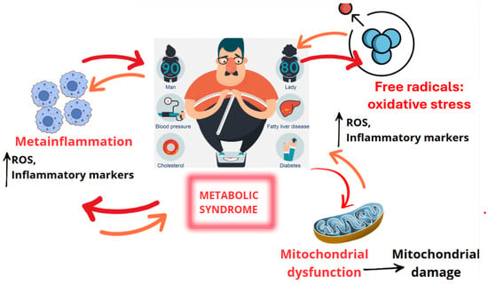
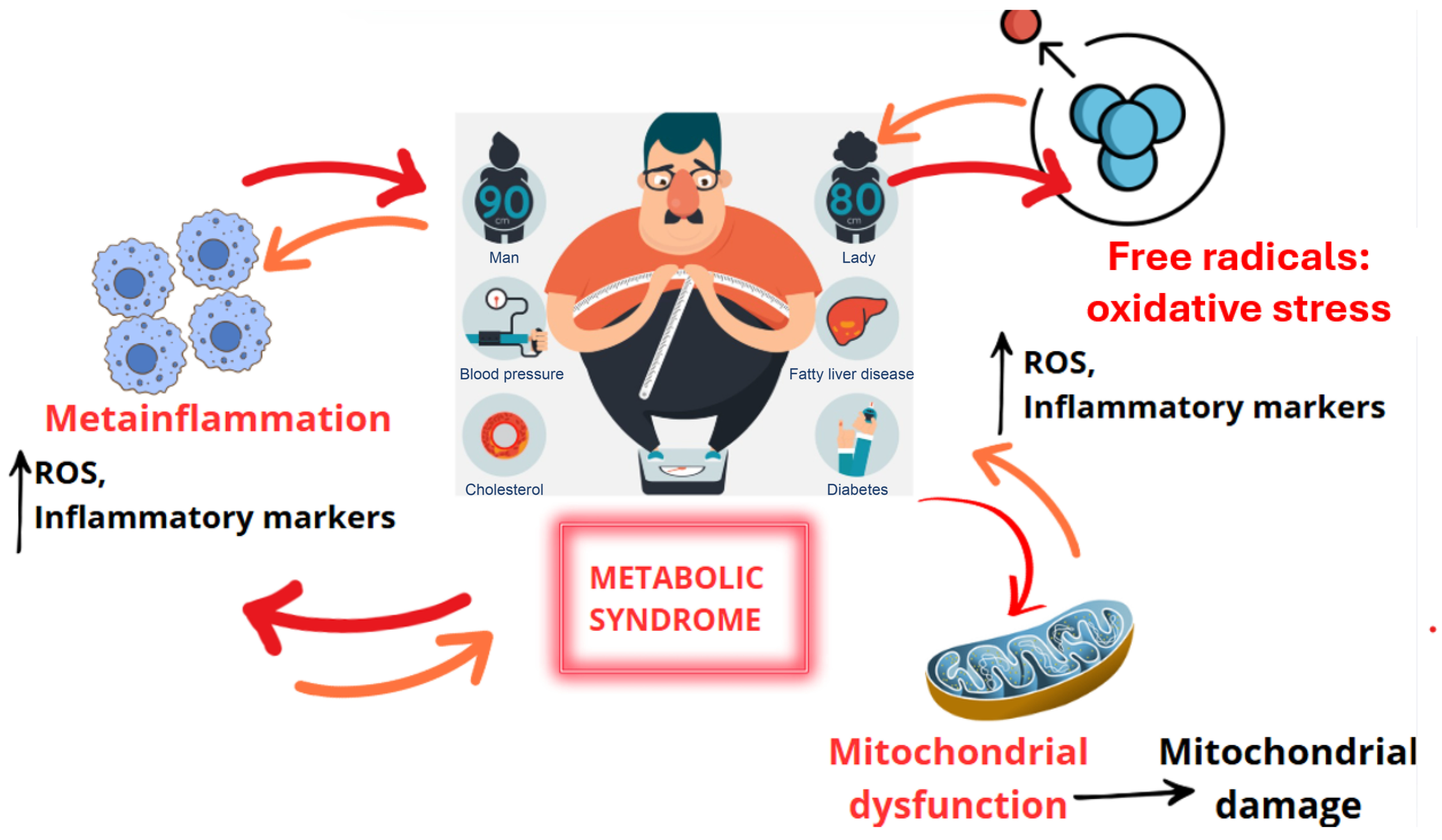
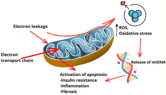
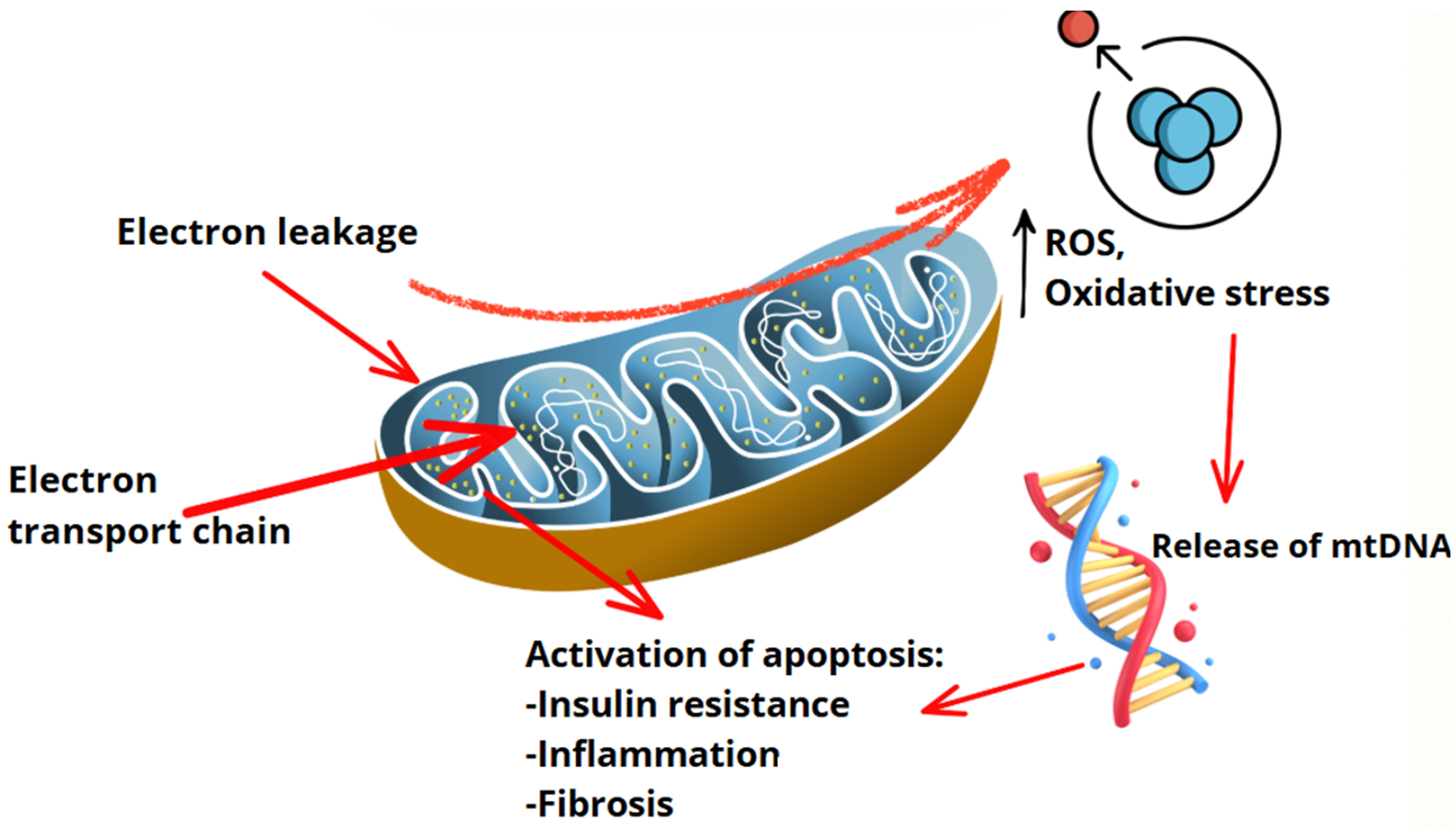
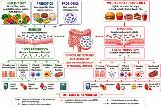
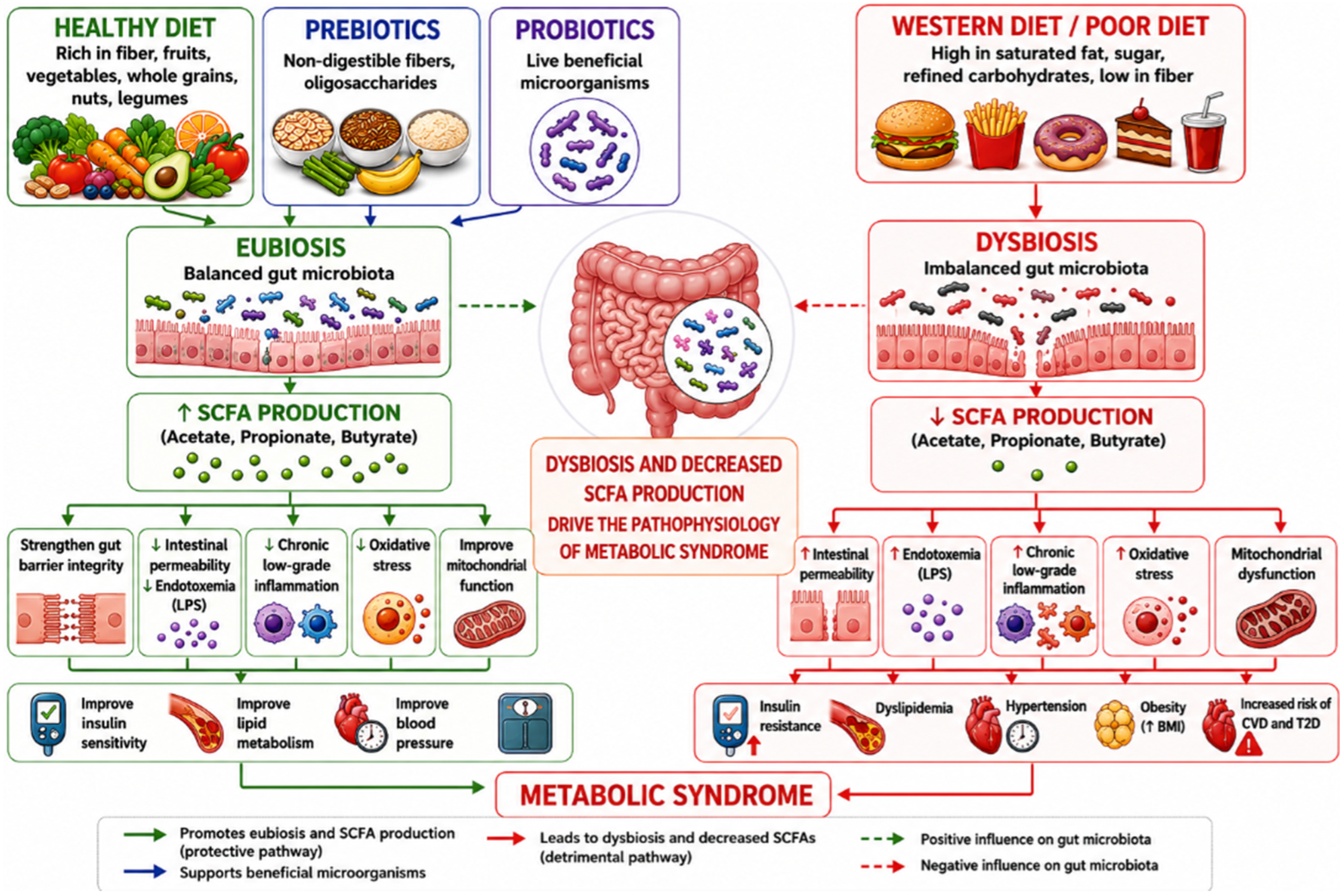
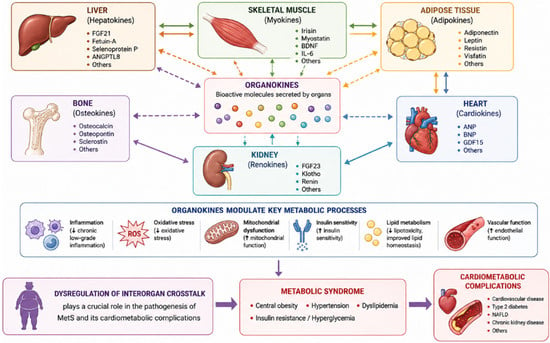
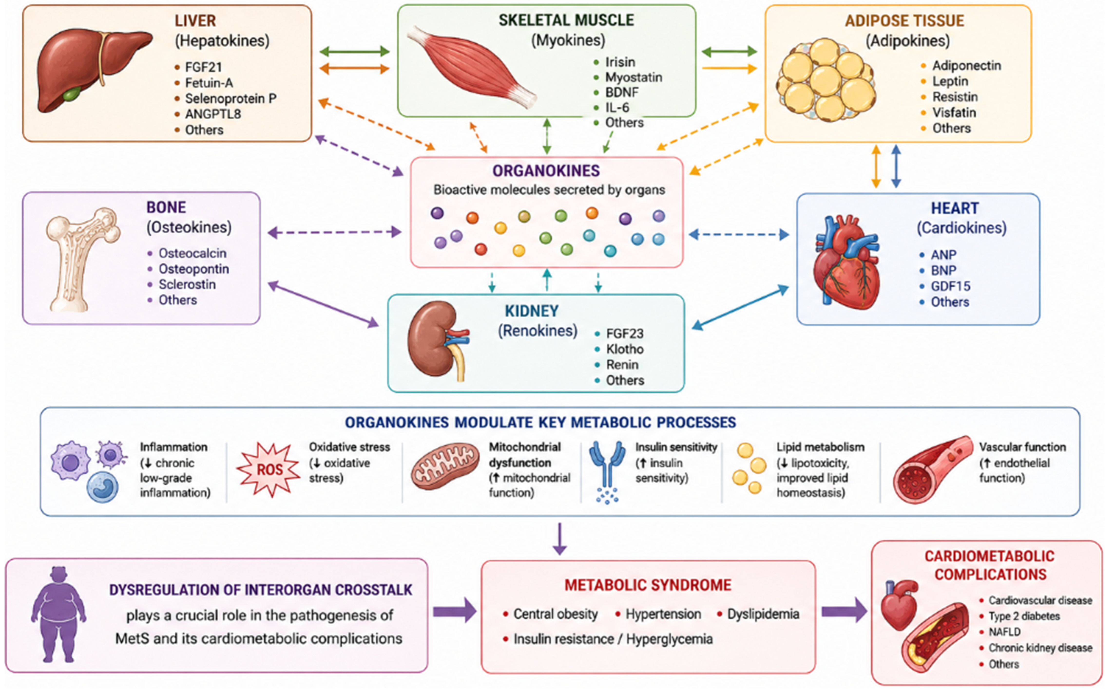
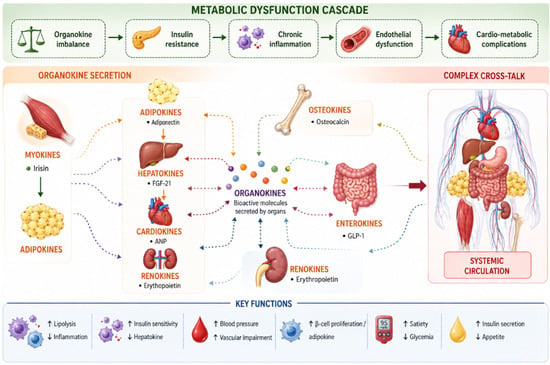
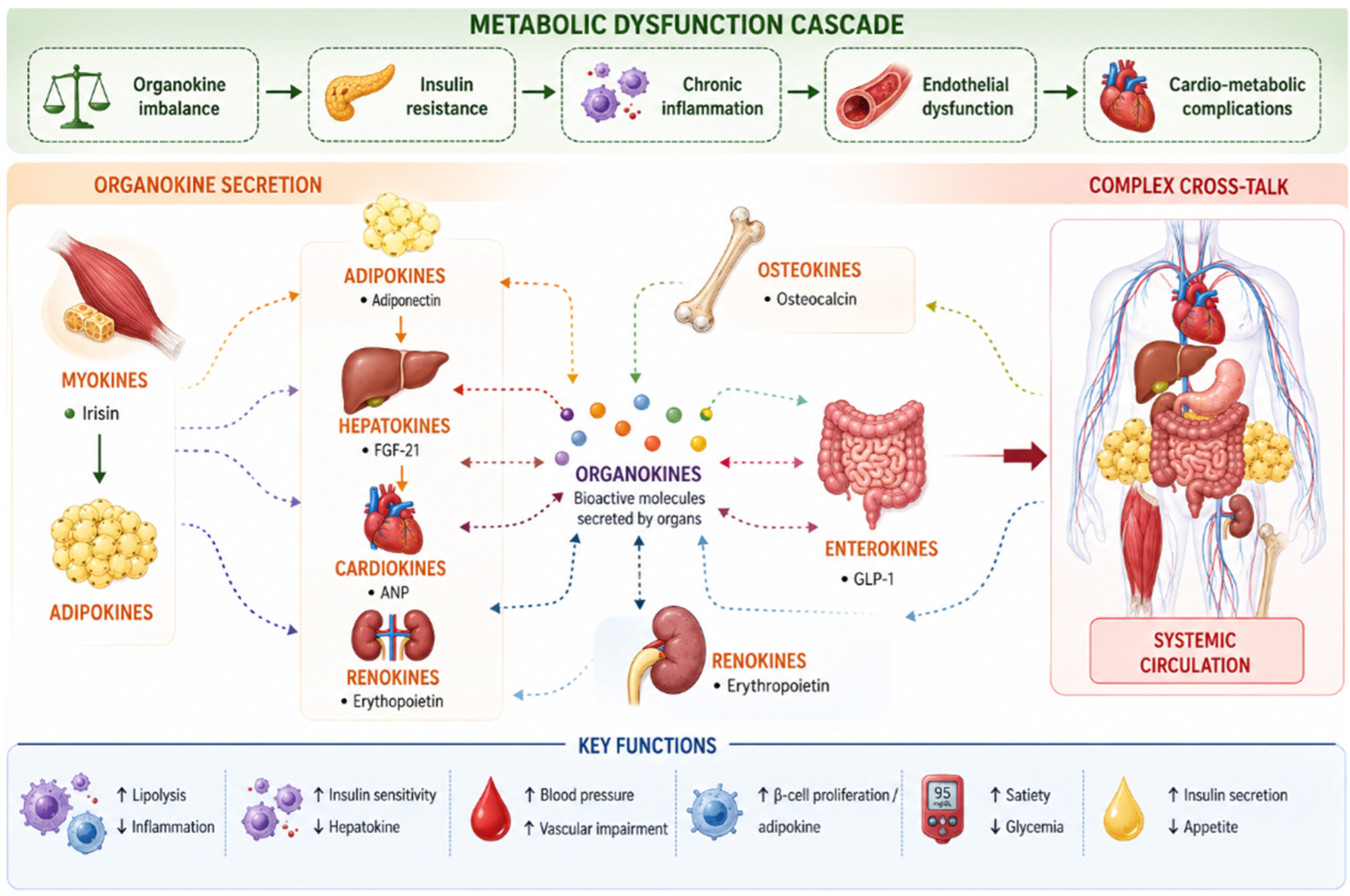

Disclaimer/Publisher’s Note: The statements, opinions and data contained in all publications are solely those of the individual author(s) and contributor(s) and not of MDPI and/or the editor(s). MDPI and/or the editor(s) disclaim responsibility for any injury to people or property resulting from any ideas, methods, instructions or products referred to in the content. |
© 2026 by the authors. Licensee MDPI, Basel, Switzerland. This article is an open access article distributed under the terms and conditions of the Creative Commons Attribution (CC BY) license.