


Abstract
Fibrosis is a common extracellular matrix pathology characterized by increased scarring, representing a critical checkpoint toward cirrhosis and hepatocellular carcinoma. Its onset involves coordinated interplay among hepatocytes, Kupffer, and hepatic stellate cells (HSCs). Extracellular ATP and its derivates act as crucial damage-associated molecular patterns when released by injured liver cells, binding to specific purinergic receptors (P2X, P2Y, and P1) to establish an autocrine–paracrine signaling loop. The hepatic fibrotic response underlies the activation of ATP receptors that generate second messengers and cationic conductance. In parallel, extracellular nucleotidases hydrolyze ATP towards less phosphorylated intermediates and adenosine. This review focuses on the role of P2X and P2Y receptors in liver injury. The P2X7 receptor regulates the NLRP3 inflammasome in Kupffer cells and HSCs, while the P2X4 receptor is upregulated in myofibroblasts, modulating migration and matrix synthesis. Among P2Y receptors, P2Y2 drives inflammation and steatosis but promotes HIF-1α-mediated DNA repair. The P2Y6 receptor promotes alcohol-induced injury but restrains metabolic-dysfunction-associated steatohepatitis. P2Y2 and P2Y4 receptors maintain biliary homeostasis in cholangiocytes, whereas the P2Y1 receptor preserves HSC quiescence by blocking YAP translocation. Finally, UDP-glucose–P2Y14 induces HSC activation. Targeting these specific purinergic receptors or ecto-nucleotidases represents a promising pharmacological frontier against hepatic fibrosis.
1. Introduction
1.1. Chronic Liver Disease and Metabolic-Dysfunction-Associated Steatotic Liver Disease
In 2023, the term non-alcoholic fatty liver disease (NAFLD) was replaced by metabolic-dysfunction-associated steatotic liver disease (MASLD). Therefore, the disease is no longer diagnosed by exclusion (ruling out alcohol) but by affirmative diagnosis; that is, based on metabolic parameters such as overweight, dyslipidemias, cardiovascular anomalies, and diabetes [1]. The pathognomonic characteristic of MASLD is the accumulation of triacylglycerides within hepatocytes, a condition known as metabolic-dysfunction-associated steatohepatitis (MASH), previously termed non-alcoholic steatohepatitis (NASH). The severity of liver conditions depends on the extent of scarring (i.e., fibrosis) and the degree of the inflammatory response, both of which alter hepatic function. Fibrosis occurs in four stages, from stage 0 (mild fatty liver) to stage 4 in which fibrosis is serious but still reversible. The onset of fibrosis and its physiopathological outcomes is a very complex and context-dependent process. The principal factor that promotes the progression from cellular damage to fibrosis, cirrhosis, and cancer is the persistence of harmful stimuli. In the liver, hepatocytes are the cells most commonly affected by pathogens, toxics, autoimmune diseases, and nutritional influences. Damaged hepatocytes communicate the stressful condition to the rest of the hepatic cells by several strategies, but one of the most relevant is the release of DAMPs to the extracellular space. Among DAMPs, ATP and UDP/glucose orchestrate the coordinated response of Kupffer cells (KCs), hepatic stellate cells (HSCs), biliary epithelial cells, and liver sinusoidal endothelial cells (LSECs). ATP and other nucleotides recognize a set of receptors (ion channels, P2X, and GPCRs, P2Y) in each cellular type activating signaling pathways that implement a global inflammatory response. A major step in this process is transdifferentiation that favors the phenotypic change from quiescent HSCs (qHSCs) to active fibrogenic myofibroblasts (MFs), which are the principal fibrogenic cells in the liver. In the presence of TGF-β, MFs enhance the formation of the extracellular matrix by promoting the synthesis of collagen I and III, fibronectin, and proteoglycans, as well as the reduction in metalloproteinase activities (MMPs) by the formation of their inhibitors (TIMPs). The fibrogenetic event is further supported by inflammatory intermediates secreted by Kupffer cells and infiltrated monocytes. Eventually, the upgraded extracellular matrix favors the formation of fibrous bands of scarring material that promote hepatic hypoxic conditions, increasing the expression of HIF-1α and the structural and metabolic alteration of liver parenchyma. In parallel, sinusoids lose their fenestrations, reducing the molecular exchange between the liver and circulating factors, the portal hypertension increases, and the onset and development of regenerative nodules are favored. As a result of this pathological process, the liver presents fibrotic septa, regenerative nodules with parenchymal hepatocytes deeply affected by stiffness conditions and severe oxidative stress. Now, the liver has become cirrhotic, and the organism is prone to present exacerbated portal hypertension, liver failure, and the risk of developing hepatocarcinoma [2,3,4]. Episodes of fibrosis involve specific characteristics of each affected system, and they depend on particular causal factors and triggers. Indeed, fibrosis is an important link related with a variety of relevant translational hepatic pathologies; however, the present review is focused mainly on the early events associated with hepatic fibrosis.
1.2. Fibrosis
Fibrosis is defined as the excessive accumulation of protein elements in the extracellular matrix (ECM). The ECM components play a physiological role in the coordinated communication among cellular types in a given tissue or organ. However, when connective proteins, such as collagens, fibronectin, elastin, laminin, and proteoglycans, are pathogenically accumulated they indicate a fibrotic process, characterized as an organic affliction that compromises cellular function [5]. The occurrence of liver fibrosis is linked to a condition of persistent cellular stress, usually related to illness (virus), intake of organic chemicals (such as alcohol), or nutritional imbalance (associated with MASLD). One of the pivotal processes leading to fibrosis is oxidative stress; in this event, NADPH oxidases (NOXs) play a relevant role (reviewed in [6]).
Fibrotic response is context-dependent, but it usually promotes changes in substrate stiffness and tissue architecture, impairment of sinusoidal exchange, incremental increase in vascular resistance, and generation of regenerative nodules. The pathological development of hepatic fibrosis requires coincidental activity among several cell types. HSCs play a central role in this process [7]. HSCs, located in the Space of Disse, are specialized pericytes rich in vitamin A droplets. Under basal conditions, qHSCs regulate sinusoidal blood flow and produce diverse molecular messengers. However, when hepatic tissue is injured, they react by sensing the release of chemical intermediates (mislocalized metabolites; see next section) from hepatocytes and KCs; transforming growth factor-β [TGF-β], platelet-derived growth factor [PDGF], and pro-inflammatory cytokines), leading to the phenotypic transformation of HSCs into MFs. MFs are responsible for the synthesis and accumulation of new ECM material, namely fibronectin, proteoglycans, and collagen 1 and 3, in the onset of the fibrotic liver [8]. It is appropriate to mention that fibrotic response may eventually compromise the integrity of all liver cell populations, resulting in a potential ATP release from each cellular type. ATP is not only released by necrotic cells; other forms by which ATP reaches the extracellular medium from damaged cells are pannexin and connexin channels, ABC transporters, and the purinergic receptor P2X7.
1.3. Reversibility of Fibrosis
Reversibility of liver fibrosis upon the removal of the causative agent has been documented in patients [9,10,11,12,13] and experimental fibrosis models [14,15,16,17]. This reversibility is characterized by a decrease in pro-inflammatory factors, including interleukin (IL)-6, IL-1β, tumor necrosis factor-alpha (TNF-α), and TGF-β. In the absence of TGF-β signaling, MFs rapidly decline and ECM overproduction is abrogated. Furthermore, the expression of matrix metalloproteinases (MMPs) that degrade collagen fibers is upregulated, surpassing the concentrations of their inhibitors (tissue inhibitors of metalloproteinase [TIMP] proteins) [18]. During the resolution of liver fibrosis, MFs are susceptible to apoptosis and can undergo senescence and death-receptor-mediated cell death caused by deprivation of fibrogenic cytokines [15,19,20].
Upon the withdrawal of fibrogenic signals, MFs increase the expression of the Fas receptor or TNF-α receptor 1 and their ligands, and undergoes a process of caspase-8/caspase-3-dependent apoptosis. Alternatively, shifting the balance toward pro-apoptotic proteins (e.g., p53, Bax) activates the intrinsic pathway, resulting in caspase-9-mediated programmed cell death [14]. Furthermore, natural killer cells and liver-specific ϒδ T cells, activated by interferon-ϒ, induce a rapid decrease in MFs [20,21,22].
On the other hand, it has been demonstrated that approximately half of hepatic MFs escape apoptosis after cessation of liver injury. These MFs return to the Space of Disse and revert to an inactivated phenotype (inactivated hepatic stellate cells, iHSCs) [17,23]. The expression of fibrogenic genes (COL1A1 and α-SMA) in iHSCs is reduced, whereas the expression of some quiescence-associated genes, such as PPARγ, is increased to similar amounts as those detected in qHSCs. However, some genes associated with qHSCs, including GFAP, ADIPOR1, ADPF, and DBP, are not re-expressed in iHSCs [17,24]. Moreover, compared to original qHSCs, iHSCs are more responsive to fibrogenic stimuli and can contribute to recurring liver fibrosis more effectively [17,24]. Additionally, the resolution of fibrosis is associated with a dynamic renewal of HSCs that emerge in the recovering liver from an unidentified source, and which possess all the characteristics of qHSCs [17,23].
Another important component of hepatic fibrosis regression is macrophage conversion. Macrophages play dual roles throughout liver fibrosis progression and resolution. Resident KCs are CD68+; MFs express CD5L, CD163, TIMD4, and VSIG4 while other resident non-KCs and MFs are also positive for CD68 but lack the other markers [25]. During the progression of fibrosis, macrophages are recruited to the liver as a consequence of the injury-induced inflammatory response and produce cytokines and chemokines that trigger the transition of qHSCs into pro-fibrotic MFs. CC-motif chemokine ligand 2 (CCL2) is secreted by HSCs and KCs to facilitate the recruitment of immature monocyte-derived Ly6Chi macrophages into the liver [26]. Ly6C is often transient; monocyte-derived KCs express all KC markers except TIM4 [25]. Pro-inflammatory macrophages are positive to CD86 and MHCII, while reparative MFs are positive to CD163 and CD206 [27]. Monocytes recruited during steatosis and fibrosis appear to have a unique identity, and have received distinct names such as lipid-associated macrophages (LAMs), scar-associated macrophages (ASMs), and NASH-associated macrophages (NAMs). All three MFs express a similar expression of markers [28]. A detailed description of MFs in fibrosis has been previously reviewed [28]. Interestingly, deletion of macrophages in the CD11b-DTR transgenic mouse line, which carries a diphtheria toxin-inducible system that depletes macrophages [29], led to reduced scarring and MFs in CCl4-induced liver fibrosis, supporting the role of macrophages in promoting this disease [29]. However, during liver fibrosis recovery, macrophages change to a Ly6Clow phenotype and stop producing fibrogenic and inflammatory factors; instead, they secrete MMPs, such as MMP9 and MMP12 [30]. MMPs are the primary enzymes capable of ECM degradation [14]. Additionally, MFs are a major source of TIMP production. The disappearance of MFs leads to reduced TIMP levels, increased MMP activity, and ECM degradation [31].
Because reversibility of liver fibrosis depends on the collagenolytic activity of ECM-degrading MMPs, sustained expression of TIMPs inhibits active MMP function. Moreover, the lack of ECM degradation may be caused by tissue transglutaminase, which prevents MF apoptosis and mediates ECM cross-linking. The latter prevents proteolytic cleavage of different collagen types [15,20,32].
1.4. Animal Models of Liver Fibrosis
Animal models of liver fibrosis are essential for investigating the pathogenesis of this disease in experimental conditions. There are currently five types of in vivo models: (a) chemical, (b) dietary, (c) surgical, (d) immune, and (e) transgenic animal models [33,34]. The most frequently utilized organisms are mice [35], rats [36], rabbits [37], Ossabaw pigs [37,38], macaques [37,38,39], and zebrafish [40]. Table 1 describes the most common experimental protocols of liver fibrosis, each one offering varying levels of efficacy and contributing differently to the overall understanding of this disease.
Table 1.
Animal models for the study of fibrosis.
1.5. Epidemiology
Chronic liver diseases are among the leading causes of death worldwide. According to the 2019 Global Burden of Disease study, mortality related to cirrhosis and other liver diseases increased by 13% between 1990 and 2019 [61]. As the final clinical stage of liver disease, cellular hepatocarcinoma is responsible for 8.3% of global cancer mortality, as reported in the 2020 Global Cancer Statistics update [62]. Viral hepatitis B and C annually cause around 1.3 million deaths [63]. Moreover, approximately 3.3 million people are diagnosed with alcohol-associated liver disease (ALD) per year, accounting for 5.9% of global deaths [64]. The rising fatalities from MASLD are also noteworthy, with an estimated 280,000 deaths in 2019 [65].
Regardless of the cause of liver disease, liver-related mortality increases exponentially with fibrosis [66]. In ALD, severe fibrosis prior to developing cirrhosis has a major impact on 10-year mortality [67]. In MASLD, higher stages of fibrosis are proven to be the strongest predictors of mortality [67,68,69,70].
Otherwise, a recent systematic review and meta-analysis have revealed a considerable rise in the prevalence of advanced liver fibrosis from before 2010 (2.0%) to after 2016 (4.7%). That study also found that advanced fibrosis was significantly more prevalent in men than in women, as well as in older individuals than in younger ones. Furthermore, the prevalence rates varied significantly across continents and countries. Advanced fibrosis had a prevalence of 4.0% in Africa, 2.6% in Asia, 3.4% in Europe, 2.2% in Latin America and the Caribbean, 4.8% in North America, and 10.6% in Oceania [71]. In this context, the study revealed that among the factors potentially associated with the risk of advanced liver fibrosis in the general population, viral hepatitis had the strongest association. Age, diabetes, excessive alcohol intake, hepatic steatosis, obesity, and sex were found to be other likely risk factors for advanced fibrosis [71].
2. Purinergic Signaling as a Damage-Associated Molecular Pattern
2.1. Damage-Associated Molecular Patterns
The correct activity of biological systems involves functional communication and coordination among their tissular and cellular components. There are several facets to sustaining this homeostatic state, such as endocrine, paracrine, autocrine, and juxtacrine signaling, which allow physiological recognition between ligands and receptors during cellular communication [72]. Another modality in this cellular dialog is the coordinated response elicited in situations of stress, damage, or cellular death, when endogenous molecules lose their compartmentalization and act as damage-associated molecular patterns (DAMPs). DAMPs are released from stressed cells and selectively recognized by pattern recognition receptors (PRRs). A similar response occurs when the molecules acting as PRR ligands are exogenous or come from pathogens such as bacteria, viruses, and other microbes. In this case, the ligands are called PAMPs (pathogen-associated molecular patterns); examples of PAMPs include lipopolysaccharides (LPSs) and peptidoglycans [73].
DAMPs are classified into three principal categories: protein-based, nucleic-acid-based, and mitochondria-derived. The most important PRRs are Toll-like receptors (TLRs), nucleotide-binding oligomerization domain-like receptors (NLRs), C-type lectin receptors (CLRs), and receptors for advanced glycation end-products (RAGEs), whereas the major signaling pathways associated with DAMP responses are NF-κB, MAPK, inflammasomes, and cyclic GMP-AMP synthase-stimulator of interferon genes (cGAS-STING) [74]. Overall, DAMPs and PAMPs promote an alert response that triggers the reversal and repair of cellular damage, and it is usually accompanied by an escalated inflammatory reaction [72].
The variety of DAMPs is extensive. Intracellular molecules that are accepted as DAMPs are: (1) nuclear proteins, like histones and the high mobility group box 1 protein (HMGB1); (2) DNA and RNA fragments, both nuclear and mitochondrial; (3) heat-shock proteins; (4) hyaluronan pieces; and (5) purines, such as uric acid and ATP. The properties and characteristics of ATP acting as a DAMP will be developed in the next section.
2.2. ATP
ATP is a multifunctional molecule and the most abundant purine nucleotide in the intracellular environment. ATP supports key cellular processes as a metabolic switch coordinating anabolic and catabolic reactions. It functions as a strategic energy currency in a dynamic equilibrium with less phosphorylated intermediates (ADP, AMP, and adenosine [ADO]) [75]. Within the intracellular milieu, ATP is one of the most concentrated molecules, being present in the 5–10 mM range [76]. It acts as the primary phosphorylating factor during biochemical energy transfer reactions in the cytoplasm and organelles. ATP also serves as an allosteric agent in metabolic pathways related to the synthesis and degradation of energy molecules (mainly sugars and fats) [77]. In contrast, the extracellular presence of ATP is very discreet (30–100 nM), as it is approximately eight orders of magnitude lower than its intracellular concentration. However, most cellular systems can use ATP as a molecular messenger. In those circumstances, ATP is released and reaches temporal concentrations near the low µM range in the extracellular space. ATP acting as a ligand in this context can recognize a variety of receptors, making it a substantial part of purinergic signaling communication [78].
2.3. Purinergic Signaling
Purinergic signaling is an interesting cellular communication system that presents two modalities: (1) ATP and other nucleotides recognize G-protein-coupled receptors and ligand-gated ion channels; (2) ADO, which is obtained by ATP degradation, acts as a ligand of a different family of G-protein-coupled receptors [79]. Purinergic receptors activated by ATP are known as P2, whereas receptors activated by ADO are termed as P1. ADO receptor subtypes are A1, A2A, A2B, and A3. Ligands for P2 receptors include ATP, ADP, UTP, UDP, and UDP-glucose, whereas P2 receptors are subdivided into P2X and seven subunits (P2X1–7), forming homo- or hetero-trimeric functional channels whose main conductance mobilizes Na+, Ca2+ and K+, and P2Y (including eight subtypes: P2Y1, 2, 4, 6, 11, 12, 13, and 14). This activity influences intracellular Ca2+ and cyclic adenosine monophosphate (cAMP) amounts [80]. Therefore, purine communication involves a complex equilibrium among the expressed ATP and ADO receptors, the activity of different extracellular ectonucleotidases, and the resulting proportion of ATP-ADP and ADO acting as ligands. The principal ectonucleotidases are CD39 and CD73; both can be regulated by chemical messengers such as TGF-β and by hypoxic conditions. The concept that encompasses and controls this dynamic molecular network is known as the purinome [81]. In the context of liver fibrosis, the purinome characteristics define whether the tissue response is inflammatory, fibrogenic, reparative, or immunosuppressive.
The ATP affinity constant (Kd) for P2Y receptors is in the low µM range (0.1–5 µM), whereas the affinity of ATP for P2X receptors is slightly higher (Kd from 5 to 20 µM), with the exception of the P2X7 receptor (see next section), since the ATP binding for this receptor shows a very high Kd of ~500 µM [82]. Interestingly, during severe cellular stress and damage, ATP is released in abundance, causing a significant increase in extracellular ATP concentrations to 10–1000 µM; under these circumstances, ATP acts as a DAMP [74]. Therefore, the P2X7 receptor plays a strategic role in cellular adaptations under stress conditions when ATP is an active DAMP [83].
The crystallographic structure of P2X receptors confirmed that functional receptors are formed by three subunits [84]. All cloned subunits, with the exception of P2X6, may form homotrimers, and all subunits, except P2X7, form heterotrimeric channels [85,86].
3. Purinergic Receptors and Liver Fibrosis
Several receptors have been identified as important in the purinergic regulation of hepatic pathologies. In this section, the most representative will be mentioned with respect to their physiopathology and pharmacological actions. However, it should be noted that some receptors are likely harmful and targetable, such as P2X7 receptor inflammasome-driven injury or the P2Y14 receptor in HSC activation, whereas others may have context-dependent or protective effects, such as the P2Y1 receptor delaying HSC activation or the P2Y13 receptor regulating the adipose tissue–liver axis.
3.1. P2X7 Receptor
The P2X7 receptor was originally described as a bifunctional protein expressed in immune cells, an ATP-gated channel mediating Ca2+/Na+ influx and K+ efflux, and a cytotoxic receptor characterized by its ability to open the “megapore” (a non-selective and irreversible conductance mediating massive entry of Ca2+), leading to the activation of the mitochondria-induced apoptotic pathway [87]. This characteristic motivated its extensive characterization in multiple cellular systems.
Recently, the ability of the ATP/P2X7 receptor to function as a DAMP ligand/receptor pair that modulates the inflammatory process by regulating NLRP3 inflammasome assembly has opened a new avenue in the understanding of the physiological role of the P2X7 receptor, highlighting its relevance in chronic inflammation processes triggered by tissue damage [83]. In this context, we will review the role of the P2X7 receptor in the fibrotic stage of chronic liver disease.
Hepatotoxic damage induced by four weeks of CCl4 administration is a well-established model of reversible liver fibrosis [88]. In whole-liver lysates, this treatment increased the expression level of the P2X7 receptor, inflammatory mediators, and the activity of the transcription factor NF-kB [89]. All the effects promoted by CCl4 were attenuated by systemic administration of A438079, a potent P2X7 receptor antagonist, suggesting a profibrotic role of this entity in the onset of hepatotoxic liver fibrosis [89].
The role of sterile inflammation in hepatic disease was studied in a mouse model of alcoholic steatohepatitis (ASH), particularly with ATP and uric acid acting as DAMPs. Null mice for P2rx7 (P2rx7−/−), transgenic mice with increased activity of uric acid metabolism, or mice treated with drugs that reduced the extracellular amounts of ATP and uric acid, showed attenuated liver damage induced by the Lieber–DeCarli alcohol diet in comparison with control mice. All the mentioned experimental models showed reduced inflammasome activity markers, including activated caspase-1 and release of IL-18. Results from in vitro experiments suggested that ATP and uric acid were released by damaged hepatocytes but not by liver mononuclear cells to promote NLRP3 inflammasome assembly [90] (Figure 1).
Figure 1.
Assembly of NLRP3 inflammasome mediated by ATP/P2X7 receptor. Activation of the P2X7 receptor induces K+ efflux, leading to NEK7 kinase activation followed by the assembly of NLRP3 and accessory protein complex. Into the complex, pro-caspase-1 is matured to process pro-IL-18 and pro-IL-1β, and Gasdermin D to split the amino-end (N) from the carboxi-end (C). Apoptosis-associated speck-like protein (ASC) is composed of the CARD and PYD domains and contains a caspase recruitment domain (CARD) and a PYD domain for homotypic interaction with a PYD-containing NLRP3 protein. Procaspase-1 features CARD and caspase (CD) domains; CARD domain mediate homotypic interactions with ASC to recruit procaspase-1 to the inflammasome complex, NLRP3 activation promotes pyroptosis and cytokine release.
On the other hand, Blassetti and colleagues analyzed the role of the P2X7 receptor-dependent inflammasome in the progression of MASLD to MASH. The disease was induced by feeding wild-type and P2rx7−/− mice with a high-fat diet for 16 weeks. P2rx7−/− mice were less sensitive to developing MASH (1/7 vs. 4/7 of control) and displayed an attenuated expression of markers for oxidative stress, inflammation, and fibrosis. Furthermore, it was demonstrated that the deletion of P2X7 receptor expression prevented NLRP3 inflammasome activation by ATP in isolated liver sinusoidal endothelial cells (LSECs) from P2rx7−/− individuals [91]. A translational study by the same research group explored the role of the P2X7 receptor–NLRP3 inflammasome pathway in MASLD, MASH, and hepatitis C virus (HCV)-related hepatic disease using samples from 46 patients. According to the findings, P2X7 and P2X4 receptors, the NLRP3 inflammasome, and caspase-1 were highly expressed in liver biopsies from patients with HCV; pro-inflammatory interleukins, such as IL-2, IL-6, and TNF-α, were also elevated [91]. Although this study is interesting, it is inconclusive because it lacked a comparison against samples from healthy donors.
Baeza Raja and collaborators investigated which hepatic cellular types express the P2X7 receptor and the effects of its pharmacological inhibition in human cells and in a non-human primate model of liver injury [92]. In accordance with previous reports [91], this study demonstrated that the P2X7 receptor was overexpressed in human biopsies from MASH patients compared with healthy donors. Components related to NLRP3 and AIM2 inflammasomes were significantly increased in those biopsies. P2X7 receptor expression and inflammasome components were mainly localized in infiltrated immune cells and KCs in comparison to hepatocytes and HSCs [92], corroborating previous observations of the functional expression and pro-inflammatory actions of the P2X7 receptor in the KC line KUP5 [93].
Thus, the P2X7 receptor/NLRP3 pathway was characterized using in vitro isolated monocytes and KCs, with both cell types responding to the double stimulus of LPS + ATP with an evident increment in IL-1β release; this effect was abolished by co-administration of the P2X7 receptor antagonist SGM-1019. Interestingly, P2X7 receptor-mediated IL-1β release by KCs induced apoptosis in hepatocytes and activated HSCs in mice treated with LPS. In a preclinical trial using non-human primates, researchers assessed the importance of the P2X7 receptor as a therapeutic target. Monkeys were treated with CCl4 for four weeks with or without administration of the P2X7 receptor antagonist SGM-1019. The histological analysis of control livers revealed signs of hepatotoxic damage, characterized by hepatocyte ballooning, inflammation, and fibrosis; in contrast, these signs were counteracted by SGM-1019 [92]. These results strongly suggest that the P2X7 receptor is a suitable pharmacological target for therapeutics in a variety of hepatotoxic protocols.
Inflammatory responses mediated by the P2X7 receptor/NLRP3 pathway have also been described in other cell types besides KCs. Two-step NLRP3 activation was also tested in HSCs. Cultures from the HSC line LX-2 responded with a significant increase in IL-1β release when challenged with LPS + ATP; the response was abolished by preincubation with A438079, indicating that it was mediated by the P2X7 receptor. Furthermore, to investigate the dual cell-to-cell regulation between KCs and HSCs, LX-2 cell cultures were stimulated with conditioned medium (CM) from LPS-activated RAW 264.7 mouse macrophages. The CM induced an increment in transcripts for the inflammatory mediators IL-1β, IL-18, P2rx7, Nlrp3, and Casp-1; in the fibrotic markers Acta2 and Col1A1; and, interestingly, in the release of ATP into the extracellular space, compared with CM from macrophages not activated with LPS. The release of IL-1β was also abolished by antagonizing the P2X7 receptor, evidencing the participation of the ATP/P2X7 receptor/NLRP3 pathway [94]; in the same study, the interplay between macrophages and HSCs orchestrated by the DAMP pair ATP/P2X7 receptor was also characterized. The sensitivity of HSCs to extracellular ATP was demonstrated in primary cultured mouse HSCs, where P2X7 receptor ligation induced NLRP3 inflammasome activity and supported inflammation in alcohol-mediated liver damage [95]. On the other hand, it has been described that exosomes from hepatocytes infected by the hepatitis B virus promoted HSC activation and, consequently, liver fibrosis; in the HSC line LX2A, it was shown that these responses were supported by the actions of METTL3 (N6-adenosine-methyltransferase 70 kDa subunit), which stabilized the P2rx7 transcript by binding N6-methyladenosine (m6A). P2X7 receptor availability supported HSC activation [96].
In MASH, the P2X7 receptor has also been associated with the enhancement of metabolic oxidative stress (MOS), which is related to autophagy induction. In a study by Das and colleagues [97], MOS was induced by treatment with a methyl-choline-deficient diet and by administering the toxin bromodichloromethane (BDCM) in a diet-induced obesity (DIO) mouse background. DIO + BDCM induced MOS hallmarks in the liver, such as increases in 4-hydroxynonenal, tyrosine nitration, and 5,5-dimethyl-1-pyrroline N-oxide-nitrone adducts. Interestingly, increased transcript and protein expression of the P2X7 receptor were detected in both models. The effects of both treatments were attenuated in the knockout mouse for the cytochrome P-450 CYP2E1, one of the enzymes mediating MOS response. MOS-induced increments in the P2X7 receptor transcript were detected in hepatocytes, KCs, and LSECs [97]. MOS induction by both pro-oxidant diets also resulted in an upregulation of transcripts coding for autophagy-related markers (Gabarap, Atg2a, and Lc3b) and proteins of lysosomal translocation complex Hsc70 and Lamp2, which were also significantly attenuated in CYP2E1KO mice. None of these changes were observed in mice lacking P2X7 receptor expression, suggesting that this receptor is an important player in the regulatory network of autophagy in MASH [97]. Furthermore, in addition to inflammation, the P2X7 receptor is an effector in a network of signals regulated by MOS through MASH in HSCs. Mechanistically, CYPE21 enhanced activity under oxidative stress, leading to high circulating leptin amounts that, in turn, upregulated P2X7 receptor expression. The receptor then triggered HSC activation and increased the intracellular concentration of glucose, thereby regulating the membrane localization of the Glut4 transporter and boosting the onset of fibrosis [98].
Recent studies characterizing the mechanisms that regulate P2X7 receptor expression in fibrotic liver have suggested that Kruppel-like factor 15 (KLF15), a transcription factor whose ablation potentiates steatosis and inflammation induced by high-fat diets [99], negatively modulates P2X7 receptor expression, and consequently, the P2X7 receptor/NLRP3 inflammatory axis [100]. On the other hand, a crosstalk between the TGF-β signaling and P2X7 receptor/NLRP3 pathways in the context of fibrosis has been described [101]. The authors assayed the feasibility of TGF-β receptor ALK5 inhibitors J-1155, J1156, and LY-2157299 to reverse thioacetamide-induced fibrotic damage. Indeed, the inhibitors prevented the increment in P2X7 receptor expression caused by thioacetamide, and hence, its inflammatory effects [101].
3.2. P2X4 Receptor
The P2X4 receptor is a member of the P2X subfamily, which is phylogenetically closer to the P2X7 receptor [86]. The P2X4 receptor is mainly homomeric and, compared with P2X7, it displays a significantly higher affinity for ATP (1–10 µM vs. 100–500 µm) and faster desensitization [102]. Experimental findings suggest that the P2X4 receptor plays an important role in the fibrogenic process.
The fibrogenic effect of BLD or the administration of a choline-deficient diet was assayed in P2X4 knockout mice (P2rx4−/−) and compared against wild-type individuals. Deletion or pharmacologic antagonism of P2X4 prevented collagen accumulation within the liver but did not affect steatosis, strongly suggesting that the P2X4 receptor is selectively expressed in liver MFs, the cells responsible for collagen I secretion. Interestingly, P2rx4−/− mice did not present any changes when fibrosis was induced by CCl4, suggesting that the role of the P2X4 receptor depends on fine-tuning mechanisms inherent to each experimental model. Moreover, upregulation of the P2rx4 transcript was observed in biopsies from human patients with HCV, primary biliary cholangitis, primary sclerosing cholangitis, alcoholic cirrhosis, and NASH. Furthermore, in cultured human or mouse MFs, ATP stimulation of the P2X4 receptor upregulated the main activation marker, αSMA. Accordingly, siRNA downregulation of the P2X4 receptor reduced basal levels of this marker in MF cell lines and the fibrogenic phenotype by regulating cell adhesion, migration, and the balance between synthesis and degradation in the ECM. This study also demonstrated that the P2X4 receptor regulates lysosomal exocytosis phenomena in MFs, potentially leading to the secretion of molecules associated with the induction or maintenance of fibrosis [103]. Similar to the controversy with the P2Y6 receptor, the “contradictory” results regarding the role of the P2X4 receptor in the pathology of liver diseases could be context-dependent. Indeed, more experiments will be beneficial to elucidate this controversy.
In a model of alcoholic liver fibrosis (ALF) induced by ethanol plus CCl4 in mice, it was shown that the P2X4 receptor is upregulated in the liver; similar findings were observed in the HSC-T6 mouse cell line upon stimulation with acetaldehyde. In ALF-induced animals, administration of the P2X4 receptor antagonist 5-BDBD prevented fibrosis induction. In the HSC-T6 line, the main pathway activated by acetaldehyde was PI3K/AKT; silencing or pharmacological antagonism of the P2X4 receptor prevented HSC-T6 activation. Interestingly, activation of HSC-T6 cells induced by the conditioned medium of acetaldehyde-stimulated RAW267.4 macrophages was prevented by preincubating macrophages with the P2X4 receptor antagonist 5-BDBD [104]. Similar increments in P2X4 receptor expression in liver tissue were demonstrated by an independent study using the ALF model. This study also reported an enhancement in the expression of CD39 ectonucleotidase and total ATP content. Similarly, pharmacological treatments in whole liver and RAW267.4 macrophages showed that P2X4 receptor activity contributed to liver steatosis and the production of pro-inflammatory cytokines, with a concomitant regulation of CD39 expression. In the in vitro model, evidence showed that P2X4 receptor stimulation positively regulated the expression of NLRP3, caspase-1, and IL-1β inflammasome components; these changes were correlated with increased intracellular Ca2+ responses. Furthermore, the conditioned medium of RAW267.4 macrophages stimulated with ethanol increased apoptosis in the hepatocyte line AML-12 [105].
On the other hand, HCV infection of HEK293 cells overexpressing the P2X4 receptor regulated the expression of the antioxidant enzymes HO-1 and Cu/Zn SOD, boosting the expression of TNF-α, TGF-β, angiotensin II, and components of the ECM, namely laminin and elastin [106]. Since the participation of other P2X receptors in liver fibrosis is unknown, the role of the P2Y receptor in fibrosis will be discussed below.
3.3. P2Y2 Receptor in Liver Fibrosis
The P2Y2 receptor belongs to the P2Y1-like subfamily of P2Y receptors. It is coupled to Gq proteins and thus associated with the turnover of phosphoinositides and Ca2+. The order of potency of natural and synthetic ligands for the human P2Y2 receptor is as follows: agonists MRS2698 = UTP ≥ ATP > Ap4A; antagonists AR-C118925 > PSB-11415 [107]. UTP and ATP naturally occur in the cell and may function as DAMPs or as autocrine/paracrine mediators [108,109]. The P2Y2 receptor plays a pivotal role in hepatic cell renewal and cell proliferation [110,111,112]. In a physiological state, this receptor is located mainly in the perivascular regions of the central and portal veins. In the fibrotic liver induced by CCl4, the P2Y2 receptor loses its zonal distribution, spreading throughout the entire hepatic parenchyma and exhibiting increased expression. This increase is related to an exacerbation of P2Y2 receptor responses in fibrotic hepatocytes [113].
The P2Y2 receptor is the only receptor involved in the induction of acute hepatitis with concanavalin A in a murine model. Ayata et al. demonstrated that its expression in bone marrow is crucial for neutrophil infiltration in the damaged liver. They also showed that activation of this receptor potentiates the TNF-α cell death response in hepatocytes. This effect was counteracted by preincubation with suramin, a classical antagonist of the P2Y2 receptor, which also reduced hepatic necrosis and circulating transaminase levels [114].
In the context of metabolic syndrome, Merz and colleagues found few signs of metabolic syndrome in adipose tissue from P2ry2−/− mice consuming a high-fat diet. The mice gained less weight, and their liver and adipose tissue occupied less volume than in wild-type mice. Plasma cholesterol and C-peptide levels were lower, and insulin resistance was improved in KO mice. Findings demonstrated that interfering with P2Y2 signaling prevents accumulation of immune cells in this model, reducing inflammation of adipose tissue and improving the metabolic phenotype [115].
In ALD, a blockade of the P2Y2 receptor with suramin prevented liver damage and lipid accumulation and decreased CD39 levels in mice. Protection with suramin proved to be dose-dependent. The best performance was identified at a high dose (20 mg/kg). This dose effectively damped circulating transaminases, free fatty acids, and inflammatory cytokines [116]. In isolated mouse hepatocytes and the RAW264.7 macrophage cell line, inhibition of the P2Y2 receptor with suramin or siRNA decreased the expression of IL-6, IL-1β, TNF-α, and CD39. In addition, exposure to 100 µM of ethanol increased P2Y2 receptor, CD39, and ATP concentrations [116]. Liu et al. suggested that the increase in CD39 ectonucleotidase represented a negative feedback mechanism for ATP-induced changes, as hydrolysis of the nucleotide limits P2Y2 receptor activation. In 2022, Liu more deeply explored the mechanism of liver inflammation mediated by the P2Y2 receptor in ALD. The authors found that the P2Y2 receptor regulated inflammation through the EGFR-ERK1/2 pathway and demonstrated that blockade with suramin or P2Y2 receptor silencing with siRNA attenuated ethanol-induced hepatocyte apoptosis and decreased pro-inflammatory cytokines, such as TNF-α and IL-1β [117].
In a murine model of hepatic fibrosis induced by CCl4, our group evaluated the role of the P2Y2 receptor. Similar to the findings above regarding the loss of zonal distributions of P2Y2 receptors in livers from CCl4-treated mice, we found that fibrotic hepatocytes presented exacerbated ERK phosphorylation in response to UTP. Furthermore, using microarrays, we observed that P2Y2 receptor activation induced significant changes in gene expression in fibrotic hepatocytes compared to healthy hepatocytes, including transcripts coding for DNA repair proteins [113]. Hepatocytes isolated from fibrotic mice showed that P2Y2 receptor activity prevented the formation of histone γH2AX foci induced by etoposide, a drug that promotes double-strand DNA damage; this effect correlated with the upregulation of a cluster of genes associated with double-strand DNA repair. We also found that the response mediated by the P2Y2 receptor required a crosstalk with hypoxia-induced factor 1 (HIF-1α) signaling [118]. The P2Y2 receptor is a multifaceted mediator in hepatic fibrosis and other inflammatory liver diseases. Overexpression and activation of this receptor in hepatic parenchyma not only facilitate inflammatory and proliferative responses, but also play a crucial role in the preservation of genomic integrity in hepatocytes by activating DNA repair mechanisms mediated by HIF-1α [118]. Pharmacological regulation of P2Y2 receptor signaling is a promising therapy for modulating the progression of fibrosis and other chronic hepatic diseases.
In another model of metabolic syndrome, Cano-Martínez et al. showed that treatment with resveratrol and quercetin downregulated the expression of the P2Y2 receptor, TLR4, and neutrophil elastase. These combined treatments reduced collagen deposition, hepatic fibrosis, and liver apoptosis. Improvement was detected especially with the normalization of circulating transaminases [119]. In this context, Dusabinama et al. studied the effect of a high-fat diet on P2ry2−/− mice; they found that P2Y2 receptor deficiency improved insulin resistance and favored the decline in serum levels of insulin and transaminases, as well as decreased expression of fatty acid synthesis mediators, such as fatty acid synthase and stearoyl-CoA desaturase 1. In addition, P2Y2 receptor deficiency enhanced the activity of AMP-activated protein kinase (AMPK), improving fatty acid oxidation [120].
In the context of inflammation caused by bacterial agents, Arunachalam A. et al. evaluated the impact of a sepsis model in P2ry2−/− mice. They reported that KO mice were protected against mortality and systemic cytokine storms. P2Y2 receptor activation depleted serum arginine, a vital part of the immunological defense against bacteria. In addition, P2ry2−/− mice presented more efficient bacterial elimination. Furthermore, P2ry2−/− mice showed attenuated inflammation and liver injury, as well as bacteremia, enhancing hepatocyte survival. Another discovery was that P2ry2−/− mice exhibited reduced hepatic MMP-9 expression [121]. It has been reported that high levels of MMP-9 promote liver fibrosis, inflammation, and macrophage recruitment, leading to epithelial–mesenchymal transition [122].
In the next two sections, we will discuss the remaining P2Y1-like receptors and the other subfamily of P2Y12-like receptors.
3.4. P2Y1-like Receptors (P2Y1, P2Y2, P2Y4, P2Y6, P2Y11)
In 2004, Dranoff et al. found that P2Y receptors, specifically UDP-sensitive P2Y6, increased procollagen-1 transcript levels by three times in rat MFs. The authors also found that P2Y2 and P2Y4 receptors were expressed in qHSCs [123]. Several years later, Yuan et al. reported in an ASH murine model that inhibition or silencing of P2Y6 receptor expression reduced liver inflammation and steatosis by decreasing TNF-α, IL-1β, and IL-6 after interrupting the p38 MAPK pathway activated by UPD-mediated calcium release [124]. The response induced by UDP was similar in RAW 264.7 cells. These results suggested that the P2Y6 receptor could act as a possible therapeutic target, but these results were not reproduced by Nishiyama’s group. Instead, they found that in P2ry6−/− mice, hepatic steatosis was not alleviated, but rather aggravated, with an increase in serum AST and C-C motif chemokine 2 mRNAs in choline-deficient rats treated with a high-fat diet. These findings suggested that the P2Y6 receptor did not contribute to disease progression [125]. Unlike the study conducted by Yuan, Nishiyama used a NASH model; this difference might account for the discrepancy in results. Certainly, more experiments are needed to define the role of the P2Y6 receptor in hepatic maladies since it is very probable that the importance of this receptor could be dependent on pathological context.
On the other hand, in normal human cholangiocytes and cholangiocarcinoma cell lines (HuCCT1, Mz-Cha1, and F258), it was shown that stimulation of the bile acid receptor TGR5 induced Gs- and Gq-mediated responses. The increase in cAMP activated PKA, which phosphorylated and activated cystic fibrosis transmembrane conductance regulator (CFTR) Cl− channels, whereas intracellular Ca2+ mobilization promoted ATP/UTP secretion in an autocrine manner, transactivating P2Y2 and P2Y4 receptors. In turn, both purinergic receptors enhanced intracellular Ca2+ dynamics, thereby activating TMEM16 and, consequently, Cl− mobilization. Cl− efflux by CFTR or TMEM16 was exchanged for HCO3− through AE2 transporters in cholangiocytes [126]. This pathway may regulate cholangiocytes during fibrosis progression.
Recently, our group identified the expression of the P2Y1 receptor in qHSCs isolated from mice; P2Y1 receptor activation by ADP induced a fast Ca2+ transient that was completely dependent on release from intracellular stores. Long-term stimulation with 10 µM ADP delayed HSC transdifferentiation to MFs and prevented the nuclear translocation of YAP [127]. We hypothesize that the P2Y1 receptor could play a protective role that maintains the quiescent status and prevents the onset of fibrosis.
3.5. P2Y12-like Receptor (P2Y12, P2Y13, P2Y14)
Finally, we will mention the role of P2Y12-like receptors in liver fibrosis. The first report relating hepatic inflammation to the P2Y12 receptor was in 2010. Sullivan and collaborators [128] tested the hypothesis that platelet-dependent coagulation contributes to cholestatic damage of the liver. In a mouse model of alpha-naphthyl isothiocyanate-induced cholestasis, conglomerates of platelets were found around necrotic foci within the liver. Systemic administration of the P2Y12 receptor antagonist clopidogrel prevented an increase in serum alanine aminotransferase, and reduced hepatocyte necrosis and neutrophil infiltration in the liver. These results were interpreted as a probe that platelets contribute to inflammatory damage in acute cholestasis [128].
In the context of type II diabetes mellitus, L. Li et al. highlighted the importance of the P2Y12 receptor in liver inflammation. In rats with this condition, the P2Y12 receptor is overexpressed, and levels of NLRP3, active caspase-1, and interleukin-1β are elevated. However, when shRNA targeting P2Y12 was used, the expression of NLRP3, caspase-1, and IL-1β decreased. Furthermore, serum levels of cholesterol and triglyceride were reduced. Liu’s group concluded that P2Y12 may contribute to hepatic inflammation by activating the NLRP3 inflammasome, and also may be involved in the regulation of hepatic glucose and lipid metabolism, indicating that the P2Y12 receptor is a possible target for the treatment of this disease [129].
The P2Y13 receptor in the KUP5 macrophage cell line was involved in pro-inflammatory action mediated by IL-6 upon LPS treatment. In this context, concomitant treatment with the P2Y13 receptor antagonist MRS2211, as well as genetic knockdown of this receptor, significantly inhibited LPS-induced IL-6 production but without affecting TNF-α production. Furthermore, it was demonstrated that the effects of LPS on macrophages involved ATP release, revealing an autocrine–paracrine feedback loop that contributed to KC activation [130].
Duparc et al. subjected wild-type and P2Y13−/− mice to an obesogenic diet containing high levels of fat, sucrose, and cholesterol. Results showed that KO mice display higher levels of IL-6 and monocyte chemoattractant protein (MCP-1) in adipose tissue; similarly, circulation levels of IL-6, MCP-1, TNF-α, and transaminases were also enhanced, suggesting liver damage and systemic inflammation. Furthermore, increased expression of profibrotic genes, such as alpha-SMA and Tgfb1, was shown in P2Y13−/− mice [131]. In summary, deficiency of the P2Y13 receptor promoted systemic inflammation and aggravated steatosis and hepatic fibrosis, suggesting a regulatory role in the adipose tissue–liver axis.
In another report, Mederacke et al. [132] explored DAMP/DAMP-receptor pairs produced by damaged hepatocytes and targeted HSCs to induce their activation in a murine model. The authors demonstrated that UDP-glucose acts as a DAMP and a ligand for the P2Y14 receptor in damaged hepatocytes. Activation of the P2Y14 receptor promotes the ERK signaling pathway and mediates HSC transdifferentiation to MFs. KO mice for this receptor showed significantly reduced fibrosis in different models of liver injury [132]. Recently, a new family of antagonists against the P2Y14 receptor was designed using computational approaches; the most potent (IC50 = 0.026 nM) and metabolically stable molecule was denominated as HDB-1 (component 47). HDB-1 was evaluated in in vivo models of liver fibrosis, including toxicity induced by CCl4 and a choline-deficient high-fat diet, as well as in LX2 cells and primary cultured HSCs. The pharmacological inhibition of the P2Y14 receptor with HDB-1 significantly reduced liver fibrosis by suppressing HSC activation through the PKA/Raf1/MEK/ERK pathway [133].
The evidence described show that P2Y receptors play a pivotal, complex role and are dependent on the context of hepatic physiology and pathology. Meanwhile, receptors P2Y6 and P2Y14 are associated with pro-inflammatory and profibrotic processes; some contradictory results between different experimental models highlight the importance of the disease context and methodological differences. On the other hand, P2Y1, P2Y2, and P2Y14 receptors could perform protective or modulating functions, especially regarding the maintenance of qHSCs and modulating biliary activity.
Collectively, these findings demonstrate that purinergic signaling is a highly coordinated and context-dependent regulatory network in the liver. The biological outcome of receptor activation depends not only on the receptor subtype but also on the responding cell type, the extracellular nucleotide involved, and the stage of liver disease. Figure 2 summarizes the principal P2-receptor-mediated signaling pathways and their contribution to inflammation, fibrosis, cirrhosis, and hepatocellular carcinoma, providing an integrated overview of the mechanisms discussed throughout this review.
Figure 2.
Cell-specific purinergic signaling networks during chronic liver disease progression. Extracellular ATP and UDP-glucose released from injured hepatocytes activate P2X and P2Y receptors expressed by HSCs, KCs, hepatocytes, and LSECs. Receptor activation triggers distinct intracellular signaling pathways that regulate inflammatory responses, HSC transdifferentiation, extracellular matrix production, tissue remodeling, and metabolic homeostasis. ATP hydrolysis by ectonucleotidases modulates these responses through the sequential generation of ADP, AMP, and adenosine. Depending on the receptor subtype and cellular context, purinergic signaling may exert either profibrotic or protective effects, ultimately influencing the progression from chronic liver injury to fibrosis, cirrhosis, and hepatocarcinoma. Images of livers and cells were sourced and adapted from NIH BioArt and Biocoins (https://bioart.niaid.nih.gov/ and https://bioicons.com/).
4. Context Determines Purinergic Outcomes in Liver Fibrosis
Nonetheless, hepatic fibrosis is a well-defined pathological state: its generation, installation, persistence, or potential reversibility is dependent upon the particular circumstances surrounding the experimental model (see Table 1), initial damage factors, ontogenic state, and associated physiological conditions (sex, species, microbiome) within a given biological system. Indeed, this context dependency will have a relevant impact on the role played by the purinergic system during the fibrotic process. Considering each of the liver cell types, the potential of ATP and UDP-glucose as DAMPs to influence the progression of fibrosis is contingent upon the changes and evolution of the corresponding purinome: the equilibrium between ATP and ADO signaling, the corresponding expressed receptors, the ligand proportion according to ectonucleotidase activities, and the intracellular signaling pathways activated in each cellular population. These considerations should be considered for any experimental model as well as for any potential translational application.
Given the great complexity in the interplay of purinergic signaling elements in the multiple responses of cellular liver types involved in the fibrotic process, the appearance of apparent contradictions or inconsistencies in the role played by ATP receptors is not unexpected. For example, Le Guilcher et al., 2018 [103], reported a different relevance of the P2X4 receptor during onset and fibrotic development according to the experimental system studied: (a) genetic-deficient animals, (b) hepatotoxic treatments (CCl4, LPS), (c) surgical procedures (BDL), (d) experimental diets (methionine/choline-deficient diet), and (e) cell lines. In this context, the fibrotic role played by the P2Y6 receptor was supported in MFs and KCs [123,124], whereas in mice lacking the expression of this receptor, it was reported to fail in improving liver injury and inflammation in non-alcoholic steatohepatitis [125]. Indeed, a putative explanation is that the outcome of each report is dependent on the cellular context in each experimental design.
Available literature regarding purinergic receptors and liver fibrosis was explored in previous sections; in the next paragraphs, we discuss the purinergic action alongside the fibrogenic process. As aforementioned, liver fibrosis is reversible and represents a strategic window for potential therapeutic interventions.
In the different types of liver damage, the first injured cells are hepatocytes [90]. A common event in liver injury is MOS that, among other effects, induces enhanced pro-oxidant reactions, including lipid peroxidation, with consequent membrane disruption and cell death [134]. In hepatocytes and most eukaryotic cells, the cytosolic concentration of ATP is high (between 5 and 10 mM) [135,136]; when the cytosolic content is released, the concentration of ATP is enough to activate any purinergic receptor present in the neighboring cells, including the low-affinity P2X7 receptor (Kd 500 uM to 1 mM) [82]. Thus, this purinergic signaling triggers diverse and complex responses.
Evidence supports that extracellular ATP acting through purinergic receptors is one of the main factors participating in different stages of the fibrotic process, including MOS and autophagy. For example, in the MASH model, high levels of leptin promote P2X7 receptor overexpression in hepatocytes, KCs, and LSECs [97]. This receptor functions as an effector of MOS signaling that, in response to high levels of extracellular ATP, induces the expression of autophagy-related genes. Autophagy protects hepatocytes but boosts HSC activation by a mechanism that has not yet been clarified [97] (see [137] for a comprehensive review about autophagy and liver disease); these events indicate a relevant role of the P2X7 receptor in the onset of hepatic disease. In addition, the establishment of an inflammatory environment during fibrosis is recognized as relevant. It has been reported that chronic inflammation is an important contributor to fibrosis onset, as well as to the progression of cirrhosis [138]. Abundant evidence supports the role of the P2X7 receptor in the induction and maintenance of inflammation in different experimental models, such as alcohol consumption [90], MASLD and MASH [91], and CCl4 administration [92]. The cell types orchestrating an ATP-induced inflammatory response in this stage of fibrogenesis are LSECs [91], KCs [92,93] and HSCs [94,95]. The main event supporting the inflammatory response is the assembly of the NLRP3 inflammasome promoted by P2X7-receptor-mediated Cl− efflux [90,91,92,93,94]. Importantly, it is well established that NLRP3 inflammasome activation induces hepatocyte pyroptosis, inflammation, and fibrogenesis, and boosts HSC activation [139] (Figure 1). NLRP3 assembly, associated with ATP release, could establish a positive inflammatory response and a fibrotic feedback loop, considering that the P2X7 receptor does not suffer desensitization in the presence of its agonist [87]. In addition, the P2X4 receptor through NLRP3 activation [105], the P2Y2 receptor activating the EGFR-ERK pathway [117] and the P2Y13 receptor [130] have also been related with inflammation in a fibrosis context.
A variety of studies support the notion that HSCs are the main source of fibrogenic MFB cells [140,141]; indeed, transdifferentiation of HSCs is of primary importance in the onset and maintenance of liver fibrosis. Although it is well recognized that HSC activation is importantly driven by the cooperative action of messengers such as TGF-β and PDGF [142], the contribution of DAMP/DAMPR in this process has recently been recognized, particularly UDP-glucose released by injured hepatocytes acting through the P2Y14 receptor in quiescent HSCs through a pathway that involves ERK signaling [132]. Regarding the relevance of roles played by MFBs during liver fibrosis, it has been reported that P2X4 receptor activity supports the fibrotic phenotype in MFBs. Its actions include upregulation of αSMA expression along with an enhanced synthesis of the ECM [103]. Potentially, the specific expression and action of the P2X4 receptor in MFBs may be exploited in clinical trials.
From this perspective, the relevance is clear of the cell specificity and temporal precision of ATP/P2 receptors in the whole fibrotic process. Another dimension of complexity is added by the concomitant conversion of ATP in its dephosphorylated metabolites, mainly in ADO by a set of ectonucleotidases. Importantly, it was reported that ALF induced overexpression of CD39, an enzyme with the ability to synthesize ADP and AMP from ATP [105]. Moreover, reported evidence has shown that the P2Y2 receptor supports CD39 expression, and that ethanol can upregulate the expression of this ectonucleotidase; hence, it was suggested that this increase limits the availability of extracellular ligands for the P2Y2 receptor [116]. On the other hand, MFBs but not quiescent HSCs express ENTPD2 [143]. Thus, it is clear that release of extracellular ATP can favor ADP synthesis and, eventually, ADO production. In an interesting purinergic balance, ADO via A2A receptors can function as a profibrotic factor [144]. Certainly, adenosinergic signaling in cellular components of the liver should be reviewed in detail in future analysis.
Collectively, these findings underscore the complexity of purinergic signaling during chronic liver disease. Although several P2X and P2Y receptors represent promising therapeutic targets, their physiological functions and context-dependent effects suggest that selective, receptor-specific interventions will be required to maximize therapeutic benefit while minimizing undesirable effects (Table 2).
Table 2.
Therapeutic targeting of purinergic receptors in liver fibrosis: experimental evidence, pharmacological strategies, expected benefits, and translational considerations.
5. Concluding Remarks
Due to the complexity of the fibrotic process, it is necessary to establish the cellular context in which it occurs. However, regardless of the affected organ or tissue, fibrosis is a problem of cellular adaptation to its surroundings since the extracellular milieu determines correct cell function. Fibrosis is a distinguishing feature in the progression of hepatic disease that may culminate in cirrhosis or cellular hepatocarcinoma. Indeed, a vast number of intermediaries and molecular messengers from diverse cell types interact to initiate, regulate, or revert fibrotic development. In this context, the role of ATP not only as an energetic metabolite but also as a cellular ligand is recognized as a key factor in adaptations to stressful circumstances associated with cellular damage. Hence, fibrosis is influenced not only by ATP-activated G-protein-coupled receptors and ligand-activated ion channels, but also by other purinergic signaling elements associated with ATP multistep enzymatic transformation. Therefore, it can be considered that purinergic signaling is a stage-dependent and cell-specific communication network that can amplify liver damage and determine fibrosis resolution versus persistence, which may become therapeutically actionable only if receptor targeting is matched to disease etiology, fibrosis stage, and cellular context. Future pharmacological and therapeutic approaches will focus on gaining a better understanding of purine’s strategic role in the fibrotic process within the liver and other organs.
Author Contributions
M.D.-M. and F.G.V.-C. conceived the article; B.V.R.-R., E.M.-M., M.D.-M., and F.G.V.-C. wrote and reviewed the text. All authors have read and agreed to the published version of the manuscript.
Funding
This work was supported by the Secretaría de Ciencia, Humanidades, Tecnología e Innovación (SECIHTI, Mexico) grant CDF-2023–2024-200 to F.G.V.-C., and by Universidad Nacional Autónoma de México (UNAM-PAPIIT) grants IN207224 to M.D.-M. and IN204926 to F.G.V.-C. Blanca Verónica Ramos-Rosillo is a student of the Maestría en Ciencias (Neurobiology) and received a fellowship from SECIHTI-Mexico for her Master’s studies. Esperanza Mata-Martínez is a postdoctoral fellow funded by SECIHTI (I1200/311/2023).
Data Availability Statement
No new data were created or analyzed in this study.
Acknowledgments
We thank Jessica González Norris for critically editing the manuscript.
Conflicts of Interest
The authors declare no conflicts of interest.
References
- Rinella, M.E.; Lazarus, J.V.; Ratziu, V.; Francque, S.M.; Sanyal, A.J.; Kanwal, F.; Romero, D.; Abdelmalek, M.F.; Anstee, Q.M.; Arab, J.P.; et al. A Multisociety Delphi Consensus Statement on New Fatty Liver Disease Nomenclature. J. Hepatol. 2023, 79, 1542–1556. [Google Scholar] [CrossRef] [PubMed]
- Hagström, H.; Shang, Y.; Hegmar, H.; Nasr, P. Natural History and Progression of Metabolic Dysfunction-Associated Steatotic Liver Disease. Lancet Gastroenterol. Hepatol. 2024, 9, 944–956. [Google Scholar] [CrossRef] [Scilit] [PubMed]
- Pafili, K.; Roden, M. Nonalcoholic Fatty Liver Disease (NAFLD) from Pathogenesis to Treatment Concepts in Humans. Mol. Metab. 2021, 50, 101122. [Google Scholar] [CrossRef] [Scilit] [PubMed]
- Llovet, J.M.; Zucman-Rossi, J.; Pikarsky, E.; Sangro, B.; Schwartz, M.; Sherman, M.; Gores, G.H. Hepatocellular Carcinoma. Nat. Rev. Dis. Primers 2016, 14, 16018. [Google Scholar]
- López-Sánchez, G.N.; Dóminguez-Pérez, M.; Uribe, M.; Nuño-Lámbarri, N. The Fibrogenic Process and the Unleashing of Acute-on-Chronic Liver Failure. Clin. Mol. Hepatol. 2020, 26, 7–15. [Google Scholar] [PubMed]
- Paik, Y.-H.; Kim, J.; Aoyama, T.; De Minicis, S.; Bataller, R.; Brenner, D.A. Role of NADPH Oxidases in Liver Fibrosis. Antioxid. Redox Signal. 2014, 20, 2854–2872. [Google Scholar] [CrossRef] [Scilit] [PubMed]
- Berumen, J.; Baglieri, J.; Kisseleva, T.; Mekeel, K. Liver Fibrosis: Pathophysiology and Clinical Implications. WIREs Mech. Dis. 2021, 13, e1499. [Google Scholar] [PubMed]
- Trivedi, P.; Wang, S.; Friedman, S.L. The Power of Plasticity-Metabolic Regulation of Hepatic Stellate Cells. Cell Metab. 2021, 33, 242–257. [Google Scholar] [PubMed]
- Arthur, M.J.P. Reversibility of Liver Fibrosis and Cirrhosis Following Treatment for Hepatitis C. Gastroenterology 2002, 122, 1525–1528. [Google Scholar] [CrossRef] [Scilit] [PubMed]
- Kweon, Y.O.; Goodman, Z.D.; Dienstag, J.L.; Schiff, E.R.; Brown, N.A.; Burchardt, E.; Schoonhoven, R.; Brenner, D.A.; Fried, M.W. Decreasing Fibrogenesis: An Immunohistochemical Study of Paired Liver Biopsies Following Lamivudine Therapy for Chronic Hepatitis B. J. Hepatol. 2001, 35, 749–755, Erratum in J. Hepatol. 2002, 36, 714. [Google Scholar] [CrossRef] [Scilit] [PubMed]
- Dixon, J.B.; Bhathal, P.S.; Hughes, N.R.; O’Brien, P.E. Nonalcoholic Fatty Liver Disease: Improvement in Liver Histological Analysis with Weight Loss. Hepatology 2004, 39, 1647–1654. [Google Scholar] [CrossRef] [Scilit] [PubMed]
- Czaja, A.J.; Carpenter, H.A. Decreased Fibrosis during Corticosteroid Therapy of Autoimmune Hepatitis. J. Hepatol. 2004, 40, 646–652. [Google Scholar] [CrossRef] [Scilit] [PubMed]
- Parés, A.; Caballería, J.; Bruguera, M.; Torres, M.; Rodés, J. Histological Course of Alcoholic Hepatitis. Influence of Abstinence, Sex and Extent of Hepatic Damage. J. Hepatol. 1986, 2, 33–42. [Google Scholar] [PubMed]
- Bataller, R.; Brenner, D.A. Liver Fibrosis. J. Clin. Investig. 2005, 115, 209–218. [Google Scholar] [CrossRef] [PubMed]
- Iredale, J.P.; Benyon, R.C.; Pickering, J.; McCullen, M.; Northrop, M.; Pawley, S.; Hovell, C.; Arthur, M.J. Mechanisms of Spontaneous Resolution of Rat Liver Fibrosis. Hepatic Stellate Cell Apoptosis and Reduced Hepatic Expression of Metalloproteinase Inhibitors. J. Clin. Investig. 1998, 102, 538–549. [Google Scholar] [CrossRef] [Scilit] [PubMed]
- Issa, R.; Williams, E.; Trim, N.; Kendall, T.; Arthur, M.J.; Reichen, J.; Benyon, R.C.; Iredale, J.P. Apoptosis of Hepatic Stellate Cells: Involvement in Resolution of Biliary Fibrosis and Regulation by Soluble Growth Factors. Gut 2001, 48, 548–557. [Google Scholar] [CrossRef] [Scilit] [PubMed]
- Kisseleva, T.; Cong, M.; Paik, Y.; Scholten, D.; Jiang, C.; Benner, C.; Iwaisako, K.; Moore-Morris, T.; Scott, B.; Tsukamoto, H.; et al. Myofibroblasts Revert to an Inactive Phenotype during Regression of Liver Fibrosis. Proc. Natl. Acad. Sci. USA 2012, 109, 9448–9453. [Google Scholar] [CrossRef] [Scilit] [PubMed]
- Schnabl, B.; Kweon, Y.O.; Frederick, J.P.; Wang, X.F.; Rippe, R.A.; Brenner, D.A. The Role of Smad3 in Mediating Mouse Hepatic Stellate Cell Activation. Hepatology 2001, 34, 89–100. [Google Scholar] [CrossRef] [Scilit] [PubMed]
- Krizhanovsky, V.; Yon, M.; Dickins, R.A.; Hearn, S.; Simon, J.; Miething, C.; Yee, H.; Zender, L.; Lowe, S.W. Senescence of Activated Stellate Cells Limits Liver Fibrosis. Cell 2008, 134, 657–667. [Google Scholar] [CrossRef] [Scilit] [PubMed]
- Kisseleva, T.; Brenner, D.A. Hepatic Stellate Cells and the Reversal of Fibrosis. J. Gastroenterol. Hepatol. 2006, 21, S84–S87. [Google Scholar] [CrossRef] [Scilit] [PubMed]
- Kisseleva, T.; Brenner, D.A. Mechanisms of Fibrogenesis. Exp. Biol. Med. 2008, 233, 109–122. [Google Scholar] [CrossRef] [Scilit]
- Gao, B.; Radaeva, S.; Park, O. Liver Natural Killer and Natural Killer T Cells: Immunobiology and Emerging Roles in Liver Diseases. J. Leukoc. Biol. 2009, 86, 513–528. [Google Scholar] [PubMed]
- Troeger, J.S.; Mederacke, I.; Gwak, G.-Y.; Dapito, D.H.; Mu, X.; Hsu, C.C.; Pradere, J.-P.; Friedman, R.A.; Schwabe, R.F. Deactivation of Hepatic Stellate Cells during Liver Fibrosis Resolution in Mice. Gastroenterology 2012, 143, 1073–1083.e22. [Google Scholar] [CrossRef] [Scilit] [PubMed]
- Liu, X.; Xu, J.; Brenner, D.A.; Kisseleva, T. Reversibility of Liver Fibrosis and Inactivation of Fibrogenic Myofibroblasts. Curr. Pathobiol. Rep. 2013, 1, 209–214. [Google Scholar] [CrossRef] [Scilit] [PubMed]
- Bonnardel, J.; T’Jonck, W.; Gaublomme, D.; Browaeys, R.; Scott, C.L.; Martens, L.; Vanneste, B.; De Prijck, S.; Nedospasov, S.A.; Kremer, A.; et al. Stellate Cells, Hepatocytes, and Endothelial Cells Imprint the Kupffer Cell Identity on Monocytes Colonizing the Liver Macrophage Niche. Immunity 2019, 51, 638–654.e9. [Google Scholar] [CrossRef] [Scilit] [PubMed]
- Baeck, C.; Wehr, A.; Karlmark, K.R.; Heymann, F.; Vucur, M.; Gassler, N.; Huss, S.; Klussmann, S.; Eulberg, D.; Luedde, T.; et al. Pharmacological Inhibition of the Chemokine CCL2 (MCP-1) Diminishes Liver Macrophage Infiltration and Steatohepatitis in Chronic Hepatic Injury. Gut 2012, 61, 416–426. [Google Scholar] [PubMed]
- Martinez, F.O.; Gordon, S. The M1 and M2 Paradigm of Macrophage Activation: Time for Reassessment. F1000Prime Rep. 2014, 6, 13. [Google Scholar] [CrossRef] [Scilit] [PubMed]
- González Del Barrio, L.; Ipsen, D.H.; Pfister, D.R.; Hancz, D.; Scott, C.L. Targeting Macrophages in Liver Fibrosis. Front. Immunol. 2026, 17, 1835489. [Google Scholar] [CrossRef] [Scilit] [PubMed]
- Duffield, J.S.; Forbes, S.J.; Constandinou, C.M.; Clay, S.; Partolina, M.; Vuthoori, S.; Wu, S.; Lang, R.; Iredale, J.P. Selective Depletion of Macrophages Reveals Distinct, Opposing Roles during Liver Injury and Repair. J. Clin. Investig. 2005, 115, 56–65. [Google Scholar] [CrossRef] [Scilit] [PubMed]
- Ramachandran, P.; Pellicoro, A.; Vernon, M.A.; Boulter, L.; Aucott, R.L.; Ali, A.; Hartland, S.N.; Snowdon, V.K.; Cappon, A.; Gordon-Walker, T.T.; et al. Differential Ly-6C Expression Identifies the Recruited Macrophage Phenotype, Which Orchestrates the Regression of Murine Liver Fibrosis. Proc. Natl. Acad. Sci. USA 2012, 109, E3186–E3195. [Google Scholar] [CrossRef] [Scilit] [PubMed]
- Iredale, J.P. Hepatic Stellate Cell Behavior during Resolution of Liver Injury. Semin. Liver Dis. 2001, 21, 427–436. [Google Scholar] [CrossRef] [Scilit] [PubMed]
- Issa, R.; Zhou, X.; Constandinou, C.M.; Fallowfield, J.; Millward-Sadler, H.; Gaca, M.D.A.; Sands, E.; Suliman, I.; Trim, N.; Knorr, A.; et al. Spontaneous Recovery from Micronodular Cirrhosis: Evidence for Incomplete Resolution Associated with Matrix Cross-Linking. Gastroenterology 2004, 126, 1795–1808. [Google Scholar] [PubMed]
- Hu, Y.; Zhang, Z.; Adiham, A.; Li, H.; Gu, J.; Gong, P. In Vivo and In Vitro Models of Hepatic Fibrosis for Pharmacodynamic Evaluation and Pathology Exploration. Int. J. Mol. Sci. 2025, 26, 696. [Google Scholar] [CrossRef] [Scilit] [PubMed]
- Lee, Y.-S.; Seki, E. In Vivo and In Vitro Models to Study Liver Fibrosis: Mechanisms and Limitations. Cell Mol. Gastroenterol. Hepatol. 2023, 16, 355–367. [Google Scholar] [CrossRef] [Scilit] [PubMed]
- Tsuchida, T.; Lee, Y.A.; Fujiwara, N.; Ybanez, M.; Allen, B.; Martins, S.; Fiel, M.I.; Goossens, N.; Chou, H.-I.; Hoshida, Y.; et al. A Simple Diet- and Chemical-Induced Murine NASH Model with Rapid Progression of Steatohepatitis, Fibrosis and Liver Cancer. J. Hepatol. 2018, 69, 385–395, Erratum in J. Hepatol. 2018, 69, 988. [Google Scholar] [CrossRef] [Scilit] [PubMed]
- Bubnov, R.V.; Drahulian, M.V.; Buchek, P.V.; Gulko, T.P. High Regenerative Capacity of the Liver and Irreversible Injury of Male Reproductive System in Carbon Tetrachloride-Induced Liver Fibrosis Rat Model. EPMA J. 2018, 9, 59–75. [Google Scholar] [PubMed]
- He, Q.; Zhang, F.; Shu, J.; Li, S.; Liang, Z.; Du, M.; Liu, X.; Liu, T.; Li, M.; Yin, X.; et al. Immunocompromised Rabbit Model of Chronic HEV Reveals Liver Fibrosis and Distinct Efficacy of Different Vaccination Strategies. Hepatology 2022, 76, 788–802. [Google Scholar] [CrossRef] [Scilit] [PubMed]
- Panasevich, M.R.; Meers, G.M.; Linden, M.A.; Booth, F.W.; Perfield, J.W., 2nd; Fritsche, K.L.; Wankhade, U.D.; Chintapalli, S.V.; Shankar, K.; Ibdah, J.A.; et al. High-Fat, High-Fructose, High-Cholesterol Feeding Causes Severe NASH and Cecal Microbiota Dysbiosis in Juvenile Ossabaw Swine. Am. J. Physiol. Endocrinol. Metab. 2018, 314, E78–E92. [Google Scholar] [PubMed]
- Matsuo, M.; Murata, S.; Hasegawa, S.; Hatada, Y.; Ohtsuka, M.; Taniguchi, H. Novel Liver Fibrosis Model in Macaca Fascicularis Induced by Thioacetamide. Sci. Rep. 2020, 10, 2450. [Google Scholar] [CrossRef] [Scilit] [PubMed]
- Wu, S.-Y.; Yang, W.-Y.; Cheng, C.-C.; Hsiao, M.-C.; Tsai, S.-L.; Lin, H.-K.; Lin, K.-H.; Yuh, C.-H. Low Molecular Weight Fucoidan Prevents Radiation-Induced Fibrosis and Secondary Tumors in a Zebrafish Model. Cancers 2020, 12, 1608. [Google Scholar] [CrossRef] [Scilit] [PubMed]
- Mackowiak, B.; Fu, Y.; Maccioni, L.; Gao, B. Alcohol-Associated Liver Disease. J. Clin. Investig. 2024, 134, e176345. [Google Scholar] [CrossRef] [Scilit] [PubMed]
- Arteel, G.E. Animal Models of Alcoholic Liver Disease. Dig. Dis. 2010, 28, 729–736. [Google Scholar] [CrossRef] [Scilit] [PubMed]
- Scholten, D.; Trebicka, J.; Liedtke, C.; Weiskirchen, R. The Carbon Tetrachloride Model in Mice. Lab. Anim. 2015, 49, 4–11. [Google Scholar] [CrossRef] [Scilit] [PubMed]
- Shin, G.-M.; Koppula, S.; Chae, Y.-J.; Kim, H.-S.; Lee, J.-D.; Kim, M.-K.; Song, M. Anti-Hepatofibrosis Effect of Allium Senescens in Activated Hepatic Stellate Cells and Thioacetamide-Induced Fibrosis Rat Model. Pharm. Biol. 2018, 56, 632–642. [Google Scholar] [PubMed]
- Chooi, K.F.; Kuppan Rajendran, D.B.; Phang, S.S.G.; Toh, H.H.A. The Dimethylnitrosamine Induced Liver Fibrosis Model in the Rat. J. Vis. Exp. 2016, 112, e54208. [Google Scholar] [CrossRef] [Scilit] [PubMed]
- Kim, N.-H.; Heo, J.-D.; Kim, T.B.; Rho, J.-R.; Yang, M.H.; Jeong, E.J. Protective Effects of Ethyl Acetate Soluble Fraction of Limonium Tetragonum on Diethylnitrosamine-Induced Liver Fibrosis in Rats. Biol. Pharm. Bull. 2016, 39, 1022–1028. [Google Scholar] [CrossRef] [Scilit] [PubMed]
- Xiao, J.; Wang, F.; Liong, E.C.; So, K.-F.; Tipoe, G.L. Lycium barbarum Polysaccharides Improve Hepatic Injury through NFkappa-B and NLRP3/6 Pathways in a Methionine Choline Deficient Diet Steatohepatitis Mouse Model. Int. J. Biol. Macromol. 2018, 120, 1480–1489. [Google Scholar] [CrossRef] [Scilit] [PubMed]
- Kohli, R.; Kirby, M.; Xanthakos, S.A.; Softic, S.; Feldstein, A.E.; Saxena, V.; Tang, P.H.; Miles, L.; Miles, M.V.; Balistreri, W.F.; et al. High-Fructose, Medium Chain Trans Fat Diet Induces Liver Fibrosis and Elevates Plasma Coenzyme Q9 in a Novel Murine Model of Obesity and Nonalcoholic Steatohepatitis. Hepatology 2010, 52, 934–944. [Google Scholar] [CrossRef] [Scilit] [PubMed]
- Charlton, M.; Krishnan, A.; Viker, K.; Sanderson, S.; Cazanave, S.; McConico, A.; Masuoko, H.; Gores, G. Fast Food Diet Mouse: Novel Small Animal Model of NASH with Ballooning, Progressive Fibrosis, and High Physiological Fidelity to the Human Condition. Am. J. Physiol. Gastrointest. Liver Physiol. 2011, 301, G825–G834. [Google Scholar] [CrossRef] [Scilit] [PubMed]
- Wei, G.; An, P.; Vaid, K.A.; Nasser, I.; Huang, P.; Tan, L.; Zhao, S.; Schuppan, D.; Popov, Y.V. Comparison of Murine Steatohepatitis Models Identifies a Dietary Intervention with Robust Fibrosis, Ductular Reaction, and Rapid Progression to Cirrhosis and Cancer. Am. J. Physiol. Gastrointest. Liver Physiol. 2020, 318, G174–G188. [Google Scholar] [CrossRef] [Scilit] [PubMed]
- Matsumoto, M.; Hada, N.; Sakamaki, Y.; Uno, A.; Shiga, T.; Tanaka, C.; Ito, T.; Katsume, A.; Sudoh, M. An Improved Mouse Model That Rapidly Develops Fibrosis in Non-Alcoholic Steatohepatitis. Int. J. Exp. Pathol. 2013, 94, 93–103. [Google Scholar] [PubMed]
- Tag, C.G.; Sauer-Lehnen, S.; Weiskirchen, S.; Borkham-Kamphorst, E.; Tolba, R.H.; Tacke, F.; Weiskirchen, R. Bile Duct Ligation in Mice: Induction of Inflammatory Liver Injury and Fibrosis by Obstructive Cholestasis. J. Vis. Exp. 2015, 96, e52438. [Google Scholar] [CrossRef] [Scilit] [PubMed]
- Song, L.-J.; Yin, X.-R.; Mu, S.-S.; Li, J.-H.; Gao, H.; Zhang, Y.; Dong, P.-P.; Mei, C.-J.; Hua, Z.-C. The Differential and Dynamic Progression of Hepatic Inflammation and Immune Responses During Liver Fibrosis Induced by or Carbon Tetrachloride in Mice. Front. Immunol. 2020, 11, 570524. [Google Scholar] [PubMed]
- Ye, L.; Yu, H.; Li, C.; Hirsch, M.L.; Zhang, L.; Samulski, R.J.; Li, W.; Liu, Z. Adeno-Associated Virus Vector Mediated Delivery of the HBV Genome Induces Chronic Hepatitis B Virus Infection and Liver Fibrosis in Mice. PLoS ONE 2015, 10, e0130052. [Google Scholar] [CrossRef] [Scilit] [PubMed]
- Bai, F.; Huang, Q.; Wei, J.; Lv, S.; Chen, Y.; Liang, C.; Wei, L.; Lu, Z.; Lin, X. Gypsophila Elegans Isoorientin-2″-O-α-l-Arabinopyranosyl Ameliorates Porcine Serum-Induced Immune Liver Fibrosis by Inhibiting NF-κB Signaling Pathway and Suppressing HSC Activation. Int. Immunopharmacol. 2018, 54, 60–67, Erratum in Int. Immunopharmacol. 2023, 122, 110697. [Google Scholar] [PubMed]
- Sharawy, M.H.; El-Kashef, D.H.; Shaaban, A.A.; El-Agamy, D.S. Anti-Fibrotic Activity of Sitagliptin against Concanavalin A-Induced Hepatic Fibrosis. Role of Nrf2 Activation/NF-κB Inhibition. Int. Immunopharmacol. 2021, 100, 108088. [Google Scholar] [PubMed]
- Martínez-Chantar, M.L.; Vázquez-Chantada, M.; Ariz, U.; Martínez, N.; Varela, M.; Luka, Z.; Capdevila, A.; Rodríguez, J.; Aransay, A.M.; Matthiesen, R.; et al. Loss of the Glycine N-Methyltransferase Gene Leads to Steatosis and Hepatocellular Carcinoma in Mice. Hepatology 2008, 47, 1191–1199. [Google Scholar] [PubMed]
- Nishio, T.; Hu, R.; Koyama, Y.; Liang, S.; Rosenthal, S.B.; Yamamoto, G.; Karin, D.; Baglieri, J.; Ma, H.-Y.; Xu, J.; et al. Activated Hepatic Stellate Cells and Portal Fibroblasts Contribute to Cholestatic Liver Fibrosis in MDR2 Knockout Mice. J. Hepatol. 2019, 71, 573–585. [Google Scholar] [CrossRef] [Scilit] [PubMed]
- Song, I.J.; Yang, Y.M.; Inokuchi-Shimizu, S.; Roh, Y.S.; Yang, L.; Seki, E. The Contribution of Toll-like Receptor Signaling to the Development of Liver Fibrosis and Cancer in Hepatocyte-Specific TAK1-Deleted Mice. Int. J. Cancer 2018, 142, 81–91. [Google Scholar] [PubMed]
- Bae, H.R.; Leung, P.S.C.; Tsuneyama, K.; Valencia, J.C.; Hodge, D.L.; Kim, S.; Back, T.; Karwan, M.; Merchant, A.S.; Baba, N.; et al. Chronic Expression of Interferon-Gamma Leads to Murine Autoimmune Cholangitis with a Female Predominance. Hepatology 2016, 64, 1189–1201. [Google Scholar] [PubMed]
- GBD 2017 Cirrhosis Collaborators. The Global, Regional, and National Burden of Cirrhosis by Cause in 195 Countries and Territories, 1990–2017: A Systematic Analysis for the Global Burden of Disease Study 2017. Lancet Gastroenterol. Hepatol. 2020, 5, 245–266. [PubMed]
- Sung, H.; Ferlay, J.; Siegel, R.L.; Laversanne, M.; Soerjomataram, I.; Jemal, A.; Bray, F. Global Cancer Statistics 2020: GLOBOCAN Estimates of Incidence and Mortality Worldwide for 36 Cancers in 185 Countries. CA Cancer J. Clin. 2021, 71, 209–249. [Google Scholar] [CrossRef] [Scilit] [PubMed]
- Stanaway, J.D.; Flaxman, A.D.; Naghavi, M.; Fitzmaurice, C.; Vos, T.; Abubakar, I.; Abu-Raddad, L.J.; Assadi, R.; Bhala, N.; Cowie, B.; et al. The Global Burden of Viral Hepatitis from 1990 to 2013: Findings from the Global Burden of Disease Study 2013. Lancet 2016, 388, 1081–1088. [Google Scholar] [CrossRef] [Scilit] [PubMed]
- Rehm, J.; Taylor, B.; Mohapatra, S.; Irving, H.; Baliunas, D.; Patra, J.; Roerecke, M. Alcohol as a Risk Factor for Liver Cirrhosis: A Systematic Review and Meta-Analysis. Drug Alcohol Rev. 2010, 29, 437–445. [Google Scholar] [PubMed]
- Le, M.H.; Yeo, Y.H.; Li, X.; Li, J.; Zou, B.; Wu, Y.; Ye, Q.; Huang, D.Q.; Zhao, C.; Zhang, J.; et al. 2019 Global NAFLD Prevalence: A Systematic Review and Meta-Analysis. Clin. Gastroenterol. Hepatol. 2022, 20, 2809–2817.e28. [Google Scholar] [CrossRef] [Scilit] [PubMed]
- Dulai, P.S.; Singh, S.; Patel, J.; Soni, M.; Prokop, L.J.; Younossi, Z.; Sebastiani, G.; Ekstedt, M.; Hagstrom, H.; Nasr, P.; et al. Increased Risk of Mortality by Fibrosis Stage in Nonalcoholic Fatty Liver Disease: Systematic Review and Meta-Analysis. Hepatology 2017, 65, 1557–1565. [Google Scholar] [PubMed]
- Lackner, C.; Spindelboeck, W.; Haybaeck, J.; Douschan, P.; Rainer, F.; Terracciano, L.; Haas, J.; Berghold, A.; Bataller, R.; Stauber, R.E. Histological Parameters and Alcohol Abstinence Determine Long-Term Prognosis in Patients with Alcoholic Liver Disease. J. Hepatol. 2017, 66, 610–618. [Google Scholar] [CrossRef] [Scilit] [PubMed]
- Angulo, P.; Kleiner, D.E.; Dam-Larsen, S.; Adams, L.A.; Bjornsson, E.S.; Charatcharoenwitthaya, P.; Mills, P.R.; Keach, J.C.; Lafferty, H.D.; Stahler, A.; et al. Liver Fibrosis, but No Other Histologic Features, Is Associated with Long-Term Outcomes of Patients with Nonalcoholic Fatty Liver Disease. Gastroenterology 2015, 149, 389–397.e10. [Google Scholar] [CrossRef] [Scilit] [PubMed]
- Ekstedt, M.; Hagström, H.; Nasr, P.; Fredrikson, M.; Stål, P.; Kechagias, S.; Hultcrantz, R. Fibrosis Stage Is the Strongest Predictor for Disease-Specific Mortality in NAFLD after up to 33 Years of Follow-Up. Hepatology 2015, 61, 1547–1554. [Google Scholar] [PubMed]
- Lackner, C.; Tiniakos, D. Fibrosis and Alcohol-Related Liver Disease. J. Hepatol. 2019, 70, 294–304. [Google Scholar] [CrossRef] [Scilit] [PubMed]
- Zamani, M.; Alizadeh-Tabari, S.; Ajmera, V.; Singh, S.; Murad, M.H.; Loomba, R. Global Prevalence of Advanced Liver Fibrosis and Cirrhosis in the General Population: A Systematic Review and Meta-Analysis. Clin. Gastroenterol. Hepatol. 2025, 23, 1123–1134. [Google Scholar] [PubMed]
- Katoh, K. Signal Transduction Mechanisms of Focal Adhesions: Src and FAK-Mediated Cell Response. Front. Biosci. 2024, 29, 392. [Google Scholar] [CrossRef] [Scilit]
- Herwald, H.; Egesten, A. On PAMPs and DAMPs. J. Innate Immun. 2016, 8, 427–428. [Google Scholar] [CrossRef] [Scilit] [PubMed]
- Lin, H.; Xiong, W.; Fu, L.; Yi, J.; Yang, J. Damage-Associated Molecular Patterns (DAMPs) in Diseases: Implications for Therapy. Mol. Biomed. 2025, 6, 60. [Google Scholar] [CrossRef] [Scilit] [PubMed]
- Oakhill, J.S.; Scott, J.W.; Kemp, B.E. AMPK Functions as an Adenylate Charge-Regulated Protein Kinase. Trends Endocrinol. Metab. 2012, 23, 125–132. [Google Scholar] [CrossRef] [Scilit] [PubMed]
- Díaz-Muñoz, M.; Campos-Contreras, A.; Juárez-Mercado, P.; Velázquez-Miranda, E.; Vázquez-Cuevas, F.G. Purinergic Signaling: A New Regulator of Ovarian Function. In Adenosine Triphosphate in Health and Disease; IntechOpen: London, UK, 2018. [Google Scholar]
- Karwi, Q.G.; Jörg, A.R.; Lopaschuk, G.D. Allosteric, Transcriptional and Post-Translational Control of Mitochondrial Energy Metabolism. Biochem. J. 2019, 476, 1695–1712. [Google Scholar] [CrossRef] [Scilit] [PubMed]
- Nuñez-Rios, J.D.; Ulrich, H.; Díaz-Muñoz, M.; Lameu, C.; Vázquez-Cuevas, F.G. Purinergic System in Cancer Stem Cells. Purinergic Signal 2025, 21, 23–38. [Google Scholar] [PubMed]
- Martínez-Ramírez, A.S.; Garay, E.; García-Carrancá, A.; Vázquez-Cuevas, F.G. The P2RY2 Receptor Induces Carcinoma Cell Migration and EMT Through Cross-Talk with Epidermal Growth Factor Receptor. J. Cell Biochem. 2016, 117, 1016–1026. [Google Scholar] [PubMed]
- Burnstock, G. Purine and Purinergic Receptors. Brain Neurosci. Adv. 2018, 2, 2398212818817494. [Google Scholar] [CrossRef] [Scilit] [PubMed]
- Volonté, C.; D’Ambrosi, N. Membrane Compartments and Purinergic Signalling: The Purinome, a Complex Interplay among Ligands, Degrading Enzymes, Receptors and Transporters. FEBS J. 2009, 276, 318–329. [Google Scholar] [PubMed]
- Dahl, G. ATP Release through Pannexon Channels. Philos. Trans. R. Soc. Lond. B Biol. Sci. 2015, 370, 20140191. [Google Scholar] [CrossRef] [Scilit] [PubMed]
- Di Virgilio, F.; Vultaggio-Poma, V.; Falzoni, S.; Giuliani, A.L. Extracellular ATP: A Powerful Inflammatory Mediator in the Central Nervous System. Neuropharmacology 2023, 224, 109333. [Google Scholar] [PubMed]
- Kawate, T.; Michel, J.C.; Birdsong, W.T.; Gouaux, E. Crystal Structure of the ATP-Gated P2X4 Ion Channel in the Closed State. Nature 2009, 460, 592–598. [Google Scholar] [PubMed]
- Torres, G.E.; Egan, T.M.; Voigt, M.M. Hetero-Oligomeric Assembly of P2X Receptor Subunits. Specificities Exist with Regard to Possible Partners. J. Biol. Chem. 1999, 274, 6653–6659. [Google Scholar] [PubMed]
- North, R.A. Molecular Physiology of P2X Receptors. Physiol. Rev. 2002, 82, 1013–1067. [Google Scholar] [CrossRef] [Scilit] [PubMed]
- Surprenant, A.; Rassendren, F.; Kawashima, E.; North, R.A.; Buell, G. The Cytolytic P2Z Receptor for Extracellular ATP Identified as a P2X Receptor (P2X7). Science 1996, 272, 735–738. [Google Scholar] [PubMed]
- Hernández-Muñoz, R.; Díaz-Muñoz, M.; Suárez-Cuenca, J.A.; Trejo-Solís, C.; López, V.; Sánchez-Sevilla, L.; Yáñez, L.; De Sánchez, V.C. Adenosine Reverses a Preestablished CCl4-Induced Micronodular Cirrhosis through Enhancing Collagenolytic Activity and Stimulating Hepatocyte Cell Proliferation in Rats. Hepatology 2001, 34, 677–687. [Google Scholar] [CrossRef] [Scilit] [PubMed]
- Huang, C.; Yu, W.; Cui, H.; Wang, Y.; Zhang, L.; Han, F.; Huang, T. P2X7 Blockade Attenuates Mouse Liver Fibrosis. Mol. Med. Rep. 2014, 9, 57–62. [Google Scholar] [PubMed]
- Iracheta-Vellve, A.; Petrasek, J.; Satishchandran, A.; Gyongyosi, B.; Saha, B.; Kodys, K.; Fitzgerald, K.A.; Kurt-Jones, E.A.; Szabo, G. Inhibition of Sterile Danger Signals, Uric Acid and ATP, Prevents Inflammasome Activation and Protects from Alcoholic Steatohepatitis in Mice. J. Hepatol. 2015, 63, 1147–1155. [Google Scholar] [CrossRef] [Scilit] [PubMed]
- Blasetti Fantauzzi, C.; Menini, S.; Iacobini, C.; Rossi, C.; Santini, E.; Solini, A.; Pugliese, G. Deficiency of the Purinergic Receptor 2X Attenuates Nonalcoholic Steatohepatitis Induced by High-Fat Diet: Possible Role of the NLRP3 Inflammasome. Oxidative Med. Cell Longev. 2017, 2017, 8962458. [Google Scholar]
- Baeza-Raja, B.; Goodyear, A.; Liu, X.; Lam, K.; Yamamoto, L.; Li, Y.; Dodson, G.S.; Takeuchi, T.; Kisseleva, T.; Brenner, D.A.; et al. Pharmacological Inhibition of P2RX7 Ameliorates Liver Injury by Reducing Inflammation and Fibrosis. PLoS ONE 2020, 15, e0234038. [Google Scholar] [CrossRef] [Scilit] [PubMed]
- Toki, Y.; Takenouchi, T.; Harada, H.; Tanuma, S.-I.; Kitani, H.; Kojima, S.; Tsukimoto, M. Extracellular ATP Induces P2X7 Receptor Activation in Mouse Kupffer Cells, Leading to Release of IL-1β, HMGB1, and PGE2, Decreased MHC Class I Expression and Necrotic Cell Death. Biochem. Biophys. Res. Commun. 2015, 458, 771–776. [Google Scholar] [PubMed]
- Jiang, S.; Zhang, Y.; Zheng, J.-H.; Li, X.; Yao, Y.-L.; Wu, Y.-L.; Song, S.-Z.; Sun, P.; Nan, J.-X.; Lian, L.-H. Potentiation of Hepatic Stellate Cell Activation by Extracellular ATP Is Dependent on P2X7R-Mediated NLRP3 Inflammasome Activation. Pharmacol. Res. 2017, 117, 82–93. [Google Scholar] [PubMed]
- Shan, L.; Jiang, T.; Ci, L.; Liu, Z.; Lv, X.; Li, J. Purine Signaling Regulating HSCs Inflammatory Cytokines Secretion, Activation, and Proliferation Plays a Critical Role in Alcoholic Liver Disease. Mol. Cell Biochem. 2020, 466, 91–102. [Google Scholar] [CrossRef] [Scilit] [PubMed]
- Li, C.; Zeng, J.; Liu, Y.; Chen, Z. Exosomes from Hepatitis B Virus-Infected Hepatocytes Induce Liver Fibrosis Through METTL3-Mediated m6A Modification of P2RX7 mRNA. J. Biochem. Mol. Toxicol. 2025, 39, e70645. [Google Scholar] [PubMed]
- Das, S.; Seth, R.K.; Kumar, A.; Kadiiska, M.B.; Michelotti, G.; Diehl, A.M.; Chatterjee, S. Purinergic Receptor X7 Is a Key Modulator of Metabolic Oxidative Stress-Mediated Autophagy and Inflammation in Experimental Nonalcoholic Steatohepatitis. Am. J. Physiol. Gastrointest. Liver Physiol. 2013, 305, G950–G963. [Google Scholar] [CrossRef] [Scilit] [PubMed]
- Chandrashekaran, V.; Das, S.; Seth, R.K.; Dattaroy, D.; Alhasson, F.; Michelotti, G.; Nagarkatti, M.; Nagarkatti, P.; Diehl, A.M.; Chatterjee, S. Purinergic Receptor X7 Mediates Leptin Induced GLUT4 Function in Stellate Cells in Nonalcoholic Steatohepatitis. Biochim. Biophys. Acta 2016, 1862, 32–45. [Google Scholar] [PubMed]
- Li, Y.; Zhao, X.; Xu, M.; Chen, M. Krüppel-like Factors in Glycolipid Metabolic Diseases. Mol. Biol. Rep. 2022, 49, 8145–8152. [Google Scholar] [CrossRef] [Scilit] [PubMed]
- Zhang, J.-J.; Wang, W.-L.; Zhong, J.-T.; Sun, H.-M.; Jiang, Y.-C.; Shang, Y.; Song, J. Calycosin Limits Hepatic Fibrogenesis via KLF15 Dependent Repression of P2X7R in Thioacetamide-Induced Hepatic Fibrosis. J. Ethnopharmacol. 2025, 352, 120194. [Google Scholar] [PubMed]
- Jiang, X.-L.; Liu, C.; Zhan, Z.-Y.; Lan, X.-Q.; Wu, Y.-L.; Nan, J.-X.; Jin, C.-H.; Lian, L.-H. Thiazole Isomers as Potential ALK5 Inhibitors Alleviate P2X7R-Mediated Inflammation during Liver Fibrosis. Int. Immunopharmacol. 2025, 153, 114472, Erratum in Int. Immunopharmacol. 2025, 157, 114776. [Google Scholar] [PubMed]
- Coddou, C.; Yan, Z.; Obsil, T.; Huidobro-Toro, J.P.; Stojilkovic, S.S. Activation and Regulation of Purinergic P2X Receptor Channels. Pharmacol. Rev. 2011, 63, 641–683. [Google Scholar] [CrossRef] [Scilit] [PubMed]
- Le Guilcher, C.; Garcin, I.; Dellis, O.; Cauchois, F.; Tebbi, A.; Doignon, I.; Guettier, C.; Julien, B.; Tordjmann, T. The P2X4 Purinergic Receptor Regulates Hepatic Myofibroblast Activation during Liver Fibrogenesis. J. Hepatol. 2018, 69, 644–653. [Google Scholar] [CrossRef] [Scilit] [PubMed]
- Li, Z.X.; Sheng, X.D.; Wang, Y.L.; Lv, X.W. Blocking P2X4 Purinergic Receptor Attenuates Alcohol-Related Liver Fibrosis by Inhibiting Hepatic Stellate Cell Activation through PI3K/AKT Signaling Pathway. Int. Immunopharmacol. 2022, 113, 109326. [Google Scholar] [PubMed]
- Xia, G.-Q.; Cai, J.-N.; Wu, X.; Fang, Q.; Zhao, N.; Lv, X.-W. The Mechanism by Which ATP Regulates Alcoholic Steatohepatitis through P2X4 and CD39. Eur. J. Pharmacol. 2022, 916, 174729. [Google Scholar] [CrossRef] [Scilit] [PubMed]
- Manzoor, S.; Khalid, M.; Idrees, M. P2X4 Receptors Mediate Induction of Antioxidants, Fibrogenic Cytokines and ECM Transcripts; in Presence of Replicating HCV in In Vitro Setting: An Insight into Role of P2X4 in Fibrosis. PLoS ONE 2022, 17, e0259727. [Google Scholar] [PubMed]
- Jacobson, K.A.; Delicado, E.G.; Gachet, C.; Kennedy, C.; von Kügelgen, I.; Li, B.; Miras-Portugal, M.T.; Novak, I.; Schöneberg, T.; Perez-Sen, R.; et al. Update of P2Y Receptor Pharmacology: IUPHAR Review 27. Br. J. Pharmacol. 2020, 177, 2413–2433. [Google Scholar] [CrossRef] [Scilit] [PubMed]
- Ralevic, V.; Burnstock, G. Receptors for Purines and Pyrimidines. Pharmacol. Rev. 1998, 50, 413–492. [Google Scholar] [CrossRef] [Scilit] [PubMed]
- Pelegrin, P. P2X7 Receptor and the NLRP3 Inflammasome: Partners in Crime. Biochem. Pharmacol. 2021, 187, 114385. [Google Scholar] [CrossRef] [Scilit] [PubMed]
- Tackett, B.C.; Sun, H.; Mei, Y.; Maynard, J.P.; Cheruvu, S.; Mani, A.; Hernandez-Garcia, A.; Vigneswaran, N.; Karpen, S.J.; Thevananther, S. P2Y2 Purinergic Receptor Activation Is Essential for Efficient Hepatocyte Proliferation in Response to Partial Hepatectomy. Am. J. Physiol. Gastrointest. Liver Physiol. 2014, 307, G1073–G1087. [Google Scholar] [CrossRef] [Scilit] [PubMed]
- Xie, R.; Xu, J.; Wen, G.; Jin, H.; Liu, X.; Yang, Y.; Ji, B.; Jiang, Y.; Song, P.; Dong, H.; et al. The P2Y2 Nucleotide Receptor Mediates the Proliferation and Migration of Human Hepatocellular Carcinoma Cells Induced by ATP. J. Biol. Chem. 2014, 289, 19137–19149. [Google Scholar] [CrossRef] [Scilit] [PubMed]
- Tak, E.; Jun, D.Y.; Kim, S.-H.; Park, G.-C.; Lee, J.; Hwang, S.; Song, G.-W.; Lee, S.-G. Upregulation of P2Y2 Nucleotide Receptor in Human Hepatocellular Carcinoma Cells. J. Int. Med. Res. 2016, 44, 1234–1247. [Google Scholar] [CrossRef] [Scilit] [PubMed]
- Velázquez-Miranda, E.; Molina-Aguilar, C.; González-Gallardo, A.; Vázquez-Martínez, O.; Díaz-Muñoz, M.; Vázquez-Cuevas, F.G. Increased Purinergic Responses Dependent on P2Y2 Receptors in Hepatocytes from CCl4-Treated Fibrotic Mice. Int. J. Mol. Sci. 2020, 21, 2305. [Google Scholar] [CrossRef] [Scilit] [PubMed]
- Ayata, C.K.; Ganal, S.C.; Hockenjos, B.; Willim, K.; Vieira, R.P.; Grimm, M.; Robaye, B.; Boeynaems, J.M.; Di Virgilio, F.; Pellegatti, P.; et al. Purinergic P2Y2 Receptors Promote Neutrophil Infiltration and Hepatocyte Death in Mice with Acute Liver Injury. Gastroenterology 2012, 143, 1620–1629.e4. [Google Scholar] [CrossRef] [Scilit] [PubMed]
- Merz, J.; Albrecht, P.; von Garlen, S.; Ahmed, I.; Dimanski, D.; Wolf, D.; Hilgendorf, I.; Härdtner, C.; Grotius, K.; Willecke, F.; et al. Purinergic Receptor Y (P2Y)- Dependent VCAM-1 Expression Promotes Immune Cell Infiltration in Metabolic Syndrome. Basic Res. Cardiol. 2018, 113, 45. [Google Scholar] [PubMed]
- Liu, Z.-N.; Jia, W.-Q.; Jiang, T.; Dai, J.-W.; Shuai, C.; Lv, X.-W. Regulation of CD39 Expression in ATP-P2Y2R-Mediated Alcoholic Liver Steatosis and Inflammation. Int. Immunopharmacol. 2019, 77, 105915. [Google Scholar] [PubMed]
- Liu, Z.-N.; Su, Q.-Q.; Wang, Y.-H.; Wu, X.; Lv, X.-W. Blockade of the P2Y2 Receptor Attenuates Alcoholic Liver Inflammation by Targeting the EGFR-ERK1/2 Signaling Pathway. Drug Des. Dev. Ther. 2022, 16, 1107–1120. [Google Scholar]
- Velázquez-Miranda, E.; Juárez-Mercado, A.P.; Mata-Martínez, E.; Ramírez-Ledesma, M.G.; González-Gallardo, A.; Ramos-Aguilar, M.E.; Vázquez-Martínez, O.; Díaz-Muñoz, M.; Vázquez-Cuevas, F.G. Purinergic Receptor P2Y2 Activity Prevents DNA Damage in CCl4-Induced Hepatic Fibrosis. J. Cell Biochem. 2025, 126, e70042. [Google Scholar] [PubMed]
- Cano-Martínez, A.; Bautista-Pérez, R.; Castrejón-Téllez, V.; Carreón-Torres, E.; Pérez-Torres, I.; Díaz-Díaz, E.; Flores-Estrada, J.; Guarner-Lans, V.; Rubio-Ruíz, M.E. Resveratrol and Quercetin as Regulators of Inflammatory and Purinergic Receptors to Attenuate Liver Damage Associated to Metabolic Syndrome. Int. J. Mol. Sci. 2021, 22, 8939. [Google Scholar] [CrossRef] [Scilit] [PubMed]
- Dusabimana, T.; Park, E.J.; Je, J.; Jeong, K.; Yun, S.P.; Kim, H.J.; Kim, H.; Park, S.W. P2Y2R Deficiency Ameliorates Hepatic Steatosis by Reducing Lipogenesis and Enhancing Fatty Acid β-Oxidation through AMPK and PGC-1α Induction in High-Fat Diet-Fed Mice. Int. J. Mol. Sci. 2021, 22, 5528. [Google Scholar] [CrossRef] [Scilit] [PubMed]
- Arunachalam, A.R.; Samuel, S.S.; Mani, A.; Maynard, J.P.; Stayer, K.M.; Dybbro, E.; Narayanan, S.; Biswas, A.; Pathan, S.; Soni, K.; et al. P2Y2 Purinergic Receptor Gene Deletion Protects Mice from Bacterial Endotoxin and Sepsis-Associated Liver Injury and Mortality. Am. J. Physiol. Gastrointest. Liver Physiol. 2023, 325, G471–G491. [Google Scholar] [PubMed]
- Wang, Y.; Jiao, L.; Qiang, C.; Chen, C.; Shen, Z.; Ding, F.; Lv, L.; Zhu, T.; Lu, Y.; Cui, X. The Role of Matrix Metalloproteinase 9 in Fibrosis Diseases and Its Molecular Mechanisms. Biomed. Pharmacother. 2024, 171, 116116. [Google Scholar] [CrossRef] [Scilit] [PubMed]
- Dranoff, J.A.; Ogawa, M.; Kruglov, E.A.; Gaça, M.D.A.; Sévigny, J.; Robson, S.C.; Wells, R.G. Expression of P2Y Nucleotide Receptors and Ectonucleotidases in Quiescent and Activated Rat Hepatic Stellate Cells. Am. J. Physiol. Gastrointest. Liver Physiol. 2004, 287, G417–G424. [Google Scholar] [CrossRef] [Scilit] [PubMed]
- Yuan, F.; Cai, J.-N.; Dai, M.; Lv, X. Inhibition of P2Y Receptor Expression in Kupffer Cells Alleviates Alcoholic Steatohepatitis in Mice. Int. Immunopharmacol. 2022, 109, 108909. [Google Scholar] [PubMed]
- Nishiyama, K.; Ariyoshi, K.; Nishimura, A.; Kato, Y.; Mi, X.; Kurose, H.; Kim, S.G.; Nishida, M. Knockout of Purinergic P2Y Receptor Fails to Improve Liver Injury and Inflammation in Non-Alcoholic Steatohepatitis. Int. J. Mol. Sci. 2023, 24, 3800. [Google Scholar] [CrossRef] [Scilit] [PubMed]
- Chen, X.; Al-Shebel, A.; Pebrier, T.; Tordjmann, T.; Dellis, O. Plasma Membrane Bile Acid TGR5 Receptor Specific Stimulation Induces Ca Mobilisation, ATP Secretion and P2Y Receptors Activation in Cholangiocytes. Biochim. Biophys. Acta Mol. Cell Res. 2026, 1873, 120067. [Google Scholar] [PubMed]
- Mata-Martínez, E.; Juárez-Mercado, A.P.; González-Gallardo, A.; Núñez-Ríos, J.D.; Díaz-Muñoz, M.; Hernández-Muñoz, R.; Vázquez-Cuevas, F.G. Purinergic-Mediated Calcium Signaling in Quiescent and Activated Hepatic Stellate Cells: Evidence That P2Y1 Receptor Delays Activation. Cells 2025, 14, 1845. [Google Scholar] [CrossRef] [Scilit] [PubMed]
- Sullivan, B.P.; Wang, R.; Tawfik, O.; Luyendyk, J.P. Protective and Damaging Effects of Platelets in Acute Cholestatic Liver Injury Revealed by Depletion and Inhibition Strategies. Toxicol. Sci. 2010, 115, 286–294. [Google Scholar] [PubMed]
- Li, L.; Yang, J.; Liu, B.; Zou, Y.; Sun, M.; Li, Z.; Yang, R.; Xu, X.; Zou, L.; Li, G.; et al. P2Y12 shRNA Normalizes Inflammatory Dysfunctional Hepatic Glucokinase Activity in Type 2 Diabetic Rats. Biomed. Pharmacother. 2020, 132, 110803. [Google Scholar] [PubMed]
- Ishimaru, M.; Yusuke, N.; Tsukimoto, M.; Harada, H.; Takenouchi, T.; Kitani, H.; Kojima, S. Purinergic Signaling via P2Y Receptors Up-Mediates IL-6 Production by Liver Macrophages/Kupffer Cells. J. Toxicol. Sci. 2014, 39, 413–423. [Google Scholar] [PubMed]
- Duparc, T.; Gore, E.; Combes, G.; Beuzelin, D.; Pires Da Silva, J.; Bouguetoch, V.; Marquès, M.-A.; Velazquez, A.; Viguerie, N.; Tavernier, G.; et al. P2Y13 Receptor Deficiency Favors Adipose Tissue Lipolysis and Worsens Insulin Resistance and Fatty Liver Disease. JCI Insight 2024, 9, e175623. [Google Scholar] [CrossRef] [Scilit] [PubMed]
- Mederacke, I.; Filliol, A.; Affo, S.; Nair, A.; Hernandez, C.; Sun, Q.; Hamberger, F.; Brundu, F.; Chen, Y.; Ravichandra, A.; et al. The Purinergic P2Y14 Receptor Links Hepatocyte Death to Hepatic Stellate Cell Activation and Fibrogenesis in the Liver. Sci. Transl. Med. 2022, 14, eabe5795. [Google Scholar] [CrossRef] [Scilit] [PubMed]
- Li, C.; Wang, H.; Liu, L.; Liu, C.; Wang, Y.; Yin, L.; Yang, Y.; Tian, S.; Qian, J.; Zhou, M.; et al. Discovery of Novel P2YR Inhibitors for Ameliorating Liver Fibrosis by Suppressing Hepatic Stellate Cell Activation. J. Med. Chem. 2025, 68, 23277–23299. [Google Scholar] [PubMed]
- Leung, T.-M.; Nieto, N. CYP2E1 and Oxidant Stress in Alcoholic and Non-Alcoholic Fatty Liver Disease. J. Hepatol. 2013, 58, 395–398. [Google Scholar] [PubMed]
- Lapashina, A.S.; Tretyakov, D.O.; Feniouk, B.A. Atp in Mitochondria: Quantitative Measurement, Regulation, and Physiological Role. Biochemistry 2025, 90, 2032. [Google Scholar] [CrossRef] [Scilit]
- Berendsen, T.A.; Izamis, M.L.; Xu, H.; Liu, Q.; Hertl, M.; Berthiaume, F.; Yarmush, M.L.; Uygun, K. Hepatocyte Viability and Adenosine Triphosphate Content Decrease Linearly over Time during Conventional Cold Storage of Rat Liver Grafts. Transplant. Proc. 2011, 43, 1484–1488. [Google Scholar] [CrossRef] [Scilit] [PubMed]
- Wei, Y.; Zhou, K.; Su, Y.; Luan, Z.; Li, P.; Chang, Y.; Han, B.; Li, H.; Qiao, Y.; Ren, J. Crosstalk between Autophagy and Ferroptosis in Liver Diseases (Review). Int. J. Mol. Med. 2026, 57, 142. [Google Scholar] [CrossRef] [Scilit] [PubMed]
- Koyama, Y.; Brenner, D.A. Liver Inflammation and Fibrosis. J. Clin. Investig. 2017, 127, 55–64. [Google Scholar] [CrossRef] [Scilit] [PubMed]
- Wree, A.; Eguchi, A.; McGeough, M.D.; Pena, C.A.; Johnson, C.D.; Canbay, A.; Hoffman, H.M.; Feldstein, A.E. NLRP3 Inflammasome Activation Results in Hepatocyte Pyroptosis, Liver Inflammation, and Fibrosis in Mice. Hepatology 2014, 59, 898–910. [Google Scholar] [PubMed]
- Wang, S.-S.; Tang, X.T.; Lin, M.; Yuan, J.; Peng, Y.J.; Yin, X.; Shang, G.; Ge, G.; Ren, Z.; Zhou, B.O. Perivenous Stellate Cells Are the Main Source of Myofibroblasts and Cancer-Associated Fibroblasts Formed after Chronic Liver Injuries. Hepatology 2021, 74, 1578–1594. [Google Scholar] [PubMed]
- Yang, W.; He, H.; Wang, T.; Su, N.; Zhang, F.; Jiang, K.; Zhu, J.; Zhang, C.; Niu, K.; Wang, L.; et al. Single-Cell Transcriptomic Analysis Reveals a Hepatic Stellate Cell-Activation Roadmap and Myofibroblast Origin during Liver Fibrosis in Mice. Hepatology 2021, 74, 2774–2790. [Google Scholar] [PubMed]
- Friedman, S.L. Molecular Regulation of Hepatic Fibrosis, an Integrated Cellular Response to Tissue Injury. J. Biol. Chem. 2000, 275, 2247–2250. [Google Scholar] [CrossRef] [Scilit] [PubMed]
- Dranoff, J.A.; Kruglov, E.A.; Toure, J.; Braun, N.; Zimmermann, H.; Jain, D.; Knowles, A.F.; Sévigny, J. Ectonucleotidase NTPDase2 Is Selectively Down-Regulated in Biliary Cirrhosis. J. Investig. Med. 2004, 52, 475–482. [Google Scholar] [CrossRef] [Scilit] [PubMed]
- Dranoff, J.A. Coffee, Adenosine, and the Liver. Purinergic Signal. 2024, 20, 21–28. [Google Scholar] [PubMed]
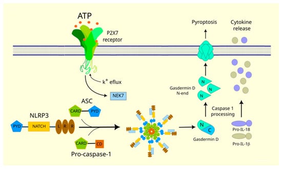
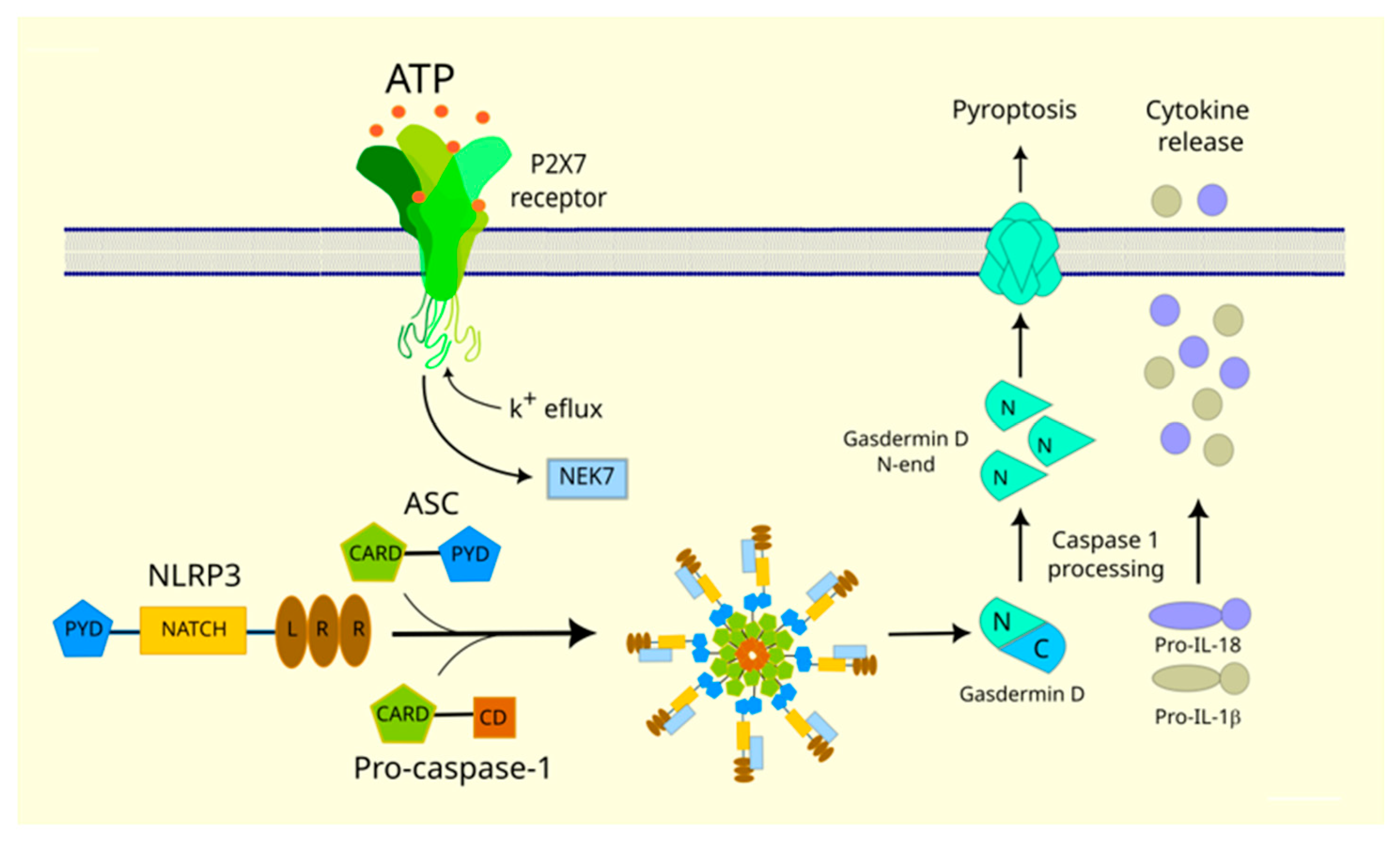
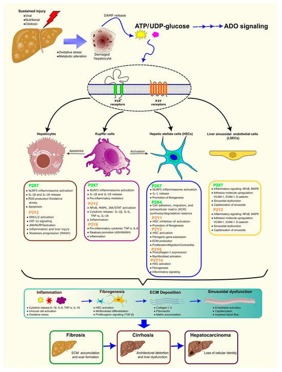
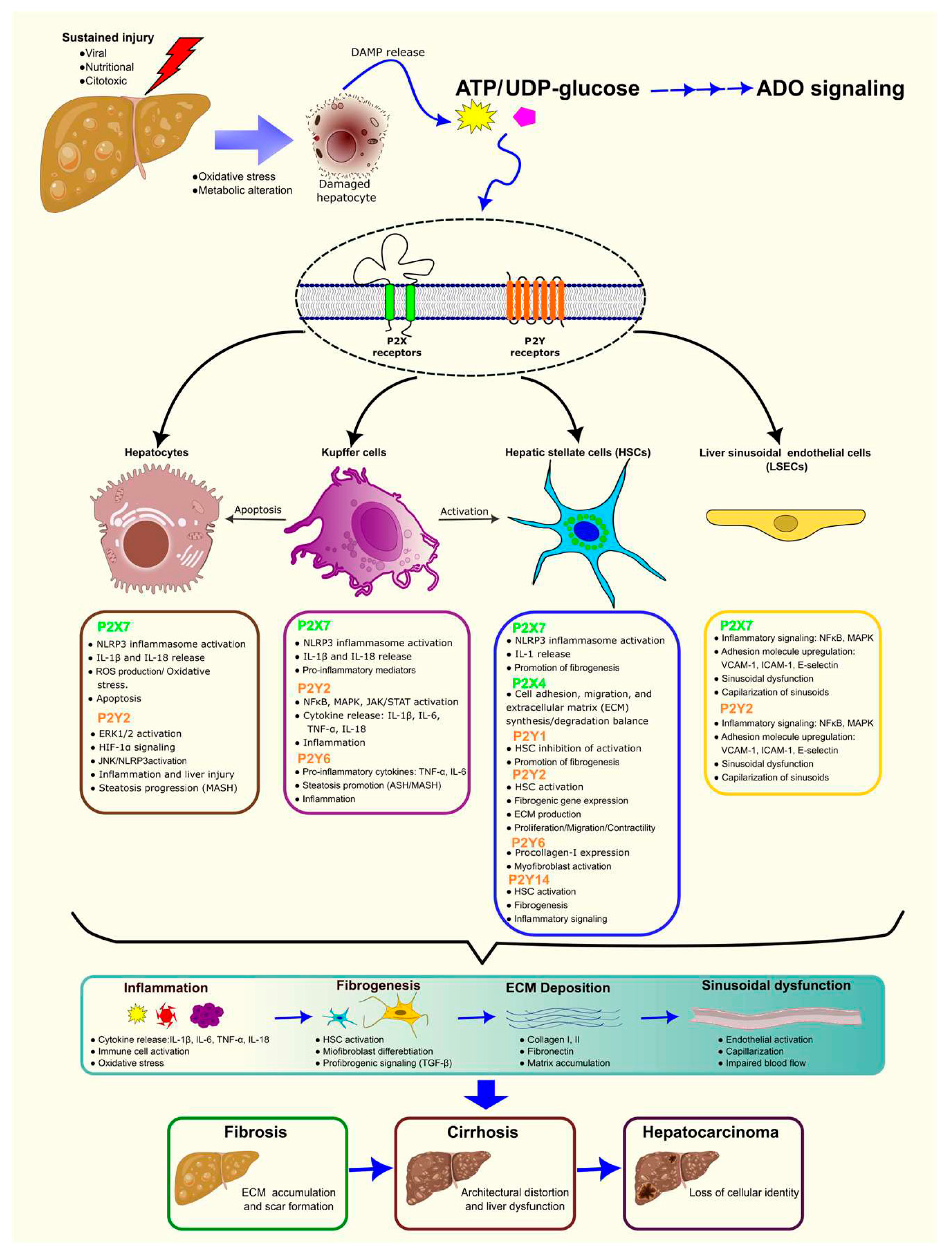

Disclaimer/Publisher’s Note: The statements, opinions and data contained in all publications are solely those of the individual author(s) and contributor(s) and not of MDPI and/or the editor(s). MDPI and/or the editor(s) disclaim responsibility for any injury to people or property resulting from any ideas, methods, instructions or products referred to in the content. |
© 2026 by the authors. Licensee MDPI, Basel, Switzerland. This article is an open access article distributed under the terms and conditions of the Creative Commons Attribution (CC BY) license.