


Abstract
A 64-year-old Asian woman diagnosed with synchronous breast ductal carcinoma in situ (DCIS) and stage IV EGFR-mutated non-small cell carcinoma of the lung (NSCLC). A decision was made to defer management of the DCIS and initiate Osimertinib for lung cancer treatment, since this was a life-limiting diagnosis. At 9 months, restaging FDG-PET CT showed an interval response in the NSCLC and complete loss of FDG avidity at the biopsy-proven DCIS site. Breast MRI confirmed complete imaging resolution of the DCIS. The clinical resolution of breast DCIS during third-generation EGFR inhibitor therapy has not been previously reported in humans and highlights a potential role for the EGFR/HER2 (ERBB) pathway in pre-invasive breast cancer.
1. Introduction
DCIS is a non-invasive breast neoplasm with heterogeneous biology and variable risk of progression [1]. Its molecular phenotype is commonly characterized by ER and HER2 status and by alterations in tumor-suppressor pathways such as p53, which correlate with histologic grade. Low-grade DCIS is predominantly ER-positive (often >90%), whereas high-grade lesions are characterized by a higher prevalence of HER2 positivity and p53 aberrations, along with lower rates of ER positivity [1].
Standard DCIS management consists of local therapy including breast-conserving surgery with adjuvant radiation for many patients or mastectomy for extensive disease [2]. Adjuvant endocrine therapy is considered for ER-positive DCIS to reduce ipsilateral and contralateral events [2]. However, targeting the HER2 pathway has not been adopted in this disease.
The ERBB (epidermal growth factor receptor) family of receptor tyrosine kinases, like EGFR (ERBB1) and HER2 (ERBB2), regulate epithelial proliferation and survival through downstream MAPK and PI3K/AKT signaling [3]. High-grade DCIS frequently expresses ERBB family receptor tyrosine kinases, including EGFR (ERBB1) and HER2 (ERBB2). Ligand-mediated EGFR activation promotes receptor homo- and heterodimerization, most notably with HER2, which serves as a preferred signaling partner, amplifying downstream kinase signaling through autophosphorylation and engagement of MAPK and pro-survival pathways. EGFR–HER2 heterodimers can further couple to SRC-family kinases and other effectors to support transformed mammary epithelial growth, and experimental models suggest ERBB heterodimer signaling is critical for tumor initiation. Consistent with this biology, EGFR-directed therapies in invasive cancer models reduce malignant epithelial proliferation and increase apoptosis in preclinical settings, providing a mechanistic rationale to interrogate ERBB network dependence in DCIS [4].
Lapatinib, a reversible dual-kinase inhibitor of EGFR (ERBB1) and HER2 (ERBB2), has demonstrated reductions in pHER2 and downstream pERK1 (RAS/MAPK) signaling, with concordant decreases in MRI-assessed lesion volume in a phase II trial with 20 patients [5]. This finding supports biologic activity of ERBB growth factor signaling in DCIS [5].
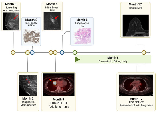
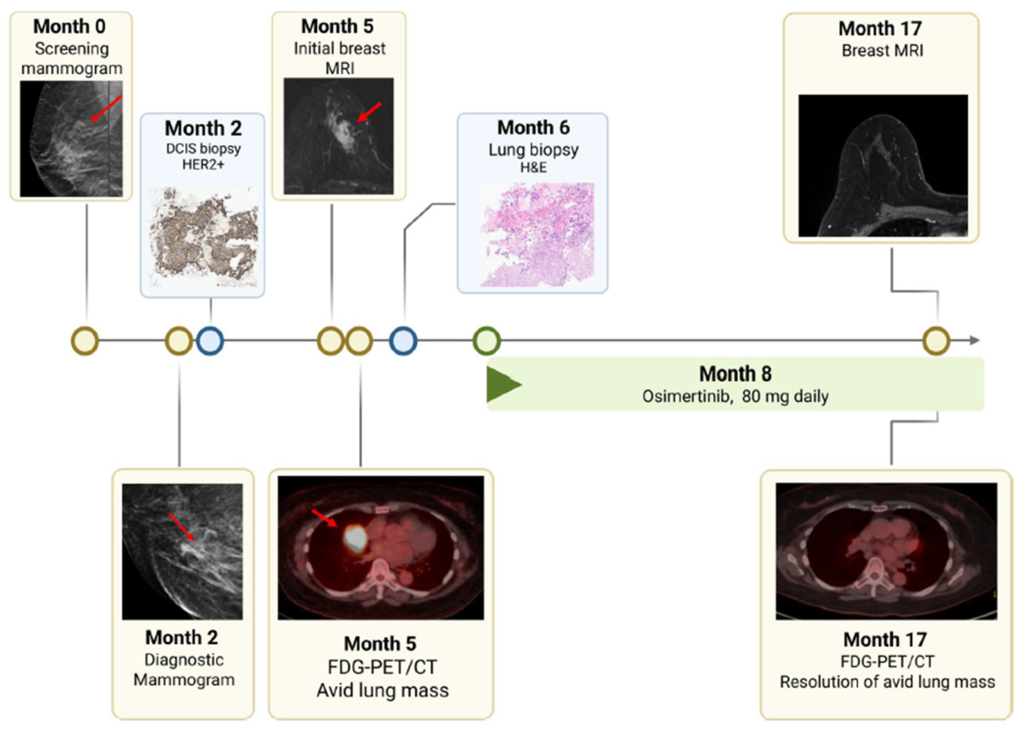
Osimertinib is a third-generation irreversible EGFR tyrosine kinase inhibitor with established efficacy in EGFR-mutant non-small-cell lung cancer (NSCLC) [6]. However, the clinical activity of EGFR-targeted therapy in DCIS has not been defined. Here, we report a patient with synchronous EGFR-mutant metastatic NSCLC and ER-positive/HER2-positive DCIS in whom the DCIS lesion demonstrated complete imaging resolution following Osimertinib therapy, raising the possibility that ERBB-pathway dependence contributed to the observed response (Figure 1).
Figure 1.
Patient timeline. The flowchart indicates the sequence of imaging and procedures performed in the presented case. Red arrows indicate the DCIS site on mammography and breast MRI and the primary lung adenocarcinoma on FDG-PET/CT.
2. Case Presentation
A 64-year-old Asian female who never smoked was referred to oncology clinic after an abnormal screening mammogram. Her previous mammograms, including from the year prior, were considered benign by the breast imaging reporting system (BI-RADS). Her medical history and family history were noncontributory.
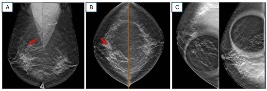
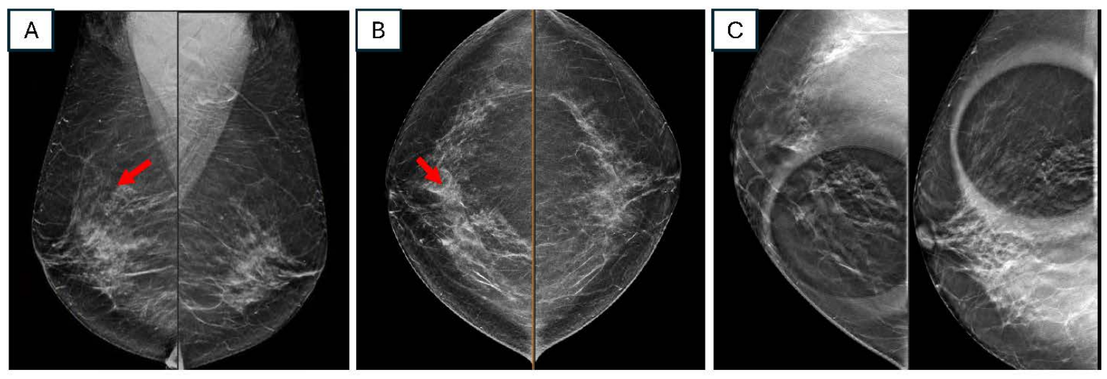
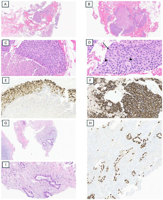
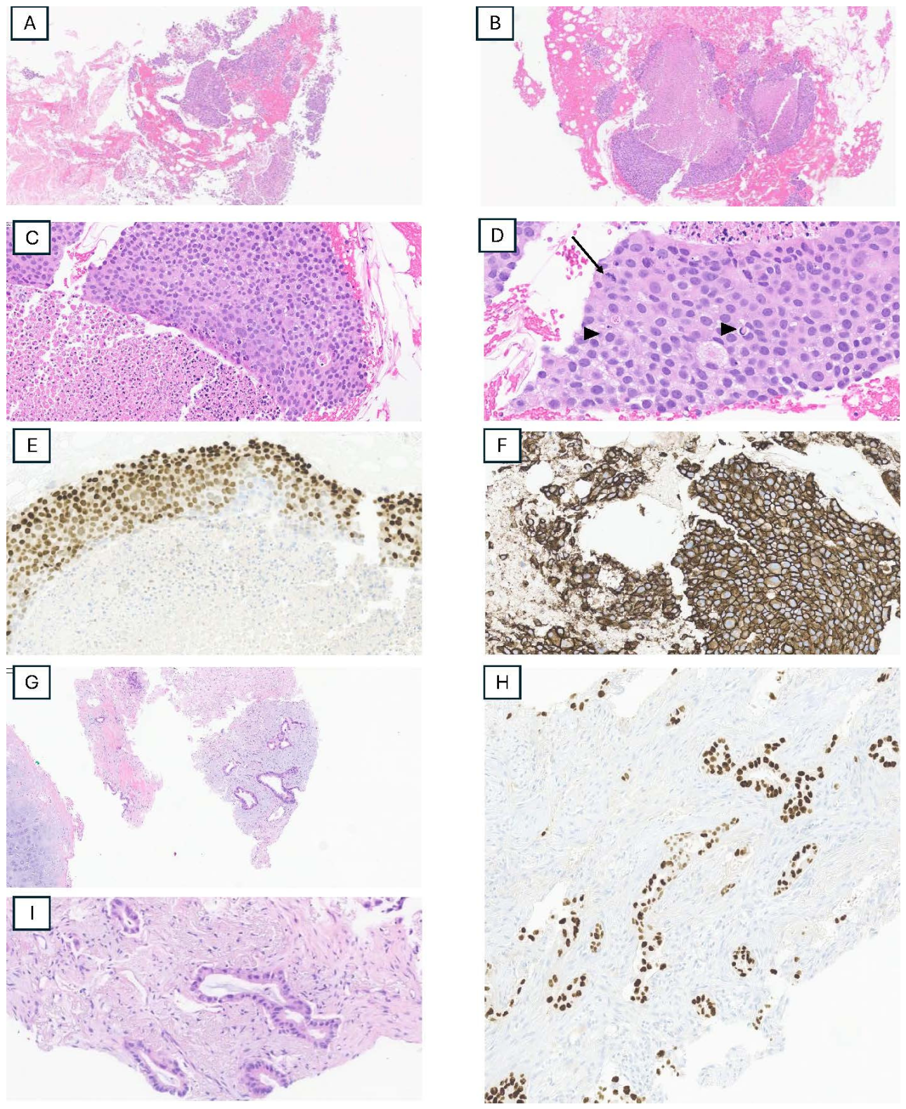
Screening mammography (Figure 2A,B) demonstrated a new focal asymmetry in the right central inner posterior breast which measured 48 mm × 22 mm × 30 mm on the follow-up diagnostic mammography (Figure 2C). Ultrasound-guided core biopsy of the breast demonstrated fragmented material with detached clusters of atypical epithelium associated with comedo necrosis. Despite multiple histologic levels, the interface between the detached epithelium and stroma was not evaluable, and the material was interpreted as DCIS, nuclear grades 2 and 3 (Figure 3A–D). By immunohistochemistry (IHC) the DCIS was ER-positive (95% with strong staining intensity) (Figure 3E) and PgR-negative. A HER2 stain performed after the observed clinical response showed the DCIS to be HER2-positive (score 3+) (Figure 3F).
Figure 2.
Images from right breast mammograms. (A): Screening mammogram CC view with red arrow pointing to breast asymmetry. (B): Screening mammogram MLO view, with red arrow demonstrates focal asymmetry in the right central inner posterior breast. (C): Diagnostic right mammogram spot compression CC/ML digital breast tomosynthesis images demonstrate an indistinct focal asymmetry measuring up to 48 mm × 22 mm × 30 mm.
Figure 3.
Pathology slides from breast biopsy. All slides were digitized at ×40 magnification. (A): Low power image of the breast core needle biopsy showing fragments of detached atypical epithelium with surrounding hemorrhage. (B): Given the detached nature of the specimen, there is no stromal interface and no definite evidence of invasion. (C): The central aspect highlights prominent comedo necrosis. (D): High power image showing a mitotic figure (arrow) and frequent apoptotic bodies (arrow heads) as well as nuclear size variability between the atypical cells, that range from nuclear grade 2 to nuclear grade 3. Without a definitive stromal interface in which to assess for invasion, the material was categorized as ductal carcinoma in situ. (E): Estrogen receptor expression within the carcinoma with 95% of nuclei showing strong staining intensity. (F): HER2 IHC performed after the imaging response in the breast was identified shows that this carcinoma has strong and diffuse circumferential membrane staining in nearly 100% of cells (positive, score 3+). (G,H): Right middle lobe biopsy showing atypical glands in a haphazard pattern within a fibrotic stroma consistent with an adenocarcinoma. (I): TTF1 IHC stain showing strong and diffuse expression within the atypical glands, supporting a lung primary.
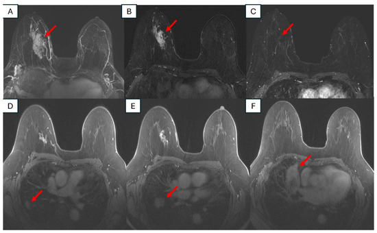
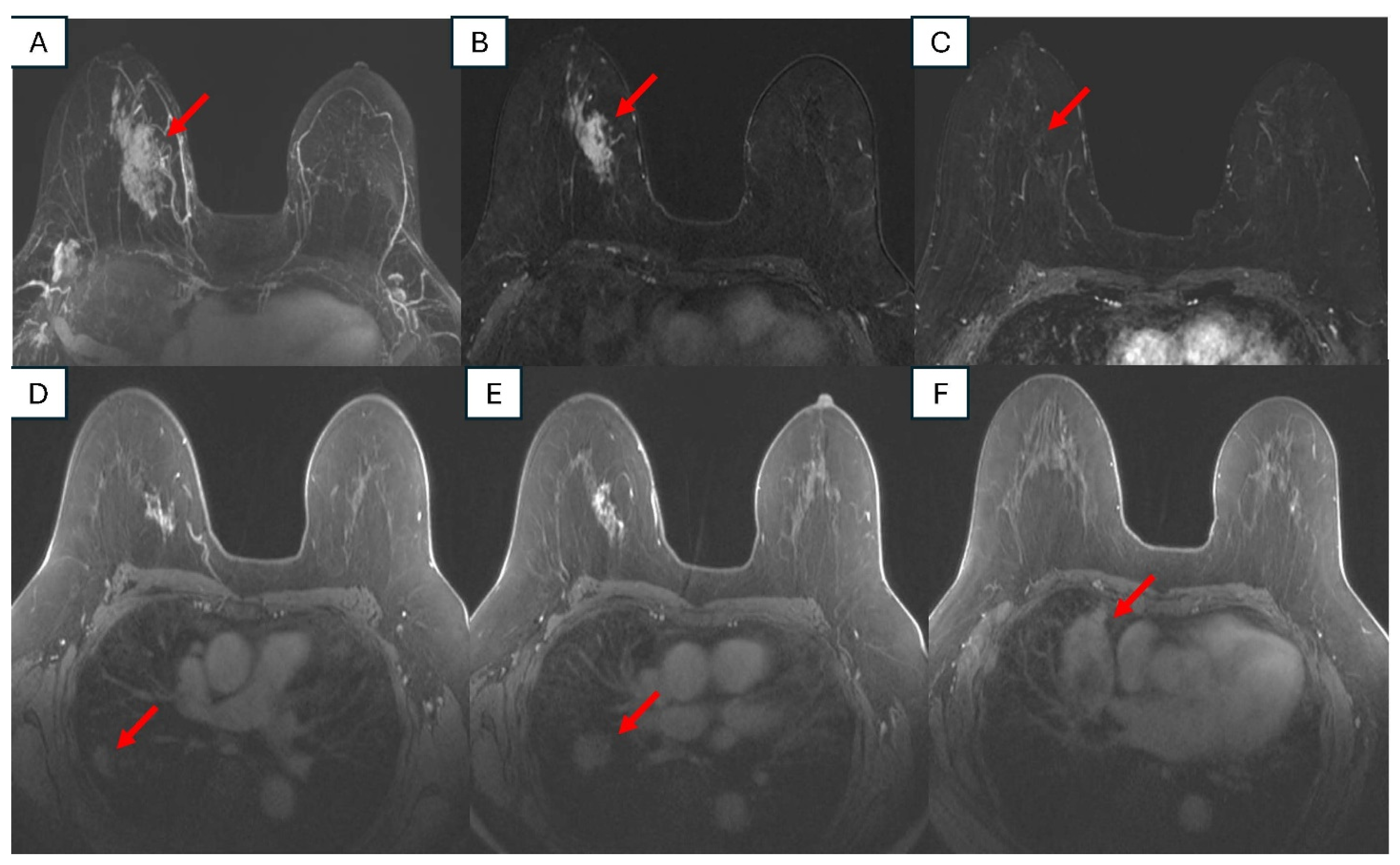
After the core biopsy results, further imaging was obtained including a breast MRI that showed segmental non-mass enhancement at the biopsy-proven DCIS site that measured 89 × 55 × 35 mm, larger than appreciated mammographically (Figure 4A,B). The extent of enhancement was considered to reflect true disease rather than post-biopsy changes, given its dense confluent morphology, absence of T2 hyperintensity, and absence of associated hemorrhage. Multiple suspicious right lung masses were also noted on the breast MRI examination, prompting further work-up (Figure 4D–F).
Figure 4.
Postcontrast T1 weighted early phase images from breast MRI before and after treatment with Osimertinib. (A) 3D maximum intensity projection axial subtraction images and (B) axial subtraction images demonstrate segmental non-mass enhancement in the right upper central mid breast, corresponding to biopsy-proven DCIS (arrows). (C) Axial subtraction images demonstrate resolution of the non-mass enhancement in the right upper central mid breast, after treatment (arrow). (D–F) Axial T1 first postcontrast images prior to treatment demonstrate multiple enhancing suspicious lung masses, as well as enhancement from biopsy-proven DCIS (arrows).
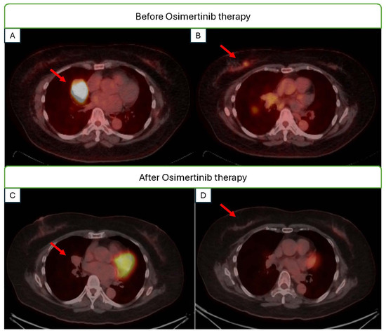
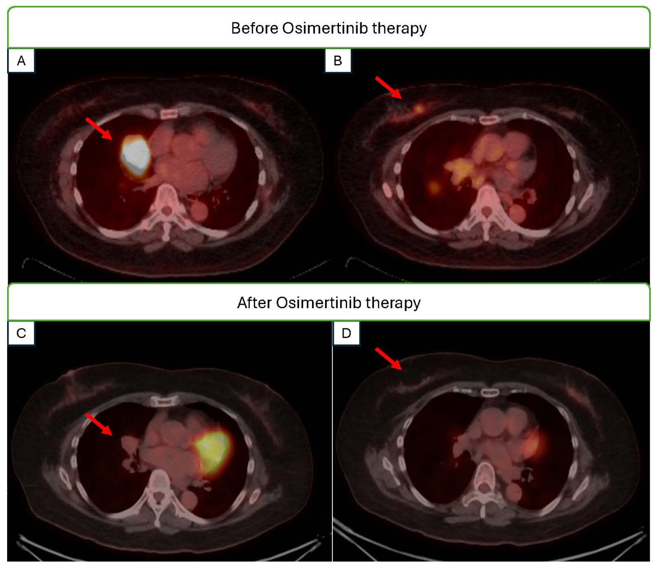
FDG-PET/CT demonstrated a 37 × 33 mm hypermetabolic right middle lobe mass (standardized uptake value [SUV] 9.0) with multifocal pulmonary and pleural-based nodules and mildly hypermetabolic right hilar lymph nodes, consistent with metastatic disease (Figure 5A). Mild uptake (SUV 2.8) was present at the right breast DCIS site (Figure 5B).
Figure 5.
Axial PETCT images acquired after administration of FDG, performed before (A,B) and after (C,D) treatment with Osimertinib. (A) Before treatment, arrow indicates FDG avid right middle lobe mass. (B) Before treatment, arrow indicates FDG avid biopsy-proven DCIS in the right breast. (C) After treatment, resolution of FDG avid right lung mass with decreased size of the underlying mass indicated by arrow. (D) After treatment, arrow indicates privious DCIS sire, showing resolution of FDG avidity at the site of biopsy-proven DCIS.
Fine needle aspiration and core biopsy of the right middle lobe lesion confirmed adenocarcinoma consistent with a lung primary (Figure 3G,I). Immunohistochemistry was positive for thyroid transcription factor 1 (TTF-1) (Figure 3H) and negative for p63. Next-generation sequencing (OmniSeq INSIGHT) identified an EGFR exon 19 deletion (E746_A750del). The 4R lymph node station fine needle aspiration (FNA) was non-diagnostic, showing only benign epithelial cells without lymphoid tissue. Brain MRI showed no evidence of intracranial metastatic disease. The patient was staged as metastatic NSCLC adenocarcinoma, stage IVA (cT2bN1M1a), EGFR exon 19 deletion-positive, with multifocal lung and pleural metastases.
Because metastatic NSCLC represented the life-limiting diagnosis, the multidisciplinary team deferred local management of DCIS and initiated first-line Osimertinib 80 mg daily. No surgery, radiation, or endocrine therapy was administered for DCIS.
Osimertinib was tolerated with expected grade 1–2 adverse effects including brittle nails, dry skin, and diarrhea, managed with supportive care. No dose reductions or treatment interruptions were required.
Restaging PET/CT performed 9 months after Osimertinib initiation demonstrated interval decrease in size and FDG avidity of pulmonary and pleural lesions (Figure 5C). At the right breast DCIS site, FDG uptake resolved and only the biopsy clip remained visible (Figure 5D). In addition, a concurrent repeat breast MRI demonstrated complete imaging resolution of the DCIS (Figure 4C).
When the patient sought re-evaluation of the untreated DCIS in the context of an ongoing lung cancer response, a follow-up breast MRI performed 9 months after Osimertinib initiation showed no residual enhancement at the biopsy-proven DCIS site (Figure 5D). The patient continues Osimertinib with planned ongoing thoracic restaging and breast surveillance imaging.
3. Discussion
This report highlights the unusual clinical observation of a complete imaging resolution at a biopsy-proven DCIS site during Osimertinib therapy for synchronous EGFR-mutant metastatic NSCLC. Although synchronous primary malignancies, especially of the breast and lung, are increasingly recognized, as in refs. [7,8,9,10], the behavior of an untreated in situ breast lesion during EGFR-targeted therapy is rarely documented.
The incidence of second primary lung cancer in breast cancer survivors has been reported to be elevated compared to the general population, with a standardized incidence ratio of 1.4 [10]. Interestingly, studies have shown a higher rate of EGFR mutations in lung cancer patients with prior breast cancer, with rates as high as 60–78% reported in some series, substantially higher than the general NSCLC population [7,10]. Retrospective data showed a poor correlation between EGFR expression and survival in ER+ breast cancer [11].
Osemartinib was chosen for this patient given the improved overall survivor rate in previous untreated EGFR-mutated NSCLCs [12]. The complete image resolution of breast DCIS during third-generation EGFR inhibitor therapy with Osimertinib targeting lung cancer is a previously unreported observation in hormonal therapy-naïve patients. While EGFR inhibitors have been studied in combination with endocrine therapy for hormone receptor-positive breast cancer, with some evidence of antiproliferative effects and hormonal therapy resistance overcome, there are no published reports of DCIS resolution with third-generation EGFR TKI inhibitor monotherapy in vivo [11,13,14].
Although this case is hypothesis-generating, it has some limitations. First, imaging resolution does not prove pathologic eradication; residual DCIS can be present despite absence of MRI enhancement. Second, baseline breast MRI and PET/CT were obtained after core biopsy and clip placement. While theoretically, post-biopsy inflammation or evolving hematoma could potentially cloud the interpretation, in this case several specific imaging features are highly suggestive that the MRI findings are from DCIS rather than post-procedural enhancement. Additionally, DCIS has a heterogeneous natural history, and some lesions may remain indolent. While spontaneous regression has been suggested, estimates from modeling suggest that true regression is uncommon [8,15].
Despite these caveats, biological plausibility exists for ERBB-pathway dependence in select DCIS. HER2 overexpression is frequent in DCIS, and short preoperative lapatinib therapy, a dual EGFR and HER2 inhibitor, in HER2-positive DCIS has been associated with inhibition of HER2/MAPK signaling and decreased MRI measured lesion volume in many patients, with complete absence of DCIS on excision in a small minority [5,9]. Osimertinib was developed as a mutant-selective EGFR inhibitor but has demonstrated preclinical antitumor activity in models driven by certain HER2 aberrations, providing a potential mechanistic bridge in a HER2-positive DCIS lesion [5,15].
From a clinical standpoint, withholding DCIS treatment was reasonable given the patient’s metastatic NSCLC. Notably, the DCIS in this case was not low-risk by COMET criteria (grade 1–2, hormone receptor-positive) [16,17], and HER2 strong positivity status has been associated with higher local recurrence risk after breast conserving therapy in observational studies [17,18,19,20]. This case adds to the hypothesis that ERBB-pathway blockade may influence proliferation of some DCIS lesions. This finding is concordant with previous ERBB blockage in DCIS [5]. Future work could include systematic collection of breast imaging outcomes in patients receiving targeted therapies for other malignancies and, when clinically appropriate, pathological confirmation to determine whether true eradication occurs.
We describe complete imaging resolution of a biopsy-proven DCIS during Osimertinib therapy for synchronous EGFR exon 19 deletion-mutant metastatic NSCLC. While causality cannot be established without pathologic confirmation, the observation is hypothesis-generating and supports further study of EGFR/HER2 pathway biology in DCIS.
Author Contributions
L.A.M.S. was responsible for manuscript preparation and clinical case review. R.E.S.J. and A.J.F. were responsible for image selection and radiological interpretation. E.D. was responsible for pathological interpretation. L.M. conceptualized the study and contributed to manuscript writing. All authors have read and agreed to the published version of the manuscript.
Funding
Mayo Clinic continued medical education funds were used to cover the article processing charges.
Institutional Review Board Statement
The Mayo Clinic Institutional Review Board (IRB) acknowledges that in accordance with the Code of Federal Regulations, 45 CFR 46.102, this study is waived from IRB review.
Informed Consent Statement
Informed consent was obtained from all subjects involved in the study.
Data Availability Statement
The original contributions presented in this study are included in the article. Further inquiries can be directed to the corresponding author.
Conflicts of Interest
The authors declare no conflicts of interest.
References
- Casasent, A.K.; Almekinders, M.M.; Mulder, C.; Bhattacharjee, P.; Collyar, D.; Thompson, A.M.; Jonkers, J.; Lips, E.H.; van Rheenen, J.; Hwang, E.S.; et al. Learning to distinguish progressive and non-progressive ductal carcinoma in situ. Nat. Rev. Cancer 2022, 22, 663–678, Erratum in Nat. Rev. Cancer 2023, 23, 112. https://doi.org/10.1038/s41568-022-00542-6. [Google Scholar] [CrossRef] [Scilit] [PubMed]
- Buchheit, J.T.; Schacht, D.; Kulkarni, S.A. Update on Management of Ductal Carcinoma in situ. Clin. Breast Cancer 2024, 24, 292–300. [Google Scholar] [PubMed]
- Lebeau, A.; Unholzer, A.; Amann, G.; Kronawitter, M.; Bauerfeind, I.; Sendelhofert, A.; Iff, A.; Löhrs, U. EGFR, HER-2/neu, cyclin D1, p21 and p53 in correlation to cell proliferation and steroid hormone receptor status in ductal carcinoma in situ of the breast. Breast Cancer Res. Treat. 2003, 79, 187–198. [Google Scholar] [CrossRef] [Scilit] [PubMed]
- Chan, K.C.; Knox, W.F.; Gee, J.M.; Morris, J.; Nicholson, R.I.; Potten, C.S.; Bundred, N.J. Effect of epidermal growth factor receptor tyrosine kinase inhibition on epithelial proliferation in normal and premalignant breast. Cancer Res. 2002, 62, 122–128. [Google Scholar] [PubMed]
- Estévez, L.G.; Suarez-Gauthier, A.; García, E.; Miró, C.; Calvo, I.; Fernández-Abad, M.; Herrero, M.; Marcos, M.; Márquez, C.; Lopez Ríos, F.; et al. Molecular effects of lapatinib in patients with HER2 positive ductal carcinoma in situ. Breast Cancer Res. 2014, 16, R76. [Google Scholar] [CrossRef] [Scilit] [PubMed]
- Hanna, N.H.; Robinson, A.G.; Temin, S.; Baker, S.; Brahmer, J.R.; Ellis, P.M.; Gaspar, L.E.; Haddad, R.Y.; Hesketh, P.J.; Jain, D.; et al. Therapy for Stage IV Non-Small-Cell Lung Cancer with Driver Alterations: ASCO and OH (CCO) Joint Guideline Update. J. Clin. Oncol. 2021, 39, 1040–1091. [Google Scholar] [PubMed]
- Wang, K.Y.; Lee, C.S.; Vempati, P.; Sharma, R.; Kohn, N.; Seetharamu, N. Characteristics of Patients with Second Primary Lung Cancer Following Breast Cancer: A Retrospective Descriptive Study. Clin. Lung Cancer 2023, 24, e198–e204. [Google Scholar] [CrossRef] [Scilit] [PubMed]
- Hao, Y.; Zhang, X.; Cui, G.; Qi, X.; Jiang, Z.; Yu, L. Clinicopathological features, prognostic factor analysis, and survival nomogram of patients with double primary cancers involving lung cancer. Cancer Med. 2024, 13, e7296. [Google Scholar] [CrossRef] [Scilit] [PubMed]
- Song, C.; Yu, D.; Wang, Y.; Wang, Q.; Guo, Z.; Huang, J.; Li, S.; Hu, W. Dual Primary Cancer Patients with Lung Cancer as a Second Primary Malignancy: A Population-Based Study. Front. Oncol. 2020, 10, 515606. [Google Scholar] [CrossRef] [Scilit] [PubMed]
- Zeng, T.; Xu, H.; Liu, Y.; Sun, C.; Yang, F.; Liang, Y.; Huang, X.; Fu, Z.; Li, W.; Yin, Y. High rate of epidermal growth factor receptor-mutated primary lung cancer in patients with primary breast cancer. Front. Oncol. 2022, 12, 985734. [Google Scholar] [CrossRef] [Scilit] [PubMed]
- Jeong, Y.; Bae, S.Y.; You, D.; Jung, S.P.; Choi, H.J.; Kim, I.; Lee, S.K.; Yu, J.; Kim, S.W.; Lee, J.E.; et al. EGFR is a Therapeutic Target in Hormone Receptor-Positive Breast Cancer. Cell Physiol. Biochem. 2019, 53, 805–819. [Google Scholar] [CrossRef] [Scilit] [PubMed]
- Soria, J.C.; Ohe, Y.; Vansteenkiste, J.; Reungwetwattana, T.; Chewaskulyong, B.; Lee, K.H.; Dechaphunkul, A.; Imamura, F.; Nogami, N.; Kurata, T.; et al. Osimertinib in Untreated EGFR-Mutated Advanced Non-Small-Cell Lung Cancer. N. Engl. J. Med. 2018, 378, 113–125. [Google Scholar] [PubMed]
- Polychronis, A.; Sinnett, H.D.; Hadjiminas, D.; Singhal, H.; Mansi, J.L.; Shivapatham, D.; Shousha, S.; Jiang, J.; Peston, D.; Barrett, N.; et al. Preoperative gefitinib versus gefitinib and anastrozole in postmenopausal patients with oestrogen-receptor positive and epidermal-growth-factor-receptor-positive primary breast cancer: A double-blind placebo-controlled phase II randomised trial. Lancet Oncol. 2005, 6, 383–391, Erratum in Lancet Oncol. 2005, 6, 357. [Google Scholar] [PubMed]
- Guix, M.; Granja, N.M.; Meszoely, I.; Adkins, T.B.; Wieman, B.M.; Frierson, K.E.; Sanchez, V.; Sanders, M.E.; Grau, A.M.; Mayer, I.A.; et al. Short preoperative treatment with erlotinib inhibits tumor cell proliferation in hormone receptor-positive breast cancers. J. Clin. Oncol. 2008, 26, 897–906. [Google Scholar] [CrossRef] [Scilit] [PubMed]
- Poelhekken, K.; Lin, Y.; Greuter, M.J.W.; van der Vegt, B.; Dorrius, M.; de Bock, G.H. The natural history of ductal carcinoma in situ (DCIS) in simulation models: A systematic review. Breast 2023, 71, 74–81. [Google Scholar] [CrossRef] [Scilit] [PubMed]
- Hwang, E.S.; Hyslop, T.; Lynch, T.; Ryser, M.D.; Weiss, A.; Wolf, A.; Norris, K.; Witten, M.; Grimm, L.; Schnitt, S.; et al. Active Monitoring With or Without Endocrine Therapy for Low-Risk Ductal Carcinoma In Situ: The COMET Randomized Clinical Trial. JAMA 2025, 333, 972–980. [Google Scholar] [PubMed]
- Kuzmiak, C.M.; Sharpe, R.E.; Lewin, A.A.; Weinstein, S.P.; Blinder, V.; Dibble, E.H.; Dodelzon, K.; Dogan, B.E.; Paulis, L.V.; Plichta, J.K.; et al. ACR Appropriateness Criteria® Imaging of Ductal Carcinoma in Situ (DCIS). J. Am. Coll. Radiol. 2025, 22, S274–S299. [Google Scholar] [CrossRef] [Scilit] [PubMed]
- Shoji, F.; Yamashita, N.; Inoue, Y.; Kozuma, Y.; Toyokawa, G.; Hirai, F.; Ito, K.; Tagawa, T.; Okamoto, T.; Maehara, Y. Surgical Resection and Outcome of Synchronous and Metachronous Primary Lung Cancer in Breast Cancer Patients. Anticancer Res. 2017, 37, 5871–5876. [Google Scholar] [CrossRef] [Scilit] [PubMed]
- Reck, M.; Rabe, K.F. Precision Diagnosis and Treatment for Advanced Non-Small-Cell Lung Cancer. N. Engl. J. Med. 2017, 377, 849–861. [Google Scholar] [PubMed]
- Yoon, H.Y.; Ryu, J.S.; Sim, Y.S.; Kim, D.; Lee, S.Y.; Choi, J.; Park, S.; Ryu, Y.J.; Lee, J.H.; Chang, J.H. Clinical significance of EGFR mutation types in lung adenocarcinoma: A multi-centre Korean study. PLoS ONE 2020, 15, e0228925. [Google Scholar] [CrossRef] [Scilit] [PubMed]
Disclaimer/Publisher’s Note: The statements, opinions and data contained in all publications are solely those of the individual author(s) and contributor(s) and not of MDPI and/or the editor(s). MDPI and/or the editor(s) disclaim responsibility for any injury to people or property resulting from any ideas, methods, instructions or products referred to in the content. |
© 2026 by the authors. Licensee MDPI, Basel, Switzerland. This article is an open access article distributed under the terms and conditions of the Creative Commons Attribution (CC BY) license.