


Abstract
Alzheimer’s disease (AD) is a progressive neurodegenerative disorder characterized by cognitive decline and significant neuropathological changes. Early and accurate diagnosis remains a major challenge, highlighting the need for reliable, minimally invasive biomarkers. MicroRNAs (miRNAs), small non-coding RNAs that regulate gene expression, have emerged as promising candidates. Their expression is altered in the brains of AD patients, reflecting disease-specific pathological processes, and they are detectable in peripheral biofluids. However, discrepancies in miRNA profiles between the brain and the circulation, and between patient populations remain a significant limitation, raising questions about their origin, transport across the blood–brain barrier, and their reliability in reflecting central nervous system pathology. This review provides a comprehensive overview of current research comparing miRNA expression profiles in brain tissue and blood in AD, with a focus on their biological relevance, mechanisms of release and transport, and diagnostic potential. We also discuss the challenges associated with cross-tissue variability, methodological inconsistencies, and the need for standardized approaches. Finally, we highlight future directions, including multi-tissue analyses and integration with other noninvasive modalities, to improve the clinical utility of miRNA-based biomarkers in AD.
1. Introduction
1.1. Importance of Early AD Diagnosis
Alzheimer’s disease (AD) and its related dementias are projected to affect 153 million individuals worldwide by 2050, imposing an immense global health burden and significant emotional, social, and economic challenges on patients, families, and healthcare systems [1,2,3]. Despite decades of research, AD remains difficult to diagnose at its earliest stages. AD pathology can occur independently or with other neurodegenerative and vascular diseases, particularly in an aging brain. The pathologic processes of amyloid beta (Aβ) accumulation, tau hyperphosphorylation, synaptic dysfunction, and neuroinflammation begin decades before clinical symptoms emerge [4,5]. Therefore, differential diagnosis in a timely manner is critical for appropriate care, support, and individualized treatment plans [6,7,8]. However, current diagnostic methods rely heavily on invasive cognitive assessments and neuroimaging or cerebrospinal fluid (CSF) analyses, which can be expensive and inaccessible in many clinical settings. As a result, there remains an urgent need for noninvasive diagnostic tools capable of detecting AD before significant neuronal damage occurs.
Because clinical disease evolves over decades, the timing of diagnosis is important for accurate AD characterization. Early diagnosis improves accuracy, reducing risk of misdiagnosis or inappropriate treatment, especially for atypical or non-classical AD [7]. While typical AD is characterized by early memory impairment, with progressive decline in other cognitive domains, atypical AD often presents with visual/spatial problems, language difficulties, executive dysfunction, and behavioral symptoms [9]. These symptoms often overlap with other neurodegenerative dementias such as frontotemporal dementia, Lewy body dementia, and progressive aphasia [7,9,10].
It is therefore critical to identify and develop methods for early detection, shifting the basis of diagnosis from symptoms to biology, so that AD pathology, rather than its consequences, are detected [7]. Modeling studies have suggested that early diagnosis and intervention may delay a person’s time to institutionalization, resulting in societal cost savings, as well as slow both cognitive and functional decline. Additionally, newer disease-modifiable treatments require early diagnosis and intervention, providing added impetus to detect AD earlier [5,11,12,13]. For example, early-stage AD patients treated with Lecanemab or Donanemab showed significant cognitive and functional benefits with greater efficacy when treatment is initiated at earlier disease stages underscoring the importance of identifying AD at the mild or prodromal stage to maximize therapeutic benefit [14,15,16].
Beyond clinical outcomes, early diagnosis provides important psychosocial advantages. Major Alzheimer’s associations advocate for early diagnosis, allowing patients and families more time to plan, make informed decisions, and access support services, ultimately reducing emotional burden and improving quality of life [17,18,19,20]. Together, these findings highlight the importance of early detection for clinical management and downstream societal burdens.
1.2. Significance of Noninvasive Diagnostic Biomarkers
AD pathology begins years before clinical symptoms appear, with Aβ accumulation and tau dysregulation detected more than 15 years prior to diagnosis [21,22,23,24]. Currently, the only diagnostic tools available for AD are cerebrospinal fluid (CSF) biomarkers (e.g., Aβ, tau) and neuroimaging (e.g., PET scans), which are effective but often invasive, expensive, and not easily scalable [25,26]. Blood-based biomarkers, in contrast, are minimally invasive, comparatively inexpensive, and can be standardized allowing for early detection, monitoring, and broader clinical application [27,28,29,30].
Advances in highly sensitive assays now allow detection of molecular signatures of AD in peripheral tissues and other biofluids (e.g., blood, saliva), providing accessible alternatives for patients unable or unwilling to undergo lumbar puncture or PET imaging [30,31,32]. Beyond clinical utility, these biomarkers facilitate drug trials, mechanistic studies, and multi-modal research approaches by integrating with imaging, genetic, and cognitive data to enhance predictive accuracy [33,34].
1.3. MiRNAs as Potential Diagnostic Biomarkers in AD
Several aspects of microRNAs (miRNAs) contribute to their strong potential as diagnostic biomarkers. They have emerged as critical regulators of a wide array of biological processes including cellular metabolism and proliferation, apoptosis, neuronal gene expression and integrity, and brain morphogenesis [35,36,37,38,39,40,41]. Significantly, their dysregulation has been implicated in multiple aspects of AD pathology, including Aβ metabolism, tau phosphorylation, neuroinflammation, and synaptic dysfunction, as well as broader disruptions in cell signaling, transcriptional regulation, and structural pathways [42,43,44,45,46,47,48,49,50]. Shifts in their expression in neurodegenerative conditions like AD potentially alter cellular pathways, contributing to disease progression [51].
The literature suggests the existence of disease-specific miRNA signatures [31,39,41]. Some miRNAs such as miR-150-5p correlate with clinical and imaging markers of disease severity [45,52,53,54,55], whereas others like miR-483-3p demonstrate functional effects in experimental models, influencing neuronal survival, apoptosis, and AD-related protein expression [42,43,44]. The consistent dysregulation of these and other miRNAs across human and animal studies underscores their potential as biomarkers and therapeutic targets, though further validation is needed [39,40,41,56].
miRNAs are influenced by factors such as subcellular localization, relative abundance of both miRNAs and target transcripts, and the binding affinity between the two [57,58,59,60]. Moreover, miRNAs are remarkably stable due to their size and packaging in exosomes, vesicles, or protein complexes. They are resistant to RNase degradation, freeze thaw cycles and pH variations. Their stability and versatile roles in many biological processes make them ideal candidates as biomarkers for disease detection [61]. Additionally, their packaging and peripheral transport via exosomes and extracellular vesicles (EVs) may reflect brain-derived signals more faithfully than free circulating miRNAs because EVs can cross the blood–brain barrier and carry central nervous system (CNS)-derived cargo (Figure 1 [40,62]). Indeed, EV-miRNAs have demonstrated superior diagnostic performance compared to serum miRNAs, with some levels correlating with brain atrophy [40].
Figure 1.
Proposed mechanisms linking brain miRNA dysregulation to peripheral biomarker detection in AD. AD-associated neuropathology, including Aβ plaque accumulation, NFTs, glial activation, and neuronal degeneration, alters miRNA expression within the CNS. These dysregulated miRNAs may enter the peripheral circulation through two non-mutually exclusive routes: (1) regulated export via EVs/exosomes and other carrier-associated transport mechanisms, and (2) passive leakage resulting from BBB dysfunction and increased permeability. Once in circulation, brain-derived miRNAs, together with other AD-associated biomolecules, may contribute to detectable peripheral biomarker signatures in blood. Created in BioRender. Shiyab, A. (2026) https://BioRender.com/mrpjwxe, accessed on 27 June 2026.
However, there are limitations to miRNAs as biomarkers, namely the context-dependent variability [63]. It is likely the variability observed in miRNA studies not only reflects isolated technical limitations, but also the context-dependent nature of miRNA expression itself. miRNA profiles are highly dynamic and shaped by multiple interacting biological variables, including tissue origin, disease state, sex, and experimental model, all of which influence their detectability and interpretation in biomarker studies.
1.4. Current Gaps, Purpose, and Scope of the Review
Despite the rapid expansion of miRNA research in neurodegenerative disease, significant knowledge gaps remain. There are inconsistent or conflicting miRNA signatures across tissues, cohorts, and experimental platforms, complicating standardization of biomarkers [45,64,65,66]. Peripheral miRNA expression does not always reflect molecular changes in the brain, where AD pathology originates [56,65,66,67,68,69,70,71]. Genetically engineered mouse models provide useful mechanistic insight but do not always fully recapitulate key features of human AD, limiting the translational validity of preclinical miRNA findings [72,73,74]. Additionally, sex, aging, and genetic variability influence miRNA expression and may contribute to inconsistent biomarker performance [75].
Standardization of sampling and analytical methods remains a major challenge. Variability in sample type, collection, processing, and quantification undermines reproducibility [75,76,77,78]. Many studies rely on small, often biased cohorts that are rarely reproduced in independent populations, with limited consideration of covariates such as sex, disease stage, or tissue type [75]. Brain miRNA studies are further complicated by under-sampling and evaluation of different brain regions [79,80].
Mechanistically, many miRNAs identified as potential candidates are derived from expression surveys with few providing possible mechanisms, such as how miRNAs affect Aβ processing, tau pathology, neuroinflammation or neuronal survival [76]. Without this data, biological relevance remains speculative. Additionally, despite growing appreciation for sex differences in AD manifestation and progression, very few studies looked at miRNA differences between men and women, and those that do are limited by unequal sex representation [75,76,81,82]. Taken together, the lack of reproducible validation and standardized protocols, limited large and balanced cohorts, and minimal mechanistic insight means that current miRNA signatures in AD must be interpreted with caution, rather than reliable diagnostic or prognostic tools. While miRNAs remain promising noninvasive biomarkers, existing research should be considered as preliminary until these gaps are addressed.
This review synthesizes current evidence on miRNA dysregulation across central and peripheral tissues in AD, with a particular focus on their potential as blood-based biomarkers (Figure 1). We compare miRNA expression patterns across different brain regions and peripheral biofluids, evaluate the consistency of these patterns between tissues, and consider possible mechanisms for how miRNAs might act across these different compartments. In addition, we examine miRNA alterations reported in commonly used AD mouse models and assess the influence of sex-specific differences on miRNA regulation. Finally, we highlight the key limitations and knowledge gaps in the field and propose future directions aimed at advancing the development of reliable and clinically useful miRNA-based diagnostic tools for AD.
2. MiRNAs and AD
MiRNAs are produced through a highly regulated biogenesis pathway involving DROSHA-, DGCR8-, and DICER-mediated processing before functioning as post-transcriptional regulators of gene expression [83,84,85]. Detailed information on miRNA biogenesis is available elsewhere [84,86,87]. These miRNAs act post-transcriptionally to regulate gene expression by binding target mRNAs and inhibiting translation or promoting mRNA decay [88,89,90].
2.1. Involvement of miRNAs in AD-Related Gene Expression
Beyond maintaining cellular homeostasis, miRNAs modulate the expression of important AD-related proteins like Amyloid Precursor Protein (APP), β-site APP-cleaving enzyme 1 (BACE1), and components of the γ-secretase complex (PSEN1/2), contributing to neuroinflammation [91,92,93,94,95].
Direct targeting of APP mRNA by miRNAs was first reported over a decade ago. Members of the miR-20a family (miR-20a, miR-17-5p, and miR-106b) regulate APP mRNA, with their expression patterns closely paralleling APP levels in the brain and in differentiating neurons, suggesting that changes in miRNA expression could influence APP levels during development and in disease [96]. Recent studies continue to show miRNA involvement in the APP amyloid pathway. For example, miR-298 reduces APP expression and Aβ production in human cell models, and miR-124 influences amyloidogenic pathways by altering APP-derived Aβ production in neuronal AD models [94,97,98]. Thus, through post-transcriptional regulation of APP expression, miRNA dysregulation could alter amyloid production and contribute to AD progression.
Multiple miRNAs exert context-dependent effects on BACE1 determined by their target and functional roles. Down-regulation of some miRNAs increases BACE1 expression to enhance Aβ production, while other miRNAs exhibit a more protective role, reducing Aβ accumulation [61,94,97,98]. MiRNAs regulate γ-secretase by targeting the 3′-UTR of PSEN1. For example, miR-3940-5p reduces PSEN1 expression, lowering PSEN1 levels. Because PSEN1 is a critical component of the γ-secretase complex that cleaves APP to generate Aβ, this miR-3940-5p-mediated downregulation of PSEN1 decreases Aβ production [91,99]. MiR-29b-2-5p directly represses PSEN1 expression and its upregulation similarly reduces Aβ and improves cognition [100].
In addition to Aβ accumulation, miRNAs are implicated in the regulation of tau pathology at multiple levels, including expression, post-translational modification and protein clearance [101,102,103,104,105,106] demonstrating that miRNAs form an integrated regulatory network governing tau expression, phosphorylation state, and clearance, underscoring their central role in tau-mediated neurodegeneration in AD.
The inflammatory response to Aβ is mediated by glial cells, including microglia and astrocytes, exacerbating AD pathology [107,108,109,110]. With neuroinflammation representing a major component of AD pathology, understanding the regulatory mechanisms of miRNAs involved in inflammatory mechanisms would facilitate future disease interventions [111]. In fact, multiple miRNAs regulate immune signaling pathways, microglial activation, and cytokine expression. Many are upregulated in AD brain and CSF, and exert opposing immunomodulatory effects depending on the context [42,61,112,113,114], demonstrating miRNAs are not simply pro- or anti-inflammatory; rather that they act to fine tune immune responses, and when their regulation is disrupted in AD, can contribute to long-lasting, damaging neuroinflammation [92]. Overall, and because changes in these miRNAs can be detected in peripheral samples such as blood, studies often highlight their potential not only as regulators of amyloid pathology but also as accessible biomarkers reflecting disease-related molecular changes in the brain.
2.2. MiRNA Stability in Peripheral Fluids
Despite shared biological processes that regulate miRNAs in tissues and the circulation, miRNAs in the circulation are generally more stable compared to those in tissue cells for several reasons including their intrinsic decay rates, post-transcriptional modifications, protein interactions, packaging in EVs, and environmental conditions [115,116,117,118].
A relatively high intracellular concentration of miRNAs is often required for functional repression of multiple target transcripts, reflecting the broad regulatory potential of a single miRNA, so their stability is critical for achieving sufficient concentrations to engage multiple targets [119,120,121]. MiRNA half-life varies from minutes [122] to several weeks [119,122,123,124], and miRNAs rank among the fastest produced and longest-lived cellular transcripts, harboring hundreds to thousands of copies per cell at a steady state. Mature miRNAs are produced within minutes, revealing tight intracellular coupling of biogenesis. This biogenesis, however, can be selectively disrupted by alterations in microprocessor activity, DICER-dependent processing, RNA-binding proteins, and splicing-associated regulatory mechanisms, resulting in reduced maturation of specific miRNAs [84,125,126,127]. And because only one strand of the miRNA duplex is efficiently loaded into the miRISC complex due to selective strand loading, this can result in a significantly less active miRNAs in the cell [128,129].
Extracellular mature miRNAs in human blood plasma and serum are highly resistant to nucleases because they are associated with AGO proteins and are often packaged in EVs (referred to as exosome microvesicles), shielding them from degradation enzymes [115,130,131,132,133,134]. As a result, EV-derived and total circulating miRNA profiles may differ [135,136,137,138,139,140]. Although circulating miRNAs are generally considered stable due to protection by AGO complexes and extracellular vesicles, variability in reported stability under conditions such as freeze–thaw and temperature exposure likely reflects differences in pre-analytical handling, sample composition, and miRNA-specific susceptibility rather than true uniform degradation [117,131,141,142,143].
Overall, enhanced stability of peripheral miRNAs contributes to their slower turnover compared to miRNAs in actively proliferating cultured cells or progenitor stem cells and why they are more often used as biomarkers for disease detection [130,144]. These findings emphasize the remarkable protection that vesicles give to miRNA stability in the blood and even with that protection, how miRNA stability can vary.
2.3. Implication of Circulating miRNAs for AD Detection
Circulating miRNAs are altered in patients with AD or mild cognitive impairment (MCI), a condition where cognitive abilities are worse than expected for age but not severe enough to be classified as dementia and may be a step in developing AD or other dementias [39,145,146]. This suggests miRNAs could serve as minimally invasive blood-based biomarkers for early disease detection and progression monitoring [145,147,148,149,150,151].
There are distinct yet variable serum and plasma miRNA signatures in AD. miRNAs such as miR-181a, miR-181c, and miR-495 differentiate AD from MCI, while other circulating miRNAs are altered in both MCI and AD, with higher levels observed in individuals who progress from MCI to AD [39,41,45,48,71]. Importantly, overlapping miRNA alterations detected in both plasma and CSF further reflect underlying brain pathology [103,152,153]. Circulating miRNAs exhibit good diagnostic performance in AD [147,154]. There are also disease specific miRNA expression patterns in plasma exosomes, with a panel of miRNAs predicting AD with an Area Under the ROC Curve (AUC) of 0.85–0.88 [66,155]. Among these, dysregulation of miRNAs involved in immune signaling pathways and microglial activation correlates with neuroinflammatory processes and AD biomarkers such as t-tau, Aβ-40, and Aβ-42, reinforcing their potential for early diagnosis [39,66,156,157,158,159,160].
Overall, evidence supports circulating miRNAs as a compelling class of blood-based biomarkers for AD detection. Their stability in peripheral biofluids, association with AD-related biological pathways, and moderate diagnostic accuracy, particularly when used in multi-miRNA panels, all underscore their potential clinical value. Despite significant challenges in their applicability, including methodological heterogeneity and limited disease specificity, they are still valuable targets with remarkable potential as biomarkers for AD detection and diagnosis.
3. Sex Differences and miRNA Regulation in AD
3.1. Role of Sex in AD
AD is sexually dimorphic in terms of disease risk and prevalence, manifestation, and pathology. We refer the reader to several recent reviews detailing these differences [161,162,163,164,165,166,167,168]. In short, women make up a disproportionate number of cases due in part to a longer lifespan and living longer following a diagnosis. They tend to exhibit cognitive reserve despite greater amyloid and tau pathology, inflammation, and a greater impact of risk factors such as APOE ε4 [169,170,171]. Importantly, sex-stratified analyses often reveal effects that are missed when men and women are analyzed together [169,170,172,173,174,175,176].
Menopause correlates with biomarker changes and brain atrophy [177,178,179]. Estrogen is neuroprotective and plays crucial roles in the brain. It enhances synaptic plasticity (especially in the AD-relevant hippocampus and prefrontal cortex), promotes mitochondrial efficiency and antioxidant defenses, regulates APP processing, reduces Aβ accumulation, and modulates tau phosphorylation, potentially slowing NFTs. When estrogen levels drop at menopause, neurons lose this protection, and the brain regions vulnerable to AD (hippocampus, temporal lobes) begin to show accelerated atrophy, with increased CSF tau, p-tau, and sometimes amyloid burden [180,181,182].
While much attention has focused on the influence of menopause and declining estrogen levels in women, hormonal changes in men may also contribute to AD risk. Testosterone, one of the major sex hormones in men, is essential for the maintenance of secondary sexual characteristics and fertility, but studies have also found that it exerts a neuroprotective effect by improving synaptic signaling and counteracting neuronal death [183]. Unlike the quick hormonal shift observed in women, men tend to experience a more gradual decline in testosterone, which has been associated with cognitive decline, and neurodegeneration [183,184,185,186].
3.2. Sex Hormones and miRNA Expression in AD
Estrogens and androgens exert widespread regulatory effects on gene expression in the brain, including modulating miRNA networks, making them highly relevant to AD [75,82,184]. Therefore, it is important to understand how these hormones shape miRNA expression and contribute to interpreting sex-specific disease mechanisms and diagnostic signatures.
Estrogen is produced throughout the body [187,188,189], and decreases significantly as people age [190,191,192]. Estrogen acts through three main receptors, the alpha, beta, and G protein coupled estrogen receptors, known to modulate miRNA profiles directly (i.e., miRNA transcription) and can influence microRNA biogenesis through regulation of components of the Drosha and Dicer processing machinery [193,194,195]. Estrogen depletion, including ovariectomy (OVX), leads to widespread alterations in brain miRNA expression, with negative downstream effects on pathways involved in cognition, stress responses, synaptic plasticity, and neuroprotection [196,197,198,199,200,201]. Specific estrogen-sensitive miRNAs such as miR-218 have been implicated in neuronal function and AD related tauopathies [202]. Others like miR-106a were shown to be neuroprotective through STAT3 suppression, which is involved in inflammation and neurodegenerative signaling [190,203]. Previously, miR-106a was observed to regulate APP expression in the brain and influences neuronal differentiation [204]. Together, these findings support a model where hormonal status shapes miRNA regulatory networks in the CNS, providing a molecular framework through which sex hormones may contribute to sex-specific vulnerability and progression in AD.
Androgens and their signaling have also been implicated in AD. Androgens may exert a protective role against AD by reducing Aβ production and accumulation, and lower levels of testosterone may be associated with an increased risk of AD [186,205,206,207]. Furthermore, androgen signaling may modulate tau pathology by indirectly regulating pathways involved in tau phosphorylation, such as intracellular kinase activity and cell signaling networks [172,208,209]. Interestingly, early experimental studies demonstrated that androgen signaling may contribute to diabetes induced AD by influencing metabolic regulation, insulin signaling, and neuroinflammatory pathways, suggesting that reduced androgen levels may exacerbate diabetes-associated brain changes, thereby increasing susceptibility to neurodegeneration and cognitive decline, although these findings have not been recently validated [210,211,212,213,214].
Overall, these findings suggest that sex hormones can and do contribute to AD risk in a bidirectional and sex-specific manner, supporting the premise that disruption of both estrogenic and androgenic signaling can shift brain homeostasis toward a more neurodegeneration-prone state.
3.3. X-Linked miRNAs and AD
The X chromosome harbors roughly 120 miRNAs, accounting for nearly 10% of all known human miRNAs, versus only a few on the Y chromosome, and represents a disproportionately high number for the X chromosome compared with most autosomes [215,216,217]. Of the X-linked miRNAs identified, about half are grouped in clusters and may have evolved faster compared to autosomal miRNAs, suggesting they may have specialized functions in sex-specific regulatory programs, including development, immunity, and brain function, though many X-linked miRNAs remain poorly characterized [218,219,220].
X-chromosome inactivation (XCI) is a fundamental epigenetic process that ensures dosage compensation between sexes by transcriptionally silencing one X chromosome in XX-bearing cells [221,222,223,224]. However, this inactivation is not always complete or consistent, where large proportions of X-linked genes escape inactivation, and inactivation patterns may become skewed or destabilized with aging, known as skewed XCI [224,225,226,227,228]. Normally, XCI is random, where roughly 50% of cells inactivate the maternal X and 50% inactivate the paternal X [227]. When one X chromosome is preferentially inactivated in a much larger proportion of cells, it gives a pattern different from the ideal 50:50 ratio [227,229,230], with different proportions of the female population typically showing skewed ratios of 70:30, 80:20 and 90:10 [229] (Figure 2). Because specific genes that reside on the X chromosome and their genetic and epigenetic regulation may play a role in AD, XCI escape and skewing may contribute to the sex bias and disease processes observed in AD [176].
Figure 2.
Random X-Chromosome Inactivation and Clonal Maintenance During Development. The figure shows X-chromosome inactivation is initiated randomly in early embryonic cells, leading to mosaic gene expression that is stably maintained through subsequent cell divisions. Created with BioRender. Shiyab, A. (2026) https://BioRender.com/zhc66p8, accessed 27 June 2026.
X-linked microRNAs have been implicated in AD through multiple expression profiling studies showing sex-dependent differences in miRNA signatures in both brain and peripheral tissues. Several X-chromosome linked miRNAs, such as miR-221/222, miR-98, and miR-532-3p, are dysregulated in AD and are associated with key disease-relevant pathways such as neuroinflammation, synaptic dysfunction, BBB integrity and neuronal stress responses [231,232,233,234,235,236,237,238,239]. These findings suggest that X-linked regulatory miRNAs may contribute to molecular heterogeneity in AD and potentially to the observed sex differences in disease susceptibility and progression. Although the functional roles of many X-linked miRNAs in AD remain incompletely defined, emerging transcriptomic and small-RNA sequencing data support their involvement in AD-related neurobiological pathways.
To address the relevance of X-linked miRNAs to AD, studies using mouse models like the Four Core Genotypes (FCG) model are particularly informative, as they allow separation of chromosomal sex effects from gonadal sex. This framework is critical for interpreting sex differences in AD-relevant molecular pathways, including miRNA regulation, as it distinguishes whether observed differences are driven by XX vs XY complement or by hormonal environment. Within this context, X-linked miRNAs can be more directly evaluated as contributors to sex-biased neurodegenerative processes rather than as purely correlative biomarkers.
4. AD Mouse Models and Translational Relevance for Biomarker Development
Mouse models are essential for studying AD pathophysiology and preclinical therapies, because they recapitulate key disease features such as Aβ accumulation, tau pathology, synaptic dysfunction, and neuroinflammation. Common models include the 5xFAD, APP/PS1, 3xTg-AD, and PS19, while sex-focused models such as the FCG, OVX, and 4-vinylcyclohexene diepoxide (VCD) allow the investigation of sex chromosome and ovarian hormone effects. Because no single model fully captures human AD, each have distinct strengths and limitations that must be considered in data interpretation. Table 1 summarizes major AD and sex-based mouse models and their translational relevance. Combining AD models with sex-focused and/or FCG models enables deconstructing the roles of gonadal hormone and sex chromosomes on miRNAs and disease processes. Collectively, these models support the identification of sex-dependent miRNA signatures and assessment of their translational relevance to human brain and peripheral biomarkers in AD.
Table 1.
Overview of Alzheimer’s disease and sex-based experimental mouse models. Relevant references in parenthesis.
5. MiRNA Expression Patterns in the Human Body
MiRNA expression patterns in the human body are tissue specific, and in the brain, region specific, reflecting differences in cellular composition, neuronal vulnerability, and pathological burden [79,324,325,326]. MiRNA dysregulation is inconsistent in AD, varying across brain regions and tissues, contributing to differential pathology [79,113,324,327,328,329].
There is, however, a degree of overlap and directionality of change in miRNA expression in peripheral blood, CSF and brain tissues from AD patients that varies across studies, reflecting both biological complexity and methodological heterogeneity [46,55,113,330,331]. The translation of peripheral miRNA profiles to CNS pathology is complicated by tissue- or disease state-specific variability, despite some miRNAs showing consistent changes. Additionally, major methodological differences further complicate direct comparisons of miRNA profiles. Finally, despite well-established sex differences in AD susceptibility, progression, neuroinflammation, and hormonal regulation, relatively few studies explore miRNA expression in a sex-specific manner [68,75,113,332]. Collectively, these findings support the need for integrated and sex-specific approaches to studying miRNA expression across the brain and periphery in AD. Selected examples in Table 2 below highlight the extent of this variability.
Table 2.
Comparison of Central and Peripheral miRNA Expression. Relevant references in parenthesis.
6. Potential Mechanisms Linking Brain and Peripheral Tissue
MiRNAs produced in the CNS can be detected in both the brain and peripheral fluids (blood, plasma, and serum), suggesting mechanistic crosstalk between the brain and periphery. In AD, several X-linked miRNAs exhibit dysregulated expression in both compartments [55,62,64]. And although direct evidence for sex-dependent miRNA trafficking between the CNS and periphery remains limited, some studies found sex-dependent differences in extracellular vesicle biology, such as increased brain extracellular vesicle secretion and altered vesicle trafficking dynamics in females relative to males, as well as increased tau-related biomarkers [172,387,388,389]. Similarly, in women undergoing the menopausal transition, neuron-enriched extracellular vesicles isolated from plasma displayed hormone-sensitive alterations in miRNA cargo associated with synaptic, inflammatory, and neurodegenerative pathways, further supporting the possibility that sex hormones modulate CNS-periphery extracellular miRNA signaling [389].
6.1. Transport Across Blood–Brain Barrier
Transport of miRNAs between the CNS and the periphery provide a mechanistic link between brain pathology and circulating biomarkers [390,391]. The blood–brain barrier (BBB) is a tightly regulated interface between the brain parenchyma and circulation [392,393] that becomes compromised in AD, facilitating altered molecular exchange [394]. MiRNAs can travel between these compartments through several mechanisms [99,395], with the strongest evidence for EVs, RNA-binding proteins, and lipoproteins, and weaker support for passive diffusion and tunneling nanotubes [396,397].
EVs are lipid bilayer–bound particles released by all cell types that mediate intercellular communication by transporting proteins, lipids, mRNAs, and miRNAs [386,398,399]. They are broadly classified into exosomes, microvesicles, and apoptotic bodies based on their biogenesis. Exosomes, the most extensively studied subtype, originate from endosomal trafficking via multivesicular bodies that fuse with the plasma membrane to release vesicles into the extracellular space [237,400,401].
As discussed above, EVs protect miRNAs from degradation and enable their stable transport in biofluids, facilitating CNS–periphery communication. In the nervous system, EV-mediated miRNA transfer contributes to the regulation of the BBB integrity, inflammation, and neuronal function, making them particularly relevant in AD [390,391,398,402,403]. In AD, there are some overlapping miRNA signatures between brain tissue, CSF and EVs, however most studies do not distinguish vesicle-associated from non-vesicle-associated miRNA populations, limiting mechanistic interpretation [62,375,400,404,405,406]. Additionally, neuronal-derived EV miRNAs in serum reflect early AD-associated brain changes, supporting their potential utility as biomarkers [70,99,407,408]. Collectively, these findings suggest that circulating miRNA profiles may partially reflect underlying brain pathology in AD; however, the extent of this “mirroring” is likely modulated by BBB integrity, selective extracellular vesicle loading, and peripheral contributions, which together shape the final circulating miRNA expression profile.
6.2. Inflammation and Immune Response on Circulating miRNAs
Recent reviews on miRNA regulation of innate immune signaling highlight the differential expression of miRNAs in the peripheral circulation that may respond to AD progression, and therefore serve as potential diagnostic biomarkers [31,55,113,409]. However, miRNAs do not simply reflect AD processes, but can actively modulate immune responses by engaging receptors such as TLRs [342,344,410,411]. Studies in AD show microglial-derived EVs contain specific miRNAs capable of activating TLR8, a key RNA sensor in innate immunity, prompting a proinflammatory response [114,412].
Additionally, several circulating inflammatory miRNAs directly influence signaling pathways downstream of TLRs, with implications for the systemic pro-inflammatory state associated with an increased risk of AD. For example, miR-146a targets key adapters like IRAK1 and TRAF6 in the TLR/NF-κB signaling cascade and modulates microglial polarization and inflammatory signaling [159,343,344,351,413,414]. Several studies exploring miRNAs modulating this process, known as “inflamma-miRNAs”, found plasma levels of several miRNAs were significantly elevated in AD patients compared with healthy matched controls [152,335,415]. And because many of these miRNAs regulate inflammatory signaling pathways implicated in AD pathology, their detection in the peripheral circulation may reflect ongoing neuroinflammatory activity within the brain. These findings suggest that circulating miRNAs may not only be passive biomarkers but may actively influence immune and inflammatory pathways with changes that may be detectable in the blood.
Together, these observations indicate that circulating miRNAs may arise from multiple sources, including CNS export, peripheral tissue expression, BBB-related transport processes, but also may contribute to or be a result of inflammation in the brain This heterogeneity complicates the direct interpretation of peripheral miRNA profiles as specific proxies for brain-derived molecular changes in AD.
7. Diagnostic Potential of miRNAs in AD Detection
As previously discussed, current diagnostic methods rely largely on clinical assessment and a limited set of biomarkers, and existing treatments provide only symptom relief without treating disease root cause or its progression. Having established that miRNAs reflect disease-specific changes in AD, their stability and accessibility in peripheral biofluids, coupled with consistent dysregulated patterns observed in blood, CSF, and brain tissues in numerous studies, it positions them as promising noninvasive diagnostic tools. Key miRNAs, including miR-132, miR-146a, miR-34a, and miR-125b, and miR-206 among others highlighted in this review, show reproducible alterations that may mimic changes observed in the brain. These reasons make them compelling candidates not only for early detection but also for potential therapeutic intervention [92,147,416].
Throughout this review, we have demonstrated that miRNAs are key functional players in AD, prompting the development of miRNA-based therapeutics aimed at correcting dysregulated gene networks [43,416,417]. There are two main strategies: (1) miRNA mimics or agonists which aim to restore downregulated protective miRNAs by mimicking the naturally occurring miRNA, and (2) antagomirs or antagonists, which inhibit or block pathogenic miRNAs contributing to disease progression [159,418,419].
Preclinical studies have demonstrated promising results. For example, miR-124, miR-188-5p, and/or miR-23b-3p mimics enhance synaptic plasticity, reduce Aβ accumulation, and improve cognitive function in AD models [420,421,422,423]. Alternately, antagonists targeting overexpressed miRNAs such as miR-206 or miR-142-3p alleviate cognitive deficits and dampen neuroinflammatory signaling [419,424,425,426]. Importantly, many of these miRNAs target multiple AD-relevant pathways simultaneously, offering the potential for multi-faceted disease modulation.
Despite this potential, several critical challenges must be addressed to enable clinical translation. Efficient delivery remains a primary obstacle, as miRNA mimics and antagomirs must cross the BBB and achieve targeted uptake in specific neural cell populations while minimizing off-target effects. Although chemical modifications and encapsulation approaches can enhance stability and delivery efficiency, the susceptibility of synthetic oligonucleotides to degradation in circulation remains a significant concern [427,428,429,430].
Specificity presents an additional limitation, given that a single miRNA can regulate hundreds of transcripts, increasing the likelihood of unintended downstream effects [431,432,433,434,435]. Furthermore, the potential for immunogenic responses, particularly with repeated or long-term administration, warrants careful evaluation [436,437,438]. To address these barriers, a range of delivery platforms are being evaluated, including lipid nanoparticles, viral vectors, and exosome-mediated systems, each offering distinct advantages in terms of stability, targeting precision, and safety profiles. Although these strategies have yet to achieve routine clinical implementation, they underscore the potential of miRNA-based therapeutic regulation as a novel, mechanism-driven approach for AD management [430,439,440,441].
In addition to clinical limitations, technical challenges remain substantial. First and foremost is the lack of methodological standardization across studies. Substantial variability exists in detection platforms, qRT-PCR, microarray technologies, and next-generation sequencing, as well as in the selection of biological matrices (e.g., plasma, serum, CFS, and exosome-derived samples). Additional inconsistencies arise from differences in RNA isolation protocols, normalization strategies, and pre-analytical variables such as sample collection procedures, storage conditions, and susceptibility to hemolysis. Collectively, these significantly influence measured miRNA expression profiles, contributing to inter-study discrepancies, reducing reproducibility, and ultimately limiting the reliability of miRNA-based diagnostics in clinical settings [40,45,66,113,147,416,442,443].
Although miRNAs show good overall diagnostic potential, their reported performance varies widely. Meta-analyses indicate pooled sensitivity and specificity values around 0.80–0.88, but individual studies often report conflicting results for the same miRNA [147,154]. Furthermore, many AD associated miRNAs are not disease specific, instead participating in common biological pathways such as neuroinflammation, oxidative stress, and apoptosis. Thus, similar miRNA expression patterns are observed in other neurodegenerative and systemic diseases and this overlap reduces their ability to reliably distinguish AD from conditions such as Parkinson’s disease or other dementias [147,444,445].
Although limited and not directly validated, there are implications that miRNA-based biomarker development in AD may be sex dependent, as several dysregulated miRNAs differed between males and females indicating the underlying molecular mechanisms in AD may vary by sex. These differences may influence biomarker reliability, diagnostic accuracy, and therapeutic targeting as well. Therefore, consideration of sex as a biological variable will be important for the development and interpretation of miRNA-based biomarkers in AD [45,75].
8. Future Directions
Despite significant advances in understanding miRNA dysregulation in AD and utility as biomarkers, several critical gaps remain. Current studies often focus on single tissues or biofluids, and methodological variability complicates the comparison of results across laboratories [150,446]. Moreover, while circulating miRNAs show promise as noninvasive detectors, their relationship to CNS pathology is not fully established [40,70]. Future research should focus on combining data across these tissues to find miRNAs in the blood that truly reflect what is occurring pathologically in the brain [64,393,447].
Although several recent studies have identified overlapping miRNA signatures between central and peripheral tissues and demonstrated associations with established AD biomarkers, larger longitudinal studies are needed to validate these findings and determine their clinical utility [55,64,152,156,448,449]. Combining miRNA profiling with complementary approaches, including transcriptomics, proteomics, neuroimaging, and other fluid biomarkers, may further improve diagnostic accuracy and provide a more comprehensive understanding of disease progression. Ultimately, translating these findings into clinical practice will require standardized analytical protocols, multicenter validation studies, and well-designed clinical trials to establish the diagnostic and therapeutic utility of miRNA-based biomarkers [450,451].
8.1. Standardize Testing Methods
Measuring miRNAs and quantifying them consistently remains a critical challenge, limiting their reproducibility and clinical translation. Studies have highlighted that pre-analytical variables significantly influence circulating miRNA profiles, contributing to variability across studies [118,446,452,453]. Furthermore, different RNA extraction protocols and analytical platforms (e.g., RT-qPCR, microarray, next-generation sequencing) result in inconsistent detection and quantification of miRNAs [454,455,456,457]. Another major unresolved issue is the lack of universally accepted normalization strategies, as commonly used reference controls can vary across experimental conditions, leading to differing results even within the same dataset [332,446,458,459,460]. Additionally, the absence of standardized reproducible protocols for circulating miRNA quantification further complicates cross-study comparisons and validation [446,461,462]. Collectively, these findings underscore the need for consistency in all the above-mentioned approaches to enable reliable comparison across studies and facilitate the clinical implementation of miRNA-based biomarkers.
8.2. Incorporate Other Noninvasive Modalities
Integration of noninvasive approaches such as saliva biomarkers, advanced imaging and proteomics should also be explored as a complement to improving the accuracy and clinical relevance of biomarker-based assessments [463,464,465,466,467]. Saliva contains miRNAs and other molecules related to AD, like Aβ or tau. While total tau is decreased, p-tau and Aβ42 are significantly increased in the saliva of AD patients [468,469,470], making them promising candidates as noninvasive indicators reflecting central pathology and warrant systematic validation in large cohorts and longitudinal designs [468,471,472].
Furthermore, advanced neuroimaging modalities, including MRI and PET, remain essential for linking peripheral miRNA profiles to structural and molecular brain changes. Multimodal approaches integrating imaging with blood-based biomarkers show improved predictive performance compared to single modalities, with associations between circulating miRNAs and amyloid, tau, and neurodegeneration imaging markers [152,473,474,475,476]. These findings highlight the value of combining imaging and peripheral biomarkers to improve early detection and refine disease characterization.
Proteomic profiling further complements miRNA-based approaches by capturing dynamic changes in protein expression associated with AD pathology. Alterations in blood and CSF proteomes have been linked to disease progression and neuronal injury, and integrating proteomic data with miRNA and imaging biomarkers enhances sensitivity and specificity for early detection and longitudinal monitoring [477,478,479,480,481,482,483,484].
This review would not be comprehensive without emphasizing the critical importance of considering sex as a biological variable as these challenges are addressed and future approaches are developed. As the field advances toward the standardization and validation of miRNA biomarkers, as well as their translation into clinical trials and therapeutic applications, sex must remain an integral component at every stage of investigation. Incorporating sex-specific analyses will be essential for improving the accuracy, reproducibility, and clinical relevance of findings, while also supporting the development of more precise and individualized diagnostic and therapeutic strategies. Ultimately, acknowledging and systematically evaluating sex-associated differences will strengthen the reliability of miRNA-based approaches and enhance their potential to advance personalized medicine in Alzheimer’s disease.
9. Conclusions
Circulating miRNAs show great promise as noninvasive biomarkers for AD with the potential to improve early detection, monitor disease progression, and guide personalized interventions. miRNAs participate in key pathogenic processes, including amyloid processing, tau pathology, inflammation, and sex-specific regulatory mechanisms, and their expression patterns differ between the CNS and peripheral tissues, often overlapping with disease-relevant brain changes. Studies in AD mouse models further support the translational relevance of miRNA research and provide mechanistic insight into how miRNAs are transported and regulated across tissues, including EVs and immune-mediated pathways.
Despite these advances, significant challenges remain. Sex differences add an additional layer of complexity, highlighting the need to consider hormonal and genetic factors when interpreting miRNA profiles. Furthermore, ethical considerations must be considered with early diagnostic strategies, particularly regarding the psychological impact of predictive biomarker testing. Together, these strategies provide a comprehensive roadmap for translating miRNA research into practical clinical applications, supporting personalized medicine approaches, and potentially improving outcomes for individuals at risk for or living with AD.
Author Contributions
Conceptualization, A.S.S. and E.G.R.; Writing—Original Draft Preparation, A.S.S.; Writing—Review & Editing, A.S.S. and E.G.R.; Supervision, E.G.R.; Funding Acquisition, E.G.R. All authors have read and agreed to the published version of the manuscript.
Funding
This work was supported by the National Institute on Aging of the National Institutes of Health (Award Number R01AG075897 to E.G. Reed) and institutional funds (to E.G. Reed).
Institutional Review Board Statement
Not applicable.
Informed Consent Statement
Not applicable.
Data Availability Statement
No new data were created or analyzed in this study. Data sharing is not applicable to this article.
Conflicts of Interest
The authors declare no conflicts of interest.
Abbreviations
The following abbreviations are used in this manuscript:
| AD | Alzheimer’s Disease |
| miRNA | microRNA |
| EV | Extracellular vesicle |
| BBB | Blood–brain barrier |
| CSF | Cerebrospinal fluid |
| APP | Amyloid precursor protein |
| Ago2 | Argonaut 2 |
| DGC8 | DiGeorge syndrome critical region gene 8 |
| PSEN | Presenilin |
| TREM2 | Triggering receptor expressed on myeloid cells 2 |
| NFTs | Neurofibrillary tangles |
| TLR | Toll like receptor |
| FCG | Four Core genotype |
| 5xFAD | Five familial AD mutations |
| CNS | Central nervous system |
| XCI | Chromosome inactivation |
| P-tau | Phosphorylated tau |
| PET | Positron Emission Tomography |
| SOCS1 | Suppressor of Cytokine Signaling |
| IRAK1 | Interleukin-1 Receptor-Associated Kinase 1 |
| IGF-1 | Insulin-like Growth Factor 1 |
| TLR8 | Toll-like Receptor 8 |
References
- Nichols, E.; Steinmetz, J.D.; Vollset, S.E.; Fukutaki, K.; Chalek, J.; Abd-Allah, F.; Abdoli, A.; Abualhasan, A.; Abu-Gharbieh, E.; Akram, T.T.; et al. Estimation of the global prevalence of dementia in 2019 and forecasted prevalence in 2050: An analysis for the Global Burden of Disease Study 2019. Lancet Public Health 2022, 7, E105–E125. [Google Scholar] [CrossRef] [PubMed]
- Chen, S.; Cao, Z.; Nandi, A.; Counts, N.; Jiao, L.; Prettner, K.; Kuhn, M.; Seligman, B.; Tortorice, D.; Vigo, D.; et al. The global macroeconomic burden of Alzheimer’s disease and other dementias: Estimates and projections for 152 countries or territories. Lancet Glob. Health 2024, 12, E1534–E1543. [Google Scholar] [CrossRef] [Scilit] [PubMed]
- Wang, R. Global burden of Alzheimer’s disease and other dementias during 1990–2021: A Global Burden of Disease and Risk Factors Study 2021-based study. Neuroepidemiology 2025, 60, 16–27. [Google Scholar] [CrossRef] [Scilit] [PubMed]
- Safiri, S.; Jolfayi, A.G.; Fazlollahi, A.; Morsali, S.; Sarkesh, A.; Sorkhabi, A.D.; Golabi, B.; Aletaha, R.; Asghari, K.M.; Hamidi, S.; et al. Alzheimer’s disease: A comprehensive review of epidemiology, risk factors, symptoms diagnosis, management, caregiving, advanced treatments and associated challenges. Front. Med. 2024, 11, 1474043. [Google Scholar] [CrossRef] [Scilit] [PubMed]
- Self, W.K.; Holtzman, D.M. Emerging diagnostics and therapeutics for Alzheimer disease. Nat. Med. 2023, 29, 2187–2199. [Google Scholar] [CrossRef] [Scilit] [PubMed]
- Jellinger, K.A. Recent update on the heterogeneity of the Alzheimer’s disease spectrum. J. Neural Transm. 2022, 129, 1–24. [Google Scholar] [CrossRef] [Scilit] [PubMed]
- Dubois, B.; Von Arnim, C.A.F.; Burnie, N.; Bozeat, S.; Cummings, J. Biomarkers in Alzheimer’s disease: Role in early and differential diagnosis and recognition of atypical variants. Alzheimer’s Res. Ther. 2023, 15, 175. [Google Scholar] [CrossRef] [Scilit] [PubMed]
- Rasmussen, J.; Langerman, H. Alzheimer’s Disease—Why We Need Early Diagnosis. Degener. Neurol. Neuromuscul. Dis. 2019, 9, 123–130. [Google Scholar] [CrossRef] [Scilit] [PubMed] [PubMed Central]
- Jones, D.; Pelak, V.; Rogalski, E. Atypical Presentations of Alzheimer Disease. Continuum 2024, 30, 1614–1641. [Google Scholar] [CrossRef] [Scilit] [PubMed]
- Sofia, L.; Massa, F.; Pardini, M.; Arnaldi, D.; Bauckneht, M.; Morbelli, S. Alzheimer’s disease (AD) co-pathology in dementia with Lewy bodies (DLB): Implications in the disease modification era. Eur. J. Nucl. Med. Mol. Imaging 2024, 51, 2151–2153. [Google Scholar] [CrossRef] [Scilit] [PubMed]
- Hicks, B.; Phipps, O.; Pusey, M.; McDonald, P.; Dennis, C.-A.; Barnard, K.; Banerjee, S.; Farina, N. Does Early or Timely Diagnosis Benefit People with Dementia and Their Carers? A Systematic Review. J. Dement. Alzheimer’s Dis. 2026, 3, 15. [Google Scholar] [CrossRef] [Scilit]
- Korologou-Linden, R.; Kalsi, J.; Kafetsouli, D.; Olawale, A.; Wingfield, D.; Mummery, D.; Hayhoe, B.; Robinson, O.; Majeed, A.; Middleton, L.T. Novel Blood-Based Biomarkers and Disease Modifying Therapies for Alzheimer’s Disease. Are We Ready for the New Era? J. Prev. Alzheimer’s Dis. 2024, 11, 897–902. [Google Scholar] [CrossRef] [Scilit] [PubMed]
- Foley, T.; Perry, M.; Robinson, D.L. Dementia, more than a diagnosis and drugs: Perspectives from primary care on the new therapeutic era for Alzheimer’s disease. Lancet Prim. Care 2025, 1, 100061. [Google Scholar] [CrossRef] [Scilit]
- Whitson, H.E.; Fowler, N.R.; Mielke, M.M. Blood-Based Biomarkers for Alzheimer Disease: Preventing Unintended Consequences. JAMA 2026, 335, 663–664. [Google Scholar] [CrossRef] [Scilit] [PubMed] [PubMed Central]
- van Dyck, C.H.; Swanson, C.J.; Aisen, P.; Bateman, R.J.; Chen, C.; Gee, M.; Kanekiyo, M.; Li, D.; Reyderman, L.; Cohen, S.; et al. Lecanemab in Early Alzheimer’s Disease. N. Engl. J. Med. 2023, 388, 9–21. [Google Scholar] [CrossRef] [Scilit] [PubMed]
- Sims, J.R.; Zimmer, J.A.; Evans, C.D.; Lu, M.; Ardayfio, P.; Sparks, J.; Wessels, A.M.; Shcherbinin, S.; Wang, H.; Monkul Nery, E.S.; et al. Donanemab in Early Symptomatic Alzheimer Disease: The TRAILBLAZER-ALZ 2 Randomized Clinical Trial. JAMA 2023, 330, 512–527. [Google Scholar] [CrossRef] [Scilit] [PubMed] [PubMed Central]
- Alzheimer’s Association. Why Get Checked for Alzheimer’s? Alzheimer’s Association. 2025 Updated. Available online: https://www.alz.org/alzheimers-dementia/diagnosis/why-get-checked?utm_source=chatgpt.com (accessed on 6 December 2025).
- Alzheimer Society of Canada. Benefits of an Early Diagnosis. Alzheimer Society of Canada. 2025 Updated. Available online: https://alzheimer.ca/en/about-dementia/do-i-have-dementia/how-get-tested-dementia-tips-individuals-families-friends/10?utm_source=chatgpt.com (accessed on 6 December 2025).
- Dubois, B.; Padovani, A.; Scheltens, P.; Rossi, A.; Dell’Agnello, G. Timely Diagnosis for Alzheimer’s Disease: A Literature Review on Benefits and Challenges. J. Alzheimer’s Dis. 2016, 49, 617–631. [Google Scholar] [CrossRef] [Scilit] [PubMed] [PubMed Central]
- Woods, B.; Arosio, F.; Diaz, A.; Gove, D.; Holmerová, I.; Kinnaird, L.; Mátlová, M.; Okkonen, E.; Possenti, M.; Roberts, J.; et al. Timely diagnosis of dementia? Family carers’ experiences in 5 European countries. Int. J. Geriatr. Psychiatry 2019, 34, 114–121. [Google Scholar] [CrossRef] [Scilit] [PubMed] [PubMed Central]
- Jack, C.R.; Bennett, D.A.; Blennow, K.; Carrillo, M.C.; Dunn, B.; Haeberlein, S.B.; Holtzman, D.M.; Jagust, W.; Jessen, F.; Karlawish, J.; et al. NIA-AA Research Framework: Toward a biological definition of Alzheimer’s disease. Alzheimer’s Dement. 2018, 14, 535–562. [Google Scholar] [CrossRef] [Scilit] [PubMed] [PubMed Central]
- Sanchez, J.S.; Hanseeuw, B.J.; Lopera, F.; Sperling, R.A.; Baena, A.; Bocanegra, Y.; Aguillon, D.; Guzmán-Vélez, E.; Pardilla-Delgado, E.; Ramirez-Gomez, L.; et al. Longitudinal amyloid and tau accumulation in autosomal dominant Alzheimer’s disease: Findings from the Colombia-Boston (COLBOS) biomarker study. Alzheimer’s Res. Ther. 2021, 13, 27. [Google Scholar] [CrossRef] [Scilit] [PubMed]
- Li, Y.; Yen, D.; Hendrix, R.D.; Gordon, B.A.; Dlamini, S.; Barthélemy, N.R.; Aschenbrenner, A.J.; Henson, R.L.; Herries, E.M.; Volluz, K.; et al. Timing of Biomarker Changes in Sporadic Alzheimer’s Disease in Estimated Years from Symptom Onset. Ann. Neurol. 2024, 95, 951–965. [Google Scholar] [CrossRef] [Scilit] [PubMed] [PubMed Central]
- Uchida, Y.; Nishimaki, K.; Soldan, A.; Pettigrew, C.; Ho, S.G.; Moghekar, A.; Wang, M.-C.; Miller, M.I.; Albert, M.; Oishi, K. Change points for dynamic biomarkers in the Alzheimer’s disease pathological cascade: A 30-year cohort study. Alzheimer’s Dement. 2025, 21, e70544. [Google Scholar] [CrossRef] [Scilit] [PubMed] [PubMed Central]
- Nakamura, A.; Kaneko, N.; Villemagne, V.L.; Kato, T.; Doecke, J.; Doré, V.; Fowler, C.; Li, Q.-X.; Martins, R.; Rowe, C.; et al. High performance plasma amyloid-β biomarkers for Alzheimer’s disease. Nature 2018, 554, 249–254. [Google Scholar] [CrossRef] [Scilit] [PubMed]
- Nasreddine, Z.; Garibotto, V.; Kyaga, S.; Padovani, A. The Early Diagnosis of Alzheimer’s Disease: A Patient-Centred Conversation with the Care Team. Neurol. Ther. 2023, 12, 11–23. [Google Scholar] [CrossRef] [Scilit] [PubMed]
- Pacoova Dal Maschio, V.; Roveta, F.; Bonino, L.; Boschi, S.; Rainero, I.; Rubino, E. The Role of Blood-Based Biomarkers in Transforming Alzheimer’s Disease Research and Clinical Management: A Review. Int. J. Mol. Sci. 2025, 26, 8564. [Google Scholar] [CrossRef] [Scilit] [PubMed] [PubMed Central]
- Palmqvist, S.; Warmenhoven, N.; Anastasi, F.; Pilotto, A.; Janelidze, S.; Tideman, P.; Stomrud, E.; Mattsson-Carlgren, N.; Smith, R.; Ossenkoppele, R.; et al. Plasma phospho-tau217 for Alzheimer’s disease diagnosis in primary and secondary care using a fully automated platform. Nat. Med. 2025, 31, 2036–2043. [Google Scholar] [CrossRef] [Scilit] [PubMed]
- Milà-Alomà, M.; Ashton, N.J.; Shekari, M.; Salvadó, G.; Ortiz-Romero, P.; Montoliu-Gaya, L.; Benedet, A.L.; Karikari, T.K.; Lantero-Rodriguez, J.; Vanmechelen, E.; et al. Plasma p-tau231 and p-tau217 as state markers of amyloid-β pathology in preclinical Alzheimer’s disease. Nat. Med. 2022, 28, 1797–1801, Erratum in Nat. Med. 2022, 28, 1965. [Google Scholar] [CrossRef] [Scilit]
- Petersen, K.K.; Milà-Alomà, M.; Li, Y.; Du, L.; Xiong, C.; Tosun, D.; Saef, B.; Saad, Z.S.; Du-Cuny, L.; Coomaraswamy, J.; et al. Predicting onset of symptomatic Alzheimerʼs disease with plasma p-tau217 clocks. Nat. Med. 2026, 32, 1085–1094. [Google Scholar] [CrossRef] [Scilit] [PubMed]
- Fattahi, F.; Asadi, M.R.; Abed, S.; Kouchakali, G.; Kazemi, M.; Mansoori Derakhshan, S.; Shekari Khaniani, M. Blood-based microRNAs as the potential biomarkers for Alzheimer’s disease: Evidence from a systematic review. Metab. Brain Dis. 2024, 40, 44. [Google Scholar] [CrossRef] [Scilit] [PubMed]
- Bayoumy, S.; Verberk, I.M.W.; Den Dulk, B.; Hussainali, Z.; Zwan, M.; Van Der Flier, W.M.; Ashton, N.J.; Zetterberg, H.; Blennow, K.; Vanbrabant, J.; et al. Clinical and analytical comparison of six Simoa assays for plasma P-tau isoforms P-tau181, P-tau217, and P-tau231. Alzheimer’s Res. Ther. 2021, 13, 198. [Google Scholar] [CrossRef] [Scilit] [PubMed]
- Iaccarino, L.; La Joie, R.; Lesman-Segev, O.H.; Lee, E.; Hanna, L.; Allen, I.E.; Hillner, B.E.; Siegel, B.A.; Whitmer, R.A.; Carrillo, M.C.; et al. Association Between Ambient Air Pollution and Amyloid Positron Emission Tomography Positivity in Older Adults With Cognitive Impairment. JAMA Neurol. 2021, 78, 197–207. [Google Scholar] [CrossRef] [Scilit] [PubMed]
- Cullen, N.C.; Leuzy, A.; Janelidze, S.; Palmqvist, S.; Svenningsson, A.L.; Stomrud, E.; Dage, J.L.; Mattsson-Carlgren, N.; Hansson, O. Plasma biomarkers of Alzheimer’s disease improve prediction of cognitive decline in cognitively unimpaired elderly populations. Nat. Commun. 2021, 12, 3555. [Google Scholar] [CrossRef] [Scilit] [PubMed]
- Jiang, L.; Li, L.; Zhang, K.; Zheng, L.; Zhang, X.; Hou, Y.; Cao, M.; Wang, Y. A systematic review and meta-analysis of microRNAs in the diagnosis of early diabetic kidney disease. Front. Endocrinol. 2025, 16, 1432652. [Google Scholar] [CrossRef] [Scilit] [PubMed]
- Veie, C.; Nielsen, I.; Frisk, N.; Dalgaard, L. Extracellular microRNAs in Relation to Weight Loss—A Systematic Review and Meta-Analysis. Non-Coding RNA 2023, 9, 53. [Google Scholar] [CrossRef] [Scilit] [PubMed]
- Khameneh, S.C.; Razi, S.; Lashanizadegan, R.; Akbari, S.; Sayaf, M.; Haghani, K.; Bakhtiyari, S. MicroRNA-mediated metabolic regulation of immune cells in cancer: An updated review. Front. Immunol. 2024, 15, 1424909. [Google Scholar] [CrossRef] [Scilit] [PubMed]
- Reza-Zaldívar, E.E.; Jacobo-Velázquez, D.A. Targeting Metabolic Syndrome Pathways: Carrot microRNAs As Potential Modulators. ACS Omega 2024, 9, 21891–21903. [Google Scholar] [CrossRef] [Scilit] [PubMed]
- Cots, A.; Riberas-Sánchez, A.; Pericot, I.; Turon, A.; García-Brito, S.; Garre-Olmo, J.; Puig, J.; Fernández-Real, J.M.; Ramos, R.; Huguet, G.; et al. A plasma miRNA signature, including miR-495, as early diagnostic biomarkers associated with cognitive decline in Alzheimer’s disease. Alzheimer’s Dement. 2025, 17, e70213. [Google Scholar] [CrossRef] [Scilit] [PubMed] [PubMed Central]
- Chai, Y.L.; Strohm, L.; Zhu, Y.; Chia, R.S.L.; Chong, J.R.; Suresh, D.D.; Zhou, L.H.; Too, H.P.; Hilal, S.; Radivoyevitch, T.; et al. Extracellular Vesicle-Enriched miRNA-Biomarkers Show Improved Utility for Detecting Alzheimer’s Disease Dementia and Medial Temporal Atrophy. J. Alzheimer’s Dis. 2024, 99, 1317–1331. [Google Scholar] [CrossRef] [Scilit] [PubMed] [PubMed Central]
- Krüger, D.M.; Pena-Centeno, T.; Liu, S.; Park, T.; Kaurani, L.; Pradhan, R.; Huang, Y.-N.; Risacher, S.L.; Burkhardt, S.; Schütz, A.-L.; et al. The plasma miRNAome in ADNI: Signatures to aid the detection of at-risk individuals. Alzheimer’s Dement. 2024, 20, 7479–7494. [Google Scholar] [CrossRef] [Scilit] [PubMed] [PubMed Central]
- Li, Y.-B.; Fu, Q.; Guo, M.; Du, Y.; Chen, Y.; Cheng, Y. MicroRNAs: Pioneering regulators in Alzheimer’s disease pathogenesis, diagnosis, and therapy. Transl. Psychiatry 2024, 14, 367. [Google Scholar] [CrossRef] [Scilit] [PubMed]
- Ramirez-Gomez, J.; Dalal, S.; Devara, D.; Sharma, B.; Rodarte, D.; Kumar, S. MicroRNA-based recent research developments in Alzheimer’s disease. J. Alzheimer’s Dis. 2025, 104, 14–31. [Google Scholar] [CrossRef] [Scilit] [PubMed]
- Luo, G.; Wang, X.; Liu, C. MiR-483-3p improves learning and memory abilities via XPO1 in Alzheimer’s disease. Brain Behav. 2022, 12, e2680. [Google Scholar] [CrossRef] [Scilit] [PubMed] [PubMed Central]
- Peña-Bautista, C.; Tarazona-Sánchez, A.; Braza-Boils, A.; Balaguer, A.; Ferré-González, L.; Cañada-Martínez, A.J.; Baquero, M.; Cháfer-Pericás, C. Plasma microRNAs as potential biomarkers in early Alzheimer disease expression. Sci. Rep. 2022, 12, 15589. [Google Scholar] [CrossRef] [Scilit] [PubMed]
- Sun, C.; Qi, X.; Liu, J.; Duan, F.; Cong, L. The role of microRNAs regulatory network in Alzheimer’s disease: A bioinformatics analysis. Arch. Med. Sci. 2021, 18, 206–222. [Google Scholar] [CrossRef] [Scilit]
- Fu, Y.; Hu, X.; Zheng, C.; Sun, G.; Xu, J.; Luo, S.; Cao, P. Intrahippocampal miR-342-3p inhibition reduces β-amyloid plaques and ameliorates learning and memory in Alzheimer’s disease. Metab. Brain Dis. 2019, 34, 1355–1363. [Google Scholar] [CrossRef] [Scilit] [PubMed]
- Siedlecki-Wullich, D.; Català-Solsona, J.; Fábregas, C.; Hernández, I.; Clarimon, J.; Lleó, A.; Boada, M.; Saura, C.A.; Rodríguez-Álvarez, J.; Miñano-Molina, A.J. Altered microRNAs related to synaptic function as potential plasma biomarkers for Alzheimer’s disease. Alzheimer’s Res. Ther. 2019, 11, 46. [Google Scholar] [CrossRef] [Scilit] [PubMed]
- Tsujimura, K.; Shiohama, T.; Takahashi, E. microRNA Biology on Brain Development and Neuroimaging Approach. Brain Sci. 2022, 12, 1366. [Google Scholar] [CrossRef] [Scilit] [PubMed] [PubMed Central]
- Huang, Y.; Chen, Y.; He, Z.; Lu, W.; Lai, H.; Wang, Y.; Wang, J. MicroRNA and Alzheimer’s disease: Diagnostic biomarkers and potential therapeutic targets. Neural Regen. Res. 2025, 21, 2863–2881. [Google Scholar] [CrossRef] [Scilit] [PubMed]
- Fernández, A.; Cuesta, P.; Marcos, A.; Montenegro-Peña, M.; Yus, M.; Rodríguez-Rojo, I.C.; Bruña, R.; Maestú, F.; López, M.E. Sex differences in the progression to Alzheimer’s disease: A combination of functional and structural markers. Geroscience 2024, 46, 2619–2640. [Google Scholar] [CrossRef] [Scilit] [PubMed] [PubMed Central]
- Chia, S.Y.; Vipin, A.; Ng, K.P.; Tu, H.; Bommakanti, A.; Wang, B.Z.; Tan, Y.J.; Zailan, F.Z.; Ng, A.S.L.; Ling, S.-C.; et al. Upregulated Blood miR-150-5p in Alzheimer’s Disease Dementia Is Associated with Cognition, Cerebrospinal Fluid Amyloid-β, and Cerebral Atrophy. J. Alzheimer’s Dis. 2022, 88, 1567–1584. [Google Scholar] [CrossRef] [Scilit] [PubMed]
- Lau, P.; Bossers, K.; Janky, R.; Salta, E.; Frigerio, C.S.; Barbash, S.; Rothman, R.; Sierksma, A.S.R.; Thathiah, A.; Greenberg, D.; et al. Alteration of the microRNA network during the progression of Alzheimer’s disease. EMBO Mol. Med. 2013, 5, 1613–1634. [Google Scholar] [CrossRef] [Scilit] [PubMed] [PubMed Central]
- Puranik, N.; Song, M. Insights into the Role of microRNAs as Clinical Tools for Diagnosis, Prognosis, and as Therapeutic Targets in Alzheimer’s Disease. Int. J. Mol. Sci. 2024, 25, 9936. [Google Scholar] [CrossRef] [Scilit] [PubMed]
- Swarbrick, S.; Wragg, N.; Ghosh, S.; Stolzing, A. Systematic Review of miRNA as Biomarkers in Alzheimer’s Disease. Mol. Neurobiol. 2019, 56, 6156–6167. [Google Scholar] [CrossRef] [Scilit] [PubMed] [PubMed Central]
- Sadlon, A.; Takousis, P.; Evangelou, E.; Prokopenko, I.; Alexopoulos, P.; Udeh-Momoh, C.-M.; Price, G.; Middleton, L.; Perneczky, R. Association of Blood MicroRNA Expression and Polymorphisms with Cognitive and Biomarker Changes in Older Adults. J. Prev. Alzheimer’s Dis. 2024, 11, 230–240. [Google Scholar] [CrossRef] [Scilit] [PubMed]
- Brancati, G.; Großhans, H. An interplay of miRNA abundance and target site architecture determines miRNA activity and specificity. Nucleic Acids Res. 2018, 46, 3259–3269. [Google Scholar] [CrossRef] [Scilit]
- Diener, C.; Keller, A.; Meese, E. The miRNA-target interactions: An underestimated intricacy. Nucleic Acids Res. 2024, 52, 1544–1557. [Google Scholar] [CrossRef] [Scilit] [PubMed]
- Corradi, E.; Dalla Costa, I.; Gavoci, A.; Iyer, A.; Roccuzzo, M.; Otto, T.A.; Oliani, E.; Bridi, S.; Strohbuecker, S.; Santos-Rodriguez, G.; et al. Axonal precursor miRNAs hitchhike on endosomes and locally regulate the development of neural circuits. EMBO J. 2020, 39, EMBJ2019102513. [Google Scholar] [CrossRef] [Scilit]
- Yang, X.; You, C.; Wang, X.; Gao, L.; Mo, B.; Liu, L.; Chen, X. Widespread occurrence of microRNA-mediated target cleavage on membrane-bound polysomes. Genome Biol. 2021, 22, 15. [Google Scholar] [CrossRef] [Scilit] [PubMed]
- Kashif, O.; Said, M. Diagnostic and therapeutic potential of microRNAs and long noncoding RNAs in the molecular mechanisms of Alzheimer disease. Discov. Med. 2025, 2, 308. [Google Scholar] [CrossRef] [Scilit]
- Li, W.; Zheng, Y. MicroRNAs in Extracellular Vesicles of Alzheimer’s Disease. Cells 2023, 12, 1378. [Google Scholar] [CrossRef] [Scilit] [PubMed]
- Zhang, J.; Liu, L.; Wei, X.; Zhao, C.; Luo, Y.; Li, J.; Le, T.D. Scanning sample-specific miRNA regulation from bulk and single-cell RNA-sequencing data. BMC Biol. 2024, 22, 218. [Google Scholar] [CrossRef] [Scilit] [PubMed]
- Takousis, P.; Sadlon, A.; Schulz, J.; Wohlers, I.; Dobricic, V.; Middleton, L.; Lill, C.M.; Perneczky, R.; Bertram, L. Differential expression of microRNAs in Alzheimer’s disease brain, blood, and cerebrospinal fluid. Alzheimer’s Dement. 2019, 15, 1468–1477. [Google Scholar] [CrossRef] [Scilit] [PubMed]
- Čarna, M.; Novotny, J.S.; Dragišić, N.; Slavik, H.; Sheardova, K.; Geda, Y.E.; Vyhnalek, M.; Laczo, J.; Hort, J.; Mao, Z.; et al. Missorting of plasma miRNAs in aging and Alzheimer’s disease. J. Neurochem. 2023, 165, 149–161. [Google Scholar] [CrossRef] [Scilit] [PubMed]
- Visconte, C.; Fenoglio, C.; Serpente, M.; Muti, P.; Sacconi, A.; Rigoni, M.; Arighi, A.; Borracci, V.; Arcaro, M.; Arosio, B.; et al. Altered Extracellular Vesicle miRNA Profile in Prodromal Alzheimer’s Disease. Int. J. Mol. Sci. 2023, 24, 14749. [Google Scholar] [CrossRef] [Scilit] [PubMed]
- Kos, M.Z.; Puppala, S.; Cruz, D.; Neary, J.L.; Kumar, A.; Dalan, E.; Li, C.; Nathanielsz, P.; Carless, M.A. Blood-Based miRNA Biomarkers as Correlates of Brain-Based miRNA Expression. Front. Mol. Neurosci. 2022, 15, 817290. [Google Scholar] [CrossRef] [Scilit] [PubMed]
- Wu, H.Z.Y.; Thalamuthu, A.; Cheng, L.; Fowler, C.; Masters, C.L.; Sachdev, P.; Mather, K.A.; The Australian Imaging Biomarkers and Lifestyle Flagship Study of Ageing. Differential blood miRNA expression in brain amyloid imaging-defined Alzheimer’s disease and controls. Alzheimer’s Res. Ther. 2020, 12, 59. [Google Scholar] [CrossRef] [Scilit] [PubMed] [PubMed Central]
- Boyer, E.; Deltenre, L.; Dourte, M.; Colmant, L.; Paître, E.; Sleegers, K.; Suelves, N.; Hanseeuw, B.; Kienlen-Campard, P. Comparison of plasma soluble and extracellular vesicles-associated biomarkers in Alzheimer’s disease patients and cognitively normal individuals. Alzheimer’s Res. Ther. 2024, 16, 141. [Google Scholar] [CrossRef] [Scilit] [PubMed] [PubMed Central]
- Manolopoulos, A.; Delgado-Peraza, F.; Mustapic, M.; Pucha, K.A.; Nogueras-Ortiz, C.; Daskalopoulos, A.; Knight, D.D.; Leoutsakos, J.-M.; Oh, E.S.; Lyketsos, C.G.; et al. Comparative assessment of Alzheimer’s disease-related biomarkers in plasma and neuron-derived extracellular vesicles: A nested case-control study. Front. Mol. Biosci. 2023, 10, 1254834. [Google Scholar] [CrossRef] [Scilit]
- Sun, Y.; Hefu, Z.; Li, B.; Lifang, W.; Zhijie, S.; Zhou, L.; Deng, Y.; Zhili, L.; Ding, J.; Li, T.; et al. Plasma Extracellular Vesicle MicroRNA Analysis of Alzheimer’s Disease Reveals Dysfunction of a Neural Correlation Network. Research 2023, 6, 0114. [Google Scholar] [CrossRef] [Scilit] [PubMed]
- Zhong, M.Z.; Peng, T.; Duarte, M.L.; Wang, M.; Cai, D. Updates on mouse models of Alzheimer’s disease. Mol. Neurodegener. 2024, 19, 23. [Google Scholar] [CrossRef] [Scilit] [PubMed]
- Lopes, P.A.; Pádua, M.S.; Guil-Guerrero, J.L. An Overview of Transgenic Mouse Models for the Study of Alzheimer’s Disease. J. Dement. Alzheimer’s Dis. 2025, 2, 2. [Google Scholar] [CrossRef] [Scilit]
- Wang, Q.; Zhu, B.-T.; Lei, P. Animal models of Alzheimer’s disease: Current strategies and new directions. Zool. Res. 2024, 45, 1385–1407. [Google Scholar] [CrossRef] [Scilit] [PubMed] [PubMed Central]
- Llera-Oyola, J.; Carceller, H.; Andreu, Z.; Hidalgo, M.R.; Soler-Sáez, I.; Gordillo, F.; Gómez-Cabañes, B.; Roson, B.; de la Iglesia-Vayá, M.; Mancuso, R.; et al. The role of microRNAs in understanding sex-based differences in Alzheimer’s disease. Biol. Sex Differ. 2024, 15, 13. [Google Scholar] [CrossRef] [Scilit] [PubMed] [PubMed Central]
- Huang, S.; Zhong, L.; Zheng, L.; Shi, J. MicroRNAs as Potential Biomarkers for Alzheimer’s Disease in Women. Genes 2025, 16, 943. [Google Scholar] [CrossRef] [Scilit] [PubMed] [PubMed Central]
- Sheinerman, K.S.; Toledo, J.B.; Tsivinsky, V.G.; Irwin, D.; Grossman, M.; Weintraub, D.; Hurtig, H.I.; Chen-Plotkin, A.; Wolk, D.A.; McCluskey, L.F.; et al. Circulating brain-enriched microRNAs as novel biomarkers for detection and differentiation of neurodegenerative diseases. Alzheimer’s Res. Ther. 2017, 9, 89. [Google Scholar] [CrossRef] [Scilit]
- Walgrave, H.; Zhou, L.; De Strooper, B.; Salta, E. The promise of microRNA-based therapies in Alzheimer’s disease: Challenges and perspectives. Mol. Neurodegener. 2021, 16, 76. [Google Scholar] [CrossRef] [Scilit] [PubMed]
- Dobricic, V.; Schilling, M.; Schulz, J.; Zhu, L.-S.; Zhou, C.-W.; Fuß, J.; Franzenburg, S.; Zhu, L.-Q.; Parkkinen, L.; Lill, C.M.; et al. Differential microRNA expression analyses across two brain regions in Alzheimer’s disease. Transl. Psychiatry 2022, 12, 352. [Google Scholar] [CrossRef] [Scilit]
- Wang, W.-X.; Huang, Q.; Hu, Y.; Stromberg, A.J.; Nelson, P.T. Patterns of microRNA expression in normal and early Alzheimer’s disease human temporal cortex: White matter versus gray matter. Acta Neuropathol. 2011, 121, 193–205. [Google Scholar] [CrossRef] [Scilit] [PubMed] [PubMed Central]
- Piscopo, P.; Bellenghi, M.; Manzini, V.; Crestini, A.; Pontecorvi, G.; Corbo, M.; Ortona, E.; Carè, A.; Confaloni, A. A Sex Perspective in Neurodegenerative Diseases: microRNAs as Possible Peripheral Biomarkers. Int. J. Mol. Sci. 2021, 22, 4423. [Google Scholar] [CrossRef] [Scilit] [PubMed] [PubMed Central]
- Vattathil, S.M.; Tan, S.S.M.; Kim, P.J.; Bennett, D.A.; Schneider, J.A.; Wingo, A.P.; Wingo, T.S. Effects of brain microRNAs in cognitive trajectory and Alzheimer’s disease. Acta Neuropathol. 2024, 148, 59. [Google Scholar] [CrossRef] [Scilit]
- Kiel, K.; Król, S.K.; Bronisz, A.; Godlewski, J. MiR-128-3p—A gray eminence of the human central nervous system. Mol. Ther. Nucleic Acids 2024, 35, 102141. [Google Scholar] [CrossRef] [Scilit] [PubMed]
- Shang, R.; Lee, S.; Senavirathne, G.; Lai, E.C. microRNAs in action: Biogenesis, function and regulation. Nat. Rev. Genet. 2023, 24, 816–833. [Google Scholar] [CrossRef] [Scilit] [PubMed] [PubMed Central]
- Hynes, C.; Kakumani, P.K. Regulatory role of RNA-binding proteins in microRNA biogenesis. Front. Mol. Biosci. 2024, 11, 1374843. [Google Scholar] [CrossRef] [Scilit] [PubMed]
- Bofill-De Ros, X.; Vang Ørom, U.A. Recent progress in miRNA biogenesis and decay. RNA Biol. 2024, 21, 36–43. [Google Scholar] [CrossRef] [Scilit] [PubMed] [PubMed Central]
- Shang, R.; Lai, E.C. Parameters of clustered suboptimal miRNA biogenesis. Proc. Natl. Acad. Sci. USA 2023, 120, e2306727120. [Google Scholar] [CrossRef] [Scilit] [PubMed] [PubMed Central]
- Biasini, A.; Abdulkarim, B.; de Pretis, S.; Tan, J.Y.; Arora, R.; Wischnewski, H.; Dreos, R.; Pelizzola, M.; Ciaudo, C.; Marques, A.C. Translation is required for miRNA-dependent decay of endogenous transcripts. EMBO J. 2021, 40, e104569. [Google Scholar] [CrossRef] [Scilit] [PubMed] [PubMed Central]
- Wang, T.; Tian, S.; Tikhonova, E.B.; Karamyshev, A.L.; Wang, J.J.; Zhang, F.; Wang, D. The Enrichment of miRNA-Targeted mRNAs in Translationally Less Active over More Active Polysomes. Biology 2023, 12, 1536. [Google Scholar] [CrossRef] [Scilit] [PubMed]
- Yang, A.; Shao, T.-J.; Bofill-De Ros, X.; Lian, C.; Villanueva, P.; Dai, L.; Gu, S. AGO-bound mature miRNAs are oligouridylated by TUTs and subsequently degraded by DIS3L2. Nat. Commun. 2020, 11, 2765. [Google Scholar] [CrossRef] [Scilit] [PubMed]
- Qi, Y.; Wang, X.; Guo, X. miR-3940-5p reduces amyloid β production via selectively targeting PSEN1. Front. Aging Neurosci. 2024, 16, 1346978. [Google Scholar] [CrossRef] [Scilit] [PubMed]
- Haśko, A.; Potocka, N.; Skrzypa, M.; Bartosik-Psujek, H.; Zawlik, I. Prospective use of miRNAs as biomarkers in the diagnosis of Alzheimer’s disease. Adv. Clin. Exp. Med. 2024, 34, 1025–1037. [Google Scholar] [CrossRef] [Scilit] [PubMed]
- Zhang, Y.; Wang, Y.; Zhao, F. Application of the Trend of miRNA Expression Levels in APP/PS1 Mice Plasma for the Early Diagnosis of Alzheimer’s Disease. Mol. Neurobiol. 2025, 62, 7792–7804. [Google Scholar] [CrossRef] [Scilit] [PubMed]
- Chopra, N.; Wang, R.; Maloney, B.; Nho, K.; Beck, J.S.; Pourshafie, N.; Niculescu, A.; Saykin, A.J.; Rinaldi, C.; Counts, S.E.; et al. MicroRNA-298 reduces levels of human amyloid-β precursor protein (APP), β-site APP-converting enzyme 1 (BACE1) and specific tau protein moieties. Mol. Psychiatry 2021, 26, 5636–5657. [Google Scholar] [CrossRef] [Scilit] [PubMed]
- Barros-Viegas, A.T.; Carmona, V.; Ferreiro, E.; Guedes, J.; Cardoso, A.M.; Cunha, P.; Pereira de Almeida, L.; Resende de Oliveira, C.; Pedro de Magalhães, J.; Peça, J.; et al. miRNA-31 Improves Cognition and Abolishes Amyloid-β Pathology by Targeting APP and BACE1 in an Animal Model of Alzheimer’s Disease. Mol. Ther. Nucleic Acids 2020, 19, 1219–1236. [Google Scholar] [CrossRef] [Scilit] [PubMed] [PubMed Central]
- Hébert, S.S.; Horré, K.; Nicolaï, L.; Bergmans, B.; Papadopoulou, A.S.; Delacourte, A.; De Strooper, B. MicroRNA regulation of Alzheimer’s Amyloid precursor protein expression. Neurobiol. Dis. 2009, 33, 422–428. [Google Scholar] [CrossRef] [Scilit] [PubMed]
- Garcia, G.; Pinto, S.; Cunha, M.; Fernandes, A.; Koistinaho, J.; Brites, D. Neuronal Dynamics and miRNA Signaling Differ between SH-SY5Y APPSwe and PSEN1 Mutant iPSC-Derived AD Models upon Modulation with miR-124 Mimic and Inhibitor. Cells 2021, 10, 2424. [Google Scholar] [CrossRef] [Scilit] [PubMed] [PubMed Central]
- Wang, R.; Lahiri, D.K. Effects of microRNA-298 on APP and BACE1 translation differ according to cell type and 3′-UTR variation. Sci. Rep. 2022, 12, 3074. [Google Scholar] [CrossRef] [Scilit] [PubMed]
- Reho, P.; Kalia, V.; Jackson, G.L.; Wang, F.; Eiten, E.; Brennan, K.; Brickman, A.M.; Mayeux, R.; Miller, G.W.; Vardarajan, B.N.; et al. Preclinical Alzheimer’s disease shows alterations in circulating neuronal-derived extracellular vesicle microRNAs in a multiethnic cohort. Alzheimer’s Dement. 2025, 21, e70050. [Google Scholar] [CrossRef] [Scilit] [PubMed]
- Wuli, W.; Lin, S.-Z.; Chen, S.-P.; Tannous, B.A.; Huang, W.-S.; Woon, P.Y.; Wu, Y.-C.; Yang, H.-H.; Chen, Y.-C.; Fleming, R.L.; et al. Targeting PSEN1 by lnc-CYP3A43-2/miR-29b-2-5p to Reduce β Amyloid Plaque Formation and Improve Cognition Function. Int. J. Mol. Sci. 2022, 23, 10554. [Google Scholar] [CrossRef] [Scilit] [PubMed] [PubMed Central]
- Bazrgar, M.; Khodabakhsh, P.; Mohagheghi, F.; Prudencio, M.; Ahmadiani, A. Brain microRNAs dysregulation: Implication for missplicing and abnormal post-translational modifications of tau protein in Alzheimer’s disease and related tauopathies. Pharmacol. Res. 2020, 155, 104729. [Google Scholar] [CrossRef] [Scilit] [PubMed]
- Zeng, L.; Jiang, H.; Ashraf, G.M.; Liu, J.; Wang, L.; Zhao, K.; Liu, M.; Li, Z.; Liu, R. Implications of miR-148a-3p/p35/PTEN signaling in tau hyperphosphorylation and autoregulatory feedforward of Akt/CREB in Alzheimer’s disease. Mol. Ther. Nucleic Acids 2022, 27, 256–275, Erratum in Mol. Ther. Nucleic Acids 2023, 34, 102059. [Google Scholar] [CrossRef] [Scilit] [PubMed]
- Piscopo, P.; Grasso, M.; Manzini, V.; Zeni, A.; Castelluzzo, M.; Fontana, F.; Talarico, G.; Castellano, A.E.; Rivabene, R.; Crestini, A.; et al. Identification of miRNAs regulating MAPT expression and their analysis in plasma of patients with dementia. Front. Mol. Neurosci. 2023, 16, 1127163. [Google Scholar] [CrossRef] [Scilit] [PubMed] [PubMed Central]
- Subramanian, M.; Hyeon, S.J.; Das, T.; Suh, Y.S.; Kim, Y.K.; Lee, J.-S.; Song, E.J.; Ryu, H.; Yu, K. UBE4B, a microRNA-9 target gene, promotes autophagy-mediated Tau degradation. Nat. Commun. 2021, 12, 3291, Correction in Nat. Commun. 2021, 12, 4257. [Google Scholar] [CrossRef] [Scilit] [PubMed] [PubMed Central]
- Nagaraj, S.; Want, A.; Laskowska-Kaszub, K.; Fesiuk, A.; Vaz, S.; Logarinho, E.; Wojda, U. Candidate Alzheimer’s Disease Biomarker miR-483-5p Lowers TAU Phosphorylation by Direct ERK1/2 Repression. Int. J. Mol. Sci. 2021, 22, 3653. [Google Scholar] [CrossRef] [Scilit] [PubMed] [PubMed Central]
- Boscher, E.; Hernandez-Rapp, J.; Petry, S.; Keraudren, R.; Rainone, S.; Loiselle, A.; Goupil, C.; Turgeon, A.; St-Amour, I.; Planel, E.; et al. Advances and Challenges in Understanding MicroRNA Function in Tauopathies: A Case Study of miR-132/212. Front. Neurol. 2020, 11, 578720. [Google Scholar] [CrossRef] [Scilit] [PubMed]
- Kwon, H.S.; Koh, S.-H. Neuroinflammation in neurodegenerative disorders: The roles of microglia and astrocytes. Transl. Neurodegener. 2020, 9, 42. [Google Scholar] [CrossRef] [Scilit] [PubMed] [PubMed Central]
- Mun, B.-R.; Park, S.-B.; Choi, W.-S. The Oligomeric Form of Amyloid Beta Triggers Astrocyte Activation, Independent of Neurons. Chonnam Med. J. 2024, 60, 27–31. [Google Scholar] [CrossRef] [Scilit] [PubMed] [PubMed Central]
- Konstantinidis, E.; Dakhel, A.; Beretta, C.; Erlandsson, A. Long-term effects of amyloid-beta deposits in human iPSC-derived astrocytes. Mol. Cell. Neurosci. 2023, 125, 103839. [Google Scholar] [CrossRef] [Scilit] [PubMed]
- Zou, J.; McNair, E.; DeCastro, S.; Lyons, S.P.; Mordant, A.; Herring, L.E.; Vetreno, R.P.; Coleman, L.G., Jr. Microglia either promote or restrain TRAIL-mediated excitotoxicity caused by Aβ1−42 oligomers. J. Neuroinflamm. 2024, 21, 215. [Google Scholar] [CrossRef] [Scilit] [PubMed]
- Kazemi, M.; Sanati, M.; Shekari Khaniani, M.; Ghafouri-Fard, S. A review on the lncRNA-miRNA-mRNA regulatory networks involved in inflammatory processes in Alzheimer’s disease. Brain Res. 2025, 1856, 149595. [Google Scholar] [CrossRef] [Scilit] [PubMed]
- Wei, W.; Wang, Z.-Y.; Ma, L.-N.; Zhang, T.-T.; Cao, Y.; Li, H. MicroRNAs in Alzheimer’s Disease: Function and Potential Applications as Diagnostic Biomarkers. Front. Mol. Neurosci. 2020, 13, 160. [Google Scholar] [CrossRef] [Scilit] [PubMed]
- Yoon, S.; Kim, S.E.; Ko, Y.; Jeong, G.H.; Lee, K.H.; Lee, J.; Solmi, M.; Jacob, L.; Smith, L.; Stickley, A.; et al. Differential expression of MicroRNAs in Alzheimer’s disease: A systematic review and meta-analysis. Mol. Psychiatry 2022, 27, 2405–2413. [Google Scholar] [CrossRef] [Scilit] [PubMed]
- Arbaciauskaite, S.; Silvestri, S.; Luo, P.; Krüger, C.; Mossmann, Z.J.; Allelein, S.; Scholz, A.; Loeffler, D.; Fiorenza, S.; Menale, A.; et al. Microglia-Derived Extracellular Vesicles from Alzheimer’s Disease Patients Carry miRNAs Driving a Neuroinflammatory Response. Mol. Neurobiol. 2026, 63, 435, Correction in Mol. Psychiatry 2022, 27, 2635. [Google Scholar] [CrossRef] [Scilit] [PubMed] [PubMed Central]
- Ma, C.; Ding, R.; Hao, K.; Du, W.; Xu, L.; Gao, Q.; Yu, C. Storage Stability of Blood Samples for miRNAs in Glycosylated Extracellular Vesicles. Molecules 2023, 29, 103. [Google Scholar] [CrossRef] [Scilit] [PubMed]
- Garbo, S.; D’Andrea, D.; Colantoni, A.; Fiorentino, F.; Mai, A.; Ramos, A.; Tartaglia, G.G.; Tancredi, A.; Tripodi, M.; Battistelli, C. m6A modification inhibits miRNAs’ intracellular function, favoring their extracellular export for intercellular communication. Cell Rep. 2024, 43, 114369. [Google Scholar] [CrossRef] [Scilit] [PubMed]
- Sandau, U.S.; Wiedrick, J.T.; McFarland, T.J.; Galasko, D.R.; Fanning, Z.; Quinn, J.F.; Saugstad, J.A. Analysis of the longitudinal stability of human plasma miRNAs and implications for disease biomarkers. Sci. Rep. 2024, 14, 2148. [Google Scholar] [CrossRef] [Scilit] [PubMed]
- Romani, C.; Baronchelli, M.; Assoni, C.; Mattavelli, D.; Calza, S.; Piazza, C.; Bossi, P. Stability of circulating miRNA in saliva: The influence of sample associated pre-analytical variables. Clin. Chim. Acta 2024, 553, 117702. [Google Scholar] [CrossRef] [Scilit] [PubMed]
- Reichholf, B.; Herzog, V.A.; Fasching, N.; Manzenreither, R.A.; Sowemimo, I.; Ameres, S.L. Time-Resolved Small RNA Sequencing Unravels the Molecular Principles of MicroRNA Homeostasis. Mol. Cell 2019, 75, 756–768.E7. [Google Scholar] [CrossRef] [Scilit] [PubMed] [PubMed Central]
- De Rooij, L.A.; Mastebroek, D.J.; Ten Voorde, N.; Van Der Wall, E.; Van Diest, P.J.; Moelans, C.B. The microRNA Lifecycle in Health and Cancer. Cancers 2022, 14, 5748. [Google Scholar] [CrossRef] [Scilit] [PubMed]
- Tian, S.; Zhao, Z.; Ren, B.; Wang, D. Non-Linear Relationship between MiRNA Regulatory Activity and Binding Site Counts on Target mRNAs. Data 2024, 9, 111. [Google Scholar] [CrossRef] [Scilit]
- Rissland, O.S.; Hong, S.-J.; Bartel, D.P. MicroRNA Destabilization Enables Dynamic Regulation of the miR-16 Family in Response to Cell-Cycle Changes. Mol. Cell 2011, 43, 993–1004. [Google Scholar] [CrossRef] [Scilit] [PubMed]
- Wang, C.; Liu, H. Factors influencing degradation kinetics of mRNAs and half-lives of microRNAs, circRNAs, lncRNAs in blood in vitro using quantitative PCR. Sci. Rep. 2022, 12, 7259. [Google Scholar] [CrossRef] [Scilit] [PubMed]
- Oikawa, S.; Yuan, S.; Kato, Y.; Akimoto, T. Skeletal muscle-enriched miRNAs are highly unstable in vivo and may be regulated in a Dicer-independent manner. FEBS J. 2023, 290, 5692–5703. [Google Scholar] [CrossRef] [Scilit] [PubMed]
- Fletcher, C.E.; Taylor, M.A.; Bevan, C.L. PLK1 Regulates MicroRNA Biogenesis through Drosha Phosphorylation. Int. J. Mol. Sci. 2023, 24, 14290. [Google Scholar] [CrossRef] [Scilit] [PubMed]
- Lee, Y.-Y.; Kim, H.; Kim, V.N. Sequence determinant of small RNA production by DICER. Nature 2023, 615, 323–330. [Google Scholar] [CrossRef] [Scilit] [PubMed]
- Downie Ruiz Velasco, A.; Parsons, A.L.; Heatley, M.C.; Martin, A.R.G.; Smart, A.D.; Shah, N.; Jopling, C.L. MicroRNA biogenesis is broadly disrupted by inhibition of the splicing factor SF3B1. Nucleic Acids Res. 2024, 52, 9210–9229. [Google Scholar] [CrossRef] [Scilit] [PubMed] [PubMed Central]
- Brosnan, C.A.; Palmer, A.J.; Zuryn, S. Cell-type-specific profiling of loaded miRNAs from Caenorhabditis elegans reveals spatial and temporal flexibility in Argonaute loading. Nat. Commun. 2021, 12, 2194. [Google Scholar] [CrossRef] [Scilit] [PubMed]
- Willkomm, S.; Jakob, L.; Kramm, K.; Graus, V.; Neumeier, J.; Meister, G.; Grohmann, D. Single-molecule FRET uncovers hidden conformations and dynamics of human Argonaute 2. Nat. Commun. 2022, 13, 3825. [Google Scholar] [CrossRef] [Scilit] [PubMed]
- Hüttenhofer, A.; Mayer, G. Circulating miRNAs as biomarkers of kidney disease. Clin. Kidney J. 2017, 10, 27–29. [Google Scholar] [CrossRef] [Scilit] [PubMed]
- Coenen-Stass, A.M.L.; Pauwels, M.J.; Hanson, B.; Martin Perez, C.; Conceição, M.; Wood, M.J.A.; Mäger, I.; Roberts, T.C. Extracellular microRNAs exhibit sequence-dependent stability and cellular release kinetics. RNA Biol. 2019, 16, 696–706. [Google Scholar] [CrossRef] [Scilit] [PubMed] [PubMed Central]
- Wang, J.; Yue, B.-L.; Huang, Y.-Z.; Lan, X.-Y.; Liu, W.-J.; Chen, H. Exosomal RNAs: Novel Potential Biomarkers for Diseases—A Review. Int. J. Mol. Sci. 2022, 23, 2461. [Google Scholar] [CrossRef] [Scilit] [PubMed]
- Groot, M.; Lee, H. Sorting Mechanisms for MicroRNAs into Extracellular Vesicles and Their Associated Diseases. Cells 2020, 9, 1044. [Google Scholar] [CrossRef] [Scilit] [PubMed] [PubMed Central]
- Conkright, W.R.; Kargl, C.K.; Hubal, M.J.; Tiede, D.R.; Beckner, M.E.; Sterczala, A.J.; Krajewski, K.T.; Martin, B.J.; Flanagan, S.D.; Greeves, J.P.; et al. Acute Resistance Exercise Modifies Extracellular Vesicle miRNAs Targeting Anabolic Gene Pathways: A Prospective Cohort Study. Med. Sci. Sports Exerc. 2024, 56, 1225–1232. [Google Scholar] [CrossRef] [Scilit] [PubMed]
- Geekiyanage, H.; Rayatpisheh, S.; Wohlschlegel, J.A.; Brown, R.; Ambros, V. Extracellular microRNAs in human circulation are associated with miRISC complexes that are accessible to anti-AGO2 antibody and can bind target mimic oligonucleotides. Proc. Natl. Acad. Sci. USA 2020, 117, 24213–24223. [Google Scholar] [CrossRef] [Scilit] [PubMed] [PubMed Central]
- Chiam, K.; Mayne, G.C.; Wang, T.; Watson, D.I.; Irvine, T.S.; Bright, T.; Smith, L.T.; Ball, I.A.; Bowen, J.M.; Keefe, D.M.; et al. Serum outperforms plasma in small extracellular vesicle microRNA biomarker studies of adenocarcinoma of the esophagus. World J. Gastroenterol. 2020, 26, 2570–2583. [Google Scholar] [CrossRef] [Scilit] [PubMed] [PubMed Central]
- Petrova, T.; Kalinina, O.; Aquino, A.; Grigoryev, E.; Dubashynskaya, N.V.; Zubkova, K.; Kostareva, A.; Golovkin, A. Topographic Distribution of miRNAs (miR-30a, miR-223, miR-let-7a, miR-let-7f, miR-451, and miR-486) in the Plasma Extracellular Vesicles. Non-Coding RNA 2024, 10, 15. [Google Scholar] [CrossRef] [Scilit] [PubMed]
- Xhuti, D.; Nilsson, M.I.; Manta, K.; Tarnopolsky, M.A.; Nederveen, J.P. Circulating exosome-like vesicle and skeletal muscle microRNAs are altered with age and resistance training. J. Physiol. 2023, 601, 5051–5073. [Google Scholar] [CrossRef] [Scilit] [PubMed]
- Tran, V.; de Oliveira, G.P., Jr.; Chidester, S.; Lu, S.; Pleet, M.L.; Ivanov, A.R.; Tigges, J.; Yang, M.; Jacobson, S.; Gonçalves, M.C.B.; et al. Choice of blood collection methods influences extracellular vesicles counts and miRNA profiling. J. Extracell. Biol. 2024, 3, e70008. [Google Scholar] [CrossRef] [Scilit] [PubMed] [PubMed Central]
- Bracht, J.W.P.; Los, M.; van der Pol, E.; Verkuijlen, S.A.W.M.; van Eijndhoven, M.A.J.; Pegtel, D.M.; Nieuwland, R. Choice of size-exclusion chromatography column affects recovery, purity, and miRNA cargo analysis of extracellular vesicles from human plasma. Extracell. Vesicles Circ. Nucleic Acids 2024, 5, 497–508. [Google Scholar] [CrossRef] [Scilit] [PubMed] [PubMed Central]
- Desgagné, V.; Lavoie, F.; Soukar, I.; Hivert, M.-F.; Bouchard, L.; O’Tierney-Ginn, P.F. Impact of ambient temperature exposure on miRNA stability in human plasma. Biotechniques 2025, 77, 271–282. [Google Scholar] [CrossRef] [Scilit] [PubMed]
- Katayama, E.S.; Hue, J.J.; Loftus, A.W.; Ali, S.A.; Graor, H.J.; Rothermel, L.D.; Londin, E.; Zarei, M.; Winter, J.M. Stability of microRNAs in serum and plasma reveal promise as a circulating biomarker. Non-Coding RNA Res. 2025, 15, 132–141. [Google Scholar] [CrossRef] [Scilit] [PubMed]
- Matias-Garcia, P.R.; Wilson, R.; Mussack, V.; Reischl, E.; Waldenberger, M.; Gieger, C.; Anton, G.; Peters, A.; Kuehn-Steven, A. Impact of long-term storage and freeze-thawing on eight circulating microRNAs in plasma samples. PLoS ONE 2020, 15, e0227648. [Google Scholar] [CrossRef] [Scilit] [PubMed] [PubMed Central]
- Nik Mohamed Kamal, N.N.S.B.; Shahidan, W.N.S. Non-Exosomal and Exosomal Circulatory MicroRNAs: Which Are More Valid as Biomarkers? Front. Pharmacol. 2019, 10, 1500. [Google Scholar] [CrossRef] [Scilit] [PubMed] [PubMed Central]
- Sandau, U.S.; Wiedrick, J.T.; Smith, S.J.; McFarland, T.J.; Lusardi, T.A.; Lind, B.; Harrington, C.A.; Lapidus, J.A.; Galasko, D.R.; Quinn, J.F.; et al. Performance of Validated MicroRNA Biomarkers for Alzheimer’s Disease in Mild Cognitive Impairment. J. Alzheimer’s Dis. 2020, 78, 245–263. [Google Scholar] [CrossRef] [Scilit] [PubMed] [PubMed Central]
- Kenny, A.; McArdle, H.; Calero, M.; Rabano, A.; Madden, S.F.; Adamson, K.; Forster, R.; Spain, E.; Prehn, J.H.M.; Henshall, D.C.; et al. Elevated Plasma microRNA-206 Levels Predict Cognitive Decline and Progression to Dementia from Mild Cognitive Impairment. Biomolecules 2019, 9, 734. [Google Scholar] [CrossRef] [Scilit] [PubMed] [PubMed Central]
- Zhang, W.T.; Zhang, G.X.; Gao, S.S. The potential diagnostic accuracy of circulating microRNAs for Alzheimer’s disease: A meta-analysis. Neurol. (Engl. Ed.) 2024, 39, 147–159. [Google Scholar] [CrossRef] [Scilit] [PubMed]
- Noor Eddin, A.; Hamsho, K.; Adi, G.; Al-Rimawi, M.; Alfuwais, M.; Abdul Rab, S.; Alkattan, K.; Yaqinuddin, A. Cerebrospinal fluid microRNAs as potential biomarkers in Alzheimer’s disease. Front. Aging Neurosci. 2023, 15, 1210191. [Google Scholar] [CrossRef] [Scilit] [PubMed]
- Zhai, W.; Zhao, M.; Wei, C.; Zhang, G.; Qi, Y.; Zhao, A.; Sun, L. Biomarker profiling to determine clinical impact of microRNAs in cognitive disorders. Sci. Rep. 2024, 14, 8270. [Google Scholar] [CrossRef] [Scilit] [PubMed]
- Kunwar, A.; Ablordeppey, K.K.; Mireskandari, A.; Sheinerman, K.; Kiefer, M.; Umansky, S.; Kumar, G. Analytical Validation of a Novel MicroRNA Panel for Risk Stratification of Cognitive Impairment. Diagnostics 2023, 13, 2170. [Google Scholar] [CrossRef] [Scilit] [PubMed]
- Liu, T.; Li, N.; Pu, J.; Zhang, C.; Xu, K.; Wang, W.; Liu, L.; Gao, L.; Xu, X.; Tan, J. The plasma derived exosomal miRNA-483-5p/502-5p serve as potential MCI biomarkers in aging. Exp. Gerontol. 2024, 186, 112355. [Google Scholar] [CrossRef] [Scilit] [PubMed]
- Liu, S.; Park, T.; Krüger, D.M.; Pena-Centeno, T.; Burkhardt, S.; Schutz, A.-L.; Huang, Y.-N.; Rosewood, T.; Chaudhuri, S.; Cho, M.; et al. Plasma miRNAs across the Alzheimer’s disease continuum: Relationship to central biomarkers. Alzheimer’s Dement. 2024, 20, 7698–7714. [Google Scholar] [CrossRef] [Scilit] [PubMed] [PubMed Central]
- Cheng, L.; Vella, L.J.; Barnham, K.J.; McLean, C.; Masters, C.L.; Hill, A.F. Small RNA fingerprinting of Alzheimer’s disease frontal cortex extracellular vesicles and their comparison with peripheral extracellular vesicles. J. Extracell. Vesicles 2020, 9, 1766822. [Google Scholar] [CrossRef] [Scilit] [PubMed] [PubMed Central]
- Shi, D.; Han, M.; Liu, W.; Tao, J.; Chen, L. Circulating MicroRNAs as Diagnostic Biomarkers of Clinical Cognitive Impairment: A Meta-Analysis. Am. J. Alzheimer’s Dis. Other Dement. 2020, 35, 1533317520951686. [Google Scholar] [CrossRef] [Scilit] [PubMed] [PubMed Central]
- Jia, L.; Zhu, M.; Yang, J.; Pang, Y.; Wang, Q.; Li, T.; Li, F.; Wang, Q.; Li, Y.; Wei, Y. Exosomal MicroRNA-Based Predictive Model for Preclinical Alzheimer’s Disease: A Multicenter Study. Biol. Psychiatry 2022, 92, 44–53. [Google Scholar] [CrossRef] [Scilit] [PubMed]
- Yaqub, A.; Mustafa, R.; Ikram, M.A.; Ghanbari, M. The intersection between circulatory microRNAs and biomarkers of neurodegeneration. Alzheimer’s Res. Ther. 2025, 17, 179. [Google Scholar] [CrossRef] [Scilit] [PubMed]
- Yashooa, R.K.; Nabi, A.Q. The miR-146a-5p and miR-125b-5p levels as biomarkers for early prediction of Alzheimer’s disease. Hum. Gene 2022, 34, 201129. [Google Scholar] [CrossRef] [Scilit]
- Liu, W.; Chen, S.; Rao, X.; Chen, X.; Yu, L.; Zhang, J.; Chen, J.; Cui, B. Exploring the role of miR-125b-5p as a pro-inflammatory factor in Alzheimer’s disease pathology. J. Alzheimer’s Dis. 2024, 102, 1224–1238. [Google Scholar] [CrossRef] [Scilit] [PubMed]
- Zhan-Qiang, H.; Hai-Hua, Q.; Chi, Z.; Miao, W.; Cui, Z.; Zi-Yin, L.; Jing, H.; Yi-Wei, W. miR-146a aggravates cognitive impairment and Alzheimer disease-like pathology by triggering oxidative stress through MAPK signaling. Neurol. (Engl. Ed.) 2023, 38, 486–494. [Google Scholar] [CrossRef] [Scilit] [PubMed]
- Jia, L.; Qiu, Q.; Zhang, H.; Chu, L.; Du, Y.; Zhang, J.; Zhou, C.; Liang, F.; Shi, S.; Wang, S.; et al. Concordance between the assessment of Aβ42, T-tau, and P-T181-tau in peripheral blood neuronal-derived exosomes and cerebrospinal fluid. Alzheimer’s Dement. 2019, 15, 1071–1080. [Google Scholar] [CrossRef] [Scilit] [PubMed]
- Lopez-Lee, C.; Torres, E.R.S.; Carling, G.; Gan, L. Mechanisms of sex differences in Alzheimer’s disease. Neuron 2024, 112, 1208–1221. [Google Scholar] [CrossRef] [Scilit] [PubMed] [PubMed Central]
- Massoumzadeh, P.; Tiemann Powles, S.; Naghashzadeh, M.; Rizzo, J.; Hu, J.; Yaeger, L.; Alkelani, H.; Wang, Q.; Chen, G.; Dolatshahi, M.; et al. Sex differences in Alzheimer’s disease: A systematic review of two decades of neuroimaging research. Br. J. Radiol. 2026, 99, 702–713. [Google Scholar] [CrossRef] [Scilit] [PubMed] [PubMed Central]
- Ajagbe, A.O.; Mobolaji, A.A.; Onigbinde, O.A.; Mosaku, T.J.; Oyeleye, B.S.; Ajenikoko-Ugbor, E.F.; Wadan, A.-H.S.; Bello, A.A.; Ajenikoko, M.K.; Abijo, A.Z. Sexual dimorphism and susceptibility to Alzheimer’s disease: Understanding genetic involvement and other risk factors. J. Steroid Biochem. Mol. Biol. 2025, 254, 106843. [Google Scholar] [CrossRef] [Scilit] [PubMed]
- Han, Y.; Huang, L.; Zhou, Y.; Zhu, Y.; Huang, S.; Huang, X.; Li, D.; Yan, J.; Su, W. Sex-specific pathogenesis of Alzheimer’s disease in elderly women: Mechanistic insights and therapeutic horizons. Aging Res. 2025, 3, 9340056. [Google Scholar] [CrossRef] [Scilit]
- Del Hoyo Soriano, L.; Wagemann, O.; Bejanin, A.; Levin, J.; Fortea, J. Sex-related differences in genetically determined Alzheimer’s disease. Front. Aging Neurosci. 2025, 17, 1522434. [Google Scholar] [CrossRef] [Scilit] [PubMed]
- Huque, H.; Eramudugolla, R.; Chidiac, B.; Ee, N.; Ehrenfeld, L.; Matthews, F.E.; Peters, R.; Anstey, K.J. Could Country-Level Factors Explain Sex Differences in Dementia Incidence and Prevalence? A Systematic Review and Meta-Analysis. J. Alzheimer’s Dis. 2023, 91, 1231–1241. [Google Scholar] [CrossRef] [Scilit] [PubMed] [PubMed Central]
- Dong, Y.; Shi, L.; Ma, Y.; Liu, T.; Sun, Y.; Jin, Q. Gender Differences in the Effects of Exercise Interventions on Alzheimer’s Disease. Brain Sci. 2025, 15, 812. [Google Scholar] [CrossRef] [Scilit] [PubMed]
- Dubal, D. Sex difference in Alzheimer’s disease: An updated, balanced and emerging perspective on differing vulnerabilities. In Handbook of Clinical Neurology; Elsevier: Amsterdam, The Netherlands, 2020; pp. 261–273. [Google Scholar] [CrossRef] [Scilit] [PubMed]
- Boccalini, C.; Peretti, D.E.; Scheffler, M.; Mu, L.; Griffa, A.; Testart, N.; Allali, G.; Prior, J.O.; Ashton, N.J.; Zetterberg, H.; et al. Sex differences in the association of Alzheimer’s disease biomarkers and cognition in a multicenter memory clinic study. Alzheimer’s Res. Ther. 2025, 17, 46. [Google Scholar] [CrossRef] [Scilit] [PubMed] [PubMed Central]
- Coughlan, G.T.; Klinger, H.M.; Boyle, R.; Betthauser, T.J.; Binette, A.P.; Christenson, L.; Chadwick, T.; Hansson, O.; Harrison, T.M.; Healy, B.; et al. Sex Differences in Longitudinal Tau-PET in Preclinical Alzheimer Disease: A Meta-Analysis. JAMA Neurol. 2025, 82, 364–375. [Google Scholar] [CrossRef] [Scilit] [PubMed] [PubMed Central]
- Boyle, R.; Townsend, D.L.; Klinger, H.M.; Scanlon, C.E.; Yuan, Z.; Coughlan, G.T.; Seto, M.; Shirzadi, Z.; Yau, W.-Y.W.; Jutten, R.J.; et al. Identifying longitudinal cognitive resilience from cross-sectional amyloid, tau, and neurodegeneration. Alzheimer’s Res. Ther. 2024, 16, 148. [Google Scholar] [CrossRef] [Scilit] [PubMed] [PubMed Central]
- Sundermann, E.E.; Panizzon, M.S.; Chen, X.; Andrews, M.; Galasko, D.; Banks, S.J. Sex differences in Alzheimer’s-related Tau biomarkers and a mediating effect of testosterone. Biol. Sex Differ. 2020, 11, 33. [Google Scholar] [CrossRef] [Scilit] [PubMed]
- Hohman, T.J.; Dumitrescu, L.; Barnes, L.L.; Thambisetty, M.; Beecham, G.; Kunkle, B.; Gifford, K.A.; Bush, W.S.; Chibnik, L.B.; Mukherjee, S.; et al. Sex-Specific Association of Apolipoprotein E With Cerebrospinal Fluid Levels of Tau. JAMA Neurol. 2018, 75, 989–998. [Google Scholar] [CrossRef] [Scilit] [PubMed] [PubMed Central]
- Yan, S.; Zheng, C.; Paranjpe, M.D.; Li, Y.; Li, W.; Wang, X.; Benzinger, T.L.S.; Lu, J.; Zhou, Y. Sex modifies APOE ε4 dose effect on brain tau deposition in cognitively impaired individuals. Brain 2021, 144, 3201–3211. [Google Scholar] [CrossRef] [Scilit] [PubMed]
- Biel, D.; Suárez-Calvet, M.; Dewenter, A.; Steward, A.; Roemer, S.N.; Dehsarvi, A.; Zhu, Z.; Pescoller, J.; Frontzkowski, L.; Kreuzer, A.; et al. Female sex is linked to a stronger association between sTREM2 and CSF p-tau in Alzheimer’s disease. EMBO Mol. Med. 2025, 17, 235–248. [Google Scholar] [CrossRef] [Scilit] [PubMed]
- Seto, M.; Clifton, M.; Gomez, M.L.; Coughlan, G.; Gifford, K.A.; Jefferson, A.L.; De Jager, P.L.; Bennett, D.A.; Wang, Y.; Barnes, L.L.; et al. Sex-specific associations of gene expression with Alzheimer’s disease neuropathology and ante-mortem cognitive performance. Nat. Commun. 2025, 16, 9466. [Google Scholar] [CrossRef] [Scilit] [PubMed]
- Mosconi, L.; Nerattini, M.; Matthews, D.C.; Jett, S.; Andy, C.; Williams, S.; Yepez, C.B.; Zarate, C.; Carlton, C.; Fauci, F.; et al. In vivo brain estrogen receptor density by neuroendocrine aging and relationships with cognition and symptomatology. Sci. Rep. 2024, 14, 12680. [Google Scholar] [CrossRef] [Scilit] [PubMed]
- Depypere, H.; Vergallo, A.; Lemercier, P.; Lista, S.; Benedet, A.; Ashton, N.; Cavedo, E.; Zetterberg, H.; Blennow, K.; Vanmechelen, E.; et al. Menopause hormone therapy significantly alters pathophysiological biomarkers of Alzheimer’s disease. Alzheimer’s Dement. 2023, 19, 1320–1330. [Google Scholar] [CrossRef] [Scilit] [PubMed]
- Mielke, M.M.; Frank, R.D.; Christenson, L.R.; Reid, R.I.; Fields, J.A.; Knyazhanskaya, Z.E.; Kara, F.; Vemuri, P.; Rocca, W.A.; Kantarci, K. Premenopausal bilateral oophorectomy and brain white matter brain integrity in later-life. Alzheimer’s Dement. 2024, 20, 5054–5061. [Google Scholar] [CrossRef] [Scilit] [PubMed] [PubMed Central]
- Nerattini, M.; Jett, S.; Andy, C.; Carlton, C.; Zarate, C.; Boneu, C.; Battista, M.; Pahlajani, S.; Loeb-Zeitlin, S.; Havryulik, Y.; et al. Systematic review and meta-analysis of the effects of menopause hormone therapy on risk of Alzheimer’s disease and dementia. Front. Aging Neurosci. 2023, 15, 1260427. [Google Scholar] [CrossRef] [Scilit] [PubMed]
- Mosconi, L.; Rahman, A.; Diaz, I.; Wu, X.; Scheyer, O.; Hristov, H.W.; Vallabhajosula, S.; Isaacson, R.S.; de Leon, M.J.; Brinton, R.D. Increased Alzheimer’s risk during the menopause transition: A 3-year longitudinal brain imaging study. PLoS ONE 2018, 13, e0207885. [Google Scholar] [CrossRef] [Scilit] [PubMed] [PubMed Central]
- Mosconi, L.; Berti, V.; Dyke, J.; Schelbaum, E.; Jett, S.; Loughlin, L.; Jang, G.; Rahman, A.; Hristov, H.; Pahlajani, S.; et al. Menopause impacts human brain structure, connectivity, energy metabolism, and amyloid-beta deposition. Sci. Rep. 2021, 11, 10867. [Google Scholar] [CrossRef] [Scilit] [PubMed]
- Tang, S.; Xiao, Z.; Lin, F.; Liang, X.; Ma, X.; Wu, J.; Zhou, X.; Zhao, Q.; Gao, J.; Xiao, Q.; et al. Joint effect of testosterone and neurofilament light chain on cognitive decline in men: The Shanghai Aging Study. Alzheimer’s Dement. 2024, 20, 5290–5298. [Google Scholar] [CrossRef] [Scilit] [PubMed] [PubMed Central]
- Ofori, E.; Solis, A.; Punjani, N.; on behalf of the Alzheimer’s Disease Neuroimaging Initiative. The Association among Hypothalamic Subnits, Gonadotropic and Sex Hormone Plasmas Levels in Alzheimer’s Disease. Brain Sci. 2024, 14, 276. [Google Scholar] [CrossRef] [Scilit] [PubMed]
- Xu, Q.; Shen, H.; Zhu, Y.; Zhang, J.; Shen, Z.; Jiang, J.; Zhou, J. Causal effects of genetically predicted testosterone on Alzheimer’s disease: A two-sample mendelian randomization study. Acta Neurol. Belg. 2024, 124, 591–601. [Google Scholar] [CrossRef] [Scilit] [PubMed]
- Kusters, C.D.J.; Paul, K.C.; Romero, T.; Sinsheimer, J.S.; Ritz, B.R. Among men, androgens are associated with a decrease in Alzheimer’s disease risk. Alzheimer’s Dement. 2023, 19, 3826–3834. [Google Scholar] [CrossRef] [Scilit] [PubMed] [PubMed Central]
- Simpson, E.R. Sources of estrogen and their importance. J. Steroid Biochem. Mol. Biol. 2003, 86, 225–230. [Google Scholar] [CrossRef] [Scilit] [PubMed]
- Hetemäki, N.; Mikkola, T.S.; Savolainen-Peltonen, H. Female Adipose Tissue Sex Steroid Biosynthesis. Semin. Reprod. Med. 2025, 43, 106–124. [Google Scholar] [CrossRef] [Scilit] [PubMed]
- Barakat, R.; Oakley, O.; Kim, H.; Jin, J.; Ko, C.J. Extra-gonadal sites of estrogen biosynthesis and function. BMB Rep. 2016, 49, 488–496. [Google Scholar] [CrossRef] [Scilit] [PubMed] [PubMed Central]
- Bahramiazar, P.; Abdollahzade, N.; Tartibian, B.; Ahmadiasl, N.; Yaghoob Nezhad, F. The Role of Estrogen in Brain MicroRNAs Regulation. Adv. Pharm. Bull. 2024, 14, 819–835. [Google Scholar] [CrossRef] [Scilit] [PubMed] [PubMed Central]
- Xu, Q.; Ji, M.; Huang, S.; Guo, W. Association between serum estradiol levels and cognitive function in older women: A cross-sectional analysis. Front. Aging Neurosci. 2024, 16, 1356791. [Google Scholar] [CrossRef] [Scilit] [PubMed]
- Grub, J.; Willi, J.; Süss, H.; Ehlert, U. The role of estrogen receptor gene polymorphisms in menopausal symptoms and estradiol levels in perimenopausal women—Findings from the Swiss Perimenopause Study. Maturitas 2024, 183, 107942. [Google Scholar] [CrossRef] [Scilit] [PubMed]
- Soota, D.; Saravanan, B.; Mann, R.; Kharbanda, T.; Notani, D. RNA fine-tunes estrogen receptor-alpha binding on low-affinity DNA motifs for transcriptional regulation. EMBO J. 2024, 43, 5186–5210. [Google Scholar] [CrossRef] [Scilit] [PubMed]
- Arnesen, S.; Polaski, J.T.; Blanchard, Z.; Osborne, K.S.; Welm, A.L.; O’Connell, R.M.; Gertz, J. Estrogen receptor alpha mutations regulate gene expression and cell growth in breast cancer through microRNAs. NAR Cancer 2023, 5, zcad027. [Google Scholar] [CrossRef] [Scilit] [PubMed] [PubMed Central]
- Pemberton, K.; Rosato, M.; Dedert, C.; DeLeon, C.; Arnatt, C.; Xu, F. Differential Effects of the G-Protein-Coupled Estrogen Receptor (GPER) on Rat Embryonic (E18) Hippocampal and Cortical Neurons. eNeuro 2022, 9, ENEURO.0475-21.2022. [Google Scholar] [CrossRef] [Scilit] [PubMed]
- Kireev, R.A.; Vara, E.; Viña, J.; Tresguerres, J.A.F. Melatonin and oestrogen treatments were able to improve neuroinflammation and apoptotic processes in dentate gyrus of old ovariectomized female rats. Age 2014, 36, 9707. [Google Scholar] [CrossRef] [Scilit] [PubMed] [PubMed Central]
- Habibi, P.; Shahidi, S.; Khajvand-Abedini, M.; Shahabi, Z.; Ahmadiasl, N.; Alipour, M.R.; Ramezani, M.; Komaki, A. Effect of Young Plasma Therapy on Cognition, Oxidative Stress, miRNA-134, BDNF, CREB, and SIRT-1 Expressions and Neuronal Survey in the Hippocampus of Aged Ovariectomized Rats with Alzheimer’s. Brain Sci. 2024, 14, 656. [Google Scholar] [CrossRef] [Scilit] [PubMed]
- Mubashir, M.; Yang, H.; Chao, X.; Zhang, C.; Chen, J.; Ding, Y.; Bi, H.; Wang, Z.; Guo, W.; Fan, J.; et al. Estradiol Reverses Ovariectomy-Induced Small RNA–mRNA Stress Signatures to Restore Neuroendocrine, Synaptic, and Immune Homeostasis in the Hypothalamus. Biomolecules 2026, 16, 354. [Google Scholar] [CrossRef] [Scilit] [PubMed]
- Nan, X.; Sun, Q.; Xu, X.; Yang, Y.; Zhen, Y.; Zhang, Y.; Zhou, H.; Fang, H. Forsythoside B ameliorates diabetic cognitive dysfunction by inhibiting hippocampal neuroinflammation and reducing synaptic dysfunction in ovariectomized mice. Front. Aging Neurosci. 2022, 14, 974690. [Google Scholar] [CrossRef] [Scilit] [PubMed]
- Batallán Burrowes, A.A.; Olajide, O.J.; Iasenza, I.A.; Shams, W.M.; Carter, F.; Chapman, C.A. Ovariectomy reduces cholinergic modulation of excitatory synaptic transmission in the rat entorhinal cortex. PLoS ONE 2022, 17, e0271131. [Google Scholar] [CrossRef] [Scilit] [PubMed]
- Yoon, E.-J.; Park, D. Effects of Resistance Exercise on mRNA Expression of Brain Neuroplasticity Related Factors in Hippocampus and Cognitive Function in Ovariectomized Rats. Brain Digit. Learn. 2021, 11, 577–587. [Google Scholar] [CrossRef] [Scilit]
- Guglielmotto, M.; Manassero, G.; Vasciaveo, V.; Venezia, M.; Tabaton, M.; Tamagno, E. Estrogens Inhibit Amyloid-β-Mediated Paired Helical Filament-Like Conformation of Tau Through Antioxidant Activity and miRNA 218 Regulation in hTau Mice. J. Alzheimer’s Dis. 2020, 77, 1339–1351. [Google Scholar] [CrossRef] [Scilit] [PubMed]
- Hu, Y.; Xu, R.; He, Y.; Zhao, Z.; Mao, X.; Lin, L.; Hu, J. Downregulation of microRNA-106a-5p alleviates ox-LDL-mediated endothelial cell injury by targeting STAT3. Mol. Med. Rep. 2020, 22, 783–791. [Google Scholar] [CrossRef] [Scilit] [PubMed] [PubMed Central]
- Patel, N.; Hoang, D.; Miller, N.; Ansaloni, S.; Huang, Q.; Rogers, J.T.; Lee, J.C.; Saunders, A.J. MicroRNAs can regulate human APP levels. Mol. Neurodegener. 2008, 3, 10. [Google Scholar] [CrossRef] [Scilit] [PubMed] [PubMed Central]
- Lei, Y.; Renyuan, Z. Effects of Androgens on the Amyloid-β Protein in Alzheimer’s Disease. Endocrinology 2018, 159, 3885–3894. [Google Scholar] [CrossRef] [Scilit] [PubMed]
- Marriott, R.J.; Murray, K.; Flicker, L.; Hankey, G.J.; Matsumoto, A.M.; Dwivedi, G.; Antonio, L.; Almeida, O.P.; Bhasin, S.; Dobs, A.S.; et al. Lower serum testosterone concentrations are associated with a higher incidence of dementia in men: The UK Biobank prospective cohort study. Alzheimer’s Dement. 2022, 18, 1907–1918. [Google Scholar] [CrossRef] [Scilit] [PubMed]
- Zhang, Z.; Kang, D.; Li, H. Testosterone and Cognitive Impairment or Dementia in Middle-Aged or Aging Males: Causation and Intervention, a Systematic Review and Meta-Analysis. J. Geriatr. Psychiatry Neurol. 2021, 34, 405–417. [Google Scholar] [CrossRef] [Scilit] [PubMed]
- Barker, R.M.; Chambers, A.; Kehoe, P.G.; Rowe, E.; Perks, C.M. Untangling the role of tau in sex hormone responsive cancers: Lessons learnt from Alzheimer’s disease. Clin. Sci. 2024, 138, 1357–1369. [Google Scholar] [CrossRef] [Scilit] [PubMed]
- Shi, Z.; Wu, L.; Zhang, C.; Zeng, X.; Yao, G.; He, X.; Hu, J.; Xie, T.; He, L. Testosterone as a mediator of APOE4-linked sex differences in Alzheimer’s disease. Int. Immunopharmacol. 2025, 166, 115588. [Google Scholar] [CrossRef] [Scilit] [PubMed]
- Tenkorang, M.A.A.; Duong, P.; Cunningham, R.L. NADPH Oxidase Mediates Membrane Androgen Receptor-Induced Neurodegeneration. Endocrinology 2019, 160, 947–963. [Google Scholar] [CrossRef] [Scilit] [PubMed] [PubMed Central]
- Moran, C.; Phan, T.G.; Chen, J.; Blizzard, L.; Beare, R.; Venn, A.; Münch, G.; Wood, A.G.; Forbes, J.; Greenaway, T.M.; et al. Brain atrophy in type 2 diabetes: Regional distribution and influence on cognition. Diabetes Care 2013, 36, 4036–4042. [Google Scholar] [CrossRef] [Scilit] [PubMed] [PubMed Central]
- Brands, A.M.A.; Kessels, R.P.C.; Hoogma, R.P.L.M.; Henselmans, J.M.L.; van der Beek Boter, J.W.; Kappelle, L.J.; de Haan, E.H.F.; Biessels, G.J. Cognitive performance, psychological well-being, and brain magnetic resonance imaging in older patients with type 1 diabetes. Diabetes 2006, 55, 1800–1806. [Google Scholar] [CrossRef] [Scilit] [PubMed]
- Jung, H.-J.; Kim, Y.-J.; Eggert, S.; Chung, K.C.; Choi, K.S.; Park, S.A. Age-dependent increases in tau phosphorylation in the brains of type 2 diabetic rats correlate with a reduced expression of p62. Exp. Neurol. 2013, 248, 441–450. [Google Scholar] [CrossRef] [Scilit] [PubMed]
- Planel, E.; Tatebayashi, Y.; Miyasaka, T.; Liu, L.; Wang, L.; Herman, M.; Yu, W.H.; Luchsinger, J.A.; Wadzinski, B.; Duff, K.E.; et al. Insulin dysfunction induces in vivo tau hyperphosphorylation through distinct mechanisms. J. Neurosci. 2007, 27, 13635–13648. [Google Scholar] [CrossRef] [Scilit] [PubMed] [PubMed Central]
- Di Palo, A.; Siniscalchi, C.; Salerno, M.; Russo, A.; Gravholt, C.H.; Potenza, N. What microRNAs could tell us about the human X chromosome. Cell. Mol. Life Sci. 2020, 77, 4069–4080, Correction in Cell. Mol. Life Sci. 2021, 78, 4067. [Google Scholar] [CrossRef] [Scilit] [PubMed]
- Geleta, U.; Prajapati, P.; Bachstetter, A.; Nelson, P.T.; Wang, W.-X. Sex-Biased Expression and Response of microRNAs in Neurological Diseases and Neurotrauma. Int. J. Mol. Sci. 2024, 25, 2648. [Google Scholar] [CrossRef] [Scilit] [PubMed]
- Wang, H. Distribution of microRNA Counts Across Human Chromosomes. Curr. Med. Chem. 2025, 32, 6380–6394. [Google Scholar] [CrossRef] [Scilit] [PubMed]
- Wang, Z.; Wang, Y.; Zhou, T.; Chen, S.; Morris, D.; Magalhães, R.D.M.; Li, M.; Wang, S.; Wang, H.; Xie, Y.; et al. The rapidly evolving X-linked MIR-506 family fine-tunes spermatogenesis to enhance sperm competition. eLife 2024, 13, RP90203. [Google Scholar] [CrossRef] [Scilit]
- Khalifa, O.; Pers, Y.-M.; Ferreira, R.; Sénéchal, A.; Jorgensen, C.; Apparailly, F.; Duroux-Richard, I. X-Linked miRNAs Associated with Gender Differences in Rheumatoid Arthritis. Int. J. Mol. Sci. 2016, 17, 1852. [Google Scholar] [CrossRef] [Scilit] [PubMed]
- Carè, A.; Bellenghi, M.; Matarrese, P.; Gabriele, L.; Salvioli, S.; Malorni, W. Sex disparity in cancer: Roles of microRNAs and related functional players. Cell Death Differ. 2018, 25, 477–485. [Google Scholar] [CrossRef] [Scilit] [PubMed]
- Dixon-McDougall, T.; Brown, C.J. Multiple distinct domains of human XIST are required to coordinate gene silencing and subsequent heterochromatin formation. Epigenet. Chromatin 2022, 15, 6. [Google Scholar] [CrossRef] [Scilit] [PubMed]
- Alfeghaly, C.; Castel, G.; Cazottes, E.; Moscatelli, M.; Moinard, E.; Casanova, M.; Boni, J.; Mahadik, K.; Lammers, J.; Freour, T.; et al. XIST dampens X chromosome activity in a SPEN-dependent manner during early human development. Nat. Struct. Mol. Biol. 2024, 31, 1589–1600. [Google Scholar] [CrossRef] [Scilit] [PubMed] [PubMed Central]
- Jachowicz, J.W.; Strehle, M.; Banerjee, A.K.; Blanco, M.R.; Thai, J.; Guttman, M. Xist spatially amplifies SHARP/SPEN recruitment to balance chromosome-wide silencing and specificity to the X chromosome. Nat. Struct. Mol. Biol. 2022, 29, 239–249. [Google Scholar] [CrossRef] [Scilit] [PubMed] [PubMed Central]
- Zito, A.; Roberts, A.L.; Visconti, A.; Rossi, N.; Andres-Ejarque, R.; Nardone, S.; El-Sayed Moustafa, J.S.; Falchi, M.; Small, K.S. Escape from X-inactivation in twins exhibits intra- and inter-individual variability across tissues and is heritable. PLoS Genet. 2023, 19, e1010556. [Google Scholar] [CrossRef] [Scilit] [PubMed] [PubMed Central]
- Song, Q.-H.; Zhao, K.-X.; Huang, S.; Chen, T.; He, L. Escape from X-chromosome inactivation and sex differences in Alzheimer’s disease. Rev. Neurosci. 2024, 35, 341–354. [Google Scholar] [CrossRef] [Scilit] [PubMed]
- Tomofuji, Y.; Edahiro, R.; Sonehara, K.; Shirai, Y.; Kock, K.H.; Wang, Q.S.; Namba, S.; Moody, J.; Ando, Y.; Suzuki, A.; et al. Quantification of escape from X chromosome inactivation with single-cell omics data reveals heterogeneity across cell types and tissues. Cell Genom. 2024, 4, 100625. [Google Scholar] [CrossRef] [Scilit] [PubMed]
- Roberts, A.L.; Morea, A.; Amar, A.; Zito, A.; El-Sayed Moustafa, J.S.; Tomlinson, M.; Bowyer, R.C.E.; Zhang, X.; Christiansen, C.; Costeira, R.; et al. Age acquired skewed X chromosome inactivation is associated with adverse health outcomes in humans. eLife 2022, 11, e78263. [Google Scholar] [CrossRef] [Scilit] [PubMed] [PubMed Central]
- Liu, Y.; Sinke, L.; Jonkman, T.H.; Slieker, R.C.; BIOS Consortium; Van Zwet, E.W.; Daxinger, L.; Heijmans, B.T. The inactive X chromosome accumulates widespread epigenetic variability with age. Clin. Epigenet. 2023, 15, 135. [Google Scholar] [CrossRef] [Scilit] [PubMed]
- Juchniewicz, P.; Kloska, A.; Portalska, K.; Jakóbkiewicz-Banecka, J.; Węgrzyn, G.; Liss, J.; Głodek, P.; Tukaj, S.; Piotrowska, E. X-chromosome inactivation patterns depend on age and tissue but not conception method in humans. Chromosome Res. 2023, 31, 4. [Google Scholar] [CrossRef] [Scilit] [PubMed] [PubMed Central]
- Fadra, N.; Schultz-Rogers, L.E.; Chanana, P.; Cousin, M.A.; Macke, E.L.; Ferrer, A.; Vairo, F.P.E.; Olson, R.J.; Oliver, G.R.; Mulvihill, L.A.; et al. Identification of skewed X chromosome inactivation using exome and transcriptome sequencing in patients with suspected rare genetic disease. BMC Genom. 2024, 25, 371. [Google Scholar] [CrossRef] [Scilit] [PubMed]
- Manzine, P.R.; Pelucchi, S.; Horst, M.A.; Vale, F.A.C.; Pavarini, S.C.I.; Audano, M.; Mitro, N.; Di Luca, M.; Marcello, E.; Cominetti, M.R. microRNA 221 Targets ADAM10 mRNA and is Downregulated in Alzheimer’s Disease. J. Alzheimer’s Dis. 2018, 61, 113–123. [Google Scholar] [CrossRef] [Scilit] [PubMed]
- Liang, T.; Liu, Y.; Liu, R.; Shen, X.; Xu, M. miR-221 activates Sox11 to reduce brain injury after intracerebral hemorrhage via inhibiting neuroinflammation. Sci. Rep. 2025, 15, 29643. [Google Scholar] [CrossRef] [Scilit] [PubMed]
- Song, J.; Ouyang, Y.; Che, J.; Li, X.; Zhao, Y.; Yang, K.; Zhao, X.; Chen, Y.; Fan, C.; Yuan, W. Potential Value of miR-221/222 as Diagnostic, Prognostic, and Therapeutic Biomarkers for Diseases. Front. Immunol. 2017, 8, 56. [Google Scholar] [CrossRef] [Scilit] [PubMed]
- Song, C.; Shi, J.; Xu, J.; Zhao, L.; Zhang, Y.; Huang, W.; Qiu, Y.; Zhang, R.; Chen, H.; Wang, H. Post-transcriptional regulation of α7 nAChR expression by miR-98-5p modulates cognition and neuroinflammation in an animal model of Alzheimer’s disease. FASEB J. 2021, 35, e21658. [Google Scholar] [CrossRef] [Scilit] [PubMed]
- Wang, C.; Liu, J.; Zheng, M.; Hu, M.; Li, Q.; Zhang, X.; Gu, L. Andrographolide mitigates neurotoxicity induced by lipopolysaccharide or amyloid-β through modulation of miR-222-mediated p62 and NF-κBp65 expression. Eur. J. Pharmacol. 2025, 988, 177224. [Google Scholar] [CrossRef] [Scilit] [PubMed]
- Dangla-Valls, A.; Molinuevo, J.L.; Altirriba, J.; Sánchez-Valle, R.; Alcolea, D.; Fortea, J.; Rami, L.; Balasa, M.; Muñoz-García, C.; Ezquerra, M.; et al. CSF microRNA Profiling in Alzheimer’s Disease: A Screening and Validation Study. Mol. Neurobiol. 2017, 54, 6647–6654. [Google Scholar] [CrossRef] [Scilit] [PubMed]
- Keske, E.; Ebrahimi, A.; Sağlam Uçar, Ö. Amyloid-β-Induced Neurotoxicity Modulates miR-98 and miR-200 Expression in SH-SY5Y Cells: A Step Toward Alzheimer’s Biomarker Discovery. Mol. Neurobiol. 2025, 62, 10201–10209. [Google Scholar] [CrossRef] [Scilit] [PubMed] [PubMed Central]
- Li, Q.; Li, X.; Wang, L.; Zhang, Y.; Chen, L. miR-98-5p Acts as a Target for Alzheimer’s Disease by Regulating Aβ Production Through Modulating SNX6 Expression. J. Mol. Neurosci. 2016, 60, 413–420. [Google Scholar] [CrossRef] [Scilit] [PubMed]
- Liang, X.; Fa, W.; Wang, N.; Peng, Y.; Liu, C.; Zhu, M.; Tian, N.; Wang, Y.; Han, X.; Qiu, C.; et al. Exosomal miR-532-5p induced by long-term exercise rescues blood-brain barrier function in 5XFAD mice via downregulation of EPHA4. Aging Cell 2023, 22, e13748. [Google Scholar] [CrossRef] [Scilit] [PubMed] [PubMed Central]
- Pádua, M.S.; Guil-Guerrero, J.L.; Lopes, P.A. Behaviour Hallmarks in Alzheimer’s Disease 5xFAD Mouse Model. Int. J. Mol. Sci. 2024, 25, 6766. [Google Scholar] [CrossRef] [Scilit] [PubMed] [PubMed Central]
- Sasaguri, H.; Nilsson, P.; Hashimoto, S.; Nagata, K.; Saito, T.; De Strooper, B.; Hardy, J.; Vassar, R.; Winblad, B.; Saido, T.C. APP mouse models for Alzheimer’s disease preclinical studies. EMBO J. 2017, 36, 2473–2487. [Google Scholar] [CrossRef] [Scilit] [PubMed]
- Nakai, T.; Yamada, K.; Mizoguchi, H. Alzheimer’s Disease Animal Models: Elucidation of Biomarkers and Therapeutic Approaches for Cognitive Impairment. Int. J. Mol. Sci. 2021, 22, 5549. [Google Scholar] [CrossRef] [Scilit] [PubMed]
- Yokoyama, M.; Kobayashi, H.; Tatsumi, L.; Tomita, T. Mouse Models of Alzheimer’s Disease. Front. Mol. Neurosci. 2022, 15, 912995. [Google Scholar] [CrossRef] [Scilit] [PubMed]
- Suloh, H.; Ojha, S.K.; Kartawy, M.; Hamoudi, W.; Tripathi, M.K.; Bazbaz, W.; Schottlender, N.; Ashery, U.; Khaliulin, I.; Amal, H. Shared early molecular mechanisms revealed in P301S and 5xFAD Alzheimer’s disease mouse models. Transl. Psychiatry 2025, 15, 97. [Google Scholar] [CrossRef] [Scilit] [PubMed]
- Nairuz, T.; Heo, J.-C.; Lee, J.-H. Differential Glial Response and Neurodegenerative Patterns in CA1, CA3, and DG Hippocampal Regions of 5XFAD Mice. Int. J. Mol. Sci. 2024, 25, 12156. [Google Scholar] [CrossRef] [Scilit] [PubMed]
- Nehra, G.; Promsan, S.; Yubolphan, R.; Chumboatong, W.; Vivithanaporn, P.; Maloney, B.J.; Lungkaphin, A.; Bauer, B.; Hartz, A.M.S. Cognitive decline, Aβ pathology, and blood–brain barrier function in aged 5xFAD mice. Fluids Barriers CNS 2024, 21, 29. [Google Scholar] [CrossRef] [Scilit] [PubMed]
- Lu, Y.; Fujioka, H.; Wang, W.; Zhu, X. Bezafibrate confers neuroprotection in the 5xFAD mouse model of Alzheimer’s disease. Biochim. Biophys. Acta (BBA)-Mol. Basis Dis. 2023, 1869, 166841. [Google Scholar] [CrossRef] [Scilit] [PubMed]
- Banerjee, P.; Wang, Y.; Carnevale, L.N.; Patel, P.; Raspur, C.K.; Tran, N.; Zhang, X.; Natarajan, R.; Roberts, A.J.; Baran, P.S.; et al. diAcCA, a Pro-Drug for Carnosic Acid That Activates the Nrf2 Transcriptional Pathway, Shows Efficacy in the 5xFAD Transgenic Mouse Model of Alzheimer’s Disease. Antioxidants 2025, 14, 293. [Google Scholar] [CrossRef] [Scilit] [PubMed]
- Lei, Y.; Nougaisse, J.; Malek, M.; Mishu, M.M.; Bai, Y.; Denney, K.; Du, Q.; Stranahan, A.M.; Garza, J.C.; Lu, X.-Y. Chronic social isolation-unpredictable stress induces early-onset cognitive deficits and exacerbates Aβ accumulation in the 5xFAD mouse model of Alzheimer’s disease. Mol. Psychiatry 2025, 30, 4720–4735. [Google Scholar] [CrossRef] [Scilit] [PubMed]
- Fang, X.; Fan, F.; Roman, R.J. Cerebrovascular Dysfunction in Alzheimer’s Disease and Transgenic Rodent Models. J. Exp. Neurol. 2024, 5, 42–64. [Google Scholar] [CrossRef] [Scilit] [PubMed]
- Sasmita, A.O.; Ong, E.C.; Nazarenko, T.; Mao, S.; Komarek, L.; Thalmann, M.; Hantakova, V.; Spieth, L.; Berghoff, S.A.; Barr, H.J.; et al. Parental origin of transgene modulates amyloid-beta plaque burden in the 5xFAD mouse model of Alzheimer’s disease. Neuron 2025, 113, 838–846. [Google Scholar] [CrossRef] [Scilit] [PubMed]
- Marshall, J.P.S.; Huynh, K.; Lancaster, G.I.; Ng, J.; Collins, J.M.; Pernes, G.; Liang, A.; Featherby, T.; Mellet, N.A.; Drew, B.G.; et al. Behavioral, metabolic, and lipidomic characterization of the 5xFADxTg30 mouse model of Alzheimer’s disease. iScience 2024, 27, 108800. [Google Scholar] [CrossRef] [Scilit] [PubMed]
- Farfara, D.; Sooliman, M.; Avrahami, L.; Royal, T.G.; Amram, S.; Rozenstein-Tsalkovich, L.; Trudler, D.; Blanga-Kanfi, S.; Eldar-Finkelman, H.; Pahnke, J.; et al. Physiological expression of mutated TAU impaired astrocyte activity and exacerbates β-amyloid pathology in 5xFAD mice. J. Neuroinflamm. 2023, 20, 174. [Google Scholar] [CrossRef] [Scilit] [PubMed]
- Sil, A.; Erfani, A.; Lamb, N.; Copland, R.; Riedel, G.; Platt, B. Sex Differences in Behavior and Molecular Pathology in the 5XFAD Model. J. Alzheimer’s Dis. 2022, 85, 755–778. [Google Scholar] [CrossRef] [Scilit] [PubMed]
- Poon, C.H.; Wong, S.T.N.; Roy, J.; Wang, Y.; Chan, H.W.H.; Steinbusch, H.; Blokland, A.; Temel, Y.; Aquili, L.; Lim, L.W. Sex Differences between Neuronal Loss and the Early Onset of Amyloid Deposits and Behavioral Consequences in 5xFAD Transgenic Mouse as a Model for Alzheimer’s Disease. Cells 2023, 12, 780. [Google Scholar] [CrossRef] [Scilit] [PubMed] [PubMed Central]
- Lansdell, T.A.; Xu, H.; Galligan, J.J.; Dorrance, A.M. Effects of Striatal Amyloidosis on the Dopaminergic System and Behavior: A Comparative Study in Male and Female 5XFAD Mice. J. Alzheimer’s Dis. 2023, 94, 1361–1375. [Google Scholar] [CrossRef] [Scilit] [PubMed] [PubMed Central]
- Calcines-Rodríguez, L.; Noyes-Martel, N.; Becker, J.L.; Nguyen, S.K.; Osabutey, M.A.; Trojanczyk, L.A.; Majewska, A.K.; O’Banion, M.K. Microglial interferon signaling and Aβ plaque pathology are enhanced in female 5xFAD Alzheimer’s disease mice, independent of estrous cycle stage. J. Neuroinflamm. 2025, 23, 41. [Google Scholar] [CrossRef] [Scilit] [PubMed]
- Zhu, C.; Liu, X. Behavioral and pathological characteristics of 5xFAD female mice in the early stage. Sci. Rep. 2025, 15, 6924. [Google Scholar] [CrossRef] [Scilit] [PubMed]
- Bouter, C.; Irwin, C.; Franke, T.N.; Beindorff, N.; Bouter, Y. Quantitative Brain Positron Emission Tomography in Female 5XFAD Alzheimer Mice: Pathological Features and Sex-Specific Alterations. Front. Med. 2021, 8, 745064. [Google Scholar] [CrossRef] [Scilit] [PubMed] [PubMed Central]
- Yarbro, J.M.; Han, X.; Dasgupta, A.; Yang, K.; Liu, D.; Shrestha, H.K.; Zaman, M.; Wang, Z.; Yu, K.; Lee, D.G.; et al. Human and mouse proteomics reveals the shared pathways in Alzheimer’s disease and delayed protein turnover in the amyloidome. Nat. Commun. 2025, 16, 1533. [Google Scholar] [CrossRef] [Scilit] [PubMed]
- Kim, D.K.; Han, D.; Park, J.; Choi, H.; Park, J.-C.; Cha, M.-Y.; Woo, J.; Byun, M.S.; Lee, D.Y.; Kim, Y.; et al. Deep proteome profiling of the hippocampus in the 5XFAD mouse model reveals biological process alterations and a novel biomarker of Alzheimer’s disease. Exp. Mol. Med. 2019, 51, 1–17. [Google Scholar] [CrossRef] [Scilit] [PubMed]
- Burton, C.P.; Chumin, E.J.; Collins, A.Y.; Persohn, S.A.; Onos, K.D.; Pandey, R.S.; Quinney, S.K.; Territo, P.R. Levetiracetam modulates brain metabolic networks and transcriptomic signatures in the 5XFAD mouse model of Alzheimer’s disease. Front. Neurosci. 2024, 17, 1336026. [Google Scholar] [CrossRef] [Scilit] [PubMed]
- Taconic Biosciences. APP/PS1 (ARTE10) Alzheimer’s Mouse Model. Taconic Biosciences. 2025; Available online: https://www.taconic.com/products/mouse-rat/gems/live-gems/arte10-app-ps1.html?utm_source=chatgpt.com (accessed on 4 January 2026).
- Zhang, J.; Ren, W.; Liu, X.; Chen, J.; Zeng, Y.; Xiang, H.; Hu, Y.; Zhang, H. A novel BODIPY-based theranostic agent for in vivo fluorescence imaging of cerebral Aβ and ameliorating Aβ-associated disorders in Alzheimer’s disease transgenic mice. RSC Med. Chem. 2024, 15, 1216–1224. [Google Scholar] [CrossRef] [Scilit] [PubMed]
- Zhang, Y.; Tang, C.; He, Y.; Zhang, Y.; Li, Q.; Zhang, T.; Zhao, B.; Tong, A.; Zhong, Q.; Zhong, Z. Semaglutide ameliorates Alzheimer’s disease and restores oxytocin in APP/PS1 mice and human brain organoid models. Biomed. Pharmacother. 2024, 180, 117540. [Google Scholar] [CrossRef] [Scilit] [PubMed]
- Bracko, O.; Vinarcsik, L.K.; Cruz Hernández, J.C.; Ruiz-Uribe, N.E.; Haft-Javaherian, M.; Falkenhain, K.; Ramanauskaite, E.M.; Ali, M.; Mohapatra, A.; Swallow, M.A.; et al. High fat diet worsens Alzheimer’s disease-related behavioral abnormalities and neuropathology in APP/PS1 mice, but not by synergistically decreasing cerebral blood flow. Sci. Rep. 2020, 10, 9884. [Google Scholar] [CrossRef] [Scilit] [PubMed]
- Han, Y.-E.; Lim, S.; Lee, S.E.; Nam, M.-H.; Oh, S.-J. Novel mouse model of Alzheimer’s disease exhibits pathology through synergistic interactions among amyloid-β, tau, and reactive astrogliosis. Zool. Res. 2025, 46, 41–53. [Google Scholar] [CrossRef] [Scilit] [PubMed] [PubMed Central]
- Benitez, M.J.; Retana, D.; Ordoñez-Gutiérrez, L.; Colmena, I.; Goméz, M.J.; Álvarez, R.; Ciorraga, M.; Dopazo, A.; Wandosell, F.; Garrido, J.J. Transcriptomic alterations in APP/PS1 mice astrocytes lead to early postnatal axon initial segment structural changes. Cell. Mol. Life Sci. 2024, 81, 444. [Google Scholar] [CrossRef] [Scilit] [PubMed]
- Ji, Q.; Yang, Y.; Xiong, Y.; Zhang, Y.-J.; Jiang, J.; Zhou, L.-P.; Du, X.-H.; Wang, C.-X.; Zhu, Z.-R. Blockade of adenosine A2A receptors reverses early spatial memory defects in the APP/PS1 mouse model of Alzheimer’s disease by promoting synaptic plasticity of adult-born granule cells. Alzheimer’s Res. Ther. 2023, 15, 187. [Google Scholar] [CrossRef] [Scilit] [PubMed]
- Lana, D.; Traini, C.; Bulli, I.; Sarti, G.; Magni, G.; Attorre, S.; Giovannini, M.G.; Vannucchi, M.G. Chronic administration of prebiotics and probiotics ameliorates pathophysiological hallmarks of Alzheimer’s disease in a APP/PS1 transgenic mouse model. Front. Pharmacol. 2024, 15, 1451114. [Google Scholar] [CrossRef] [Scilit] [PubMed]
- Chen, J.; Xia, Y.; Chen, K.; Yao, D. Pathological and Structural Alterations of the Visual Pathway in APP/PS1 Mice: A Spatiotemporal Analysis. Diagnostics 2025, 15, 2768. [Google Scholar] [CrossRef] [Scilit] [PubMed]
- Metaxas, A.; Thygesen, C.; Kempf, S.J.; Anzalone, M.; Vaitheeswaran, R.; Petersen, S.; Landau, A.M.; Audrain, H.; Teeling, J.L.; Darvesh, S.; et al. Ageing and amyloidosis underlie the molecular and pathological alterations of tau in a mouse model of familial Alzheimer’s disease. Sci. Rep. 2019, 9, 15758. [Google Scholar] [CrossRef] [Scilit] [PubMed]
- Chen, Y.; Zhao, S.; Fan, Z.; Li, Z.; Zhu, Y.; Shen, T.; Li, K.; Yan, Y.; Tian, J.; Liu, Z.; et al. Metformin attenuates plaque-associated tau pathology and reduces amyloid-β burden in APP/PS1 mice. Alzheimer’s Res. Ther. 2021, 13, 40. [Google Scholar] [CrossRef] [Scilit] [PubMed]
- Mifflin, M.A.; Winslow, W.; Surendra, L.; Tallino, S.; Vural, A.; Velazquez, R. Sex differences in the IntelliCage and the Morris water maze in the APP/PS1 mouse model of amyloidosis. Neurobiol. Aging 2021, 101, 130–140. [Google Scholar] [CrossRef] [Scilit] [PubMed] [PubMed Central]
- Adak, S.; Singh, S.; Jain, R.; Tiwari, A.; Singh, B.; Kumar, S.; Dadhwal, A.; Chakroborty, A.; Sharma, A.K.; Singh, R.P.; et al. Advancement in modeling of Alzheimer’s disease: A comprehensive review of preclinical screening platforms. Front. Aging Neurosci. 2025, 17, 1646551. [Google Scholar] [CrossRef] [Scilit] [PubMed]
- Liu, E.; Zhang, Y.; Wang, J.-Z. Updates in Alzheimer’s disease: From basic research to diagnosis and therapies. Transl. Neurodegener. 2024, 13, 45. [Google Scholar] [CrossRef] [Scilit] [PubMed]
- Schweighauser, M.; Murzin, A.G.; Macdonald, J.; Lavenir, I.; Crowther, R.A.; Scheres, S.H.W.; Goedert, M. Cryo-EM structures of tau filaments from the brains of mice transgenic for human mutant P301S Tau. Acta Neuropathol. Commun. 2023, 11, 160. [Google Scholar] [CrossRef] [Scilit] [PubMed]
- Longo, S.; Messi, M.L.; Wang, Z.; Meeker, W.; Delbono, O. Accelerated sarcopenia precedes learning and memory impairments in the P301S mouse model of tauopathies and Alzheimer’s disease. J. Cachexia Sarcopenia Muscle 2024, 15, 1358–1375. [Google Scholar] [CrossRef] [Scilit] [PubMed]
- Wang, S.; Ponnusamy, M.; Patel, O.; Hansen, M.; Collier, L.; Collier, S.; Thinakaran, G. Spatiotemporal transcriptomic profiling reveals upregulation of glycolysis pathway genes before overt tauopathy in the PS19 mouse model. Exp. Mol. Med. 2026, 58, 548–561. [Google Scholar] [CrossRef] [Scilit] [PubMed]
- Ono, M.; Komatsu, M.; Ji, B.; Takado, Y.; Shimojo, M.; Minamihisamatsu, T.; Warabi, E.; Yanagawa, T.; Matsumoto, G.; Aoki, I.; et al. Central role for p62/SQSTM1 in the elimination of toxic tau species in a mouse model of tauopathy. Aging Cell 2022, 21, e13615. [Google Scholar] [CrossRef] [Scilit] [PubMed]
- Williams, T.; Bathe, T.; Vo, Q.; Sacilotto, P.; McFarland, K.; Ruiz, A.J.; Hery, G.P.; Sullivan, P.; Borchelt, D.R.; Prokop, S.; et al. Humanized APOE genotypes influence lifespan independently of tau aggregation in the P301S mouse model of tauopathy. Acta Neuropathol. Commun. 2023, 11, 99. [Google Scholar] [CrossRef] [Scilit] [PubMed]
- Hussong, S.A.; Banh, A.Q.; Van Skike, C.E.; Dorigatti, A.O.; Hernandez, S.F.; Hart, M.J.; Ferran, B.; Makhlouf, H.; Gaczynska, M.; Osmulski, P.A.; et al. Soluble pathogenic tau enters brain vascular endothelial cells and drives cellular senescence and brain microvascular dysfunction in a mouse model of tauopathy. Nat. Commun. 2023, 14, 2367. [Google Scholar] [CrossRef] [Scilit] [PubMed]
- Walker, C.K.; Greathouse, K.M.; Boros, B.D.; Poovey, E.H.; Clearman, K.R.; Ramdas, R.; Muhammad, H.M.; Herskowitz, J.H. Dendritic Spine Remodeling and Synaptic Tau Levels in PS19 Tauopathy Mice. Neuroscience 2021, 455, 195–211. [Google Scholar] [CrossRef] [Scilit] [PubMed] [PubMed Central]
- Ahmad, F.; Mein, H.; Jing, Y.; Zhang, H.; Liu, P. Behavioural Functions and Cerebral Blood Flow in a P301S Tauopathy Mouse Model: A Time-Course Study. Int. J. Mol. Sci. 2021, 22, 9727. [Google Scholar] [CrossRef] [Scilit] [PubMed]
- Kron, J.O.Z.J.; Metha, J.A.; Daykin, H.J.; Walker, L.C.; Potts, Y.; Allocca, G.; Keenan, R.J.; Hoyer, D.; Jacobson, L.H. PS19 mouse tauopathy is associated with sex-dependent sleep loss and hyperarousal, and predicts cognitive performance. Neurobiol. Aging 2026, 159, 69–87. [Google Scholar] [CrossRef] [Scilit] [PubMed]
- Soni, D.M.; Lin, P.B.; Lee-Gosselin, A.; Lloyd, C.D.; Mason, E.; Ingraham, C.M.; Perkins, A.; Moutinho, M.; Lamb, B.T.; Chu, S.; et al. Inpp5d haplodeficiency alleviates tau pathology in the PS19 mouse model of Tauopathy. Alzheimer’s Dement. 2024, 20, 4985–4998. [Google Scholar] [CrossRef] [Scilit] [PubMed]
- Graves, S.I.; Meyer, C.F.; Jeganathan, K.B.; Baker, D.J. p16-expressing microglia and endothelial cells promote tauopathy and neurovascular abnormalities in PS19 mice. Neuron 2025, 113, 2251–2264.E4. [Google Scholar] [CrossRef] [Scilit] [PubMed]
- Mirbaha, H.; Chen, D.; Mullapudi, V.; Terpack, S.J.; White, C.L.; Joachimiak, L.A.; Diamond, M.I. Seed-competent tau monomer initiates pathology in a tauopathy mouse model. J. Biol. Chem. 2022, 298, 102163. [Google Scholar] [CrossRef] [Scilit] [PubMed] [PubMed Central]
- Sahara, N.; Yanai, R. Limitations of human tau-expressing mouse models and novel approaches of mouse modeling for tauopathy. Front. Neurosci. 2023, 17, 1149761. [Google Scholar] [CrossRef] [Scilit] [PubMed] [PubMed Central]
- Javonillo, D.I.; Tran, K.M.; Phan, J.; Hingco, E.; Kramár, E.A.; Da Cunha, C.; Forner, S.; Kawauchi, S.; Milinkeviciute, G.; Gomez-Arboledas, A.; et al. Systematic Phenotyping and Characterization of the 3xTg-AD Mouse Model of Alzheimer’s Disease. Front. Neurosci. 2022, 15, 785276. [Google Scholar] [CrossRef] [Scilit] [PubMed]
- Pattanayak, A.; Firdous, S.M. The 3xTg-AD Mouse Model: A Comprehensive Tool for Understanding Alzheimer’s Disease. Cell. Mol. Neurobiol. 2026, 46, 41. [Google Scholar] [CrossRef] [Scilit] [PubMed]
- Fernandes, A.; Caldeira, C.; Cunha, C.; Ferreiro, E.; Vaz, A.R.; Brites, D. Differences in Immune-Related Genes Underlie Temporal and Regional Pathological Progression in 3xTg-AD Mice. Cells 2022, 11, 137. [Google Scholar] [CrossRef] [Scilit] [PubMed]
- Belfiore, R.; Rodin, A.; Ferreira, E.; Velazquez, R.; Branca, C.; Caccamo, A.; Oddo, S. Temporal and regional progression of Alzheimer’s disease-like pathology in 3xTg-AD mice. Aging Cell 2019, 18, e12873. [Google Scholar] [CrossRef] [Scilit] [PubMed]
- Dionisio-Santos, D.A.; Karaahmet, B.; Belcher, E.K.; Owlett, L.D.; Trojanczyk, L.A.; Olschowka, J.A.; O’Banion, M.K. Evaluating Effects of Glatiramer Acetate Treatment on Amyloid Deposition and Tau Phosphorylation in the 3xTg Mouse Model of Alzheimer’s Disease. Front. Neurosci. 2021, 15, 758677. [Google Scholar] [CrossRef] [Scilit] [PubMed]
- Barber, A.J.; del Genio, C.L.; Swain, A.B.; Pizzi, E.M.; Watson, S.C.; Tapiavala, V.N.; Zanazzi, G.J.; Gaur, A.B. Age, sex and Alzheimer’s disease: A longitudinal study of 3xTg-AD mice reveals sex-specific disease trajectories and inflammatory responses mirrored in postmortem brains from Alzheimer’s patients. Alzheimer’s Res. Ther. 2024, 16, 134. [Google Scholar] [CrossRef] [Scilit] [PubMed]
- Robison, L.S.; Gannon, O.J.; Salinero, A.E.; Abi-Ghanem, C.; Kelly, R.D.; Riccio, D.A.; Mansour, F.M.; Zuloaga, K.L. Sex differences in metabolic phenotype and hypothalamic inflammation in the 3xTg-AD mouse model of Alzheimer’s disease. Biol. Sex Differ. 2023, 14, 51. [Google Scholar] [CrossRef] [Scilit] [PubMed]
- Robison, L.S.; Gannon, O.J.; Thomas, M.A.; Salinero, A.E.; Abi-Ghanem, C.; Poitelon, Y.; Belin, S.; Zuloaga, K.L. Role of sex and high-fat diet in metabolic and hypothalamic disturbances in the 3xTg-AD mouse model of Alzheimer’s disease. J. Neuroinflamm. 2020, 17, 285. [Google Scholar] [CrossRef] [Scilit] [PubMed]
- Jagadeesan, N.; Roules, G.C.; Chandrashekar, D.V.; Yang, J.; Kolluru, S.; Sumbria, R.K. Modulation of hippocampal protein expression by a brain penetrant biologic TNF-α inhibitor in the 3xTg Alzheimer’s disease mice. J. Transl. Med. 2024, 22, 291. [Google Scholar] [CrossRef] [Scilit] [PubMed]
- Li, Y.-Y.; Yu, K.-Y.; Cui, Y.-J.; Wang, Z.-J.; Cai, H.-Y.; Cao, J.-M.; Wu, M.-N. Orexin-A aggravates cognitive deficits in 3xTg-AD mice by exacerbating synaptic plasticity impairment and affecting amyloid β metabolism. Neurobiol. Aging 2023, 124, 71–84. [Google Scholar] [CrossRef] [Scilit] [PubMed]
- Itoh, Y.; Mackie, R.; Kampf, K.; Domadia, S.; Brown, J.D.; O’Neill, R.; Arnold, A.P. Four Core Genotypes mouse model: Localization of the Sry transgene and bioassay for testicular hormone levels. BMC Res. Notes 2015, 8, 69. [Google Scholar] [CrossRef] [Scilit] [PubMed]
- Wiese, C.B.; Soliman, B.; Reue, K. The Four Core Genotypes mouse model: Evaluating the impact of a recently discovered translocation. Biol. Sex Differ. 2024, 15, 90. [Google Scholar] [CrossRef] [Scilit] [PubMed]
- Casali, B.T.; Lin, L.; Benedict, O.; Zuppe, H.; Marsico, E.; Reed, E.G. Sex chromosomes and gonads modify microglial-mediated pathology in a mouse model of Alzheimer’s disease. J. Neuroinflamm. 2025, 22, 81. [Google Scholar] [CrossRef] [Scilit] [PubMed]
- Panten, J.; Del Prete, S.; Cleland, J.P.; Saunders, L.M.; van Riet, J.; Schneider, A.; Ginno, P.; Schneider, N.; Koch, M.-L.; Chen, X.; et al. Four Core Genotypes mice harbour a 3.2MB X-Y translocation that perturbs Tlr7 dosage. Nat. Commun. 2024, 15, 8814. [Google Scholar] [CrossRef] [Scilit] [PubMed] [PubMed Central]
- Arnold, A.P. Four Core Genotypes and XY* mouse models: Update on impact on SABV research. Neurosci. Biobehav. Rev. 2020, 119, 1–8. [Google Scholar] [CrossRef] [Scilit] [PubMed] [PubMed Central]
- Bobotis, B.C.; Khakpour, M.; Braniff, O.; de Andrade, E.G.; Gargus, M.; Allen, M.; Carrier, M.; Baillargeon, J.; Rangachari, M.; Tremblay, M.-È. Sex chromosomes and sex hormones differently shape microglial properties during normal physiological conditions in the adult mouse hippocampus. J. Neuroinflamm. 2025, 22, 18. [Google Scholar] [CrossRef] [Scilit] [PubMed] [PubMed Central]
- Ghosh, M.K.; Chen, K.-H.E.; Dill-Garlow, R.; Ma, L.J.; Yonezawa, T.; Itoh, Y.; Rivera, L.; Radecki, K.C.; Wu, Q.P.; Arnold, A.P.; et al. Sex Differences in the Immune System Become Evident in the Perinatal Period in the Four Core Genotypes Mouse. Front. Endocrinol. 2021, 12, 582614. [Google Scholar] [CrossRef] [Scilit] [PubMed] [PubMed Central]
- Ocañas, S.R.; Ansere, V.A.; Tooley, K.B.; Hadad, N.; Chucair-Elliott, A.J.; Stanford, D.R.; Rice, S.; Wronowski, B.; Pham, K.D.; Hoffman, J.M.; et al. Differential Regulation of Mouse Hippocampal Gene Expression Sex Differences by Chromosomal Content and Gonadal Sex. Mol. Neurobiol. 2022, 59, 4669–4702. [Google Scholar] [CrossRef] [Scilit] [PubMed] [PubMed Central]
- Davis, E.J.; Lobach, I.; Dubal, D.B. Female XX sex chromosomes increase survival and extend lifespan in aging mice. Aging Cell 2019, 18, e12871. [Google Scholar] [CrossRef] [Scilit] [PubMed] [PubMed Central]
- Fujii, K.; Koshidaka, Y.; Yanagibashi, Y.; Adachi, M.; Matsuo, M.; Kimura, K.; Saito, T.; Saido, T.C.; Takao, K. Ovariectomy attenuates phenotypes related to Alzheimer’s disease in a preclinical mouse model and in C57BL/6 J mice. Sci. Rep. 2025, 15, 36995. [Google Scholar] [CrossRef] [Scilit] [PubMed]
- Baek, D.-C.; Kang, J.-Y.; Lee, J.-S.; Lee, E.-J.; Son, C.-G. Linking alterations in estrogen receptor expression to memory deficits and depressive behavior in an ovariectomy mouse model. Sci. Rep. 2024, 14, 6854. [Google Scholar] [CrossRef] [Scilit] [PubMed]
- Abi-Ghanem, C.; Opiela, A.K.; Paul, A.S.; Comito, M.L.; Hao, L.; Martino, G.; Kyaw, N.-R.; Salinero, A.E.; Mansour, F.M.; Kelly, R.D.; et al. Loss of ovarian hormones is detrimental in early disease stages of mouse models of Alzheimer’s disease and multi-etiology dementia. Biol. Sex Differ. 2025, 17, 4. [Google Scholar] [CrossRef] [Scilit] [PubMed]
- Farkas, S.; Szabó, A.; Török, B.; Sólyomvári, C.; Fazekas, C.L.; Bánrévi, K.; Correia, P.; Chaves, T.; Zelena, D. Ovariectomy-induced hormone deprivation aggravates Aβ1-42 deposition in the basolateral amygdala and cholinergic fiber loss in the cortex but not cognitive behavioral symptoms in a triple transgenic mouse model of Alzheimer’s disease. Front. Endocrinol. 2022, 13, 985424. [Google Scholar] [CrossRef] [Scilit] [PubMed] [PubMed Central]
- Mohammad, A.; Finch, M.S.; Rouhana, S.; Mashouri, P.; Barry, C.; Hubbard, E.F.; Sze, N.; Beaudette, S.M.; Pyle, W.G.; Power, G.A.; et al. Accelerated ovarian failure results in brain alterations related to Alzheimer’s disease that are not recovered by high-intensity interval training in mice. Alzheimer’s Dement. 2025, 21, e14463. [Google Scholar] [CrossRef] [Scilit] [PubMed]
- Cao, F.; Liu, J.; Zuo, H.; Lin, X.; Su, Y.; Wang, C.; Cui, H.; Du, J.; Zhang, Y. Estradiol ameliorates AD pathology and cognitive deficits by SORLA-mediated APP endosomal trafficking. Alzheimer’s Res. Ther. 2026, 18, 100. [Google Scholar] [CrossRef] [Scilit] [PubMed]
- Zhang, Y.-N.; Chen, X.-L.; Guo, L.-Y.; Jiang, P.-R.; Lu, H.; Pan, K.; Guo, L.; Hu, Y.-T.; Bao, A.-M. Downregulation of peripheral luteinizing hormone rescues ovariectomy-associated cognitive deficits in APP/PS1 mice. Neurobiol. Aging 2024, 135, 60–69. [Google Scholar] [CrossRef] [Scilit] [PubMed]
- Marongiu, R.; Platholi, J.; Park, L.; Yu, F.; Sommer, G.; Woods, C.; Milner, T.A.; Glass, M.J. Promotion of neuroinflammation in select hippocampal regions in a mouse model of perimenopausal Alzheimer’s disease. Front. Mol. Biosci. 2025, 12, 1597130, Correction in Front. Mol. Biosci. 2025, 12, 1684511. [Google Scholar] [CrossRef] [Scilit] [PubMed]
- Kumari, R.; Ponte, M.E.; Franczak, E.; Prom, J.C.; O’Neil, M.F.; Sardiu, M.E.; Lutkewitte, A.J.; Christenson, L.K.; Shankar, K.; Morris, E.M.; et al. VCD-induced menopause mouse model reveals reprogramming of hepatic metabolism. Mol. Metab. 2024, 82, 101908. [Google Scholar] [CrossRef] [Scilit] [PubMed]
- Saeki, K.; Ha, D.; Chang, G.; Mori, H.; Yoshitake, R.; Wu, X.; Wang, J.; Wang, Y.-Z.; Wang, X.; Tzeng, T.; et al. Perimenopausal and Menopausal Mammary Glands In A 4-Vinylcyclohexene Diepoxide Mouse Model. J. Mammary Gland. Biol. Neoplasia 2024, 29, 15. [Google Scholar] [CrossRef] [Scilit] [PubMed]
- Li, Y.; He, R.; Qin, X.; Zhu, Q.; Ma, L.; Liang, X. Transcriptome analysis during 4-vinylcyclohexene diepoxide exposure-induced premature ovarian insufficiency in mice. PeerJ 2024, 12, e17251. [Google Scholar] [CrossRef] [Scilit] [PubMed] [PubMed Central]
- Niu, Y.; Miao, Y.; Wan, W.; Wang, Q.; Ma, Y.; Pan, M.; Ma, B.; Wei, Q. The 4-vinylcyclohexene dioxide induced mouse ovarian premature failure is caused by down regulation of IGF1R and triggering excessive autophagy. Stress Biol. 2025, 5, 21. [Google Scholar] [CrossRef] [Scilit]
- Dennison, J.L.; Miller, M.A.; Sharma, A.; Cherry, A.M.; Djuraskovic, I.; Chapple, J.P.; Timmons, J.A.; Pitsillides, A.A.; Wahlestedt, C.; Volmar, C.-H. Experimental menopause in 3xTg-AD mice exacerbates metabolic, inflammatory, and osteologic phenotypes aligned with Alzheimer’s disease pathology. Res. Sq. 2025. [Google Scholar] [CrossRef] [Scilit] [PubMed] [PubMed Central]
- Vrontou, S.; Bédécarrats, A.; Wei, X.; Ayodeji, M.; Brassai, A.; Molnár, L.; Mody, I. Altered brain rhythms and behaviour in the accelerated ovarian failure mouse model of human menopause. Brain Commun. 2022, 4, fcac166. [Google Scholar] [CrossRef] [Scilit] [PubMed] [PubMed Central]
- Yu, S.; Zhang, L.; Wang, Y.; Yan, J.; Wang, Q.; Bian, H.; Huang, L. Mood, hormone levels, metabolic and sleep across the menopausal transition in VCD-induced ICR mice. Physiol. Behav. 2023, 265, 114178. [Google Scholar] [CrossRef] [Scilit] [PubMed]
- Han, S.-W.; Pyun, J.-M.; Bice, P.J.; Bennett, D.A.; Saykin, A.J.; Kim, S.Y.; Park, Y.H.; Nho, K. miR-129-5p as a biomarker for pathology and cognitive decline in Alzheimer’s disease. Alzheimer’s Res. Ther. 2024, 16, 5. [Google Scholar] [CrossRef] [Scilit] [PubMed]
- Alberro, A.; Bravo-Miana, R.D.C.; Iñiguez, S.G.; Iribarren-López, A.; Arroyo-Izaga, M.; Matheu, A.; Muñoz-Culla, M.; Otaegui, D. Age-Related sncRNAs in Human Hippocampal Tissue Samples: Focusing on Deregulated miRNAs. Int. J. Mol. Sci. 2024, 25, 12872. [Google Scholar] [CrossRef] [Scilit] [PubMed]
- Peeples, E.S.; Sahar, N.; Snyder, W.; Mirnics, K. Early Brain microRNA/mRNA Expression is Region-Specific After Neonatal Hypoxic-Ischemic Injury in a Mouse Model. Front. Genet. 2022, 13, 841043. [Google Scholar] [CrossRef] [Scilit] [PubMed]
- Ianni, M.; Corraliza-Gomez, M.; Costa-Coelho, T.; Ferreira-Manso, M.; Inteiro-Oliveira, S.; Alemãn-Serrano, N.; Sebastião, A.M.; Garcia, G.; Diógenes, M.J.; Brites, D. Spatiotemporal Dysregulation of Neuron-Glia Related Genes and Pro-/Anti-Inflammatory miRNAs in the 5xFAD Mouse Model of Alzheimer’s Disease. Int. J. Mol. Sci. 2024, 25, 9475. [Google Scholar] [CrossRef] [Scilit] [PubMed] [PubMed Central]
- Canoy, R.J.; Sy, J.C.; Deguit, C.D.; Castro, C.B.; Dimaapi, L.J.; Panlaqui, B.G.; Perian, W.; Yu, J.; Velasco, J.M.; Sevilleja, J.E.; et al. Non-coding RNAs involved in the molecular pathology of Alzheimer’s disease: A systematic review. Front. Neurosci. 2024, 18, 1421675. [Google Scholar] [CrossRef] [Scilit] [PubMed]
- Nagaraj, S.; Quintanilla-Sánchez, C.; Ando, K.; Lopez-Gutierrez, L.; Gansemans, Y.; Doeraene, E.; Kosa, A.-C.; Aydin, E.; Van Nieuwerburgh, F.; Brion, J.-P.; et al. Differentially expressed miRNAs in the temporal cortex of Alzheimer’s disease patients and their association to tau pathology. Commun. Biol. 2026, 9, 253. [Google Scholar] [CrossRef] [Scilit] [PubMed]
- Abidin, S.Z.; Mat Pauzi, N.A.; Mansor, N.I.; Mohd Isa, N.I.; Hamid, A.A. A new perspective on Alzheimer’s disease: microRNAs and circular RNAs. Front. Genet. 2023, 14, 1231486. [Google Scholar] [CrossRef] [Scilit] [PubMed]
- Arora, T.; Prashar, V.; Singh, R.; Barwal, T.S.; Changotra, H.; Sharma, A.; Parkash, J. Dysregulated miRNAs in Progression and Pathogenesis of Alzheimer’s Disease. Mol. Neurobiol. 2022, 59, 6107–6124. [Google Scholar] [CrossRef] [Scilit] [PubMed]
- Dakterzada, F.; Targa, A.; Benítez, I.D.; Romero-ElKhayat, L.; de Gonzalo-Calvo, D.; Torres, G.; Moncusí-Moix, A.; Huerto, R.; Sánchez-de-la-Torre, M.; Barbé, F.; et al. Identification and validation of endogenous control miRNAs in plasma samples for normalization of qPCR data for Alzheimer’s disease. Alzheimer’s Res. Ther. 2020, 12, 163. [Google Scholar] [CrossRef] [Scilit] [PubMed] [PubMed Central]
- Zhao, W.; Spiers, J.G.; Vassileff, N.; Khadka, A.; Jaehne, E.J.; Van Den Buuse, M.; Hill, A.F. microRNA-146a modulates behavioural activity, neuroinflammation, and oxidative stress in adult mice. Mol. Cell. Neurosci. 2023, 124, 103820. [Google Scholar] [CrossRef] [Scilit] [PubMed]
- Kim, S.J.; Russell, A.E.; Wang, W.; Gemoets, D.E.; Sarkar, S.N.; Simpkins, J.W.; Brown, C.M. miR-146a Dysregulates Energy Metabolism During Neuroinflammation. J. Neuroimmune Pharmacol. 2022, 17, 228–241. [Google Scholar] [CrossRef] [Scilit] [PubMed] [PubMed Central]
- Maffioletti, E.; Milanesi, E.; Ansari, A.; Zanetti, O.; Galluzzi, S.; Geroldi, C.; Gennarelli, M.; Bocchio-Chiavetto, L. miR-146a Plasma Levels Are Not Altered in Alzheimer’s Disease but Correlate With Age and Illness Severity. Front. Aging Neurosci. 2019, 11, 366. [Google Scholar] [CrossRef] [Scilit] [PubMed] [PubMed Central]
- Ansari, A.; Maffioletti, E.; Milanesi, E.; Marizzoni, M.; Frisoni, G.B.; Blin, O.; Richardson, J.C.; Bordet, R.; Forloni, G.; Gennarelli, M.; et al. miR-146a and miR-181a are involved in the progression of mild cognitive impairment to Alzheimer’s disease. Neurobiol. Aging 2019, 82, 102–109. [Google Scholar] [CrossRef] [Scilit] [PubMed]
- Kiko, T.; Nakagawa, K.; Tsuduki, T.; Furukawa, K.; Arai, H.; Miyazawa, T. MicroRNAs in plasma and cerebrospinal fluid as potential markers for Alzheimer’s disease. J. Alzheimer’s Dis. 2014, 39, 253–259. [Google Scholar] [CrossRef] [Scilit] [PubMed]
- Nakano, M.; Kubota, K.; Hashizume, S.; Kobayashi, E.; Chikenji, T.S.; Saito, Y.; Fujimiya, M. An enriched environment prevents cognitive impairment in an Alzheimer’s disease model by enhancing the secretion of exosomal microRNA-146a from the choroid plexus. Brain Behav. Immun. Health 2020, 9, 100149. [Google Scholar] [CrossRef] [Scilit] [PubMed] [PubMed Central]
- Islam, M.R.; Kaurani, L.; Berulava, T.; Heilbronner, U.; Budde, M.; Centeno, T.P.; Elerdashvili, V.; Zafieriou, M.-P.; Benito, E.; Sertel, S.M.; et al. A microRNA signature that correlates with cognition and is a target against cognitive decline. EMBO Mol. Med. 2021, 13, e13659. [Google Scholar] [CrossRef] [Scilit] [PubMed] [PubMed Central]
- Jiao, Y.; Ding, Y.; Luo, N.; Niu, M.; Zhong, M.; Liu, J.; Zhao, A.; Li, Y.; Alzheimer’s Disease Neuroimaging Initiative. MicroRNA biomarkers in cerebrospinal fluid and plasma predicting cognitive decline in Alzheimer’s disease. J. Alzheimer’s Dis. 2025, 108, 1916–1928. [Google Scholar] [CrossRef] [Scilit] [PubMed]
- Acosta-Martínez, M.; Carter, V.; Nessim, A.; Panchal, R.; John, D.; Dhawan, J.; Beach, T.G.; Serrano, G.E.; Sundermann, E.E.; Biegon, A. Sex- and region-dependent neuroinflammation in Alzheimer’s disease. Alzheimer’s Dement. 2025, 21, e14603. [Google Scholar] [CrossRef] [Scilit] [PubMed] [PubMed Central]
- Yin, Z.; Herron, S.; Silveira, S.; Kleemann, K.; Gauthier, C.; Mallah, D.; Cheng, Y.; Margeta, M.A.; Pitts, K.M.; Barry, J.-L.; et al. Identification of a protective microglial state mediated by miR-155 and interferon-γ signaling in a mouse model of Alzheimer’s disease. Nat. Neurosci. 2023, 26, 1196–1207. [Google Scholar] [CrossRef] [Scilit] [PubMed] [PubMed Central]
- Aloi, M.S.; Prater, K.E.; Sánchez, R.E.A.; Beck, A.; Pathan, J.L.; Davidson, S.; Wilson, A.; Keene, C.D.; De La Iglesia, H.; Jayadev, S.; et al. Microglia specific deletion of miR-155 in Alzheimer’s disease mouse models reduces amyloid-β pathology but causes hyperexcitability and seizures. J. Neuroinflamm. 2023, 20, 60. [Google Scholar] [CrossRef] [Scilit] [PubMed]
- Aloi, M.S.; Prater, K.E.; Sopher, B.; Davidson, S.; Jayadev, S.; Garden, G.A. The pro-inflammatory microRNA miR-155 influences fibrillar β-Amyloid1-42 catabolism by microglia. Glia 2021, 69, 1736–1748. [Google Scholar] [CrossRef] [Scilit] [PubMed] [PubMed Central]
- Shi, H.; Yin, Z.; Koronyo, Y.; Fuchs, D.-T.; Sheyn, J.; Davis, M.R.; Wilson, J.W.; Margeta, M.A.; Pitts, K.M.; Herron, S.; et al. Regulating microglial miR-155 transcriptional phenotype alleviates Alzheimer’s-induced retinal vasculopathy by limiting Clec7a/Galectin-3+ neurodegenerative microglia. Acta Neuropathol. Commun. 2022, 10, 136. [Google Scholar] [CrossRef] [Scilit] [PubMed]
- Chen, X.; Wang, X.; Cui, Z.; Luo, Q.; Jiang, Z.; Huang, Y.; Jiang, J.; Qiu, J.; Li, Y.; Yu, K.; et al. M1 Microglia-derived Exosomes Promote Activation of Resting Microglia and Amplifies Proangiogenic Effects through Irf1/miR-155-5p/Socs1 Axis in the Retina. Int. J. Biol. Sci. 2023, 19, 1791–1812. [Google Scholar] [CrossRef] [Scilit] [PubMed] [PubMed Central]
- Readhead, B.; Haure-Mirande, J.-V.; Mastroeni, D.; Audrain, M.; Fanutza, T.; Kim, S.H.; Blitzer, R.D.; Gandy, S.; Dudley, J.T.; Ehrlich, M.E. miR155 regulation of behavior, neuropathology, and cortical transcriptomics in Alzheimer’s disease. Acta Neuropathol. 2020, 140, 295–315. [Google Scholar] [CrossRef] [Scilit] [PubMed] [PubMed Central]
- Liu, D.; Zhao, D.; Zhao, Y.; Wang, Y.; Zhao, Y.; Wen, C. Inhibition of microRNA-155 Alleviates Cognitive Impairment in Alzheimer’s Disease and Involvement of Neuroinflammation. Curr. Alzheimer Res. 2019, 16, 473–482. [Google Scholar] [CrossRef] [Scilit] [PubMed]
- Ryan, B.; Williams, J.M.; Curtis, M.A. Plasma MicroRNAs Are Altered Early and Consistently in a Mouse Model of Tauopathy. Neuroscience 2019, 411, 164–176. [Google Scholar] [CrossRef] [Scilit] [PubMed]
- Liu, J.; Long, Y.; Xu, P.; Guo, H.; Cui, G. Pathogenesis of miR-155 on nonmodifiable and modifiable risk factors in Alzheimer’s disease. Alzheimer’s Res. Ther. 2023, 15, 122. [Google Scholar] [CrossRef] [Scilit] [PubMed]
- Nikanfar, M.; Nouri, M.; Hassanpour, M.; Rezaei, J.; Nourazarian, A. Elevated miRNA-21, miRNA-155, and miRNA-182 levels correlate with cytokine dysregulation in neurological disorders and indicate potential for biomarker and therapy development. Sci. Rep. 2025, 15, 23523. [Google Scholar] [CrossRef] [Scilit] [PubMed]
- Rovčanin Dragović, I.; Popović, N.; Ždralević, M.; Radulović, L.; Vuković, T.; Marzano, F.; Tullo, A.; Radunović, M. Inflammation-Related microRNAs-146a and -155 Are Upregulated in Mild Cognitive Impairment Subjects Among Older Age Population in Montenegro. J. Alzheimer’s Dis. 2022, 90, 625–638. [Google Scholar] [CrossRef] [Scilit] [PubMed]
- Yoshida, K.; Saito, N.; Takahashi, R.; Ando, S.; Kashibayashi, T.; Fujita, J.; Takano, K.; Kowa, H.; Hashiramoto, A. Inflammation-related miR-155-5p as an APOE ε4-modulated biomarker for amyloid pathology in mild cognitive impairment. J. Alzheimer’s Dis. 2026, 111, 878–889. [Google Scholar] [CrossRef] [Scilit] [PubMed]
- Zingale, V.D.; Gugliandolo, A.; Mazzon, E. MiR-155: An Important Regulator of Neuroinflammation. Int. J. Mol. Sci. 2021, 23, 90. [Google Scholar] [CrossRef] [Scilit] [PubMed]
- Malmhäll, C.; Weidner, J.; Rådinger, M. MicroRNA-155 expression suggests a sex disparity in innate lymphoid cells at the single-cell level. Cell. Mol. Immunol. 2020, 17, 544–546. [Google Scholar] [CrossRef] [Scilit] [PubMed]
- Kanach, C.; Blusztajn, J.K.; Fischer, A.; Delalle, I. MicroRNAs as Candidate Biomarkers for Alzheimer’s Disease. Non-Coding RNA 2021, 7, 8. [Google Scholar] [CrossRef] [Scilit] [PubMed]
- Raia, T.; Cavallaro, R.A.; Borges, L.D.F.; Cinti, S.; Bizzarri, M.; Ferrer, I.; Lucarelli, M.; Fuso, A. One-carbon metabolism modulates miR-29a-DNA methylation crosstalk in Alzheimer’s disease. Alzheimer’s Dement. 2025, 21, e70703. [Google Scholar] [CrossRef] [Scilit] [PubMed] [PubMed Central]
- Miglione, A.; Raucci, A.; Amato, J.; Marzano, S.; Pagano, B.; Raia, T.; Lucarelli, M.; Fuso, A.; Cinti, S. Printed Electrochemical Strip for the Detection of miRNA-29a: A Possible Biomarker Related to Alzheimer’s Disease. Anal. Chem. 2022, 94, 15558–15563. [Google Scholar] [CrossRef] [Scilit] [PubMed] [PubMed Central]
- Alzarea, S.I. Non-coding RNA-mediated gene regulation in Alzheimer’s disease pathogenesis: Molecular insights and emerging innovations. Saudi Pharm. J. 2025, 33, 33. [Google Scholar] [CrossRef] [Scilit] [PubMed]
- Abuelezz, N.Z.; Nasr, F.E.; Abdel Aal, W.M.; Molokhia, T.; Zaky, A. Sera miR-34a, miR-29b and miR-181c as potential novel diagnostic biomarker panel for Alzheimers in the Egyptian population. Exp. Gerontol. 2022, 169, 111961. [Google Scholar] [CrossRef] [Scilit] [PubMed]
- Mei, Z.; Liu, J.; Schroeder, J.P.; Weinshenker, D.; Duong, D.M.; Seyfried, N.T.; Li, Y.; Jin, P.; Wingo, A.P.; Wingo, T.S. Lowering Hippocampal miR-29a Expression Slows Cognitive Decline and Reduces Beta-Amyloid Deposition in 5×FAD Mice. Mol. Neurobiol. 2024, 61, 3343–3356. [Google Scholar] [CrossRef] [Scilit] [PubMed] [PubMed Central]
- Li, H.; Liu, Q.; Xue, X.; Lu, X.; Song, J.; He, C.; Hao, Y.; Nie, J.; Zhang, Q.; Zhao, Y.; et al. miR-29a/b1 Regulates BACE1 in Aluminum-Induced Aβ Deposition in Vitro. ACS Chem. Neurosci. 2021, 12, 3250–3265. [Google Scholar] [CrossRef] [Scilit] [PubMed]
- Hébert, S.S.; Horré, K.; Nicolaï, L.; Papadopoulou, A.S.; Mandemakers, W.; Silahtaroglu, A.N.; Kauppinen, S.; Delacourte, A.; De Strooper, B. Loss of microRNA cluster miR-29a/b-1 in sporadic Alzheimer’s disease correlates with increased BACE1/beta-secretase expression. Proc. Natl. Acad. Sci. USA 2008, 105, 6415–6420. [Google Scholar] [CrossRef] [Scilit] [PubMed] [PubMed Central]
- Swahari, V.; Nakamura, A.; Hollville, E.; Stroud, H.; Simon, J.M.; Ptacek, T.S.; Beck, M.V.; Flowers, C.; Guo, J.; Plestant, C.; et al. MicroRNA-29 is an essential regulator of brain maturation through regulation of CH methylation. Cell Rep. 2021, 35, 108946. [Google Scholar] [CrossRef] [Scilit] [PubMed] [PubMed Central]
- Olufunmilayo, E.O.; Holsinger, R.M.D. Roles of Non-Coding RNA in Alzheimer’s Disease Pathophysiology. Int. J. Mol. Sci. 2023, 24, 12498. [Google Scholar] [CrossRef] [Scilit] [PubMed] [PubMed Central]
- Dungan, C.M.; Valentino, T.; Vechetti, I.J.; Zdunek, C.J.; Murphy, M.P.; Lin, A.-L.; McCarthy, J.J.; Peterson, C.A. Exercise-mediated alteration of hippocampal Dicer mRNA and miRNAs is associated with lower BACE1 gene expression and Aβ1-42 in female 3xTg-AD mice. J. Neurophysiol. 2020, 124, 1571–1577. [Google Scholar] [CrossRef] [Scilit] [PubMed] [PubMed Central]
- Nagaraj, S.; Quintanilla-Sánchez, C.; Ando, K.; Lopez-Gutierrez, L.; Doeraene, E.; Kosa, A.-C.; Aydin, E.; Brion, J.-P.; Leroy, K. Downregulation of hsa-miR-132 and hsa-miR-129: Non-coding RNA molecular signatures of Alzheimer’s disease. Front. Mol. Neurosci. 2024, 17, 1423340. [Google Scholar] [CrossRef] [Scilit] [PubMed]
- De Souza, P.C.; Warren Bezerra, T.P.; De Oliveira, I.L.R.; De Morais Gomes, P.A.T.; De Melo Rêgo, M.J.B.; Pereira, M.C.; Da Rocha Pitta, M.G.; Da Rosa, M.M. MicroRNAs in neuroplasticity: A comprehensive review of mechanisms and therapeutic strategies for neurodegenerative diseases. Neuroscience 2025, 585, 97–106. [Google Scholar] [CrossRef] [Scilit] [PubMed]
- Wingo, A.P.; Wang, M.; Liu, J.; Breen, M.S.; Yang, H.-S.; Tang, B.; Schneider, J.A.; Seyfried, N.T.; Lah, J.J.; Levey, A.I.; et al. Brain microRNAs are associated with variation in cognitive trajectory in advanced age. Transl. Psychiatry 2022, 12, 47. [Google Scholar] [CrossRef] [Scilit] [PubMed] [PubMed Central]
- El Fatimy, R.; Li, S.; Chen, Z.; Mushannen, T.; Gongala, S.; Wei, Z.; Balu, D.T.; Rabinovsky, R.; Cantlon, A.; Elkhal, A.; et al. MicroRNA-132 provides neuroprotection for tauopathies via multiple signaling pathways. Acta Neuropathol. 2018, 136, 537–555. [Google Scholar] [CrossRef] [Scilit] [PubMed] [PubMed Central]
- Zhang, M.; Bian, Z. Alzheimer’s Disease and microRNA-132: A Widespread Pathological Factor and Potential Therapeutic Target. Front. Neurosci. 2021, 15, 687973. [Google Scholar] [CrossRef] [Scilit] [PubMed] [PubMed Central]
- Smith, P.Y.; Hernandez-Rapp, J.; Jolivette, F.; Lecours, C.; Bisht, K.; Goupil, C.; Dorval, V.; Parsi, S.; Morin, F.; Planel, E.; et al. miR-132/212 deficiency impairs tau metabolism and promotes pathological aggregation in vivo. Hum. Mol. Genet. 2015, 24, 6721–6735. [Google Scholar] [CrossRef] [Scilit] [PubMed]
- Zeng, C.; Meng, X.; Mai, D.; Xu, K.; Qu, S. Overexpression of miR-132-3p contributes to neuronal protection in in vitro and in vivo models of Alzheimer’s disease. Behav. Brain Res. 2022, 417, 113584. [Google Scholar] [CrossRef] [Scilit] [PubMed]
- Sbriscia, M.; Spadoni, T.; Ambrogini, P.; Guescini, M.; Agostini, R.; Graciotti, L.; Piacenza, F.; Giuli, C.; Cecati, M.; Bonfigli, A.R.; et al. miR-132–3p is down-regulated in plasma and CD171+ extracellular vesicles isolated from patients with mild Alzheimer’s disease. Mech. Ageing Dev. 2025, 225, 112063. [Google Scholar] [CrossRef] [Scilit] [PubMed]
- Cha, D.J.; Mengel, D.; Mustapic, M.; Liu, W.; Selkoe, D.J.; Kapogiannis, D.; Galasko, D.; Rissman, R.A.; Bennett, D.A.; Walsh, D.M. miR-212 and miR-132 Are Downregulated in Neurally Derived Plasma Exosomes of Alzheimer’s Patients. Front. Neurosci. 2019, 13, 1208. [Google Scholar] [CrossRef] [Scilit] [PubMed] [PubMed Central]
- Walgrave, H.; Balusu, S.; Snoeck, S.; Vanden Eynden, E.; Craessaerts, K.; Thrupp, N.; Wolfs, L.; Horré, K.; Fourne, Y.; Ronisz, A.; et al. Restoring miR-132 expression rescues adult hippocampal neurogenesis and memory deficits in Alzheimer’s disease. Cell Stem Cell 2021, 28, 1805–1821.e8. [Google Scholar] [CrossRef] [Scilit] [PubMed]
- Qu, J.; Xiong, X.; Hujie, G.; Ren, J.; Yan, L.; Ma, L. MicroRNA-132-3p alleviates neuron apoptosis and impairments of learning and memory abilities in Alzheimer’s disease by downregulation of HNRNPU stabilized BACE1. Cell Cycle 2021, 20, 2309–2320. [Google Scholar] [CrossRef] [Scilit] [PubMed] [PubMed Central]
- Ren, Q.-X.; Zhao, Q.; Wu, N.; Du, W.; Liu, Z.; Zhang, W.J.; Ren, A.-J. MicroRNA-221: A Context-Dependent Mediator in Human Diseases—Highlights from Molecular Mechanisms to Clinical Translation. Cells 2025, 14, 1896. [Google Scholar] [CrossRef] [Scilit] [PubMed]
- Yang, H.; Zhou, H.; Zhao, K.; Shi, L.; Li, Y.; Zhao, Q.; Ya, J.; Wu, C. PDGF-BB mitigates pericyte injury by activating the PHF19-PRC2 complex via the miR-221/BRCA1 signaling axis in Alzheimer’s disease. Brain Res. Bull. 2026, 237, 111819. [Google Scholar] [CrossRef] [Scilit] [PubMed]
- Oh, S.E.; Park, H.-J.; He, L.; Skibiel, C.; Junn, E.; Mouradian, M.M. The Parkinson’s disease gene product DJ-1 modulates miR-221 to promote neuronal survival against oxidative stress. Redox Biol. 2018, 19, 62–73. [Google Scholar] [CrossRef] [Scilit] [PubMed] [PubMed Central]
- Yao, Y.; Zhao, Z.; Zhang, F.; Miao, N.; Wang, N.; Xu, X.; Yang, C. microRNA-221 rescues the loss of dopaminergic neurons in a mouse model of Parkinson’s disease. Brain Behav. 2023, 13, e2921. [Google Scholar] [CrossRef] [Scilit] [PubMed] [PubMed Central]
- Chen, F.-Z.; Zhao, Y.; Chen, H.-Z. MicroRNA-98 reduces amyloid β-protein production and improves oxidative stress and mitochondrial dysfunction through the Notch signaling pathway via HEY2 in Alzheimer’s disease mice. Int. J. Mol. Med. 2019, 43, 91–102. [Google Scholar] [CrossRef] [Scilit] [PubMed] [PubMed Central]
- Yang, Q.; Wei, B.; Peng, C.; Wang, L.; Li, C. Identification of serum exosomal miR-98-5p, miR-183-5p, miR-323-3p and miR-19b-3p as potential biomarkers for glioblastoma patients and investigation of their mechanisms. Curr. Res. Transl. Med. 2022, 70, 103315. [Google Scholar] [CrossRef] [Scilit] [PubMed]
- Kumar, S.; Reddy, P.H. Are circulating microRNAs peripheral biomarkers for Alzheimer’s disease? Biochim. Biophys. Acta 2016, 1862, 1617–1627. [Google Scholar] [CrossRef] [Scilit] [PubMed] [PubMed Central]
- Satoh, J.-I.; Kino, Y.; Niida, S. MicroRNA-Seq Data Analysis Pipeline to Identify Blood Biomarkers for Alzheimer’s Disease from Public Data. Biomark. Insights 2015, 10, 21–31. [Google Scholar] [CrossRef] [Scilit] [PubMed] [PubMed Central]
- Tan, L.; Yu, J.-T.; Tan, M.-S.; Liu, Q.-Y.; Wang, H.-F.; Zhang, W.; Jiang, T.; Tan, L. Genome-wide serum microRNA expression profiling identifies serum biomarkers for Alzheimer’s disease. J. Alzheimer’s Dis. 2014, 40, 1017–1027. [Google Scholar] [CrossRef] [Scilit] [PubMed]
- Kim, Y.; Pérez-González, R.; Miller, C.; Kurz, M.; D’Acunzo, P.; Goulbourne, C.N.; Levy, E. Sex Differentially Alters Secretion of Brain Extracellular Vesicles During Aging: A Potential Mechanism for Maintaining Brain Homeostasis. Neurochem. Res. 2022, 47, 3428–3439. [Google Scholar] [CrossRef] [Scilit] [PubMed] [PubMed Central]
- Elsherbini, A.; Zhu, Z.; Quadri, Z.; Crivelli, S.M.; Ren, X.; Vekaria, H.J.; Tripathi, P.; Zhang, L.; Zhi, W.; Bieberich, E. Novel Isolation Method Reveals Sex-Specific Composition and Neurotoxicity of Small Extracellular Vesicles in a Mouse Model of Alzheimer’s Disease. Cells 2023, 12, 1623. [Google Scholar] [CrossRef] [Scilit] [PubMed] [PubMed Central]
- Frye, R.L.; Lapato, D.M.; Sikes-Keilp, C.; Pinkerton, J.V.; Payne, J.L.; Yakovlev, V.; Roberson-Nay, R. Neuron-Enriched Extracellular Vesicle MicroRNAs Reflect Hormone-Sensitive Neural Pathway Changes from Early to Late Perimenopause. Sexual and Reproductive Health. 2026. Available online: http://medrxiv.org/lookup/doi/10.64898/2026.04.24.26351678 (accessed on 25 May 2026).
- Zaman, A.; Drake, S.S.; Fournier, A.E. Extracellular Vesicle-Derived microRNAs as Fluid Biomarkers in Neurodegenerative Diseases: A Systematic Review. J. Neurochem. 2026, 170, e70323. [Google Scholar] [CrossRef] [Scilit] [PubMed] [PubMed Central]
- Hamdalla, R.H.; Bhaskar, V.B.; Tian, C. Brain-derived extracellular vesicles potentially mediate crosstalk with peripheral organs in neurodegenerative diseases. Front. Cell Dev. Biol. 2025, 13, 1710150. [Google Scholar] [CrossRef] [Scilit] [PubMed] [PubMed Central]
- Hanafy, A.S.; Steinlein, P.; Pitsch, J.; Silva, M.H.; Vana, N.; Becker, A.J.; Graham, M.E.; Schoch, S.; Lamprecht, A.; Dietrich, D. Subcellular analysis of blood-brain barrier function by micro-impalement of vessels in acute brain slices. Nat. Commun. 2023, 14, 481. [Google Scholar] [CrossRef] [Scilit] [PubMed]
- Kumar, A.; Su, Y.; Sharma, M.; Singh, S.; Kim, S.; Peavey, J.J.; Suerken, C.K.; Lockhart, S.N.; Whitlow, C.T.; Craft, S.; et al. MicroRNA expression in extracellular vesicles as a novel blood-based biomarker for Alzheimer’s disease. Alzheimer’s Dement. 2023, 19, 4952–4966. [Google Scholar] [CrossRef] [Scilit] [PubMed] [PubMed Central]
- Sugiyama, S.; Sasaki, T.; Tanaka, H.; Yan, H.; Ikegami, T.; Kanki, H.; Nishiyama, K.; Beck, G.; Gon, Y.; Okazaki, S.; et al. The tight junction protein occludin modulates blood–brain barrier integrity and neurological function after ischemic stroke in mice. Sci. Rep. 2023, 13, 2892. [Google Scholar] [CrossRef] [Scilit] [PubMed]
- Hosseinkhani, B.; Duran, G.; Hoeks, C.; Hermans, D.; Schepers, M.; Baeten, P.; Poelmans, J.; Coenen, B.; Bekar, K.; Pintelon, I.; et al. Cerebral microvascular endothelial cell-derived extracellular vesicles regulate blood − brain barrier function. Fluids Barriers CNS 2023, 20, 95. [Google Scholar] [CrossRef] [Scilit] [PubMed]
- Zhao, C.; Sun, X.; Li, L. Biogenesis and function of extracellular miRNAs. ExRNA 2019, 1, 38. [Google Scholar] [CrossRef] [Scilit]
- Zampetaki, A.; Willeit, P.; Drozdov, I.; Kiechl, S.; Mayr, M. Profiling of circulating microRNAs: From single biomarkers to re-wired networks. Cardiovasc. Res. 2012, 93, 555–562. [Google Scholar] [CrossRef] [Scilit] [PubMed]
- Zanirati, G.; Dos Santos, P.G.; Alcará, A.M.; Bruzzo, F.; Ghilardi, I.M.; Wietholter, V.; Xavier, F.A.C.; Gonçalves, J.I.B.; Marinowic, D.; Shetty, A.K.; et al. Extracellular Vesicles: The Next Generation of Biomarkers and Treatment for Central Nervous System Diseases. Int. J. Mol. Sci. 2024, 25, 7371. [Google Scholar] [CrossRef] [Scilit] [PubMed]
- Ragni, E. Extracellular Vesicles: Recent Advances and Perspectives. Front. Biosci. 2025, 30, 36405. [Google Scholar] [CrossRef] [Scilit] [PubMed]
- Burgos, K.; Malenica, I.; Metpally, R.; Courtright, A.; Rakela, B.; Beach, T.; Shill, H.; Adler, C.; Sabbagh, M.; Villa, S.; et al. Profiles of extracellular miRNA in cerebrospinal fluid and serum from patients with Alzheimer’s and Parkinson’s diseases correlate with disease status and features of pathology. PLoS ONE 2014, 9, e94839, Correction in PLoS ONE 2014, 9, e106174. [Google Scholar] [CrossRef] [Scilit] [PubMed] [PubMed Central]
- Alexandrov, P.N.; Dua, P.; Hill, J.M.; Bhattacharjee, S.; Zhao, Y.; Lukiw, W.J. microRNA (miRNA) speciation in Alzheimer’s disease (AD) cerebrospinal fluid (CSF) and extracellular fluid (ECF). Int. J. Biochem. Mol. Biol. 2012, 3, 365–373. [Google Scholar] [PubMed] [PubMed Central]
- Bekris, L.M.; Lutz, F.; Montine, T.J.; Yu, C.E.; Tsuang, D.; Peskind, E.R.; Leverenz, J.B. MicroRNA in Alzheimer’s disease: An exploratory study in brain, cerebrospinal fluid and plasma. Biomarkers 2013, 18, 455–466. [Google Scholar] [CrossRef] [Scilit] [PubMed] [PubMed Central]
- Bravo-Miana, R.D.C.; Arizaga-Echebarria, J.K.; Otaegui, D. Central nervous system-derived extracellular vesicles: The next generation of neural circulating biomarkers? Transl. Neurodegener. 2024, 13, 32. [Google Scholar] [CrossRef] [Scilit] [PubMed]
- Hébert, S.S.; Wang, W.-X.; Zhu, Q.; Nelson, P.T. A study of small RNAs from cerebral neocortex of pathology-verified Alzheimer’s disease, dementia with lewy bodies, hippocampal sclerosis, frontotemporal lobar dementia, and non-demented human controls. J. Alzheimer’s Dis. 2013, 35, 335–348. [Google Scholar] [CrossRef] [Scilit] [PubMed] [PubMed Central]
- Banzhaf-Strathmann, J.; Benito, E.; May, S.; Arzberger, T.; Tahirovic, S.; Kretzschmar, H.; Fischer, A.; Edbauer, D. MicroRNA-125b induces tau hyperphosphorylation and cognitive deficits in Alzheimer’s disease. EMBO J. 2014, 33, 1667–1680. [Google Scholar] [CrossRef] [Scilit] [PubMed]
- McKeever, P.M.; Schneider, R.; Taghdiri, F.; Weichert, A.; Multani, N.; Brown, R.A.; Boxer, A.L.; Karydas, A.; Miller, B.; Robertson, J.; et al. MicroRNA Expression Levels Are Altered in the Cerebrospinal Fluid of Patients with Young-Onset Alzheimer’s Disease. Mol. Neurobiol. 2018, 55, 8826–8841. [Google Scholar] [CrossRef] [Scilit] [PubMed]
- Eitan, E.; Thornton-Wells, T.; Elgart, K.; Erden, E.; Gershun, E.; Levine, A.; Volpert, O.; Azadeh, M.; Smith, D.G.; Kapogiannis, D. Synaptic proteins in neuron-derived extracellular vesicles as biomarkers for Alzheimer’s disease: Novel methodology and clinical proof of concept. Extracell. Vesicles Circ. Nucleic Acids 2023, 4, 133–150. [Google Scholar] [CrossRef] [Scilit] [PubMed] [PubMed Central]
- He, W.; Belsham, D.D. RNA-Binding Protein Motifs Predict microRNA Secretion and Cellular Retention in Hypothalamic and Other Cell Types. Biomedicines 2024, 12, 857. [Google Scholar] [CrossRef] [Scilit] [PubMed]
- Liang, Y.; Wang, L. Inflamma-MicroRNAs in Alzheimer’s Disease: From Disease Pathogenesis to Therapeutic Potentials. Front. Cell. Neurosci. 2021, 15, 785433. [Google Scholar] [CrossRef] [Scilit] [PubMed] [PubMed Central]
- Lukiw, W.J.; Zhao, Y.; Cui, J.G. An NF-kappaB-sensitive micro RNA-146a-mediated inflammatory circuit in Alzheimer disease and in stressed human brain cells. J. Biol. Chem. 2008, 283, 31315–31322. [Google Scholar] [CrossRef] [Scilit] [PubMed] [PubMed Central]
- Burgaletto, C.; Platania, C.B.M.; Di Benedetto, G.; Munafò, A.; Giurdanella, G.; Federico, C.; Caltabiano, R.; Saccone, S.; Conti, F.; Bernardini, R.; et al. Targeting the miRNA-155/TNFSF10 network restrains inflammatory response in the retina in a mouse model of Alzheimer’s disease. Cell Death Dis. 2021, 12, 905. [Google Scholar] [CrossRef] [Scilit] [PubMed]
- Weidling, H.; Motta, E.; Kuhrt, L.D.; Krüger, C.; Figueiredo, C.A.; Wallach, T.; Frahm, S.; Diecke, S.; Wolf, S.A.; Kettenmann, H.; et al. Extracellular microRNAs modulate human microglial function through TLR8. Front. Immunol. 2025, 16, 1645062. [Google Scholar] [CrossRef] [Scilit] [PubMed]
- Long, Y.; Liu, J.; Wang, Y.; Guo, H.; Cui, G. The complex effects of miR-146a in the pathogenesis of Alzheimer’s disease. Neural Regen. Res. 2025, 20, 1309–1323. [Google Scholar] [CrossRef] [Scilit] [PubMed] [PubMed Central]
- Liu, G.-J.; Zhang, Q.-R.; Gao, X.; Wang, H.; Tao, T.; Gao, Y.-Y.; Zhou, Y.; Chen, X.-X.; Li, W.; Hang, C.-H. MiR-146a Ameliorates Hemoglobin-Induced Microglial Inflammatory Response via TLR4/IRAK1/TRAF6 Associated Pathways. Front. Neurosci. 2020, 14, 311. [Google Scholar] [CrossRef] [Scilit] [PubMed] [PubMed Central]
- Giuliani, A.; Gaetani, S.; Sorgentoni, G.; Agarbati, S.; Laggetta, M.; Matacchione, G.; Gobbi, M.; Rossi, T.; Galeazzi, R.; Piccinini, G.; et al. Circulating Inflamma-miRs as Potential Biomarkers of Cognitive Impairment in Patients Affected by Alzheimer’s Disease. Front. Aging Neurosci. 2021, 13, 647015. [Google Scholar] [CrossRef] [Scilit] [PubMed] [PubMed Central]
- Cai, M.; Yan, S.; Sun, Y.; Huo, Q.; Dai, X. miRNAs: Promising Biomarkers for Alzheimer’s Diagnosis and Treatment. Curr. Alzheimer Res. 2026, 23, 123–137. [Google Scholar] [CrossRef] [Scilit] [PubMed]
- Wang, L.; Shui, X.; Diao, Y.; Chen, D.; Zhou, Y.; Lee, T.H. Potential Implications of miRNAs in the Pathogenesis, Diagnosis, and Therapeutics of Alzheimer’s Disease. Int. J. Mol. Sci. 2023, 24, 16259. [Google Scholar] [CrossRef] [Scilit] [PubMed]
- Sparmann, A.; Vogel, J. RNA-based medicine: From molecular mechanisms to therapy. EMBO J. 2023, 42, e114760. [Google Scholar] [CrossRef] [Scilit] [PubMed]
- Peng, D.; Liu, T.; Lu, H.; Zhang, L.; Chen, H.; Huang, Y.; Hu, B.; Zhang, Q. Intranasal delivery of engineered extracellular vesicles loaded with miR-206-3p antagomir ameliorates Alzheimer’s disease phenotypes. Theranostics 2024, 14, 7623–7644. [Google Scholar] [CrossRef] [Scilit] [PubMed]
- Ke, J.; Ding, J.; Xu, Y.; Yu, C.; Hong, Y.; Li, S.; Meng, T.; Ping, Y.; Yuan, H.; Hu, F. Engineering microglial exosome-mediated microRNA-124-3p delivery for Alzheimer’s disease combinational therapy. Biomater. Sci. 2026, 14, 186–197. [Google Scholar] [CrossRef] [Scilit] [PubMed]
- Jiang, H.; Liu, J.; Guo, S.; Zeng, L.; Cai, Z.; Zhang, J.; Wang, L.; Li, Z.; Liu, R. miR-23b-3p rescues cognition in Alzheimer’s disease by reducing tau phosphorylation and apoptosis via GSK-3β signaling pathways. Mol. Ther. Nucleic Acids 2022, 28, 539–557. [Google Scholar] [CrossRef] [Scilit] [PubMed] [PubMed Central]
- Zhou, Y.; Deng, J.; Chu, X.; Zhao, Y.; Guo, Y. Role of Post-Transcriptional Control of Calpain by miR-124-3p in the Development of Alzheimer’s Disease. J. Alzheimer’s Dis. 2019, 67, 571–581. [Google Scholar] [CrossRef] [Scilit] [PubMed]
- Lee, K.; Kim, H.; An, K.; Kwon, O.-B.; Park, S.; Cha, J.H.; Kim, M.-H.; Lee, Y.; Kim, J.-H.; Cho, K.; et al. Replenishment of microRNA-188-5p restores the synaptic and cognitive deficits in 5XFAD Mouse Model of Alzheimer’s Disease. Sci. Rep. 2016, 6, 34433. [Google Scholar] [CrossRef] [Scilit] [PubMed]
- Peng, D.; Wang, Y.; Xiao, Y.; Peng, M.; Mai, W.; Hu, B.; Jia, Y.; Chen, H.; Yang, Y.; Xiang, Q.; et al. Extracellular vesicles derived from astrocyte-treated with haFGF14-154 attenuate Alzheimer phenotype in AD mice. Theranostics 2022, 12, 3862–3881. [Google Scholar] [CrossRef] [Scilit] [PubMed]
- Liang, W.; Xie, Z.; Liao, D.; Li, Y.; Li, Z.; Zhao, Y.; Li, X.; Dong, M. Inhibiting microRNA-142–5p improves learning and memory in Alzheimer’s disease rats via targeted regulation of the PTPN1-mediated Akt pathway. Brain Res. Bull. 2023, 192, 107–114. [Google Scholar] [CrossRef] [Scilit] [PubMed]
- Gupta, N.; Jadhav, S.; Tan, K.-L.; Saw, G.; Mallilankaraman, K.B.; Dheen, S.T. miR-142-3p Regulates BDNF Expression in Activated Rodent Microglia Through Its Target CAMK2A. Front. Cell. Neurosci. 2020, 14, 132. [Google Scholar] [CrossRef] [Scilit] [PubMed]
- Zhou, S.; Huangfu, S.; Yang, H.; Wang, D.; Zheng, X.; Bi, J.; Xu, S. MiR-137-3p mechanisms of action and blood-brain barrier penetrating and neuron-targeting lipid nanoparticles deliver miR-137-3p for the treatment of Alzheimer’s disease. J. Drug Deliv. Sci. Technol. 2024, 100, 106100. [Google Scholar] [CrossRef] [Scilit]
- Israel, L.L.; Sun, T.; Braubach, O.; Cox, A.; Shatalova, E.S.; Rashid, H.-M.; Galstyan, A.; Grodzinski, Z.; Song, X.Y.; Chepurna, O.; et al. β-Amyloid targeting nanodrug for neuron-specific delivery of nucleic acids in Alzheimer’s disease mouse models. J. Control. Release 2023, 361, 636–658. [Google Scholar] [CrossRef] [Scilit] [PubMed]
- Ke, J.; Yu, C.; Li, S.; Hong, Y.; Xu, Y.; Wang, K.; Meng, T.; Ping, Y.; Fu, Q.; Yuan, H.; et al. Combining Multifunctional Delivery System with Blood-Brain Barrier Reversible Opening Strategy for the Enhanced Treatment of Alzheimer’s Disease. Adv. Healthc. Mater. 2024, 13, 2302939. [Google Scholar] [CrossRef] [Scilit] [PubMed]
- Lee, D.; Shen, A.M.; Shah, M.; Garbuzenko, O.B.; Minko, T. In Vivo Evaluation of Nose-to-Brain Delivery of Liposomal Donepezil, Memantine, and BACE-1 siRNA for Alzheimer’s Disease Therapy. Int. J. Mol. Sci. 2024, 25, 10357. [Google Scholar] [CrossRef] [Scilit] [PubMed]
- Kobayashi, H.; Singer, R.H. Single-molecule imaging of microRNA-mediated gene silencing in cells. Nat. Commun. 2022, 13, 1435. [Google Scholar] [CrossRef] [Scilit] [PubMed]
- Agarwal, S.; Taft, E.; Gauthier, M.; Darcy, J.; Buckowing, K.; Berman, D.; Davis, W.P.; Rogers, A.B.; Janas, M.M. Mechanistic Insights into Hybridization-Based Off-Target Activity of GalNAc-siRNA Conjugates. Nucleic Acid. Ther. 2025, 35, 125–136. [Google Scholar] [CrossRef] [Scilit] [PubMed]
- Hofmeister, A.; Jahn-Hofmann, K.; Brunner, B.; Helms, M.; Metz-Weidmann, C.; Krack, A.; Kurz, M.; Heubel, C.; Scheidler, S. Small Interfering RNAs Containing Dioxane- and Morpholino-Derived Nucleotide Analogues Show Improved Off-Target Profiles and Chirality-Dependent In Vivo Knock-Down. J. Med. Chem. 2022, 65, 13736–13752. [Google Scholar] [CrossRef] [Scilit] [PubMed]
- Yasuhara, H.; Yoshida, T.; Sasaki, K.; Obika, S.; Inoue, T. Reduction of Off-Target Effects of Gapmer Antisense Oligonucleotides by Oligonucleotide Extension. Mol. Diagn. Ther. 2022, 26, 117–127. [Google Scholar] [CrossRef] [Scilit] [PubMed]
- Plotnikova, O.; Baranova, A.; Skoblov, M. Comprehensive Analysis of Human microRNA-mRNA Interactome. Front. Genet. 2019, 10, 933. [Google Scholar] [CrossRef] [Scilit] [PubMed] [PubMed Central]
- Seyhan, A.A. Trials and Tribulations of MicroRNA Therapeutics. Int. J. Mol. Sci. 2024, 25, 1469. [Google Scholar] [CrossRef] [Scilit] [PubMed] [PubMed Central]
- Zhao, Y.; Liu, F.; Dong, J.; Fu, H.; Luo, J.; Ji, J.; Gao, X.; Guo, W. Inducible downregulation of miR-93 feedback promotes innate responses against RNA virus by amplifying interferon signaling. Am. J. Transl. Res. 2022, 14, 7689–7704. [Google Scholar] [PubMed] [PubMed Central]
- Hong, D.S.; Kang, Y.-K.; Borad, M.; Sachdev, J.; Ejadi, S.; Lim, H.Y.; Brenner, A.J.; Park, K.; Lee, J.-L.; Kim, T.-Y.; et al. Phase 1 study of MRX34, a liposomal miR-34a mimic, in patients with advanced solid tumours. Br. J. Cancer 2020, 122, 1630–1637. [Google Scholar] [CrossRef] [Scilit] [PubMed] [PubMed Central]
- Su, D.; Chen, Z.; An, X.; Yang, J.; Yang, J.; Wang, X.; Qu, Y.; Gong, C.; Chai, Y.; Liu, X.; et al. MicroRNA-195 liposomes for therapy of Alzheimer’s disease. J. Control. Release 2024, 365, 583–601. [Google Scholar] [CrossRef] [Scilit] [PubMed]
- Iyaswamy, A.; Thakur, A.; Guan, X.-J.; Krishnamoorthi, S.; Fung, T.Y.; Lu, K.; Gaurav, I.; Yang, Z.; Su, C.-F.; Lau, K.-F.; et al. Fe65-engineered neuronal exosomes encapsulating corynoxine-B ameliorate cognition and pathology of Alzheimer’s disease. Signal Transduct. Target. Ther. 2023, 8, 404. [Google Scholar] [CrossRef] [Scilit] [PubMed]
- Jiang, S.; Cai, G.; Yang, Z.; Shi, H.; Zeng, H.; Ye, Q.; Hu, Z.; Wang, Z. Biomimetic Nanovesicles as a Dual Gene Delivery System for the Synergistic Gene Therapy of Alzheimer’s Disease. ACS Nano 2024, 18, 11753–11768. [Google Scholar] [CrossRef] [Scilit] [PubMed]
- Pichet Binette, A.; Janelidze, S.; Cullen, N.; Dage, J.L.; Bateman, R.J.; Zetterberg, H.; Blennow, K.; Stomrud, E.; Mattsson-Carlgren, N.; Hansson, O. Confounding factors of Alzheimer’s disease plasma biomarkers and their impact on clinical performance. Alzheimer’s Dement. 2023, 19, 1403–1414. [Google Scholar] [CrossRef] [Scilit] [PubMed] [PubMed Central]
- Guévremont, D.; Tsui, H.; Knight, R.; Fowler, C.J.; Masters, C.L.; Martins, R.N.; Abraham, W.C.; Tate, W.P.; Cutfield, N.J.; Williams, J.M. Plasma microRNA vary in association with the progression of Alzheimer’s disease. Alzheimer’s Dement. 2022, 14, e12251. [Google Scholar] [CrossRef] [Scilit] [PubMed]
- Van Der Auwera, S.; Ameling, S.; Wittfeld, K.; Bülow, R.; Nauck, M.; Völzke, H.; Völker, U.; Grabe, H.J. Circulating miRNAs modulating systemic low-grade inflammation and affecting neurodegeneration. Prog. Neuro-Psychopharmacol. Biol. Psychiatry 2024, 135, 111130. [Google Scholar] [CrossRef] [Scilit] [PubMed]
- Oliveira, U.L. Role of Micrornas in the Regulation of Genes Associated with Neurodegenerative Diseases: A Scoping Review. RevistaFT 2025, 29, 08–09. [Google Scholar] [CrossRef] [Scilit]
- Want, A.; Staniak, K.; Grabowska-Pyrzewicz, W.; Fesiuk, A.; Barczak, A.; Gabryelewicz, T.; Kulczyńska-Przybik, A.; Mroczko, B.; Wojda, U. Optimized RT-qPCR and a novel normalization method for validating circulating miRNA biomarkers in ageing-related diseases. Sci. Rep. 2023, 13, 20869. [Google Scholar] [CrossRef] [Scilit] [PubMed] [PubMed Central]
- Jia, L.; Zhu, M.; Yang, J.; Pang, Y.; Wang, Q.; Li, Y.; Li, T.; Li, F.; Wang, Q.; Li, Y.; et al. Prediction of P-tau/Aβ42 in the cerebrospinal fluid with blood microRNAs in Alzheimer’s disease. BMC Med. 2021, 19, 264. [Google Scholar] [CrossRef] [Scilit] [PubMed]
- Devara, D.; Sharma, B.; Goyal, G.; Rodarte, D.; Kulkarni, A.; Tinu, N.; Pai, A.; Kumar, S. MiRNA-501-3p and MiRNA-502-3p: A promising biomarker panel for Alzheimer’s disease. Clin. Transl. Med. 2025, 15, e70389. [Google Scholar] [CrossRef] [Scilit] [PubMed] [PubMed Central]
- Cleary, J.A.; Kumar, A.; Su, Y.; Singh, S.; Hsu, F.-C.; Craft, S.; Harrison, W.; Keene, C.D.; Howard, M.; Hayden, K.M.; et al. MicroRNA cargo in neuron-derived vesicles as peripheral biomarkers of brain insulin dysregulation. Alzheimer’s Dement. 2025, 21, e70597. [Google Scholar] [CrossRef] [Scilit] [PubMed] [PubMed Central]
- Shi, L.; Xu, J.; Green, R.; Wretlind, A.; Homann, J.; Buckley, N.J.; Tijms, B.M.; Vos, S.J.B.; Lill, C.M.; Kate, M.T.; et al. Multiomics profiling of human plasma and cerebrospinal fluid reveals ATN-derived networks and highlights causal links in Alzheimer’s disease. Alzheimer’s Dement. 2023, 19, 3350–3364. [Google Scholar] [CrossRef] [Scilit] [PubMed]
- Zeng, X.; Lafferty, T.K.; Sehrawat, A.; Chen, Y.; Ferreira, P.C.L.; Bellaver, B.; Povala, G.; Kamboh, M.I.; Klunk, W.E.; Cohen, A.D.; et al. Multi-analyte proteomic analysis identifies blood-based neuroinflammation, cerebrovascular and synaptic biomarkers in preclinical Alzheimer’s disease. Mol. Neurodegener. 2024, 19, 68. [Google Scholar] [CrossRef] [Scilit] [PubMed] [PubMed Central]
- Zendjabil, M. Preanalytical, analytical and postanalytical considerations in circulating microRNAs measurement. Biochem. Medica 2024, 34, 020501. [Google Scholar] [CrossRef] [Scilit] [PubMed] [PubMed Central]
- Faraldi, M.; Sansoni, V.; Perego, S.; Gomarasca, M.; Kortas, J.; Ziemann, E.; Banfi, G.; Lombardi, G. Study of the preanalytical variables affecting the measurement of clinically relevant free-circulating microRNAs: Focus on sample matrix, platelet depletion, and storage conditions. Biochem. Med. 2020, 30, 010703. [Google Scholar] [CrossRef] [Scilit] [PubMed] [PubMed Central]
- Androvic, P.; Benesova, S.; Rohlova, E.; Kubista, M.; Valihrach, L. Small RNA-Sequencing for Analysis of Circulating miRNAs: Benchmark Study. J. Mol. Diagn. 2022, 24, 386–394. [Google Scholar] [CrossRef] [Scilit] [PubMed]
- Satake, E.; Krolewski, B.; Kobayashi, H.; Md Dom, Z.I.; Ricca, J.; Wilson, J.M.; Hoon, D.S.B.; Duffin, K.L.; Pezzolesi, M.G.; Krolewski, A.S. Preanalytical considerations in quantifying circulating miRNAs that predict end-stage kidney disease in diabetes. JCI Insight 2024, 9, e174153. [Google Scholar] [CrossRef] [Scilit] [PubMed]
- Chan, S.-F.; Cheng, H.; Goh, K.K.-R.; Zou, R. Preanalytic Methodological Considerations and Sample Quality Control of Circulating miRNAs. J. Mol. Diagn. 2023, 25, 438–453. [Google Scholar] [CrossRef] [Scilit] [PubMed]
- Chekka, L.M.S.; Langaee, T.; Johnson, J.A. Comparison of Data Normalization Strategies for Array-Based MicroRNA Profiling Experiments and Identification and Validation of Circulating MicroRNAs as Endogenous Controls in Hypertension. Front. Genet. 2022, 13, 836636. [Google Scholar] [CrossRef] [Scilit] [PubMed]
- Petracci, I.; Bellini, S.; Goljanek-Whysall, K.; Quinlan, L.R.; Fiszer, A.; Cakmak, A.; Njume, C.M.; Borroni, B.; Ghidoni, R. Exploring the Role of microRNAs as Blood Biomarkers in Alzheimer’s Disease and Frontotemporal Dementia. Int. J. Mol. Sci. 2025, 26, 3399. [Google Scholar] [CrossRef] [Scilit] [PubMed]
- Siguemoto, J.T.; Motta Neri, C.; de Godoy Torso, N.; de Souza Nicoletti, A.; Berlofa Visacri, M.; Regina da Silva Correa da Ronda, C.; Perroud, M.W.; Oliveira Reis, L.; Dos Santos, L.A.; Durán, N.; et al. Data normalization of plasma miRNA profiling from patients with COVID-19. Sci. Rep. 2024, 14, 26791. [Google Scholar] [CrossRef] [Scilit] [PubMed] [PubMed Central]
- Levitis, D.L.; Si, J.; Ravishankar, K.; Toborek, M.; Park, M. Identification of Stable Reference miRNAs for miRNA Expression Analysis in Adult Neurogenesis Across Mouse and Human Tissues. Cells 2024, 13, 2060. [Google Scholar] [CrossRef] [Scilit] [PubMed]
- Lakkisto, P.; Dalgaard, L.T.; Belmonte, T.; Pinto-Sietsma, S.-J.; Devaux, Y.; De Gonzalo-Calvo, D. Development of circulating microRNA-based biomarkers for medical decision-making: A friendly reminder of what should NOT be done. Crit. Rev. Clin. Lab. Sci. 2023, 60, 141–152. [Google Scholar] [CrossRef] [Scilit] [PubMed]
- Rogacz, Z.; Pacuła, W.; Strzałka-Mrozik, B.; Turek, A. Molecular Testing in Early Diagnosis and Clinical Assessment of Alzheimer’s Disease: A Narrative Review. Appl. Sci. 2026, 16, 2554. [Google Scholar] [CrossRef] [Scilit]
- Xu, A.; Kouznetsova, V.L.; Tsigelny, I.F. Alzheimer’s Disease Diagnostics Using miRNA Biomarkers and Machine Learning. J. Alzheimer’s Dis. 2022, 86, 841–859. [Google Scholar] [CrossRef] [Scilit] [PubMed]
- Pomilio, A.B.; Vitale, A.A.; Lazarowski, A.J. Uncommon Noninvasive Biomarkers for the Evaluation and Monitoring of the Etiopathogenesis of Alzheimer’s Disease. Curr. Pharm. Des. 2022, 28, 1152–1169. [Google Scholar] [CrossRef] [Scilit] [PubMed]
- Navas-Carrillo, D.; Rivera-Caravaca, J.M.; Sampedro-Andrada, A.; Orenes-Piñero, E. Novel biomarkers in Alzheimer’s disease using high resolution proteomics and metabolomics: miRNAS, proteins and metabolites. Crit. Rev. Clin. Lab. Sci. 2021, 58, 167–179. [Google Scholar] [CrossRef] [Scilit] [PubMed]
- Kitamura, Y.; Usami, R.; Ichihara, S.; Kida, H.; Satoh, M.; Tomimoto, H.; Murata, M.; Oikawa, S. Plasma protein profiling for potential biomarkers in the early diagnosis of Alzheimer’s disease. Neurol. Res. 2017, 39, 231–238. [Google Scholar] [CrossRef] [Scilit] [PubMed]
- Latifi-Navid, H.; Mokhtari, S.; Taghizadeh, S.; Moradi, F.; Poostforoush-Fard, D.; Alijanpour, S.; Aghanoori, M.-R. AI-assisted multi-OMICS analysis reveals new markers for the prediction of AD. Biochim. Biophys. Acta Mol. Basis Dis. 2025, 1871, 167925. [Google Scholar] [CrossRef] [Scilit] [PubMed]
- Nijakowski, K.; Owecki, W.; Jankowski, J.; Surdacka, A. Salivary Biomarkers for Alzheimer’s Disease: A Systematic Review with Meta-Analysis. Int. J. Mol. Sci. 2024, 25, 1168. [Google Scholar] [CrossRef] [Scilit] [PubMed] [PubMed Central]
- Bermejo-Pareja, F.; Del Ser, T.; Valentí, M.; de la Fuente, M.; Bartolome, F.; Carro, E. Salivary lactoferrin as biomarker for Alzheimer’s disease: Brain-immunity interactions. Alzheimer’s Dement. 2020, 16, 1196–1204. [Google Scholar] [CrossRef] [Scilit] [PubMed] [PubMed Central]
- Marksteiner, J.; Defrancesco, M.; Humpel, C. Saliva tau and phospho-tau-181 measured by Lumipulse in patients with Alzheimer’s disease. Front. Aging Neurosci. 2022, 14, 1014305. [Google Scholar] [CrossRef] [Scilit] [PubMed] [PubMed Central]
- Ryu, I.S.; Kim, D.H.; Ro, J.-Y.; Park, B.-G.; Kim, S.H.; Im, J.-Y.; Lee, J.-Y.; Yoon, S.J.; Kang, H.; Iwatsubo, T.; et al. The microRNA-485-3p concentration in salivary exosome-enriched extracellular vesicles is related to amyloid β deposition in the brain of patients with Alzheimer’s disease. Clin. Biochem. 2023, 118, 110603. [Google Scholar] [CrossRef] [Scilit] [PubMed]
- Guo, H.; Yang, R.; Cheng, W.; Li, Q.; Du, M. An Update of Salivary Biomarkers for the Diagnosis of Alzheimer’s Disease. Int. J. Mol. Sci. 2025, 26, 2059. [Google Scholar] [CrossRef] [Scilit] [PubMed] [PubMed Central]
- Villa, C.; Lavitrano, M.; Salvatore, E.; Combi, R. Molecular and Imaging Biomarkers in Alzheimer’s Disease: A Focus on Recent Insights. J. Pers. Med. 2020, 10, 61. [Google Scholar] [CrossRef] [Scilit] [PubMed] [PubMed Central]
- Muthuchetty, S. A proposed framework for multimodal diagnosis in Alzheimer’s disease. Discov. Neurosci. 2026, 21, 1. [Google Scholar] [CrossRef] [Scilit]
- Kim, D.; Ottesen, J.A.; Kumar, A.; Ho, B.C.; Bismuth, E.; Young, C.B.; Mormino, E.; Zaharchuk, G. Deep Learning-Based Prediction of PET Amyloid Status Using Multi-Contrast MRI. Am. J. Neuroradiol. 2025, 46, 2590–2598. [Google Scholar] [CrossRef] [Scilit] [PubMed]
- Tripathi, S.M.; Chutia, P.; Murray, A.D. Neuroimaging Biomarkers in Alzheimer’s Disease. J. Dement. Alzheimer’s Dis. 2025, 2, 37. [Google Scholar] [CrossRef] [Scilit]
- Patel, N.; Agrawal, N.; Mishra, R.; Rekha, M.M.; Nayak, P.P.; Kaur, M.; Khachi, A.; Goyal, K.; Rekha, A.; Rana, M.; et al. Emerging blood biomarkers in Alzheimer’s disease: A proteomic perspective. Clin. Chim. Acta 2025, 576, 120397. [Google Scholar] [CrossRef] [Scilit] [PubMed]
- Dong, J.M.; Zhong, H. Systematic Review: Proteomics-Driven Multi-Omics Integration for Alzheimer’s Disease Pathology and Precision Medicine. Neurol. Int. 2025, 17, 197. [Google Scholar] [CrossRef] [Scilit] [PubMed]
- Dammer, E.B.; Ping, L.; Duong, D.M.; Modeste, E.S.; Seyfried, N.T.; Lah, J.J.; Levey, A.I.; Johnson, E.C.B. Multi-platform proteomic analysis of Alzheimer’s disease cerebrospinal fluid and plasma reveals network biomarkers associated with proteostasis and the matrisome. Alzheimer’s Res. Ther. 2022, 14, 174. [Google Scholar] [CrossRef] [Scilit] [PubMed]
- Magnelli, V.; Pagano, C.A.; Sabbatini, M. Proteome-Based Biomarkers for Alzheimer’s Disease: Old Acquisitions and Innovative Proposals. Int. J. Mol. Sci. 2025, 26, 11654. [Google Scholar] [CrossRef] [Scilit] [PubMed] [PubMed Central]
- Tijms, B.M.; Vromen, E.M.; Mjaavatten, O.; Holstege, H.; Reus, L.M.; Van Der Lee, S.; Wesenhagen, K.E.J.; Lorenzini, L.; Vermunt, L.; Venkatraghavan, V.; et al. Cerebrospinal fluid proteomics in patients with Alzheimer’s disease reveals five molecular subtypes with distinct genetic risk profiles. Nat. Aging 2024, 4, 33–47. [Google Scholar] [CrossRef] [Scilit] [PubMed]
- Weiner, S.; Sauer, M.; Montoliu-Gaya, L.; Benedet, A.L.; Ashton, N.J.; Gonzalez-Ortiz, F.; Simrén, J.; Rahmouni, N.; Tissot, C.; Therriault, J.; et al. Cerebrospinal fluid proteome profiling across the Alzheimer’s disease continuum: A step towards solving the equation for ‘X’. Mol. Neurodegener. 2025, 20, 52. [Google Scholar] [CrossRef] [Scilit] [PubMed]
- Souchet, B.; Michaïl, A.; Heuillet, M.; Dupuy-Gayral, A.; Haudebourg, E.; Pech, C.; Berthemy, A.; Autelitano, F.; Billoir, B.; Domoto-Reilly, K.; et al. Multiomics Blood-Based Biomarkers Predict Alzheimer’s Predementia with High Specificity in a Multicentric Cohort Study. J. Prev. Alzheimer’s Dis. 2024, 11, 567–581, Corrigendum in J. Prev. Alzheimer’s Dis. 2025, 12, 100026. [Google Scholar] [CrossRef] [Scilit] [PubMed]
- Ali, M.; Timsina, J.; Western, D.; Liu, M.; Beric, A.; Budde, J.; Do, A.; Heo, G.; Wang, L.; Gentsch, J.; et al. Multi-cohort cerebrospinal fluid proteomics identifies robust molecular signatures across the Alzheimer disease continuum. Neuron 2025, 113, 1363–1379.E9. [Google Scholar] [CrossRef] [Scilit] [PubMed]
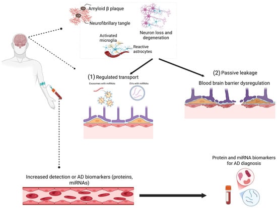
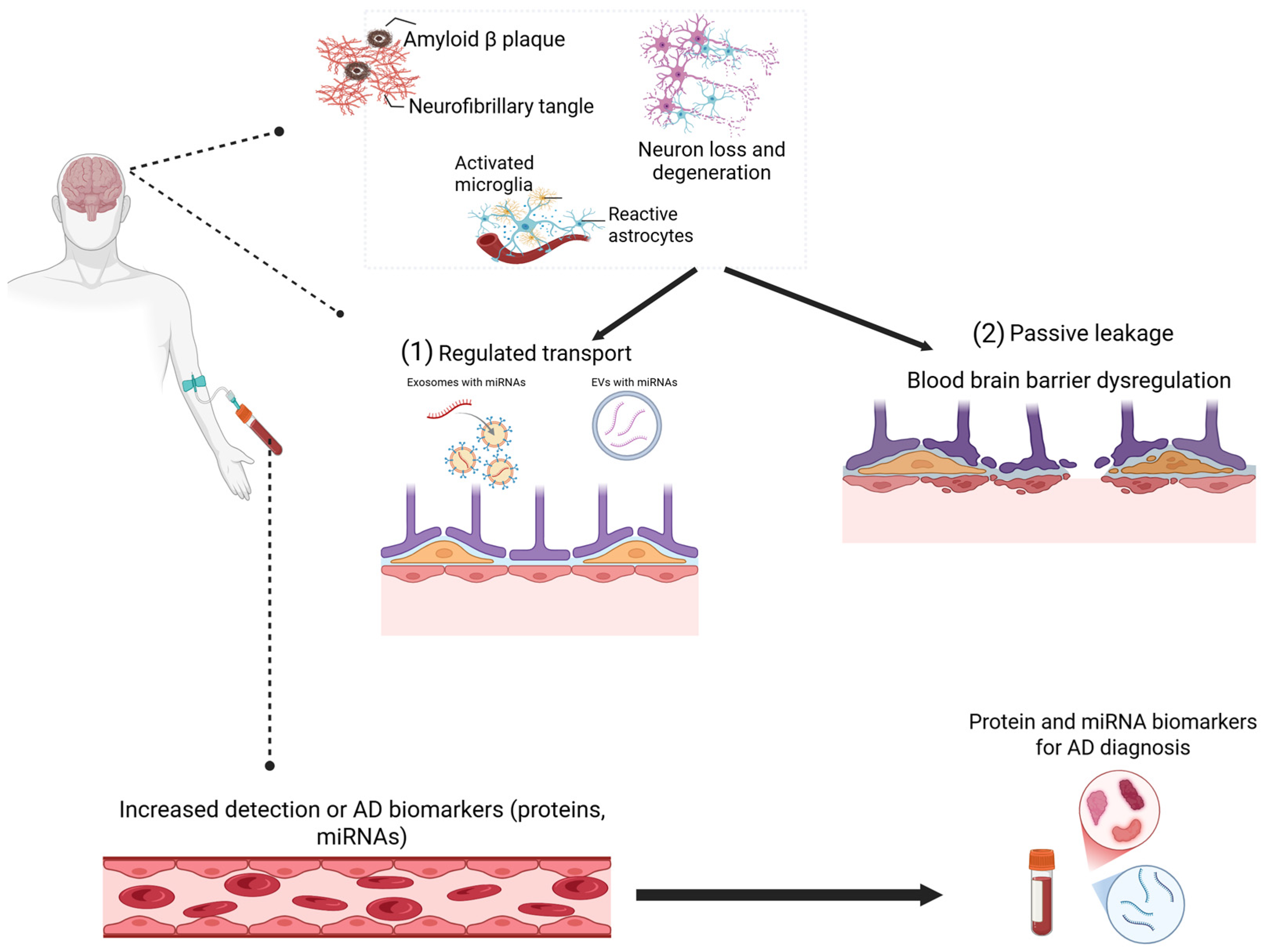
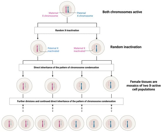
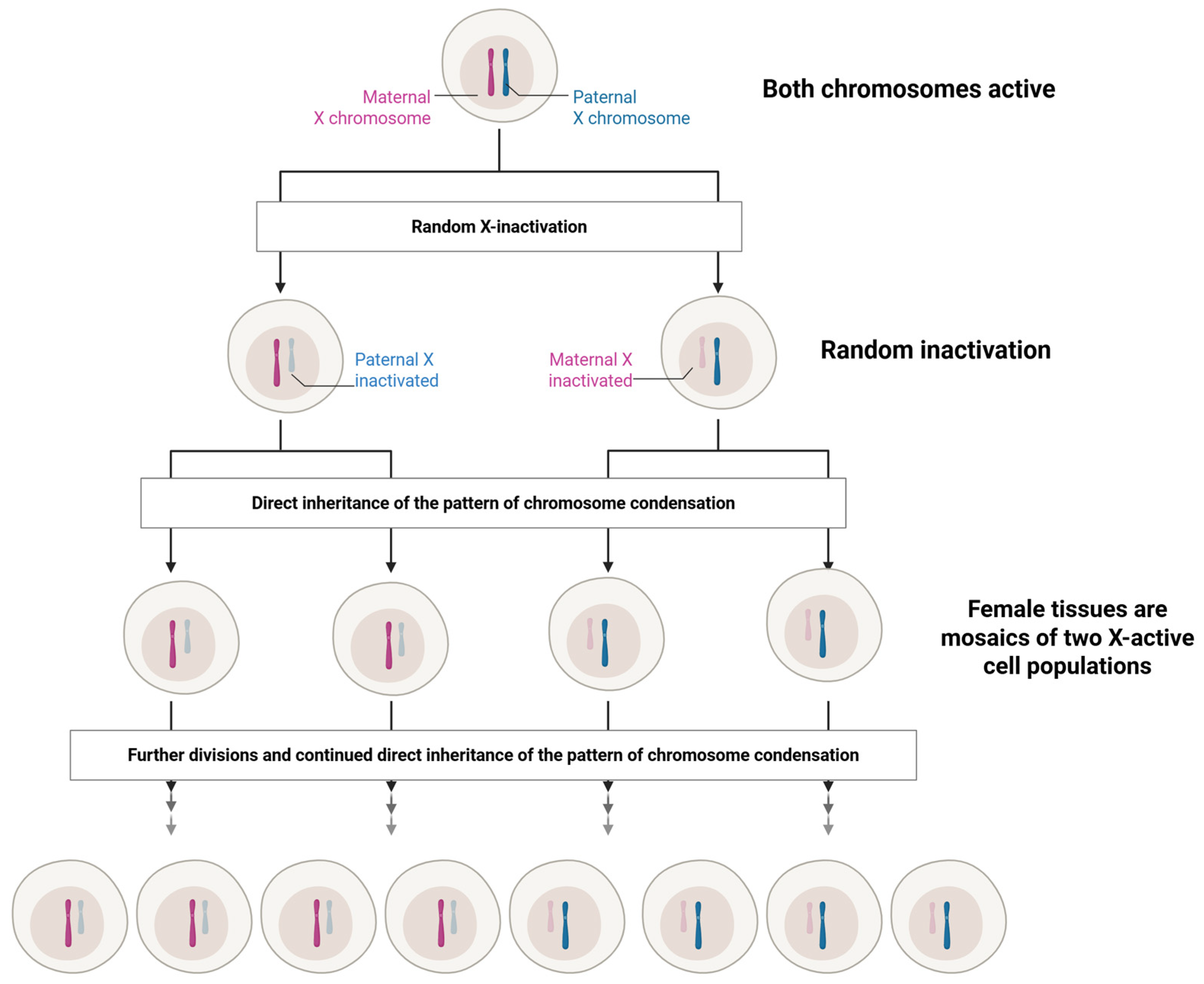

Disclaimer/Publisher’s Note: The statements, opinions and data contained in all publications are solely those of the individual author(s) and contributor(s) and not of MDPI and/or the editor(s). MDPI and/or the editor(s) disclaim responsibility for any injury to people or property resulting from any ideas, methods, instructions or products referred to in the content. |
© 2026 by the authors. Licensee MDPI, Basel, Switzerland. This article is an open access article distributed under the terms and conditions of the Creative Commons Attribution (CC BY) license.