


Abstract
The voltage-dependent anion channel (VDAC) is the primary conduit for ion and metabolite transport across the mitochondrial outer membrane. Positioned at the interface between the cytosol and the mitochondrial compartment, VDAC is uniquely accessible to proteins on both sides of the membrane, making it an interaction hub whose biophysical properties and signaling functions are shaped by protein complexation in addition to its intrinsic pore specialization. Mammals express three isoforms—VDAC1, VDAC2, and VDAC3—sharing a conserved β-barrel scaffold with about 70% identity. However, minor differences in the sequence lead to drastic changes in VDAC isoform affinity with other proteins. Here, we review the molecular mechanisms and physiological consequences of VDAC complexation with a set of well-characterized partners: hexokinase, dimeric tubulin, α-synuclein, mitochondria-associated membrane proteins, B-cell lymphoma 2 (BCL-2) family proteins, and the translocase of the outer membrane (TOM) protein import complex. For each complex, we evaluate the available structural, biophysical, and genetic evidence for isoform specificity, highlight where mechanistic understanding is most advanced, and identify open questions. A consistent principle emerges across all complexes: functionally nonredundant isoform contributions are primarily governed by differential partner affinity and complexation, rather than by differences in pore architecture alone. This framework has direct implications for mitochondria-associated pathologies, including cancer, cardiovascular disease, and neurodegeneration, as well as for the rational design of VDAC-targeting therapeutics.
1. Introduction
Voltage-dependent anion channels (VDACs) are the principal channels of the mitochondrial outer membrane (MOM) for small-molecule transport. They are among the most abundant integral membrane proteins in the MOM [1]. Localized at the interface between the cytosol and mitochondria, VDACs do more than just conduct water-soluble metabolites and small ions: they also serve as docking platforms for other proteins. Mammals express three VDAC isoforms, VDAC1, VDAC2, and VDAC3, which share a conserved pore-forming scaffold but differ in sequence features that are likely responsible for the differences in partner recognition and regulation. Comparative analyses highlight an extended N-terminus in VDAC2 and marked differences in cysteine content and distribution among the three isoforms, despite their overall conservation [2,3] (Figure 1). Only the structure of VDAC1 has been solved [4,5,6]; however, sequence homology and computational studies suggest a largely conserved three-dimensional framework (Figure 1). Recently, a cryo-electron microscopy (EM) study of the VDAC2 complex with translocase of the outer membrane 40 (TOM40) and PTEN-induced kinase 1 (PINK1) confirmed the ß-barrel structure of VDAC2 with the N-terminus extending into the intermembrane space (IMS) [7] (Figure 1).
Figure 1.
Structural comparison of mammalian VDAC isoforms. Top: Cartoon representation of human VDAC1 (hVDAC1, PDB: 6G6U) derived from X-ray crystallography, colored in rainbow colors with N-terminal in blue and C-terminal in red. Structures of hVDAC2 and hVDAC3 were generated based on sequence homology using ColabFold [8]. Bottom: IMS view of hVDAC1 and hVDAC2, with the latter obtained from cryo-EM of VDAC2-TOM-PINK complex (PDB: 9EIH), shows similar conformations with the N-terminal helix in the lumen of ß-barrel. Isoform-specific differences in cysteine residues and membrane-facing glutamate residues, Glu73 (VDAC1) and Glu84 (VDAC2), and corresponding glutamine (Gln73) in VDAC3 are labeled. (Oxygen: red; nitrogen: blue; sulfur: yellow). Structures derived from X-ray crystallography [9] and cryo-EM data [7] are shown in boxes, along with their corresponding PDB codes. Created on BioRender.
Since the discovery of VDAC in 1976 [10,11,12] by Colombini, Mannella, and coauthors, for another 20 years, this mitochondrial channel was conventionally regarded as a simple molecular sieve. Only in the mid-90s was VDAC experimentally shown to translocate ATP by passive diffusion and to regulate ATP flux by voltage-gating [13]. Since then, VDAC is increasingly viewed not as a passive β-barrel mitochondrial channel, but as a regulated hub whose permeability and signaling functions are shaped by interactions with other proteins. The unique VDAC location at the intersection between mitochondria and cytosol allows VDAC to regulate communication between mitochondria and the rest of the cell by complexing with a plethora of proteins involved in cell signaling and metabolism [14,15,16]. This view is supported by both targeted studies and broader interactome approaches. Roman et al. used a membrane-protein-adapted phage display strategy to identify many previously known ligands and numerous additional VDAC-interacting epitopes [17]. For instance, VDAC was found to be associated with different pro- and anti-apoptotic proteins [18,19,20,21,22], multiple kinases [23,24,25,26,27,28], cytoskeleton [29,30,31], neuronal [32,33,34], and mitochondrial proteins [7,35]. In addition, distinct protein-partner repertoires provide a mechanistic basis for the nonredundant contributions of VDAC1, VDAC2, and VDAC3 to metabolism, apoptosis, redox regulation, and inter-organelle communication [36]. Thus, the strongest current framework for understanding isoform-specific VDAC biology is not only through its channel properties but also differential complexation with protein partners.
This review approaches VDAC primarily as an interaction hub and treats isoform-specific complexation as the key to understanding how structurally similar ß-barrel mitochondrial channels acquire distinct cellular functions.
2. Hexokinase–VDAC Complex
VDAC complexation with hexokinase (HK) at the MOM links mitochondrial metabolism to glycolysis [37]. Early work showed that a substantial fraction of brain HK is associated with particulate matter, which was identified as the mitochondrial fraction [38,39]. Felgner and colleagues extended this finding by purifying an HK-binding protein from MOM, which was later identified as VDAC [26,40,41].
Around the same time, Polakis and Wilson showed that the hydrophobic N-terminal sequence of rat brain HK is required for mitochondrial targeting [42]. There are four major HK isoforms, but only HK1 and HK2 have an N-terminal membrane-binding domain that associates with the MOM (Figure 2A) [37]. HK2-specific mutational analysis further confirmed that residues within the N-terminus are critical for mitochondrial binding, with substitution of His5 abolishing outer-membrane association [43]. While the N-terminal peptide is necessary for mitochondrial targeting, the N-terminal binding domain may not be sufficient since fusion experiments showed that the HK1 binding domain could not target HK4 to mitochondria, indicating that adjacent structural elements also contribute to docking competence [44]. Additional work arguing for multiple mitochondrial HK-binding modes or sites, including distinctions between glucose-6-phosphate-sensitive and -insensitive interactions, also suggests that HK isoform specificity may intersect with binding heterogeneity rather than reflecting a single uniform docking mechanism [45,46].
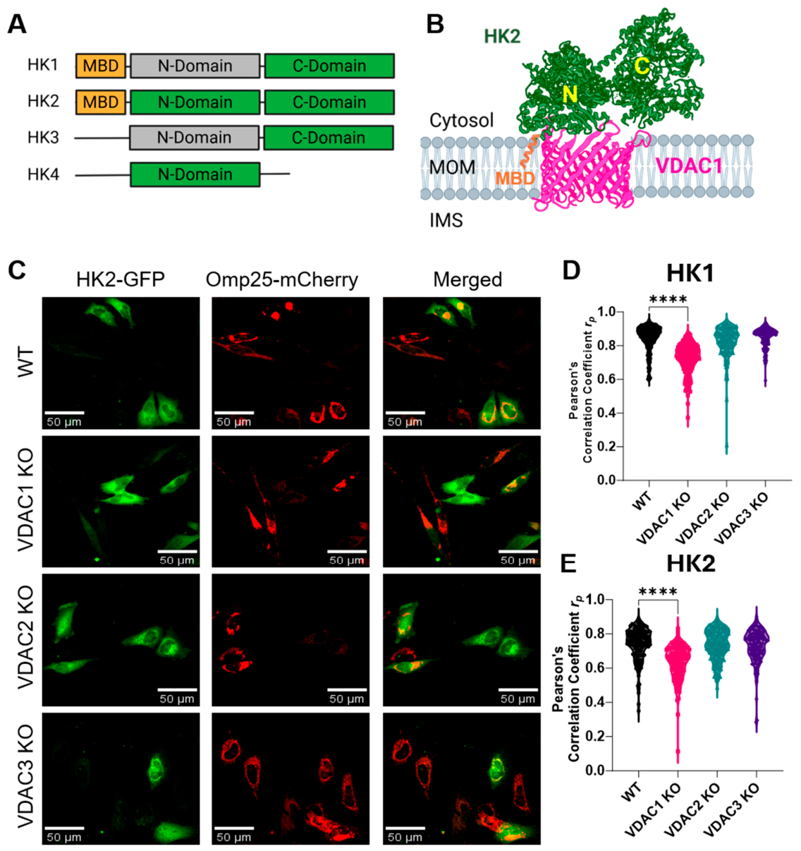
VDAC isoform specificity further complicates our understanding of HK-VDAC complexation. Earlier work often treated VDAC as a single receptor entity, but later studies began to resolve the interaction more explicitly at the isoform level. Deletion of VDAC1 reduced mitochondria-bound HK activity in oxidative tissues such as heart and soleus muscle and is associated with impaired glucose tolerance, supporting VDAC1 as a physiologically important mitochondrial HK-binding site in vivo [47]. Later cell-based work further showed that VDAC1 loss reduces mitochondrial HK2 binding under oxidative stress conditions in the rat myoblast cell line (H9c2), extending the relevance of the interaction beyond HK1 alone [48]. More recent comparative analysis by our group strengthened the VDAC1-centered view in HeLa cells [49]. VDAC1 knockout (KO) decreased glycolysis rate, whereas VDAC2 and VDAC3 KO did not produce the same glycolytic defect, linking VDAC1 most directly to glycolytic support in HeLa cells. We also showed that mitochondrial localization of both HK1-GFP and HK2-GFP, measured by colocalization with the outer-membrane marker Omp25-mCherry (Figure 2C–E), is significantly reduced in VDAC1 KO cells, further supporting a privileged role for VDAC1 in recruiting or stabilizing mitochondrial HK in this cellular context (Figure 2B) [49]. Using molecular dynamics (MD) simulations, Haloi et al. proposed a membrane-first model in which HK2 initially binds the MOM via its N-terminal segment and then engages VDAC1 at the membrane interface [50]. Recently, Bieker et al. suggested that HK1 binds VDAC1 and VDAC2 through the interaction of the N-terminal of HK1 with the membrane-facing glutamate in VDAC1 (Glu73) and VDAC2 (Glu84) [51]. The dissociation of HK1 from mitochondria upon expression of VDAC1 and VDAC2, with the glutamate residue mutated, was used as evidence for direct binding of the HK1 N-terminus to the membrane-facing glutamate residue. However, mutating Glu73 may disrupt other protein–protein interactions and cellular processes, which may indirectly affect HK1 binding. In addition, HK1 showed a higher degree of colocalization with VDAC3, the isoform lacking Glu73 but carrying neutral Gln73, at the spatial resolution achieved by two-color STED microscopy. A substantial fraction of mitochondria-bound HK1 did not colocalize with any of the three isoforms, suggesting a more complex spatial relationship than previously anticipated [52]. Therefore, future work is needed to resolve the structure of the HK complex with each VDAC isoform.
Figure 2.
VDAC complexation with hexokinase (HK). (A) Schematic representation of the four major HK isoforms with the mitochondrial binding domain (MBD) shown in orange for HK1 and HK2. Except HK4, all isoforms have two domains, with the catalytically active domain shown in green. (B) Putative VDAC-HK complexation after Haloi et al. [50]. HK MBD inserts into MOM and interacts with VDAC1 at the membrane interface. (Created on BioRender) (C) Colocalization of HK2-GFP (green) and Omp25-mCherry (red) shown in the merged image for HeLa WT, VDAC1, 2, and 3 KO cell lines. Pearson’s Correlation Coefficient analysis of HK1 (D) and HK2 (E) colocalization with Omp25-mCherry reveals a significant decrease in mitochondrial localization of HK1 and HK2 in VDAC1 KO cell lines. Significance was tested using one-way ANOVA followed by Dunnett’s multiple comparison test (**** p < 0.0001) (C–E) were adapted with permission from [49].
Historically, mitochondrially bound HK was suggested to preferentially phosphorylate glucose using intramitochondrially generated ATP in hepatoma mitochondria, providing direct evidence that the VDAC-HK complex can channel ATP from oxidative phosphorylation into the first step of glycolysis [37,53]—the so-called channeling model. More recently, Huggler et al. reported that cytosolic HK, rather than outer-membrane-docked HK, better supports aerobic glycolysis and cell growth in their system, directly challenging the broadest version of the classic channeling model [54]. The VDAC-HK complex has also been suggested to act as a “battery” leading to metabolic regulation of MOM potential and therefore permeability for metabolites through VDAC regulation by voltage [55]. Thus, the controversy in the field is no longer whether HK can bind VDAC, but how general the metabolic consequences of that binding are and whether channeling models apply uniformly across systems.
A separate line of work has linked HK-VDAC to cell death control, but the protective role of a defined HK-VDAC complex remains incompletely established. Perturbation studies show that disrupting the HK2-VDAC1 axis increases susceptibility to mitochondrial injury under stress conditions [23,48] and can also promote inflammasome signaling [56] or stress-induced mitochondrial remodeling [57], yet these results remain largely indirect and do not by themselves demonstrate that HK-VDAC binding is the primary protective determinant against apoptosis.
3. Tubulin–VDAC Complex
Beyond its canonical role as the building block of microtubules, free dimeric tubulin also associates with mitochondria. Early evidence came from Bernier-Valentin et al., who demonstrated high-affinity tubulin binding to purified mitochondrial membranes [58], and from Saetersdal et al., who used immunofluorescence and immunogold EM to show β-tubulin association with MOM in adult cardiac myocytes [59]. Carré et al. later provided direct biochemical evidence for a VDAC–tubulin complex at MOM, showing that tubulin is an inherent component of mitochondrial membranes and accounts for ~2% of total cellular tubulin. It is likely organized as an α/β heterodimer, enriched in βIII-tubulin, and specifically co-immunoprecipitates with VDAC [60]. However, the physiological role of free cytosol-solubilized dimeric tubulin, other than being a supply for microtubules, was unknown until recently.
Dimeric tubulin is a heterodimer of around 110 kDa with α- and ß-subunits with well-defined structures of their globular parts [61]. Each tubulin subunit also has an unstructured, highly negatively charged C-terminal tail (CTT) composed of 10–27 amino acids [62] that are essentially poly-glutamate polypeptides exposed at the protein surface (Figure 3A). Higher eukaryotes encode six to nine tubulin isotypes of each subunit with cell-specific expression [63]. Notably, the seven α-tubulin and eight ß-tubulin known isotypes in humans vary mostly in their CTTs [64].
Our group showed that dimeric tubulin interacts with VDAC reconstituted into planar lipid membrane (PLM) and induces characteristic fast reversible blockages of VDAC conductance on the 1 ms to 1000 ms time scale [30,65]. A representative experiment in which 20 nM of dimeric tubulin isolated from the brain induces fluctuations in channel conductance between high conducting “open” and low conducting “blocked” states is shown in Figure 3B. Dimeric tubulin induces measurable blockage events only when negative voltage is applied to the side of tubulin addition (Figure 3C). This observation and the fact that tubulin with proteolytically cleaved CTT does not induce channel blockages lead to the conclusion that anionic CTTs are responsible for pore blockage [30]. The blockage time—the time when the channel is blocked by tubulin—is highly voltage dependent and increases exponentially with applied voltage [30,65,66]. Another piece of experimental evidence that the anionic CTT of tubulin is captured inside the VDAC pore, driven there by negative voltage, is that the tubulin-blocked state has reversed, cationic selectivity, in contrast to the anionic selectivity of the open state [67]. Equilibrium and nonequilibrium MD simulations, together with the Rosetta protein–protein docking algorithm, provided further structural evidence of the tubulin–VDAC complex with the anionic CTT captured inside the pore and reversing its selectivity [68]. Another important feature of the tubulin–VDAC interaction is that the CTT alone is not sufficient to produce long-lived VDAC blockages. CTT should be attached to an “anchor” that localizes CTT near the channel entrance and prevents its free translocation through the pore (Figure 3C).
Figure 3.
VDAC reconstitution experiments. Dimeric tubulin (A–C) and αSyn (D–F) induce fast, transient, partial blockages of VDAC conductance. (A,D) Both VDAC inhibitors, tubulin and αSyn, have two domains: a membrane binding domain (green frame), which can be structured or not, and a disordered polyanionic pore-blocking domain (red frame). Records of fluctuating conductance of single VDAC channels reconstituted into PLM before (left traces) and after addition of 20 nM tubulin (B) or 50 nM αSyn (E) to one side of the membrane at −25 mV applied voltage. Potential is more negative at the side of tubulin or αSyn addition. The channel conductance fluctuates between the open and blocked states, as indicated by the blue and red dashed lines, respectively, with well-time-resolved blockage events. Long dashed black lines indicate the zero-current corresponding to negligible conductance. Models of VDAC complexation with dimeric tubulin (C) and αSyn (F), in which the negatively charged C-terminal tails (CTT, shown in red) of either tubulin or αSyn, driven into the VDAC pore by the applied negative potential, partially block the channel. The relatively short α- and the longer β-tubulin CTT (A) can be captured by the pore only when the α-tubulin subunit is membrane-bound [69]. Due to its bulkiness, tubulin does not translocate through VDAC. The membrane-bound N-terminal domain of αSyn (D) prevents translocation of the whole molecule through the pore only up to a certain voltage [32,70]. (G) Each interaction of blocker with channel is characterized by the open time (topen) and the blockage time (tblock). The open state is anion-selective, allowing ATP/ADP fluxes while exhibiting limited Ca2+ permeability. The blocked state is cation-selective and impermeable to ATP/ADP, but it has higher permeability for Ca2+. Adapted with permission from [66].
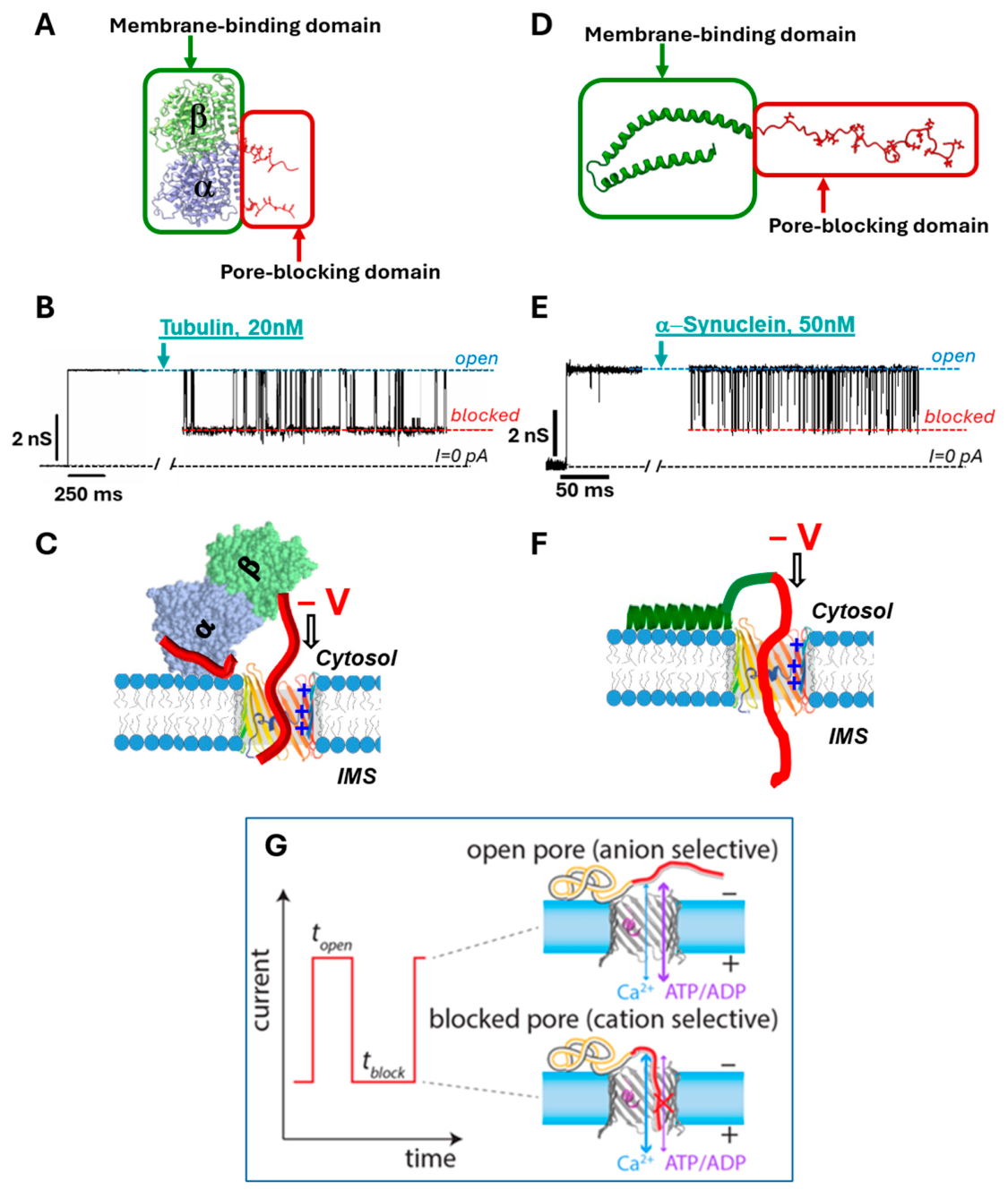
Therefore, the first step of the tubulin–VDAC interaction is tubulin binding to the lipid surface, as confirmed by several biophysical methods and MD simulations [69]. Furthermore, using a recombinant yeast tubulin construct with either α- or ß-tubulin CTT in experiments with reconstituted VDAC, we showed that both α- and ß-CTTs could be captured by the VDAC pore [71]. Taken together, these data allowed us to propose a model of the tubulin–VDAC interaction where anionic tubulin CTT partially and transiently blocks the positively charged VDAC pore. This causes reversal of its ionic selectivity, and complete blocking of ATP transport through the pore while still allowing for transport of small ions, including Ca2+ [66,67,68] (Figure 3C).
The most important physiological consequence of VDAC’s reversible blockage by tubulin CTTs for mitochondria is that the tubulin-blocked state of the channel is essentially impermeable for ATP, as has been shown by direct experiments on reconstituted channels [67] and MD simulations [68]. Thus, tubulin can effectively regulate the fluxes of metabolites (most of which are negatively charged) through VDAC and, consequently, MOM permeability and mitochondria respiration. Indeed, these predictions were confirmed in experiments with isolated mitochondria [30,72] and intact cancer cells [73,74]. It was shown that when a cytosolic pool of free dimeric tubulin in human hepatoma (HepG2) cells was increased after the treatment with the microtubule-targeting drugs (MTD) causing microtubule destabilization (such as nocodazole), the mitochondrial potential decreased [73]. In contrast, treatment with a microtubule-stabilizing agent (such as paclitaxel) decreased free cytosolic tubulin and led to a rise in mitochondrial potential [73]. The subsequent genetic evidence of VDAC’s involvement in the modulation of mitochondrial potential by MTDs was obtained using the siRNA knockdown of each VDAC isoform in HepG2 cells [74]. These experiments unambiguously showed that knockdown of each VDAC isoform led to an isoform-dependent decrease in mitochondrial potential. The observed changes in mitochondrial potential were suggested to be caused by a decrease in ATP/ADP uptake and an increase in Ca2+ uptake by VDAC when blocked by free dimeric tubulin. The close proximity of ßIII-tubulin to VDAC1 was later confirmed using a proximity ligation assay (PLA) in neuroblastoma cells [71].
Together, these findings support a model of the tubulin–VDAC complex, which dynamically modulates VDAC permeability at the MOM, thereby regulating metabolite fluxes in and out of mitochondria, mitochondrial membrane potential, and respiration. More recent work by Parker et al. further links βIII-tubulin to mitochondrial biology. Parker et al. showed that βIII-tubulin promotes a more extended reticular mitochondrial network, whereas βIII-tubulin suppression shifts mitochondria toward a more perinuclear distribution [75], and that βIII-tubulin alters cellular metabolism and stress-response signaling under glucose limitation [76].
4. α-Synuclein–VDAC Complex
α-Synuclein (αSyn) is a small, 140-residue, 14.4 kDa intrinsically disordered protein that is highly expressed in neurons, particularly at presynaptic terminals of neurons [77]. It is central to Parkinson’s disease (PD) pathology [78]. αSyn is a major constituent of Lewy bodies, and SNCA point mutations, multiplications, and regulatory variants link altered αSyn sequence or dosage to familial and sporadic synucleinopathies [79]. αSyn toxicity is often discussed in the context of its oligomer and fibril formation [78], but the detailed mechanisms by which αSyn is involved in neurodegeneration, particularly in its monomeric form, remain largely unresolved.
Early studies showed that a fraction of αSyn localizes to both outer and inner mitochondrial membranes [80,81], causing impairment of the mitochondrial respiratory complexes I [82,83], IV [84], and V [85], increasing oxidative stress [86,87] and fission [88] and leading to neuronal cell death (for the recent review, see [89,90]). More direct evidence for a VDAC-centered pathway came from neuronally differentiated SH-SY5Y cells overexpressing wild-type αSyn, where PLA detected αSyn in close proximity to both VDAC1 at the outer membrane and COX IV at the inner membrane; importantly, VDAC1 knockdown markedly reduced αSyn proximity to COX IV [91], supporting VDAC as a leading route for αSyn passage across the outer membrane.
Mechanistically, αSyn resembles tubulin in combining a membrane-binding domain with a highly acidic, disordered CTT capable of pore engagement (Figure 3D). Structural studies show that upon binding to an anionic membrane, the N-terminal region of αSyn becomes α-helical and acts as a membrane anchor, whereas the C-terminal region remains disordered and only weakly associated with the membrane [92,93], thus leaving it available for interaction with VDAC. Consistent with this model, the biophysical work demonstrated that monomeric αSyn at nanomolar concentrations reversibly blocks reconstituted VDAC in planar bilayers [32] (Figure 3E). The single-channel recordings show markedly similar fluctuations between the open VDAC state and the blocked state, the latter with the conductance ~0.4 of the open state conductance, in the presence of either αSyn or tubulin (Figure 3B,E). Importantly, similar to tubulin, increasing the applied negative voltage keeps the anionic CTT of αSyn inside the pore longer, which is seen as an exponential increase in the blockage time, and selectivity of the αSyn blocked state shifts from anionic to cationic [70,94] (Figure 3F,G). At relatively low voltages, the pore-blocking domain—CTT of αSyn or tubulin—is retracted back to the membrane surface after some time spent inside the pore. At higher voltages, the αSyn blockage time decreases, consistent with a transition from reversible C-terminal capture to αSyn translocation through the pore [32,70]. The translocation regime for αSyn was demonstrated in experiments on VDAC reconstituted in PLM in a salt gradient, using direct single-channel, real-time monitoring of αSyn translocation through the pore [70]. Evidently, tubulin, whose bulky body prohibits its translocation through the pore, does not show a reduction in blockage time even at voltages higher than 40 mV. This framework suggests several consequences for mitochondrial physiology. First, αSyn may regulate MOM permeability by dynamically restricting VDAC-mediated flux of ATP/ADP and other anionic respiratory substrates. Second, despite lowering overall conductance, the αSyn-blocked state strongly favors Ca2+ flux, offering a mechanistic link to reports that αSyn expression enhances mitochondrial Ca2+ uptake [95] (Figure 3G). Third, under conditions of αSyn overexpression or stress, VDAC-mediated translocation provides a plausible route by which cytosolic αSyn reaches intramitochondrial targets and contributes to respiratory dysfunction and loss of mitochondrial membrane potential. Taken together, these observations broaden the discussion beyond the toxic role of aggregated αSyn and support a model in which membrane-bound monomeric αSyn directly modulates VDAC function and mitochondrial metabolism in both healthy and pathological conditions.
A further layer of complexity is introduced by VDAC isoform specificity. Single-channel studies demonstrated that αSyn can block and translocate through all three mammalian VDAC isoforms, but with quantitatively distinct kinetics: VDAC3 displays 10- to 100-fold lower on-rates for αSyn-induced block than VDAC1 [96], and the VDAC2-αSyn interaction is ~ 10 times less effective than that of VDAC1-αSyn [97]. This finding supports the idea that differential affinity for cytosolic disordered proteins may contribute to nonredundant isoform function in cells, although the physiological consequences of these kinetic differences still need to be defined more directly.
5. VDAC-Mitochondria-Associated Membrane Protein Complexes
Close contact sites between mitochondria and other organelles, such as endoplasmic reticulum (ER), sarcoplasmic reticulum (SR), and lysosomes, known as mitochondria-associated membranes (MAMs), have been implicated in sterol homeostasis, Ca2+ signaling, and ER stress pathways.
Ca2+ acts as a secondary signaling molecule to coordinate cell activity with mitochondrial ATP production. This requires rapid Ca2+ uptake into the mitochondrial matrix by mitochondrial Ca2+ uniporters (MCUs) located at the inner membrane. However, MCUs have low affinity for Ca2+ [98] compared to the speed of Ca2+ accumulation observed in live cells. MAMs link VDAC to other Ca2+ channels, such as inositol-1,4,5-triphosphate-sensitive channels (IP3R) of ER [99], Ryanodine-sensitive channels (RYRs) of SR [100], and transient receptor potential mucolipin 1 (TRPML1) of lysosomes [101] (Figure 4). These Ca2+ microdomains facilitate rapid Ca2+ uptake via VDAC and MCU.
Figure 4.
VDAC complexation at mitochondria-associated membranes (MAMs). Cartoon representation of VDAC complexes at the contact sites with other organelles such as endoplasmic reticulum (ER), sarcoplasmic reticulum (SR), and lysosome (cartoon at the left). VDAC1 has been shown to form complexes with Ca2+ transporter TRPML1 at the lysosome and with multiple proteins at the ER/SR contact sites (zoomed red box). VDAC1 complex with ER Ca2+ transporter IP3R through GRP75 allow rapid Ca2+ uptake from ER to mitochondria via the formation of Ca2+ microdomains at the interface. This complex can be modulated by Parkinson’s disease-related proteins, such as PARKIN and DJ1, and is linked to ER stress. STIM1, an ER Ca2+ sensor, promotes ER-mitochondria Ca2+ flux through interaction with the IP3R-GRP75-VDAC1 complex. TMEM43, an ER/nuclear protein, was shown to promote ER–mitochondria contact sites through interaction with VDAC1 and VDAC2. Lamin B receptor (LBR) promotes Ca2+ flux through the IP3R-VDAC2 complex, while CKAP4 competes with IP3R for binding VDAC2, reducing the IP3R-VDAC2 interaction. VDAC2 also forms a complex with RyR2 in SR. Created on BioRender.
Early work established VDAC as an important part of Ca2+ complexes at contact sites to promote the transfer of local ER Ca2+ signals to mitochondria [102]. Later, VDAC1 was identified as a core component of the IP3R-GRP75-VDAC1 axis [103,104]. In particular, despite all three VDAC isoforms being involved in mitochondrial Ca2+ uptake upon IP3R stimulation [105], only VDAC1 was reported to form complexes with IP3R and to selectively support transmission of low-amplitude apoptotic Ca2+ signals to mitochondria. However, more recent studies re-established the role of VDAC2 in Ca2+ signaling. Harada et al. identified palmitoylated CKAP4 as a VDAC2-binding partner at ER–mitochondria contact sites that restrains IP3R–VDAC2 coupling and mitochondrial Ca2+ loading [106], whereas Zhao et al. showed that the lamin B receptor (LBR) organizes a mitosis-specific IP3R–LBR–VDAC2 contact that enhances mitochondrial Ca2+ influx to support ATP production and cell division [107]. Furthermore, the RyR2-VDAC2 association at SR–mitochondria junctions has been linked directly to mitochondrial Ca2+ uptake in cardiomyocytes [108]. Cardiac-specific deletion of VDAC2 caused impaired mitochondrial Ca2+ uptake, altered intracellular Ca2+ transients, defective excitation–contraction coupling, and dilated cardiomyopathy, indicating that VDAC2 has a nonredundant role in maintaining mitochondrial Ca2+ handling in the heart [109].
Comparative cardiac experiments showed that VDAC3 is less effective than VDAC1 or VDAC2 in supporting mitochondrial Ca2+ transfer, and isoform-specific mutational analysis pointed specifically to the membrane-facing glutamate, showing that the native Gln73 in VDAC3 contributes to its weaker support of mitochondrial Ca2+ transfer relative to the glutamate found at the corresponding position in VDAC1 and VDAC2 [110]. However, considering that the Ca2+ ion cannot reach membrane-facing glutamate through the hydrophobic lipid core (Figure 1), the mechanism by which Glu73 regulates mitochondrial Ca2+ uptake in HL-1 cardiomyocytes remains unknown. We can only speculate that, in cardiac cells, there is an ER Ca2+ channel that forms a putative complex with the VDAC2 hydrophobic exterior via Glu84.
Beyond mitochondrial Ca2+ flux itself, MAM-associated VDAC complexes are increasingly implicated in ER stress pathways. ER stress activates the unfolded protein response (UPR) and ER-associated degradation (ERAD) to restore proteostasis, but prolonged stress can remodel ER–mitochondria contact sites, where altered Ca2+ transfer can amplify mitochondrial dysfunction. In this context, the classical IP3R–GRP75–VDAC1 complex is increasingly understood not simply as a Ca2+ conduit, but as an ER stress signaling hub. Meng et al. identified a STIM1-regulated IP3R–GRP75–VDAC1–MCU axis. STIM1, an ER Ca2+ sensor, promotes assembly or stabilization of the ER–mitochondria Ca2+-transport complex, thereby enhancing mitochondrial Ca2+ loading, oxidative stress, and downstream injury during inflammation [111]. This idea was reinforced by Yan et al., who showed that an ERAD modulator (NCATS-SM0225) binds all three VDAC isoforms, strengthens VDAC1–IP3R coupling, and activates UPR response to selectively kill cancer cells [112]. Along with this model, Orantos-Aguilera et al. showed that STIM1-containing contact sites also promote direct ER-to-mitochondria Ca2+ flux through GRP75- and PTPIP51-associated MAMs, extending the classical IP3R–VDAC1 framework to a broader contact-site model for mitochondrial Ca2+ uptake [113]. In cancer, chronic ER stress can drive a more adaptive UPR/ERAD program that helps sustain survival and metabolic remodeling. In contrast, ERAD is more often linked to the accumulation of misfolded proteins in neurodegeneration. Two PD-linked proteins, Parkin and DJ-1, regulate VDAC1-centered ER–mitochondria contact: Xue et al. showed that Parkin modulates the IP3R–GRP75–VDAC1 complex to influence ER–mitochondria association and Ca2+ homeostasis [114], whereas Liu et al. identified DJ-1 as a stabilizing partner of the IP3R3–GRP75–VDAC1 complex [115]. These studies suggest that disruption of VDAC1-centered MAM signaling may be one route by which PD-associated genes perturb mitochondrial homeostasis under stress.
TMEM43/LUMA—an ER and nuclear-envelope membrane protein [116,117]—has also been identified as a component of an ER-membrane E3 ubiquitin ligase (RNF26) complex, linking TMEM43 more broadly to the ERAD pathway [116,117,118]. Mutations in this protein are associated with muscular dystrophy [119], arrhythmogenic right ventricular cardiomyopathy [120], and cancer [121]. TMEM43/LUMA has been linked more broadly to cardiac and metabolic regulatory networks, as systems-genetics analysis and Tmem43-S358L mouse studies have associated altered TMEM43 with cardiomyopathy- and mitochondria-related pathways [122]. In particular, the TMEM43p.S358L mutant weakens the interaction with VDAC1 and VDAC2 and destabilizes ER/SR–mitochondria contact sites, leading to impaired mitochondrial function and cardiac cell death [123,124]. Zhang et al. showed that TMEM43 can promote hepatocellular carcinoma by enhancing VDAC1 activity through Ubiquitin-specific-processing protease 7 (USP7)-dependent deubiquitination, suggesting that TMEM43–VDAC coupling can also be co-opted to support metabolic adaptation and survival in cancer [125]. Together, these studies suggest that ER stress phenotypes can arise not only from altered Ca2+ flux through canonical IP3R–VDAC complexes, but also from disruption of the contact-site scaffolds that organize those complexes.
6. VDAC-BCL-2 Family Protein Complexes
Apoptosis, a complex cell death pathway, often leads to MOM permeabilization (MOMP), driven by the oligomerization of the effector proteins such as BAX and BAK into cytochrome c releasing pores. This process is governed by the BCL-2 protein family, whose pro-survival members (BCL-2, BCL-XL, MCL-1) suppress MOMP by sequestering pro-apoptotic effectors and BH3-only proteins, while BH3-only proteins (BIM, tBid, BAD) tip the balance toward cell death [126]. The founding observation linking VDAC to this pathway initially came from Shimizu’s lab, based on experiments with VDAC-containing proteoliposome, that BAX and BAK supposedly create a novel large pore with VDAC that allows cytochrome c passage [127]. Based on extensive work from the Shimizu lab, VDAC was hypothesized to be part of MOMP [128]. However, subsequent studies contradict this model (see review [129]), and Baines et al. showed that mouse embryonic fibroblast (MEF) cells can undergo apoptosis even in the absence of all three VDACs [130].
Recently, VDAC1 has been suggested to form a homo-oligomeric “mega-pore” that facilitates the release of cytochrome c and mitochondrial DNA, thereby inducing apoptosis in many diseases, such as lupus-like diseases [131], diabetes [132,133], and neurodegenerative diseases [134,135]. These studies extrapolate the presence of higher-molecular-weight bands in SDS-PAGE upon cross-linking to “mega-pore” formation. VDAC’s tendency to assemble into oligomeric structures in native mitochondrial membranes has been known since 1984 from pioneering EM work by Carmen Mannella’s group [136,137]. Later, VDAC’s ability to form higher-order complexes was confirmed by atomic force microscopy and cryo-EM [27,138,139,140]. Notably, VDAC1 oligomeric assemblies are sensitive to lipid composition, as shown on giant proteoliposomes with reconstituted VDAC1 by fluorescence correlation spectroscopy [141] and by atomic force microscopy [142] (see review by Di Pinto et al. [143] for a comprehensive discussion).
The loss of higher-molecular-weight bands upon treatment with the VDAC1 oligomerization inhibitor VBIT-4 has been routinely used to confirm the involvement of VDAC “mega-pores” in apoptosis and inflammation. However, it was recently shown that VBIT-4 does not interact with VDAC1 but may affect membrane proteins, especially mitochondrial proteins, via membrane modulation [144]. In fact, VBIT-4 was also shown to inhibit ETC complexes (I, III, and IV) and thus decrease mitochondrial potential and increase oxidative stress, leading to cell death [145]. In addition, VDAC oligomeric arrays may be important for rapid Ca2+ uptake from ER or SR. Therefore, changes in VDAC oligomerization can affect Ca2+ uptake, leading to oxidative stress and apoptosis without forming a “mega-pore”. Furthermore, the VDAC “mega-pore” has never been visualized, unlike BAK/BAX pores [146]. BAX/BAK have been shown to form proteolipid pores independently of VDAC [146,147,148], where VDAC serves as a context-dependent organizer or regulator of BCL-2 family proteins at the MOM.
VDAC, specifically VDAC1, has been reported to interact with BCL-XL [149], promoting the open state of VDAC to sustain mitochondrial metabolite flux as a pro-survival bioenergetic function [18]. However, to date, the physiological relevance of direct VDAC1–BCL-2 family complexes and whether VDAC1 plays an indispensable role in apoptosis remain under debate.
Unlike VDAC1, VDAC2 has emerged as the isoform with the most clearly established role in apoptotic regulation. Mice KO studies showed that only VDAC2 KO led to embryonic lethality [19]. This was linked to the pro-survival function of VDAC2 due to its complexation with monomeric BAK, which is localized to MOM through its C-terminal transmembrane anchor [150]. Upon apoptotic stimulation, according to one model, BH3-only activators such as tBID displace VDAC2 from BAK, releasing BAK to oligomerize and permeabilize the MOM [151]. Using VDAC1-VDAC2 chimeric constructs in VDAC2 KO MEF cells, it was shown that the cytosolic loop between β-strands 10-11 of VDAC2 is a BAK docking site, with residues T168 and D170 being critical [21]. A small molecule, WEHI-9625, was shown to stabilize this interaction to prevent BAK-driven apoptosis [152]. Deep mutational scanning confirmed these findings and suggested that the cytosolic loop between β-strands 10-11 occupies the hydrophobic groove of BAK to stabilize the VDAC2-BAK complex [153]. Recent studies highlight VDAC2 as a mitochondrial checkpoint in cancer therapy through complexation with BAK [154,155,156]. Normal hepatocytes express low levels of both VDAC2 and BAK relative to hepatocellular carcinoma cells, rendering them resistant to VDAC2-directed pro-apoptotic interventions [157]. This raises a new challenging conundrum in the field: the possibility of distinct physiological roles for VDAC2 in normal versus cancer cells, further complicating the VDAC-involving signaling network. Furthermore, these studies emphasize the pharmacological disruption of the VDAC2–BAK interface as a novel therapeutic target [158].
In contrast to its pro-survival function through complexation with BAK, VDAC2 KO was reported to inhibit BAX apoptotic function, suggesting its pro-apoptotic role [22]. BAX is predominantly cytosolic due to sequestration of its C-terminal transmembrane domain in its hydrophobic groove [159]. VDAC2-BAX complexation has been suggested to facilitate BAX translocation to mitochondria upon apoptotic simulation [160]. However, BAX retained the ability to accumulate in MOM and mediate cell death upon apoptotic stimulation in VDAC2-deficient cells and required BAK for mitochondrial localization [161]. The conflicting results in the literature highlight the complex regulation of BAX recruitment [162] and require further investigation to clarify the role of VDAC2 in pro-apoptotic recruitment of BAX.
7. VDAC2-TOM Complex
VDAC is increasingly recognized as a functional partner of the TOM protein import machinery. Sakaue et al. found that yeast VDAC ortholog, porin (Por1), binds newly imported Tom22 and acts as a chaperone for unassembled Tom22, thereby regulating the balance between trimeric and dimeric TOM complexes and influencing mitochondrial protein import [163]. Takeda et al. confirmed that the Tom22-Por1 complex facilitates protein import into the IMS [140]. Ellenrieder et al. extended this concept by showing that Por1 also promotes carrier biogenesis after TOM-mediated entry, interacting with precursor proteins in the IMS and recruiting TIM22 to support transfer to the inner membrane [164].
Most recently, Callegari et al. resolved a human TOM-VDAC2 array in which two TOM core complexes flank a central VDAC2 dimer to stabilize PINK1 at depolarized mitochondria, providing direct structural evidence that VDAC2 can be incorporated into higher-order TOM assemblies in mammalian cells [7] (Figure 5). Taken together, these recent studies support a model in which VDAC2 is not simply adjacent to TOM, but can function as a dynamic organizer and coupling factor for TOM-dependent protein import.
Figure 5.
Cryo-EM structure of VDAC2 supercomplex with mitochondrial TOM complexes and cytosolic dimeric PINK1. Top: Cartoon representation of VDAC2 (teal), TOM complex (orange), and PINK1 (pink). Bottom: IMS view of the supercomplex derived from cryo-EM (PDB: 9EIH).
8. Other VDAC Complexes
VDAC has been found to interact with other proteins not discussed in this review, and new complexes are constantly being identified through recent advances in proteomics. In addition to the proteins discussed above, Table 1 lists other proteins suggested to interact with VDAC and regulate cellular processes such as metabolism (Mitochondrial creatine kinase and glycerol kinase), steroidogenesis (TSPO and StAR), and apoptosis (MCL1 and tBid). Many proteins linked to neurodegeneration have also been shown to interact with VDAC (Tau, Amyloid beta, Parkin, and MIRO-1). However, further studies are needed to confirm the direct complexation and regulation of VDAC by these proteins and their isoform specificity.
Table 1.
List of VDAC isoform-specific protein complexes.
9. Summary
The simultaneous access of VDAC to otherwise strictly compartmentalized proteins on both sides of the MOM helps explain the growing interest in the physiological consequences of VDAC complex formation with its many cytosolic and mitochondrial partners. This emerging role as a cellular communication hub—distinct from, yet intrinsically linked to, its conventional function as a metabolite channel—explains how such a “simple” ß-barrel channel can contribute to a remarkably wide range of mitochondria-associated pathologies, including diabetes, cardiovascular diseases, various cancers, and neurodegenerative disorders.
Given the higher expression levels of VDAC1 in most cells and the availability of specific antibodies, it is the most well-characterized isoform. Many proteins have been identified as targeting VDAC1 by proteomics screening, and in a number of cases, further studies have confirmed the specificity of such binding and its impact on the channel function and cell physiology. VDAC2 has recently emerged as pivotal in regulating Ca2+ signaling and apoptosis in cardiac cells and cancer. VDAC3 interacts with αSyn and dimeric tubulin but with 10-100 times lower affinity than VDAC1 [96] (Figure 6). A proteomic study identified multiple VDAC3-interacting proteins [175]; however, the specificity of these interactions remains unconfirmed. Therefore, systematic isoform-resolved interactomics and structural studies to understand the molecular mechanisms of VDAC isoforms’ function and the identification of their natural regulators and synthetic inhibitors are vital for developing targeted therapies to improve outcomes in mitochondria-associated pathologies.
Figure 6.
From passive holes to the cellular hub through complexation with cytosolic protein partners. The cartoon shows the array of cytosolic proteins reported to form complexes with all three VDAC isoforms. Created on BioRender.
Author Contributions
Conceptualization, M.R. and T.K.R.; writing—original draft preparation, M.R.; writing—review and editing, M.R., T.K.R. and S.M.B.; visualization, M.R. and T.K.R.; supervision, T.K.R. and S.M.B.; funding acquisition, S.M.B. All authors have read and agreed to the published version of the manuscript.
Funding
Research is supported by the Intramural Research Program of the Eunice Kennedy Shriver National Institute of Child Health and Human Development, National Institutes of Health (grants ZIA HD000072 to S.M.B.). The contributions of the NIH authors are considered works of the United States Government. The findings and conclusions presented in this paper are those of the authors and do not necessarily reflect the views of the National Institutes of Health or the U.S. Department of Health and Human Services.
Data Availability Statement
No new data were created or analyzed in this study. Data sharing is not applicable to this article.
Acknowledgments
During the preparation of this manuscript/study, the authors used ChatGPT version 5.4 developed by OpenAI and Claude Sonnet 4.6 developed by Anthropic to assist in initial drafting of this manuscript. The authors have reviewed and edited the output and take full responsibility for the content of this publication.
Conflicts of Interest
The authors declare no conflicts of interest.
Abbreviations
The following abbreviations are used in this manuscript:
| VDAC | Voltage-Dependent Anion Channel |
| MOM | Mitochondrial outer membrane |
| IMS | Intermembrane space |
| HK | Hexokinase |
| αSyn | Alpha-synuclein |
| KO | Knockout |
| MBD | Mitochondria Binding Domain |
| CTT | C-Terminal Tail |
| PLM | Planar lipid membrane |
| MD | Molecular Dynamics |
| PLA | Proximity ligation assay |
| ER | Endoplasmic Reticulum |
| SR | Sarcoplasmic Reticulum |
| MAM | Mitochondria associated membranes |
| ERAD | ER-associated degradation |
| UPR | Unfolded Protein Response |
| PD | Parkinson’s Disease |
| MOMP | MOM permeabilization |
References
- Mannella, C.A. Conformational changes in the mitochondrial channel protein, VDAC, and their functional implications. J. Struct. Biol. 1998, 121, 207–218. [Google Scholar] [CrossRef] [Scilit] [PubMed]
- Messina, A.; Reina, S.; Guarino, F.; De Pinto, V. VDAC isoforms in mammals. Biochim. Biophys. Acta 2012, 1818, 1466–1476. [Google Scholar] [CrossRef] [Scilit] [PubMed]
- De Pinto, V.; Reina, S.; Gupta, A.; Messina, A.; Mahalakshmi, R. Role of cysteines in mammalian VDAC isoforms’ function. Biochim. Biophys. Acta 2016, 1857, 1219–1227. [Google Scholar] [CrossRef] [Scilit] [PubMed]
- Bayrhuber, M.; Meins, T.; Habeck, M.; Becker, S.; Giller, K.; Villinger, S.; Vonrhein, C.; Griesinger, C.; Zweckstetter, M.; Zeth, K. Structure of the human voltage-dependent anion channel. Proc. Natl. Acad. Sci. USA 2008, 105, 15370–15375. [Google Scholar] [CrossRef] [Scilit] [PubMed]
- Hiller, S.; Garces, R.G.; Malia, T.J.; Orekhov, V.Y.; Colombini, M.; Wagner, G. Solution structure of the integral human membrane protein VDAC-1 in detergent micelles. Science 2008, 321, 1206–1210. [Google Scholar] [CrossRef] [Scilit] [PubMed]
- Ujwal, R.; Cascio, D.; Colletier, J.P.; Faham, S.; Zhang, J.; Toro, L.; Ping, P.; Abramson, J. The crystal structure of mouse VDAC1 at 2.3 A resolution reveals mechanistic insights into metabolite gating. Proc. Natl. Acad. Sci. USA 2008, 105, 17742–17747. [Google Scholar] [CrossRef] [Scilit] [PubMed]
- Callegari, S.; Kirk, N.S.; Gan, Z.Y.; Dite, T.; Cobbold, S.A.; Leis, A.; Dagley, L.F.; Glukhova, A.; Komander, D. Structure of human PINK1 at a mitochondrial TOM-VDAC array. Science 2025, 388, 303–310. [Google Scholar] [CrossRef] [Scilit] [PubMed]
- Mirdita, M.; Schutze, K.; Moriwaki, Y.; Heo, L.; Ovchinnikov, S.; Steinegger, M. ColabFold: Making protein folding accessible to all. Nat. Methods 2022, 19, 679–682. [Google Scholar] [CrossRef] [Scilit] [PubMed]
- Razeto, A.; Gribbon, P.; Loew, C. The dynamic nature of the VDAC1 channels in bilayers: Human VDAC1 at 2.7 Angstrom resolution. wwPDB, 2019; to be published. [CrossRef]
- Schein, S.J.; Colombini, M.; Finkelstein, A. Reconstitution in planar lipid bilayers of a voltage-dependent anion-selective channel obtained from paramecium mitochondria. J. Membr. Biol. 1976, 30, 99–120. [Google Scholar] [CrossRef] [Scilit] [PubMed]
- Colombini, M. A candidate for the permeability pathway of the outer mitochondrial membrane. Nature 1979, 279, 643–645. [Google Scholar] [CrossRef] [Scilit] [PubMed]
- Mannella, C.A.; Colombini, M.; Frank, J. Structural and functional evidence for multiple channel complexes in the outer membrane of Neurospora crassa mitochondria. Proc. Natl. Acad. Sci. USA 1983, 80, 2243–2247. [Google Scholar] [CrossRef] [Scilit] [PubMed]
- Rostovtseva, T.; Colombini, M. ATP flux is controlled by a voltage-gated channel from the mitochondrial outer membrane. J. Biol. Chem. 1996, 271, 28006–28008. [Google Scholar] [CrossRef] [Scilit] [PubMed]
- Colombini, M. VDAC: The channel at the interface between mitochondria and the cytosol. Mol. Cell. Biochem. 2004, 256–257, 107–115. [Google Scholar] [CrossRef] [Scilit] [PubMed]
- Lemasters, J.J.; Holmuhamedov, E. Voltage-dependent anion channel (VDAC) as mitochondrial governator—Thinking outside the box. Biochim. Biophys. Acta 2006, 1762, 181–190. [Google Scholar] [CrossRef] [Scilit] [PubMed]
- Mannella, C.A. VDAC—A Primal Perspective. Int. J. Mol. Sci. 2021, 22, 1685. [Google Scholar] [CrossRef] [Scilit] [PubMed]
- Roman, I.; Figys, J.; Steurs, G.; Zizi, M. Hunting interactomes of a membrane protein: Obtaining the largest set of voltage-dependent anion channel-interacting protein epitopes. Mol. Cell. Proteom. 2006, 5, 1667–1680. [Google Scholar] [CrossRef] [Scilit] [PubMed]
- Vander Heiden, M.G.; Li, X.X.; Gottleib, E.; Hill, R.B.; Thompson, C.B.; Colombini, M. Bcl-xL promotes the open configuration of the voltage-dependent anion channel and metabolite passage through the outer mitochondrial membrane. J. Biol. Chem. 2001, 276, 19414–19419. [Google Scholar] [CrossRef] [Scilit] [PubMed]
- Cheng, E.H.; Sheiko, T.V.; Fisher, J.K.; Craigen, W.J.; Korsmeyer, S.J. VDAC2 inhibits BAK activation and mitochondrial apoptosis. Science 2003, 301, 513–517. [Google Scholar] [CrossRef] [Scilit] [PubMed]
- Rostovtseva, T.K.; Antonsson, B.; Suzuki, M.; Youle, R.J.; Colombini, M.; Bezrukov, S.M. Bid, but not Bax, regulates VDAC channels. J. Biol. Chem. 2004, 279, 13575–13583. [Google Scholar] [CrossRef] [Scilit] [PubMed]
- Naghdi, S.; Varnai, P.; Hajnoczky, G. Motifs of VDAC2 required for mitochondrial Bak import and tBid-induced apoptosis. Proc. Natl. Acad. Sci. USA 2015, 112, E5590–E5599. [Google Scholar] [CrossRef] [Scilit] [PubMed]
- Chin, H.S.; Li, M.X.; Tan, I.K.L.; Ninnis, R.L.; Reljic, B.; Scicluna, K.; Dagley, L.F.; Sandow, J.J.; Kelly, G.L.; Samson, A.L.; et al. VDAC2 enables BAX to mediate apoptosis and limit tumor development. Nat. Commun. 2018, 9, 4976. [Google Scholar] [CrossRef] [Scilit] [PubMed]
- Pastorino, J.G.; Hoek, J.B.; Shulga, N. Activation of glycogen synthase kinase 3beta disrupts the binding of hexokinase II to mitochondria by phosphorylating voltage-dependent anion channel and potentiates chemotherapy-induced cytotoxicity. Cancer Res. 2005, 65, 10545–10554. [Google Scholar] [CrossRef] [Scilit] [PubMed]
- Arzoine, L.; Zilberberg, N.; Ben-Romano, R.; Shoshan-Barmatz, V. Voltage-dependent anion channel 1-based peptides interact with hexokinase to prevent its anti-apoptotic activity. J. Biol. Chem. 2009, 284, 3946–3955. [Google Scholar] [CrossRef] [Scilit] [PubMed]
- Sheldon, K.L.; Maldonado, E.N.; Lemasters, J.J.; Rostovtseva, T.K.; Bezrukov, S.M. Phosphorylation of voltage-dependent anion channel by serine/threonine kinases governs its interaction with tubulin. PLoS ONE 2011, 6, e25539. [Google Scholar] [CrossRef] [Scilit] [PubMed]
- Felgner, P.L.; Messer, J.L.; Wilson, J.E. Purification of a hexokinase-binding protein from the outer mitochondrial membrane. J. Biol. Chem. 1979, 254, 4946–4949. [Google Scholar] [CrossRef] [Scilit]
- Leung, M.R.; Zenezini Chiozzi, R.; Roelofs, M.C.; Hevler, J.F.; Ravi, R.T.; Maitan, P.; Zhang, M.; Henning, H.; Bromfield, E.G.; Howes, S.C.; et al. In-cell structures of conserved supramolecular protein arrays at the mitochondria-cytoskeleton interface in mammalian sperm. Proc. Natl. Acad. Sci. USA 2021, 118, e2110996118. [Google Scholar] [CrossRef] [Scilit] [PubMed]
- Heslop, K.A.; Rovini, A.; Hunt, E.G.; Fang, D.; Morris, M.E.; Christie, C.F.; Gooz, M.B.; DeHart, D.N.; Dang, Y.; Lemasters, J.J.; et al. JNK activation and translocation to mitochondria mediates mitochondrial dysfunction and cell death induced by VDAC opening and sorafenib in hepatocarcinoma cells. Biochem. Pharmacol. 2020, 171, 113728. [Google Scholar] [CrossRef] [Scilit] [PubMed]
- Xu, X.; Forbes, J.G.; Colombini, M. Actin modulates the gating of Neurospora crassa VDAC. J. Membr. Biol. 2001, 180, 73–81. [Google Scholar] [CrossRef] [Scilit] [PubMed]
- Rostovtseva, T.K.; Sheldon, K.L.; Hassanzadeh, E.; Monge, C.; Saks, V.; Bezrukov, S.M.; Sackett, D.L. Tubulin binding blocks mitochondrial voltage-dependent anion channel and regulates respiration. Proc. Natl. Acad. Sci. USA 2008, 105, 18746–18751. [Google Scholar] [CrossRef] [Scilit] [PubMed]
- Viola, H.M.; Adams, A.M.; Davies, S.M.; Fletcher, S.; Filipovska, A.; Hool, L.C. Impaired functional communication between the L-type calcium channel and mitochondria contributes to metabolic inhibition in the mdx heart. Proc. Natl. Acad. Sci. USA 2014, 111, E2905–E2914. [Google Scholar] [CrossRef] [Scilit] [PubMed]
- Rostovtseva, T.K.; Gurnev, P.A.; Protchenko, O.; Hoogerheide, D.P.; Yap, T.L.; Philpott, C.C.; Lee, J.C.; Bezrukov, S.M. alpha-Synuclein Shows High Affinity Interaction with Voltage-dependent Anion Channel, Suggesting Mechanisms of Mitochondrial Regulation and Toxicity in Parkinson Disease. J. Biol. Chem. 2015, 290, 18467–18477. [Google Scholar] [CrossRef] [Scilit] [PubMed]
- Magri, A.; Belfiore, R.; Reina, S.; Tomasello, M.F.; Di Rosa, M.C.; Guarino, F.; Leggio, L.; De Pinto, V.; Messina, A. Hexokinase I N-terminal based peptide prevents the VDAC1-SOD1 G93A interaction and re-establishes ALS cell viability. Sci. Rep. 2016, 6, 34802. [Google Scholar] [CrossRef] [Scilit] [PubMed]
- Manczak, M.; Reddy, P.H. Abnormal interaction of VDAC1 with amyloid beta and phosphorylated tau causes mitochondrial dysfunction in Alzheimer’s disease. Hum. Mol. Genet. 2012, 21, 5131–5146. [Google Scholar] [CrossRef] [Scilit] [PubMed]
- McEnery, M.W.; Snowman, A.M.; Trifiletti, R.R.; Snyder, S.H. Isolation of the mitochondrial benzodiazepine receptor: Association with the voltage-dependent anion channel and the adenine nucleotide carrier. Proc. Natl. Acad. Sci. USA 1992, 89, 3170–3174. [Google Scholar] [CrossRef] [Scilit] [PubMed]
- Caterino, M.; Ruoppolo, M.; Mandola, A.; Costanzo, M.; Orru, S.; Imperlini, E. Protein-protein interaction networks as a new perspective to evaluate distinct functional roles of voltage-dependent anion channel isoforms. Mol. Biosyst. 2017, 13, 2466–2476. [Google Scholar] [CrossRef] [Scilit] [PubMed]
- Wilson, J.E. Isozymes of mammalian hexokinase: Structure, subcellular localization and metabolic function. J. Exp. Biol. 2003, 206, 2049–2057. [Google Scholar] [CrossRef] [Scilit] [PubMed]
- Crane, R.K.; Sols, A. The Association of Hexokinase with Particulate Fractions of Brain and Other Tissue Homogenates. J. Biol. Chem. 1953, 203, 273–292. [Google Scholar] [CrossRef] [Scilit]
- Rose, I.A.; Warms, J.V.B. Mitochondrial hexokinase: Release, rebinding, and location. J. Biol. Chem. 1967, 242, 1635–1645. [Google Scholar] [CrossRef] [Scilit]
- Fiek, C.; Benz, R.; Roos, N.; Brdiczka, D. Evidence for identity between the hexokinase-binding protein and the mitochondrial porin in the outer membrane of rat liver mitochondria. Biochim. Biophys. Acta 1982, 688, 429–440. [Google Scholar] [CrossRef] [Scilit] [PubMed]
- Nakashima, R.A.; Mangan, P.S.; Colombini, M.; Pedersen, P.L. Hexokinase receptor complex in hepatoma mitochondria: Evidence from N,N’-dicyclohexylcarbodiimide-labeling studies for the involvement of the pore-forming protein VDAC. Biochemistry 1986, 25, 1015–1021. [Google Scholar] [CrossRef] [Scilit] [PubMed]
- Polakis, P.G.; Wilson, J.E. An intact hydrophobic N-terminal sequence is critical for binding of rat brain hexokinase to mitochondria. Arch. Biochem. Biophys. 1985, 236, 328–337. [Google Scholar] [CrossRef] [Scilit] [PubMed]
- Bryan, N.; Raisch, K.P. Identification of a mitochondrial-binding site on the N-terminal end of hexokinase II. Biosci. Rep. 2015, 35, e00205. [Google Scholar] [CrossRef] [Scilit] [PubMed]
- Sui, D.; Wilson, J.E. Structural determinants for the intracellular localization of the isozymes of mammalian hexokinase: Intracellular localization of fusion constructs incorporating structural elements from the hexokinase isozymes and the green fluorescent protein. Arch. Biochem. Biophys. 1997, 345, 111–125. [Google Scholar] [CrossRef] [Scilit] [PubMed]
- Golestani, A.; Ramshini, H.; Nemat-Gorgani, M. A study on the two binding sites of hexokinase on brain mitochondria. BMC Biochem. 2007, 8, 20. [Google Scholar] [CrossRef] [Scilit] [PubMed]
- Kabir, F.; Wilson, J.E. Mitochondrial hexokinase in brain of various species: Differences in sensitivity to solubilization by glucose 6-phosphate. Arch. Biochem. Biophys. 1993, 300, 641–650. [Google Scholar] [CrossRef] [Scilit] [PubMed]
- Anflous-Pharayra, K.; Cai, Z.J.; Craigen, W.J. VDAC1 serves as a mitochondrial binding site for hexokinase in oxidative muscles. Biochim. Biophys. Acta 2007, 1767, 136–142. [Google Scholar] [CrossRef] [Scilit] [PubMed]
- Yang, M.; Sun, J.; Stowe, D.F.; Tajkhorshid, E.; Kwok, W.M.; Camara, A.K.S. Knockout of VDAC1 in H9c2 Cells Promotes Oxidative Stress-Induced Cell Apoptosis through Decreased Mitochondrial Hexokinase II Binding and Enhanced Glycolytic Stress. Cell Physiol. Biochem. 2020, 54, 853–874. [Google Scholar] [CrossRef] [Scilit] [PubMed]
- Rajendran, M.; Rosencrans, W.M.; Bautista, N.A.; Fitzgerald, W.; Huynh, D.; Beyene, B.G.; Quan, B.; Petersen, J.D.; Hwang, J.; Chou, T.F.; et al. Three mammalian VDAC isoforms distinctly regulate mitochondrial function and proteome to maintain cell metabolism. J. Biol. Chem. 2026, 302, 111009. [Google Scholar] [CrossRef] [Scilit] [PubMed]
- Haloi, N.; Wen, P.C.; Cheng, Q.; Yang, M.; Natarajan, G.; Camara, A.K.S.; Kwok, W.M.; Tajkhorshid, E. Structural basis of complex formation between mitochondrial anion channel VDAC1 and Hexokinase-II. Commun. Biol. 2021, 4, 667. [Google Scholar] [CrossRef] [Scilit] [PubMed]
- Bieker, S.; Timme, M.; Woge, N.; Hassan, D.G.; Brown, C.M.; Marrink, S.J.; Melo, M.N.; Holthuis, J.C.M. Hexokinase-I directly binds to a charged membrane-buried glutamate of mitochondrial VDAC1 and VDAC2. Commun. Biol. 2025, 8, 212. [Google Scholar] [CrossRef] [Scilit] [PubMed]
- Neumann, D.; Buckers, J.; Kastrup, L.; Hell, S.W.; Jakobs, S. Two-color STED microscopy reveals different degrees of colocalization between hexokinase-I and the three human VDAC isoforms. PMC Biophys. 2010, 3, 4. [Google Scholar] [CrossRef] [Scilit] [PubMed]
- Arora, K.K.; Pedersen, P.L. Functional significance of mitochondrial bound hexokinase in tumor cell metabolism. Evidence for preferential phosphorylation of glucose by intramitochondrially generated ATP. J. Biol. Chem. 1988, 263, 17422–17428. [Google Scholar] [CrossRef] [Scilit]
- Huggler, K.S.; Flickinger, K.M.; Forsberg, M.H.; Mellado Fritz, C.A.; Chang, G.R.; McGuire, M.F.; Capitini, C.M.; Cantor, J.R. Hexokinase detachment from mitochondria drives the Warburg effect to support compartmentalized ATP production. Nat. Metab. 2026, 8, 215–236. [Google Scholar] [CrossRef] [Scilit] [PubMed]
- Lemeshko, V.V. The Mitochondrial Outer Membrane Potential as an Electrical Feedback Control of Cell Energy Metabolism. In Molecular Basis for Mitochondrial Signaling; Biological and Medical Physics, Biomedical Engineering; Springer: Cham, Switzerland, 2017; pp. 217–250. [Google Scholar]
- Baik, S.H.; Ramanujan, V.K.; Becker, C.; Fett, S.; Underhill, D.M.; Wolf, A.J. Hexokinase dissociation from mitochondria promotes oligomerization of VDAC that facilitates NLRP3 inflammasome assembly and activation. Sci. Immunol. 2023, 8, eade7652. [Google Scholar] [CrossRef] [Scilit] [PubMed]
- Pilic, J.; Gottschalk, B.; Bourgeois, B.; Habisch, H.; Koshenov, Z.; Oflaz, F.E.; Erdogan, Y.C.; Miri, S.M.; Yigit, E.N.; Aydin, M.S.; et al. Hexokinase 1 forms rings that regulate mitochondrial fission during energy stress. Mol. Cell 2024, 84, 2732–2746. [Google Scholar] [CrossRef] [Scilit] [PubMed]
- Bernier-Valentin, F.; Aunis, D.; Rousset, B. Evidence for tubulin-binding sites on cellular membranes: Plasma membranes, mitochondrial membranes, and secretory granule membranes. J. Cell Biol. 1983, 97, 209–216. [Google Scholar] [CrossRef] [Scilit] [PubMed]
- Saetersdal, T.; Greve, G.; Dalen, H. Associations between beta-tubulin and mitochondria in adult isolated heart myocytes as shown by immunofluorescence and immunoelectron microscopy. Histochemistry 1990, 95, 1–10. [Google Scholar] [CrossRef] [Scilit] [PubMed]
- Carre, M.; Andre, N.; Carles, G.; Borghi, H.; Brichese, L.; Briand, C.; Braguer, D. Tubulin is an inherent component of mitochondrial membranes that interacts with the voltage-dependent anion channel. J. Biol. Chem. 2002, 277, 33664–33669. [Google Scholar] [CrossRef] [Scilit] [PubMed]
- Nogales, E.; Wolf, S.G.; Downing, K.H. Structure of the alpha beta tubulin dimer by electron crystallography. Nature 1998, 391, 199–203. [Google Scholar] [CrossRef] [Scilit] [PubMed]
- Westermann, S.; Weber, K. Post-translational modifications regulate microtubule function. Nat. Rev. Mol. Cell Biol. 2003, 4, 938–947. [Google Scholar] [CrossRef] [Scilit] [PubMed]
- Gasic, I. Regulation of Tubulin Gene Expression: From Isotype Identity to Functional Specialization. Front. Cell Dev. Biol. 2022, 10, 898076. [Google Scholar] [CrossRef] [Scilit] [PubMed]
- Redeker, V. Mass spectrometry analysis of C-terminal posttranslational modifications of tubulins. Methods Cell Biol. 2010, 95, 77–103. [Google Scholar] [CrossRef] [Scilit] [PubMed]
- Rostovtseva, T.K.; Bezrukov, S.M. VDAC regulation: Role of cytosolic proteins and mitochondrial lipids. J. Bioenerg. Biomembr. 2008, 40, 163–170. [Google Scholar] [CrossRef] [Scilit] [PubMed]
- Rostovtseva, T.K.; Bezrukov, S.M.; Hoogerheide, D.P. Regulation of Mitochondrial Respiration by VDAC Is Enhanced by Membrane-Bound Inhibitors with Disordered Polyanionic C-Terminal Domains. Int. J. Mol. Sci. 2021, 22, 7358. [Google Scholar] [CrossRef] [Scilit] [PubMed]
- Gurnev, P.A.; Rostovtseva, T.K.; Bezrukov, S.M. Tubulin-blocked state of VDAC studied by polymer and ATP partitioning. FEBS Lett. 2011, 585, 2363–2366. [Google Scholar] [CrossRef] [Scilit] [PubMed]
- Noskov, S.Y.; Rostovtseva, T.K.; Bezrukov, S.M. ATP transport through VDAC and the VDAC-tubulin complex probed by equilibrium and nonequilibrium MD simulations. Biochemistry 2013, 52, 9246–9256. [Google Scholar] [CrossRef] [Scilit] [PubMed]
- Hoogerheide, D.P.; Noskov, S.Y.; Jacobs, D.; Bergdoll, L.; Silin, V.; Worcester, D.L.; Abramson, J.; Nanda, H.; Rostovtseva, T.K.; Bezrukov, S.M. Structural features and lipid binding domain of tubulin on biomimetic mitochondrial membranes. Proc. Natl. Acad. Sci. USA 2017, 114, E3622–E3631. [Google Scholar] [CrossRef] [Scilit] [PubMed]
- Hoogerheide, D.P.; Gurnev, P.A.; Rostovtseva, T.K.; Bezrukov, S.M. Real-Time Nanopore-Based Recognition of Protein Translocation Success. Biophys. J. 2018, 114, 772–776. [Google Scholar] [CrossRef] [Scilit] [PubMed]
- Rostovtseva, T.K.; Gurnev, P.A.; Hoogerheide, D.P.; Rovini, A.; Sirajuddin, M.; Bezrukov, S.M. Sequence diversity of tubulin isotypes in regulation of the mitochondrial voltage-dependent anion channel. J. Biol. Chem. 2018, 293, 10949–10962. [Google Scholar] [CrossRef] [Scilit] [PubMed]
- Monge, C.; Beraud, N.; Kuznetsov, A.V.; Rostovtseva, T.; Sackett, D.; Schlattner, U.; Vendelin, M.; Saks, V.A. Regulation of respiration in brain mitochondria and synaptosomes: Restrictions of ADP diffusion in situ, roles of tubulin, and mitochondrial creatine kinase. Mol. Cell Biochem. 2008, 318, 147–165. [Google Scholar] [CrossRef] [Scilit] [PubMed]
- Maldonado, E.N.; Patnaik, J.; Mullins, M.R.; Lemasters, J.J. Free Tubulin Modulates Mitochondrial Membrane Potential in Cancer Cells. Cancer Res. 2011, 70, 10192–10201. [Google Scholar]
- Maldonado, E.N.; Sheldon, K.L.; DeHart, D.N.; Patnaik, J.; Manevich, Y.; Townsend, D.M.; Bezrukov, S.M.; Rostovtseva, T.K.; Lemasters, J.J. Voltage-dependent anion channels modulate mitochondrial metabolism in cancer cells: Regulation by free tubulin and erastin. J. Biol. Chem. 2013, 288, 11920–11929. [Google Scholar] [CrossRef] [Scilit] [PubMed]
- Parker, A.L.; Teo, W.S.; Brayford, S.; Moorthi, U.K.; Arumugam, S.; Ferguson, C.; Parton, R.G.; McCarroll, J.A.; Kavallaris, M. βIII-Tubulin Structural Domains Regulate Mitochondrial Network Architecture in an Isotype-Specific Manner. Cells 2022, 11, 776. [Google Scholar] [CrossRef] [Scilit] [PubMed]
- Parker, A.L.; Turner, N.; McCarroll, J.A.; Kavallaris, M. βIII-Tubulin alters glucose metabolism and stress response signaling to promote cell survival and proliferation in glucose-starved non-small cell lung cancer cells. Carcinogenesis 2016, 37, 787–798. [Google Scholar] [CrossRef] [Scilit] [PubMed]
- Kruger, R.; Muller, T.; Riess, O. Involvement of alpha-synuclein in Parkinson’s disease and other neurodegenerative disorders. J. Neural Transm. 2000, 107, 31–40. [Google Scholar] [CrossRef] [Scilit] [PubMed]
- Spillantini, M.G.; Schmidt, M.L.; Lee, V.M.; Trojanowski, J.Q.; Jakes, R.; Goedert, M. Alpha-synuclein in Lewy bodies. Nature 1997, 388, 839–840. [Google Scholar] [CrossRef] [Scilit] [PubMed]
- Nussbaum, R.L. Genetics of Synucleinopathies. Cold Spring Harb. Perspect. Med. 2018, 8, a024109. [Google Scholar] [CrossRef] [Scilit] [PubMed]
- Li, W.W.; Yang, R.; Guo, J.C.; Ren, H.M.; Zha, X.L.; Cheng, J.S.; Cai, D.F. Localization of alpha-synuclein to mitochondria within midbrain of mice. Neuroreport 2007, 18, 1543–1546. [Google Scholar] [CrossRef] [Scilit] [PubMed]
- Robotta, M.; Gerding, H.R.; Vogel, A.; Hauser, K.; Schildknecht, S.; Karreman, C.; Leist, M.; Subramaniam, V.; Drescher, M. Alpha-synuclein binds to the inner membrane of mitochondria in an alpha-helical conformation. Chembiochem 2014, 15, 2499–2502. [Google Scholar] [CrossRef] [Scilit] [PubMed]
- Devi, L.; Raghavendran, V.; Prabhu, B.M.; Avadhani, N.G.; Anandatheerthavarada, H.K. Mitochondrial import and accumulation of alpha-synuclein impair complex I in human dopaminergic neuronal cultures and Parkinson disease brain. J. Biol. Chem. 2008, 283, 9089–9100. [Google Scholar] [CrossRef] [Scilit] [PubMed]
- Chinta, S.J.; Mallajosyula, J.K.; Rane, A.; Andersen, J.K. Mitochondrial alpha-synuclein accumulation impairs complex I function in dopaminergic neurons and results in increased mitophagy in vivo. Neurosci. Lett. 2010, 486, 235–239. [Google Scholar] [CrossRef] [Scilit] [PubMed]
- Elkon, H.; Don, J.; Melamed, E.; Ziv, I.; Shirvan, A.; Offen, D. Mutant and wild-type alpha-synuclein interact with mitochondrial cytochrome C oxidase. J. Mol. Neurosci. 2002, 18, 229–238. [Google Scholar] [CrossRef] [Scilit] [PubMed]
- Ludtmann, M.H.R.; Angelova, P.R.; Horrocks, M.H.; Choi, M.L.; Rodrigues, M.; Baev, A.Y.; Berezhnov, A.V.; Yao, Z.; Little, D.; Banushi, B.; et al. α-synuclein oligomers interact with ATP synthase and open the permeability transition pore in Parkinson’s disease. Nat. Commun. 2018, 9, 2293. [Google Scholar] [CrossRef] [Scilit] [PubMed]
- Parihar, M.S.; Parihar, A.; Fujita, M.; Hashimoto, M.; Ghafourifar, P. Mitochondrial association of alpha-synuclein causes oxidative stress. Cell. Mol. Life Sci. 2008, 65, 1272–1284. [Google Scholar] [CrossRef] [Scilit] [PubMed]
- Parihar, M.S.; Parihar, A.; Fujita, M.; Hashimoto, M.; Ghafourifar, P. Alpha-synuclein overexpression and aggregation exacerbates impairment of mitochondrial functions by augmenting oxidative stress in human neuroblastoma cells. Int. J. Biochem. Cell Biol. 2009, 41, 2015–2024. [Google Scholar] [CrossRef] [Scilit] [PubMed]
- Nakamura, K.; Nemani, V.M.; Azarbal, F.; Skibinski, G.; Levy, J.M.; Egami, K.; Munishkina, L.; Zhang, J.; Gardner, B.; Wakabayashi, J.; et al. Direct membrane association drives mitochondrial fission by the Parkinson disease-associated protein alpha-synuclein. J. Biol. Chem. 2011, 286, 20710–20726. [Google Scholar] [CrossRef] [Scilit] [PubMed]
- Vicario, M.; Cieri, D.; Brini, M.; Cali, T. The Close Encounter Between Alpha-Synuclein and Mitochondria. Front. Neurosci. 2018, 12, 388. [Google Scholar] [CrossRef] [Scilit] [PubMed]
- Sohrabi, T.; Mirzaei-Behbahani, B.; Zadali, R.; Pirhaghi, M.; Morozova-Roche, L.A.; Meratan, A.A. Common Mechanisms Underlying alpha-Synuclein-Induced Mitochondrial Dysfunction in Parkinson’s Disease. J. Mol. Biol. 2023, 435, 167992. [Google Scholar] [CrossRef] [Scilit] [PubMed]
- Rovini, A.; Gurnev, P.A.; Beilina, A.; Queralt-Martin, M.; Rosencrans, W.; Cookson, M.R.; Bezrukov, S.M.; Rostovtseva, T.K. Molecular mechanism of olesoxime-mediated neuroprotection through targeting alpha-synuclein interaction with mitochondrial VDAC. Cell. Mol. Life Sci. 2020, 77, 3611–3626. [Google Scholar] [CrossRef] [Scilit] [PubMed]
- Fusco, G.; De Simone, A.; Gopinath, T.; Vostrikov, V.; Vendruscolo, M.; Dobson, C.M.; Veglia, G. Direct observation of the three regions in alpha-synuclein that determine its membrane-bound behaviour. Nat. Commun. 2014, 5, 3827. [Google Scholar] [CrossRef] [Scilit] [PubMed]
- Ulmer, T.S.; Bax, A.; Cole, N.B.; Nussbaum, R.L. Structure and dynamics of micelle-bound human alpha-synuclein. J. Biol. Chem. 2005, 280, 9595–9603. [Google Scholar] [CrossRef] [Scilit] [PubMed]
- Hoogerheide, D.P.; Gurnev, P.A.; Rostovtseva, T.K.; Bezrukov, S.M. Mechanism of alpha-synuclein translocation through a VDAC nanopore revealed by energy landscape modeling of escape time distributions. Nanoscale 2017, 9, 183–192. [Google Scholar] [CrossRef] [Scilit] [PubMed]
- Rosencrans, W.M.; Aguilella, V.M.; Rostovtseva, T.K.; Bezrukov, S.M. α-Synuclein emerges as a potent regulator of VDAC-facilitated calcium transport. Cell Calcium 2021, 95, 102355. [Google Scholar] [CrossRef] [Scilit] [PubMed]
- Queralt-Martin, M.; Bergdoll, L.; Teijido, O.; Munshi, N.; Jacobs, D.; Kuszak, A.J.; Protchenko, O.; Reina, S.; Magri, A.; De Pinto, V.; et al. A lower affinity to cytosolic proteins reveals VDAC3 isoform-specific role in mitochondrial biology. J. Gen. Physiol. 2020, 152, e201912501. [Google Scholar] [CrossRef] [Scilit] [PubMed]
- Rosencrans, W.M.; Khuntia, H.; Ghahari Larimi, M.; Mahalakshmi, R.; Yu, T.Y.; Bezrukov, S.M.; Rostovtseva, T.K. Conformational plasticity of mitochondrial VDAC2 controls the kinetics of its interaction with cytosolic proteins. Sci. Adv. 2025, 11, eadv4410. [Google Scholar] [CrossRef] [Scilit] [PubMed]
- Boyman, L.; Lederer, W.J. How the mitochondrial calcium uniporter complex (MCUcx) works. Proc. Natl. Acad. Sci. USA 2020, 117, 22634–22636. [Google Scholar] [CrossRef] [Scilit] [PubMed]
- Lock, J.T.; Parker, I. IP3 mediated global Ca2+ signals arise through two temporally and spatially distinct modes of Ca2+ release. Elife 2020, 9, e55008. [Google Scholar] [CrossRef] [Scilit] [PubMed]
- Pacher, P.; Thomas, A.P.; Hajnoczky, G. Ca2+ marks: Miniature calcium signals in single mitochondria driven by ryanodine receptors. Proc. Natl. Acad. Sci. USA 2002, 99, 2380–2385. [Google Scholar] [CrossRef] [Scilit] [PubMed]
- Peng, W.; Wong, Y.C.; Krainc, D. Mitochondria-lysosome contacts regulate mitochondrial Ca2+ dynamics via lysosomal TRPML1. Proc. Natl. Acad. Sci. USA 2020, 117, 19266–19275. [Google Scholar] [CrossRef] [Scilit] [PubMed]
- Rapizzi, E.; Pinton, P.; Szabadkai, G.; Wieckowski, M.R.; Vandecasteele, G.; Baird, G.; Tuft, R.A.; Fogarty, K.E.; Rizzuto, R. Recombinant expression of the voltage-dependent anion channel enhances the transfer of Ca2+ microdomains to mitochondria. J. Cell Biol. 2002, 159, 613–624. [Google Scholar] [CrossRef] [Scilit] [PubMed]
- Szabadkai, G.; Bianchi, K.; Varnai, P.; De Stefani, D.; Wieckowski, M.R.; Cavagna, D.; Nagy, A.I.; Balla, T.; Rizzuto, R. Chaperone-mediated coupling of endoplasmic reticulum and mitochondrial Ca2+ channels. J. Cell Biol. 2006, 175, 901–911. [Google Scholar] [CrossRef] [Scilit] [PubMed]
- Atakpa-Adaji, P.; Ivanova, A. IP3R at ER-Mitochondrial Contact Sites: Beyond the IP3R-GRP75-VDAC1 Ca2+ Funnel. Contact 2023, 6, 25152564231181020. [Google Scholar] [CrossRef] [Scilit] [PubMed]
- De Stefani, D.; Bononi, A.; Romagnoli, A.; Messina, A.; De Pinto, V.; Pinton, P.; Rizzuto, R. VDAC1 selectively transfers apoptotic Ca2+ signals to mitochondria. Cell Death Differ. 2012, 19, 267–273. [Google Scholar] [CrossRef] [Scilit] [PubMed]
- Harada, T.; Sada, R.; Osugi, Y.; Matsumoto, S.; Matsuda, T.; Hayashi-Nishino, M.; Nagai, T.; Harada, A.; Kikuchi, A. Palmitoylated CKAP4 regulates mitochondrial functions through an interaction with VDAC2 at ER-mitochondria contact sites. J. Cell Sci. 2020, 133, jcs249045. [Google Scholar] [CrossRef] [Scilit] [PubMed]
- Zhao, G.; Jia, M.; Zhu, S.; Ren, H.; Wang, G.; Xin, G.; Sun, M.; Wang, X.; Lin, Q.; Jiang, Q.; et al. Mitotic ER-mitochondria contact enhances mitochondrial Ca2+ influx to promote cell division. Cell Rep. 2024, 43, 114794. [Google Scholar] [CrossRef] [Scilit] [PubMed]
- Min, C.K.; Yeom, D.R.; Lee, K.E.; Kwon, H.K.; Kang, M.; Kim, Y.S.; Park, Z.Y.; Jeon, H.; Kim, D.H. Coupling of ryanodine receptor 2 and voltage-dependent anion channel 2 is essential for Ca2+ transfer from the sarcoplasmic reticulum to the mitochondria in the heart. Biochem. J. 2012, 447, 371–379. [Google Scholar] [CrossRef] [Scilit] [PubMed]
- Shankar, T.S.; Ramadurai, D.K.A.; Steinhorst, K.; Sommakia, S.; Badolia, R.; Thodou Krokidi, A.; Calder, D.; Navankasattusas, S.; Sander, P.; Kwon, O.S.; et al. Cardiac-specific deletion of voltage dependent anion channel 2 leads to dilated cardiomyopathy by altering calcium homeostasis. Nat. Commun. 2021, 12, 4583. [Google Scholar] [CrossRef] [Scilit] [PubMed]
- Shimizu, H.; Huber, S.; Langenbacher, A.D.; Crisman, L.; Huang, J.; Wang, K.; Wilting, F.; Gudermann, T.; Schredelseker, J.; Chen, J.N. Glutamate 73 Promotes Anti-arrhythmic Effects of Voltage-Dependent Anion Channel Through Regulation of Mitochondrial Ca2+ Uptake. Front. Physiol. 2021, 12, 724828. [Google Scholar] [CrossRef] [Scilit] [PubMed]
- Meng, M.; Jiang, Y.; Wang, Y.; Huo, R.; Ma, N.; Shen, X.; Chang, G. β-carotene targets IP3R/GRP75/VDAC1-MCU axis to renovate LPS-induced mitochondrial oxidative damage by regulating STIM1. Free Radic. Biol. Med. 2023, 205, 25–46. [Google Scholar] [CrossRef] [Scilit] [PubMed]
- Yan, W.; Talley, D.C.; Syam, A.; Danchik, C.; Zhong, Y.; Zhang, X.; Liu, X.; Baljinnyam, B.; Kales, S.C.; Cyr, M.G.; et al. A small molecule VDAC ligand inhibits ERAD and induces selective cancer cell death via disruption of calcium homeostasis. Nat. Commun. 2026, 17, 1058. [Google Scholar] [CrossRef] [Scilit] [PubMed]
- Orantos-Aguilera, Y.; Sanchez-Lopez, I.; Pascual-Caro, C.; Gomez-Suaga, P.; Area-Gomez, E.; Pozo-Guisado, E.; Montesinos, J.; Martin-Romero, F.J. STIM1-containing contact sites promote direct calcium flux from the endoplasmic reticulum to mitochondria. EMBO J. 2026, 45, 1760–1793. [Google Scholar] [CrossRef] [Scilit] [PubMed]
- Xue, N.J.; Liu, Y.; Lin, Z.H.; Huang, W.H.; Zhang, F.; Zheng, R.; Si, X.L.; Gu, L.Y.; Fan, Y.; Pu, J.L.; et al. Parkin Deficiency Impairs ER-Mitochondria Associations and calcium homeostasis via IP3R-Grp75-VDAC1 Complex. Int. J. Biol. Sci. 2026, 22, 731–749. [Google Scholar] [CrossRef] [Scilit] [PubMed]
- Liu, Y.; Ma, X.; Fujioka, H.; Liu, J.; Chen, S.; Zhu, X. DJ-1 regulates the integrity and function of ER-mitochondria association through interaction with IP3R3-Grp75-VDAC1. Proc. Natl. Acad. Sci. USA 2019, 116, 25322–25328. [Google Scholar] [CrossRef] [Scilit] [PubMed]
- Bengtsson, L.; Otto, H. LUMA interacts with emerin and influences its distribution at the inner nuclear membrane. J. Cell Sci. 2008, 121, 536–548. [Google Scholar] [CrossRef] [Scilit] [PubMed]
- Dreger, M.; Bengtsson, L.; Schoneberg, T.; Otto, H.; Hucho, F. Nuclear envelope proteomics: Novel integral membrane proteins of the inner nuclear membrane. Proc. Natl. Acad. Sci. USA 2001, 98, 11943–11948. [Google Scholar] [CrossRef] [Scilit] [PubMed]
- Fenech, E.J.; Lari, F.; Charles, P.D.; Fischer, R.; Laetitia-Thezenas, M.; Bagola, K.; Paton, A.W.; Paton, J.C.; Gyrd-Hansen, M.; Kessler, B.M.; et al. Interaction mapping of endoplasmic reticulum ubiquitin ligases identifies modulators of innate immune signalling. Elife 2020, 9, e57306. [Google Scholar] [CrossRef] [Scilit] [PubMed]
- Mukai, T.; Mori-Yoshimura, M.; Nishikawa, A.; Hokkoku, K.; Sonoo, M.; Nishino, I.; Takahashi, Y. Emery-Dreifuss muscular dystrophy-related myopathy with TMEM43 mutations. Muscle Nerve 2019, 59, E5–E7. [Google Scholar] [CrossRef] [Scilit] [PubMed]
- Merner, N.D.; Hodgkinson, K.A.; Haywood, A.F.; Connors, S.; French, V.M.; Drenckhahn, J.D.; Kupprion, C.; Ramadanova, K.; Thierfelder, L.; McKenna, W.; et al. Arrhythmogenic right ventricular cardiomyopathy type 5 is a fully penetrant, lethal arrhythmic disorder caused by a missense mutation in the TMEM43 gene. Am. J. Hum. Genet. 2008, 82, 809–821. [Google Scholar] [CrossRef] [Scilit] [PubMed]
- Toma, C.; Diaz-Gay, M.; Soares de Lima, Y.; Arnau-Collell, C.; Franch-Exposito, S.; Munoz, J.; Overs, B.; Bonjoch, L.; Carballal, S.; Ocana, T.; et al. Identification of a Novel Candidate Gene for Serrated Polyposis Syndrome Germline Predisposition by Performing Linkage Analysis Combined With Whole-Exome Sequencing. Clin. Transl. Gastroenterol. 2019, 10, e00100. [Google Scholar] [CrossRef] [Scilit] [PubMed]
- Gu, Q.; Xu, F.; Orgil, B.O.; Khuchua, Z.; Munkhsaikhan, U.; Johnson, J.N.; Alberson, N.R.; Pierre, J.F.; Black, D.D.; Dong, D.; et al. Systems genetics analysis defines importance of TMEM43/LUMA for cardiac- and metabolic-related pathways. Physiol. Genom. 2022, 54, 22–35. [Google Scholar] [CrossRef] [Scilit] [PubMed]
- Jurgens, K.; Menzel, L.; Klinke, N.; Schaper, L.; Breitsprecher, L.; Holtmannspotter, M.; Psathaki, O.E.; Walter, S.; Ratnavadivel, S.; Malmendal, A.; et al. The ARVC-5-associated protein TMEM43 controls mitochondrial energy metabolism by stabilising ER-mitochondrial contact sites. Cell. Mol. Life Sci. 2025, 82, 400. [Google Scholar] [CrossRef] [Scilit] [PubMed]
- Zhu, Q.; Zheng, G.; Lu, Y.; Jiang, Y.; Li, Y.; Chen, H.; Huang, L.; Tang, N.; Li, B.; Lu, Y.; et al. Proteomic screening of TMEM43 binding partners identifies VDAC leading to mitochondrial dysfunction. PLoS ONE 2025, 20, e0339129. [Google Scholar] [CrossRef] [Scilit] [PubMed]
- Zhang, N.; Wang, F.; Yang, X.; Wang, Q.; Chang, R.; Zhu, L.; Feitelson, M.A.; Chen, Z. TMEM43 promotes the development of hepatocellular carcinoma by activating VDAC1 through USP7 deubiquitination. Transl. Gastroenterol. Hepatol. 2024, 9, 9. [Google Scholar] [CrossRef] [Scilit] [PubMed]
- Czabotar, P.E.; Garcia-Saez, A.J. Mechanisms of BCL-2 family proteins in mitochondrial apoptosis. Nat. Rev. Mol. Cell Biol. 2023, 24, 732–748. [Google Scholar] [CrossRef] [Scilit] [PubMed]
- Shimizu, S.; Narita, M.; Tsujimoto, Y. Bcl-2 family proteins regulate the release of apoptogenic cytochrome c by the mitochondrial channel VDAC. Nature 1999, 399, 483–487, Erratum in Nature 2000, 407, 767. [Google Scholar] [CrossRef] [Scilit] [PubMed]
- Tsujimoto, Y.; Shimizu, S. VDAC regulation by the Bcl-2 family of proteins. Cell Death Differ. 2000, 7, 1174–1181. [Google Scholar] [CrossRef] [Scilit] [PubMed]
- Rostovtseva, T.K.; Tan, W.; Colombini, M. On the role of VDAC in apoptosis: Fact and fiction. J. Bioenerg. Biomembr. 2005, 37, 129–142. [Google Scholar] [CrossRef] [Scilit] [PubMed]
- Baines, C.P.; Kaiser, R.A.; Sheiko, T.; Craigen, W.J.; Molkentin, J.D. Voltage-dependent anion channels are dispensable for mitochondrial-dependent cell death. Nat. Cell Biol. 2007, 9, 550–555. [Google Scholar] [CrossRef] [Scilit]
- Kim, J.; Gupta, R.; Blanco, L.P.; Yang, S.; Shteinfer-Kuzmine, A.; Wang, K.; Zhu, J.; Yoon, H.E.; Wang, X.; Kerkhofs, M.; et al. VDAC oligomers form mitochondrial pores to release mtDNA fragments and promote lupus-like disease. Science 2019, 366, 1531–1536. [Google Scholar] [CrossRef] [Scilit] [PubMed]
- Yao, C.; Zeng, X.; Huang, S.; Cheng, H.; Huang, H.; Zou, Z.; Mou, B.; Youssef, M.; Liu, M.; Wei, Y.; et al. Inhibition of voltage-dependent anion channel 1 aggregation protects pancreatic beta-cell mitochondria from glucose toxicity. Int. J. Biol. Macromol. 2026, 356, 151598. [Google Scholar] [CrossRef] [Scilit] [PubMed]
- Belosludtsev, K.N.; Serov, D.A.; Ilzorkina, A.I.; Starinets, V.S.; Dubinin, M.V.; Talanov, E.Y.; Karagyaur, M.N.; Primak, A.L.; Belosludtseva, N.V. Pharmacological and Genetic Suppression of VDAC1 Alleviates the Development of Mitochondrial Dysfunction in Endothelial and Fibroblast Cell Cultures upon Hyperglycemic Conditions. Antioxidants 2023, 12, 1459. [Google Scholar] [CrossRef] [Scilit] [PubMed]
- Shteinfer-Kuzmine, A.; Argueti-Ostrovsky, S.; Leyton-Jaimes, M.F.; Anand, U.; Abu-Hamad, S.; Zalk, R.; Shoshan-Barmatz, V.; Israelson, A. Targeting the Mitochondrial Protein VDAC1 as a Potential Therapeutic Strategy in ALS. Int. J. Mol. Sci. 2022, 23, 9946. [Google Scholar] [CrossRef] [Scilit] [PubMed]
- Verma, A.; Shteinfer-Kuzmine, A.; Kamenetsky, N.; Pittala, S.; Paul, A.; Nahon Crystal, E.; Ouro, A.; Chalifa-Caspi, V.; Pandey, S.K.; Monsonego, A.; et al. Targeting the overexpressed mitochondrial protein VDAC1 in a mouse model of Alzheimer’s disease protects against mitochondrial dysfunction and mitigates brain pathology. Transl. Neurodegener. 2022, 11, 58. [Google Scholar] [CrossRef] [Scilit] [PubMed]
- Mannella, C.A.; Colombini, M. Evidence that the crystalline arrays in the outer membrane of Neurospora mitochondria are composed of the voltage-dependent channel protein. Biochim. Biophys. Acta 1984, 774, 206–214. [Google Scholar] [CrossRef] [Scilit] [PubMed]
- Mannella, C.A.; Frank, J. Negative staining characteristics of arrays of mitochondrial pore protein: Use of correspondence analysis to classify different staining patterns. Ultramicroscopy 1984, 13, 93–102. [Google Scholar] [CrossRef] [Scilit] [PubMed]
- Goncalves, R.P.; Buzhynskyy, N.; Prima, V.; Sturgis, J.N.; Scheuring, S. Supramolecular assembly of VDAC in native mitochondrial outer membranes. J. Mol. Biol. 2007, 369, 413–418. [Google Scholar] [CrossRef] [Scilit] [PubMed]
- Hoogenboom, B.W.; Suda, K.; Engel, A.; Fotiadis, D. The supramolecular assemblies of voltage-dependent anion channels in the native membrane. J. Mol. Biol. 2007, 370, 246–255. [Google Scholar] [CrossRef] [Scilit] [PubMed]
- Takeda, H.; Shinoda, S.; Goto, C.; Tsutsumi, A.; Sakaue, H.; Zhang, C.; Hirashima, T.; Konishi, Y.; Ono, H.; Yamamori, Y.; et al. Oligomer-based functions of mitochondrial porin. Nat. Commun. 2025, 16, 6854. [Google Scholar] [CrossRef] [Scilit] [PubMed]
- Betaneli, V.; Petrov, E.P.; Schwille, P. The role of lipids in VDAC oligomerization. Biophys. J. 2012, 102, 523–531. [Google Scholar] [CrossRef] [Scilit] [PubMed]
- Lafargue, E.; Duneau, J.P.; Buzhinsky, N.; Ornelas, P.; Ortega, A.; Ravishankar, V.; Sturgis, J.; Casuso, I.; Bergdoll, L. Membrane lipid composition modulates the organization of VDAC1, a mitochondrial gatekeeper. Commun. Biol. 2025, 8, 936. [Google Scholar] [CrossRef] [Scilit] [PubMed]
- De Pinto, V.; Battiato, G.; Conti-Nibali, S.; Cubisino, S.A.M. A pore is a pore is a pore (or a hub?): VDAC oligomerization in mitochondrial connectivity and modulation. Biochem. Soc. Trans. 2026, 54, 651–664. [Google Scholar] [CrossRef] [Scilit] [PubMed]
- Ravishankar, V.; Borges-Araujo, L.; Lafargue, E.; Byrne, D.; Buzhinsky, N.; Wolfe, M.S.; Bautista, N.A.; Beyene, B.G.; Larimi, M.G.; Duneau, J.P.; et al. A Membrane-Disruptive Action of VBIT-4 Challenges Its Role as a Widely Used VDAC Oligomerization Inhibitor. bioRxiv 2025. [Google Scholar] [CrossRef] [Scilit] [PubMed]
- Belosludtsev, K.N.; Ilzorkina, A.I.; Matveeva, L.A.; Chulkov, A.V.; Semenova, A.A.; Dubinin, M.V.; Belosludtseva, N.V. Effect of VBIT-4 on the functional activity of isolated mitochondria and cell viability. Biochim. Biophys. Acta Biomembr. 2024, 1866, 184329. [Google Scholar] [CrossRef] [Scilit] [PubMed]
- Salvador-Gallego, R.; Mund, M.; Cosentino, K.; Schneider, J.; Unsay, J.; Schraermeyer, U.; Engelhardt, J.; Ries, J.; Garcia-Saez, A.J. Bax assembly into rings and arcs in apoptotic mitochondria is linked to membrane pores. EMBO J. 2016, 35, 389–401. [Google Scholar] [CrossRef] [Scilit] [PubMed]
- Cosentino, K.; Garcia-Saez, A.J. Bax and Bak Pores: Are We Closing the Circle? Trends Cell Biol. 2017, 27, 266–275. [Google Scholar] [CrossRef] [Scilit] [PubMed]
- Cosentino, K.; Hertlein, V.; Jenner, A.; Dellmann, T.; Gojkovic, M.; Pena-Blanco, A.; Dadsena, S.; Wajngarten, N.; Danial, J.S.H.; Thevathasan, J.V.; et al. The interplay between BAX and BAK tunes apoptotic pore growth to control mitochondrial-DNA-mediated inflammation. Mol. Cell 2022, 82, 933–949 e939. [Google Scholar] [CrossRef] [Scilit] [PubMed]
- Malia, T.J.; Wagner, G. NMR structural investigation of the mitochondrial outer membrane protein VDAC and its interaction with antiapoptotic Bcl-xL. Biochemistry 2007, 46, 514–525. [Google Scholar] [CrossRef] [Scilit] [PubMed]
- Lazarou, M.; Stojanovski, D.; Frazier, A.E.; Kotevski, A.; Dewson, G.; Craigen, W.J.; Kluck, R.M.; Vaux, D.L.; Ryan, M.T. Inhibition of Bak activation by VDAC2 is dependent on the Bak transmembrane anchor. J. Biol. Chem. 2010, 285, 36876–36883. [Google Scholar] [CrossRef] [Scilit] [PubMed]
- Roy, S.S.; Ehrlich, A.M.; Craigen, W.J.; Hajnoczky, G. VDAC2 is required for truncated BID-induced mitochondrial apoptosis by recruiting BAK to the mitochondria. EMBO Rep. 2009, 10, 1341–1347. [Google Scholar] [CrossRef] [Scilit] [PubMed]
- van Delft, M.F.; Chappaz, S.; Khakham, Y.; Bui, C.T.; Debrincat, M.A.; Lowes, K.N.; Brouwer, J.M.; Grohmann, C.; Sharp, P.P.; Dagley, L.F.; et al. A small molecule interacts with VDAC2 to block mouse BAK-driven apoptosis. Nat. Chem. Biol. 2019, 15, 1057–1066. [Google Scholar] [CrossRef] [Scilit] [PubMed]
- Yuan, Z.; van Delft, M.F.; Li, M.X.; Sumardy, F.; Smith, B.J.; Huang, D.C.S.; Lessene, G.; Khakam, Y.; Jin, R.; He, S.; et al. Key residues in the VDAC2-BAK complex can be targeted to modulate apoptosis. PLoS Biol. 2024, 22, e3002617. [Google Scholar] [CrossRef] [Scilit] [PubMed]
- Yuan, S.; Sun, R.; Shi, H.; Chapman, N.M.; Hu, H.; Guy, C.; Rankin, S.; Kc, A.; Palacios, G.; Meng, X.; et al. VDAC2 loss elicits tumour destruction and inflammation for cancer therapy. Nature 2025, 640, 1062–1071. [Google Scholar] [CrossRef] [Scilit] [PubMed]
- Cosentino, K.; Verduijn, J.; Galluzzi, L. VDAC2 enforces a mitochondrial checkpoint to cancer immunity. Nat. Immunol. 2025, 26, 808–809. [Google Scholar] [CrossRef] [Scilit] [PubMed]
- Patel, S.J.; Chen, Z.J. VDAC2 brake release: Unleashing inflammation via IFNγ. Trends Pharmacol. Sci. 2025, 46, 695–696. [Google Scholar] [CrossRef] [Scilit] [PubMed]
- Naghdi, S.; Mishra, P.; Roy, S.S.; Weaver, D.; Walter, L.; Davies, E.; Antony, A.N.; Lin, X.; Moehren, G.; Feitelson, M.A.; et al. VDAC2 and Bak scarcity in liver mitochondria enables targeting hepatocarcinoma while sparing hepatocytes. Nat. Commun. 2025, 16, 2416. [Google Scholar] [CrossRef] [Scilit] [PubMed]
- Champion, O.; Maiga, S.; Antier, C.; Dousset, C.; Moreau-Aubry, A.; Bellanger, C.; Guillonneau, F.; Descamps, G.; Moreno, J.A.; Kwon, O.; et al. VDAC2 primes myeloma cells for BAK-dependent apoptosis and represents a novel therapeutic target. Leukemia 2025, 39, 995–1000. [Google Scholar] [CrossRef] [Scilit] [PubMed]
- Nechushtan, A.; Smith, C.L.; Hsu, Y.T.; Youle, R.J. Conformation of the Bax C-terminus regulates subcellular location and cell death. EMBO J. 1999, 18, 2330–2341. [Google Scholar] [CrossRef] [Scilit] [PubMed]
- Lauterwasser, J.; Todt, F.; Zerbes, R.M.; Nguyen, T.N.; Craigen, W.; Lazarou, M.; van der Laan, M.; Edlich, F. The porin VDAC2 is the mitochondrial platform for Bax retrotranslocation. Sci. Rep. 2016, 6, 32994. [Google Scholar] [CrossRef] [Scilit] [PubMed]
- Ma, S.B.; Nguyen, T.N.; Tan, I.; Ninnis, R.; Iyer, S.; Stroud, D.A.; Menard, M.; Kluck, R.M.; Ryan, M.T.; Dewson, G. Bax targets mitochondria by distinct mechanisms before or during apoptotic cell death: A requirement for VDAC2 or Bak for efficient Bax apoptotic function. Cell Death Differ. 2014, 21, 1925–1935. [Google Scholar] [CrossRef] [Scilit] [PubMed]
- Edlich, F.; Banerjee, S.; Suzuki, M.; Cleland, M.M.; Arnoult, D.; Wang, C.; Neutzner, A.; Tjandra, N.; Youle, R.J. Bcl-xL retrotranslocates Bax from the mitochondria into the cytosol. Cell 2011, 145, 104–116. [Google Scholar] [CrossRef] [Scilit] [PubMed]
- Sakaue, H.; Shiota, T.; Ishizaka, N.; Kawano, S.; Tamura, Y.; Tan, K.S.; Imai, K.; Motono, C.; Hirokawa, T.; Taki, K.; et al. Porin Associates with Tom22 to Regulate the Mitochondrial Protein Gate Assembly. Mol. Cell 2019, 73, 1044–1055 e1048. [Google Scholar] [CrossRef] [Scilit] [PubMed]
- Ellenrieder, L.; Dieterle, M.P.; Doan, K.N.; Martensson, C.U.; Floerchinger, A.; Campo, M.L.; Pfanner, N.; Becker, T. Dual Role of Mitochondrial Porin in Metabolite Transport across the Outer Membrane and Protein Transfer to the Inner Membrane. Mol. Cell 2019, 73, 1056–1065 e1057. [Google Scholar] [CrossRef] [Scilit] [PubMed]
- Schlattner, U.; Dolder, M.; Wallimann, T.; Tokarska-Schlattner, M. Mitochondrial creatine kinase and mitochondrial outer membrane porin show a direct interaction that is modulated by calcium. J. Biol. Chem. 2001, 276, 48027–48030. [Google Scholar] [CrossRef] [Scilit] [PubMed]
- Liu, J.; Rone, M.B.; Papadopoulos, V. Protein-protein interactions mediate mitochondrial cholesterol transport and steroid biosynthesis. J. Biol. Chem. 2006, 281, 38879–38893. [Google Scholar] [CrossRef] [Scilit] [PubMed]
- Gatliff, J.; East, D.; Crosby, J.; Abeti, R.; Harvey, R.; Craigen, W.; Parker, P.; Campanella, M. TSPO interacts with VDAC1 and triggers a ROS-mediated inhibition of mitochondrial quality control. Autophagy 2014, 10, 2279–2296, Erratum in Autophagy 2016, 12, 442. http://doi.org/10.1080/15548627.2015.1122968. [Google Scholar] [CrossRef] [Scilit] [PubMed]
- Bose, M.; Whittal, R.M.; Miller, W.L.; Bose, H.S. Steroidogenic activity of StAR requires contact with mitochondrial VDAC1 and phosphate carrier protein. J. Biol. Chem. 2008, 283, 8837–8845. [Google Scholar] [CrossRef] [Scilit] [PubMed]
- Satoh, A.O.; Fujioka, Y.; Kashiwagi, S.; Yoshida, A.; Fujioka, M.; Sasajima, H.; Nanbo, A.; Amano, M.; Ohba, Y. Interaction between PI3K and the VDAC2 channel tethers Ras-PI3K-positive endosomes to mitochondria and promotes endosome maturation. Cell Rep. 2023, 42, 112229. [Google Scholar] [CrossRef] [Scilit] [PubMed]
- Huang, H.; Hu, X.; Eno, C.O.; Zhao, G.; Li, C.; White, C. An interaction between Bcl-xL and the voltage-dependent anion channel (VDAC) promotes mitochondrial Ca2+ uptake. J. Biol. Chem. 2013, 288, 19870–19881. [Google Scholar] [CrossRef] [Scilit] [PubMed]
- Huang, H.; Shah, K.; Bradbury, N.A.; Li, C.; White, C. Mcl-1 promotes lung cancer cell migration by directly interacting with VDAC to increase mitochondrial Ca2+ uptake and reactive oxygen species generation. Cell Death Dis. 2014, 5, e1482. [Google Scholar] [CrossRef] [Scilit] [PubMed]
- Diokmetzidou, A.; Soumaka, E.; Kloukina, I.; Tsikitis, M.; Makridakis, M.; Varela, A.; Davos, C.H.; Georgopoulos, S.; Anesti, V.; Vlahou, A.; et al. Desmin and alphaB-crystallin interplay in the maintenance of mitochondrial homeostasis and cardiomyocyte survival. J. Cell Sci. 2016, 129, 3705–3720. [Google Scholar] [CrossRef] [Scilit] [PubMed]
- Sun, Y.; Vashisht, A.A.; Tchieu, J.; Wohlschlegel, J.A.; Dreier, L. Voltage-dependent anion channels (VDACs) recruit Parkin to defective mitochondria to promote mitochondrial autophagy. J. Biol. Chem. 2012, 287, 40652–40660. [Google Scholar] [CrossRef] [Scilit] [PubMed]
- Ren, X.; Zhou, H.; Sun, Y.; Fu, H.; Ran, Y.; Yang, B.; Yang, F.; Bjorklund, M.; Xu, S. MIRO-1 interacts with VDAC-1 to regulate mitochondrial membrane potential in Caenorhabditis elegans. EMBO Rep. 2023, 24, e56297. [Google Scholar] [CrossRef] [Scilit] [PubMed]
- Messina, A.; Reina, S.; Guarino, F.; Magri, A.; Tomasello, F.; Clark, R.E.; Ramsay, R.R.; De Pinto, V. Live cell interactome of the human voltage dependent anion channel 3 (VDAC3) revealed in HeLa cells by affinity purification tag technique. Mol. Biosyst. 2014, 10, 2134–2145. [Google Scholar] [CrossRef] [Scilit] [PubMed]
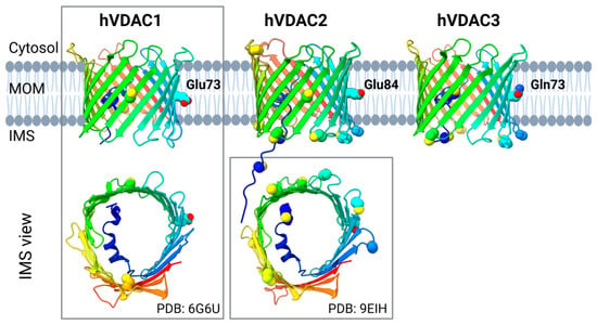
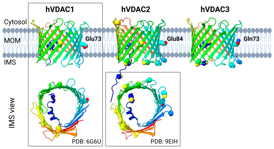
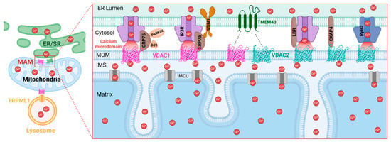
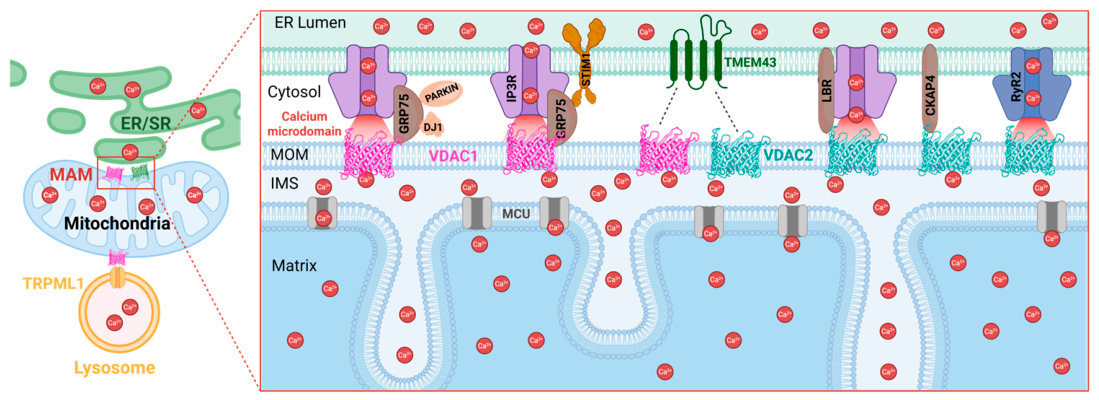
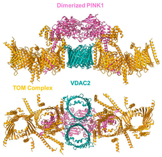
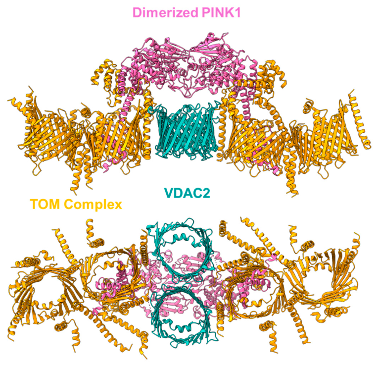
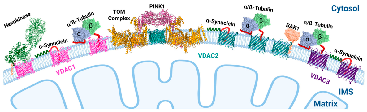

Disclaimer/Publisher’s Note: The statements, opinions and data contained in all publications are solely those of the individual author(s) and contributor(s) and not of MDPI and/or the editor(s). MDPI and/or the editor(s) disclaim responsibility for any injury to people or property resulting from any ideas, methods, instructions or products referred to in the content. |
© 2026 by the authors. Licensee MDPI, Basel, Switzerland. This article is an open access article distributed under the terms and conditions of the Creative Commons Attribution (CC BY) license.