


Abstract
Rare genetic disorders of the central nervous system (CNS) remain some of the most complex and challenging diseases to treat for several reasons. Targeting the CNS, especially the brain, presents one of the greatest obstacles in gene therapy using adeno-associated virus (AAV) vectors. Although various AAVs have been identified for their ability to transduce different cells in the CNS, their effectiveness and efficiency are significantly limited by the presence of neutralising antibodies (NAbs) and restricted cargo capacity. Despite these challenges, our understanding of AAV structure and technological advances continue to enable researchers to develop innovative strategies that have resulted in groundbreaking, FDA-approved therapeutic products now available for Leber congenital amaurosis (LCA) (Luxturna®), spinal muscular atrophy (SMA) (Zolgensma®), and the two recent gene therapy products for aromatic L-amino acid decarboxylase (AADC) deficiency, Kebilidi® and Upstaza®, which currently hold FDA and EMA approval, respectively. This review aims to highlight recent advances in the field of AAV gene therapy for neurological disorders, identify research gaps, and suggest areas for future investigation to enable potential breakthroughs particularly in neurodegenerative, neurodevelopmental, and neuromuscular disorders. We foresee that more tissue- and cell-specific AAV vectors designed using AI-powered platforms will emerge to precisely and efficiently target specific brain regions, transforming how CNS disorders are treated.
1. Introduction
For millions, the simple act of holding a pen, sitting properly or recalling loved one’s name becomes a challenge as the system that coordinates these functions begins to fray. To others, these malfunctions start early in life due to genetic mutations. Neurological conditions account for some of the greatest medical diseases, many of which have effective long-term treatments imposing serious financial burdens. Disorders such as spinal muscular atrophy (SMA), aromatic L-amino acid decarboxylase (AADC) deficiency, Parkinson’s disease (PD), gangliosidoses (GM1 and GM2), mucopolysaccharidoses (MPSs), lysosomal storage diseases, and many others are marked by progressive neuronal dysfunction, degeneration, or biochemical imbalances that severely impact quality of life and reduce lifespan. Recently, it was estimated that 40% of the world population, totalling about 3.4 billion, had a condition affecting the central nervous system [1]. Traditional therapies, including enzyme replacement therapy (ERT), small molecules, and symptomatic management, have yielded limited success due to poor blood–brain barrier (BBB) penetration, temporary effects, or their inability to halt disease progression. Furthermore, these standard pharmacotherapies are often associated with side effects, lack disease-modifying activity, and do not prevent long-term neurological decline in diseases like Parkinson’s and Alzheimer’s [2]. These limitations underscore the need for a more targeted approach that will provide long-term, reliable therapeutic benefits to patients. Among the candidates that have shown the possibility of delivering effective therapy to the CNS is adeno-associated virus (AAV)-based gene therapy.
AAV vectors continue to gain attention as potential platforms for CNS-directed gene therapy, owing to their favourable safety profile, capacity for long-term transgene expression in post-mitotic neurons, and versatility across delivery routes (intravenous, intrathecal, intracisternal, and direct parenchymal injection). Natural virus serotypes, such as AAV9, AAVrh8, and AAVrh10, and engineered capsids (e.g., AAV-PHP.B, AAV-PHP.eB, and CAP-B10) display broad CNS tropism and the clinical potential to overcome the BBB, particularly in neonatal or infant patients [3,4]. Clinical translation of AAV therapy in the CNS is exemplified by the landmark approval of onasemnogene abeparvovec (Zolgensma®) for SMA, which demonstrated that systemic AAV9 delivery of SMN1 can reliably alter disease course and survival [5,6,7]. Similarly, eladocagene exuparvovec (Upstaza®), an AAV2-based therapy for AADC deficiency, established the feasibility of intraputaminal delivery of a dopamine biosynthetic enzyme gene to restore neurotransmitter function and improve motor development [8]. These clinical successes underscore the transformative potential of AAV vectors for neurogenetic disorders.
Additionally, a growing pipeline of investigational AAV programmes targets a diverse range of CNS disorders. For example, AAV-mediated delivery of dopamine-synthesising enzymes (TH, AADC, and GCH1) and neurotrophic factors for Parkinson’s disease has shown sustained transgene expression and motor improvements in early trials [9,10], though further optimisation of vectors and clinical endpoints remains necessary. Lysosomal storage disorders (LSDs) present some of the most debilitating behavioural and neuropsychiatric disorders in infants, although there are also juvenile manifestations [11,12]. In LSDs such as gangliosidoses (GM1 and GM2) and mucopolysacharidoses (MPS II, IIIA and C), AAV vectors are employed to restore lysosomal enzyme activity either via systemic AAV9 delivery or CNS-targeted injection routes (e.g., bi-thalamic rAAVrh8 for GM2) [13,14]. Early clinical studies have also reported biochemical correction (restored enzyme activity, reduced ganglioside or glycosaminoglycan accumulation) and signs of clinical stabilisation [13,15,16,17]. Recently, efforts have also been extended to rare neurotransmitter and metabolic disorders, including DRD, where strategies using AAV to deliver GCH1 or combinations of dopamine biosynthetic enzymes show strong preclinical promise [18,19]. All of these examples demonstrate the applicability of AAV gene therapy in both neurodevelopmental and neurodegenerative diseases.
Despite these landmark achievements, significant challenges still exist. Achieving efficient, targeted CNS transduction at clinically acceptable vector doses is hindered by the BBB, vector biodistribution, and pre-existing immunity to AAV capsids [20,21]. Safety concerns, such as hepatotoxicity, neuroinflammation, and dorsal root ganglia pathology in animal models, require careful dose selection, immune modulation, and long-term follow-up to properly evaluate the therapeutic potential of any AAV vector. Additionally, issues like the durability of expression, re-dosing constraints due to immunogenicity, production costs, and manufacturing scale-up pose other major translational obstacles.
Nevertheless, the convergence of capsid engineering, promoter optimisation, and new delivery methods, along with a rapidly growing body of preclinical and clinical data, positions AAV gene therapy at the forefront of a therapeutic revolution that will benefit patients with neurodegenerative disorders worldwide. As the field progresses, the challenges are not only to improve safety and efficacy but also to incorporate biomarker-driven outcome measures, equitable access strategies, and long-term follow-up to ensure lasting and global impact. For this review, we searched databases such as PubMed and Google Scholar for relevant articles with a focus on gene therapy clinical reports. Search keywords included ‘gene therapy’, ‘adeno-associated virus’ and/or ‘AAV’, ‘central nervous system’, ‘CNS disorders’, ‘Huntington’s disease’, Alzheimer’s disease’, ‘SMA’, and/or ‘clinical trial’. Where clinical data are not available, we consider experimental reports from open sources. In some cases, we also searched companies’ websites for clinical trial reports. Hence, we provide an overview of recent advances in adeno-associated virus (AAV)-mediated gene therapy for central nervous system (CNS) disorders, with particular emphasis on key clinical milestones, emerging mechanistic understanding, and critical knowledge gaps that must be addressed to realise the full therapeutic potential of these evolving modalities. We also offer a considered perspective on priority areas for future investigation that are likely to be essential for achieving durable and clinically meaningful outcomes.
2. Adeno-Associated Virus Vectors with Tropism to the CNS
AAV vectors continue to attract attention, emerging as the leading platform for gene delivery to the CNS, offering promise for treating various neurological disorders due to their favourable safety profile, long-term transgene expression, and ability to transduce non-dividing cells such as neurons [21,22,23]. They are non-enveloped viruses with an icosahedral capsid approximately 25 nm in diameter, composed of three viral proteins (VP1, VP2, and VP3) in a 1:1:10 ratio [21]. The capsids encapsulate a single-stranded DNA (ssDNA) genome of approximately 4.7 kb, flanked by inverted terminal repeats (ITRs) that serve as origins of replication and packaging signals [21,24]. For CNS applications, certain AAV serotypes demonstrate superior tropism and transduction efficiency. AAV9 and AAVrh10 are particularly notable for their ability to cross the BBB following systemic delivery, making them leading vectors for CNS gene delivery [25]. Other serotypes, including AAV1, AAV2, AAV5, AAV6, AAV8, and AAVrh8 (see Figure 1), also exhibit affinity for neural tissues but with varying cellular specificities within the CNS [25]. Each AAV serotype has a specific primary receptor that facilitates its transduction and cellular trafficking [26] (see Figure 2). The recent FDA approval of Kebilidi and EMA-backed Upstaza® for the treatment of AADC deficiency demonstrates remarkable progress in the fight against rare genetic diseases in different areas (capsid engineering and novel delivery options). As clinical trials advance and vector design technologies evolve, AAV-based intervention may soon become a cornerstone of precision neurotherapeutics.
Figure 1.
(A) Some notable wild-type (natural) and engineered AAV vectors that have demonstrated significant tropism in the central nervous system (represented by the brain). (B) Different injection routes to deliver AAV vectors into the brain and the CNS.
Figure 2.
Schematic illustration of AAV transduction and nuclear trafficking. (A) Recombinant/engineered AAV vector with transgene and promoter. (B) (1) The initial step in the AAV transduction and intracellular trafficking is the binding of the AAV vector to both the primary receptors, like HSPG for AAV2 or LamR for AAV8, and other secondary receptors. (2) For cell entry, the viruses undergo clathrin-mediated endocytosis via clathrin-coated vesicles. (3) The formation of early and late endosomes and viral maturation. Mature viruses are then released from the endosome. (4) Released viruses form perinuclear accumulation and nuclear entry by interacting with importin-β and other karyopherins. (5) The viruses that escaped ubiquitination degradation enter the nucleolus as a temporary sequestration site that helps to facilitate viral uncoating. (6) Viral uncoating and genome release. (7) The crucial step of the dsDNA genome formation is facilitated by the host’s DNA polymerase. (8) The double-stranded AAV genome is transcribed and leads to eventual transgene expression.
2.1. Advances in AAV Design for CNS Delivery
Ongoing efforts focus on improving the transduction efficiency of AAV vectors throughout the CNS, targeting different brain regions and cell types. Despite their small size and cargo capacity, AAV vectors continue to attract attention for several reasons: (i) the ability to modify AAV capsids to enhance their cell type or tissue specificity; (ii) combining these advanced capsids with modern gene regulatory elements, reporters, sensors, and effector cargos enables physiologically relevant transgene expression; (iii) they facilitate detailed understanding of how different brain regions respond to gene therapy. For comprehensive reviews of recent advances in AAV technology, including capsid modifications, regulatory elements, and functional cargos used to improve targeting and expression control, we direct readers to Challis et al. and Kolesnik et al. [27,28].
2.2. AAV Transduction and Intracellular Trafficking
The foundation of AAV vector engineering relies on a comprehensive understanding of the AAV pathway and cellular trafficking (see Figure 2). This host-factor-dependent process begins with attachment to abundant cell-surface glycans (e.g., heparan sulfate proteoglycan (HSPG) for AAV2, an uncharacterised type-I transmembrane protein (KIAA0319L) named AAV receptor (AAVR)) [29]. See Table 1 for other serotype-specific AAV receptors. Also, using genome-wide screening, several conserved host factors required for productive AAV transduction, such as GPR108 (a GPCR-family protein) [30], and CRISPR-identified TM9SF2 [31] modulating AAV uptake and trafficking have been identified. AAV binding to the cells is followed by endocytosis and early intracellular trafficking, often involving clathrin-coated pits, but alternative pathways (caveolar or micropinocytosis-like routes) have been reported for particular serotypes/cell types [32]. Acidification of early/late endosomes induces capsid rearrangements that expose internal regions of VP1/VP2 (the VP1u unique region) with a functional phospholipase A2 (PLA2) motif; this activity (and associated conformational changes) is important for destabilising endosomal membranes and enabling escape to the cytosol [33]. The mature capsids must then escape the endosome and undergo proteasomal degradation and ubiquitination. Only a minority of internalised particles escape productive endosomal compartments: the capsid sequence and host endosomal proteases. Enhancing escape (e.g., by modifying capsids or transiently inhibiting degradative pathways) improves transduction [34,35]. Then, AAV capsids interact with nuclear import machinery (e.g., importin-β and other karyopherins) and are thought to enter the nucleus either as intact particles through nuclear pores or after partial uncoating near the nuclear envelope. The transduction efficiency of AAV vectors can be modulated by manipulating nuclear import signals [36]. After nuclear entry, uncoating releases the single-stranded AAV genomes, which require host DNA polymerases to synthesise the complementary strand. This step is regarded as a rate-limiting step for many serotypes and/or cell types. The invention of self-complementary AAV (scAAV) vectors bypasses this bottleneck by packaging an inverted repeat genome that folds into double-stranded DNA, markedly accelerating transgene expression in non-dividing cells [37,38]. All of these fundamental understandings and discoveries in the AAV transduction pathway and intracellular trafficking are shaping the development and engineering of new and more efficient vectors.
Table 1.
Comparison of transduction efficiency of natural AAV capsids for CNS/brain delivery with respect to injection routes.
Recently, Drouyer et al. developed customised AAV testing kits that contain mixtures of various AAV capsids tagged with unique barcodes for high-throughput next-generation sequencing (NGS) analysis. Their goal was to identify capsids that can effectively transduce human neural cells using human induced pluripotent stem cell (hiPSC)-derived brain organoids, as well as mouse and non-human primate (NHP) in vivo models after cerebrospinal fluid (CSF) delivery [42]. The study identified top-performing AAV variants, including AAV2.7m8, AAV-SYD12, AAV2-M1, and AAV2-L5, among others, which showed efficient transduction in human brain organoids. These variants exhibited specificity for neurons with minimal targeting of astrocytes. Validation in a mouse model confirmed efficient CNS transduction near injection sites, with AAV2-M1 demonstrating widespread brain distribution, including in cerebellar neurons. Such delivery methods are gaining traction because the intravenous injection route faces systemic barriers and complications [43,44,45]. This set of problems involves toxicity and mortality associated with high-dose systemic AAV administration [46]. Meta-analyses encompassing more than 250 clinical trials indicate that hepatotoxicity, thrombotic microangiopathy, and neurotoxicity constitute the predominant categories of serious treatment-related adverse events, with 11 deaths reported across eight trials and approximately one-third of studies documenting serious adverse events attributed to vector administration [47,48]. Intravenous AAV delivery, particularly at doses exceeding 1 × 10¹⁴ vector genomes per kilogram, frequently elicits dose-dependent hepatic injury, characterised by elevations in serum transaminases emerging 1 to 4 weeks post-infusion and, in the most severe instances, acute liver failure driven by immune-mediated sinusoidal endothelial cell injury and microthrombosis. Clinically, this has been underscored by fatal liver failure events following high-dose AAV gene therapy for muscular dystrophy, which have prompted regulatory investigations and renewed scrutiny of dose-escalation paradigms [46]. Collectively, these observations underscore the need for alternative delivery methods that would be more targeted while avoiding systemic injury and vector clearance. In Table 2, we present a comparison of performance metrics of natural capsids vs. engineered AAV vectors. These suggest the reasons why engineered vectors continue to attract more attention.
Table 2.
Core performance metrics: natural AAV capsids versus recombinant AAVs.
Recombinant AAV vectors such as AAV-DJ, AAV-DJB, AAV-PHP.B, AAV-PHP.eB, AAV-PHP.S, AAV-CAP-B10, AAV-CAP-Mac, AAV-MG1.1, AAV-MG1.2, and rAAV-TGX-007 continue to demonstrate improved transduction across major CNS cell types (neurons, astrocytes, oligodendrocytes, microglia, and endothelial) and brain regions (cortex, hippocampus, striatum, and cerebellum) [55,56,57,58,59,60]. The delivery encompasses all the routes of administration mentioned above. Figure 2 shows general intracellular trafficking of AAV vectors to protein synthesis.
A study on NHPs showed that AAV2-M1 and AAV-SYD12 achieved effective CNS transduction across multiple brain and spinal cord regions [42]. Although this approach is promising, several questions remain. The study focused on direct CNS delivery via CSF routes to bypass the BBB, but challenges persist for achieving effective systemic intravenous delivery capable of crossing the BBB without requiring high vector doses or causing off-target toxicity. While human brain organoids provide a valuable human-relevant system for testing AAV tropism and efficiency, they lack many in vivo biological complexities, such as the BBB, immune system interactions, and complete brain cytoarchitecture, which may limit their predictive accuracy for in vivo human CNS transduction. Additionally, even with CSF delivery, none of the AAV variants demonstrated deep parenchymal penetration of the brain in vivo, indicating a need to engineer vectors or delivery methods capable of wider CNS distribution. An additional concern is that rodent-selected AAV vectors often do not translate efficiently to NHPs or humans, as seen with AAV-PHP.B/PHP.eB. Although this study attempted to address this through multi-species screening, the use of only one NHP limits statistical robustness; larger NHP cohorts are necessary to confirm cross-species efficacy. Finally, it is important to consider how pre-existing immunity or neutralising antibodies may influence the effectiveness of these bioengineered capsids in clinical settings. The long-term expression stability, potential off-target effects, and safety profiles of these new AAV variants still require further clarification through preclinical and clinical studies.
2.3. Promoter Considerations in AAV Vector Design
Promoters are the “software” of AAV therapy because they determine what and how much therapeutic gene is expressed. Another area of interest in AAV vector design for CNS delivery has been the incorporation of various promoters. Different classes of promoters have been tested for AAV vector delivery with varying degrees of gene expression cascades and associated toxicities. Strong, ubiquitous, and constitutive promoters like CMV (human or murine cytomegalovirus immediate-early promoter), CAG/sCAG (CMV-enhancer + chicken β-actin promoter ± intron), EF1α (elongated factor 1 alpha), and CBh/CBA (chicken β-actin hybrid variants) are very good choices considering their strong, early expression capabilities [61,62,63]. However, studies have shown that CMV can be prone to transcriptional silencing in some tissues [64]. Besides the diminished durability of some of these promoters, another concern is the promoter–capsid interactions. Several studies have found that the same promoter may produce different expression patterns depending on the serotype/capsid (e.g., AAV9 vs. AAV1 vs. AAV2 vs. engineered capsids like PHP.eB) [65].
Some promoters are neuron-specific, such as hSyn (human synapsin I), which is widely used for pan-neuronal expression. The compact, neuron-restricted CaMKIIα promoter (calcium/calmodulin-dependent kinase II alpha) is considered to be strong in excitatory forebrain neurons. NSE (neuron-specific enolase) is another commonly used promoter for CNS delivery [66,67]. NSE is a ~1.8 kb regulatory element used in AAV vectors to drive strong, sustained, and specific transgene expression in mature neurons and neuroendocrine cells [68]. It is often considered for CNS gene delivery to avoid off-target expression in glia, but its large size limits the capacity for larger transgenes in AAV. GFAP (an astrocyte-specific promoter), MBP/MOG/PLP (oligodendrocyte lineage promoters) and Iba1/CD68 are glia-specific promoters, though not very common, but are also available for AAV vector design and delivery [69]. Pol III promoters, particularly for small RNAs such as U6 and H1 for shRNA and gRNA, respectively, are used for gene-editing or RNAi expression. However, they are not considered suitable for protein expression.
Recently, synthetic and engineered promoters, also called micro-promoters, have emerged. After reporting the creation of a hybrid form of CBA promoter (CBh) that demonstrated long-term and robust expression in many cells including motor neurons, Gray et al. also reported the development of 229 bp mouse methyl-CpG-binding protein-2 (MeCP2), which not only demonstrates neuron-specific expression but also allows an extra 570 bp packaging capacity in AAV compared to the 800 bp CMV promoter [62]. Similarly, Syn1-minCMV is another hybrid promoter designed for highly specific and robust gene expression in neurons. This promoter combines the neuron-specific regulatory elements of the human or rat synapsin I (SYN1) gene with minimal cytomegalovirus (CMV) core promoter (often ~50–60 bp). The Neuron-Restrictive Silencer Elements (NRSE/RE1) in the SYN1 promoter that bind to Neuron-Restrictive Silencer Factor (NRSF/REST) in non-neuronal cells particularly suppress expression outside the CNS [70]. Synthetic tissue-specific promoters and ultra-small micro-promoters (MP-84 and MP-135) have been developed to reduce cassette size while preserving strong expression [71]. These emerging promoters could be candidates of choice, especially considering the limited cargo capacity of AAV vectors.
3. AAV Gene Therapies in CNS Disorders
3.1. Neurodegenerative Disorders
Neurodegenerative disorders are conditions involving progressive degeneration of neurons and are often linked to toxic protein accumulation or impaired cellular clearance mechanisms. This loss of neurological functions results in some of the debilitating mental disorders affecting millions of people across the world today, with Alzheimer’s and Parkinson’s diseases presenting the most challenging pathological and clinical features.
3.1.1. Alzheimer’s Disease (AD)
AD is considered one of the most challenging neurodegenerative disorders to treat. To date, no approved therapy can halt or reverse its progression. However, with advancements in engineering technology for AAV vector design, there is a gradual shift from conceptualisation to clinical application of AAV-dependent gene therapy targeting the underlying molecular drivers of AD, especially by leveraging the ability of recombinant AAVs (rAAVs) to deliver the transgene directly to the CNS for long-term expression. The genetic risk factor for AD is APOE ε4, and it is characterised by amyloid-β (Aβ) plaques, tau neurofibrillary tangles, neuroinflammation, synaptic dysfunction, and progressive neuronal loss. Following a landmark phase 1/2 clinical trial involving an intrathecal AAV therapy delivering APOE2 to APOE4 homozygous AD patients, and with incredible data showing detectable APOE2 in CSF, reductions in tau biomarkers (T-tau, P-tau) at 12 months, and a favourable safety profile with no serious adverse effects (NCT03634007), Yang et al. have recently identified some key gaps in APOE2 replacement strategy. They argued that it is unclear if APOE2 supplementation as a standalone therapy is sufficient to suppress APOE4-driven pathology in AD or if a co-repressor is needed to achieve sustained therapeutic benefits. Also, it is unclear whether introducing this therapy at the early or late stage of the disease will significantly improve therapeutic outcome. Hence, they developed an inducible AAV platform for APOE isoform replacement strategies, enabling temporary replacement of APOE4 with APOE2 in 5xFAD mice. Their data showed that long-term APOE2 expression with concurrent APOE4 silencing reversed Aβ deposition, glial activation, and cognitive decline, while short-term replacement potentially worsened pathology, linked to RAB24-mediated rebound adaptation affecting Aβ clearance and cholesterol metabolism [72]. This underscores the importance of timing and sustained expression in APOE-targeted therapy. Other notable approaches include neurotrophic factor delivery (AAV-BDNF) that aims to restore brain-derived neurotrophic factor in the entorhinal cortex and hippocampus, regions affected by AD, with a preclinical model demonstrating reversal of synaptic loss and neuroprotection [73,74], as well as the tau silencing approach, anti-amyloid gene therapy, and Klotho gene therapy. Currently, Voyager Therapeutics (VY1706) uses a BBB-penetrant TRACERTM capsid to deliver a vectorised pri-amiRNA targeting all tau isoforms. In non-human primates, IV administration achieved 73% tau mRNA knockdown across brain regions. In mouse models, tauopathy treatment reduced pathological tau, preserved neurons, and improved function [75]. Also, the company is developing vectorised anti-amyloid antibodies for sustained CNS expression [76]. This approach could overcome the limitations of repeated monoclonal antibody infusions by providing continuous target engagement. KLTO-202, developed by Klotho Neurosciences, is an AAV-delivered form of the “anti-ageing” Klotho gene (s-KL). In preclinical AD models, Klotho protein has neuroprotective and cognitive-enhancing effects. Engineered capsids (AAVnerGene’s ATHENA platform) aim to improve CNS targeting and reduce liver tropism [77], which remains a major problem in AAV gene therapies. For details about other strategies and platforms for designing AAV vectors for effective CNS transduction, we refer readers to the article by Sharma et al. [40].
Furthermore, TREM2 (triggering receptor expressed on myeloid cells 2), a pattern-recognition receptor that is expressed almost exclusively by microglia in the central nervous system, where it regulates phagocytosis, lipid metabolism, chemotaxis, and cell survival [78,79], has been identified as a key driver in the “disease-associated microglia” (DAM) phenotype that clusters around amyloid-β (Aβ) plaques in AD models. Rare coding variants in TREM2, most notably R47H, particularly the p.R47H [80] variant as well as several other missense changes, are associated with a threefold increase in the risk of late-onset Alzheimer’s disease (AD) [81], yielding odds ratios similar to those conferred by the APOE ε4 allele [79,80]. Collectively, these studies support a causal role for microglia-driven innate immune processes in AD pathogenesis. In addition, loss-of-function mutations in TREM2 cause Nasu–Hakola disease [78,82], a presenile dementia accompanied by bone cysts, further highlighting the critical importance of TREM2 in preserving normal brain homeostasis.
Many phase 3 Alzheimer’s trials targeting amyloid (solanezumab (NCT02008357), gantenerumab, bapineuzumab, aducanumab ENGAGE/EMERGE, crenezumab (NCT01998841), etc.) have failed to show convincing clinical benefits despite target engagement [83,84]. Recurrent themes are: treatment started too late (mild–moderate dementia, when large-scale synaptic and neuronal loss is already present), heterogeneous populations (including amyloid-negative “Alzheimer’s” by clinical criteria), and modest effect sizes drowned out by variability and placebo response. In many cases, amyloid burden was reduced, but cognition did not improve meaningfully over 18–24 months [84], suggesting either that amyloid is an upstream trigger that must be treated pre-symptomatically, or that parallel pathologies (tau, inflammation, and vascular disease) dominate once dementia is established.
Despite these breakthroughs, there is a need to develop capsids with enhanced cell-type specificity, immune evasion capabilities, and equally offer transient immunosuppression as the use of magnetic resonance imaging (MRI)-dependent delivery continues to grow (NCT05040217 and NCT04120493) in clinical trials following the FDA approval of Kelibidi (eladocagene exuparvovec-tnaq) in 2024, which is administered directly to the brain. This method ensures accurate targeting, monitoring the distribution in real time, and overcoming the blood–brain barrier [85]. Another technology that will be very useful is the incorporation of PET reporter genes for dynamic tracking [86,87]. PET imaging allows for quantification, measurement of intensity and regional distribution of the expressed gene. Several PET reporter genes such as dopamine 2 receptor (D2R) [87,88], D2R mutant [89], type 2 cannabinoid receptor mutant, sodium iodide symporter, somatostatin receptor 2 [90,91] and herpes simplex virus type 1 thymidine kinase (HSV1-tk) [92,93] have been used. This method involves binding a reporter gene with a specific radiolabelled probe to measure the level of radioactivity, which directly reflects the gene expression level. This will help to evaluate the pharmacokinetics of AAV longitudinally beyond delivery monitoring [94].
3.1.2. Parkinson’s Disease (PD)
Progressive loss of dopaminergic neurons in the substantia nigra is the main genetic deviation responsible for the development of PD. Although dopamine replacement therapies like levodopa (L-dopa) offer some relief symptomatically, they do not completely result in disease regression. Hence, researchers have focused on approaches that provide a long-term solution using AAV gene therapies.
One such approach is the attempt to restore GABAergic balance in patients [95]. MeiraGTx AAV-GAD therapy is based on this mechanism to deliver the glutamic acid decarboxylase (GAD) encoding gene to the subthalamic nucleus (STN), thereby improving local GABA production and rebalancing the circuitry of the basal ganglia. STN motor dysfunction is a hallmark of PD, and reducing hyperactivity in this region by the conversion of glutamate to GABA through GAD is a reliable approach. AAV-GAD is administered as a single-dose, minimally invasive intracerebral infusion delivered directly into the subthalamic nucleus (STN) [96]. The phase 2 clinical trial (NCT00643890), which included 45 PD patients, was a sham-controlled bilateral infusion. A high dose (2.1 × 10¹¹ vector genomes) of AAV-GAD led to an 18-point improvement in Unified Parkinson’s Disease Rating Scale (UPDRS) Part III scores and an 8-point gain in PDQ-39 quality-of-life scores [97]. It will be worth following up to see the outcome of the trial (NCT05894343).
Furthermore, a recent NIH-sponsored clinical trial was designed to deliver a copy of the glial-cell-line-derived neurotrophic factor (AAV-GDNF). This open-label dose-escalation trial with convention-enhanced delivery to the putamen demonstrated safety and tolerability with long-term follow-up underway (NCT01621581).
Although AAV-GAD and AAV-GDNF remain the two primary approaches in efforts to restore motor neuron function in patients with PD, there has recently been a rise in other clinical techniques focusing on circuit-level reprogramming. For example, Chen et al. engineered AAV vectors with DREADDs (designer receptors exclusively activated by designer drugs) to selectively activate D1-MSNs in the striatum [98]. This method restored motor function in both mouse and primate PD models without causing dyskinesia [9,98]. In another study, Lee and his colleagues developed a self-complementary AAV vector encoding miR142-3p target sequences to suppress transgene expression in haematopoietic cells, thereby minimising peripheral toxicity [44]. Meanwhile, MeiraGTx is progressing its work on riboswitch-controlled AAV platforms that enable drug-inducible control of transgene expression, which could provide dynamic regulation and enhance safety, as previously mentioned.
Finally, in a large cohort study conducted by Sidransky et al. on Parkinson’s disease patients, it was discovered that a mutation in GBA1, the gene that encodes the lysosomal enzyme glucocerebrosidase (GCase) (primarily associated with the development of Gaucher disease), is also implicated in the development of PD [99]. According to one study, carriers of GBA1 variants have a potential risk of developing PD, with an odds ratio ranging from 2.2 to 30 [100]. Following some successes in restoring or normalising GCase pathway activities after injection of AAV-PHP.B-GBA1 into the A53 T-SCNA transgenic mouse model [101] or AAV-GBA1 and AAV-A53T injection into transgenic mice [102] (see Figure 3), Prevail Therapeutics has launched a phase 1/2a (NCT04127578), multicentre, open-label, ascending-dose PROPEL study to evaluate the safety of AAV-GBA1 (LY3884961) intracisternal administration to restore GCase activity and reverse PD. This opens a new frontier in the treatment of PD.
Figure 3.
(A) Current clinical strategies aimed at treating AD (Alzheimer’s disease): neurotrophic factor delivery, APOE isoform replacement strategies replacing APOE4 with APOE2 in patients, approaches aimed at silencing tau protein, anti-amyloid gene therapy, and novel Klotho gene therapy approach; (B) PD (Parkinson’s disease): enhancing dopamine synthesis, restoring GABAergic balance, and neurotrophic support strategies. (C) Schematic illustration of the milestones in clinical translation of AAV-GAD, AAV-GDNF, and AAV-GBA1 therapeutic strategies for PD treatments, highlighting some important clinical trials [96,97,98,101,102]. STN—Subthalamic nucleus; CED—Convection-enhanced delivery; iMRI—interventional MRI, GAD—Glutamic acid decarboxylase; GCase- glucocerebrosidase.
3.1.3. Amyotrophic Lateral Sclerosis (ALS)
Amyotrophic lateral sclerosis (ALS) is a progressive, uniformly fatal, neurodegenerative disorder characterised by gradual loss of both upper and lower motor neurons, leading to muscle weakness, paralysis, and eventual respiratory failure with a very low survival rate (only 2–5 years post-diagnosis) [103,104]. While approximately 10% of cases are familial (fALS), linked to mutations in genes such as SOD1, C9orf72, FUS, and TARDBP, the majority (90%) are sporadic (sALS) with no known genetic cause [104]. SOD1 and C9orf72 are two of the most studied genetic causes of ALS, and they illustrate two different disease mechanisms [105,106]. SOD1 mutations cause a toxic gain-of-function: the mutant protein misfolds, aggregates, and disrupts multiple pathways, including proteostasis, axonal transport, mitochondrial function, lipid metabolism, and synaptic transmission [107]. These aggregates are a pathological hallmark of SOD1-familial ALS, and misfolded SOD1 inclusions are also found in some cases with other genetic mutations [106,107], suggesting that SOD1 misfolding can be a common downstream event. Clinically, SOD1 mutations account for roughly 10–20% of familial ALS and a smaller fraction of apparently sporadic cases, including de novo mutations, and motivated the development of SOD1-lowering antisense therapies. Contrariwise, the massive expansion of hexanucleotide repeats (GGGGCC) in the first intron of C9orf72 is the most commonly known genetic cause of ALS [105]. This expansion leads to three interacting mechanisms: haploinsufficiency of normal C9orf72 protein, accumulation of toxic RNA foci, and production of dipeptide repeat proteins through repeat-associated non-ATG (RAN) translation [108]. Masrori et al. recently showed that C9orf72 haploinsufficiency in microglia and astrocytes impairs endolysosomal pathways and blunts the normal transition of microglia into reactive, disease-associated states, altering neuroimmune responses in ALS [109].
Despite the heterogeneity in the disease development, 97% of all ALS cases are said to share common pathological hallmarks: the cytoplasmic mislocalisation and aggregation of TAR DNA-binding protein 43 (TDP-43) [110]. This proteinopathy disrupts RNA metabolism, promotes neuroinflammation, and ultimately drives motor neuron death [110,111]. The current FDA-approved therapies, riluzole, edavarone, and tofersen (for SOD1-ALS), offer only modest benefits, underscoring the urgent need for targeted, disease-modifying treatment like AAV-based gene therapy due to their ability to cross the blood–brain barrier and transduce the cells of the CNS.
New technologies are emerging to target ALS. Researchers at the University of Pennsylvania and CHOP (Children’s Hospital of Philadelphia) recently developed a peptide-modifying AAV9 (PM-AAV9) to deliver RNA interference (RNAi) for gene silencing (for example, miRNAs targeting ATXN2 or SOD1), which demonstrates superior transduction in motor cortex layer V and spinal cord anterior horn cells compared to wild-type AAV9 and AAV-PHP.eB [110]. This capsid variant achieved 54% knockdown of target genes in the frontal cortex and 23–25% knockdown of brainstem and spinal regions after intracerebroventricular (ICV) injection in mice [110]. The TDP-43 mouse models showed prolonged survival by 54%, improved motor function (rotarod performance and gait scores), reduced neuroinflammation, and evidence of rescued transcriptomic dysregulation in >450 genes linked to ALS pathways [110]. The translation of these results to NHPs or human trials will be valuable to determine the prospect of this technology. Also, the use of neuroprotective genes, for example TRIM72 (Tripartite Motif Protein 72), to counteract oxidative stress and mitochondrial dysfunction has been reported [112]. SinuaGene’s therapy named SNUG01 that delivers TRIM72 for ALS treatment has been granted Orphan Drug Designation (ODD) by the FDA according to the company’s report (https://www.sineugene.com/). AMT-162 by uniQure, an AAV5-based therapy encoding miRNA against SOD1, is currently in phase I/II trials (NCT06100276), with no safety concerns reported yet in the first cohort.
3.1.4. Prion Diseases
Prion diseases, which include Creutzfeldt–Jakob disease (CJD), variant CJD, Gerstmann–Sträussler–Scheinker syndrome (GSS), fatal familial insomnia (FFI), iatrogenic CJD (iCJD), and other variants, are caused by the accumulation of misfolded prion protein (PrP) encoded by the PRNP gene, eventually leading to neurodegeneration of the brain [113,114]. This group of rare, progressive, and fatal neurodegenerative diseases are also called transmissible spongiform encephalopathies (TSEs) and affect both humans and animals. The accumulation of the abnormally folded protein (PrP^Sc^) causes neuronal dysfunction and cellular death [115]. Unlike other pathogens, prions contain no nucleic acid (DNA or RNA), which makes treatment attempts even more challenging. Recent research has identified specific intermediate conformations that drive this misfolding process, particularly involving structural changes in the α-helix H1 region and the highly conserved TVTTTT motif [116], an understanding that is crucial for tackling the disease. Current treatments are limited, and there is a substantial need for innovative therapeutic approaches. To date, the most advanced clinical trial for prion disease is Ionis Pharmaceuticals’ ION717 (PrProfile study), a non-AAV-based trial which completed enrolment in December 2024. This Phase 1/2 trial (NCT06153966) enrolled 56 symptomatic prion disease patients across 16 global sites. The trial uses intrathecal delivery of antisense oligonucleotides to reduce PrP mRNA. While this is promising, the therapy focuses on safety rather than efficacy. The need for repeated intrathecal dosing will be too burdensome for patients and families.
The therapeutic approach for prion diseases using AAV gene therapy centres on reducing or eliminating prion protein (PrP) expression. There are currently four significant AAV-related approaches targeting prion diseases. These strategies are based on the compelling evidence that PrP reduction halts disease progression in animal models [114,117,118]. The most advanced AAV-based approach involves the Coupled Histone tail for Autoinhibition Release of Methyltransferase (CHARM) system developed by researchers at the Broad Institute and Whitehead Institute. CHARM is a compact epigenetic editor that uses a histone H3 tail-Dnmt3l fusion to recruit and activate endogenous DNA methyltransferases. The strategy focuses on programmable DNA methylation to silence the PRNP gene. It was packaged into AAV vectors with zinc finger proteins (ZFPs) or other DNA-binding domains and demonstrates a 70–90% reduction in PRNP transcripts and a 60–80% reduction in PRNP protein levels in mouse brains [118]. The second method focuses on base editing technologies. Recent research has demonstrated AAV-delivered base editing systems that create stop codons in the PRNP gene using BE3.9max base editor. This experimental work showed 37% edit frequency at the target site, a 50% reduction in PrP levels, and a 52% increase in survival in humanised PRNP mice infected with pathogenic prions [117]. Furthermore, Sangamo Therapeutics has developed zinc finger repressor approaches that bind to PRNP DNA and shut down gene expression. The AAV-ZFR system showed dose-dependent PrP reduction up to 50% in C57BL/6 mice, extended survival from median 170 days to up to 494 days in prion-infected mice, and up to 95% PRNP repression in key brain areas like the hippocampus, thalamus, and putamen [119]. The new STAC-BBB capsid platform successfully delivered the vectors to 35 brain regions in non-human primates (NHPs) [120]. While these are laudable advancement in the treatment of prion diseases, it would worth following up to see how these translate into human clinical trials and, eventually, therapeutic treatment because of the BBB problem that may limit the effectiveness of these approaches. Also, there is the issue that the high dose requirement of (1 × 10¹⁴ vector genomes/kg) raises safety concerns and potential immune response.
3.2. Neurodevelopmental and Metabolic Disorder
3.2.1. Aromatic L-Amino Acid Decarboxylase Deficiency (AADCD)
The recent FDA approval of KEBILIDITM (Eladocagene Exuparvovec) in November 2024 as the first AAV-based gene therapy for AADCD [121] is a milestone achievement for paediatric neurological disorders and CNS gene therapy. The technology behind Kebilidi® involves the use of recombinant AAV2 (rAAV2) encoding the functional human DDC gene (which encodes the AADC enzyme) infused directly into the putamen via stereotactic neurosurgery. The SmartFlow Neuro Cannula has received FDA approval for precise intracerebral delivery of Kebilidi® [121]. The open-label study involved 13 children with genetically confirmed AADCD who had no gross motor function at baseline. At 48 weeks post-treatment, 66.66% (8 out of 12) evaluable patients showed motor improvement, including head control, sitting, and standing. Dopamine production was restored, leading to improved mood, movement, and energy levels. One of the successfully treated children was a 3-year-old girl at Texas Children’s Hospital who was reported to show early signs of increased vitality and reduced oculogyric crises [122].
Several other AAV-based AADC gene transfer programmes beyond Kebilidi®/Upstaza are (or have been) in clinical trials, with significant progress towards addressing the debilitating deficiency. Midbrain-targeted AAV2-hAADC (NCT02852213), unlike Kebilidi®, which delivers functional gene to the putamen, infuses hAADC directly into the midbrain dopaminergic nuclei (bilateral substantia nigra pars compacta and ventral tegmental area) using intraoperative MRI-guided conventional-enhanced delivery (CED). This was aimed at restoring AADC, where it is normally expressed, so rescued neurons can supply dopamine along intact nigrostriatal projections [8]. Two cohorts of children aged 4–9 years old with genetically confirmed AADC deficiency received ~1.3 × 10¹¹ vector genomes or 4.2 × 10¹¹ vector genomes total; intraoperative gadolinium co-infusion documented ~98% SN and ~70% VTA coverage on MRI in aggregate analyses with remarkable results, showing resolution of oculogyric crises in six out of seven children by three months post-treatment, increased FDOPA uptake in midbrain/striatal targets, and motor skill gains through 12–24 months [8].
3.2.2. Rett Syndrome
Rett Syndrome is a debilitating X-linked neurodevelopmental disorder caused primarily by mutations in the MECP2 gene. This gene encodes a transcriptional regulator critical for neuronal maturation and synaptic plasticity. The disease leads to severe motor, cognitive, and autonomic dysfunction in children between 6 and 18 months of age.
The REVEAL phase 1/2 trial in adolescent, adult, and paediatric patients across the US, Canada, and the UK with TSHA-102 (Taysha Gene Therapy), which delivers a shortened MECP2 “mini-gene” using AAV9 via intrathecal injection, was shown to improve motor skills, communication, autonomic function, and seizure activity as early as 4 weeks after injection. While Taysha delivered a MECP2 mini-gene, their counterpart, Neurogene, in their trial designated NGN-401, delivered a full-length MECP2 gene. The vector encoding the gene was administered as a single injection into the cerebral ventricles, the fluid-filled space deep within the brain, to promote wider distribution across the brain. Both trials incorporated biological switches, miRARE and EXACT, respectively, that help regulate MECP2 protein production, since overproduction has negative effects. According to available data and an ongoing clinical trial (NCT05898620), the therapy is administered to female children, raising questions about whether it will have the same effects in male children.
3.3. Neuromuscular and Motor Neuron Diseases
Spinal Muscular Atrophy (SMA)
Spinal muscular atrophy (SMA) is a group of recessive neuromuscular disorders caused by mutations in the survival motor neuron 1 (SMN1) gene. It mainly affects nerve cells that control motor neurons, leading to progressive muscle loss and weakness due to wasting [123,124]. Most SMA types impact muscles involved in walking, arm movement, sitting, and head control. However, the disease progression in some types can cause difficulty in breathing and swallowing [125]. Importantly, SMA is considered a multisystem disorder affecting organs beyond the nervous system, including the heart, liver, kidneys, and pancreas [126,127]. The treatment options for SMA have changed significantly, with three approved therapies: nusinersen (antisense oligonucleotide SMN2 splicing modulator), risdiplam (oral SMN2 splicing modifier), and onasemnogene abeparvovec (AAV9-based SMN1 gene replacement therapy, marketed as Zolgensma®) [128,129]. While nusinersen and risdiplam need to be administered multiple times, onasemnogene abeparvovec offers the benefit of a single dose but has safety concerns, especially hepatotoxicity and dose-limiting toxicities, although it could be mitigated with prophylactic prednisolone [130].
Recent research has centred on optimising promoter systems to achieve tissue-preferential expression without suppressing essential peripheral expression, addressing the multisystem nature of SMA. Song and colleagues developed EXG001-307, an AAV9-based vector that incorporates a hybrid neuron-preferential promoter combining synapsin and cytomegalovirus (CMV) regulatory elements. This design takes advantage of the neuronal specificity of synapsin, while the CMV enhancer provides enough peripheral expression to address non-neuronal disease manifestations as mentioned previously. In preclinical studies, EXG001-307 showed notable safety benefits, allowing administration at doses up to 6 × 10¹⁴ vector genomes/kg without treatment-related mortality, which is a considerable improvement over benchmark vectors that cause significant toxicity at lower doses. At 4.0 × 10¹⁴ vector genomes/kg, animals treated with EXG001-307 achieved a median survival of 191 days, compared to 32.5 days in animals receiving the benchmark vector. Tissue analysis of expression revealed reduced SMN levels in the liver and heart, while maintaining therapeutic levels in the brain and spinal cord, aligning with the intended preferential targeting [131,132].
A groundbreaking advancement in vector technology is the development of covalently closed-end AAV (cceAAV) vectors, which are part of a new generation of self-complementary AAV vectors that do not rely on mutant inverted terminal repeats (ITRs) for production. Unlike traditional scAAVs, which often contain incomplete genomes, cceAAV vectors consistently package more intact genomes. NKG-001, a cceAAV-based therapy, shares the same genetic sequence as onasemnogene abeparvovec except at the terminal level, where the mITR is replaced by TeIN, leading to enhanced transduction efficiency and transgene expression. Early clinical data from two children with SMA (aged one and two years) who previously received RNA-splicing-modifying drugs (nusinersen or risdiplam) show that NKG-001 was administered intravenously at relatively high doses (6 × 10¹³ to 1.2 × 10¹⁴ vector genomes/kg). The report indicates that the treatment was well-tolerated with no serious side effects, and both children experienced rapid motor function improvements. This vector technology addresses key manufacturing challenges associated with conventional scAAV vectors and may provide increased consistency and potency. However, despite promising results, the small sample size limits the ability to generalise findings or draw definitive conclusions. Larger, controlled trials are needed to validate these results. Additionally, the study involved older infants (12–24 months) who had already been treated with RNA-splicing modifiers, leaving uncertainty about the vector’s performance in treatment-naïve patients, older children, or adults, and whether gene therapy offers benefits beyond existing treatments.
Other clinical trials that have shown remarkable results include OAV101 IT (NCT05089656), which demonstrated a 2.39-point HFMSE improvement vs. 0.51 with placebo [133], GC101 (NCT05824169, NCT05901987, and NCT06421831) for SMA types 1, 2, and 3, which has entered phase 3, and LT01-101 by Lantu Biopharma, which is currently on phase 1/2. For detailed information about ongoing clinical trials, see Table 3. For a detailed review of the treatment of neuromuscular disorders with gene therapy and the target genes, we refer our readers to Wu and colleagues [134].
Table 3.
Selected ongoing clinical trials for AAV gene therapy in different CNS disorders.
3.4. Lysosomal Storage Disorder
3.4.1. Neuronal Ceroid Lipofuscinosis (Batten Disease)
Neuronal ceroid lipofuscinoses (NCLs), commonly known as Batten disease, are a group of serious inherited lysosomal storage disorders that primarily affect the nervous system. They result from mutations in various CLN genes (such as CLN2, CLN3, and CLN6), leading to defective lysosomal enzymes or membrane proteins. These defects cause progressive vision loss, seizures, deterioration of motor and cognitive functions, and early death. Although Cerliponase alfa (BioMarin Pharmaceuticals Inc., CA, USA), a recombinant proenzyme administered via intracerebroventricular (ICV) infusion every two weeks, is reported to significantly slow neurological decline and improve survival in children, it does not impact retinal dystrophy.
In early 2024, Henderson and his colleagues reported the first-in-human clinical trial of RGX-381 (AAV9.CB7.hCLN2), delivering human CLN2 via subretinal injection to treat Batten-associated retinopathy. The results of the phase 1/2 showed robust expression of tripeptidyl peptidase 1 (TPP1) enzyme in aqueous humour and preservation of photoreceptor structure at day 90 post-treatment [135]. AAV2-CLN2 CNS delivery showed dramatically extended survival in a subset of treated children up to 23.4 years in comparison to the typical 8–12 years [136]. This suggests that age at treatment and cranial volume may influence therapeutic efficacy. Furthermore, Amicus Therapeutics has launched two clinical trials for juvenile NCL (CLN3 disease) and CLN6 named AT-GTX-502 and AAV-CLN6, respectively. Both therapies are aimed at delivering functional CLN3 and CLN6 genes intrathecally. In a phase 1/2 study (NCT03770572), children treated with AT-GTX-502 showed stabilisation in physical impairment scores over 15 months, compared to an expected decline [137]. Similarly, 87.5% (seven out of eight) children who participated in the AAV-CLN6 clinical trial (NCT02725580) were reported to show stabilisation or improvement in motor and language scores over 24 months [138,139].
3.4.2. Mucopolysaccharidoses
Mucopolysaccharidoses (MPSs) represent a group of 11 inherited lysosomal storage disorders caused by deficiencies in enzymes required for glycosaminoglycan (GAG) degradation, leading to progressive accumulation of undegraded substrates in multiple tissues [140]. The majority of MPS disorders (MPS I, MPS IIIA-D, MPS VII, and MPS IX) are autosomal recessive, while MPS II is X-linked recessive. These conditions manifest with multisystem involvement, including hepatosplenomegaly, skeletal dysplasia, cardiorespiratory complications, and in many cases, progressive neurodegeneration [140]. There are two traditional treatment options available for MPSs: enzyme replacement therapy (ERT) and haematopoietic stem cell transplantation (HSCT). While ERT has shown efficacy for somatic symptoms, its effectiveness is limited by its inability to cross the BBB, thus failing to address neurological manifestations. The drawbacks of these therapies call for alternative and more effective therapies like AAV gene therapy and a precise injection route that will target the disease’s pathological basis. Several AAV programmes for MPSs have progressed to human trials, with IV scAAV9 approaches (for early infants) and CNS-directed strategies (IT/ICT or intraparenchymal) being the most prominent [141,142].
Although AAV9 and AAVrh10 have consistently demonstrated efficient BBB crossing across different species compared to other natural variants (see Table 1), engineered capsids represent the next frontier in AAV technology, with AI-assisted design enabling enhanced tissue specificity, BBB crossing, and reduced immunogenicity. In MPS gene therapy, more attention has been given to the delivery strategy. For MPSs with primary neurological involvement (MPSs I, II, III, and VII), direct intracisternal delivery strategies have been developed: REGENXBIO recently announced their innovations for MPSs, RGX-111 (for MPSIIIA) and RGX-121 (for MPS II), which are both AA9-based therapies [143,144]. Both therapies are in phase I/II trials, and RGX-121 was initially considered for priority review. RGX-111 delivers a functional copy of the α-L-iduronidase (IDUA) gene to the CNS. The clinical trial (NCT03580083) enrolled 21 participants with two dose (1 × 10¹⁰ GC/g brain mass and 5 × 10¹⁰ GC/g brain mass) cohorts, which were administered using an investigational device by intracisternal or intracerebroventricular injection. For RGX-121 therapy on the other hand, there are two studies (NCT803566043 and NCT04571970) investigating the tolerability of the therapy in patients with the severe form of MPS II in children from 4 months to 5 years, with central nervous system involvement, and patients with severe Hunter syndrome aged 5 to 17, respectively [145]. However, a recent update on clinicaltrials.gov showed that these trials have been suspended following a single case of neoplasm (intraventricular CNS tumour) in a participant treated in a phase I/II study (NCT03580083) four years earlier.
Following pre-clinical successes in mouse models [146,147], Ultragenyx has recently completed a phase III clinical trial (NCT02716246) for UX111 (scAAV9 for MPS IIIA, also known as Sanfilippo syndrome), which uses IV delivery. The clinical data showed over 50% median reduction in CSF heparan sulfate levels (p < 0.0001) in the group of all patients who received the study’s high dose (3 × 10¹³ vector genomes/kg, n = 17), and improved neurodevelopmental outcomes [148]. However, the clinical trial’s data were met with the FDA’s Complete Response Letter (CRL) (BL 125845/0), citing specific chemistry, manufacturing, and controls (CMC)-related observations that are resolvable [149]. Moreover, we believe that there is a need for extended post-treatment follow-up beyond 34 months to properly evaluate the long-term effectiveness of the therapy and possible side effects, especially for patients who received high doses.
Rossi and colleagues recently reported their findings from a clinical trial of liver-directed gene therapy using AAV2/8 vectors to deliver the arylsulfatase B (ARSB) gene (AAV2/8.TBG.hARSB) in patients with mucopolysaccharidosis type VI (MPS VI) under a liver-specific promoter. In patients who receive a high dose (6.0 × 10¹² vector genomes/kg), there is evidence of sustained serum ARSB enzyme activity at 38–67% of the mean healthy values after gene therapy, with no significant changes observed in endurance, cardiac, or pulmonary functions across patients. Overall, the treatment was well tolerated and suggested disease stabilisation with multi-year enzyme expression after a single gene therapy dose. However, the small patient cohort involved in the trial limits broad conclusions about efficacy and safety. Additionally, despite enzyme expression, some patients still had increased urinary GAG levels, similar to a previous report by Brunetti-Pierri et al., indicating incomplete biochemical correction or variable therapeutic response. Moreover, it is unclear whether the therapy will benefit naïve MPS patients who have no prior exposure to ERT or any other therapy, since the participants are those who discontinued ERT therapy. Finally, there is a need to clarify the impact of age at treatment and disease severity on therapeutic benefit, as the trial cohort had been exposed to traditional treatments.
3.4.3. Gangliosidoses
Gangliosidoses are a group of inherited lysosomal storage disorders characterised by the accumulation of gangliosides in neural tissues due to deficiencies in specific lysosomal enzymes. The two main categories are GM1 gangliosidosis (caused by mutations on the GLB1 gene encoding β-galactosidase) and GM2 gangliosidosis (including Tay–Sachs and Sandhoff diseases resulting from deficiencies in hexosaminidase A) [150]. These progressively devastating and ultimately fatal disorders currently have no approved disease-modifying treatments [13,150].
Currently, researchers are exploring various AAV serotypes with different tropisms for CNS targeting. AAV9 and AAVrh8 have demonstrated particularly favourable properties for gangliosidosis therapy: while AAV9 shows efficient BBB crossing capabilities when administered systemically, enabling widespread CNS transduction [151], AAVrh8 on the other hand demonstrates excellent distribution through CSF and axonal transport when delivered intracranially [13]. These recent advances include the development of dual-vector approaches for GM2 gangliosidosis, where separate AAVrh8 vectors deliver HEXA and HEXB genes simultaneously to restore hexosaminidase A function [13]. A phase 1/2 clinical trial (NCT04669535) investigating dual AAVrh8 vectors encoding HEXA and HEXB genes demonstrated promising results, with CSF HexA activity reaching ~13% of normal activity (0.59 nmol/h/mL) with peak effects at 12 weeks [13]. The CSF GM2 levels were also reported to decrease by up to 52.5% in four out of six infant patients, improved oral feeding ability (beyond 25 months), delayed seizure onset, and reduced seizure severity [152]. However, the therapeutic effects showed a decline by 24 weeks post-treatment, according to the report [13], indicating potential durability challenges.
For GM1 gangliosidosis, research has progressed along two main directions: one is the systemic delivery of AAV9 in mice, which has shown partial restoration of β-galactosidase activity in the CNS, 36–76% reduction in brain GM1-ganglioside content, and significantly extended lifespan (316–576 vs. 250–264 days in control) [151]. A recent clinical trial in type II GM1 patients (NCT03952637) involving a single infusion of AAV9 encoding β-galactosidase was well tolerated with increased CSF β-galactosidase and decreased GM1 ganglioside in all patients, stabilisation on Vineland Expressive Communication and Gross Motor domains, and improved myelination on MRIO and reduced cerebral atrophy [153].
However, several complications were reported in the clinical trial data. In both GM1 (NCT03952637) and GM2 (NCT04669535) trials, there was an occurrence of elevated liver enzymes, but they were manageable with corticosteroid regimens. The surgical procedure for GM2 resulted in wound dehiscence following intracranial delivery, although this was addressed through protocol modifications. Another concerning report was the differential responses observed in the GM2 trial. Juvenile patients with GM2 experienced worsening dystonia, leading to their exclusion from ongoing trials. The worsening dystonia in juvenile GM2 patients calls for a mechanistic understanding and tailored approaches. Despite detailed characterisation of immune responses indicating immunosuppression, immune reactions limit efficacy and require better mitigation strategies. For example, there is evidence of neutralising antibodies against AAV capsid in serum (9/9 patients) and CSF (7/9 patients), capsid-specific T-cell responses in all patients that emerge 2–3 weeks post-injection, and upregulation of inflammatory chemokines (CXCL8, CXCL9, CXCL10) in CSF [154].
3.5. Neurotransmitter and Monoamine Disorder
Dopamine-Responsive Dystonia
Dopamine-responsive dystonia (DRD), also known as Segawa syndrome, is a rare genetic movement disorder primarily caused by mutations in genes involved in dopamine synthesis, such as guanosine triphosphate cyclohydrolase 1, GCH1 (encoding GTP cyclohydrolase I), TH (tyrosine hydrolase), and SR (sepiapterin reductase) [155,156,157]. These mutations lead to dopamine deficiency in the nigrostriatal pathway, resulting in symptoms like dystonia, Parkinsonism, and diurnal fluctuation of symptoms [158]. Although the disease is highly treatable with levodopa therapy, some patients have been reported to experience incomplete responses or long-term complications [158].
Preclinical studies focus on using AAV serotypes with high tropism for dopaminergic neurons, such as AAV2, AAV9, and AAVrh10, for targeted gene therapy [159,160]. These serotypes enable efficient transduction of the striatum and substantia nigra, critical regions for dopamine synthesis. The goal is to either deliver GCH1 by restoring tetrahydrobiopterin (BH4) production or by seeking to directly enhance dopamine synthesis in striatal neurons by providing the TH gene [158,161], or a combination of both [160]. For example, AAV2-mediated delivery of GCH1 or TH genes in rodent models of DRD has demonstrated partial restoration of dopamine levels and improved motor function [160,161]. Currently, there are no publicly available data on a clinical trial exclusively for DRD; however, it will be interesting to see some of the preclinical studies translating to a human clinical trial in the coming years. DRD therapy may leverage strategies from Parkinson’s disease trials, such as intraparenchymal AAV2-GAD delivery to modulate neuronal circuitry [97,160,162].
4. Challenges
4.1. Species-Specific Barriers to CNS Tropism
The translation of laboratory research evidence to clinical applications remains a major hurdle today in combating several diseases. Data have shown that some AAV capsids that demonstrate effective transduction in the mouse CNS or brain have failed to demonstrate similar performance in other animal models. For example, AAV-PHP.B and related capsids show robust brain transduction in mice, but they fail to deliver similar efficiency in non-human primates and humans. The root cause is often the reliance on mouse-specific receptors like LY6A and LY6C1, which are absent or divergent in primate vasculature. Huang and colleagues engineered peptide-modified AAVs to bind LY6A/LY6C1 for mouse BBB crossing, then discovered that nearly all published “mouse-CNS” capsids exploit these proteins, rendering them ineffective in primates [163]. In an effort to address this problem, Ghauri and Ou reviewed how directed evolution and rational design can overcome such dependencies by selecting capsids on human cells or primate models rather than rodent brains [164]. Yet, translating capsid discoveries from rodents to primates remains a major bottleneck.
4.2. Cell-Type Specificity Within the CNS
Most wild-type serotypes (for example, AAV2 or AAV9) broadly transduce neurons and astrocytes, but actual targeting of microglia, oligodendrocytes, or distinct neuronal subtypes remains unclear. O’Carroll et al. note that promoter choice and serotype selection can modestly bias transduction toward astrocytes or oligodendrocytes, but no vector yet achieves exclusive, high-efficiency in vivo microglial transduction. This makes neurological diseases originating in these cells difficult to target [165].
Brown et al. deployed single-cell RNA sequencing to map AAV capsid tropism at cellular resolution, revealing that variants such as CAP-B10 preferentially infect glutamatergic neurons over astrocytes, and that subpopulations (Myoc+ astrocytes and pericytes) remain largely untargeted. These cell-type tropism maps underscore the need for continued capsid and promoter engineering tuned to proteomic and transcriptomic signatures of each CNS cell type [166].
4.3. Dynamic Tropism Under Physiological and Pathological States
To date, all AAV tropism data are derived from healthy animals. Yet, neuroinflammatory, BBB disruption or disease-specific brain tissue remodelling likely alter AAV distribution. Brown et al. observed that three days after systemic administration of AAV-PHP.eB, endothelial cells transiently upregulate p53 pathway genes. However, this acute response waned by day 25, suggesting that vascular stress reshapes cell susceptibility over time [166].
4.4. Delivery Method and Long-Term Expression Monitoring
In addition to capsid engineering for effective and efficient CNS tropism, another area of focus in AAV gene therapy for the CNS is therapeutic delivery strategies. The direct infusion of therapy into the putamen via stereotactic neurosurgery, for example Kebilidi®, is contraindicated in patients without radiographic evidence of skull maturity, excluding infants and many young children from treatment [167]. Preclinical rodent studies report stable transgene expression for 6–12 months, but long-term human data are sparse. Xu et al. found that while AAV delivers persistent gene expression in non-dividing neurons, suboptimal vector design and host immunity can erode expression over time, especially if the transgene provokes cytotoxic T cells or antibody responses.
Longitudinal analyses in non-human primates and multi-year follow-up of clinical cases (e.g., SMA gene therapy) suggest durability over 5 years; comprehensive datasets tracking transgene levels, immune markers, and functional outcomes beyond this window are lacking. Understanding the kinetics of vector genome silencing, immune clearance, or epigenetic regulation is critical for truly one-and-done CNS therapies. Secondly, it is unclear whether the therapeutic benefits persist beyond 2–3 years and provide long-term durability. Hence, there is a need for longitudinal follow-up studies.
What are the implications of tropism variability for re-dosing and immune responses? Do tropism changes directly or indirectly relate to disease stage or inflammation?
Every innovation comes with new challenges that must be carefully addressed. With new gene delivery methods gaining attraction, there is need to properly evaluate the long-term effects of these methods to ensure patients’ safety.
4.5. Off-Target Toxicity
Several AAV-based gene therapies have been shown to present off-target toxicity in different organs post-injection. Systemically (IV) delivered AAV9 and AAVrh10 penetrate the BBB, but their broad tropism also drives high ectopic expression in the liver, heart, and muscle. Ghauri and Ou document strategies to “de-target” peripheral organs, modifying surface loops or adding molecular “detours”, but stress that peripheral leakage remains problematic, contributing to hepatotoxicity and thrombocytopenia at high doses [164]. Daci and Flotte recently compared CSF, intraparenchymal, and IV delivery doses, showing that systemic administration requires vector loads ~10–100× higher than direct CNS injection, correlating with off-target organ exposures and immune activation [168]. Biology Insights further quantifies AAV9’s native affinity for hepatocytes and cardiomyocytes, cautioning that high IV doses induce dose-limiting toxicities in these tissues.
Several AAV-based gene therapies have been shown to present off-target toxicity in different organs post-injection. A recent study in Signal Transduction and Targeted Therapy used human-stem-cell-derived kidney organoids to model nephrotoxicity from AAV-mediated genome editing. Even without delivering a CRISPR/Cas9 payload, AAV2 alone triggered a robust NF-κB-dependent inflammatory cascade, leading to DNA damage, fibrotic remodelling, and cellular senescence [169]. After infecting the differentiated kidney organoid at 21 days with AAV serotypes with a multiplicity of infection (MOI) optimised by live fluorescence imaging, the data showed that AAV2 transduced ~46% of LTL+ proximal tubules and 46% of CDH1+ distal tubules, outperforming AAV8 and AAV9, which showed minimal uptake [169].
4.6. Failure to Meet the Expected Endpoint in Clinical Trials
Multiple AAV-mediated gene therapy clinical programmes have either failed to meet predefined efficacy endpoints or have been discontinued due to safety concerns, thereby highlighting critical translational limitations of this platform. In Parkinson’s disease, the AAV2-neurturin (CERE-120) trial did not demonstrate efficacy in a phase 2 study, as it failed to confer a clinically meaningful benefit, most likely due to suboptimal vector biodistribution and administration at relatively advanced stages of neurodegeneration [170,171]. Similarly, the CUPID2 trial evaluating AAV1–SERCA2a for the treatment of heart failure showed no improvement in clinical outcomes, with subsequent analyses implicating insufficient myocardial transduction and the negative impact of pre-existing neutralising antibodies [172]. In X-linked myotubular myopathy, high-dose systemic administration of AAV8–MTM1 (AT132) was associated with fatal hepatotoxicity, underscoring the risk of dose-dependent liver injury [173]. Likewise, systemic AAV micro-dystrophin gene transfer strategies for Duchenne muscular dystrophy have been associated with serious adverse events, including complement activation and acute hepatic injury, which highlights immune-mediated toxicities at higher vector doses [174]. Collectively, these outcomes have been attributed to suboptimal tissue transduction, host immune responses, dose-limiting toxicities, and challenges in translating preclinical efficacy to human subjects, as already discussed. These insights are now shaping contemporary approaches that emphasise advanced capsid engineering, targeted immune modulation, and optimised clinical trial design.
4.7. Cost and Access Barriers to AAV Gene Therapies in Low-Income Countries
The high prices of gene therapies and complex delivery requirements place AAV gene therapies out of reach for low-income countries. Manufacturing costs for clinical-grade vectors are enormous, requiring specialised facilities and rigorous quality control. The manufacturing complexity drives unit cost through expensive GMP facilities, high-value reagents, and low-yield viral vectors. In addition, technology transfer, local or indigenous capacity building, and partnerships (public–private and philanthropic) to ensure regional production and clinical deployment are a bottleneck [175,176]. See Figure 4 for other relevant challenges.
Figure 4.
Schematic illustration of notable challenges in AAV gene therapy for neurological diseases.
5. Future Perspectives
5.1. Capsid Engineering Strategies
The future of AAV gene therapy for the CNS is likely to be defined by a combination of advances in capsid engineering, delivery strategies, payload design, and immunological control, all aimed at overcoming the intrinsic barriers that have limited the breadth and durability of clinical success. Although, current vectors have demonstrated meaningful efficacy in some cases, particularly through intrathecal or intraparenchymal delivery [177,178], the next generation of approaches will need to achieve efficient, widespread, and cell-specific transduction across the complex architecture of the human brain and spinal cord following minimally invasive administration (Figure 5).
Figure 5.
Summary of different approaches that transform the AAV gene therapy landscape to the CNS.
The challenge of off-target and organ toxicity of AAV vectors is often the result of large doses of AAVs. The researchers at the CHOP recently developed novel AAV capsids with enhanced CNS tropism named AAV-Ep+ and AAV-DB-3. The AAV-Ep+ targets ventricular lining cells and cerebral neurons for lifelong enzyme secretion at lower doses, while AAV-DB-3 efficiently transduced deep brain and cortical structures in large animal models, reducing required vector doses significantly [179,180]. These innovations may address key limitations on potency and safety concerns, paving the way for one-time therapies. Also, there is a need for capsids that bind conserved human entry factors (e.g., AAVR/GPR108) and avoid reliance on species-restricted receptors like lymphocyte antigen 6 complex, locus A (Ly6a), using directed evolution, structure-guided design, and AI/ML to optimise tropism, immune evasion, and reduced ubiquitination [181].
Although several AAV serotypes and engineered variants have demonstrated relatively favourable biodistribution and transduction profile in the brain, their performance is highly dependent on both capsid properties and the route of administration. Efficient delivery to the CNS remains a central challenge in AAV gene therapy, owing to cellular complexity of neural tissues and the restrictive nature of the blood–brain barrier.
At the time of this review, White Lab Genomics announced at the American Society of Gene & Cell Therapy meeting in 2026 that their proprietary AI platform, ALFRED, enabled the design of a novel AAV vector with approximately 50-fold enhanced brain delivery compared to AAV9. This vector showed no detectable signal in the liver of wild-type mice and achieved functional targeting following standard intravenous administration (www.whitelabgx.com). Similarly, another AI-powered platform, REACH, showed promising results in AAV capsid engineering to penetrate the BBB in a non-human primate model with a 600–2000-fold increase in RNA expression level in the brain [182].
5.2. Cell-Specific Expression and mRNA De-Targeting
Equally important will be the refinement of AAV cellular specificity within the CNS. The ability to target neurons, astrocytes, oligodendrocytes, or microglia will be critical for addressing the diverse range of neurological disorders under investigation. Engineered vectors for controlled and inducible expression may be necessary for safety-first expression systems (inducible promoters, miRNA de-targeting, and protease-activatable switches) to avoid overexpression-related toxicity by resisting transgene expression to defined cell populations, even when transduction is widespread, and to enhance post-marketing modulation [164]. This dual-layer targeting approach may reduce off-target effects and improve therapeutic indices, particularly in diseases where aberrant expression in non-target cells could have deleterious consequences. Furthermore, addressing the dorsal root ganglion (DRG) and PNS neurotoxicity would be valuable for the future of AAV gene therapy for the CNS. Mechanistic studies and mitigation approaches (microRNA de-targeting, regulated expression, and dose optimisation) have been employed to reduce DRG pathology observed in NHPs after high-dose or intrathecal delivery, and to define biomarkers (e.g., NfL) for early detection [183,184].
5.3. Dual-Vector System
As technology advances, different approaches to AAV gene delivery to the brain continue to emerge. Examples include trans-splicing and the dual-vector approaches to delivering therapeutic genes, especially large genes. The hybrid approach was first described by Ghosh et al. in 2008 [185] and has been considered a promising approach that counters major concerns in the trans-splicing method [186]. Experimental data have shown the efficiency of this approach in delivering large transgenes in inherited retinal diseases [187,188,189,190,191] and muscle [192]. Researchers have also engineered advanced dual AAV platforms that exploit RNA trans-splicing and protein reconstitution strategies. For instance, Reidmayr et al. developed dual AAV vectors employing mRNA trans-splicing (REVeRT) that enable the reassembly of segmentally encoded, oversized expression cassettes at the mRNA level following co-transduction, thereby supporting functional expression of large or multiplexed modalities such as CRISPR-based transcriptional activators or base editors [193]. This strategy enhances flexibility in choosing split sites and improves the efficiency and robustness of functional reconstitution across co-delivered vector components.
Although the majority of these approaches remain at the preclinical stage, the overarching concept of combining distinct vector systems is being actively explored as a means to deliver larger genome-editing machineries or to coordinate multiple, independent therapeutic activities within the same cellular context, and this concept can be explored for CNS diseases as well. Although these strategies are considered promising, care must be taken to avoid immune activation, as every increase in viral load increases the chances of liver or other organ toxicity.
5.4. Combination Therapy
Pharmacological co-interventions also represent a promising avenue for enhancing AAV efficacy. Small molecules that modulate neuroinflammation, proteostasis, and cellular stress response can improve neuronal survival and transgene performance. Importantly, immunomodulatory regimens, including corticosteroids and other immunosuppressive agents such as prednisolone, methylprednisolone, and prednisone have been used clinically to mitigate immune responses to AAV capsids and transgene products, thereby improving safety and persistence of expression [194,195,196]. Recently, Wave Life Sciences demonstrated how combination therapy could be a way forward. They developed WVE-004, a gene-silencing therapy targeting C90rf72, and ION363, an Ionis therapy targeting FUS, both of which hold promise for future clinical translation. Both therapies incorporate antisense oligonucleotide (ASO) and are currently in phase II and III trials, respectively [104]. Hence, designing AAV vectors that incorporate this nucleotide may provide therapeutic benefits for millions of patients globally. Another area of interest would be the use of organoids to model CNS organs [197]. Humanised xenografts, organoids, and NHPs with human receptor expression (or receptor knock-ins) can be used to reduce mouse-to-human translational failures and to test immune/DRG safety in a clinically relevant context [181].
Additionally, from a systems perspective, combination therapies acknowledge that many CNS disorders involve complex interactions between neurons, glial cells, and immune pathways, and AAV-mediated delivery of a single gene may not be sufficient to address this complexity or produce gene expression capable of restoring normal functioning. By simultaneously targeting multiple pathways or cell types, combination approaches could produce synergistic effects and improve functional recovery. This is particularly relevant in neurodegenerative diseases, where both neuroprotection and regeneration may be required for meaningful clinical benefit.
For the TREM2 target in AD, most published TREM2 overexpression studies in Alzheimer’s disease models have actually used lentiviral or transgenic approaches rather than AAV. In APPswe/PS1dE9 mice, upregulated TREM2 was achieved by using lentiviral vectors for delivery into the brain parenchyma. For example, Ruganzu et al. used a lentiviral construct carrying TREM2 to overexpress the receptor in the APP/PS1 hippocampus and reported reduced amyloid deposition, decreased neuroinflammation via JAK/STAT/SOCS signalling, and improved cognition [198]. Jiang et al. similarly employed lentiviral vectors for TREM2 (overexpression and knockdown) in primary microglia and in APP/PS1 mouse brains, showing that increased TREM2 enhanced Aβ phagocytosis and reduced Aβ-induced pro-inflammatory responses [199]. The human TREM2 cDNA is approximately 820 base pairs long and encodes a 230-amino acid type 1 transmembrane glycoprotein with a theoretical molecular weight (MW) of 25.4 kDa (Uniprot: Q9NZC2) [200,201,202]. A minimal AAV expression cassette for TREM2—including a CNS- or microglia-active promoter (such as CX3CR1, CD68, or short synthetic promoters around 200–400 bp), the TREM2 transgene, a polyA signal (SV40 or bGH, ~200 bp), and ITRs—fits comfortably within the standard AAV packaging limit of 4.7 kb ssDNA. Even with added regulatory elements like miRNA detargeting for neuron sparing (~100–200 bp) or a WPRE for enhanced expression (~600 bp), the total cassette size would remain under 4 kb, leaving ample headroom and avoiding the inefficiencies or truncations seen with oversized payloads above 5.2 kb [203]. For future work, this gap defines a clear opportunity. However, designing an AAV-TREM2 system will require making first-principles choices about capsid (for microglial versus general CNS targeting), promoter strength and specificity, and dosing window, rather than simply borrowing a “standard” AAV-TREM2 vector system.
5.5. Improved Large-Scale Manufacturing Process
From a translational perspective, improvement in manufacturing, vector quality, and dose standardisation will be essential to support broader clinical applications of AAV gene therapy. Across all manufacturing platforms, downstream processing of rAAV remains a critical determinant of vector quality [204,205]. Purification methods such as caesium chloride or iodixanol density gradient ultracentrifugation are commonly used at laboratory scale to separate full from empty capsids, but these methods are difficult to scale and are labour-intensive. More recently, tangential flow filtration (TFF), and mass photometry (MP) techniques were developed as alternative methods for fast, efficient and high-quality AAV viral purification [206,207]. On the other hand, chromatography-based approaches, including ion exchange and affinity chromatography, have been developed for large-scale purification and offer improved scalability and reproducibility [208,209,210]. Although MP techniques present some advantages over ion exchange and affinity chromatography such as the ability to resolve and quantify not only empty and full capsid populations but also to identify partially packaged capsid impurities [206], the scalability of such techniques at industrial scale may present another layer of challenge. These manufacturing bottlenecks add to the problems of vector quality, quantity and cost.
The integration of high-resolution chromatography techniques and novel affinity ligands capable of discriminating between empty and full capsids could transform the AAV purification process.
Despite increasing knowledge of AAV biology, the relationships between vector dose, biodistribution, effect of empty capsid and toxicity remain incompletely understood, particularly in the context of systemic delivery to the CNS.
Finally, there is a need for parallel investment in decentralised delivery infrastructure, cost-reducing manufacturing, and payer engagement so that AAV CNS therapies can move beyond single-centre, ultra-high-cost treatment to multicentre clinical trials that take into account the genetic diversity across the global population that might hinder therapeutic efficacy and broad clinical usage [211]. In Figure 6, we summarised the process of the AAV production cycle, clinical evaluation and regulatory approval, which might increase the efficacy and validity of AAV gene therapy to the CNS.
Figure 6.
Schematic illustration of the process of AAV production cycle, clinical trial and regulatory approval. ADME (adsorbtion, distribution, metabolism, excretion)—the four core stages of pharmacokinetics—absorption, distribution, metabolism, and excretion; GMP—good manufacturing practice; LIC—low-income countries.
6. Conclusions
The field of AAV gene therapy for genetic diseases of the central nervous system has evolved significantly from first-generation approaches to advanced vector engineering strategies that enhance safety, efficacy, and accessibility. Next-generation promoters, such as the hybrid synapsin/CMV system, for example in EXG001-307, enable tissue-preferential expression, thereby expanding the therapeutic window. Novel vector technologies such as cceAAV offer improved manufacturing consistency and transduction efficiency. There is no doubt that further advances in protein language models (pLMs), combined with machine learning paradigms such as reinforcement learning (RL), will enable the engineering of novel, enhanced AAV capsids for precise delivery to targeted brain cells. Alternative delivery routes, especially intrathecal administration and CSF delivery, extend treatment potential to older patients while reducing systemic toxicity.
Despite these advances, significant challenges remain regarding optimal time of intervention, tissue-specific expression control to reduce off-target and organ toxicity, and global access to these transformative therapies. The development of cost-effective alternatives for resource-limited, low-income, and developing countries, and the exploration of combination therapies addressing both dependent and independent pathways, will represent promising future directions. The Global Gene Therapy Initiative (GGTI) and other stakeholders must prioritise regulating tiered pricing linked to country income levels and outcomes-based payment models that spread cost over time, conditional on patient benefit, and ensuring technology transfer and capacity building partnerships to establish regional GMP vector manufacturing hubs that lower unit cost via scale and proximity.
As the field continues to evolve, ongoing research must focus not only on technological innovations but also on implementation strategies that ensure the equitable distribution of these life-changing therapies to all sufferers of these debilitating disorders worldwide. The remarkable progress in AAV gene therapy for CNS disorders serves as a paradigm shift for the treatment of monogenic neurological disorders and offers hope for increasingly effective and accessible treatments in the coming years.
Author Contributions
Conceptualisation, S.O.; writing—original draft preparation, writing—review and editing, A.M.; S.O. and A.M. writing, supervision, reviewing, and manuscript editing. All authors have read and agreed to the published version of the manuscript.
Funding
The Molecular virology laboratory is supported by the Academic leadership program Priority 2030 by the Federal State Autonomous Educational Institution of Higher Education I.M. Sechenov, First Moscow State Medical University of the Ministry of Health of the Russian Federation (Sechenov University).
Institutional Review Board Statement
Not applicable.
Informed Consent Statement
Not applicable.
Data Availability Statement
No new data were created or analyzed in this study. Data sharing is not applicable to this article.
Acknowledgments
The authors would like to thank Alexander N. Lukashev for his insightful comments during the preparation of the manuscript.
Conflicts of Interest
The authors declare no conflicts of interest.
References
- The Lancet Neurology. Increasing awareness of the need for brain health. Lancet Neurol. 2024, 23, 325. [Google Scholar] [CrossRef] [Scilit] [PubMed]
- Ney, J.P.; Steinmetz, J.D.; Anderson-benge, E.; Gillespie, C.W.; Becker, A.; Steele, X.; Esper, G.J. US Burden of Disorders Affecting the Nervous System: From the Global Burden of Disease 2021 Study. JAMA Neurol. 2026, 83, 20–34. [Google Scholar] [CrossRef] [Scilit] [PubMed]
- Chen, W.; Hu, Y.; Ju, D. Gene therapy for neurodegenerative disorders: Advances, insights and prospects. Acta Pharm. Sin. B 2020, 10, 1347–1359. [Google Scholar] [CrossRef] [Scilit] [PubMed]
- Terstappen, G.C.; Meyer, A.H.; Bell, R.D.; Zhang, W. Strategies for delivering therapeutics across the blood–brain barrier. Nat. Rev. Drug Discov. 2021, 20, 362–383. [Google Scholar] [CrossRef] [Scilit] [PubMed]
- Mendell, J.R.; Al-Zaidy, S.A.; Rodino-Klapac, L.R.; Goodspeed, K.; Gray, S.J.; Kay, C.N.; Boye, S.L.; Boye, S.E.; George, L.A.; Salabarria, S.; et al. Current Clinical Applications of In Vivo Gene Therapy with AAVs. Mol. Ther. 2021, 29, 464–488. [Google Scholar] [CrossRef] [Scilit] [PubMed]
- Mendell, J.R.; Al-Zaidy, S.; Shell, R.; Arnold, W.D.; Rodino-Klapac, L.R.; Prior, T.W.; Lowes, L.; Alfano, L.; Berry, K.; Church, K.; et al. Single-Dose Gene-Replacement Therapy for Spinal Muscular Atrophy. N. Engl. J. Med. 2017, 377, 1713–1722. [Google Scholar] [CrossRef] [Scilit] [PubMed]
- Hinderer, C.; Katz, N.; Buza, E.L.; Dyer, C.; Goode, T.; Bell, P.; Richman, L.K.; Wilson, J.M. Severe Toxicity in Nonhuman Primates and Piglets Following High-Dose Intravenous Administration of an Adeno-Associated Virus Vector Expressing Human SMN. Hum. Gene Ther. 2018, 29, 285–298. [Google Scholar] [CrossRef] [Scilit] [PubMed]
- Pearson, T.S.; Gupta, N.; San Sebastian, W.; Imamura-Ching, J.; Viehoever, A.; Grijalvo-Perez, A.; Fay, A.J.; Seth, N.; Lundy, S.M.; Seo, Y.; et al. Gene therapy for aromatic L-amino acid decarboxylase deficiency by MR-guided direct delivery of AAV2-AADC to midbrain dopaminergic neurons. Nat. Commun. 2021, 12, 4251. [Google Scholar] [CrossRef] [Scilit] [PubMed]
- Kingwell, K. Gene therapy zeroes in on Parkinson disease brain circuits. Nat. Rev. Drug Discov. 2024, 23, 20. [Google Scholar] [CrossRef] [Scilit] [PubMed]
- Kumagai, S.; Nakajima, T.; Muramatsu, S. Intraparenchymal delivery of adeno-associated virus vectors for the gene therapy of neurological diseases. Expert Opin. Biol. Ther. 2024, 24, 773–785. [Google Scholar] [CrossRef] [Scilit] [PubMed]
- Kobayashi, H. Gene therapy for lysosomal storage diseases. Brain Dev. 2025, 47, 104399. [Google Scholar] [CrossRef] [Scilit] [PubMed]
- Burlina, A.P.; Manara, R.; Gueraldi, D. Lysosomal storage diseases. In Handbook of Clinical Neurology; Elsevier: Amsterdam, The Netherlands, 2024; Volume 204, pp. 147–172. [Google Scholar]
- Eichler, F.; Cataltepe, O.I.; Daci, R.; Puri, A.S.; Taghian, T.; Jiang, X.; Shazeeb, M.S.; Kuhn, A.; Hader, A.; Celik, H.; et al. Dual-vector rAAVrh8 gene therapy for GM2 gangliosidosis: A phase 1/2 trial. Nat. Med. 2025, 31, 2927–2935. [Google Scholar] [CrossRef] [Scilit] [PubMed]
- Lahey, H.G.; Webber, C.J.; Golebiowski, D.; Izzo, C.M.; Horn, E.; Taghian, T.; Rodriguez, P.; Batista, A.R.; Ellis, L.E.; Hwang, M.; et al. Pronounced Therapeutic Benefit of a Single Bidirectional AAV Vector Administered Systemically in Sandhoff Mice. Mol. Ther. 2020, 28, 2150–2160. [Google Scholar] [CrossRef] [Scilit] [PubMed]
- Hocquemiller, M.; Hemsley, K.M.; Douglass, M.L.; Tamang, S.J.; Neumann, D.; King, B.M.; Beard, H.; Trim, P.J.; Winner, L.K.; Lau, A.A.; et al. AAVrh10 Vector Corrects Disease Pathology in MPS IIIA Mice and Achieves Widespread Distribution of SGSH in Large Animal Brains. Mol. Ther.—Methods Clin. Dev. 2020, 17, 174–187. [Google Scholar] [CrossRef] [Scilit] [PubMed]
- Bobo, T.A.; Samowitz, P.N.; Robinson, M.I.; Montes, L.I.; Forsberg, L.J.; Feng, R.; Nicely, N.I.; Fu, H. IgG-cleavage protein allows therapeutic AAV gene delivery in passively immunized MPS IIIA mice. Gene Ther. 2023, 30, 377–385. [Google Scholar] [CrossRef] [Scilit] [PubMed]
- Bobo, T.A.; Robinson, M.; Tofade, C.; Sokolski-Papkov, M.; Nichols, P.; Vorobiov, S.; Fu, H. AAV gene replacement therapy for treating MPS IIIC: Facilitating bystander effects via EV-mRNA cargo. J. Extracell. Vesicles 2024, 13, e12464. [Google Scholar] [CrossRef] [Scilit] [PubMed]
- Chang, H.; Lee, K.J.; Park, M.; Woo, H.-N.; Kim, J.H.; Kang, I.K.; Park, H.; Chon, C.H.; Lee, H.; Jung, H.H. Cross-species RNAi therapy via AAV delivery alleviates neuropathic pain by targeting GCH1. Neurotherapeutics 2025, 22, e00511. [Google Scholar] [CrossRef] [Scilit] [PubMed]
- Nagatsu, T. Catecholamines and Parkinson’s disease: Tyrosine hydroxylase (TH) over tetrahydrobiopterin (BH4) and GTP cyclohydrolase I (GCH1) to cytokines, neuromelanin, and gene therapy: A historical overview. J. Neural Transm. 2024, 131, 617–630. [Google Scholar] [CrossRef] [Scilit] [PubMed]
- Harkins, A.L.; Ambegaokar, P.P.; Keeler, A.M. Immune responses to central nervous system directed adeno-associated virus gene therapy: Does direct CNS delivery make a difference? Neurotherapeutics 2024, 21, e00435. [Google Scholar] [CrossRef] [Scilit] [PubMed]
- Wang, J.-H.; Gessler, D.J.; Zhan, W.; Gallagher, T.L.; Gao, G. Adeno-associated virus as a delivery vector for gene therapy of human diseases. Signal Transduct. Target. Ther. 2024, 9, 78. [Google Scholar] [CrossRef] [Scilit] [PubMed]
- Wang, D.; Tai, P.W.L.; Gao, G. Adeno-associated virus vector as a platform for gene therapy delivery. Nat. Rev. Drug Discov. 2019, 18, 358–378. [Google Scholar] [CrossRef] [Scilit] [PubMed]
- Liebscher Vidal, L.E.; Martinez-Navio, J.M. AAV-mediated gene therapy for the CNS: The case for early treatment. Mol. Ther.—Methods Clin. Dev. 2025, 33, 101640. [Google Scholar] [CrossRef] [Scilit] [PubMed]
- Zwi-Dantsis, L.; Mohamed, S.; Massaro, G.; Moeendarbary, E. Adeno-Associated Virus Vectors: Principles, Practices, and Prospects in Gene Therapy. Viruses 2025, 17, 239. [Google Scholar] [CrossRef] [Scilit] [PubMed]
- Wang, S.; Xiao, L. Progress in AAV-Mediated In Vivo Gene Therapy and Its Applications in Central Nervous System Diseases. Int. J. Mol. Sci. 2025, 26, 2213. [Google Scholar] [CrossRef] [Scilit] [PubMed]
- Issa, S.S.; Shaimardanova, A.A.; Solovyeva, V.V.; Rizvanov, A.A. Various AAV Serotypes and Their Applications in Gene Therapy: An Overview. Cells 2023, 12, 785. [Google Scholar] [CrossRef] [Scilit] [PubMed]
- Challis, R.C.; Ravindra Kumar, S.; Chen, X.; Goertsen, D.; Coughlin, G.M.; Hori, A.M.; Chuapoco, M.R.; Otis, T.S.; Miles, T.F.; Gradinaru, V. Adeno-Associated Virus Toolkit to Target Diverse Brain Cells. Annu. Rev. Neurosci. 2022, 45, 447–469. [Google Scholar] [CrossRef] [Scilit] [PubMed]
- Kolesnik, V.V.; Nurtdinov, R.F.; Oloruntimehin, E.S.; Karabelsky, A.V.; Malogolovkin, A.S. Optimization strategies and advances in the research and development of AAV-based gene therapy to deliver large transgenes. Clin. Transl. Med. 2024, 14, e1607. [Google Scholar] [CrossRef] [Scilit] [PubMed]
- Meyer, N.L.; Chapman, M.S. Adeno-associated virus (AAV) cell entry: Structural insights. Trends Microbiol. 2022, 30, 432–451. [Google Scholar] [CrossRef] [Scilit] [PubMed]
- Dudek, A.M.; Zabaleta, N.; Zinn, E.; Pillay, S.; Zengel, J.; Porter, C.; Franceschini, J.S.; Estelien, R.; Carette, J.E.; Zhou, G.L.; et al. GPR108 Is a Highly Conserved AAV Entry Factor. Mol. Ther. 2020, 28, 367–381. [Google Scholar] [CrossRef] [Scilit] [PubMed]
- Meisen, W.H.; Nejad, Z.B.; Hardy, M.; Zhao, H.; Oliverio, O.; Wang, S.; Hale, C.; Ollmann, M.M.; Collins, P.J. Pooled Screens Identify GPR108 and TM9SF2 as Host Cell Factors Critical for AAV Transduction. Mol. Ther.—Methods Clin. Dev. 2020, 17, 601–611. [Google Scholar] [CrossRef] [Scilit] [PubMed]
- Bartlett, J.S.; Wilcher, R.; Samulski, R.J. Infectious Entry Pathway of Adeno-Associated Virus and Adeno-Associated Virus Vectors. J. Virol. 2000, 74, 2777–2785. [Google Scholar] [CrossRef] [Scilit] [PubMed]
- Stahnke, S.; Lux, K.; Uhrig, S.; Kreppel, F.; Hösel, M.; Coutelle, O.; Ogris, M.; Hallek, M.; Büning, H. Intrinsic phospholipase A2 activity of adeno-associated virus is involved in endosomal escape of incoming particles. Virology 2011, 409, 77–83. [Google Scholar] [CrossRef] [Scilit] [PubMed]
- Monahan, P.E.; Lothrop, C.D.; Sun, J.; Hirsch, M.L.; Kafri, T.; Kantor, B.; Sarkar, R.; Tillson, D.M.; Elia, J.R.; Samulski, R.J. Proteasome Inhibitors Enhance Gene Delivery by AAV Virus Vectors Expressing Large Genomes in Hemophilia Mouse and Dog Models: A Strategy for Broad Clinical Application. Mol. Ther. 2010, 18, 1907–1916. [Google Scholar] [CrossRef] [Scilit] [PubMed]
- Riyad, J.M.; Weber, T. Intracellular trafficking of adeno-associated virus (AAV) vectors: Challenges and future directions. Gene Ther. 2021, 28, 683–696, Erratum in Gene Ther. 2021, 28, 771. [Google Scholar] [CrossRef] [Scilit] [PubMed]
- Nicolson, S.C.; Samulski, R.J. Recombinant Adeno-Associated Virus Utilizes Host Cell Nuclear Import Machinery to Enter the Nucleus. J. Virol. 2014, 88, 4132–4144. [Google Scholar] [CrossRef] [Scilit] [PubMed]
- McCarty, D.; Monahan, P.; Samulski, R. Self-complementary recombinant adeno-associated virus (scAAV) vectors promote efficient transduction independently of DNA synthesis. Gene Ther. 2001, 8, 1248–1254. [Google Scholar] [CrossRef] [Scilit] [PubMed]
- McCarty, D.M. Self-complementary AAV Vectors; Advances and Applications. Mol. Ther. 2008, 16, 1648–1656. [Google Scholar] [CrossRef] [Scilit] [PubMed]
- McGinnis, J.; Ortiz-Guzman, J.; Guevara, M.C.; Mallannagari, S.; Belfort, B.D.W.; Bao, S.; Srivastava, S.; Morkas, M.; Ji, E.; Addison, A.; et al. Common AAV gene therapy vectors show nonselective transduction of ex vivo human brain tissue. Mol. Ther.—Methods Clin. Dev. 2025, 33, 101494. [Google Scholar] [CrossRef] [Scilit] [PubMed]
- Sharma, S.; Joshi, V.; Kumar, V. Implications of AAV Serotypes in Neurological Disorders: Current Clinical Applications and Challenges. Clin. Transl. Neurosci. 2025, 9, 32. [Google Scholar] [CrossRef] [Scilit]
- Estipona, D. A Review of Viral Vectors in Gene Therapy. 2025. Available online: https://www.biocompare.com/Editorial-Articles/619487-A-Review-of-Viral-Vectors-in-Gene-Therapy/ (accessed on 6 May 2026).
- Drouyer, M.; Merjane, J.; Nedelkoska, T.; Westhaus, A.; Scott, S.; Lee, S.; Burke, P.G.R.; McMullan, S.; Lanciego, J.L.; Vicente, A.F.; et al. Enhanced AAV transduction across preclinical CNS models: A comparative study in human brain organoids with cross-species evaluations. Mol. Ther.—Nucleic Acids 2024, 35, 102264. [Google Scholar] [CrossRef] [Scilit] [PubMed]
- De, B.P.; Rosenberg, J.B.; Selvan, N.; Wilson, I.; Yusufzai, N.; Greco, A.; Kaminsky, S.M.; Heier, L.A.; Ricart Arbona, R.J.; Miranda, I.C.; et al. Assessment of Safety and Biodistribution of AAVrh.10hCLN2 Following Intracisternal Administration in Nonhuman Primates for the Treatment of CLN2 Batten Disease. Hum. Gene Ther. 2023, 34, 905–916. [Google Scholar] [CrossRef] [Scilit] [PubMed]
- Lee, M.H.; Kang, S.; Um, K.-H.; Lee, S.W.; Hwang, H.; Baek, K.; Choi, J.W. Brain-targeted delivery of neuroprotective survival gene minimizing hematopoietic cell contamination: Implications for Parkinson’s disease treatment. J. Transl. Med. 2024, 22, 53. [Google Scholar] [CrossRef] [Scilit] [PubMed]
- Chen, X.; Lim, D.A.; Lawlor, M.W.; Dimmock, D.; Vite, C.H.; Lester, T.; Tavakkoli, F.; Sadhu, C.; Prasad, S.; Gray, S.J. Biodistribution of Adeno-Associated Virus Gene Therapy Following Cerebrospinal Fluid-Directed Administration. Hum. Gene Ther. 2023, 34, 94–111. [Google Scholar] [CrossRef] [Scilit] [PubMed]
- Duan, D. Lethal immunotoxicity in high-dose systemic AAV therapy. Mol. Ther. 2023, 31, 3123–3126. [Google Scholar] [CrossRef] [Scilit] [PubMed]
- Shen, W.; Liu, S.; Ou, L. rAAV immunogenicity, toxicity, and durability in 255 clinical trials: A meta-analysis. Front. Immunol. 2022, 13, 1001263, Correction in Front. Immunol. 2023, 13, 1104646. [Google Scholar] [CrossRef] [Scilit] [PubMed]
- FDA. FDA Investigating Deaths Due to Acute Liver Failure Following Treatment with Sarepta’s AAVrh74 Gene Therapies; FDA: Silver Spring, MD, USA, 2025. [Google Scholar]
- Mathiesen, S.N.; Lock, J.L.; Schoderboeck, L.; Abraham, W.C.; Hughes, S.M. CNS Transduction Benefits of AAV-PHP.eB over AAV9 Are Dependent on Administration Route and Mouse Strain. Mol. Ther.—Methods Clin. Dev. 2020, 19, 447–458. [Google Scholar] [CrossRef] [Scilit] [PubMed]
- Goertsen, D.; Flytzanis, N.C.; Goeden, N.; Chuapoco, M.R.; Cummins, A.; Chen, Y.; Fan, Y.; Zhang, Q.; Sharma, J.; Duan, Y.; et al. AAV capsid variants with brain-wide transgene expression and decreased liver targeting after intravenous delivery in mouse and marmoset. Nat. Neurosci. 2022, 25, 106–115. [Google Scholar] [CrossRef] [Scilit] [PubMed]
- Aoki, R.; Konno, A.; Hosoi, N.; Kawabata, H.; Hirai, H. AAV vectors for specific and efficient gene expression in microglia. Cell Rep. Methods 2025, 5, 101116. [Google Scholar] [CrossRef] [Scilit] [PubMed]
- Cabanes-Creus, M.; Navarro, R.G.; Liao, S.H.Y.; Scott, S.; Carlessi, R.; Roca-Pinilla, R.; Knight, M.; Baltazar, G.; Zhu, E.; Jones, M.; et al. Characterization of the humanized FRG mouse model and development of an AAV-LK03 variant with improved liver lobular biodistribution. Mol. Ther.—Methods Clin. Dev. 2023, 28, 220–237. [Google Scholar] [CrossRef] [Scilit] [PubMed]
- Rico, A.J.; Jolinon, N.; Torre-Muruzabal, T.; Linden, R.M.; Henckaerts, E.; Lanciego, J.L. Biodistribution of AAV-TT and AAV9 in the Nonhuman Primate Brain. Hum. Gene Ther. 2026, 10430342261427733. [Google Scholar] [CrossRef] [Scilit] [PubMed]
- Huang, Q.; Chan, K.Y.; Wu, J.; Botticello-Romero, N.R.; Zheng, Q.; Lou, S.; Keyes, C.; Svanbergsson, A.; Johnston, J.; Mills, A.; et al. An AAV capsid reprogrammed to bind human transferrin receptor mediates brain-wide gene delivery. Science 2024, 384, 1220–1227. [Google Scholar] [CrossRef] [Scilit] [PubMed]
- Chan, K.Y.; Jang, M.J.; Yoo, B.B.; Greenbaum, A.; Ravi, N.; Wu, W.-L.; Sánchez-Guardado, L.; Lois, C.; Mazmanian, S.K.; Deverman, B.E.; et al. Engineered AAVs for efficient noninvasive gene delivery to the central and peripheral nervous systems. Nat. Neurosci. 2017, 20, 1172–1179. [Google Scholar] [CrossRef] [Scilit] [PubMed]
- Deverman, B.E.; Pravdo, P.L.; Simpson, B.P.; Kumar, S.R.; Chan, K.Y.; Banerjee, A.; Wu, W.-L.; Yang, B.; Huber, N.; Pasca, S.P.; et al. Cre-dependent selection yields AAV variants for widespread gene transfer to the adult brain. Nat. Biotechnol. 2016, 34, 204–209. [Google Scholar] [CrossRef] [Scilit] [PubMed]
- Hordeaux, J.; Wang, Q.; Katz, N.; Buza, E.L.; Bell, P.; Wilson, J.M. The Neurotropic Properties of AAV-PHP.B Are Limited to C57BL/6J Mice. Mol. Ther. 2018, 26, 664–668. [Google Scholar] [CrossRef] [Scilit] [PubMed]
- Hanlon, K.S.; Meltzer, J.C.; Buzhdygan, T.; Cheng, M.J.; Sena-Esteves, M.; Bennett, R.E.; Sullivan, T.P.; Razmpour, R.; Gong, Y.; Ng, C.; et al. Selection of an Efficient AAV Vector for Robust CNS Transgene Expression. Mol. Ther.—Methods Clin. Dev. 2019, 15, 320–332. [Google Scholar] [CrossRef] [Scilit] [PubMed]
- Li, C.; Samulski, R.J. Engineering adeno-associated virus vectors for gene therapy. Nat. Rev. Genet. 2020, 21, 255–272. [Google Scholar] [CrossRef] [Scilit] [PubMed]
- György, B.; Lööv, C.; Zaborowski, M.P.; Takeda, S.; Kleinstiver, B.P.; Commins, C.; Kastanenka, K.; Mu, D.; Volak, A.; Giedraitis, V.; et al. CRISPR/Cas9 Mediated Disruption of the Swedish APP Allele as a Therapeutic Approach for Early-Onset Alzheimer’s Disease. Mol. Ther.—Nucleic Acids 2018, 11, 429–440. [Google Scholar] [CrossRef] [Scilit] [PubMed]
- Nieuwenhuis, B.; Haenzi, B.; Hilton, S.; Carnicer-Lombarte, A.; Hobo, B.; Verhaagen, J.; Fawcett, J.W. Optimization of adeno-associated viral vector-mediated transduction of the corticospinal tract: Comparison of four promoters. Gene Ther. 2021, 28, 56–74. [Google Scholar] [CrossRef] [Scilit] [PubMed]
- Gray, S.J.; Foti, S.B.; Schwartz, J.W.; Bachaboina, L.; Taylor-Blake, B.; Coleman, J.; Ehlers, M.D.; Zylka, M.J.; McCown, T.J.; Samulski, R.J. Optimizing Promoters for Recombinant Adeno-Associated Virus-Mediated Gene Expression in the Peripheral and Central Nervous System Using Self-Complementary Vectors. Hum. Gene Ther. 2011, 22, 1143–1153. [Google Scholar] [CrossRef] [Scilit] [PubMed]
- Qin, J.Y.; Zhang, L.; Clift, K.L.; Hulur, I.; Xiang, A.P.; Ren, B.-Z.; Lahn, B.T. Systematic Comparison of Constitutive Promoters and the Doxycycline-Inducible Promoter. PLoS ONE 2010, 5, e10611. [Google Scholar] [CrossRef] [Scilit] [PubMed]
- Bäck, S.; Dossat, A.; Parkkinen, I.; Koivula, P.; Airavaara, M.; Richie, C.T.; Chen, Y.-H.; Wang, Y.; Harvey, B.K. Neuronal Activation Stimulates Cytomegalovirus Promoter-Driven Transgene Expression. Mol. Ther.—Methods Clin. Dev. 2019, 14, 180–188. [Google Scholar] [CrossRef] [Scilit] [PubMed]
- Bohlen, M.O.; McCown, T.J.; Powell, S.K.; El-Nahal, H.G.; Daw, T.; Basso, M.A.; Sommer, M.A.; Samulski, R.J. Adeno-Associated Virus Capsid-Promoter Interactions in the Brain Translate from Rat to the Nonhuman Primate. Hum. Gene Ther. 2020, 31, 1155–1168. [Google Scholar] [CrossRef] [Scilit] [PubMed]
- Hollidge, B.S.; Carroll, H.B.; Qian, R.; Fuller, M.L.; Giles, A.R.; Mercer, A.C.; Danos, O.; Liu, Y.; Bruder, J.T.; Smith, J.B. Kinetics and durability of transgene expression after intrastriatal injection of AAV9 vectors. Front. Neurol. 2022, 13, 1051559. [Google Scholar] [CrossRef] [Scilit] [PubMed]
- Radhiyanti, P.T.; Konno, A.; Matsuzaki, Y.; Hirai, H. Comparative study of neuron-specific promoters in mouse brain transduced by intravenously administered AAV-PHP.eB. Neurosci. Lett. 2021, 756, 135956. [Google Scholar] [CrossRef] [Scilit] [PubMed]
- Chesnokova, E.; Bal, N.; Alhalabi, G.; Balaban, P. Regulatory Elements for Gene Therapy of Epilepsy. Cells 2025, 14, 236. [Google Scholar] [CrossRef] [Scilit] [PubMed]
- Zhao, X.; Jin, C.; Dong, T.; Sun, Z.; Zheng, X.; Feng, B.; Cheng, Z.; Li, X.; Tao, Y.; Wu, H. Characterization of promoters for adeno-associated virus mediated efficient Cas9 activation in adult Cas9 knock-in murine cochleae. Hear. Res. 2020, 394, 107999. [Google Scholar] [CrossRef] [Scilit] [PubMed]
- Ede, C.; Chen, X.; Lin, M.-Y.; Chen, Y.Y. Quantitative Analyses of Core Promoters Enable Precise Engineering of Regulated Gene Expression in Mammalian Cells. ACS Synth. Biol. 2016, 5, 395–404. [Google Scholar] [CrossRef] [Scilit] [PubMed]
- Chai, S.; Wakefield, L.; Norgard, M.; Li, B.; Enicks, D.; Marks, D.L.; Grompe, M. Strong ubiquitous micro-promoters for recombinant adeno-associated viral vectors. Mol. Ther.—Methods Clin. Dev. 2023, 29, 504–512. [Google Scholar] [CrossRef] [Scilit] [PubMed]
- Yang, R.; Li, Y.; Zhao, X.; Wang, L.; Wu, J.; Sima, J. AAV-based temporal APOE4-to-APOE2 replacement reveals rebound adaptation and RAB24-mediated Aβ and cholesterol dysregulation. bioRxiv 2025. [Google Scholar] [CrossRef] [Scilit]
- Tang, S.; Luo, W.; Wu, S.; Yuan, M.; Wen, J.; Zhong, G.; Shen, L.; Jiang, W.; Cheng, C.; Wu, X.; et al. Hippocampus-targeted BDNF gene therapy to rescue cognitive impairments of Alzheimer’s disease in multiple mouse models. Genes Dis. 2026, 13, 101649. [Google Scholar] [CrossRef] [Scilit] [PubMed]
- Queen, N.J.; Zou, X.; Anderson, J.M.; Huang, W.; Appana, B.; Komatineni, S.; Wevrick, R.; Cao, L. Hypothalamic AAV-BDNF gene therapy improves metabolic function and behavior in the Magel2-null mouse model of Prader-Willi syndrome. Mol. Ther.—Methods Clin. Dev. 2022, 27, 131–148. [Google Scholar] [CrossRef] [Scilit] [PubMed]
- Sivasankaran, R. Intravenous delivery of VY1706, a CNS penetrant AAV gene therapy for Alzheimer’s disease, provides broad Tau lowering in NHP. In Proceedings of the ASGCT 2025, New Orleans, LA, USA, 13–17 May 2025. [Google Scholar]
- Retzlaff, C.; Wimalasena, N.; Calitri, R.; Rappoli, D.; Tsolias, A.; Ren, X.; Shah, I.; O’Brien, J.; Chen, J.; Nonnenmacher, M.; et al. One-time Delivery of a Vectorized Anti-amyloid Antibody for Increased and Sustained CNS Expression and Target Engagement. In Proceedings of the ASGCT 2025, New Orleans, LA, USA, 13–17 May 2025. [Google Scholar]
- PHARMABIZ. Klotho Neurosciences Begins Manufacturing of KLTO-202 Product Candidate Using AAVnerGene’s Platform Technology. 2025. Available online: https://www.pharmabiz.com/NewsDetails.aspx?aid=180674&sid=2 (accessed on 22 August 2025).
- Jin, S.C.; Benitez, B.A.; Karch, C.M.; Cooper, B.; Skorupa, T.; Carrell, D.; Norton, J.B.; Hsu, S.; Harari, O.; Cai, Y.; et al. Coding variants in TREM2 increase risk for Alzheimer’s disease. Hum. Mol. Genet. 2014, 23, 5838–5846. [Google Scholar] [CrossRef] [Scilit] [PubMed]
- Morini, R.; Tagliatti, E.; Bizzotto, M.; Matteoli, M. Microglial and TREM2 dialogues in the developing brain. Immunity 2025, 58, 1068–1084. [Google Scholar] [CrossRef] [Scilit] [PubMed]
- Jonsson, T.; Stefansson, H.; Steinberg, S.; Jonsdottir, I.; Jonsson, P.V.; Snaedal, J.; Bjornsson, S.; Huttenlocher, J.; Levey, A.I.; Lah, J.J.; et al. Variant of TREM2 Associated with the Risk of Alzheimer’s Disease. N. Engl. J. Med. 2013, 368, 107–116. [Google Scholar] [CrossRef] [Scilit] [PubMed]
- Wang, S.; Mustafa, M.; Yuede, C.M.; Salazar, S.V.; Kong, P.; Long, H.; Ward, M.; Siddiqui, O.; Paul, R.; Gilfillan, S.; et al. Anti-human TREM2 induces microglia proliferation and reduces pathology in an Alzheimer’s disease model. J. Exp. Med. 2020, 217, e20200785. [Google Scholar] [CrossRef] [Scilit] [PubMed]
- Paloneva, J.; Mandelin, J.; Kiialainen, A.; Böhling, T.; Prudlo, J.; Hakola, P.; Haltia, M.; Konttinen, Y.T.; Peltonen, L. DAP12/TREM2 Deficiency Results in Impaired Osteoclast Differentiation and Osteoporotic Features. J. Exp. Med. 2003, 198, 669–675. [Google Scholar] [CrossRef] [Scilit] [PubMed]
- Ma, Y.; Hu, X.; Karako, K.; Song, P.; Tang, W.; Xia, Y. The potential and challenges of TREM2-targeted therapy in Alzheimer’s disease: Insights from the INVOKE-2 study. Front. Aging Neurosci. 2025, 17, 1576020. [Google Scholar] [CrossRef] [Scilit] [PubMed]
- Kim, C.K.; Lee, Y.R.; Ong, L.; Gold, M.; Kalali, A.; Sarkar, J. Alzheimer’s Disease: Key Insights from Two Decades of Clinical Trial Failures. J. Alzheimer’s Dis. 2022, 87, 83–100. [Google Scholar] [CrossRef] [Scilit] [PubMed]
- Bermudez, D.H.; Lilieholm, T.; Block, W.F. MR-Guidance of Gene Therapy for Brain Diseases: Moving from Palliative Treatment to Cures. J. Magn. Reson. Imaging 2025, 62, 1280–1295. [Google Scholar] [CrossRef] [Scilit] [PubMed]
- Yoon, S.Y.; Gay-Antaki, C.; Ponde, D.E.; Poptani, H.; Vite, C.H.; Wolfe, J.H. Quantitative, noninvasive, in vivo longitudinal monitoring of gene expression in the brain by co-AAV transduction with a PET reporter gene. Mol. Ther.—Methods Clin. Dev. 2014, 1, 14016. [Google Scholar] [CrossRef] [Scilit] [PubMed]
- Ray, P.; Bauer, E.; Iyer, M.; Barrio, J.R.; Satyamurthy, N.; Phelps, M.E.; Herschman, H.R.; Gambhir, S.S. Monitoring gene therapy with reporter gene imaging. Semin. Nucl. Med. 2001, 31, 312–320. [Google Scholar] [CrossRef] [Scilit] [PubMed]
- MacLaren, D.C.; Gambhir, S.S.; Satyamurthy, N.; Barrio, J.R.; Sharfstein, S.; Toyokuni, T.; Wu, L.; Berk, A.J.; Cherry, S.R.; Phelps, M.E.; et al. Repetitive, non-invasive imaging of the dopamine D2 receptor as a reporter gene in living animals. Gene Ther. 1999, 6, 785–791. [Google Scholar] [CrossRef] [Scilit] [PubMed]
- Liang, Q.; Satyamurthy, N.; Barrio, J.R.; Toyokuni, T.; Phelps, M.P.; Gambhir, S.S.; Herschman, H.R. Noninvasive, quantitative imaging in living animals of a mutant dopamine D2 receptor reporter gene in which ligand binding is uncoupled from signal transduction. Gene Ther. 2001, 8, 1490–1498. [Google Scholar] [CrossRef] [Scilit] [PubMed]
- Vandeputte, C.; Evens, N.; Toelen, J.; Deroose, C.M.; Bosier, B.; Ibrahimi, A.; Van der Perren, A.; Gijsbers, R.; Janssen, P.; Lambert, D.M.; et al. A PET brain reporter gene system based on type 2 cannabinoid receptors. J. Nucl. Med. 2011, 52, 1102–1109. [Google Scholar] [CrossRef] [Scilit] [PubMed]
- Groot-Wassink, T.; Aboagye, E.O.; Wang, Y.; Lemoine, N.R.; Reader, A.J.; Vassaux, G. Quantitative imaging of Na/I symporter transgene expression using positron emission tomography in the living animal. Mol. Ther. 2004, 9, 436–442. [Google Scholar] [CrossRef] [Scilit] [PubMed]
- Gambhir, S.S.; Bauer, E.; Black, M.E.; Liang, Q.; Kokoris, M.S.; Barrio, J.R.; Iyer, M.; Namavari, M.; Phelps, M.E.; Herschman, H.R. A mutant herpes simplex virus type 1 thymidine kinase reporter gene shows improved sensitivity for imaging reporter gene expression with positron emission tomography. Proc. Natl. Acad. Sci. USA 2000, 97, 2785–2790. [Google Scholar] [CrossRef] [Scilit] [PubMed]
- Tjuvajev, J.G.; Avril, N.; Oku, T.; Sasajima, T.; Miyagawa, T.; Joshi, R.; Safer, M.; Beattie, B.; DiResta, G.; Daghighian, F.; et al. Imaging herpes virus thymidine kinase gene transfer and expression by positron emission tomography. Cancer Res. 1998, 58, 4333–4341. [Google Scholar] [PubMed]
- Bansal, A.; Sharma, S.; Kethamreddy, M.; Pandey, M.K. PET imaging of AAV9 and AAVBR1 trafficking in normal mice. Sci. Rep. 2025, 15, 4257. [Google Scholar] [CrossRef] [Scilit] [PubMed]
- Muramatsu, S. The current status of gene therapy for Parkinson’s disease. Ann. Neurosci. 2010, 17, 92–95. [Google Scholar] [CrossRef] [Scilit] [PubMed]
- Meglio, M. Gene Therapy AAV-GAD Gains Regenerative Medicine Advanced Therapy Designation as Potential Parkinson Treatment. NeurologyLive 2025. [Google Scholar]
- Taylor, P. MeiraGTx Plans to Take Parkinson’s Gene Therapy into Phase 3. Precis. Med. Online 2024. [Google Scholar]
- Chen, Y.; Hong, Z.; Wang, J.; Liu, K.; Liu, J.; Lin, J.; Feng, S.; Zhang, T.; Shan, L.; Liu, T.; et al. Circuit-specific gene therapy reverses core symptoms in a primate Parkinson’s disease model. Cell 2023, 186, 5394–5410.e18. [Google Scholar] [CrossRef] [Scilit] [PubMed]
- Sidransky, E.; Nalls, M.A.; Aasly, J.O.; Aharon-Peretz, J.; Annesi, G.; Barbosa, E.R.; Bar-Shira, A.; Berg, D.; Bras, J.; Brice, A.; et al. Multicenter Analysis of Glucocerebrosidase Mutations in Parkinson’s Disease. N. Engl. J. Med. 2009, 361, 1651–1661. [Google Scholar] [CrossRef] [Scilit] [PubMed]
- Menozzi, E.; Del Pozo, S.L.; Macnaughtan, J.; Mezabrovschi, R.; Koletsi, S.; Mitrotti, P.; Gallo, L.; Calabrese, R.; Toffoli, M.; Loefflad, N.; et al. Severe GBA1 variants drive the GBA1-PD clinical phenotype: Implications for counselling and clinical trials. npj Park. Dis. 2025, 11, 281. [Google Scholar] [CrossRef] [Scilit] [PubMed]
- Morabito, G.; Giannelli, S.G.; Ordazzo, G.; Bido, S.; Castoldi, V.; Indrigo, M.; Cabassi, T.; Cattaneo, S.; Luoni, M.; Cancellieri, C.; et al. AAV-PHP.B-Mediated Global-Scale Expression in the Mouse Nervous System Enables GBA1 Gene Therapy for Wide Protection from Synucleinopathy. Mol. Ther. 2017, 25, 2727–2742. [Google Scholar] [CrossRef] [Scilit] [PubMed]
- Rocha, E.M.; Smith, G.A.; Park, E.; Cao, H.; Brown, E.; Hayes, M.A.; Beagan, J.; McLean, J.R.; Izen, S.C.; Perez-Torres, E.; et al. Glucocerebrosidase gene therapy prevents α-synucleinopathy of midbrain dopamine neurons. Neurobiol. Dis. 2015, 82, 495–503. [Google Scholar] [CrossRef] [Scilit] [PubMed]
- Ticozzi, N.; Padovani, A.; Filosto, M.; on behalf of the Motor Neuron Disease Study Group and the Neurogenetic and Rare Diseases Study Group of the Italian Neurological Society. ALS global day 2025: Research and clinical advances. Neurol. Sci. 2025, 46, 3359–3361. [Google Scholar] [CrossRef] [Scilit] [PubMed]
- Xie, Q.; Li, K.; Chen, Y.; Li, Y.; Jiang, W.; Cao, W.; Yu, H.; Fan, D.; Deng, B. Gene therapy breakthroughs in ALS: A beacon of hope for 20% of ALS patients. Transl. Neurodegener. 2025, 14, 19. [Google Scholar] [CrossRef] [Scilit] [PubMed]
- Mizielinska, S.; Hautbergue, G.M.; Gendron, T.F.; Van Blitterswijk, M.; Hardiman, O.; Ravits, J.; Isaacs, A.M.; Rademakers, R. Amyotrophic lateral sclerosis caused by hexanucleotide repeat expansions in C9orf72: From genetics to therapeutics. Lancet Neurol. 2025, 24, 261–274. [Google Scholar] [CrossRef] [Scilit] [PubMed]
- Suthar, S.K.; Lee, S.-Y. The Role of Superoxide Dismutase 1 in Amyotrophic Lateral Sclerosis: Identification of Signaling Pathways, Regulators, Molecular Interaction Networks, and Biological Functions through Bioinformatics. Brain Sci. 2023, 13, 151. [Google Scholar] [CrossRef] [Scilit] [PubMed]
- Peggion, C.; Scalcon, V.; Massimino, M.L.; Nies, K.; Lopreiato, R.; Rigobello, M.P.; Bertoli, A. SOD1 in ALS: Taking Stock in Pathogenic Mechanisms and the Role of Glial and Muscle Cells. Antioxidants 2022, 11, 614. [Google Scholar] [CrossRef] [Scilit] [PubMed]
- Alzheimer’s Disease Neuroimaging Initiative; Iacoangeli, A.; Al Khleifat, A.; Jones, A.R.; Sproviero, W.; Shatunov, A.; Opie-Martin, S.; Morrison, K.E.; Shaw, P.J.; Shaw, C.E.; et al. C9orf72 intermediate expansions of 24–30 repeats are associated with ALS. Acta Neuropathol. Commun. 2019, 7, 115. [Google Scholar] [CrossRef] [Scilit] [PubMed]
- Masrori, P.; Bijnens, B.; Fumagalli, L.; Davie, K.; Poovathingal, S.K.; Meese, T.; Storm, A.; Hersmus, N.; Fazal, R.; Van Den Biggelaar, D.; et al. C9orf72 hexanucleotide repeat expansions impair microglial response in ALS. Nat. Neurosci. 2025, 28, 2217–2230. [Google Scholar] [CrossRef] [Scilit] [PubMed]
- Amado, D.A.; Robbins, A.B.; Whiteman, K.R.; Smith, A.R.; Chillon, G.; Chen, Y.; Fuller, J.A.; Patty, N.A.; Izda, A.; Cheng, C.; et al. AAV-based delivery of RNAi targeting ataxin-2 improves survival and pathology in TDP-43 mice. Nat. Commun. 2025, 16, 5334, Correction in Nat. Commun. 2025, 16, 9356. [Google Scholar] [CrossRef] [Scilit] [PubMed]
- Watanabe, Y.; Suzuki, N.; Nakagawa, T.; Hosogane, M.; Akiyama, T.; Kageyama, N.; Funayama, Y.; Warita, H.; Morimoto, S.; Okano, H.; et al. ALS-associated RNA-binding proteins promote UNC13A transcription through REST downregulation. EMBO J. 2025, 44, 4745–4771. [Google Scholar] [CrossRef] [Scilit] [PubMed]
- Zhang, X.; He, J.; Song, N.; Li, X.; Wang, W.; Hu, Y.; Li, Q.; Hu, Y.; Yin, C.; Zhao, P.; et al. Cell-autonomous and non-cell-autonomous effect of TRIM72 on ALS disease progression. bioRxiv 2025. [Google Scholar] [CrossRef] [Scilit]
- Prusiner, S.B. Prions. Proc. Natl. Acad. Sci. USA 1998, 95, 13363–13383. [Google Scholar] [CrossRef] [Scilit] [PubMed]
- Minikel, E.V.; Zhao, H.T.; Le, J.; O’Moore, J.; Pitstick, R.; Graffam, S.; Carlson, G.A.; Kavanaugh, M.P.; Kriz, J.; Kim, J.B.; et al. Prion protein lowering is a disease-modifying therapy across prion disease stages, strains and endpoints. Nucleic Acids Res. 2020, 48, 10615–10631. [Google Scholar] [CrossRef] [Scilit] [PubMed]
- Moore, R.A.; Taubner, L.M.; Priola, S.A. Prion protein misfolding and disease. Curr. Opin. Struct. Biol. 2009, 19, 14–22. [Google Scholar] [CrossRef] [Scilit] [PubMed]
- Sanz-Hernández, M.; Barritt, J.D.; Sobek, J.; Hornemann, S.; Aguzzi, A.; De Simone, A. Mechanism of misfolding of the human prion protein revealed by a pathological mutation. Proc. Natl. Acad. Sci. USA 2021, 118, e2019631118. [Google Scholar] [CrossRef] [Scilit] [PubMed]
- An, M.; Davis, J.R.; Levy, J.M.; Serack, F.E.; Harvey, J.W.; Brauer, P.P.; Pirtle, C.P.; Berríos, K.N.; Newby, G.A.; Yeh, W.-H.; et al. Author Correction: In vivo base editing extends lifespan of a humanized mouse model of prion disease. Nat. Med. 2025, 31, 1369, Correction in Nat. Med. 2025, 31, 1319–1328. [Google Scholar] [CrossRef] [Scilit] [PubMed]
- Neumann, E.N.; Bertozzi, T.M.; Wu, E.; Serack, F.; Harvey, J.W.; Brauer, P.P.; Pirtle, C.P.; Coffey, A.; Howard, M.; Kamath, N.; et al. Brainwide silencing of prion protein by AAV-mediated delivery of an engineered compact epigenetic editor. Science 2024, 384, ado7082. [Google Scholar] [CrossRef] [Scilit] [PubMed]
- Chou, S.-W.; Mortberg, M.A.; Marlen, K.; Ojala, D.S.; Parman, T.; Howard, M.; DeSouza-Lenz, K.; Lian, Y.; Mehrabian, M.; Tiffany, M.; et al. Zinc Finger Repressors mediate widespread PRNP lowering in the nonhuman primate brain and profoundly extend survival in prion disease mice. bioRxiv 2025. [Google Scholar] [CrossRef] [Scilit]
- Ilic, D.; Liovic, M. Industry updates from the field of stem cell research and regenerative medicine in March 2024. Regen. Med. 2024, 19, 355–363. [Google Scholar] [CrossRef] [Scilit]
- Food and Drug Administration (FDA). FDA Approves First Gene Therapy for Treatment of Aromatic L-amino Acid Decarboxylase Deficiency; Food and Drug Administration (FDA): Silver Spring, MD, USA, 2024. [Google Scholar]
- Texas Children’s Hospital. Texas Children’s Provides New Breakthrough Treatment for Patient with Rare Neurological Disorder; Texas Children’s Hospital: Houston, TX, USA, 2025. [Google Scholar]
- Mercuri, E.; Sumner, C.J.; Muntoni, F.; Darras, B.T.; Finkel, R.S. Spinal muscular atrophy. Nat. Rev. Dis. Primers 2022, 8, 52. [Google Scholar] [CrossRef] [Scilit] [PubMed]
- Byrne, B.J.; Flanigan, K.M.; Matesanz, S.E.; Finkel, R.S.; Waldrop, M.A.; D’Ambrosio, E.S.; Johnson, N.E.; Smith, B.K.; Bönnemann, C.; Carrig, S.; et al. Current clinical applications of AAV-mediated gene therapy. Mol. Ther. 2025, 33, 2479–2516. [Google Scholar] [CrossRef] [Scilit] [PubMed]
- Martí, Y.; Aponte Ribero, V.; Batson, S.; Mitchell, S.; Gorni, K.; Gusset, N.; Oskoui, M.; Servais, L.; Deconinck, N.; McGrattan, K.E.; et al. A Systematic Literature Review of the Natural History of Respiratory, Swallowing, Feeding, and Speech Functions in Spinal Muscular Atrophy (SMA). J. Neuromuscul. Dis. 2024, 11, 889–904. [Google Scholar] [CrossRef] [Scilit] [PubMed]
- Shababi, M.; Lorson, C.L.; Rudnik-Schöneborn, S.S. Spinal muscular atrophy: A motor neuron disorder or a multi-organ disease? J. Anat. 2014, 224, 15–28. [Google Scholar] [CrossRef] [Scilit] [PubMed]
- Angilletta, I.; Ferrante, R.; Giansante, R.; Lombardi, L.; Babore, A.; Dell’Elice, A.; Alessandrelli, E.; Notarangelo, S.; Ranaudo, M.; Palmarini, C.; et al. Spinal Muscular Atrophy: An Evolving Scenario through New Perspectives in Diagnosis and Advances in Therapies. Int. J. Mol. Sci. 2023, 24, 14873. [Google Scholar] [CrossRef] [Scilit] [PubMed]
- Oskoui, M.; Servais, L. Spinal Muscular Atrophy. Continuum 2023, 29, 1564–1584. [Google Scholar] [CrossRef] [Scilit] [PubMed]
- Chaytow, H.; Faller, K.M.E.; Huang, Y.-T.; Gillingwater, T.H. Spinal muscular atrophy: From approved therapies to future therapeutic targets for personalized medicine. Cell Rep. Med. 2021, 2, 100346. [Google Scholar] [CrossRef] [Scilit] [PubMed]
- Blair, H.A. Onasemnogene Abeparvovec: A Review in Spinal Muscular Atrophy. CNS Drugs 2022, 36, 995–1005. [Google Scholar] [CrossRef] [Scilit] [PubMed]
- Groen, E.J.N.; Wadman, R.I. Safety through design: Expanding options for spinal muscular atrophy gene therapy. Mol. Ther.—Methods Clin. Dev. 2025, 33, 101550. [Google Scholar] [CrossRef] [Scilit] [PubMed]
- Song, C.; Wang, Q.; Zhu, P.; Li, J.; Dai, L.; Hu, W.; Yang, W.; Zhang, R.; Wang, D.; Li, Z.; et al. Efficacy and preliminary safety assessment of EXG001-307 AAV gene therapy for spinal muscular atrophy. Mol. Ther.—Methods Clin. Dev. 2025, 33, 101475. [Google Scholar] [CrossRef] [Scilit] [PubMed]
- Stansfield, N. Spinal Muscular Atrophy Month 2025: Looking Back at News and Expert Insights; CGTLive: Monroe Township, NJ, USA, 2025. [Google Scholar]
- Wu, Y.-F.; Chen, J.-A.; Jong, Y.-J. Treating neuromuscular diseases: Unveiling gene therapy breakthroughs and pioneering future applications. J. Biomed. Sci. 2025, 32, 30. [Google Scholar] [CrossRef] [Scilit] [PubMed]
- Henderson, R.; Gissen, P.; Prise, K.; Vithlani, A.; Cornelius, S.; Roche, D.; Thompson, D.A.; Buss, N.; Bailey, A.; Paulo, F.; et al. Interim results for RGX-381: A first in human phase 1/2 clinical trial of AAV.CB7.hCLN2 gene therapy for CLN2 Batten disease-associated retinopathy. Investig. Ophthalmol. Vis. Sci. 2024, 65, 1025. [Google Scholar]
- Sondhi, D.; Kaminsky, S.M.; Rosenberg, J.B.; Rostami, M.R.; Hackett, N.R.; Crystal, R.G. Twenty-Year Survival Analysis of Adeno-Associated Virus Vector Serotype 2-Mediated Gene Therapy to the Central Nervous System for CLN2 Disease. Hum. Gene Ther. 2025, 36, 28–35. [Google Scholar] [CrossRef] [Scilit] [PubMed]
- de los Reyes, E.; Aylward, S.; Islam, M.; Meyer, K.; Stefanelli, E.; Jiang, H.; Weimer, J.; Goldman, M. An Open-Label, Phase 1/2a, AAV9-CLN3 Gene Transfer Clinical Trial for Juvenile Neuronal Ceroid Lipofuscinosis. In Proceedings of the WORLDSymposium 2021, Virtual Meeting, 8–12 February 2021. [Google Scholar]
- Gujjarlapudi, S. AAV Gene Therapy Shows Positive Results in Batten Disease Trial. 2019. Available online: https://www.clinicaltrialsarena.com/news/aav-gene-therapy-shows-positive-results-in-batten-disease-trial/?cf-view&cf-closed (accessed on 5 May 2026).
- Payesko, J. Gene Therapy for CLN6 Batten Disease Shows Positive Interim Results. NeurologyLive 2019. [Google Scholar]
- Wood, S.R.; Bigger, B.W. Delivering gene therapy for mucopolysaccharide diseases. Front. Mol. Biosci. 2022, 9, 965089. [Google Scholar] [CrossRef] [Scilit] [PubMed]
- Brunetti-Pierri, N.; Ferla, R.; Ginocchio, V.M.; Rossi, A.; Fecarotta, S.; Romano, R.; Parenti, G.; Yildiz, Y.; Zancan, S.; Pecorella, V.; et al. Liver-Directed Adeno-Associated Virus–Mediated Gene Therapy for Mucopolysaccharidosis Type VI. NEJM Evid. 2022, 1, EVIDoa2200052. [Google Scholar] [CrossRef] [Scilit] [PubMed]
- Rossi, A.; Romano, R.; Fecarotta, S.; Dell’Anno, M.; Pecorella, V.; Passeggio, R.; Zancan, S.; Parenti, G.; Santamaria, F.; Borgia, F.; et al. Multi-year enzyme expression in patients with mucopolysaccharidosis type VI after liver-directed gene therapy. Med 2025, 6, 100544. [Google Scholar] [CrossRef] [Scilit] [PubMed]
- Belur, L.R.; Huber, A.K.; Mantone, H.; Robertson, M.; Smith, M.C.; Karlen, A.D.; Kitto, K.F.; Ou, L.; Whitley, C.B.; Braunlin, E.; et al. Intrathecal or intravenous AAV9-IDUA/RGX-111 at minimal effective dose prevents cardiac, skeletal and neurologic manifestations of murine MPS I. Mol. Ther.—Methods Clin. Dev. 2024, 32, 101369. [Google Scholar] [CrossRef] [Scilit] [PubMed]
- Nevoret, M.-L.; Escolar, M.; Ficicioglu, C.; Giugliani, R.; Harmatz, P.; Cho, Y.; Phillips, D.; Falabella, P. RGX-121 gene therapy for severe mucopolysaccharidosis type II (MPS II): Interim results of an ongoing first in human trial. Mol. Genet. Metab. 2021, 132, S76. [Google Scholar] [CrossRef] [Scilit]
- Zapolnik, P.; Pyrkosz, A. Gene Therapy for Mucopolysaccharidosis Type II—A Review of the Current Possibilities. Int. J. Mol. Sci. 2021, 22, 5490. [Google Scholar] [CrossRef] [Scilit] [PubMed]
- Le, S.Q.; Sorensen, A.; Sukupolvi, S.; Jewhurst, G.; Austin, G.; Doray, B.; Cooper, J.D.; Dickson, P.I. Reduced heparan sulfate levels in cerebrospinal fluid reflect brain neuron correction in Sanfilippo B mice. J. Clin. Investig. 2025, 135, e195268. [Google Scholar] [CrossRef] [Scilit] [PubMed]
- Sohn, Y.B.; Ko, A.; Seong, M.; Lee, S.; Kim, M.R.; Cho, S.Y.; Kim, J.; Sakaguchi, M.; Nakazawa, T.; Kosuga, M.; et al. The efficacy of intracerebroventricular idursulfase-beta enzyme replacement therapy in mucopolysaccharidosis II murine model: Heparan sulfate in cerebrospinal fluid as a clinical biomarker of neuropathology. J. Inherit. Metab. Dis. 2018, 41, 1235–1246. [Google Scholar] [CrossRef] [Scilit] [PubMed]
- Lau, H.; Patra, K.; Wolf, M.; Smith, N.J.C.; Couce, M.L.; Rajan, D.S.; Truxal, K.V.; De Castro, M.J.; Fuller, M.; Monteagudo, E.; et al. Treatment with UX111 Gene Therapy Rapidly Reduced Heparan Sulfate (HS) Exposure in Cerebrospinal Fluid (CSF) and Improved Long-term Cognitive Function in Children with Mucopolysaccharidosis IIIA (MPS IIIA) (S31.001). Neurology 2025, 104, 4415. [Google Scholar] [CrossRef] [Scilit]
- U.S Food & Drug Administration. Complete Response Letter For UX111; U.S Food & Drug Administration: Silver Spring, MD, USA, 2025. [Google Scholar]
- Maguire, A.S.; Martin, D.R. White Matter Pathology as a Barrier to Gangliosidosis Gene Therapy. Front. Cell. Neurosci. 2021, 15, 682106. [Google Scholar] [CrossRef] [Scilit] [PubMed]
- Weismann, C.M.; Ferreira, J.; Keeler, A.M.; Su, Q.; Qui, L.; Shaffer, S.A.; Xu, Z.; Gao, G.; Sena-Esteves, M. Systemic AAV9 gene transfer in adult GM1 gangliosidosis mice reduces lysosomal storage in CNS and extends lifespan. Hum. Mol. Genet. 2015, 24, 4353–4364. [Google Scholar] [CrossRef] [Scilit] [PubMed]
- Hoffman, M. Dual-Vector AAV Gene Therapy for GM2 Gangliosidosis Shows Promising Early Outcomes; CGTLive: Monroe Township, NJ, USA, 2025. [Google Scholar]
- Lewis, C.J.; D’Souza, P.; Johnston, J.M.; Acosta, M.T.; Farmer, C.; Baker, E.H.; Crowell, A.; Mojica, Y.; Rahman, S.; Joseph, L.; et al. AAV9 Gene Therapy in GM1 Gangliosidosis Type II: A Phase 1/2 Trial. medRxiv 2025. [Google Scholar] [CrossRef] [Scilit] [PubMed]
- Flotte, T.R.; Blackwood, M.; Arjomandnejad, M.; Harkins, A.; Sylvia, K.; Iyer, S.; Kokoski, D.; Artinian, R.; Keeler, A.M. Characterization of Immune Responses to rAAVrh8 Gene Therapy for GM2 Gangliosidosis in Phase 1/2 Trial. medRxiv 2025. [Google Scholar] [CrossRef] [Scilit]
- Nygaard, G.; Szigetvari, P.D.; Grindheim, A.K.; Ruoff, P.; Martinez, A.; Haavik, J.; Kleppe, R.; Flydal, M.I. Personalized Medicine to Improve Treatment of Dopa-Responsive Dystonia-A Focus on Tyrosine Hydroxylase Deficiency. J. Pers. Med. 2021, 11, 1186. [Google Scholar] [CrossRef] [Scilit] [PubMed]
- Pitton, J.; Caprara, A. Dopa-responsive dystonia: Guanosine triphosphate cyclohydrolase 1, tyrosine hydroxylase, and sepiapterin reductase. Ibnosina J. Med. Biomed. Sci. 2021, 13, 44–45. [Google Scholar] [CrossRef] [Scilit] [PubMed]
- Weissbach, A.; Pauly, M.G.; Herzog, R.; Hahn, L.; Halmans, S.; Hamami, F.; Bolte, C.; Camargos, S.; Jeon, B.; Kurian, M.A.; et al. Relationship of Genotype, Phenotype, and Treatment in Dopa-Responsive Dystonia: MDSGene Review. Mov. Disord. 2022, 37, 237–252. [Google Scholar] [CrossRef] [Scilit] [PubMed]
- Lütschgg, J.; Zaugg, C.; Hassink, R.; Morris, M. Dopamine-responsive dystonia: Clinical and therapeutic variability with identical genetic defect. Neuropediatrics 2013, 44, FV15_04. [Google Scholar] [CrossRef] [Scilit]
- Zhao, Q.; Peng, H.; Ma, Y.; Yuan, H.; Jiang, H. In vivo applications and toxicities of AAV-based gene therapies in rare diseases. Orphanet J. Rare Dis. 2025, 20, 368. [Google Scholar] [CrossRef] [Scilit] [PubMed]
- Sartorelli, J.; Ng, J.; Rahim, A.A.; Waddington, S.N.; Kurian, M.A. Genetic therapies for movement disorders-current status. J. Neurol. 2025, 272, 220. [Google Scholar] [CrossRef] [Scilit] [PubMed]
- Ng, J.; Barral, S.; Waddington, S.N.; Kurian, M.A. Dopamine Transporter Deficiency Syndrome (DTDS): Expanding the Clinical Phenotype and Precision Medicine Approaches. Cells 2023, 12, 1737. [Google Scholar] [CrossRef] [Scilit] [PubMed]
- Phielipp, N.; Henchcliffe, C. Investigational Gene Therapies for Parkinson’s Disease. CNS Drugs 2025, 39, 725–737. [Google Scholar] [CrossRef] [Scilit] [PubMed]
- Huang, Q.; Chen, A.T.; Chan, K.Y.; Sorensen, H.; Barry, A.J.; Azari, B.; Zheng, Q.; Beddow, T.; Zhao, B.; Tobey, I.G.; et al. Targeting AAV vectors to the central nervous system by engineering capsid–receptor interactions that enable crossing of the blood–brain barrier. PLoS Biol. 2023, 21, e3002112. [Google Scholar] [CrossRef] [Scilit] [PubMed]
- Ghauri, M.S.; Ou, L. AAV Engineering for Improving Tropism to the Central Nervous System. Biology 2023, 12, 186. [Google Scholar] [CrossRef] [Scilit] [PubMed]
- O’Carroll, S.J.; Cook, W.H.; Young, D. AAV Targeting of Glial Cell Types in the Central and Peripheral Nervous System and Relevance to Human Gene Therapy. Front. Mol. Neurosci. 2021, 13, 618020. [Google Scholar] [CrossRef] [Scilit] [PubMed]
- Brown, D.; Altermatt, M.; Dobreva, T.; Chen, S.; Wang, A.; Thomson, M.; Gradinaru, V. Deep Parallel Characterization of AAV Tropism and AAV-Mediated Transcriptional Changes via Single-Cell RNA Sequencing. Front. Immunol. 2021, 12, 730825. [Google Scholar] [CrossRef] [Scilit] [PubMed]
- Golikeri, A.; Yi, S.; Fashoyin-Aje, L. Eladocagene Exuparvovec for Aromatic L-Amino Acid Decarboxylase Deficiency. JAMA 2025, 333, 1449. [Google Scholar] [CrossRef] [Scilit] [PubMed]
- Daci, R.; Flotte, T.R. Delivery of Adeno-Associated Virus Vectors to the Central Nervous System for Correction of Single Gene Disorders. Int. J. Mol. Sci. 2024, 25, 1050. [Google Scholar] [CrossRef] [Scilit] [PubMed]
- Gupta, N.; Zhang, K.; Sabbisetti, V.; Shu, J.; Morizane, R. AAV for gene therapy drives a nephrotoxic response via NFκB in kidney organoids. Signal Transduct. Target. Ther. 2025, 10, 252. [Google Scholar] [CrossRef] [Scilit] [PubMed]
- Marks, W.J.; Bartus, R.T.; Siffert, J.; Davis, C.S.; Lozano, A.; Boulis, N.; Vitek, J.; Stacy, M.; Turner, D.; Verhagen, L.; et al. Gene delivery of AAV2-neurturin for Parkinson’s disease: A double-blind, randomised, controlled trial. Lancet Neurol. 2010, 9, 1164–1172. [Google Scholar] [CrossRef] [Scilit] [PubMed]
- Bartus, R.T.; Baumann, T.L.; Siffert, J.; Herzog, C.D.; Alterman, R.; Boulis, N.; Turner, D.A.; Stacy, M.; Lang, A.E.; Lozano, A.M.; et al. Safety/feasibility of targeting the substantia nigra with AAV2-neurturin in Parkinson patients. Neurology 2013, 80, 1698–1701. [Google Scholar] [CrossRef] [Scilit] [PubMed]
- Greenberg, B.; Butler, J.; Felker, G.M.; Ponikowski, P.; Voors, A.A.; Desai, A.S.; Barnard, D.; Bouchard, A.; Jaski, B.; Lyon, A.R.; et al. Calcium upregulation by percutaneous administration of gene therapy in patients with cardiac disease (CUPID 2): A randomised, multinational, double-blind, placebo-controlled, phase 2b trial. Lancet 2016, 387, 1178–1186. [Google Scholar] [CrossRef] [Scilit] [PubMed]
- Shieh, P.B.; Kuntz, N.L.; Dowling, J.J.; Müller-Felber, W.; Bönnemann, C.G.; Seferian, A.M.; Servais, L.; Smith, B.K.; Muntoni, F.; Blaschek, A.; et al. Safety and efficacy of gene replacement therapy for X-linked myotubular myopathy (ASPIRO): A multinational, open-label, dose-escalation trial. Lancet Neurol. 2023, 22, 1125–1139. [Google Scholar] [CrossRef] [Scilit] [PubMed]
- Mendell, J.R.; Sahenk, Z.; Lehman, K.; Nease, C.; Lowes, L.P.; Miller, N.F.; Iammarino, M.A.; Alfano, L.N.; Nicholl, A.; Al-Zaidy, S.; et al. Assessment of Systemic Delivery of rAAVrh74.MHCK7.micro-dystrophin in Children With Duchenne Muscular Dystrophy: A Nonrandomized Controlled Trial. JAMA Neurol. 2020, 77, 1122. [Google Scholar] [CrossRef] [Scilit] [PubMed]
- Doxzen, K.; Cornetta, K.; Hongeng, S.; Kityo, C.; Mahlangu, J.; Makani, J.; Mathews, V. Accelerating Global Access to Gene Therapies: Case Studies from Low- and Middle-Income Countries; World Economic Forum: Geneva, Switzerland, 2022. [Google Scholar]
- Harlow, E.M.; Adair, J.E. Make gene therapies more available by manufacturing them in lower-income nations. Nature 2024, 631, 502–504. [Google Scholar] [CrossRef] [Scilit] [PubMed]
- Christine, C.W.; Bankiewicz, K.S.; Van Laar, A.D.; Richardson, R.M.; Ravina, B.; Kells, A.P.; Boot, B.; Martin, A.J.; Nutt, J.; Thompson, M.E.; et al. Magnetic resonance imaging–guided phase 1 trial of putaminal AADC gene therapy for Parkinson’s disease. Ann. Neurol. 2019, 85, 704–714. [Google Scholar] [CrossRef] [Scilit] [PubMed]
- Perera, A.; Brock, O.; Ahmed, A.; Shaw, C.; Ashkan, K. Taking the knife to neurodegeneration: A review of surgical gene therapy delivery to the CNS. Acta Neurochir. 2024, 166, 136. [Google Scholar] [CrossRef] [Scilit] [PubMed]
- Leib, D.E.; Chen, Y.H.; Tecedor, L.; Ranum, P.T.; Keiser, M.S.; Lewandowski, B.C.; Carrell, E.M.; Arora, S.; Huerta-Ocampo, I.; Lai, D.; et al. Optimized AAV capsids for basal ganglia diseases show robust potency and distribution. Nat. Commun. 2025, 16, 4653. [Google Scholar] [CrossRef] [Scilit] [PubMed]
- Tecedor, L.; Chen, Y.H.; Leib, D.E.; Ranum, P.T.; Keiser, M.S.; Lewandowski, B.C.; Carrell, E.M.; Lysenko, E.; Huerta-Ocampo, I.; Arora, S.; et al. An AAV variant selected through NHP screens robustly transduces the brain and drives secreted protein expression in NHPs and mice. Sci. Transl. Med. 2025, 17, eadr2531. [Google Scholar] [CrossRef] [Scilit] [PubMed]
- Eid, F.-E.; Chen, A.T.; Chan, K.Y.; Huang, Q.; Zheng, Q.; Tobey, I.G.; Pacouret, S.; Brauer, P.P.; Keyes, C.; Powell, M.; et al. Systematic multi-trait AAV capsid engineering for efficient gene delivery. Nat. Commun. 2024, 15, 6602. [Google Scholar] [CrossRef] [Scilit] [PubMed]
- Wang, Z.; Li, H.; Xu, X.; Sun, Z.; He, R.; Zhang, L.; Yu, M.; Wang, S.; Hu, C.; Liu, L.; et al. Novel Engineered AAV Variants Demonstrate Superior Blood-Brain Barrier Penetration and Safety in Non-Human Primates. bioRxiv 2026. [Google Scholar] [CrossRef] [Scilit]
- Buss, N.; Lanigan, L.; Zeller, J.; Cissell, D.; Metea, M.; Adams, E.; Higgins, M.; Kim, K.H.; Budzynski, E.; Yang, L.; et al. Characterization of AAV-mediated dorsal root ganglionopathy. Mol. Ther.—Methods Clin. Dev. 2022, 24, 342–354. [Google Scholar] [CrossRef] [Scilit] [PubMed]
- Yin, L.; He, H.; Zhang, H.; Shang, Y.; Fu, C.; Wu, S.; Jin, T. Revolution of AAV in Drug Discovery: From Delivery System to Clinical Application. J. Med. Virol. 2025, 97, e70447. [Google Scholar] [CrossRef] [Scilit] [PubMed]
- Ghosh, A.; Yue, Y.; Lai, Y.; Duan, D. A Hybrid Vector System Expands Adeno-associated Viral Vector Packaging Capacity in a Transgene-independent Manner. Mol. Ther. 2008, 16, 124–130. [Google Scholar] [CrossRef] [Scilit] [PubMed]
- McClements, M.E.; MacLaren, R.E. Adeno-associated Virus (AAV) Dual Vector Strategies for Gene Therapy Encoding Large Transgenes. Yale J. Biol. Med. 2017, 90, 611–623. [Google Scholar] [PubMed]
- Trapani, I.; Colella, P.; Sommella, A.; Iodice, C.; Cesi, G.; De Simone, S.; Marrocco, E.; Rossi, S.; Giunti, M.; Palfi, A.; et al. Effective delivery of large genes to the retina by dual AAV vectors. EMBO Mol. Med. 2014, 6, 194–211. [Google Scholar] [CrossRef] [Scilit] [PubMed]
- Patel, A.; Zhao, J.; Duan, D.; Lai, Y. Design of AAV Vectors for Delivery of Large or Multiple Transgenes. In Adeno-Associated Virus Vectors; Castle, M.J., Ed.; Methods in Molecular Biology; Springer: New York, NY, USA, 2019; Volume 1950, pp. 19–33. [Google Scholar]
- Dyka, F.M.; Boye, S.L.; Chiodo, V.A.; Hauswirth, W.W.; Boye, S.E. Dual adeno-associated virus vectors result in efficient in vitro and in vivo expression of an oversized gene, MYO7A. Hum. Gene Ther. Methods 2014, 25, 166–177. [Google Scholar] [CrossRef] [Scilit] [PubMed]
- Trapani, I.; Toriello, E.; de Simone, S.; Colella, P.; Iodice, C.; Polishchuk, E.V.; Sommella, A.; Colecchi, L.; Rossi, S.; Simonelli, F.; et al. Improved dual AAV vectors with reduced expression of truncated proteins are safe and effective in the retina of a mouse model of Stargardt disease. Hum. Mol. Genet. 2015, 24, 6811–6825. [Google Scholar] [CrossRef] [Scilit] [PubMed]
- Colella, P.; Trapani, I.; Cesi, G.; Sommella, A.; Manfredi, A.; Puppo, A.; Iodice, C.; Rossi, S.; Simonelli, F.; Giunti, M.; et al. Efficient gene delivery to the cone-enriched pig retina by dual AAV vectors. Gene Ther. 2014, 21, 450–456. [Google Scholar] [CrossRef] [Scilit] [PubMed]
- Pryadkina, M.; Lostal, W.; Bourg, N.; Charton, K.; Roudaut, C.; Hirsch, M.L.; Richard, I. A comparison of AAV strategies distinguishes overlapping vectors for efficient systemic delivery of the 6.2 kb Dysferlin coding sequence. Mol. Ther.—Methods Clin. Dev. 2015, 2, 15009. [Google Scholar] [CrossRef] [Scilit] [PubMed]
- Riedmayr, L.M.; Hinrichsmeyer, K.S.; Thalhammer, S.B.; Mittas, D.M.; Karguth, N.; Otify, D.Y.; Böhm, S.; Weber, V.J.; Bartoschek, M.D.; Splith, V.; et al. mRNA trans-splicing dual AAV vectors for (epi)genome editing and gene therapy. Nat. Commun. 2023, 14, 6578. [Google Scholar] [CrossRef] [Scilit] [PubMed]
- Prasad, S.; Dimmock, D.P.; Greenberg, B.; Walia, J.S.; Sadhu, C.; Tavakkoli, F.; Lipshutz, G.S. Immune Responses and Immunosuppressive Strategies for Adeno-Associated Virus-Based Gene Therapy for Treatment of Central Nervous System Disorders: Current Knowledge and Approaches. Hum. Gene Ther. 2022, 33, 1228–1245. [Google Scholar] [CrossRef] [Scilit] [PubMed]
- Vrellaku, B.; Sethw Hassan, I.; Howitt, R.; Webster, C.P.; Harriss, E.; McBlane, F.; Betts, C.; Schettini, J.; Lion, M.; Mindur, J.E.; et al. A systematic review of immunosuppressive protocols used in AAV gene therapy for monogenic disorders. Mol. Ther. 2024, 32, 3220–3259. [Google Scholar] [CrossRef] [Scilit] [PubMed]
- Tardieu, M.; Zérah, M.; Husson, B.; De Bournonville, S.; Deiva, K.; Adamsbaum, C.; Vincent, F.; Hocquemiller, M.; Broissand, C.; Furlan, V.; et al. Intracerebral Administration of Adeno-Associated Viral Vector Serotype rh.10 Carrying Human SGSH and SUMF1 cDNAs in Children with Mucopolysaccharidosis Type IIIA Disease: Results of a Phase I/II Trial. Hum. Gene Ther. 2014, 25, 506–516. [Google Scholar] [CrossRef] [Scilit] [PubMed]
- Kaiser, V.M.; Gonzalez-Cordero, A. Organoids–the future of pre-clinical development of AAV gene therapy for CNS disorders. Gene Ther. 2025. [Google Scholar] [CrossRef] [Scilit] [PubMed]
- Ruganzu, J.B.; Zheng, Q.; Wu, X.; He, Y.; Peng, X.; Jin, H.; Zhou, J.; Ma, R.; Ji, S.; Ma, Y.; et al. TREM2 overexpression rescues cognitive deficits in APP/PS1 transgenic mice by reducing neuroinflammation via the JAK/STAT/SOCS signaling pathway. Exp. Neurol. 2021, 336, 113506. [Google Scholar] [CrossRef] [Scilit] [PubMed]
- Jiang, T.; Tan, L.; Zhu, X.-C.; Zhang, Q.-Q.; Cao, L.; Tan, M.-S.; Gu, L.-Z.; Wang, H.-F.; Ding, Z.-Z.; Zhang, Y.-D.; et al. Upregulation of TREM2 Ameliorates Neuropathology and Rescues Spatial Cognitive Impairment in a Transgenic Mouse Model of Alzheimer’s Disease. Neuropsychopharmacology 2014, 39, 2949–2962. [Google Scholar] [CrossRef] [Scilit] [PubMed]
- Qin, Q.; Teng, Z.; Liu, C.; Li, Q.; Yin, Y.; Tang, Y. TREM2, microglia, and Alzheimer’s disease. Mech. Ageing Dev. 2021, 195, 111438. [Google Scholar] [CrossRef] [Scilit] [PubMed]
- Kober, D.L.; Brett, T.J. TREM2-Ligand Interactions in Health and Disease. J. Mol. Biol. 2017, 429, 1607–1629. [Google Scholar] [CrossRef] [Scilit] [PubMed]
- Deczkowska, A.; Weiner, A.; Amit, I. The Physiology, Pathology, and Potential Therapeutic Applications of the TREM2 Signaling Pathway. Cell 2020, 181, 1207–1217. [Google Scholar] [CrossRef] [Scilit] [PubMed]
- Chamberlain, K.; Riyad, J.M.; Weber, T. Expressing Transgenes That Exceed the Packaging Capacity of Adeno-Associated Virus Capsids. Hum. Gene Ther. Methods 2016, 27, 1–12. [Google Scholar] [CrossRef] [Scilit] [PubMed]
- Srivastava, A.; Mallela, K.M.G.; Deorkar, N.; Brophy, G. Manufacturing Challenges and Rational Formulation Development for AAV Viral Vectors. J. Pharm. Sci. 2021, 110, 2609–2624, Erratum in J. Pharm. Sci. 2021, 110, 3324. [Google Scholar] [CrossRef] [Scilit] [PubMed]
- Schnödt, M.; Büning, H. Improving the Quality of Adeno-Associated Viral Vector Preparations: The Challenge of Product-Related Impurities. Hum. Gene Ther. Methods 2017, 28, 101–108. [Google Scholar] [CrossRef] [Scilit] [PubMed]
- Wu, D.; Hwang, P.; Li, T.; Piszczek, G. Rapid characterization of adeno-associated virus (AAV) gene therapy vectors by mass photometry. Gene Ther. 2022, 29, 691–697. [Google Scholar] [CrossRef] [Scilit] [PubMed]
- Miyaoka, R.; Tsunekawa, Y.; Kurosawa, Y.; Sasaki, T.; Onodera, A.; Sakamoto, K.; Kakiuchi, Y.; Wada, M.; Nitahara-Kasahara, Y.; Hayashita-Kinoh, H.; et al. Development of a novel purification method for AAV vectors using tangential flow filtration. Biotech. Bioeng. 2023, 120, 3311–3321. [Google Scholar] [CrossRef] [Scilit] [PubMed]
- Hejmowski, A.L.; Boenning, K.; Huato, J.; Kavara, A.; Schofield, M. Novel anion exchange membrane chromatography method for the separation of empty and full adeno-associated virus. Biotechnol. J. 2022, 17, 2100219. [Google Scholar] [CrossRef] [Scilit] [PubMed]
- Kowshik, N.C.S.S.; Singh, P. Advancing AAV vector manufacturing: Challenges, innovations, and future directions for gene therapy. Front. Mol. Med. 2025, 5, 1709095. [Google Scholar] [CrossRef] [Scilit] [PubMed]
- Duzenli, O.F.; Aslanidi, G. Consecutive Affinity and Ion-Exchange Chromatography for AAV9 Vectors Purification. Biomedicines 2025, 13, 361. [Google Scholar] [CrossRef] [Scilit] [PubMed]
- Jiang, Z.; Dalby, P.A. Challenges in scaling up AAV-based gene therapy manufacturing. Trends Biotechnol. 2023, 41, 1268–1281. [Google Scholar] [CrossRef] [Scilit] [PubMed]
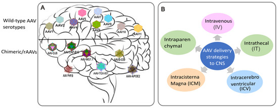
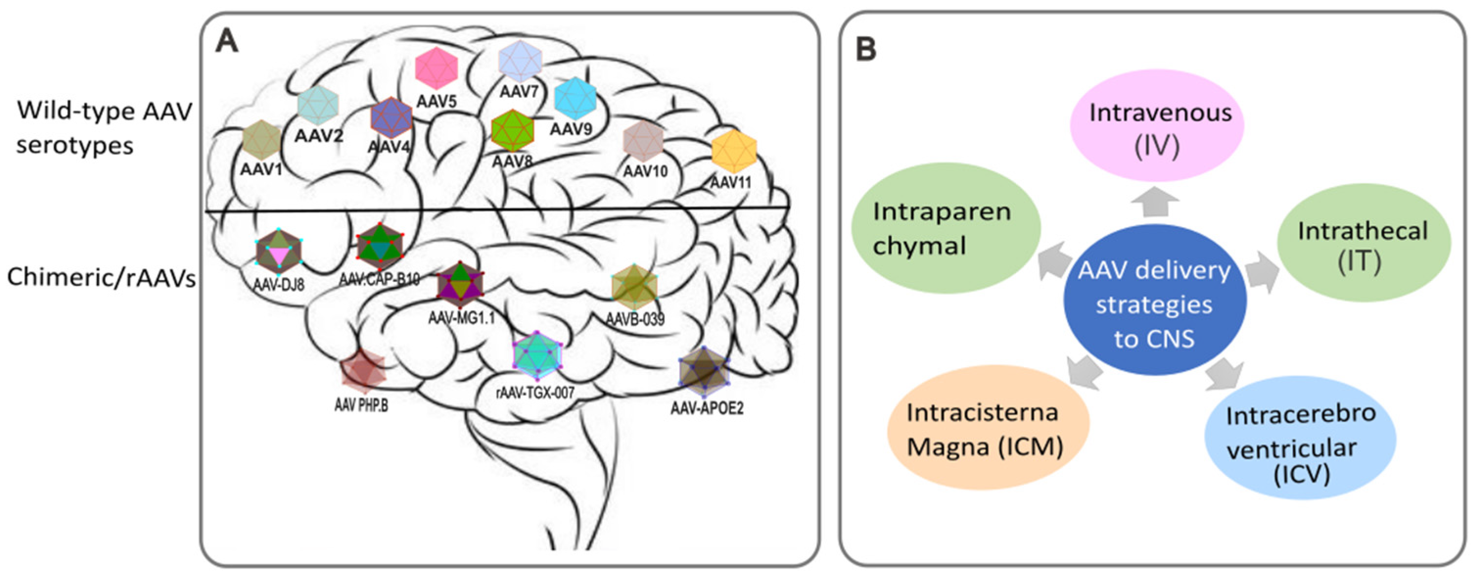
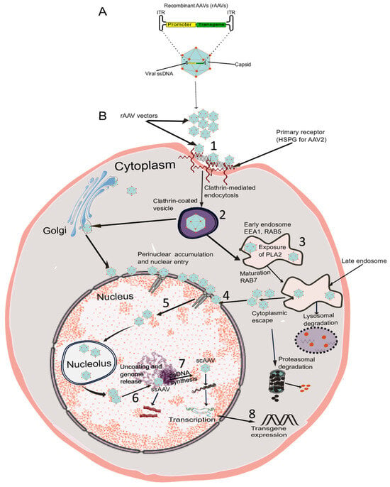
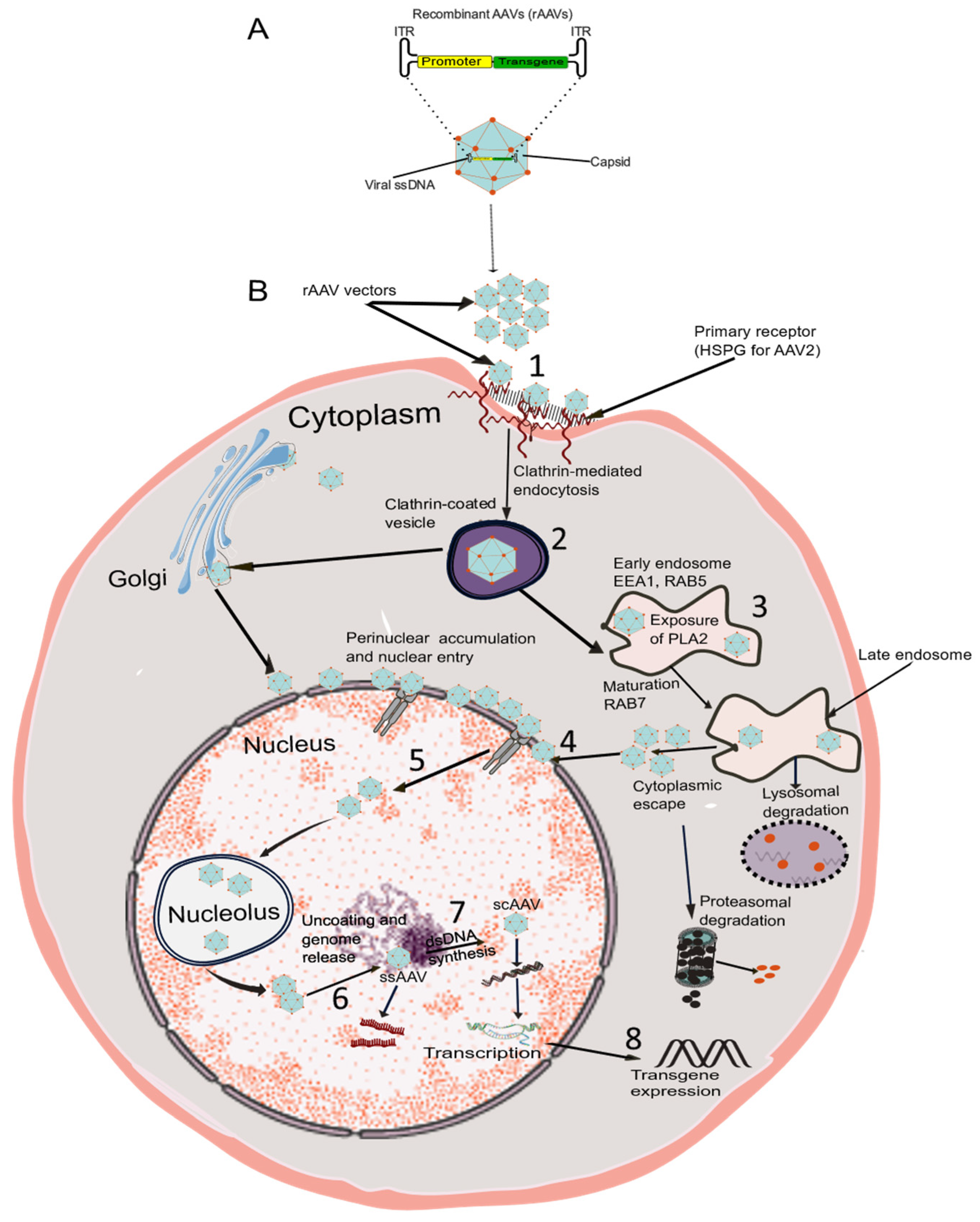
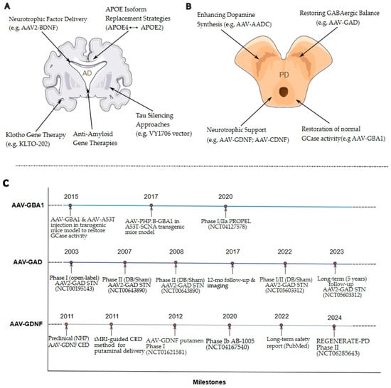
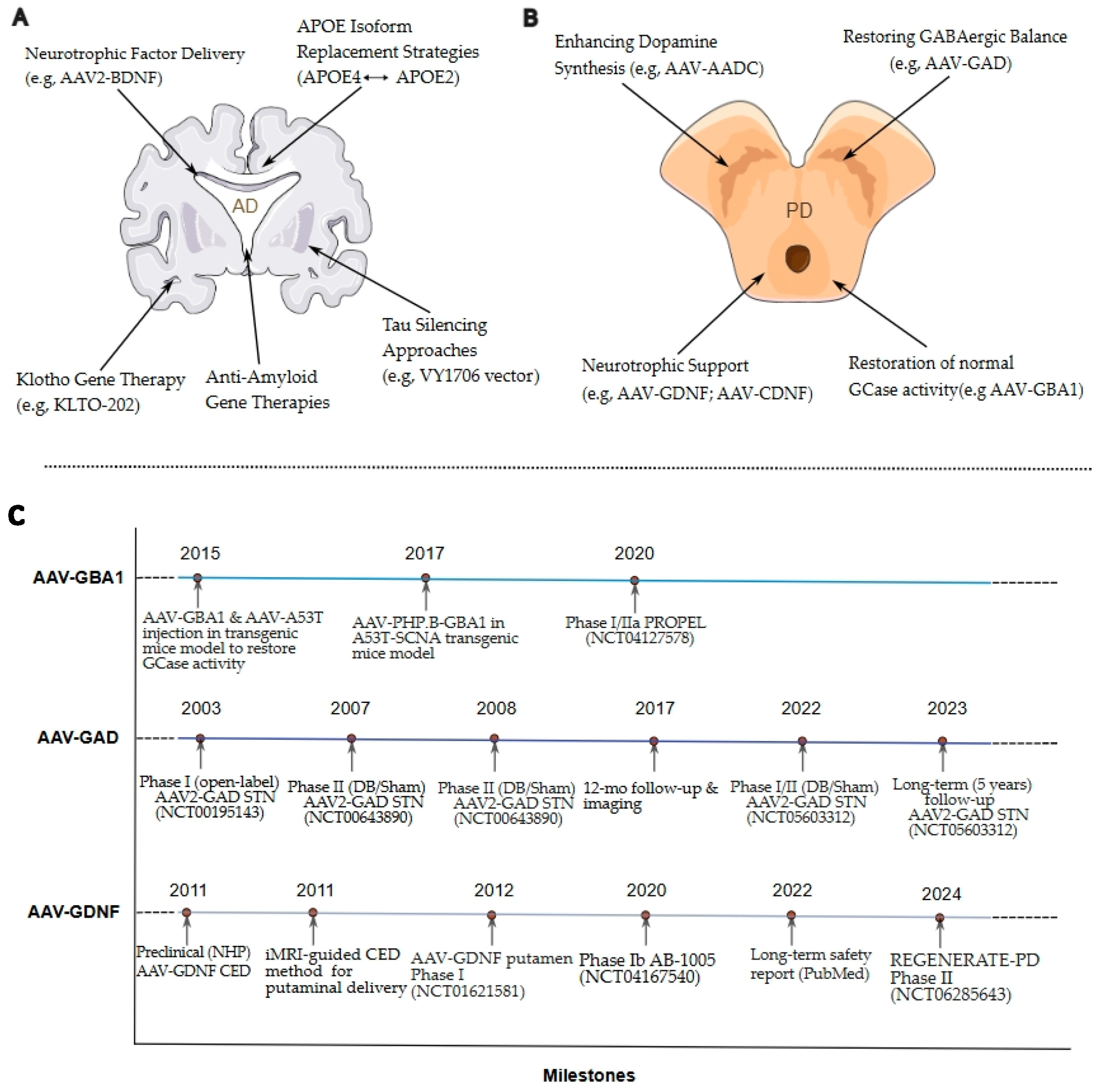
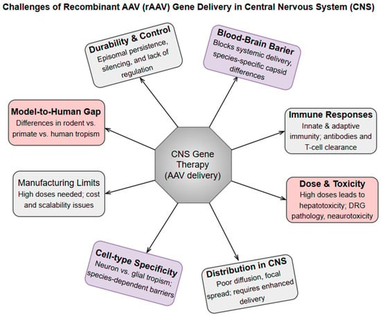
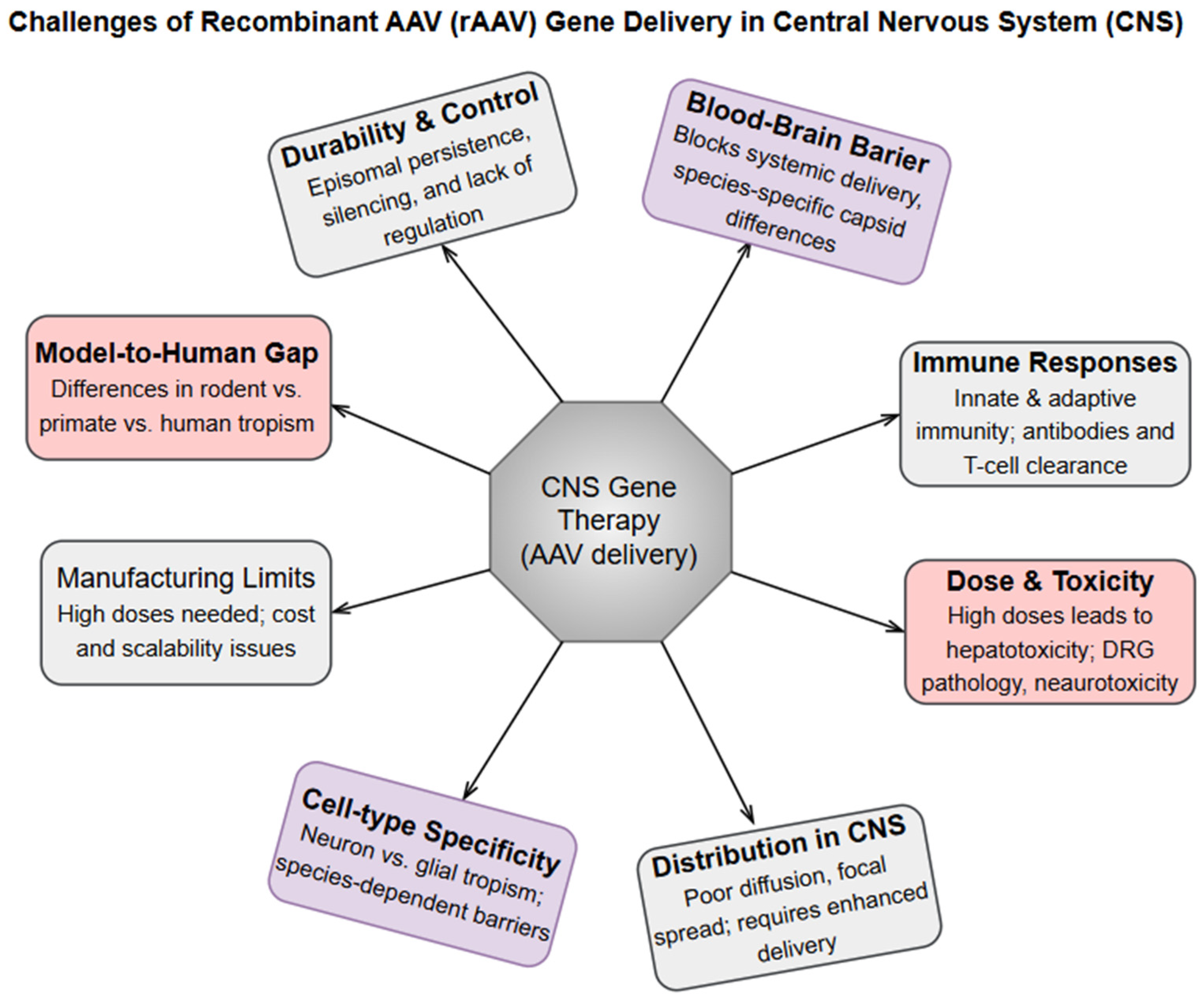
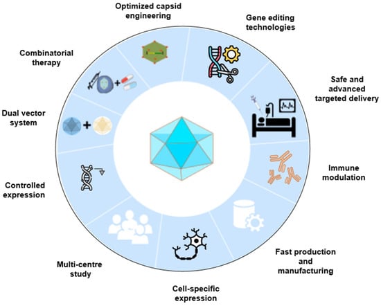
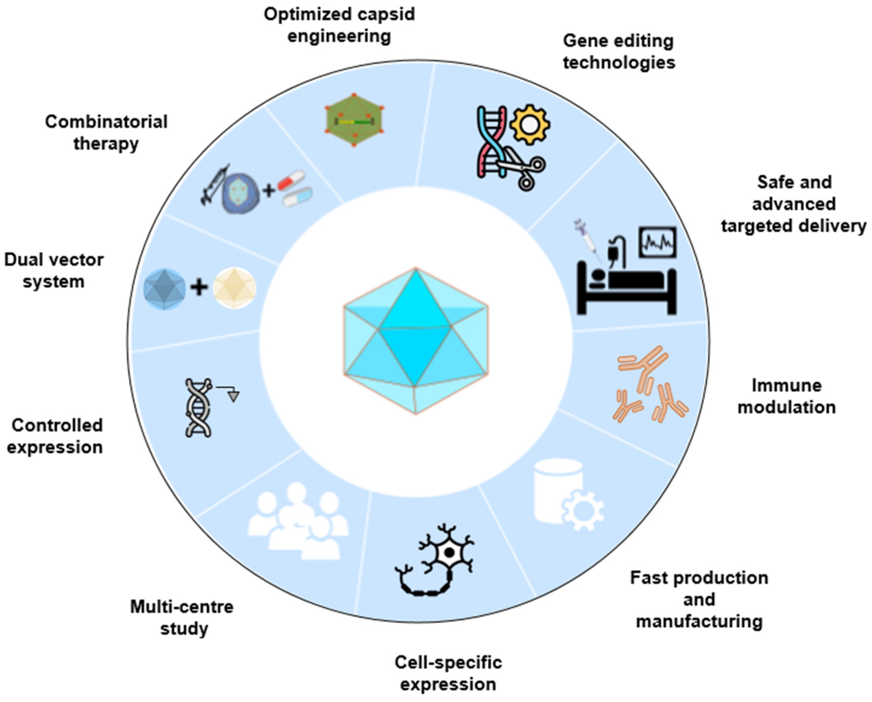
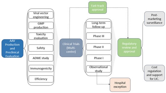
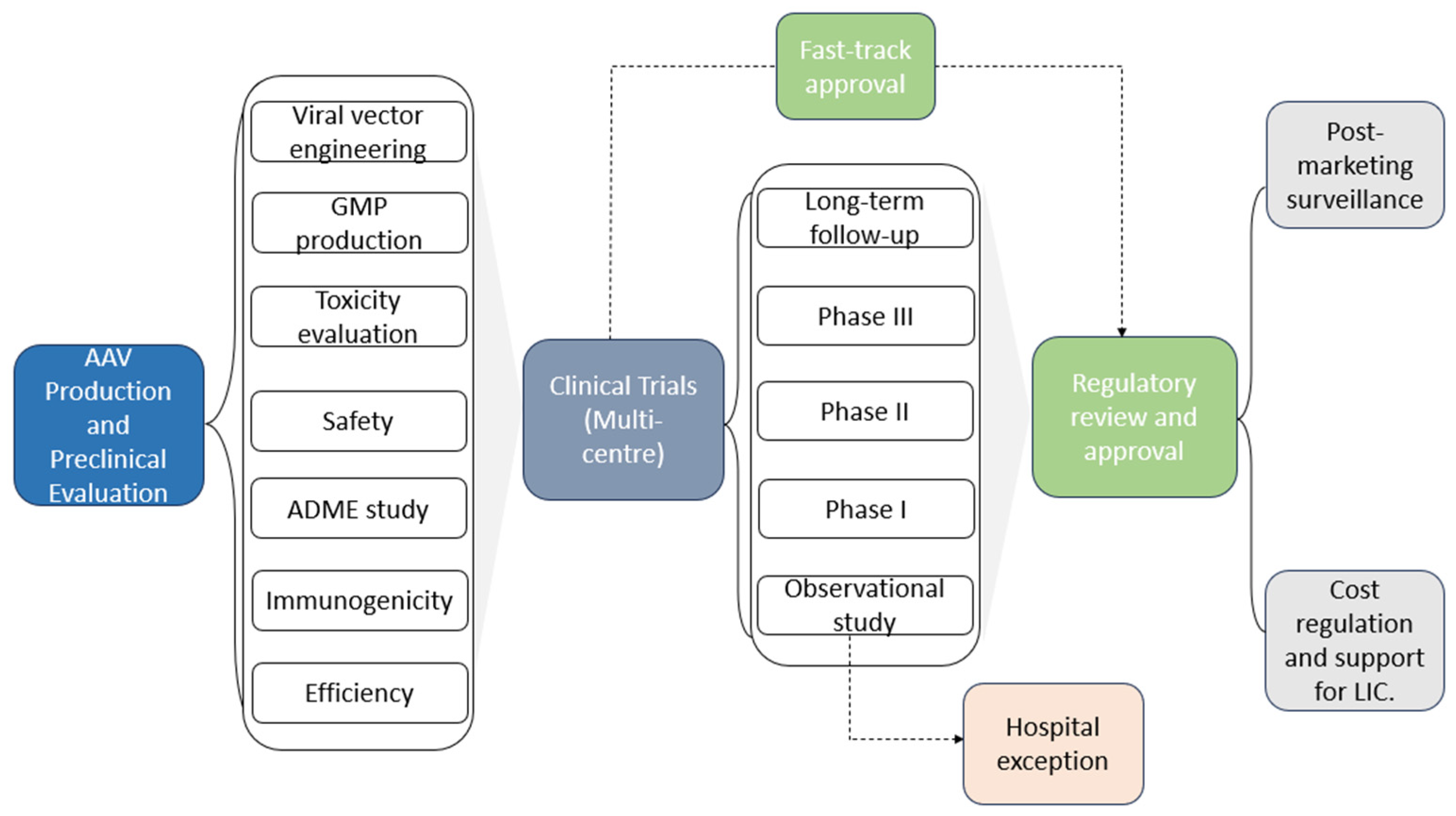

Disclaimer/Publisher’s Note: The statements, opinions and data contained in all publications are solely those of the individual author(s) and contributor(s) and not of MDPI and/or the editor(s). MDPI and/or the editor(s) disclaim responsibility for any injury to people or property resulting from any ideas, methods, instructions or products referred to in the content. |
© 2026 by the authors. Licensee MDPI, Basel, Switzerland. This article is an open access article distributed under the terms and conditions of the Creative Commons Attribution (CC BY) license.