


Abstract
Amaranthin is a major red-violet betacyanin of Amaranthaceae and an increasingly relevant natural pigment for food, cosmetic, nutraceutical, and biotechnological applications. This review integrates knowledge from over 100 studies, addressing amaranthin as a chemically defined betalain, distinguishing it from other scientific uses of the term, and evaluates its natural sources, analytical methods, extraction strategies, in vitro production systems, biosynthetic regulation, and biological activity. Cultivated Amaranthus species are among the richest plant sources, with total betacyanins of 46.1–199 mg/100 g fresh weight and amaranthin comprising, on average, 80.9% of the pigment fraction. Reliable identification and quantification rely on high-performance liquid chromatography coupled with a diode array detector (HPLC-DAD), liquid chromatography-tandem mass spectrometry (LC-MS/MS), and ultraviolet–visible (UV–Vis) spectrophotometry. Microwave- and ultrasound-assisted extraction can improve pigment recovery under optimized conditions, although its stability depends strongly on pH, temperature, solvent, time and storage parameters. While plant in vitro cultures, including callus, suspension, and shoot systems, have clarified biosynthetic regulation and offer controlled production platforms, engineered yeast systems have recently expanded production options, with Yarrowia lipolytica reaching 2.97 ± 0.029 g L−1 amaranthin in fed-batch fermentation. Amaranthin-rich extracts and amaranthin-type pigments show antioxidantand anti-inflammatory potential, while antimicrobial and antiviral activities have mainly been reported for mixed betacyanin fractions; direct mechanistic, bioavailability, and in vivo evidence for purified amaranthin remains limited. Standardized analytical protocols, further investigation of stable high-yield sources, physicochemical stability assessment, and structure–activity studies are identified as priorities for advancing future application-oriented research on this multipotential pigment.
1. Introduction
Amaranthin, the predominant betacyanin pigment in the Amaranthus genus, is a natural red-violet colorant with promising commercial applications across the food, pharmaceutical, and cosmetic industries [1,2,3,4]. Betacyanins, a subclass of betalains, are water-soluble nitrogenous pigments responsible for the characteristic coloration in Amaranthaceae species, which have been cultivated for centuries for their ornamental, nutritional, and medicinal properties [5,6,7,8].
Research on amaranthin content in both wild and cultivated Amaranthaceae species, with a simultaneous focus on plant in vitro systems, has emerged as a critical area of inquiry due to the increasing global demand for natural food colorants and antioxidants [1,9,10,11]. The practical significance of this research lies in the potential of amaranth-derived betacyanins as natural colorants with antioxidant properties, offering viable alternatives to synthetic dyes while contributing to innovation in the food industry [1,2,12,13]. The increasing consumer demand for natural additives, coupled with the estimated growth of the natural colorant market, underscores the importance of exploring amaranthin-rich plant tissues, as they may offer advantages over both synthetic dyes and other natural pigments like anthocyanins [1,14,15,16]. Global market trends consistently indicate a growing preference for natural pigments over synthetic alternatives, further emphasizing the importance of developing efficient extraction and analytical methods for amaranthin-like compounds [1,13,17,18].
Over the past two decades, significant advances in chromatographic and mass spectrometric techniques have enabled detailed profiling of betalain compounds, highlighting the predominance of amaranthin and isoamaranthin in cultivated amaranths [8,19,20,21]. High-resolution liquid chromatography–tandem mass spectrometry (LC-MS/MS), together with complementary spectroscopic and chromatographic approaches has substantially improved betalain identification and quantification, providing comprehensive compound catalogs and analytical protocols for profiling amaranthin and related betalains in diverse Amaranthus species [19,20,21,22].
Recent advances in genomics and analytical techniques have expanded our understanding of betalain pigment biosynthesis and regulation, revealing the molecular mechanisms underlying their production and accumulation [23,24,25,26]. Genomic resources and functional studies have identified key pathway enzymes, regulatory genes, and tissue-specific control mechanisms in Amaranthus, with chromosome-scale genome assemblies enabling the isoform-resolved annotation and mapping of betalain regulation across different tissues [23,24,25]. These discoveries have elucidated the core betalain biosynthesis pathway and identified transcription factors involved in betalain metabolism, further highlighting the significance of these pigments in food, pharmaceutical, and cosmetic applications [23,24,25,26].
Beyond summarizing the available literature, the specific objective of this review is to integrate these fragmented data into a coherent framework that highlights conceptual and technological bottlenecks in amaranthin research and production. The intention is to distill from this synthesis both an updated view of amaranthin as a multipotential pigment and a set of concrete, experimentally tractable questions that can guide future basic and applied research.
2. Literature Search Strategy and Thematic Organization
Because this article is a narrative and integrative review rather than a formal systematic review, the present section briefly describes the literature search strategy and the criteria used to identify and thematically organize publications dealing specifically with amaranthin as a betacyanin pigment.
To comprehensively investigate the research devoted to amaranthin as a betacyanin pigment, a multi-stage literature search was conducted, utilizing primarily the Scopus and Web of Science databases. The goal was to identify and compile publications detailing the plant sources of amaranthin as a betacyanin pigment, with particular emphasis on the plant in vitro culture techniques used to obtain this compound.
The initial literature search involved the keywords: “amaranthin”, “amaranthine”, “amarantin”, and “amarantine”. These were applied to the title, abstract, and keyword fields in the above-mentioned databases, resulting, in both cases, in over 200 publications. This preliminary search revealed that terms connected with amaranthin are used variably across different disciplines, indicating the need for a more refined search. A focused screening was then applied to capture literature specifically linking amaranthin to its identity as a betacyanin pigment by pairing the core terms with “betacyanin”, “betanin”, and “betalain” phrases. To identify experimental work involving controlled in vitro production systems, another search strategy was implemented using the core terms with additional keywords, such as “callus”, “suspension culture”, “hairy roots”, “transgenic roots”, “transformed roots”, “micropropagation”, and “bioreactor”. This collection of over 100 gathered and selected publications was further enriched with relevant records obtained by forward and backward citation chaining, supported by the AI-assisted SciSpace Deep Review tool, with final selection and verification performed by the author. Original experimental articles, reviews, or book chapters written in English and with an available full-text version were chosen for analysis, excluding those lacking essential experimental details. Full-text versions of publications were accessed through institutional subscriptions to platforms including ScienceDirect, PubMed/PubMed Central, and SpringerLink. Additional open-access articles were identified through a search of online resources and publisher repositories.
Finally, 136 of the most relevant publications and sources were selected and grouped into four thematic sections: (1) natural sources of amaranthin—documented occurrence of amaranthin in wild and cultivated species of the genus Amaranthus and other genera; (2) analytical and biosynthetic approaches—methods for the extraction, quantification, structural elucidation, and biosynthetic pathway analysis of amaranthin-type betacyanins; (3) plant in vitro systems—studies on amaranthin biosynthesis and production in cell, tissue, and organ cultures; and (4) biological activity—investigations of antioxidant, anti-inflammatory, and other bioactivities attributed to amaranthin as a betacyanin pigment. In addition, a brief section clarifies the different uses of the term “amaranthin” in scientific research that emerged from this extended literature survey.
3. Terminology Note: ‘Amaranthin’ in the Literature
Before focusing exclusively on amaranthin as a betacyanin, it is important to note that the term “amaranthin” has been used in the scientific literature to describe chemically and functionally distinct entities, including a plant betacyanin, a lectin, a seed storage protein, and a synthetic azo dye, which makes explicit clarification of the terminology essential. As the subject of this review, amaranthin refers specifically to the plant betacyanin pigment: betanidin 5-O-[β-D-glucuronosyl-(1→2)-β-D-glucoside] with full IUPAC name (2S)-5-[(2S,3R,4S,5S,6R)-3-[(2R,3R,4S,5S,6S)-6-carboxy-3,4,5-trihydroxyoxan-2-yl]oxy-4,5-dihydroxy-6-(hydroxymethyl)oxan-2-yl]oxy-1-[(2E)-2-[(2S)-2,6-dicarboxy-2,3-dihydro-1H-pyridin-4-ylidene]ethylidene]-6-hydroxy-2,3-dihydroindol-1-ium-2-carboxylate (PubChem CID: 6325284) [27]. However, across a broad spectrum of literature, the terms ‘amaranthin’, ‘amaranthine’, ‘amarantin’, and ‘amarantine’ appear with some limited spelling consistency in reported scientific papers. Therefore, being careful in this regard when reviewing the literature but taking into account all possible spellings ensures that some reports on the investigated topic are not overlooked.
In most cases, ‘amaranthin’ represents a vivid natural pigment of the betacyanin type [28], a lectin protein involved in plant defense mechanisms and biochemical studies [29,30], and a seed storage protein with applications in genetically engineered crops [31,32,33]. To supplement this data, it is worth mentioning here a synthetic red coloring known as Amaranth dye (E 123, Red Dye No.2, Food Red 9, Acid Red 27, Azorubin S, or Color Index (C.I.) number 16185), which is a modified red azo dye used as a food and beverage colorant and also in the cosmetics and textile industries [34]. Amaranth dye was banned as a food additive in the United States by the U.S. Food and Drug Administration (FDA) in 1976 as a suspected carcinogen but is still allowed in some countries for use, for example, in the European Union (EU) (as E 123), with quantity restrictions [35,36]. To complete this list, the term ‘amaranthine’ has also been metaphorically invoked in more abstract applications, retaining its adjectival sense of “everlasting”, “immortal”, or “imperishable”; examples include “amaranthine adventure” and “amaranthine scale” in biology [37,38], as well as its adoption as the name of a 3D-printed humanoid robot in robotics [39]. In Table 1, examples of various amaranthin contexts in scientific publications are listed.
Table 1.
Various uses and related contexts of ‘amaranthin’ term in scientific literature.
The following sections of this review are devoted exclusively to amaranthin as a betacyanin pigment, using the nomenclature and structure details as presented by the NCBI PubChem databases [27].
4. Amaranthin Characteristics and Its Natural Sources
Research on the methods and techniques of amaranthin betacyanin separation, identification, and quantification has emerged as a critical area of inquiry due to the increasing demand for natural food colorants and bioactive compounds with health benefits. Betalains, including betacyanins and betaxanthins, are nitrogen-containing pigments responsible for the red-violet and yellow-orange colors in plants of the Caryophyllales order, such as Amaranthus species [19,43]. Since the early isolation of betanin from red beetroot in 1918, research has expanded to characterize betalains in various Amaranthaceae species, recognizing amaranthin as the predominant betacyanin in the Amaranthus genus [1,19] and also present in other genera of this family, for example: Chenopodium, Celosia, Atriplex, Alternanthera, Gomphrena, and Iresine [44,45,46,47,48,49,50].
4.1. Structure Characteristics and Analytical Methodology
Amaranthin is a disaccharide derivative that is a betanidin in which a beta-D-glucuronosyl-(1->2)-beta-D-glucosyl moiety is attached at position 5 (Figure 1a) [27]. It presents a characteristic amaranth color belonging to the betacyanin group of compounds found in Amaranthus sp., among others (Figure 1b).
Figure 1.
Characteristic amaranthin features: (a) amaranthin chemical structure [27]; (b) young Amaranthus sp. shoots presenting the typical color of the amaranthin presence (Photograph by D. Kucz).
The C-15 epimer of amaranthin–isoamaranthin is also found to be an accompanying compound in investigated plant tissues [2,11,51]. As for separation, identification, and quantification techniques, high-performance liquid chromatography (HPLC), LC-MS/MS, and high-speed counter-current chromatography (HSCCC) are primary methods for separating and identifying amaranthin and related betacyanins. These techniques enable precise molecular formula determination and fragmentation pattern elucidation, which are essential for distinguishing structural isomers and diastereomers [19,21,52,53,54]. UV–Vis spectrophotometry (typical λmax ≈ 535 nm for amaranthin) is widely used for screening and routine quantification [43]. In complex matrices, it should be complemented by LC-MS/MS for definitive identification and isomer resolution. Comparative studies indicate that HPLC methods are preferred when interfering substances are present, as spectrophotometric methods may show discrepancies of up to 15% with degraded samples [55].
Matrix-assisted laser desorption/ionization quadrupole ion trap time-of-flight mass spectrometry (MALDI-QIT-TOF MS) has also been applied for rapid identification without prior purification [52]. Recently, an attenuated total reflectance–Fourier transform infrared (ATR–FTIR) spectroscopy method was proposed by López et al. (2023) as a simple and efficient protocol for amaranth betalain extraction and stability analysis [20]. Recent studies emphasize tandem MS and NMR for confident structure confirmation [53,56]. Structural characterization among betacyanins and, more broadly, betalains is based on amaranthin’s characteristic unique glucuronosylglucosyl moiety and acylation patterns, which distinguish it from the well-known betacyanin betanin (beetroot red; E 162). Comparative evaluations of analytical methods highlight the complementarity of chromatographic and spectrometric approaches for amaranthin analysis. HPLC coupled with diode array and mass spectrometric detection (DAD/MS) enables accurate quantification and identification [19,47,57]. The integration of spectral data and fragmentation patterns enhances reliability in complex plant matrices [52,58]. HSCCC is recognized as complementary, especially for isolating diastereomers and enriching minor betacyanins [53,54]. Several studies demonstrate that structural variation within betalains modulates color-related properties, stability, and radical-scavenging capacity [45,59,60,61,62]. In particular, Cai et al. established a structure-antiradical activity series for purified Amaranthaceae betalains, revealing that celosianins exhibit higher antiradical potency than amaranthin/isoamaranthin by 2,2-Diphenyl-1-picrylhydrazyl radical (DPPH) assay, with glycosylation of the aglycone and hydroxyl group number as key structural determinants [62]. For acylated amaranthin-type betacyanins, oxidation generated structurally distinct derivatives with modulated antiradical activity; importantly, such oxidized products retained significant cardioprotective potential in cardiomyoblasts [61]. When purified native pigments were directly compared, celosianin exhibited markedly higher antioxidant potency than amaranthin (IC50 21.5 vs. 32.2 μg/mL, respectively) [45]. By contrast, studies on betalain-rich plant extracts, including those from amaranth, cactus pear, and beetroot, support their broad bioactivity and potential as natural colorants, but do not provide systematic structure-activity comparisons of individual pigments [4,63]. Taken together, the evidence indicates that structural diversity among betalains, including amaranthin-related pigments and their oxidation products, governs physicochemical and antiradical properties at the molecular level, while extract-based functional assays further support their relevance as natural food colorants and health-promoting agents [4,45,59,60,61,62,63,64].
4.2. Amaranthin in Wild and Cultivated Plant Species
Research demonstrates substantial variation in amaranthin content across the Amaranthaceae family. Table 2 presents a multi-exemplary list of research on plant species in which the presence of amaranthin was tested, and it is composed to present worldwide works on plants of wild and cultivated origin. The main goal of this summary is to present the range and the origin of the Amaranthaceae plants examined for the presence of amaranthin, to highlight the extraction methods used for plant material analysis, and to summarize the analytical methods applied to quantify this specific metabolite. However, works listed in Table 2 were not solely focused on the amaranthin content but also often concerned the determination of various betalains and other metabolite groups. Authors often also investigated the antioxidant properties or other biological activity potential of whole extracts. Variability in sample preparation, diversified extraction conditions, and differences in instrumentation sensitivity contribute to divergence across studies, which significantly limits cross-study comparability in amaranthin quantity among various species and plant material of diverse origin. Matrix complexity and pigment instability during handling introduce extra quantification challenges. Despite certain limitations in the direct comparison of results, all these works provide rich documentation of the dynamically developing topic focused on searching for sources of natural dyes, such as amaranthin, with a wide potential of applications.
Some of the research works listed in Table 2 are exceptionally significant. In a comprehensive study of 21 genotypes from seven Amaranthus species, conducted by Cai et al. (1998), total betacyanins ranged from 46.1 to 199 mg/100 g of fresh plant material (FW), with amaranthin comprising an average of 80.9% of the total betacyanins [7]. Cultivated species exhibited higher betacyanin concentrations than wild species and had substantially greater biomass [7]. A broader survey by Cai et al. (2001) [65], covering 37 species across eight genera in the Amaranthaceae family, revealed total betacyanin content ranging from 0.08 to 1.36 mg/g fresh weight [58]. Among amaranth accessions grown as leafy vegetables, total betacyanin concentrations varied markedly from 4.7 to 478.8 mg/100 g dry weight, with amaranthin and isoamaranthin identified as major constituents [1]. Specific amaranth cultivars demonstrated notable differences in betacyanin profiles. In A. gangeticus accessions, four betacyanin compounds were identified: amaranthin, isoamaranthin, betanin, and iso-betanin [2]. Similarly, A. tricolor genotypes contained the same four betacyanin compounds, with genotypes VA14 and VA16 showing particularly high amaranthin and isoamaranthin contents [11]. Weedy Amaranthus species also showed significant potential, with the A. viridis genotype WAV7 and A. spinosus genotype WAS13 demonstrating the highest concentrations of betacyanins among the accessions tested by Sarker and Oba (2019) [66].
Table 2.
Natural sources of amaranthin: Documented occurrences of amaranthin in worldwide research studies—wild and cultivated plant species.
To expand and facilitate the comparison between the taxa and tissues of the research presented in Table 2, the following Figure 2 shows the plant material from 43 species of 10 genera in which amaranthin has been investigated. There is a clear, significant predominance of research evidence supporting the presence of amaranthin in seedlings, primarily Amaranthus sp., and in leaves, here visibly expanding, for example, for Alternanthera sp.
Figure 2.
Plant species and plant materials investigated for amaranthin presence, including: seedlings, leaves, stems (incl. branches), flowers (incl. inflorescences), and seeds (incl. grains). The information supplements and extends the entries summarized in Table 2; all citations are as listed in Table 2.
4.3. Impact of Postharvest and Extraction Protocols on Amaranthin Yield and Stability
Postharvest and extraction protocols influence amaranthin stability during storage and processing, with temperature, pH, and antioxidants playing key roles [7,20,76]. Therefore, treatments such as freeze-drying, solar drying, and cold storage markedly affect betacyanin retention. Freeze-drying and low-temperature storage preserve pigment content better than air or oven drying [76,77]. Thermal and light exposure accelerate degradation, necessitating optimized handling protocols [59,76]. Visible/near-infrared spectroscopy can aid in the non-destructive monitoring of pigment stability [77]. Khandaker et al. (2009) examined seven cultivars of red amaranth (Amaranthus tricolor L.) [78]. They proved how variable factors such as the type of red amaranth cultivar, its degree of maturity, and food processing factors (temperature, light exposure, oxidant presence) quantitatively influenced betacyanin yields and preservation [78].
Conventional solvent extraction with water or ethanol is often optimized by multivariate analysis and remains widely used for amaranthin recovery [15,76]. Emerging methods such as microwave-assisted and ultrasound-assisted extraction can enhance pigment yield and purity while preserving its stability [63,79,80]. Applying spectroscopic methods, such as attenuated total reflectance–Fourier transform infrared (ATR-FTIR) spectroscopy, assists in monitoring pigment stability post-extraction [20]. These gradually implemented methods represent advances toward greener, more efficient pigment recovery and quality evaluation, which are crucial for amaranthin application.
Despite advances in betalain research, challenges still remain in the reliable separation, identification, and quantification of amaranthin due to its chemical diversity and instability under processing conditions [19,57]. Discrepancies in extraction protocols, solvent systems, and analytical parameters persist [12,52,53]. Moreover, the structural complexity of amaranthin, characterized by glucuronosylglucoside moieties and acylation patterns, complicates its differentiation from other betacyanins and betalains [53,77]. Discussion regarding the optimal methods for preserving pigment stability during extraction and storage, which impacts quantification accuracy and application potential, is also present [20,76]. The lack of standardized, high-throughput approaches limits the comprehensive understanding and commercial exploitation of amaranthin-rich extracts [1,15].
5. Plant In Vitro Systems for Amaranthin Biosynthesis
Research on amaranthin in plant in vitro cultures has emerged as a critical area of inquiry due to the multifaceted potential significance of this betacyanin pigment in the nutrition, medicine, and cosmetic industries. A summary of experimental studies on the biosynthesis of amaranthin in organ and tissue in vitro cultures is presented below in Table 3. Presenting the predominant plant in vitro systems (shoot cultures and callus and suspension cultures), it also outlines various research foci within the still-developing multi-directional trend of identifying efficient methods of obtaining valuable plant metabolites through biotechnological approaches. Figure 3 shows Celosia sp. shoots obtained from seeds on solid culture medium (based on the author’s own unpublished research).
Table 3.
Comparative Overview of Amaranthin-Focused In Vitro Systems.
Figure 3.
Flowering in vitro shoots of Celosia sp. (Photograph by M. Jeziorek; author’s own unpublished research).
5.1. Diverse In Vitro Culture Platforms
Researchers have employed a wide array of in vitro systems—callus cultures, cell suspensions, organogenic shoots, immobilized beads and even heterologous tobacco bright yellow-2 (BY-2) line—to investigate and enhance betalain biosynthesis. Early foundational work by Berlin et al. (1986) focused on Chenopodium rubrum suspension cultures as a reference system [87], while later studies extended to calli of Amaranthus tricolor [84], Alternanthera spp. [81,82,83] and Celosia spp. [56,85,86]. Immobilization (alginate–chitosan beads) and physical permeabilization (electric fields, high pressure) of C. rubrum cells were introduced as green extraction methods to recover intracellular amaranthin with up to 85% release efficiencies by Dörnenburg and Knorr (1993) [89]; analogous chitosan/DMSO treatments used by Knorr and Berlin (1987) also liberated pigment but revealed rapid degradation if not stabilized [92].
It is worth noting the successful attempts at genetic transformation to intensify amamrantin production [93,94]. One of the recent advances reconstitutes the amaranthin pathway in tobacco BY-2 cells by over-expressing four Chenopodium quinoa genes, including the newly isolated amaranthin synthetase. This chassis produced 13.7 µM amaranthin and 26.6 µM betanin, demonstrating proof-of-concept for microbial-style pigment factories [93]. Glitz et al. (2025) successfully engineered yeast cell factories for recombinant amaranthin production, achieving remarkable yields of 2.97 g L−1 ± 29.3 mg L−1 in fed-batch fermentation using Yarrowia lipolytica strains equipped with glucuronosyltransferases and UDP-glucose dehydrogenase from Arabidopsis thaliana for de novo UDP-glucuronic acid synthesis [94]. Hairy root in vitro cultures of Beta vulgaris have additionally proved to be a system of betalains production, which justifies undertaking similar attempts to obtain amaranthin from species in which it naturally occurs [95,96].
5.2. Media Composition and Precursor Feeding
Most studies utilize Murashige and Skoog (MS) basal salts, originally described in 1962, with variations in sucrose (3–6%), auxins [2,4-dichlorophenoxyacetic acid (2,4-D) and 1-naphthaleneacetic acid (NAA)], cytokinins [kinetin, 6-benzylaminopurine (BAP), and thidiazuron (TDZ)] and, increasingly, precursor feeds such as L-tyrosine. Tyrosine feeding emerged as a simple yet effective booster in C. rubrum suspensions—raising total betacyanins to ~100 mg L−1 (~1% DW), with amaranthin comprising ~80% of the pool (Berlin et al., 1986) [87]—and in Alternanthera shoots, delivering up to 51 mg 100 g−1 FM (Kleinowski et al., 2014) [82]. Carbon-source trials in Celosia calli revealed that sucrose remains superior to hexoses for maximal pigment yield, while also guiding the discovery of malonylated amaranthin derivatives [56].
5.3. Elicitation and Light Quality
Beyond simple precursors, biotic elicitors and light spectra have been harnessed to tailor pigment output. Elicitation studies using methyl jasmonate in Alternanthera sessilis have demonstrated the potential to enhance bioactive compound accumulation through transcriptomic approaches [97]. A Fusarium oxysporum cell-wall lysate at 0.125 ‰ w/v significantly elevated both amaranthin and betanin in Celosia cristata suspensions, without growth penalties [86]. Regarding light quality, LED light treatments (red, white, and blue) applied to Alternanthera microshoots showed red/white light to be most stimulatory for betalain accumulation [83].
5.4. Enzymology and Biosynthetic Pathways
The molecular understanding of betalain biosynthesis has advanced through plant tissue culture studies, with research on Amaranthus tricolor elucidating core biosynthetic pathways and identifying key regulatory genes [6,98,99]. The transcriptomic and enzymatic analyses shedding light on the biosynthetic genes responsible for glycosylation and pigment accumulation in Amaranthaceae species direct the potential to increase bioactive compound production [6,97,98,99]. A parallel thread of Bokern et al. studies dissected the enzymatic machinery: crude protein preps from Chenopodium rubrum were shown to acylate amaranthin to celosianin I/II [100], and the responsible hydroxycinnamoyl transferase was purified ~500-fold, characterized kinetically [91]. Growth-curve studies further correlated transferase activity with temporal betacyanin buildup [90].
The comparative analysis reveals that while plant in vitro systems have provided crucial foundational knowledge for understanding amaranthin biosynthesis and have demonstrated various production approaches, engineered yeast cell factories currently represent a more efficient and scalable platform for amaranthin production. The significantly higher yields achieved through microbial fermentation, combined with the stability and industrial scalability of yeast systems, position recombinant production as the preferred method for commercial amaranthin production in biotechnological applications.
6. Regulation of Amaranthin Biosynthesis
6.1. Amaranthin Biosynthetic Pathway
The amaranthin biosynthetic pathway indicating the key enzymes involved in the process is presented in Figure 4.
Figure 4.
Proposed biosynthetic pathway and regulatory control of amaranthin.
In betalain-producing Amaranthus species, amaranthin biosynthesis proceeds through the general betacyanin pathway from the aromatic amino acid L-tyrosine (Figure 4). Tyrosine is first hydroxylated to 3,4-Dihydroxy-L-phenylalanine (L-DOPA) by CYP76AD-type cytochrome P450 enzymes, which may also participate in the formation of cyclo-3,4-dihydroxyphenylalanine (cyclo-DOPA). L-DOPA can be cleaved by DOPA 4,5-dioxygenase (DODA) to produce betalamic acid, the chromophoric precursor common to all betalains, while a parallel oxidation step yields cyclo-DOPA. Condensation between betalamic acid and cyclo-DOPA gives rise to the betacyanin aglycone betanidin. Subsequent 5-O-glucosylation of betanidin and glucuronylation at the glucose moiety result in the formation of amaranthin (betanidin 5-O-[β-D-glucuronosyl-(1→2)-β-D-glucoside]). This sequence has been substantiated by classical feeding experiments with labeled tyrosine and intermediates in Amaranthus, as well as by the molecular characterization of CYP76AD, DODA, and glucosyltransferase genes in beet and amaranth [6,18,101,102,103].
It was proven that in Amaranthus spp., amaranthin synthesis is tightly regulated by light, particularly through the phytochrome system, where red light stimulates pigment production and far-red light reverses this effect [104]. Blue and UV light further enhance amaranthin biosynthesis via cryptochrome activation, revealing light-quality-dependent regulation [105]. Pigment accumulation can also be induced in complete darkness with exogenous kinetin, a cytokinin that activates transcription of the biosynthetic enzymes [106,107]. However, gibberellic acid (GA3) antagonizes this effect, inhibiting both light- and cytokinin-induced synthesis, likely by modulating precursor allocation or repressing gene expression [108,109]. The need for active transcription and translation was demonstrated by the complete inhibition of pigment synthesis by actinomycin D and puromycin [110,111]. DOPA and tyrosine feeding studies confirmed the importance of precursor supply, with DOPA being more effective and light-enhanced, which indicates that a key enzymatic conversion step is light-dependent [112]. Further biochemical elucidation in Celosia plumosa showed that cyclo-DOPA and its glucoside serve as more efficient amaranthin precursors than betanidin or betanin, validating the specific biosynthetic route [113]. Cyclic nucleotides such as dibutyryl cyclic adenosine monophosphate (db-cAMP) and N6-substituted adenines also stimulated pigment production but lacked an amplifying effect with light, implying that they mimic cytokinins rather than act as second messengers in light pathways [114]. This type of experimental system is being continued. Similarly, Zhu et al. (2016) performed experiments based on methods developed by Köhler et al. (1980) and, with minor modifications, investigated the antagonistic effect of indole-3-acetic acid on kinetin-stimulated amaranthin accumulation in the cotyledons of Amaranthus mangostanus seedlings [115,116]. Transcriptomic analyses of Amaranthus tricolor tissues performed by Liu et al. (2019) and Liu et al. (2023) provided additional insights into betalain biosynthesis and regulation [99,117]. Together, these studies generated a detailed catalog of genes and a tissue-specific expression map for betalain biosynthesis in A. tricolor and showed that environmental cues such as blue light influence pigment accumulation by increasing the transcription of biosynthetic genes, likely through a GA–DELLA-associated regulatory pathway.
6.2. Classical Studies on Hormonal and Physical Factors Influencing Amaranthin Biosynthesis
This section reviews noteworthy studies conducted on seedlings of Amaranthaceae plants and examining amaranthin biosynthesis, including the influence of physical factors and growth regulators. The findings derived from these investigations, predominantly conducted during the 1970s and early 1980s, established a fundamental comprehension of the influences exerted by variables such as light and kinetin on the biosynthesis of amaranthin within plant tissues [110,111,112]. This segment of classical research was focused on species such as Amaranthus caudatus, A. tricolor, and also Celosia plumosa [108,109,110,113].
The consistent applied methodology enabled precise control of the experimental variables and ensured comparability across light and hormone treatment regimes. Seedlings were cultivated under specific controlled laboratory conditions: seeds were typically germinated on moistened filter paper in Petri dishes and maintained in darkness at temperatures ranging from 25 to 28 °C. After an initial dark growth period (48–72 h), seedlings were exposed to various light treatments (white, red, far-red, blue, UV) or incubated with hormonal, nucleotide, or biochemical precursor solutions.
Collectively, the studies affirm that amaranthin biosynthesis is governed by light-dependent photoreceptors, hormone signaling, and transcriptional activity, offering a detailed model for pigment regulation in plants. The experimental consistency and physiological relevance of these findings positioned Amaranthus sp. as providing a cornerstone system for exploring light–hormone interactions in secondary metabolism.
7. Biological Activity of Amaranthin
The literature on the biological activity of amaranthin highlights several major themes, mainly its antioxidant and anti-inflammatory properties. Emerging studies have examined molecular mechanisms, bioavailability, and potential therapeutic applications, illustrating a growing integration of biochemical, pharmacological, and food science perspectives.
7.1. Antioxidant Properties
Amaranthin and related betacyanins exhibit strong antioxidant activity, efficiently scavenging free radicals such as DPPH, hydroxyl radicals, and superoxide anions. Multiple studies consistently demonstrate the strong antioxidant and radical scavenging activities of amaranthin-containing extracts, with evidence of effective quenching of reactive oxygen species and free radicals [62,63,80,118,119,120]. Comparative analyses performed by Fernando et al. (2023) indicated that betacyanins exhibit superior radical scavenging compared to betaxanthins [120]. Associations between pigment content and DPPH- and ABTS-based radical scavenging capacity have been reported in various Amaranthus genotypes by Sarker et al. (2020) [11], Sarker et al. (2021) [2], and Sarker et al. (2022) [119,121].
Amaranthin and isoamaranthin exhibit antioxidant activity through radical scavenging and cellular redox modulation, though with lower potency than other betacyanins. In comparative DPPH assays, amaranthin/isoamaranthin ranked among the least active betalains tested (EC50 ≈ 8.0 μM), approximately 2.2-fold less potent than gomphrenins (EC50 ≈ 3.7 μM) and 1.5-fold less active than betanin (EC50 ≈ 5.5 μM) [62]. Amaranthus caudatus flower extracts rich in amaranthin (171 mg/g of extract) and isoamaranthin (38 mg/g of extract) demonstrated OxHLIA IC50 values of 29.0 μg/mL (60 min) [63]. At the cellular level, amaranthin-type betacyanins protected H9c2 cardiomyoblasts against H2O2-induced damage at 0.1–10 μg/mL, increasing intracellular glutathione levels [45,61].
7.2. Anti-Inflammatory Effects
Betalains demonstrate anti-inflammatory activity by suppressing pro-inflammatory cytokines such as interleukin-6 (IL-6) and tumor necrosis factor-alpha (TNF-α), and enzymes, including inducible nitric oxide synthase (iNOS) and cyclooxygenase-2 (COX-2), thereby modulating immune responses in vitro and in vivo. Evidence from cellular and animal models supports their anti-inflammatory potential, including suppression of pro-inflammatory cytokines and enzymes, modulation of signaling pathways such as MAPK/NF-κB, and enhancement of antioxidant defenses [120,122]. Betalain extracts have been shown to reduce leukocyte recruitment and oxidative stress markers in inflammation models, which suggests their therapeutic relevance [123].
Atriplex hortensis extracts and purified amaranthin-type pigments reduced prostaglandin E2 (PGE2) production in LPS-stimulated RAW264.7 macrophages [75]. Tyszka-Czochara et al. (2016) also investigated selenium supplementation and reported that amaranth sprouts with the highest amount of betacyanins (19.30 ± 0.57–28.85 ± 2.23 mg of amaranthin/100 g of fresh weight) and a high total selenium content exerted an anti-inflammatory effect, decreasing inflammatory interleukin-6 production in activated RAW264.7 macrophages [124].
7.3. Antimicrobial and Antiviral Activities Studies
Amaranthin has been investigated for its antimicrobial activity primarily as a component of betacyanin-rich fractions rather than as an isolated compound. Studies by Yong et al. demonstrated that betacyanin fractions from red spinach (Amaranthus dubius) and red pitahaya (Hylocereus polyrhizus) exhibited inhibitory effects against Staphylococcus aureus and Pseudomonas aeruginosa, including biofilm formation on various polymer surfaces [72,125,126]. Comparative analyses indicated stronger antimicrobial activity of red spinach betacyanin fractions, which contain amaranthin-type pigments, in planctonic assays [72]. However, anti-biofilm activity was pathogen-dependent, with red spinach fractions showing stronger inhibition against P. aeruginosa biofilms [125]. These findings suggest that amaranthin-type betacyanins may contribute to the observed antimicrobial effects in red spinach fractions, though the specific contribution of amaranthin versus other betacyanins in the fractions remains to be fully elucidated.
Recent investigations have expanded the bioactivities of betacyanin fractions to include antiviral activity. Lim et al. (2024) reported inhibitory effects of red pitahaya betacyanin fractions against the influenza A virus [127], while Chang et al. demonstrated the antiviral activity of betacyanin fractions from both red pitahaya and red spinach against dengue virus type 2 [71].
The variability in reported antimicrobial efficacy may be attributed to several factors, including differences in betacyanin fraction composition, microbial strain susceptibility, and assay methodologies. The enhanced biofilm-inhibiying activity observed for combined red spinach and red pitahaya betacyanin fractions compared with single-source fractions suggests possible additive or synergistic effects among fraction constituents; however, the specific contribution of amaranthin was not determined [126].
7.4. Comparative Bioactivity with Other Betacyanins
Comparative analyses reveal that amaranthin shares many bioactivities with other betacyanins, but they may differ in potency and mechanism. Amaranthin and isoamaranthin demonstrate measurable antioxidant, anti-inflammatory, and cardioprotective activities at low micromolar concentrations, though they are less potent than betanin or gomphrenins in direct assays [45,61,62,63,75]. Amaranthin’s biological effects involve ROS scavenging, glutathione elevation, NF-κB inhibition, and pro-inflammatory mediator suppression [45,61,75,124]. Most anti-inflammatory studies focus on betanin or mixed betalain extracts rather than isolated amaranthin, which limits direct attribution of effects. More specific data for isolated amaranthin is still in demand, and the dose–response relationships are also to be well established [120,122,123,128,129]. The lower antioxidant potency is attributed to glycosylation patterns and hydroxyl group positioning that reduce hydrogen-donation capacity compared to more active betalains [62]. Direct comparative studies specifically including amaranthin are limited, and many focus on betanin or other betacyanins from different plant sources, which complicates extrapolation [62,130]. The diversity of betacyanin structures and their derivatives necessitates more systematic studies of structure–activity relationships to clarify differences [61,120].
Research gaps include: (i) the absence of in vivo data for purified compounds, (ii) limited bioavailability studies, and (iii) insufficient evidence for neuroprotective, hepatoprotective, or anticancer activities. Despite a lower per-molecule potency, amaranthin’s abundance in Amaranthus species and synergistic contributions to whole-food bioactivity warrant further investigation, particularly regarding oxidized derivatives that may exhibit enhanced biological activities [45,61]. Mentioned previously innovative extraction techniques such as microwave-assisted and ultrasound-assisted methods have improved amaranthin recovery and purity and, thus, may also enable reliable bioactivity research [63,79,80].
Some studies have begun to elucidate the molecular pathways affected by amaranthin and related betacyanins, including modulation of oxidative stress enzymes, inflammatory mediators, and apoptotic proteins [120,122,128,129]. Detailed mechanistic studies remain scarce, particularly for amaranthin specifically. The complexity of cellular responses and the interplay with other phytochemicals complicate interpretation. Data derived from in vitro or animal models still have limited translation to human physiology [120,129]. The bioavailability and metabolism of amaranthin in vivo are not well characterized, which hinders our understanding of its systemic effects, and the impact of oxidation and degradation products on bioactivity is not fully understood [61,129].
There is a scarcity of animal models or human clinical studies evaluating the efficacy and safety of amaranthin, which limits the evidence base for its therapeutic potential and external validity in real-world applications [61,93,122]. Most research focuses on antioxidant and anti-inflammatory properties, with less attention to other bioactivities such as antiviral, cytoprotective, or metabolic effects, which restricts the comprehensive understanding of amaranthin’s full biological profile. Although some areas require further investigation, current research identifies amaranthin as a potentially valuable bioactive compound worth detailed focus not only as a natural food and cosmetic colorant but also in terms of its nutraceutical and possible therapeutic applications.
8. Practical Implications
Together with the aspects mentioned earlier, there are multiple areas of developing application potential for amaranthin and amaranthin-like natural pigments. To name some of the significant ones, a few threads from this area are pointed out below.
The strong antioxidant and anti-inflammatory properties of amaranthin suggest its potential application as a natural therapeutic agent or dietary supplement for managing oxidative stress-related and inflammatory diseases, supporting its development in the nutraceutical and pharmaceutical industries [120,122,123]. Amaranthin-rich extracts have already been initially tested as natural food colorants in various matrices including yogurt, pasta, and confectionery [13,47,131]. The functional benefits, including antioxidant and antimicrobial activities, can additionally enhance the final product value [4,47]. The demonstrated antimicrobial and anti-biofilm activities of amaranthin-containing extracts against clinically relevant pathogens highlight their potential for incorporation into food preservation systems and medical device coatings to reduce infection risks [71,125,126].
Advances in extraction and analytical techniques, including ultrasound-assisted and microwave-assisted methods, improve the yield, purity, and stability assessment of amaranthin, facilitating its practical use as a natural colorant and bioactive ingredient in food and cosmetic products [63,79,80].
Insights into the biosynthetic genes and enzymatic pathways responsible for amaranthin production enable biotechnological approaches such as transgenic cell cultures for scalable pigment production, which can meet industrial demands for natural colorants and bioactives [92,97].
The identification of amaranthin’s antiviral effects against influenza A virus and its non-cytotoxic profile in vitro suggest promising avenues for developing plant-based antiviral agents, which is particularly relevant for public health and pharmaceutical innovation [127].
The correlation between pigment content, antioxidant capacity, and environmental factors such as light exposure informs agricultural practices and cultivar selection for optimizing amaranthin yield and bioactivity, benefiting functional food development and crop improvement programs [2,11,116,119,121].
Amaranthin is also known for its application in the “amaranthin reduction test” in plant-focused research. This method relies on the pigment production in seedlings of Amaranthus under normal conditions. When gibberellins (such as GA3) are applied, they inhibit the synthesis of amaranthin. By measuring the decrease in its content (usually via spectrophotometry), researchers can estimate the gibberellin activity or concentration in a sample [132]. Additionally, not directly amaranthin-dependent but based on betalain synthesis and worth mentioning is the RUBY reporter system developed for gene expression and transformation monitoring in plants. RUBY is a genetic construct/cassette containing three betalain-biosynthetic genes, CYP76AD1, DODA, and glucosyltransferase, originally derived from Beta vulgaris. When introduced into a target plant’s genome during transformation, it triggers production of a vivid red-purple pigment in the tissues where it is expressed. Because betalains are not normally present in most plant species, pigment accumulation is a clear, easily visible indicator that transformation was successful—no staining, microscopy, or chemical assays required [133].
9. Conclusions
The integrated evidence from the studies reviewed provides an updated perspective on amaranthin as a betacyanin pigment with multifunctional potential. This review focuses on its plant sources and also summarizes in vitro production systems, as well as the analytical methods used for its identification and quantification, extraction protocols, and currently reported biological activities. Among natural sources, cultivated Amaranthus species demonstrate significantly higher amaranthin concentrations than wild species, with total betacyanin contents reported to range from 46.1 to 199 mg/100 g fresh weight, and amaranthin comprising, on average, 80.9% of the betacyanin fraction [7]. With regard to in vitro culture techniques, the review indicates that selected in vitro plant tissues and transgenic organisms with elevated amaranthin concentrations could serve as viable alternatives to synthetic colorants, although specific quantitative data for the highest-yielding systems would require further detailed analysis of the sections discussing tissue culture methodologies. Collectively, the reviewed studies identify Amaranthus sp. (cultivated), Chenopodium sp. (in vitro suspensions), and Alternanthera sp. (in vitro shoots) as the three most productive genera for amaranthin biosynthesis, with additional contributions from Celosia sp., Atriplex sp., and genetically engineered Nicotiana sp. systems. The highest reported amaranthin titer to date was achieved through microbial fermentation, reaching 2.97 ± 0.029 g L−1 in fed-batch cultivation of engineered Yarrowia lipolytica strain ST14102 expressing CcAmaSy1 [94]. Together with the robustness and scalability of yeast platforms, these results support recombinant production as a promising route toward biotechnology-oriented amaranthin manufacturing.
These studies indicate that amaranthin is not only a promising natural pigment, but also a valuable model compound for integrating betalain biosynthesis, secondary metabolite bioactivity, and scalable production strategies for industrially relevant natural products. Selected areas of significance are highlighted below.
9.1. Development of Betacyanin Extraction
The extraction of betacyanins (including amaranthin and isoamaranthin) from plant materials has evolved significantly from traditional aqueous and alcoholic solvent methods toward advanced green extraction techniques, with ultrasound-assisted extraction optimized through response surface methodology representing the current state-of-the-art, achieving amaranthin concentrations of 171 ± 1 mg/g extract (isoamaranthin at 38 ± 1 mg/g extract) from Amaranthus caudatus flowers [63]. Traditional methods employing water, methanol, or aqueous ethanol remain widely used, with reported amaranthin yields ranging from 19.30 to 28.85 mg/100 g fresh weight (FW) in sprouts [124], though these conventional approaches typically require longer extraction times (1–24 h) and may result in lower efficiency compared to assisted extraction methods [134,135]. Biotechnological production through engineered cell suspension cultures represents an emerging alternative, with transgenic tobacco BY-2 cells producing amaranthin at 13.67 ± 4.13 μM, which demonstrates the feasibility of in vitro biosynthesis independent of agricultural constraints [93].The field has witnessed a clear trend toward sustainability and process intensification, with recent studies emphasizing green chemistry principles, reduced solvent consumption, and systematic optimization using design of experiments (DoE) approaches [63,134], while advanced analytical techniques including LC-Orbitrap-MS and NMR have enabled precise structural characterization of amaranthin-type betacyanins and their oxidized derivatives [45,61]. Key optimization parameters include pH (optimal 3–5), temperature (35–45 °C), ultrasound power, and solid-to-liquid ratio, with acidified solvents enhancing both extraction efficiency and pigment stability [63,134,135].
9.2. Current Research Domains
The available data position amaranthin as a versatile plant-derived betacyanin with multiple functional roles and a broad range of potential applications. Beyond its role as a natural pigment, amaranthin is increasingly considered both as a natural colorant and as a bioactive compound, which supports its relevance for food, cosmetic, nutraceutical, and biotechnological applications. Current research is particularly concentrated in several areas:
- -
- Commercial colorant development: studies assessing Amaranthus species and related sources as natural colorants [1,7,12,136];
- -
- Bioactivity assessment: investigations of antioxidant and other health-related properties of amaranthin-rich extracts and amaranthin-type pigments [2,65];
- -
- Extraction optimization: studies aimed at improving extraction efficiency, pigment recovery, and stability [47,63];
- -
- Genetic and biosynthetic pathways: research on betalain biosynthesis, pathway enzymes, and regulatory mechanisms involved in amaranthin production [24].
9.3. Priority Areas for Future Investigation
This analysis highlights several priority areas for future research, including: (i) harmonization of extraction, quantification, and reporting protocols for amaranthin to enable cross-study comparisons; (ii) systematic screening and further genetic or biotechnological improvement of both field-grown and in vitro sources to obtain high and stable pigment production; (iii) detailed structure–stability–bioavailability–activity studies, particularly in relevant in vivo models, to underpin health-related claims; and (iv) translational research on formulation, processing stability, and safety to support regulatory approval and industrial implementation. Above all, the stability of amaranthin under various processing conditions remains a critical factor influencing its bioavailability and broader application potential. Therefore, this area is likely to progress rapidly with more detailed and targeted studies.
Funding
This research received no external funding.
Institutional Review Board Statement
Not applicable.
Informed Consent Statement
Not applicable.
Data Availability Statement
No new data were created or analyzed in this study.
Acknowledgments
The author would like to thank S. Daniela Kucz CSP (Marienburg, Ofteringen, Germany) for providing the amaranth photograph for this publication. During the preparation of this manuscript, the author used SciSpace AI-Deep Review Model (v1.5.4) for the purposes of extended research of the scientific data collection. The author has reviewed and edited the output and takes full responsibility for the content of this publication.
Conflicts of Interest
The author declares no conflicts of interest.
Abbreviations
The following abbreviations are used in this manuscript:
| 2,4-D | 2,4-Dichlorophenoxyacetic acid |
| AA | Ascorbic acid |
| ABTS | 2,2′-azino-bis(3-ethylbenzothiazoline-6-sulfonic acid) |
| AI | Artificial intelligence |
| AMBR | Automated micro-bioreactor system |
| ATR-FTIR | Attenuated total reflectance–Fourier transform infrared |
| AtUGD1 | Arabidopsis thaliana UDP-glucose dehydrogenase 1 |
| BAP | 6-Benzylaminopurine |
| BY-2 | Bright Yellow 2 (Nicotiana tabacum cell line) |
| db-cAMP | Dibutyryl cyclic adenosine monophosphate |
| CH and CHN | Chenopodium rubrum cell lines |
| C.I. | Color index |
| CIM | Callus induction medium |
| COX-2 | Cyclooxygenase-2 |
| cyclo-DOPA | Cyclo-3,4-dihydroxyphenylalanine |
| CYP76AD | Cytochrome P450 family 76 subfamily AD |
| DAD | Diode array detector |
| DE | Dry extract |
| DI-IM-MS | Direct infusion–ion mobility–mass spectrometry |
| DM | Dry mass |
| DMSO | Dimethyl sulfoxide |
| DODA | DOPA 4,5-dioxygenase |
| DoE | Design of experiments |
| L-DOPA | 3,4-Dihydroxy-L-phenylalanine |
| DPPH | 2,2-Diphenyl-1-picrylhydrazyl |
| DT | Dry tissue |
| DW | Dry weight |
| E123/E162 | EU “E-number” of food colorants: E123, Amaranth synthetic azo dye; E162, Beetroot Red/betanin-rich beetroot extract |
| EC50 | Half-maximal effective concentration |
| EDTA | Ethylenediaminetetraacetic acid |
| ESI | Electrospray ionization |
| EU | European Union |
| FDA | U.S. Food and Drug Administration |
| FM | Fresh mass |
| FTIR | Fourier-transform infrared spectroscopy |
| FW | Fresh weight |
| GA3 | Gibberellic acid |
| GA–DELLA | Gibberellin–DELLA signaling pathway |
| GlcAT/GlcATs | Glucuronosyltransferase(s) |
| H9c2 | Rat cardiomyoblast cell line |
| HIV-1 | Human Immunodeficiency Virus type 1 |
| HPLC | High-performance liquid chromatography |
| HSCCC | High-speed counter-current chromatography |
| IAA | Indole-3-acetic acid |
| IC50 | Half-maximal inhibitory concentration |
| IL-6 | Interleukin-6 (cytokine) |
| iNOS | Inducible nitric oxide synthase (enzyme) |
| IP-HSCCC | Ion-pair high-speed counter-current chromatography |
| LC | Liquid chromatography |
| LC-MS/MS | Liquid chromatography–tandem mass spectrometry |
| LED | Light-emitting diode |
| LPS | Lipopolysaccharide |
| LS | Linsmaier and Skoog plant tissue culture medium (1965) |
| LTQ-Orbitrap Elite MS | Linear trap quadrupole–Orbitrap Elite mass spectrometer |
| MALDI-QIT-TOF MS | Matrix-assisted laser desorption/ionization quadrupole ion trap time-of-flight mass spectrometry |
| MAPK | Mitogen-activated protein kinase |
| MAPK/NF-κB | Mitogen-activated protein kinase/Nuclear Factor kappa-light-chain-enhancer of activated B cells (signaling pathways) |
| MeOH/EtOH/H2O | Methanol/Ethanol/Water |
| MM | minimal/mineral medium |
| MS | Mass spectrometry/Murashige and Skoog plant tissue culture medium (1962)—context-dependent |
| NAA | 1-Naphthaleneacetic acid |
| NF-κB | Nuclear Factor kappa-B |
| n.g. | Not given |
| NMR | Nuclear magnetic resonance |
| OxHLIA | Oxidative hemolysis inhibition assay |
| pABA | p-aminobenzoic acid |
| PAD or PDA | Photodiode array detector |
| PGE2 | Prostaglandin E2 |
| PLE | Pressurized liquid extraction |
| QTOF | Quadrupole time-of-flight (mass spectrometer) |
| RAW264.7 | Murine macrophage cell line |
| ROS | Reactive oxygen species |
| RSM | Response surface methodology |
| RUBY | Betalain-based visible reporter system |
| SCC | Suspension cell culture |
| SPE | Solid-phase extraction |
| TDZ | Thidiazuron |
| TNF-α | Tumor necrosis factor alpha |
| TOF/Q-TOF MS | Time-of-flight/quadrupole time-of-flight mass spectrometry |
| UDP | Uridine diphosphate |
| UHPLC | Ultra-high-performance liquid chromatography |
| UV–Vis | Ultraviolet–visible |
| WAX | Weak anion exchange |
References
- Howard, J.E.; Villamil, M.B.; Riggins, C.W. Amaranth as a natural food colorant source: Survey of germplasm and optimization of extraction methods for betalain pigments. Front. Plant Sci. 2022, 13, 932440. [Google Scholar] [CrossRef] [Scilit] [PubMed]
- Sarker, U.; Oba, S. Color attributes, betacyanin, and carotenoid profiles, bioactive components, and radical quenching capacity in selected Amaranthus gangeticus leafy vegetables. Sci. Rep. 2021, 11, 11559. [Google Scholar] [CrossRef] [Scilit] [PubMed]
- Xu, F.; Yun, D.; Huang, X.; Sun, B.; Tang, C.; Liu, J. Preparation, Characterization, and Application of pH-Response Color-Changeable Films Based on Pullulan, Cooked Amaranth (Amaranthus tricolor L.) Juice, and Bergamot Essential Oil. Foods 2023, 12, 2779. [Google Scholar] [CrossRef] [Scilit] [PubMed]
- Sigwela, V.; De Wit, M.; du Toit, A.; Osthoff, G.; Hugo, A. Bioactive betalain extracts from cactus pear fruit pulp, beetroot tubers, and amaranth leaves. Molecules 2021, 26, 5012. [Google Scholar] [CrossRef] [Scilit] [PubMed]
- Packard, E.E.; Alves da Costa Ribeiro Quintans, I.L.; Adhikary, D. Genetics of Betalain Pigments in Amaranth Species. In The Amaranth Genome; Compendium of Plant Genomes; Springer: Cham, Switzerland, 2021; ISBN 9783030723651. [Google Scholar]
- Chang, Y.C.; Chiu, Y.C.; Tsao, N.W.; Chou, Y.L.; Tan, C.M.; Chiang, Y.H.; Liao, P.C.; Lee, Y.C.; Hsieh, L.C.; Wang, S.Y.; et al. Elucidation of the core betalain biosynthesis pathway in Amaranthus tricolor. Sci. Rep. 2021, 11, 6086, Correction in Sci. Rep. 2021, 11, 15634. https://doi.org/10.1038/s41598-021-85486-x. [Google Scholar] [CrossRef] [Scilit] [PubMed]
- Cai, Y.; Sun, M.; Wu, H.; Huang, R.; Corke, H. Characterization and Quantification of Betacyanin Pigments from Diverse Amaranthus Species. J. Agric. Food Chem. 1998, 46, 2063–2070. [Google Scholar] [CrossRef] [Scilit]
- Biswas, M.; Dey, S.; Sen, R. Betalains from Amaranthus tricolor L. J. Pharmacogn. Phytochem. 2013, 1, 87–95. [Google Scholar]
- Biswas, M.; Das, S.S.; Dey, S. Establishment of a stable Amaranthus tricolor callus line for production of food colorant. Food Sci. Biotechnol. 2013, 22, 1–8. [Google Scholar] [CrossRef] [Scilit]
- Shcherban, A.B. Physiological, biochemical and genetic bases of amaranth (Amaranthus L.) breeding for food and feed purposes (a review). Proc. Appl. Bot. Genet. Breed. 2021, 181, 213–221. [Google Scholar] [CrossRef]
- Sarker, U.; Oba, S. Leaf pigmentation, its profiles and radical scavenging activity in selected Amaranthus tricolor leafy vegetables. Sci. Rep. 2020, 10, 18617. [Google Scholar] [CrossRef] [Scilit] [PubMed]
- Cai, Y.-Z.; Sun, M.; Corke, H. Characterization and application of betalain pigments from plants of the Amaranthaceae. Trends Food Sci. Technol. 2005, 16, 370–376. [Google Scholar] [CrossRef] [Scilit]
- Roriz, C.L.; Carocho, M.; Alves, M.J.; Rodrigues, P.; Morales, P.; Ferreira, I.C.F.R.; Heleno, S.A.; Barros, L. Betacyanins obtained from alternative novel sources as natural food colorant additives: Incorporated in savory and sweet food products. Food Funct. 2023, 14, 8775–8784. [Google Scholar] [CrossRef] [Scilit] [PubMed]
- Pavokovi, D.; Krsnik-Rasol, M. Complex biochemistry and biotechnological production of betalains. Food Technol. Biotechnol. 2011, 49, 145–155. [Google Scholar]
- Chong, P.H.; Yusof, Y.A.; Aziz, M.G.; Mohd Nazli, N.; Chin, N.L.; Syed Muhammad, S.K. Evaluation of solvent extraction of Amaranth betacyanins using multivariate analysis. Int. Food Res. J. 2014, 21, 1569–1573. [Google Scholar]
- Khan, M.I.; Giridhar, P. Plant betalains: Chemistry and biochemistry. Phytochemistry 2015, 117, 267–295. [Google Scholar] [CrossRef] [Scilit] [PubMed]
- Gandía-Herrero, F.; García-Carmona, F. Biosynthesis of betalains: Yellow and violet plant pigments. Trends Plant Sci. 2013, 18, 334–343. [Google Scholar] [CrossRef] [Scilit] [PubMed]
- Polturak, G.; Aharoni, A. “La Vie en Rose”: Biosynthesis, Sources, and Applications of Betalain Pigments. Mol. Plant 2018, 11, 7–22. [Google Scholar] [CrossRef] [Scilit] [PubMed]
- Araujo-León, J.A.; Aguilar-Hernández, V.; del Pino, I.S.; Brito-Argáez, L.; Peraza-Sánchez, S.R.; Xingú-López, A.; Ortiz-Andrade, R. Analysis of Red Amaranth (Amaranthus cruentus L.) Betalains by LC-MS. J. Mex. Chem. Soc. 2023, 67, 227–239. [Google Scholar] [CrossRef] [Scilit]
- López, M.A.H.; Luna-Suárez, S.; Macuil, R.J.D.; Cárdenas, F.d.F.R. Simple and efficient protocol for amaranth betalains extraction and stability analysis by ATR-FTIR spectroscopy. J. Cereal Sci. 2023, 113, 103745. [Google Scholar] [CrossRef] [Scilit]
- Cai, Y.; Sun, M.; Corke, H. HPLC characterization of betalains from plants in the Amaranthaceae. J. Chromatogr. Sci. 2005, 43, 454–460. [Google Scholar] [CrossRef] [Scilit] [PubMed]
- Gins, M.S.; Gins, V.K.; Motyleva, S.M.; Kulikov, I.M.; Medvedev, S.M.; Pivovarov, V.F.; Mertvishcheva, M.E. Metabolites with antioxidant and protective functions from leaves of vegetable amaranth (Amaranthus tricolor L.). Agric. Biol. 2017, 52, 1030–1040. [Google Scholar] [CrossRef] [Scilit]
- Wang, H.; Xu, D.; Wang, S.; Wang, A.; Lei, L.; Jiang, F.; Yang, B.; Yuan, L.; Chen, R.; Zhang, Y.; et al. Chromosome-scale Amaranthus tricolor genome provides insights into the evolution of the genus Amaranthus and the mechanism of betalain biosynthesis. DNA Res. 2022, 30, dsac050. [Google Scholar] [CrossRef] [Scilit] [PubMed]
- Winkler, T.S.; Vollmer, S.K.; Dyballa-Rukes, N.; Metzger, S.; Stetter, M.G. Isoform-resolved genome annotation enables mapping of tissue-specific betalain regulation in amaranth. New Phytol. 2024, 243, 1082–1100. [Google Scholar] [CrossRef] [Scilit] [PubMed]
- Yang, R.; Huang, T.; Song, W.; An, Z. Identification of WRKY gene family members in amaranth based on a transcriptome database and functional analysis of AtrWRKY42-2 in betalain metabolism. Front. Plant Sci. 2023, 14, 1300522. [Google Scholar] [CrossRef] [Scilit] [PubMed]
- Vara, D.; De, J.P.; Gonza, L.; Casique-arroyo, G.; Martı, N. Betacyanin Biosynthetic Genes and Enzymes Are Differentially Induced by (a)biotic Stress in Amaranthus hypochondriacus. PLoS ONE 2014, 9, e99012. [Google Scholar] [CrossRef] [Scilit] [PubMed]
- Amaranthin 2D-Structure (PubChem). Available online: https://pubchem.ncbi.nlm.nih.gov/compound/Amaranthin#section=2D-Structure (accessed on 19 October 2025).
- Tabio-García, D.; Paraguay-Delgado, F.; Sánchez-Madrigal, M.; Quintero-Ramos, A.; Espinoza-Hicks, J.C.; Meléndez-Pizarro, C.O.; Ruiz-Gutiérrez, M.G.; Espitia-Rangel, E. Optimisation of the ultrasound-assisted extraction of betalains and polyphenols from Amaranthus hypochondriacus var. Nutrisol. Ultrason. Sonochem. 2021, 77, 105680. [Google Scholar] [CrossRef] [Scilit] [PubMed]
- Rinderle, S.J.; Goldstein, I.J.; Matta, K.L.; Ratcliffe, R.M. Isolation and characterization of amaranthin, a lectin present in the seeds of Amaranthus caudatus, that recognizes the T- (or cryptic T)-antigen. J. Biol. Chem. 1989, 264, 16123–16131. [Google Scholar] [CrossRef] [Scilit]
- Sata, T.; Roth, J.; Zuber, C.; Stamm, B.; Rinderle, S.J.; Goldstein, I.J.; Heitz, P.U. Studies on the Thomsen-Friedenreich antigen in human colon with the lectin Amaranthin: Normal and neoplastic epithelium express only cryptic T antigen. Lab. Investig. 1992, 66, 175–186. [Google Scholar] [PubMed]
- Reyes-Moreno, C.; Ayala-Rodríguez, A.E.; Milán-Carrillo, J.; Mora-Rochín, S.; López-Valenzuela, J.A.; Valdez-Ortiz, A.; Paredes-López, O.; Gutiérrez-Dorado, R. Production of nixtamalized flour and tortillas from amarantin transgenic maize lime-cooked in a thermoplastic extruder. J. Cereal Sci. 2013, 58, 465–471. [Google Scholar] [CrossRef] [Scilit]
- Santos-Ballardo, D.U.; Germán-Báez, L.J.; Cruz-Mendívil, A.; Fuentes-Gutiérrez, C.I.; Milán-Carrillo, J.; Reyes-Moreno, C.; Valdez-Ortiz, A. Expression of the acidic-subunit of amarantin, carrying the antihypertensive biopeptides VY, in cell suspension cultures of Nicotiana tabacum NT1. Plant Cell Tissue Organ Cult. 2013, 113, 315–322. [Google Scholar] [CrossRef] [Scilit]
- Santos-Ballardo, D.U.; Germán-Báez, L.J.; Ambriz-Pérez, D.L.; Meza-Ayala, K.A.; Luna-Avelar, K.D.; Valdez-Ortiz, A. Optimizing the particle bombardment conditions in cell suspension cultures of Nicotiana tabacum and expression of the recombinant antihypertensive amarantin. S. Afr. J. Bot. 2019, 125, 329–336. [Google Scholar] [CrossRef] [Scilit]
- Amaranth(E123) Market Report. 2024. Available online: https://www.verifiedmarketreports.com/product/amaranth-e123-market/ (accessed on 19 October 2025).
- Downham, A.; Collins, P. Colouring our foods in the last and next millennium. Int. J. Food Sci. Technol. 2000, 35, 5–22. [Google Scholar] [CrossRef] [Scilit]
- European Food Safety Authority (EFSA). Scientific Opinion on the re-evaluation of Amaranth (E 123) as a food additive. EFSA J. 2010, 8, 1649. [Google Scholar] [CrossRef] [Scilit] [PubMed]
- Small, G.R.; Ruddy, T.D. PET imaging of aortic atherosclerosis: Is combined imaging of plaque anatomy and function an amaranthine quest or conceivable reality? J. Nucl. Cardiol. 2011, 18, 717–728. [Google Scholar] [CrossRef] [Scilit] [PubMed]
- Wu, N.C.; Wilson, I.A. Structural biology of influenza hemagglutinin: An amaranthine adventure. Viruses 2020, 12, 1053. [Google Scholar] [CrossRef] [Scilit] [PubMed]
- Asthana, S.; Karna, S.R.; Shelby, I.A. Amaranthine: Humanoid Robot Kinematics. Int. J. High Speed Electron. Syst. 2020, 29, 2040015. [Google Scholar] [CrossRef] [Scilit]
- Snehalatha, M.; Ravikumar, C.; Sekar, N.; Jayakumar, V.S.; Hubert Joe, I. FT-Raman, IR and UV-visible spectral investigations and ab initio computations of a nonlinear food dye amaranth. J. Raman Spectrosc. 2008, 39, 928–936. [Google Scholar] [CrossRef] [Scilit]
- Shuai, P.; Guo, Q.; Liao, L.; Su, K.; Ding, J.; An, N.; Mei, L.; Woźny, P.; Runowski, M. Na3GaF6:Ho3+,Yb3+@SiO2: A Novel Upconversion Sensor Material for Food Pigment Detection Application. Cryst. Growth Des. 2024, 24, 8203−8212. [Google Scholar] [CrossRef] [Scilit]
- Moulya, K.P.; Manjunatha, J.G.; Aldossari, S.A.; Ataollahi, N. Advanced graphene composite paste electrode for amaranth dye detection with tartrazine. Monatsh. Chem. 2025, 156, 595–605. [Google Scholar] [CrossRef] [Scilit]
- Coy-Barrera, E. Analysis of betalains (betacyanins and betaxanthins). In Recent Advances in Natural Products Analysis; Elsevier: Amsterdam, The Netherlands, 2020; pp. 593–619. ISBN 9780128164556. [Google Scholar]
- Jerz, G.; Gebers, N.; Szot, D.; Szaleniec, M.; Winterhalter, P.; Wybraniec, S. Separation of amaranthine-type betacyanins by ion-pair high-speed countercurrent chromatography. J. Chromatogr. A 2014, 1344, 42–50. [Google Scholar] [CrossRef] [Scilit] [PubMed]
- Kumorkiewicz-Jamro, A.; Górska, R.; Krok-Borkowicz, M.; Reczyńska-Kolman, K.; Mielczarek, P.; Popenda, Ł.; Spórna-Kucab, A.; Tekieli, A.; Pamuła, E.; Wybraniec, S. Betalains isolated from underexploited wild plant Atriplex hortensis var. rubra L. exert antioxidant and cardioprotective activity against H9c2 cells. Food Chem. 2023, 414, 135641. [Google Scholar] [CrossRef] [Scilit] [PubMed]
- Schliemann, W.; Cai, Y.; Degenkolb, T.; Schmidt, J.; Corke, H. Betalains of Celosia argentea. Phytochemistry 2001, 58, 159–165. [Google Scholar] [CrossRef] [Scilit] [PubMed]
- Schneider-Teixeira, A.; Molina-García, A.D.; Alvarez, I.; Dello Staffolo, M.; Deladino, L. Application of betacyanins pigments from Alternanthera brasiliana as yogurt colorant. LWT-Food Sci. Technol. 2022, 159, 113237. [Google Scholar] [CrossRef] [Scilit]
- Escribano, J.; Cabanes, J.; Jiménez-Atiénzar, M.; Ibañez-Tremolada, M.; Gómez-Pando, L.R.; García-Carmona, F.; Gandía-Herrero, F. Characterization of betalains, saponins and antioxidant power in differently colored quinoa (Chenopodium quinoa) varieties. Food Chem. 2017, 234, 285–294. [Google Scholar] [CrossRef] [Scilit] [PubMed]
- Spórna-Kucab, A.; Hołda, E.; Wybraniec, S. High-speed counter-current chromatography in separation of betacyanins from flowers of red Gomphrena globosa L. cultivars. J. Chromatogr. B Anal. Technol. Biomed. Life Sci. 2016, 1033–1034, 421–427. [Google Scholar] [CrossRef] [Scilit] [PubMed]
- Sasaki, N.; Abe, Y.; Wada, K.; Koda, T.; Goda, Y.; Adachi, T.; Ozeki, Y. Amaranthin in feather cockscombs is synthesized via glucuronylation at the cyclo-DOPA glucoside step in the betacyanin biosynthetic pathway. J. Plant Res. 2005, 118, 439–442. [Google Scholar] [CrossRef] [Scilit] [PubMed]
- Stintzing, F.C.; Kammerer, D.; Schieber, A.; Adama, H.; Nacoulma, O.G.; Carle, R. Betacyanins and Phenolic Compounds from Amaranthus spinosus L. and Boerhavia erecta L. Z. Naturforsch.-Sect. C J. Biosci. 2004, 59, 1–8. [Google Scholar] [CrossRef] [Scilit] [PubMed]
- Cai, Y.-Z.; Xing, J.; Sun, M.; Corke, H. Rapid identification of betacyanins from Amaranthus tricolor, Gomphrena globosa, and Hylocereus polyrhizus by matrix-assisted laser desorption/ionization quadrupole ion trap time-of-flight mass spectrometry (MALDI-QIT-TOF MS). J. Agric. Food Chem. 2006, 54, 6520–6526. [Google Scholar] [CrossRef] [Scilit] [PubMed]
- Spórna-Kucab, A.; Kumorkiewicz, A.; Szmyr, N.; Szneler, E.; Wybraniec, S. Separation of betacyanins from flowers of Amaranthus cruentus L. in a polar solvent system by high-speed counter-current chromatography. J. Sep. Sci. 2019, 42, 1676–1685. [Google Scholar] [CrossRef] [Scilit] [PubMed]
- Spórna-Kucab, A.; Jerz, G.; Kumorkiewicz-Jamro, A.; Tekieli, A.; Wybraniec, S. High-speed countercurrent chromatography for isolation and enrichment of betacyanins from fresh and dried leaves of Atriplex hortensis L. var. “Rubra”. J. Sep. Sci. 2021, 44, 4222–4236. [Google Scholar] [CrossRef] [Scilit] [PubMed]
- Schwarz, S.J.; Hildenbrand, B.E.; Von Elbe, J.H. Comparison of Spectrophotometric and HPLC Methods to Quantify Betacyanins. J. Food Sci. 1981, 46, 1648–1649. [Google Scholar] [CrossRef] [Scilit]
- Lystvan, K.; Kumorkiewicz, A.; Szneler, E.; Wybraniec, S. Study on Betalains in Celosia cristata Linn. Callus Culture and Identification of New Malonylated Amaranthins. J. Agric. Food Chem. 2018, 66, 3870–3879. [Google Scholar] [CrossRef] [Scilit] [PubMed]
- Hussain, E.A.; Sadiq, Z.; Zia-Ul-Haq, M. Betalains: Biomolecular Aspects; Springer: Cham, Switzerland, 2018; ISBN 9783319956244. [Google Scholar]
- Cai, Y.; Sun, M.; Corke, H. Identification and distribution of simple and acylated betacyanins in the Amaranthaceae. J. Agric. Food Chem. 2001, 49, 1971–1978. [Google Scholar] [CrossRef] [Scilit] [PubMed]
- Esquivel, P. Chapter 6—Betalains. In Handbook on Natural Pigments in Food and Beverages, 2nd ed.; Elsevier: Amsterdam, The Netherlands, 2024; pp. 147–167. ISBN 9780323996082. [Google Scholar]
- Gandía-Herrero, F.; Escribano, J.; García-Carmona, F. Structural implications on color, fluorescence, and antiradical activity in betalains. Planta 2010, 232, 449–460. [Google Scholar] [CrossRef] [Scilit] [PubMed]
- Kumorkiewicz-Jamro, A.; Górska, R.; Krok-Borkowicz, M.; Mielczarek, P.; Popenda, Ł.; Lystvan, K.; Pamuła, E.; Wybraniec, S. Unveiling Alternative Oxidation Pathways and Antioxidant and Cardioprotective Potential of Amaranthin-Type Betacyanins from Spinach-like Atriplex hortensis var. ‘Rubra’. J. Agric. Food Chem. 2023, 71, 15017–15034. [Google Scholar] [CrossRef] [Scilit] [PubMed]
- Cai, Y.; Sun, M.; Corke, H. Antioxidant activity of betalains from plants of the Amaranthaceae. J. Agric. Food Chem. 2003, 51, 2288–2294. [Google Scholar] [CrossRef] [Scilit] [PubMed]
- Roriz, C.L.; Xavier, V.; Heleno, S.A.; Pinela, J.; Dias, M.I.; Calhelha, R.C.; Morales, P.; Ferreira, I.C.F.R.; Barros, L. Chemical and Bioactive Features of Amaranthus caudatus L. Flowers and Optimized Ultrasound-Assisted Extraction of Betalains. Foods 2021, 10, 779. [Google Scholar] [CrossRef] [Scilit] [PubMed]
- Sadowska-Bartosz, I.; Bartosz, G. Biological Properties and Applications of Betalains. Molecules 2021, 26, 2520. [Google Scholar] [CrossRef] [Scilit] [PubMed]
- Cai, Y.; Sun, M.; Corke, H. Colorant Properties and Stability of Amaranthus Betacyanin Pigments. J. Agric. Food Chem. 1998, 46, 4491–4495. [Google Scholar] [CrossRef] [Scilit]
- Sarker, U.; Oba, S. Nutraceuticals, antioxidant pigments, and phytochemicals in the leaves of Amaranthus spinosus and Amaranthus viridis weedy species. Sci. Rep. 2019, 9, 20413. [Google Scholar] [CrossRef] [Scilit] [PubMed]
- Deladino, L.; Alvarez, I.; De Ancos, B.; Sánchez-Moreno, C.; Molina-García, A.D.; Schneider Teixeira, A. Betalains and phenolic compounds of leaves and stems of Alternanthera brasiliana and Alternanthera tenella. Food Res. Int. 2017, 97, 240–249. [Google Scholar] [CrossRef] [Scilit] [PubMed]
- Chen, X.W.; Shao, L.; Song, L.Y.; Chen, Y.J.; Peng, C.L.; Zhang, Q. Amaranthine plays an important role in photoprotection for Alternanthera sessilis under photooxidative stress. Biotechnol. Biotechnol. Equip. 2013, 27, 3791–3797. [Google Scholar] [CrossRef] [Scilit]
- Yap, C.H.; Mat Junit, S.; Abdul Aziz, A.; Kong, K.W. Multiple extraction conditions to produce phytochemical- and antioxidant-rich Alternanthera sessilis (red) extracts that attenuate lipid accumulation in steatotic HepG2 cells. Food Biosci. 2019, 32, 100489. [Google Scholar] [CrossRef] [Scilit]
- Mohd Hazli, U.H.A.; Abdul-Aziz, A.; Mat-Junit, S.; Chee, C.F.; Kong, K.W. Solid-liquid extraction of bioactive compounds with antioxidant potential from Alternanthera sesillis (red) and identification of the polyphenols using UHPLC-QqQ-MS/MS. Food Res. Int. 2019, 115, 241–250. [Google Scholar] [CrossRef] [Scilit] [PubMed]
- Chang, Y.J.; Pong, L.Y.; Hassan, S.S.; Choo, W.S. Antiviral activity of betacyanins from red pitahaya (Hylocereus polyrhizus) and red spinach (Amaranthus dubius) against dengue virus type 2 (GenBank accession no. MH488959). Access Microbiol. 2020, 2, e000073. [Google Scholar] [CrossRef] [Scilit] [PubMed]
- Yong, Y.Y.; Dykes, G.; Lee, S.M.; Choo, W.S. Comparative Study of Betacyanin Profile and Antimicrobial Activity of Red Pitahaya (Hylocereus polyrhizus) and Red Spinach (Amaranthus dubius). Plant Foods Hum. Nutr. 2017, 72, 41–47. [Google Scholar] [CrossRef] [Scilit] [PubMed]
- Niveyro, S.L.; Mortensen, A.G.; Fomsgaard, I.S.; Salvo, A. Differences among five amaranth varieties (Amaranthus spp.) regarding secondary metabolites and foliar herbivory by chewing insects in the field. Arthropod. Plant. Interact. 2013, 7, 235–245, Erratum in Arthropod. Plant. Interact. 2013, 7, 247–248. https://doi.org/10.1007/s11829-012-9219-y. [Google Scholar] [CrossRef] [Scilit]
- Gins, E.M.; Goryunova, S.V.; Motyleva, S.M.; Khasanova, S.D.; Gins, V.K.; Pivovarov, V.F.; Kulikov, I.M.; Baikov, A.A.; Gins, M.S. Modulation of Low-Molecular-Weight Antioxidants in Amaranthus tricolor Leaves Exposed to Cold Stress During the Ripening Stage. SABRAO J. Breed. Genet. 2024, 56, 1424–1436. [Google Scholar] [CrossRef] [Scilit]
- Kumorkiewicz-Jamro, A.; Pachulicz, R.J.; Fitter, S.; Górska, R.; Duggan, J.; Vandyke, K.; Pukala, T.L.; Wybraniec, S.; Zannettino, A.C.W. Atriplex hortensis var. “rubra” extracts and purified amaranthin-type pigments reduce oxidative stress and inflammatory response in LPS-stimulated RAW264.7 cells. Food Chem. 2025, 462, 140920. [Google Scholar] [CrossRef] [Scilit] [PubMed]
- Das, M.; Saeid, A.; Hossain, M.F.; Jiang, G.H.; Eun, J.B.; Ahmed, M. Influence of extraction parameters and stability of betacyanins extracted from red amaranth during storage. J. Food Sci. Technol. 2019, 56, 643–653. [Google Scholar] [CrossRef] [Scilit] [PubMed]
- Cai, Y.Z.; Corke, H. Effect of postharvest treatments on Amaranthus betacyanin degradation evaluated by visible/near-infrared spectroscopy. J. Food Sci. 2001, 66, 1112–1118. [Google Scholar] [CrossRef] [Scilit]
- Khandaker, L.; Ali, M.B.; Oba, S. Influence of cultivar and growth stage on pigments and processing factors on betacyanins in Red Amaranth (Amaranthus tricolor L.). Food Sci. Technol. Int. 2009, 15, 259–265. [Google Scholar] [CrossRef] [Scilit]
- Sharma, A.; Mazumdar, B.; Keshav, A. Valorization of unsalable Amaranthus tricolour leaves by microwave-assisted extraction of betacyanin and betaxanthin. Biomass Convers. Biorefin. 2023, 13, 1251–1267. [Google Scholar] [CrossRef] [Scilit]
- Ahmed, M.; Ramachandraiah, K.; Jiang, G.; Eun, J.B. Effects of Ultra-Sonication and Agitation on Bioactive Compounds and Structure of Amaranth Extract. Foods 2020, 9, 1116. [Google Scholar] [CrossRef] [Scilit] [PubMed]
- Reis, A.; Kleinowski, A.M.; Klein, F.R.S.; de Souza, R.T.T.; do Amarante, L.; Braga, E.J.B. Callus induction and betacyanin quantification by HPLC/MS-MS in Alternanthera brasiliana (L.) Kuntze. Hoehnea 2017, 44, 90–95. [Google Scholar] [CrossRef] [Scilit]
- Kleinowski, A.M.; Brandão, I.R.; Einhardt, A.M.; Ribeiro, M.V.; Peters, J.A.; Braga, E.J.B. Pigment production and growth of Alternanthera plants cultured in vitro in the presence of tyrosine. Braz. Arch. Biol. Technol. 2014, 57, 253–260. [Google Scholar] [CrossRef] [Scilit]
- Reis, A.; Kleinowski, A.M.; Klein, F.R.S.; Telles, R.T.; Do Amarante, L.; Braga, E.J.B. Light quality on the in vitro growth and production of pigments in the genus Alternanthera. J. Crop Sci. Biotechnol. 2015, 18, 349–357. [Google Scholar] [CrossRef] [Scilit]
- Bianco-Colomas, J.; Hugues, M. Establishment and Characterization of a Betacyanin Producing Cell Line of Amaranthus tricolor: Inductive effects of Light and Cytokinin. J. Plant Physiol. 1990, 136, 734–739. [Google Scholar] [CrossRef] [Scilit]
- Sang, A.; Roon, T.; Klanrit, P.; Klanrit, P.; Thanonkeo, P.; Apiraksakorn, J.; Thanonkeo, S.; Klanrit, P. Establishment of Betalain-Producing Cell Line and Optimization of Pigment Production in Cell Suspension Cultures of Celosia argentea var. plumosa. Plants 2024, 13, 3225. [Google Scholar] [CrossRef] [Scilit] [PubMed]
- Warhade, M.I.; Badere, R.S. Fusarium oxysporum cell elicitor enhances betalain content in the cell suspension culture of Celosia cristata. Physiol. Mol. Biol. Plants 2018, 24, 285–293. [Google Scholar] [CrossRef] [Scilit] [PubMed]
- Berlin, J.; Sieg, S.; Strack, D.; Bokern, M.; Harms, H. Production of betalains by suspension cultures of Chenopodium rubrum L. Plant Cell Tissue Organ Cult. 1986, 5, 163–174. [Google Scholar] [CrossRef] [Scilit]
- Beaumont, M.; Pandya, Y.; Knorr, D. Chitosan immobilization and permeabilization of cultured Apium graveolens, Chenopodium rubrum, and Daucus carota cells. Food Biotechnol. 1989, 3, 71–87. [Google Scholar] [CrossRef] [Scilit]
- Dörnenburg, H.; Knorr, D. Cellular permeabilization of cultured plant tissues by high electric field pulses or ultra high pressure for the recovery of secondary metabolites. Food Biotechnol. 1993, 7, 35–48. [Google Scholar] [CrossRef] [Scilit]
- Bokern, M.; Wray, V.; Strack, D. Accumulation of phenolic conjugates and betacyanins, and changes in the activities of enzymes involved in feruloylglucose metabolism in cell-suspension cultures of Chenopodium rubrum L. Planta 1991, 184, 261–270. [Google Scholar] [CrossRef] [PubMed]
- Bokern, M.; Heuer, S.; Strack, D. Hydroxycinnamic Acid Transferases in the Biosynthesis of Acylated Betacyanins: Purification and Characterization from Cell Cultures of Chenopodium rubrum and Occurrence in Some Other Members of the Caryophyllales. Bot. Acta 1992, 105, 146–151. [Google Scholar] [CrossRef] [Scilit]
- Knorr, D.; Berlin, J. Effects of Immobilization and Permeabilization Procedures on Growth of Chenopodium rubrum Cells and Amaranthin Concentration. J. Food Sci. 1987, 52, 1397–1400. [Google Scholar] [CrossRef] [Scilit]
- Imamura, T.; Isozumi, N.; Higashimura, Y.; Miyazato, A.; Mizukoshi, H.; Ohki, S.; Mori, M. Isolation of amaranthin synthetase from Chenopodium quinoa and construction of an amaranthin production system using suspension-cultured tobacco BY-2 cells. Plant Biotechnol. J. 2019, 17, 969–981. [Google Scholar] [CrossRef] [Scilit] [PubMed]
- Glitz, C.; Dyekjær, J.D.; Solimando, G.M.C.; Avila Neto, P.M.; Rago, D.; Babaei, M.; Borodina, I. Recombinant production of amaranthin and other betalain variants with yeast cell factories. Synth. Syst. Biotechnol. 2025, 10, 1127–1139. [Google Scholar] [CrossRef] [Scilit] [PubMed]
- Thimmaraju, R.; Bhagyalakshmi, N.; Narayan, M.S.; Ravishankar, G.A. Kinetics of pigment release from hairy root cultures of Beta vulgaris under the influence of pH, sonication, temperature and oxygen stress. Process Biochem. 2003, 38, 1069–1076. [Google Scholar] [CrossRef] [Scilit]
- Pavlov, A.; Bley, T. Betalains biosynthesis by Beta vulgaris L. hairy root culture in a temporary immersion cultivation system. Process Biochem. 2006, 41, 848–852. [Google Scholar] [CrossRef] [Scilit]
- Milech, C.; do Amaral, M.N.; Auler, P.A.; Lucho, S.R.; Kleinowski, A.M.; da Maia, L.C.; Bianchi, V.J.; Braga, E.J.B. Betalain accumulation and de novo transcriptome sequencing reveal the potential to increase bioactive compounds in Alternanthera sessilis elicited by methyl jasmonate. Acta Physiol. Plant. 2023, 45. [Google Scholar] [CrossRef] [Scilit]
- Zheng, X.; Liu, S.; Cheng, C.; Guo, R.; Chen, Y.; Xie, L.; Mao, Y.; Lin, Y.; Zhang, Z.; Lai, Z. Cloning and expression analysis of betalain biosynthesis genes in Amaranthus tricolor. Biotechnol. Lett. 2016, 38, 723–729. [Google Scholar] [CrossRef] [Scilit] [PubMed]
- Liu, S.; Zheng, X.; Pan, J.; Peng, L.; Cheng, C.; Wang, X.; Zhao, C.; Zhang, Z.; Lin, Y.; XuHan, X.; et al. RNA-sequencing analysis reveals betalains metabolism in the leaf of Amaranthus tricolor L. PLoS ONE 2019, 14, e0216001. [Google Scholar] [CrossRef] [Scilit] [PubMed]
- Bokern, M.; Strack, D. Synthesis of hydroxycinnamic acid esters of betacyanins via 1-O-acylglucosides of hydroxycinnamic acids by protein preparations from cell suspension cultures of Chenopodium rubrum and petals of Lampranthus sociorum. Planta 1988, 174, 101–105. [Google Scholar] [CrossRef] [Scilit] [PubMed]
- Garay, A.S.; Towers, G.H.N. Studies on the Biosynthesis of Amaranthin. Can. J. Bot. 1966, 44, 231–236. [Google Scholar] [CrossRef] [Scilit]
- Sunnadeniya, R.; Bean, A.; Brown, M.; Akhavan, N.; Hatlestad, G.; Gonzalez, A.; Symonds, V.V.; Lloyd, A. Tyrosine hydroxylation in betalain pigment biosynthesis is performed by cytochrome P450 enzymes in beets (Beta vulgaris). PLoS ONE 2016, 11, e0149417. [Google Scholar] [CrossRef] [Scilit] [PubMed]
- Xie, H.; Zeng, J.; Feng, W.; Gao, W.; Lai, Z.; Liu, S. Differential Expression of Amaranth AtrDODA Gene Family Members in Betalain Synthesis and Functional Analysis of AtrDODA1-1 Promoter. Plants 2025, 14, 454. [Google Scholar] [CrossRef] [Scilit] [PubMed]
- Piattelli, M.; Giudici De Nicola, M.; Castrogiovanni, V. Photocontrol of amaranthin synthesis in Amaranthus tricolor. Phytochemistry 1969, 8, 731–736. [Google Scholar] [CrossRef] [Scilit]
- Kochhar, V.K.; Kochhar, S.; Mohr, H. An Analysis of the Action of Light on Betalain Synthesis in the Seedling of Amaranthus caudatus, var. viridis. Planta 1981, 151, 81–87. [Google Scholar] [CrossRef] [Scilit] [PubMed]
- Piattelli, M.; de Nicola, M.G.; Castrogiovanni, V. The effect of kinetin on amaranthin synthesis in Amaranthus tricolor in darkness. Phytochemistry 1971, 10, 289–293. [Google Scholar] [CrossRef] [Scilit]
- Giudici De Nicola, M.; Piattelli, M.; Castrogiovanni, V.; Amico, V. The effects of light and kinetin on amaranthin synthesis in relation to phytochrome. Phytochemistry 1972, 11, 1011–1017. [Google Scholar] [CrossRef] [Scilit]
- Stobart, A.K.; Pinfield, N.J.; Kinsman, L.T. The effects of hormones and inhibitors on amaranthin synthesis in seedlings of Amaranthus tricolor. Planta 1970, 94, 152–155. [Google Scholar] [CrossRef] [Scilit] [PubMed]
- Kinsman, L.T.; Pinfield, N.J.; Stobart, A.K. The hormonal control of amaranthin synthesis in Amaranthus caudatus seedlings. Planta 1975, 127, 207–212. [Google Scholar] [CrossRef] [Scilit] [PubMed]
- Giudici de Nicola, M.; Amico, V.; Piattelli, M. Effects of light and kinetin on amaranthin synthesis induced by cAMP. Phytochemistry 1975, 14, 989–991. [Google Scholar] [CrossRef] [Scilit]
- Piattelli, M.; Giudici de Nicola, M.; Castrogiovanni, V. The inhibition by actinomycin D and puromycin of light-stimulated amaranthin synthesis. Phytochemistry 1970, 9, 785–789. [Google Scholar] [CrossRef] [Scilit]
- French, C.J.; Pecket, R.C.; Smith, H. Effect of light and exogenously applied precursors on amaranthin synthesis in Amaranthus caudatus. Phytochemistry 1973, 12, 2887–2891. [Google Scholar] [CrossRef] [Scilit]
- Sciuto, S.; Oriente, G.; Piattelli, M.; Impellizzeri, G.; Amico, V. Biosynthesis of amaranthin in Celosia plumosa. Phytochemistry 1974, 13, 947–951. [Google Scholar] [CrossRef] [Scilit]
- Rajagopal, R. Significance of cyclic nucleotides in amaranthin synthesis by Amaranthus caudatus seedlings. Ann. Bot. 1980, 46, 641–647. [Google Scholar] [CrossRef] [Scilit]
- Köhler, K.H.; Dörfler, M.; Göring, H. The influence of light on the cytokinin content of Amaranthus seedlings. Biol. Plant. 1980, 22, 128–134. [Google Scholar] [CrossRef] [Scilit]
- Zhu, C.; Jin, H.; Guo, Z.; Li, G.; Xia, K.; Xu, Z.; Ng, D.; Gan, L. Antagonistic effect of indole-3-acetic acid on kinetin-stimulated amaranthin accumulation in the cotyledons of Amaranthus mangostanus seedlings. J. Hortic. Sci. Biotechnol. 2016, 91, 196–202. [Google Scholar] [CrossRef] [Scilit]
- Liu, S.; Wang, X.; Peng, L. Comparative Transcriptomic Analysis of the Metabolism of Betalains and Flavonoids in Red Amaranth Hypocotyl under Blue Light and Dark Conditions. Molecules 2023, 28, 5627. [Google Scholar] [CrossRef] [Scilit] [PubMed]
- Nana, F.W.; Hilou, A.; Millogo, J.F.; Nacoulma, O.G. Phytochemical composition, antioxidant and xanthine oxidase inhibitory activities of Amaranthus cruentus L. and Amaranthus hybridus L. Extracts. Pharmaceuticals 2012, 5, 613–628. [Google Scholar] [CrossRef] [Scilit]
- Sarker, U.; Oba, S.; Alsanie, W.F.; Gaber, A. Characterization of Phytochemicals, Nutrients, and Antiradical Potential in Slim Amaranth. Antioxidants 2022, 11, 1089. [Google Scholar] [CrossRef] [Scilit] [PubMed]
- Fernando, G.S.N.; Sergeeva, N.N.; Vagkidis, N.; Chechik, V.; Marshall, L.J.; Boesch, C. Differential Effects of Betacyanin and Betaxanthin Pigments on Oxidative Stress and Inflammatory Response in Murine Macrophages. Mol. Nutr. Food Res. 2023, 67, 2200583. [Google Scholar] [CrossRef] [Scilit] [PubMed]
- Sarker, U.; Rabbani, M.G.; Oba, S.; Eldehna, W.M.; Al-Rashood, S.T.; Mostafa, N.M.; Eldahshan, O.A. Phytonutrients, Colorant Pigments, Phytochemicals, and Antioxidant Potential of Orphan Leafy Amaranthus Species. Molecules 2022, 27, 2899. [Google Scholar] [CrossRef] [Scilit] [PubMed]
- He, Y.; Hussain, S.A.; Dai, W. Betanin Mitigates Inflammation and Ankle Joint Damage by Subduing the MAPK/NF-κB Pathway in Arthritis Triggered by Type II Collagen in Rats. Comb. Chem. High Throughput Screen. 2025, 28, 3250–3259. [Google Scholar] [CrossRef] [Scilit] [PubMed]
- Martinez, R.M.; Longhi-Balbinot, D.T.; Zarpelon, A.C.; Staurengo-Ferrari, L.; Baracat, M.M.; Georgetti, S.R.; Sassonia, R.C.; Verri, W.A.; Casagrande, R. Anti-inflammatory activity of betalain-rich dye of Beta vulgaris: Effect on edema, leukocyte recruitment, superoxide anion and cytokine production. Arch. Pharm. Res. 2015, 38, 494–504. [Google Scholar] [CrossRef] [Scilit] [PubMed]
- Tyszka-Czochara, M.; Pasko, P.; Zagrodzki, P.; Gajdzik, E.; Wietecha-Posluszny, R.; Gorinstein, S. Selenium Supplementation of Amaranth Sprouts Influences Betacyanin Content and Improves Anti-Inflammatory Properties via NFκB in Murine RAW 264.7 Macrophages. Biol. Trace Elem. Res. 2016, 169, 320–330. [Google Scholar] [CrossRef] [Scilit]
- Yong, Y.Y.; Dykes, G.; Lee, S.M.; Choo, W.S. Biofilm inhibiting activity of betacyanins from red pitahaya (Hylocereus polyrhizus) and red spinach (Amaranthus dubius) against Staphylococcus aureus and Pseudomonas aeruginosa biofilms. J. Appl. Microbiol. 2019, 126, 68–78. [Google Scholar] [CrossRef] [Scilit] [PubMed]
- Yong, Y.Y.; Ong, M.W.K.; Dykes, G.; Choo, W.S. Betacyanin-inhibited biofilm formation of co-culture of Staphylococcus aureus and Pseudomonas aeruginosa on different polymer surfaces. FEMS Microbiol. Lett. 2021, 368, fnaa214. [Google Scholar] [CrossRef] [Scilit] [PubMed]
- Lim, C.M.; Lal, S.K.; Isa, N.M.; Omar, A.R.; Choo, W.S. Betacyanins from red pitahaya (Hylocereus polyrhizus) exhibit antiviral response against influenza A virus. Heliyon 2024, 10, e33049. [Google Scholar] [CrossRef] [Scilit] [PubMed]
- Ahmadi, H.; Nayeri, Z.; Minuchehr, Z.; Sabouni, F.; Mohammadi, M. Betanin purification from red beetroots and evaluation of its anti-oxidant and anti-inflammatory activity on LPS-activated microglial cells. PLoS ONE 2020, 15, e0233088. [Google Scholar] [CrossRef] [Scilit] [PubMed]
- Wang, Y.; Fernando, G.S.N.; Sergeeva, N.N.; Vagkidis, N.; Chechik, V.; Do, T.; Marshall, L.J.; Boesch, C. Uptake and Immunomodulatory Properties of Betanin, Vulgaxanthin I and Indicaxanthin towards Caco-2 Intestinal Cells. Antioxidants 2022, 11, 1627. [Google Scholar] [CrossRef] [Scilit] [PubMed]
- Xu, X.; Jiang, Y.; Yeo, Q.X.; Zhou, W. Purification and characterization of betacyanin monomers from Hylocereus polyrhizus peel: A comparative study of their antioxidant and antidiabetic activities with mechanistic insights. Food Chem. 2024, 451, 139467. [Google Scholar] [CrossRef] [Scilit] [PubMed]
- Cai, Y.; Corke, H. Amaranthus betacyanin pigments applied in model food systems. J. Food Sci. 1999, 64, 869–873. [Google Scholar] [CrossRef] [Scilit]
- Khan, M.I. Gibberellic acid bioassay based on the inhibition of anthocyanin production in tomato seedlings. Biol. Plant. 1980, 22, 401–403. [Google Scholar] [CrossRef] [Scilit]
- Yu, J.; Deng, S.; Huang, H.; Mo, J.; Xu, Z.F.; Wang, Y. Exploring the Potential Applications of the Noninvasive Reporter Gene RUBY in Plant Genetic Transformation. Forests 2023, 14, 637. [Google Scholar] [CrossRef] [Scilit]
- Azeredo, H.M.C. Betalains: Properties, sources, applications, and stability—A review. Int. J. Food Sci. Technol. 2009, 44, 2365–2376. [Google Scholar] [CrossRef] [Scilit]
- Khan, M.I. Plant Betalains: Safety, Antioxidant Activity, Clinical Efficacy, and Bioavailability. Compr. Rev. Food Sci. Food Saf. 2016, 15, 316–330. [Google Scholar] [CrossRef] [Scilit] [PubMed]
- Klanrit, P.; Thanonkeo, S.; Klanrit, P.; Klanrit, P.; Mueangnak, K.; Thanonkeo, P. Celosia argentea: Towards a Sustainable Betalain Source—A Critical Review and Future Prospects. Plants 2025, 14, 1940. [Google Scholar] [CrossRef] [Scilit] [PubMed]
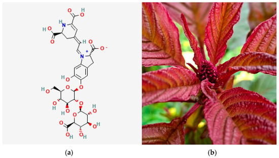
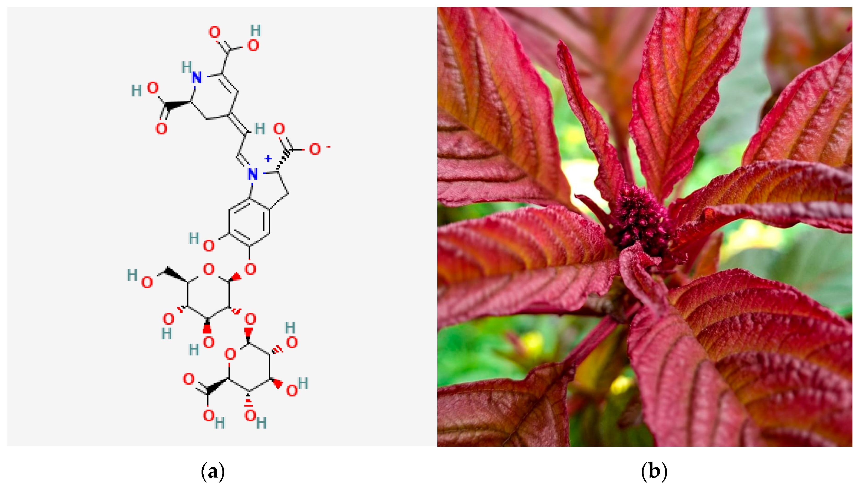
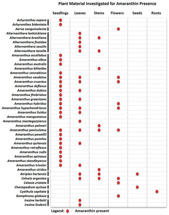
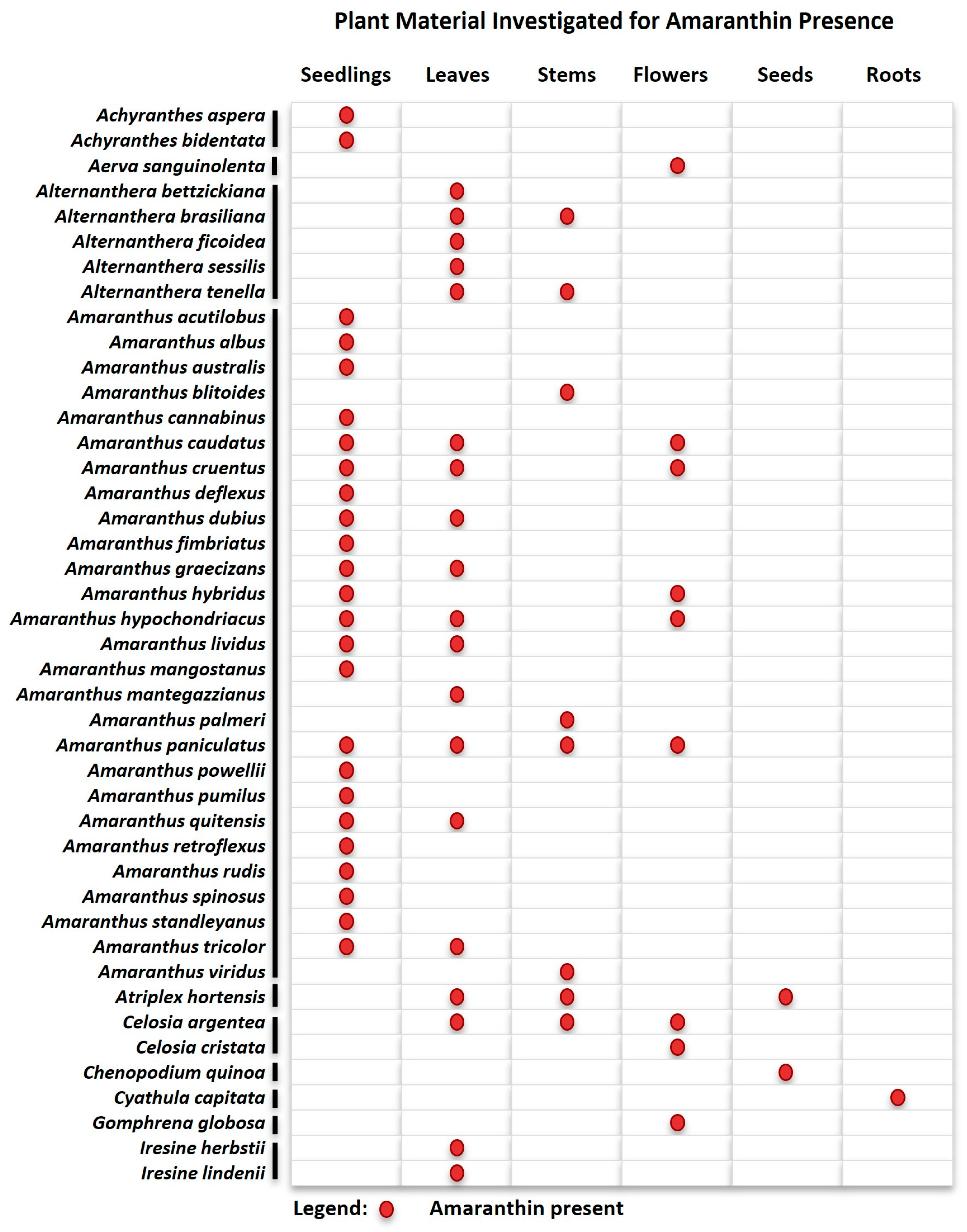
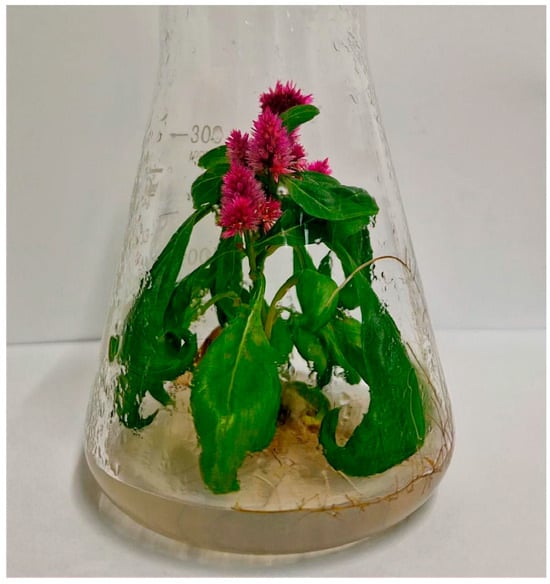
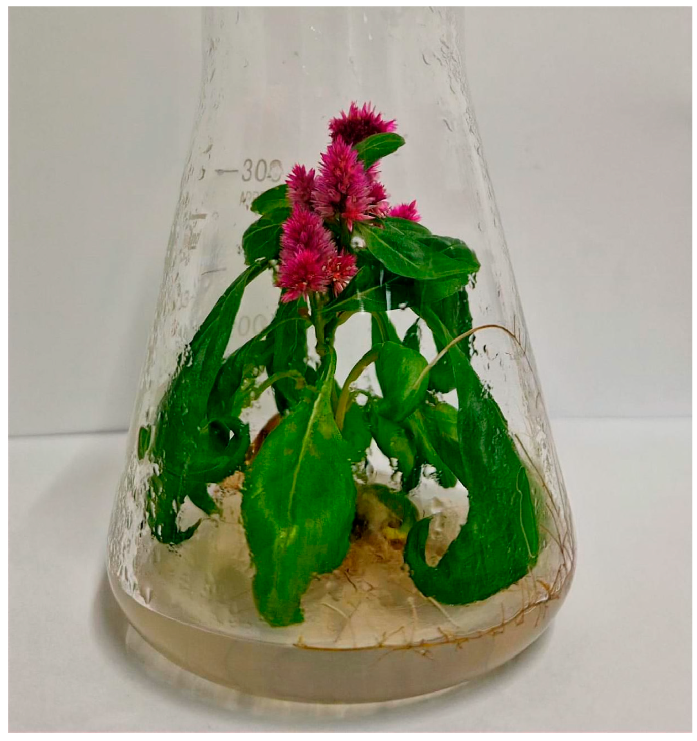
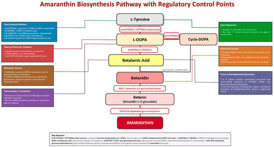
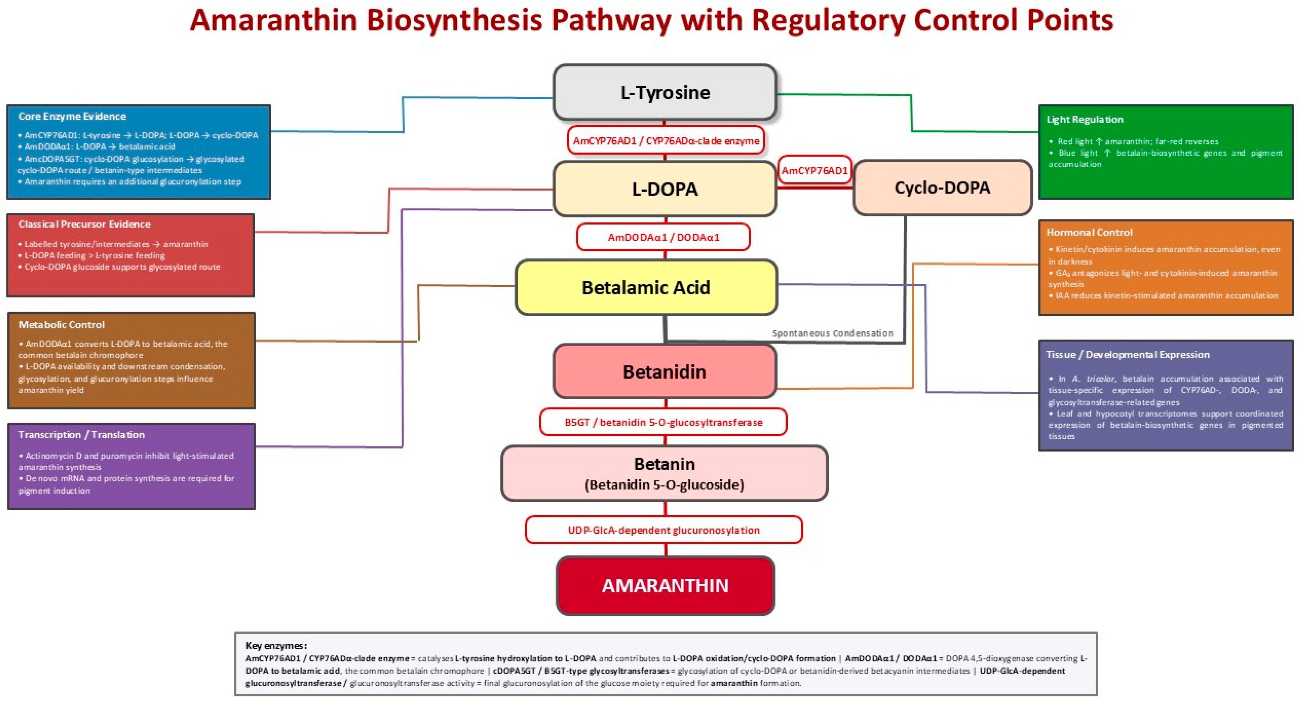

Disclaimer/Publisher’s Note: The statements, opinions and data contained in all publications are solely those of the individual author(s) and contributor(s) and not of MDPI and/or the editor(s). MDPI and/or the editor(s) disclaim responsibility for any injury to people or property resulting from any ideas, methods, instructions or products referred to in the content. |
© 2026 by the author. Licensee MDPI, Basel, Switzerland. This article is an open access article distributed under the terms and conditions of the Creative Commons Attribution (CC BY) license.