


Abstract
The incidence of various inflammatory diseases has remained high. Inula cappa is a kind of Chinese herbal medicine with a wide range of pharmacological uses and application value. It has anti-inflammatory, antibacterial, antioxidant, hepatoprotective and other pharmacological activities. The monomeric compounds that have been confirmed to have anti-inflammatory effects are luteolin, chrysoerilol, artemetin, chlorogenic acid, neochlorogenic acid, cryptchlorogenic acid, isochlorogenic acid A, isochlorogenic acid B, isochlorogenic acid C and 1,3-O-dicaffeoylquinic acid. This article introduces the relationship between Inula cappa and inflammation, the anti-inflammatory components of I. cappa, the modulation of each component on the inflammatory transduction signal pathway, and the TLR2/MyD88/NF-KB anti-inflammatory signaling pathway, providing a theoretical basis for anti-inflammatory research on and clinical medication using Inula cappa.
1. Introduction
Inflammation is a complex defensive response of the body to external damage. It stimulates cells to produce a variety of cytokines and regulates the balance between pro-inflammatory and anti-inflammatory systems. Inflammation plays an important role in protecting the body and defending against infections of foreign pathogens. It is generally believed that inflammation is a beneficial host defense system, but excessive and dysfunctional inflammatory responses can be harmful. In the dysregulated inflammatory response, complex inflammatory mediators, including pro-inflammatory cytokines and cytotoxic cytokines, are associated with the development of different diseases, including pneumonia [1], arthritis, chronic bronchitis, rheumatism [2] and even some neurodegenerative diseases, such as Alzheimer’s disease [3]. In recent years, the incidence of various inflammatory diseases has continued to high and is on the rise, even becoming a global disease [4]. The demand for safe and effective drugs to treat inflammatory diseases is rising.
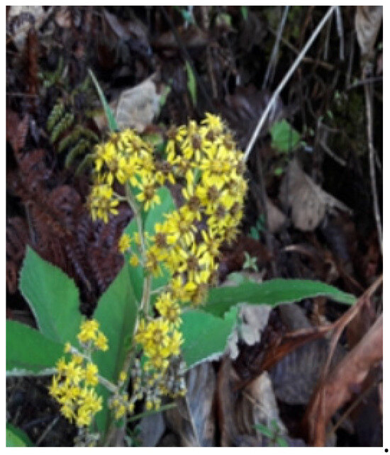
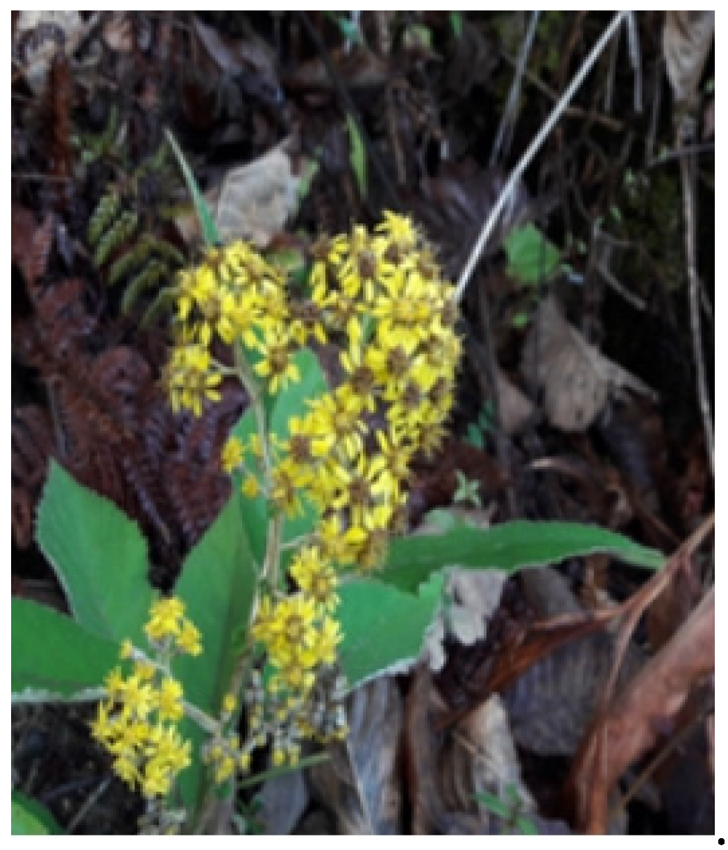
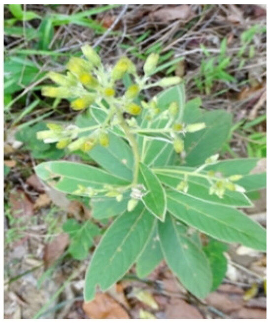
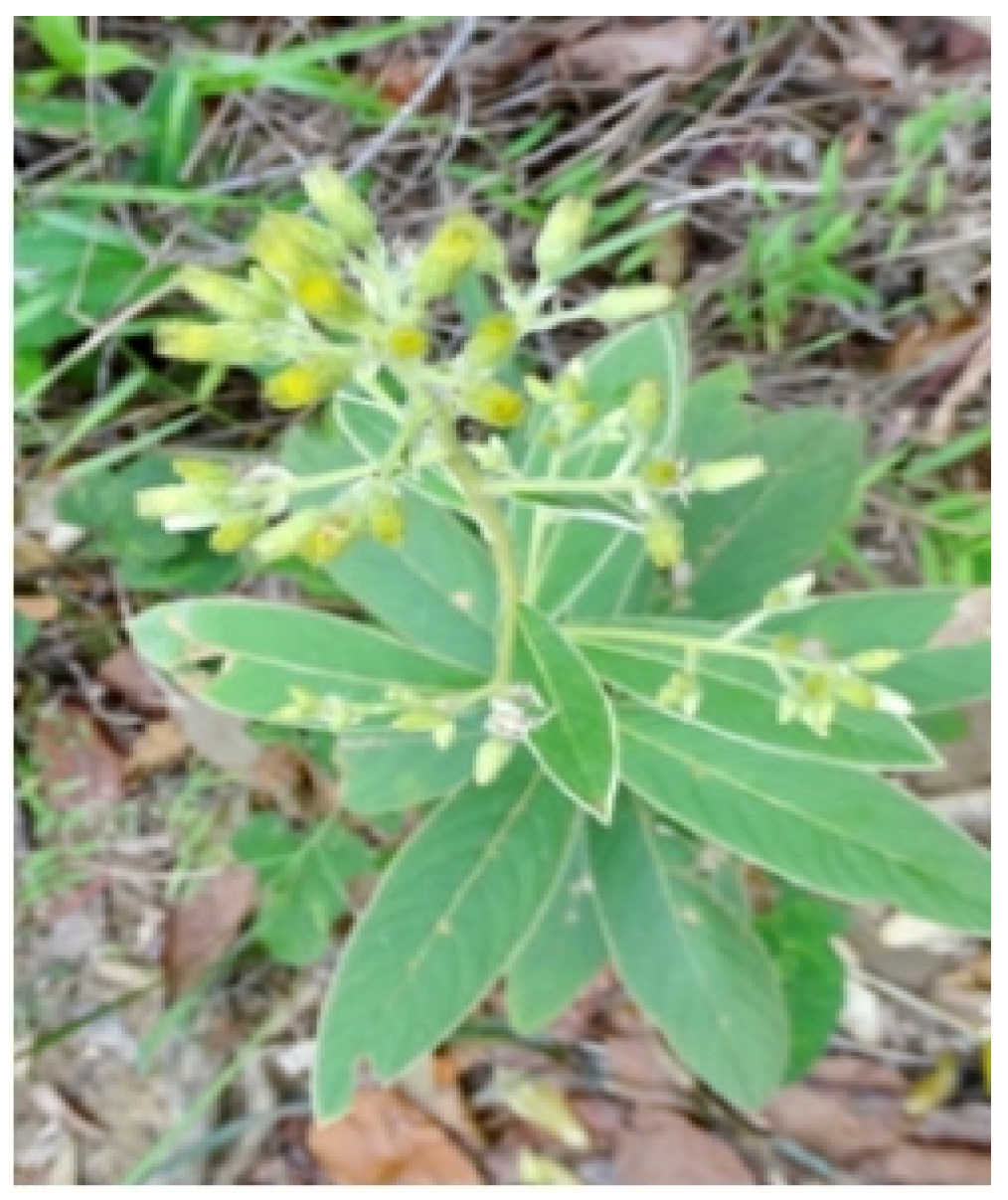
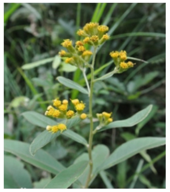
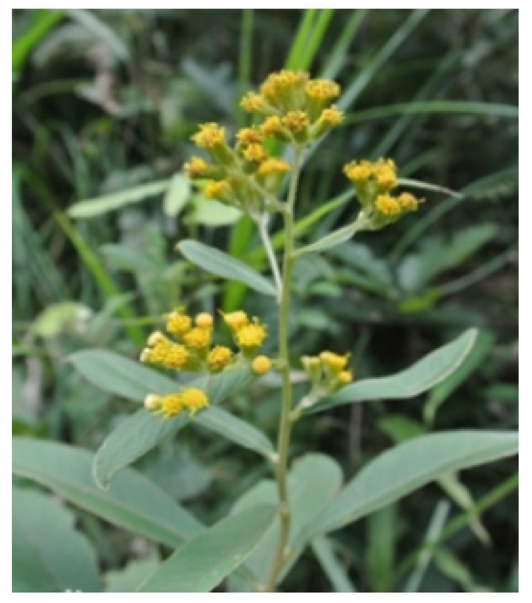
Inula cappa has a long history of medical use in China and has a wide range of pharmacological uses and application values. The plant is a subshrub, with stout rhizomes, erect multi-branched stems, 70–200 cm high, and white or light brown silky or cottony dense hairs. It has florets 4–5.5 mm long, marginal floret ligule short, with 3–4 lobes, or without ligule and with four staminodes; central floret tubular, with triangular-ovoid lobes distally; corolla hairs white, as long as tubular corolla, with more than 20 rough hairs. The flowering period is from June to October, and the fruit period is from August to December. The fruit is long and cylindrical, about 1.8 mm in length, with white, long sericeous hairs. Pictures of Inula cappa are shown in Figure 1, Figure 2 and Figure 3. All pictures are from the following website address: https://baike.baidu.com/item/%E7%BE%8A%E8%80%B3%E8%8F%8A/5009988#reference-2 (accessed on 19 January 2025).
Figure 1.
This is a picture of Inula cappa.
Figure 2.
This is a picture of Inula cappa.
Figure 3.
This is a picture of Inula cappa.
More detailed information about I. cappa is shown in Table 1.
Table 1.
Detailed information about synonyms, growing environment, distribution areas, medicinal parts, medicinal value and folk medication experience of Inula cappa.
Modern studies have shown that I. cappa contains sesquiterpenes, inositols, triterpenes, flavonoids, caffeoylquinic acids and other chemical components, and the plant has a fundamental role in anti-inflammatory, antibacterial, antioxidant, liver protection and other pharmacological effects [7]. In this paper, the chemical constituents and mechanism of the anti-inflammatory effect of Inula cappa were studied and discussed in order to provide a theoretical basis for further research and the development and utilization of the herb.
2. Anti-Inflammatory Effect of Inula cappa
Modern pharmacological studies have shown that the extract of I. cappa has anti-inflammatory factors, which is bound to have a repair effect on inflammatory tissues and organs [11].
Mo Jiajia et al. measured the anti-inflammatory effect of the ethanol extract of the herb by the xylene-induced mouse ear swelling method, mouse abdominal capillary permeability hyperfunction method, acetic acid writhing method and hot plate method. The results showed that the ethanol extract of the plant root could significantly inhibit xylene-induced mouse ear swelling and acetic acid-induced capillary permeability hyperfunction in mice, and had a certain anti-inflammatory effect. The mechanism may be related to the improvement in local blood circulation, reduction in exudation and acceleration of absorption [5]. Based on the in vitro and in vivo inflammation model, Gong Zipeng found that the extract of the herb had anti-inflammatory activity [12]. Zhang Wanyou conducted an experimental study with the alcoholic extract of the herb roots and found that it has anti-inflammatory and analgesic effects, which provides a reference for the clinical use of the drug [13]. Kalola Jyoti et al. used the carrageenan-induced rat paw swelling method and cotton ball granuloma method to determine the anti-inflammatory and immunomodulatory activity of the root extract. The results showed that the methanol extract had the greatest inhibitory effect on rat paw swelling and had a significant inhibitory effect on rat cotton ball granuloma. The root extract of Inula cappa may inhibit chronic inflammation and acute inflammation by inhibiting the enzyme activity of arachidonic acid metabolic enzymes [14]. Huang Jing et al. found that the anti-inflammatory active ingredients in the extract were easily recognized and absorbed by inflammatory cells in vivo [15]. Wang Yunfei et al. found that the aqueous extract of the plant could effectively improve osteoarthritis [16]. Wang et al. found that the extract of the herb could effectively inhibit the release of NO from RAW264.7 cells stimulated by LPS in a certain range [17]. It was confirmed that the active ingredients of the herb could inhibit the secretion of TNF-α in addition to inhibiting the release of NO [18]. Various experimental studies have demonstrated that Inula cappa has certain anti-inflammatory effects.
3. Anti-Inflammatory Components of Inula cappa
So far, according to domestic and international research reports, the chemical constituents isolated and identified from the roots, stems, leaves and skins of Inula cappa mainly include sesquiterpenes, inositols, triterpenoids, flavonoids, caffeoylquinic acids, etc. [19,20,21].
Inula cappa has a strong inhibitory effect on a variety of inflammatory reactions. Sesquiterpenes, inositols, flavonoids and caffeoylquinic acids may be its anti-inflammatory active ingredients [7,22]. Sesquiterpenoids are the characteristic components of Inula plants. It has been found that most of the sesquiterpenoids isolated from Inula plants have obvious anti-inflammatory and anti-tumor effects [23,24]. Inositol is a cyclic polyol with six hydroxyl groups. After epimerization, nine isomers are formed. The most common ones are myoinositol (MI) and D-chiral inositol (DCI). Inositol has been shown to have the potential to treat polycystic ovary syndrome [25], inflammatory conditions [26], and hypoglycemia [27]. Flavonoids are a class of low-molecular natural plant components widely found in plants. Flavonoids are ubiquitous in Inula plants. Flavonoids have antibacterial, anti-inflammatory, antiviral and antioxidant effects [7]. Caffeoylquinic acid compounds are natural phenolic compounds that are synthesized by the condensation of quinic acid with one or more caffeic acids through an esterification reaction. They have antibacterial, anti-inflammatory, antiviral, and free radical scavenging effects, among others [28].
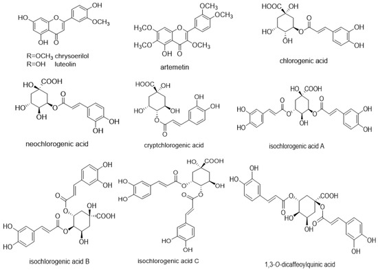
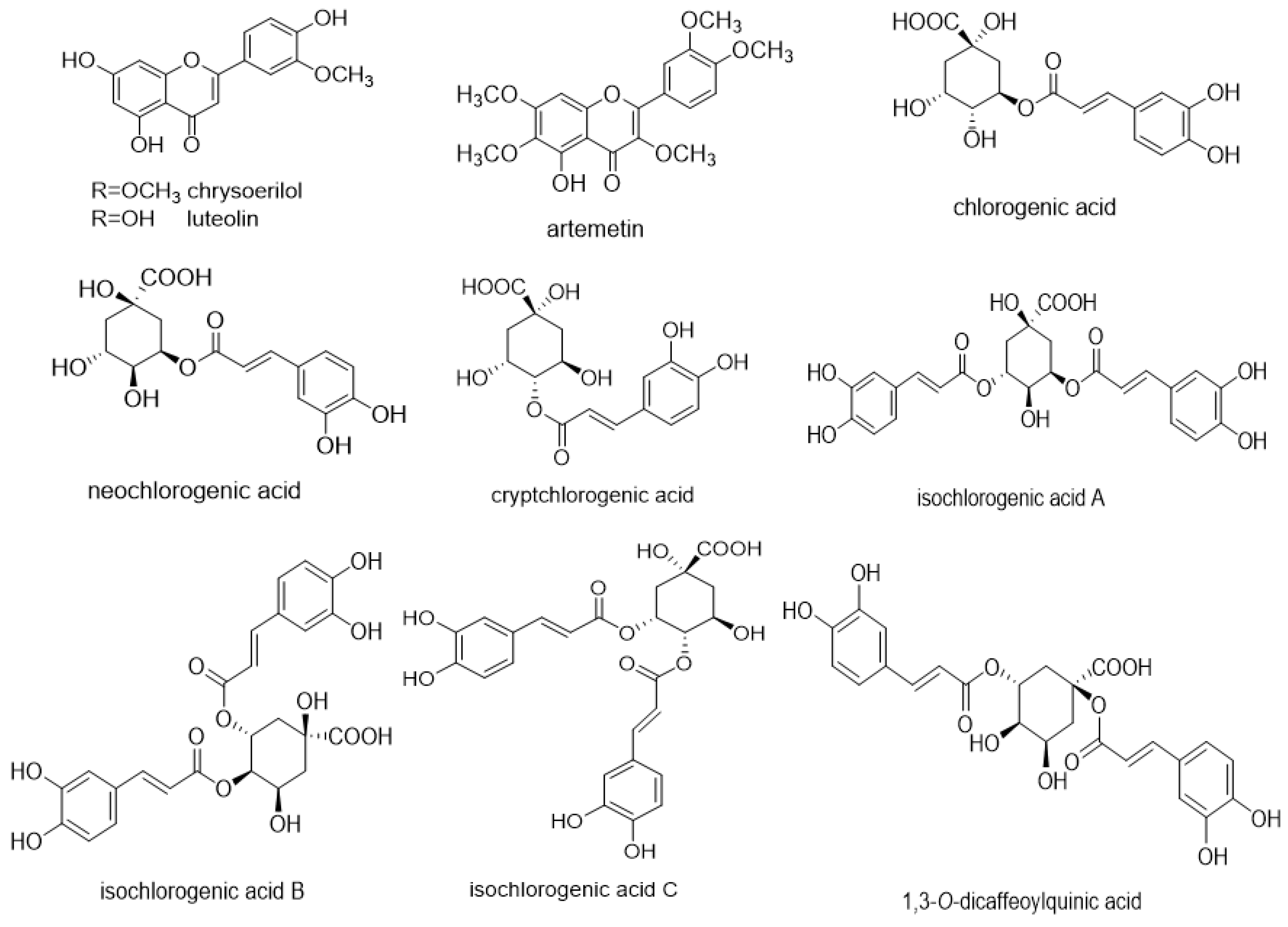
At present, the compounds with anti-inflammatory activity extracted from I. cappa have been reported to be luteolin, artemetin, chrysoerilol, chlorogenic acid, neochlorogenic acid, cryptchlorogenic acid, isochlorogenic acid A, isochlorogenic acid B, isochlorogenic acid C and 1,3-O-dicaffeoylquinic acid [15,18,29,30,31,32,33]. The structural formula of the related monomer compounds is shown in the following Figure 4. In addition, there are few studies on the anti-inflammatory mechanism of these 10 monomer compounds extracted from I. cappa. The modulation of these 10 monomeric compounds extracted from other plants on the inflammatory signal transduction pathway is shown in Table 2. It provides more ideas for the study of the anti-inflammatory mechanism of I. cappa.
Figure 4.
Monomer compounds with anti-inflammatory activity extracted from Inula cappa are luteolin, chrysoerilol, artemetin, chlorogenic acid, neochlorogenic acid, cryptchlorogenic acid, isochlorogenic acid A, isochlorogenic acid B, isochlorogenic acid C and 1,3-O-dicaffeoylquinic acid. The corresponding chemical structure formulas of each monomeric compound are also shown in the figure.
Table 2.
Modulation of the inflammation transduction signal pathways by luteolin, chrysoerilol, artemetin, chlorogenic acid, neochlorogenic acid, cryptchlorogenic acid, isochlorogenic acid A, isochlorogenic acid B, isochlorogenic acid C and 1,3-O-dicaffeoylquinic acid.
4. Related Pathways of Inula cappa’s Anti-Inflammatory Effect
Inula cappa has a strong inhibitory effect on a variety of inflammatory responses. Caffeoylquinic acids, flavonoids, inositols and sesquiterpenes may be its anti-inflammatory active ingredients. These components have been shown to ameliorate inflammation by regulating signaling pathways, such as MAPK, TLR2 and NF-KB, and modulating the secretion of inflammatory factors such as TNF-α, IL-6, IL-1β and NO [61,62].
It has been proven by experiments that the extract of Inula cappa can reduce the inflammatory injury of pneumonia caused by Klebsiella pneumoniae by affecting the MAPK signaling pathway and the NF-KB signaling pathway [12]. In addition, another experimental result showed that the aqueous extract of Inula cappa may improve the inflammation of rats with severe pneumonia by inhibiting the TLR2/MyD88/NF-KB signaling pathway [63]. Therefore, the MAPK, NF-KB and TLR2 pathways are described in this article.
4.1. MAPK Pathway
Mitogen-activated protein kinase (MAPK) is a kind of serine/threonine protein kinase discovered by Sturgill and Ray in 3T3-L1 adipocyte extract in 1986, which is mainly distributed in the cytoplasm [64]. MAPK has a serine/threonine kinase domain, with different nitrogen terminal and carbon terminal regions on both sides. There are different additional domains, including the trans-activation domain (TAD), nuclear localization sequence (NLS) and conserved domain (C34) structural domain in ERK3/4 [65].
The MAPK pathway is a fundamental signal transduction pathway in mammalian cells. It is a key bridge connecting inside and outside the cell, which is stimulated by diverse extracellular signaling molecules (growth factors, neurotransmitters, cytokines, hormones, etc.). After the upstream activator protein binds to a specific receptor, it triggers a three-level enzymatic cascade reaction (MAPKKK-MAPKK-MAPK) [66]. MAPK kinase kinase (MAPKKK) is activated by mitogen-stimulated phosphorylation. On this basis, MAPKKK is phosphorylated to activate MAPK kinase (MAPKK). Finally, MAPKK phosphorylates MAPK and transmits signals to downstream response molecules through continuous activation. Ultimately, activated MAPK transduces extracellular stimuli into cells and their nuclei and causes cell responses, leading to proliferation, migration, differentiation, inflammation, stress, survival or apoptosis, thereby promoting the occurrence and development of diseases [67,68]. The related MAPK pathway kinases are shown in Table 3.
Table 3.
Specific information about MAPK pathway-related kinases, including the full name of the kinase, the subtribe of the kinase and the number of subfamilies.
The MAPK superfamily can be divided into four members: extracellular signal-regulated kinase 1/2 (ERK1/2), c-Jun NH2-terminal kinase (JNK), p38 mitogen-activated protein kinase (p38MAPK) and ERK5. The most studied proteins in the MAPK pathway are ERK1/2, JNK and p38MAPK [69].
4.1.1. ERK Signaling Pathway
ERK (extracellular signal-regulated kinase) pathway is a classical MAPK signal transduction pathway. ERK1/2 kinase is composed of ERK1 kinase and ERK2 kinase. ERK1/2 can be activated by various external signals such as cell growth factors, T hormones, G protein-coupled receptor ligands, osmotic pressure, neurotransmitters and neurotrophic factors [70].
The activation pathway of the ERK pathway is Ras→ Raf→ MEK1/2→ ERK1/2. After being activated by cytokines and other stimuli, Ras binds to Raf, and the activated Raf then moves from the cytoplasm to the cell membrane. MEK is a special bispecific kinase on the cell membrane. MEK binds to activated Raf and is activated by phosphorylation, resulting in ERK1/2 activation. Activated ERK1/2 translocates into the nucleus and activates downstream transcription factors and kinases [71]. The downstream related transcription factors are Ets-1, ATF-2, ElK-1, NF-KB, c-Fos and c-Myc. Cytoplasmic and nuclear kinases include MNKs, MPKAP-2, RSK and MSKs. This pathway plays a significant role in regulating gene transcription, cell growth, proliferation, cell cycle regulation, maintaining cell morphology, immune response, apoptosis and inflammatory response [43,67].
4.1.2. JNK Pathway
In 1990, JNK (c-Jun N-terminal kinase) was first identified as a phosphorylated c-Jun kinase, hence the name c-Jun N-terminal kinase [72]. JNK was isolated from the liver of mice injected with cycloheximide, and there are three subtypes (JNK1, JNK2, JNK3). JNK1 and JNK2 are widely present in the body, and JNK3 is mainly expressed in brain tissue [66]. JNK is a kind of serine/threonine kinase that can be activated by various extracellular stimuli, including growth factors, cytokines and cellular stress, such as ultraviolet radiation, high osmotic pressure, heat shock and ischemia–reperfusion [73].
The activation of the JNK pathway is MEKK1-4, TAK, MLKs, ASKs → MKK4, MKK7 → JNK. In the JNK pathway, MEKK1-4, mixed-ligation kinases (MLKs), transforming growth factor-β-activated kinases (TAKs), apoptosis signal-regulated kinases (ASKs) in MAPKKK, MKK4 and MKK7 in MAPKK are activated. MKK4 and MKK7 are the bispecific kinases of JNK, but the way of activation is different. MKK4 is mainly activated by environmental stress, while MKK7 is mainly activated by cytokines. The activation mode of the JNK signaling pathway is that the activation of MEKK1-4 can phosphorylate MKK4 and MKK7. The activated MKK4 and MKK7 can activate JNK by phosphorylating the ThR and TyR sites of JNK [74]. JNK can activate different transcription factors (AP-1, c-Jun, ATF-2, ElK-1, c-Myc, p53, Bad, MLK2) and some members of the Bcl-2 family [75]. The JNK signaling pathway is involved in regulating a variety of cellular processes, including apoptosis, proliferation, transduction pathways, differentiation, inflammatory response and oxidative stress [69,76].
4.1.3. P38MAPK Pathway
P38 mitogen-activated protein kinase (p38MAPK) was first discovered by Han et al. in 1994. It is one of the signaling pathways in the MAPK family that can be activated by various extracellular stimuli (cytokines, physiological stress, and ultraviolet osmotic pressure changes). It is a material pathway that mediates the survival and death of nerve cells [77,78]. There are four major subtypes of P38MAPK, including p38α, p38β, p38γ, and p38δ. P38α and p38β are widely distributed and can be expressed in almost all tissues and cells. The distribution of p38δ and p38γ is relatively tissue-specific.
The activation of P38MAPK depends on a typical three-level enzymatic cascade. The activation pathways of p38MAPK pathway is as follows: MLKs, ASK1, MEKKs, TAK→ MKK3, MKK4, MKK6→ p38MAPK. Ten MAP3Ks (ASK1, DLK, MEKK3, MEKK4, MLK3, TAK1, TAO1, TAO2, TPL2, ZAK1) contribute to the activation of P38 kinase, although some of them can also trigger other MAPK activation pathways, such as JNK [79]. MAP3K phosphorylates MKK3, MKK4 and MKK6 in MAP2K [79]. MKK3, MKK4 and MKK6 are known upstream-specific protein kinases of p38. Different p38 MAPK subtypes can be activated by different MKKs. MKK3 can activate p38α, p38γ and p38δ, MKK4 only activates p38α, and MKK6 is a common activator of different subtypes. Different subtypes can activate different downstream substrates, including other protein kinases, phospholipases and cytoskeleton-binding proteins. After being activated, p38MAPK enters the nucleus or transfers to other parts and regulates cell growth, cytoskeleton remodeling, apoptosis, inflammatory response and hyperalgesia by controlling the gene expression of various transcription factors [80].
4.2. NF-KB Pathway
Nuclear factor-KB (NF-KB) is a general name for a family of transcription factors. Its function is to coordinate a variety of physiological and pathological processes as a dimer. Members of the NF-KB family include p50, p52, RElA (p65), RElB and cREl. NF-KB can be divided into two groups. One group is p50 and p52, which are produced, respectively, by the cleavage of p105 and p100 precursors. The other group was RElA (p65), RElB and cREl, without precursors. The NF-KB protein refers to the NF-KB1 dimer protein formed by the p65/p50 subunit and the NF-KB2 dimer protein formed by the RElB/p52 subunit.
In the resting state, it binds to the inhibitor of NF-KB (IKB) and exists in the cytoplasm in an inactive form [81]. IKB is a complex composed of IKKα and IKKβ and the basic modulator (NEMO) of NF-KB [82].
The classical NF-KB signaling pathway is induced by a variety of immune mediators. When activated by upstream stimulation signals such as LPS, TNF-α, IL-1 and IL-6, IKB kinase (IKK) is activated and can activate IKBα/β phosphorylation at specific N-terminal serine residues, causing IKB to be phosphorylated and degraded, which in turn enables p65/p50 dimers normally present in the cytoplasm to be transferred to the nucleus and bind to the KB sequence in the promoter region of the relevant target gene sequence to exert its biological effects [82,83].
The bypass pathway mainly refers to the activation of NF-KB containing pl00 or pl05 dimers. In some cells, after being stimulated by extracellular signals, IKKa is phosphorylated and activated under the action of NF-KB-induced kinase (NIK) and further activates p100, resulting in the phosphorylation of p100 and subsequent cleavage by corresponding enzymes to form an active RElB/p52 complex. The RElB/p52 complex enters the nucleus of the cell and binds to the target gene to regulate the expression of the corresponding gene [84,85].
In addition, the mechanism of UV-activated NF-KB is different from the above pathways. Ultraviolet light causes the phosphorylation of the C-terminus of IKBα by activating casein kinase 2 (CK2), which leads to the ubiquitination and degradation of IKBα. The activation of CK2 is not dependent on IKK but through P38 mitogen-activated protein kinase, so the p38-CK2-IKBα pathway is also one of the ways to activate NF-KB [86].
NF-KB protein is a member of the transcription factor family, which is stimulated by chemokines, ECM degradation products, stress-related factors and pro-inflammatory cytokines. More and more studies have shown that NF-KB is a transcription factor involved in the regulation of immune and inflammatory responses. The activation of NF-KB is accompanied by inflammatory responses, such as asthma, rheumatoid arthritis, psoriasis, and enteritis. In addition, NF-KB also plays an important regulatory role in expanding inflammatory responses [87]. Therefore, inhibiting the activation of the NF-KB signaling pathway is of great significance for the prevention and treatment of inflammatory or immune diseases [38].
4.3. Toll-TLR2 Pathway
4.3.1. Introduction of Toll-like Receptors
Toll-like receptors (TLRs) are usually a group of evolutionarily conserved type I transmembrane proteins, which is a family of pattern recognition receptors, mainly involved in cellular immune response and inflammatory response. At present, 13 different TLRs have been found in mammals, of which 11 TLRs play a role in the human body [88]. TLRs exist in more than 20 kinds of cells, among which TLR2 and TLR4 are the most studied members [89].
TLRs are composed of three parts: extracellular, cytoplasmic and transmembrane regions, which consist of an extracellular domain at the N-terminal end of the membrane, a single transmembrane structural domain and an intramembrane C-terminal structural domain [90]. The N-terminal extracellular domain contains a leucine-rich repeat (LRR) and selectively recognizes PAMP, and each TLR can recognize a specific molecular pattern. The transmembrane region has a simple structure and is composed of a cysteine-rich domain that connects the intracellular and extracellular regions of TLRs across the cell membrane. The C-terminal domain, also known as the cytoplasmic domain, contains an evolutionarily conserved Toll/IL-1 receptor (TIR) homology domain, and each TLR can recognize a specific molecular pattern and is responsible for signal transduction [91].
The TLR signaling pathway is divided into MyD88-dependent and MyD88-independent signaling pathways. MyD88 is a cytoplasmic soluble protein that consists of three functional regions: the N-terminal death domain, the intermediate region and the C-terminal homeodomain [92]. The MyD88-dependent signaling pathway mainly mediates the production of cytokines. In addition to TLR3, all of the currently identified TLR family members can activate the MyD88-dependent pathway by binding to agonists, thereby activating downstream signaling molecules, causing cascades and activating related signaling pathways [89].
TLRs recruit adaptor protein myeloid differentiation factor 88 (MyD88) to form a complex with IL-1 receptor-associated kinase (IRAK) family molecules. After the formation of the complex, IRAK4 phosphorylates IRAK1. Phosphorylated IRAK1 binds to tumor necrosis factor receptor-associated factor-6 (TRAF6), and then ubiquitination initiates nuclear factor kappa-B (NF-KB), cAMP response element-binding protein (CREB) and activating protein-1 (AP-1), thereby triggering an inflammatory response. The MyD88-independent pathway is also known as the TRIF pathway. TLRs recruit and activate TIR domain-containing adaptor-inducing interferon β (TRIF) and utilize TRAF3 and TRAF6 to activate interferon-regulatory factor (IRF) 3 and 7 and NF-KB. This leads to the expression of type I interferons and the production of inflammatory cytokines [93,94].
The specific process of the signaling pathway is as follows [95].
MyD88 signaling pathway: TLRs recruit the adaptor protein MyD88 to its TIR domain. MyD88 contains two domains: one binds to the TIR domain, and the other is called the death domain to recruit IRAK 1,2,4 proteins. IRAK protein recruits TRAF6 to the receptor complex, and IRAK4 phosphorylates IRAK1 after the formation of the complex. Then, phosphorylated IRAK1 and TRAF6 are dissociated from the complex and form a complex with TAK1 (TGF-β-activated kinase 1), TAB1 and TAB2 on the plasma membrane, thereby phosphorylating all three. Subsequently, IRAK1 is degraded on the plasma membrane, and the remaining complex (composed of TRAF6, TAK1, TAB1 and TAB2) is translocated to the cytosol, resulting in TRAF6 ubiquitination and TAK1 activation. Activated TAK1 acts on the IKB kinase (IKK) complex (composed of IKKα, IKKβ and IKKγ) to activate IKK, thereby phosphorylating IKB and leading to IKB ubiquitination and proteasome degradation, and then releases NF-KB to the nucleus, inducing the transcription of a variety of related proteins, including various pro-inflammatory cytokines, such as IL-6, IL-12p40 and TNF family-related molecules. AK1 can also activate the MKK3/6-p38 signaling cascade and the MKK4/7-Jun N-terminal kinase (JNK) pathway, leading to the production of CREB and AP-1, respectively.
TRIF signaling pathway: TLRs recruit TRIF to the TIR domain, and TLR4 can also recruit TRAM (TRIF-related adaptor molecule) to TIR. Subsequently, IKKε, TBK1 and TRAF3 are recruited into TRIF/TIR or TRAM/TRIF/TIR complexes, and TBK1 phosphorylated IRF3 and IRF7, which combined with p300 and CBP (CREB binding protein) to activate interferon-induced gene expression. TRIF can also bind to TRAF6 and produce pro-inflammatory cytokines by activating NF-KB.
4.3.2. TLR2 Pathway
TLR2 is one of the important members of the Toll-like receptor family, which is involved in the immune response and inflammatory response of cells, accompanied by the occurrence and development of various diseases. TLR2 usually forms heterodimers with TLR1 or TLR6. The TLR2-TLR1 heterodimer can specifically recognize triacylglycerols of Gram-negative bacteria and mycoplasma, while the TLR2-TLR6 heterodimer can specifically recognize diacylglycerols of Gram-positive bacteria and mycoplasma. In addition, TLR2 can work together with other co-receptors on the cell surface. These co-receptors include CD36, which mediates the sensing of some agonists of TLR2 together with TLR2-TLR6 heterodimers [96].
The TLR2 pathway includes the classical TLR2-MyD88-NF-KB signaling pathway, the non-classical TLR2-PI3K/AKt-NF-KB and other pathways.
TLR2 signaling is mainly mediated by the MyD88-dependent pathway. After TLR2 binds to the adaptor protein molecule MyD88, it activates the downstream target gene protein molecules IRAK-4, IRAK-1, TRAF6, IKK-β, TGF-β, TAK1, IKK-γ, NF-KB, etc., forming the TLR2-MyD88-NF-KB pathway. After the interaction of TLR2 and its related adaptor proteins, the IRAK complex is activated to recruit TRAF6. Activated TRAF6 triggers the activity of the transforming growth factor β-activated kinase 1 (TAK1)/TAK1-binding protein (TAB) complex and stimulates the activation of MAPK and nuclear factor KB inhibitor kinase complexes (IKK1,2 and IKK-γ), also known as NF-KB Essential Modulator (NEMO). The MAPK family involved includes JNK and p38MAPK. The IKK complex promotes the nuclear translocation of NF-KB. This leads to the production of pro-inflammatory cytokines by activator protein-1 (AP-1) and NF-KB, thereby controlling inflammation and regulating cell survival and proliferation [97].
The specific process of non-classical TLR2-PI3K/AKt-NF-KB is as follows. After being activated by endogenous DAMPs released by other cells, TLR2 can transmit signals to phosphatidylinositol-3-kinase (PI3K) and catalyze the phosphorylation of phosphatidylinositol diphosphate (PIP2) to phosphatidylinositol triphosphate (PIP3). PIP3 binds to protein kinase D1 (PKD1) and protein kinase B (PKB/AKt) containing the PH domain to promote PKD1 to phosphorylate 308 serine of AKt. Like TAK-1, activated AKt can also phosphorylate a variety of proteins, such as the IKK complex, thereby activating the NF-KB signaling pathway and the transcription of a variety of target genes [98].
5. Conclusions and Discussion
Various experiments have shown that Inula cappa has a strong inhibitory effect on a variety of inflammatory reactions. Caffeoylquinic acids, flavonoids, inositols and sesquiterpenes in the plant may contain anti-inflammatory active ingredients. The specific anti-inflammatory components are known to be luteolin, artemetin, chrysoerilol, chlorogenic acid, neochlorogenic acid, cryptchlorogenic acid, isochlorogenic acid A, isochlorogenic acid B, isochlorogenic acid C and 1,3-O-dicaffeoylquinic acid. Inula cappa can alleviate inflammation by harmonizing MAPK, TLR2 and NF-KB signaling pathways and regulating the secretion of inflammatory factors such as TNF-α, IL-6, IL-1β and NO.
At present, there are few studies revealing the anti-inflammatory active components of Inula cappa and how this herb affects the potential targets of inflammation. The anti-inflammatory effects and mechanisms of other components of this herb are not deep enough.
More than 100 monomeric compounds have been extracted from I. cappa [11]. The anti-inflammatory substances in I. cappa are not only the 10 species listed in this paper; more in-depth research needs to be conducted on the other extracted components to prove whether they have anti-inflammatory effects. In addition, there are more studies on the modulation targets of the 10 monomer compounds extracted from other herbal medicine plants, providing a certain idea for research on the anti-inflammatory targets of I. cappa. However, to determine whether the anti-inflammatory targets of these 10 monomer compounds extracted from I. cappa and extracted from other plants are consistent, further experimental studies are needed.
Therefore, in the future, it is significant to further explore the anti-inflammatory active components of Inula cappa, the mechanisms and the specific targets of action to provide ideas for subsequent research and lay a foundation for its development and utilization.
Author Contributions
Conceptualization, N.W. and S.W. (Siqi Wang); writing—original draft, N.W. and S.W. (Siqi Wang); writing—review and editing, N.W., Y.Z. and S.W. (Siming Wang); supervision, S.W. (Siming Wang). All authors have read and agreed to the published version of the manuscript.
Funding
This study was supported by the Natural Science Foundation of Hebei Province, China (H2024201050); the Science Research Project of Hebei Education Department, Hebei Province, China (No. QN2024041); and the Key Talent Project of Hebei Province, China (Overseas Study Returning Platform; No. B2024029).
Institutional Review Board Statement
Not applicable.
Informed Consent Statement
Not applicable.
Data Availability Statement
No new data were created or analyzed in this study. Data sharing is not applicable to this article.
Conflicts of Interest
The authors declare no conflicts of interest.
References
- Mizgerd, J.P. Inflammation and pneumonia: Why are some more susceptible than others? Clin. Chest Med. 2018, 39, 669–676. [Google Scholar] [CrossRef] [Scilit] [PubMed]
- Joosten, L.A.B.; Abdollahi-Roodsaz, S.; Dinarello, C.A.; O’Neill, L.; Netea, M.G. Toll-like receptors and chronic inflammation in rheumatic diseases: New developments. Nat. Rev. Rheumatol. 2016, 12, 344–357. [Google Scholar] [CrossRef] [Scilit]
- Glass, C.K.; Saijo, K.; Winner, B.; Marchetto, M.C.; Gage, F.H. Mechanisms underlying inflammation in neurodegeneration. Cell 2010, 140, 918–934. [Google Scholar] [CrossRef] [Scilit] [PubMed]
- Ng, S.C.; Shi, H.Y.; Hamidi, N.; Underwood, F.E.; Tang, W.; Benchimol, E.I.; Panaccione, R.; Ghosh, S.; Wu, J.C.Y.; Chan, F.K.L.; et al. Worldwide incidence and prevalence of inflammatory bowel disease in the 21st century: A systematic review of population-based studies. Lancet 2017, 390, 2769–2778. [Google Scholar] [CrossRef] [Scilit] [PubMed]
- Mo, J.J.; Xu, M.; Yang, D.D. Experimental study on anti-inflammatory and analgesic effects of ethanol extracts of dong-medicine Inula cappa. Chin. J. Exp. Tradit. Med. Formulae 2012, 18, 258–260. [Google Scholar]
- Hu, L.; He, Z.S. Progress of constituents and pharmacological activities of Inula cappa. Chin. J. Mod. Appl. Pharm. 2012, 29, 889–894. [Google Scholar]
- Guo, J.T.; Li, H.Y.; Cheng, C.; Shi, J.X.; Ruan, H.N.; Li, J.; Liu, C.M. Isochlorogenic acid A ameliorated lead-induced anxiety-like behaviors in mice by inhibiting ferroptosis-mediated neuroinflammation via the BDNF/Nrf2/GPX4 pathways. Food Chem. Toxicol. 2024, 190, 114814. [Google Scholar] [CrossRef] [Scilit] [PubMed]
- Liu, Y.; Tang, L.; Mou, J.L.; Lu, Y. Study on the antiviral and immunologic effects of Juhuangshangqing buccal lozenge. Chin. J. Hosp. Pharm. 2013, 33, 1673–1677. [Google Scholar]
- Pu, H.H.; Liang, Z.X.; Jin, H.; Yang, H.J. Quantitative evaluation and verification of the prescription of the Pearl Dropping Pill treating oral ulcers in rats. Pharmacol. Clin. Chin. Mater. Med. 2012, 28, 95–98. [Google Scholar]
- Tao, L.; Wang, Y.L.; Wang, A.M.; He, X. Determination of bergenin in Qishen Houbitong Granules by HPLC. Chin. Tradit. Herb. Drugs 2005, 36, 218–219. [Google Scholar]
- Huang, S.J.; Zhuang, Z.B.; Peng, B.L.; King, J.J.; He, C.M. Research progress of the medicinal plant Duhaldea cappa. For. Environ. Sci. 2023, 39, 122–126. [Google Scholar]
- Gong, Z.P.; Li, M.; Xiong, D.F.F.; Wu, L.L. Evaluation of anti-inflammatory activity of Inula cappa extract based on inflammation model in vitro and in vivo. Nat. Prod. Res. Dev. 2017, 29, 2050–2055. [Google Scholar]
- Zhang, W.Y. Experimental study on anti-inflammatory and analgesic effects of ethanol extract of Inula cappa. Mod. Anim. Husb. 2018, 7. [Google Scholar] [CrossRef]
- Kalola, J.; Shah, R.; Patel, A.; Lahiri, S.K.; Shah, M.B. Anti-inflammatory and immunomodulatory activities of Inula cappa roots (Compositae). J. Complement. Integr. Med. 2017, 14, 20160083. [Google Scholar] [CrossRef] [Scilit] [PubMed]
- Huang, J.; Chen, R.; Zhou, J.; Zhang, Q.; Xue, C.; Li, Y.; Zheng, L.; Huang, Y.; Wang, Q.; Chen, Y.; et al. Comparative pharmacokinetic study of the five anti-inflammatory active ingredients of Inula cappa in a normal and an LPS-induced inflammatory cell model. Front. Pharmacol. 2022, 13, 981112. [Google Scholar] [CrossRef] [Scilit] [PubMed]
- Wang, Y.F.; Qian, L.G.; Jiao, J.B.; Chen, T.P. Protective effect of aqueous extract of Inula cappa on ACLT-induced osteoarthritis in rats by alleviating immune disorders and cartilage degeneration. J. Med. Mol. Biol. 2021, 18, 211–216. [Google Scholar]
- Wang, A.M.; Li, M.; Sun, J.; Xiong, D. Study on antibacterial and anti-inflammatory activity of Inula cappa extract. Lishizhen Med. Mater. Med. Res. 2018, 29, 1580–1584. [Google Scholar]
- Zhou, J.; Zhang, Q.; Chen, Y.; Xue, C. Establishment of PK-PD model in anti-inflammatory active components in Inula cappa extract based on lipopolysaccharide-induced in vitro inflammation model. China J. Chin. Mater. Med. 2022, 47, 6308–6319. [Google Scholar]
- Wang, Y.L.; Li, Y.J.; Wang, A.M.; He, X.; Liao, S.G.; Lan, Y.Y. Two new phenolic glycosides from Inula cappa. J. Asian Nat. Prod. Res. 2010, 12, 765–769. [Google Scholar] [CrossRef] [Scilit] [PubMed]
- Guan, H.Y.; Lan, Y.Y.; Liao, S.G.; Liu, J.H. Caffeoylquinic acid derivatives from Inula cappa. Nat. Prod. Res. Dev. 2014, 26, 1948–1952. [Google Scholar]
- Zhou, W.; Wang, X.; Fu, S.H.; Sun, X. Chemical constituents of Inula cappa. Chin. Pharm. J. 2017, 52, 25–30. [Google Scholar]
- Nikomtat, J.; Meepowpan, P.; Tragoolpua, Y. Inhibition of Inula cappa (Ham. ex D. Don) DC. extracts on herpes simplex virus infection in vitro. Afr. J. Microbiol. Res. 2011, 5, 4049–4058. [Google Scholar]
- Fan, L.L.; Cheng, J.N.; Zhang, T.; Guo, J.T. Chemical constituents in xuanfuhuan (Inula L.) and their pharmacological activities. Guid. J. Tradit. Chin. Med. Pharm. 2017, 23, 40–43. [Google Scholar]
- Guo, Q.L.; Yang, J.S. Sesquiterpenes in Inula L. plants and their pharmacological activities. Nat. Prod. Res. Dev. 2005, 804–809. [Google Scholar]
- Greff, D.; Juhász, A.E.; Váncsa, S.; Váradi, A.; Sipos, Z.; Szinte, J.; Park, S.; Hegyi, P.; Nyirády, P.; Ács, N.; et al. Inositol is an effective and safe treatment in polycystic ovary syndrome: A systematic review and meta-analysis of randomized controlled trials. Reprod. Biol. Endocrinol. 2023, 21, 10. [Google Scholar] [CrossRef] [Scilit] [PubMed]
- Baldassarre, M.P.A.; Di Tomo, P.; Centorame, G.; Pandolfi, A.; Di Pietro, N.; Consoli, A.; Formoso, G. Myoinositol reduces inflammation and oxidative stress in human endothelial cells exposed in vivo to chronic hyperglycemia. Nutrients 2021, 13, 2210. [Google Scholar] [CrossRef] [Scilit] [PubMed]
- DiNicolantonio, J.J.; O’Keefe, J.H. Myo-inositol for insulin resistance, metabolic syndrome, polycystic ovary syndrome and gestational diabetes. Open Heart 2022, 9, e001989. [Google Scholar] [CrossRef] [Scilit] [PubMed]
- Wu, Q.Z. Advances in research of caffeoylquinic acid compounds. Chin. Wild Plant Resour. 2020, 39, 48–53+60. [Google Scholar]
- Zhang, Z.Y.; Hu, H.J.; Xiong, D.F.F.; Li, Y.T. Study on quality control of Miao medicine Inula cappa based on anti-inflammatory active ingredients. Chin. Tradit. Herb. Drugs 2019, 50, 5571–5576. [Google Scholar]
- Bao, H.S.; Hou, J.Y.; Hu, H.J.; Li, Y.T. Determination of plasma protein binding rates of nine compounds of Inula cappa extraction based on method of equilibrium dialysis. China J. Chin. Mater. Med. 2019, 44, 1475–1484. [Google Scholar]
- Zhang, W.M.; Zhang, S.L.; Zhong, G. The study survey of luteolin. China Food Addit. 2005, 2, 11–16. [Google Scholar]
- Yang, Y.; Zhou, X.; Xiao, M.; Hong, Z.; Gong, Q.; Jiang, L.; Zhou, J. Discovery of chrysoeriol, a PI3K-AKT-mTOR pathway inhibitor with potent antitumor activity against human multiple myeloma cells in vitro. J. Huazhong Univ. Sci. Technol. 2010, 30, 734–740. [Google Scholar] [CrossRef] [Scilit]
- Yang, X.H.; Tian, L.; Cao, J.G.; Luo, Q. Extraction and separation of artemetin and in vitro anti-human cervical cancer activity. Cent. South Pharm. 2010, 8, 568–571. [Google Scholar]
- Aziz, N.; Kim, M.Y.; Cho, J.Y. Anti-inflammatory effects of luteolin: A review of in vitro, in vivo, and in silico studies. J. Ethnopharmacol. 2018, 225, 342–358. [Google Scholar] [CrossRef] [Scilit] [PubMed]
- Ntalouka, F.; Tsirivakou, A. Luteolin: A promising natural agent in management of pain in chronic conditions. Front. Pain Res. 2023, 4, 1114428. [Google Scholar] [CrossRef] [Scilit] [PubMed]
- Huang, L.; Kim, M.Y.; Cho, J.Y. Immunopharmacological activities of luteolin in chronic diseases. Int. J. Mol. Sci. 2023, 24, 2136. [Google Scholar] [CrossRef] [Scilit]
- Aboulaghras, S.; Sahib, N.; Bakrim, S.; Benali, T.; Charfi, S.; Guaouguaou, F.E.; Omari, N.E.; Gallo, M.; Montesano, D.; Zengin, G.; et al. Health benefits and pharmacological aspects of chrysoeriol. Pharmaceuticals 2022, 15, 973. [Google Scholar] [CrossRef] [Scilit] [PubMed]
- Wu, J.Y.; Chen, Y.J.; Bai, L.; Liu, Y.X.; Fu, X.Q.; Zhu, P.L.; Li, J.K.; Chou, J.Y.; Yin, C.L.; Wang, Y.P.; et al. Chrysoeriol ameliorates TPA-induced acute skin inflammation in mice and inhibits NF-κB and STAT3 pathways. Phytomedicine 2020, 68, 153173. [Google Scholar] [CrossRef] [Scilit] [PubMed]
- Wee, H.N.; Neo, S.Y.; Singh, D.; Yew, H.C.; Qiu, Z.Y.; Tsai, X.C.; How, S.Y.; Yip, K.C.; Tan, C.H.; Koh, H.L. Effects of Vitex trifolia L. leaf extracts and phytoconstituents on cytokine production in human U937 macrophages. BMC Complement. Med. Ther. 2020, 20, 91. [Google Scholar] [CrossRef] [Scilit] [PubMed]
- Nguyen, V.; Taine, E.G.; Meng, D.; Cui, T.; Tan, W. Chlorogenic Acid: A systematic review on the biological functions, mechanistic actions, and therapeutic potentials. Nutrients 2024, 16, 924. [Google Scholar] [CrossRef] [Scilit]
- Bagdas, D.; Gul, Z.; Meade, J.A.; Cam, B.; Cinkilic, N.; Gurun, M.S. Pharmacologic overview of chlorogenic acid and its metabolites in chronic pain and Inflammation. Curr. Neuropharmacol. 2020, 18, 216–228. [Google Scholar] [CrossRef] [Scilit] [PubMed]
- Lv, B.; Guo, J.; Du, Y.; Chen, Y.; Zhao, X.; Yu, B.; Liu, J.; Cui, T.; Mao, H.; Wang, X.; et al. Chlorogenic acid reduces inflammation by inhibiting the elevated expression of KAT2A to ameliorate lipopolysaccharide-induced acute lung injury. Br. J. Pharmacol. 2023, 180, 2156–2171. [Google Scholar] [CrossRef] [Scilit]
- Xu, X.; Chang, J.; Wang, P.; Yin, Q.; Liu, C.; Li, M.; Song, A.; Zhu, Q.; Lu, F. Effect of chlorogenic acid on alleviating inflammation and apoptosis of IPEC-J2 cells induced by deoxyniyalenol. Ecotoxicol. Environ. Saf. 2020, 205, 111376. [Google Scholar] [CrossRef] [Scilit] [PubMed]
- Cheng, S.C.; Liou, C.J.; Wu, S.J.; Lin, C.F.; Huang, T.H.; Huang, W.C. Neochlorogenic acid ameliorates allergic airway inflammation by suppressing type 2 immunity and upregulating HO-1 expression. Int. Immunopharmacol. 2025, 146, 113867. [Google Scholar] [CrossRef] [Scilit]
- Kim, M.; Choi, S.Y.; Lee, P.; Hur, J. Neochlorogenic acid inhibits lipopolysaccharide-induced activation and pro-inflammatory responses in BV2 microglial cells. Neurochem. Res. 2015, 40, 1792–1798. [Google Scholar] [CrossRef] [Scilit] [PubMed]
- Park, S.Y.; Jin, M.L.; Yi, E.H.; Kim, Y.; Park, G. Neochlorogenic acid inhibits against LPS-activated inflammatory responses through up-regulation of Nrf2/HO-1 and involving AMPK pathway. Environ. Toxicol. Pharmacol. 2018, 62, 1–10. [Google Scholar] [CrossRef] [Scilit] [PubMed]
- Ma, X.; Okyere, S.K.; Hu, L.; Wen, J.; Ren, Z.; Deng, J.; Hu, Y. Anti-inflammatory activity and mechanism of cryptochlorogenic acid from ageratina adenophora. Nutrients 2022, 14, 439. [Google Scholar] [CrossRef] [Scilit] [PubMed]
- Zhao, X.L.; Yu, L.; Zhang, S.D.; Ping, K.; Ni, H.Y.; Qin, X.Y.; Zhao, C.J.; Wang, W.; Efferth, T.; Fu, Y.J. Cryptochlorogenic acid attenuates LPS-induced inflammatory response and oxidative stress via upregulation of the Nrf2/HO-1 signaling pathway in RAW 264.7 macrophages. Int. Immunopharmacol. 2020, 83, 106436. [Google Scholar] [CrossRef] [Scilit] [PubMed]
- Yang, Y.J.; Lu, L.J.; Wang, J.J.; Ma, S.Y.; Xu, B.L.; Lin, R.; Chen, Q.S.; Ma, Z.G.; Mo, Y.L.; Wang, D.T. Tubson-2 decoction ameliorates rheumatoid arthritis complicated with osteoporosis in CIA rats involving isochlorogenic acid A regulating IL-17/MAPK pathway. Phytomedicine 2023, 116, 154875. [Google Scholar] [CrossRef] [Scilit] [PubMed]
- Tang, S.; Zhong, W.; Li, T.; Li, Y.; Song, G. Isochlorogenic acid A alleviates dextran sulfate sodium-induced ulcerative colitis in mice through STAT3/NF-kB pathway. Int. Immunopharmacol. 2023, 118, 109989. [Google Scholar] [CrossRef] [Scilit]
- Shi, J.X.; Cheng, C.; Ruan, H.N.; Li, J.; Liu, C.M. Isochlorogenic acid B alleviates lead-induced anxiety, depression and neuroinflammation in mice by the BDNF pathway. Neurotoxicology 2023, 98, 1–8. [Google Scholar] [CrossRef] [Scilit]
- Lv, X.; Xie, Z.; Wang, H.; Lu, G.; Li, M.; Chen, D.; Lin, T.; Jiang, C. In vivo and in vitro anti-inflammation of Rhapontici Radix extract on mastitis via TMEM59 and GPR161. J. Ethnopharmacol. 2024, 333, 118462. [Google Scholar] [CrossRef] [Scilit] [PubMed]
- Zheng, L.; Lin, G.; Li, R.; Gan, H.; Huang, X.; Yao, N.; Cai, D.; Zhao, Z.; Hu, Z.; Li, M.; et al. Isochlorogenic acid C alleviates high-fat diet-induced hyperlipemia by promoting cholesterol reverse transport. Front. Pharmacol. 2022, 13, 881078. [Google Scholar] [CrossRef] [Scilit] [PubMed]
- Cao, Z.; Ding, Y.; Cao, L.; Ding, G.; Wang, Z.; Xiao, W. Isochlorogenic acid C prevents enterovirus 71 infection via modulating redox homeostasis of glutathione. Sci. Rep. 2017, 7, 16278. [Google Scholar] [CrossRef] [Scilit]
- Jang, G.; Lee, S.; Hong, J.; Park, B.; Kim, D.; Kim, C. Anti-inflammatory effect of 4,5-dicaffeoylquinic acid on RAW264.7 Cells and a rat model of inflammation. Nutrients 2021, 13, 3537. [Google Scholar] [CrossRef] [Scilit] [PubMed]
- Jang, G.; Lee, S.A.; Hong, J.H.; Park, B.R.; Kim, D.K.; Kim, C.S. Chondroprotective effects of 4,5-Dicaffeoylquinic acid in osteoarthritis through NF-κB signaling inhibition. Antioxidants 2022, 11, 487. [Google Scholar] [CrossRef] [Scilit] [PubMed]
- Kim, D.B.; Unenkhuu, B.; Kim, G.J.; Kim, S.W.; Kim, H.S. Cynarin attenuates LPS-induced endothelial inflammation via upregulation of the negative regulator MKP-3. Anim. Cells Syst. 2022, 26, 119–128. [Google Scholar] [CrossRef] [Scilit] [PubMed]
- Zhang, B.; Yu, J.; Bao, L.; Feng, D.; Qin, Y.; Fan, D.; Hong, X.; Chen, Y. Cynarin inhibits microglia-induced pyroptosis and neuroinflammation via Nrf2/ROS/NLRP3 axis after spinal cord injury. Inflamm. Res. 2024, 73, 1981–1994. [Google Scholar] [CrossRef] [Scilit] [PubMed]
- Song, C.; Wang, K.; Qian, B.; Lu, J.; Qiao, M.; Qiu, Y.; Wang, B.; Yu, Y. Nrf-2/ROS/NF-κB pathway is modulated by cynarin in human mesenchymal stem cells in vitro from ankylosing spondylitis. Clin. Transl. Sci. 2024, 17, e13748. [Google Scholar] [CrossRef] [Scilit] [PubMed]
- Chen, S.; Tang, S.; Zhang, C.; Li, Y. Cynarin ameliorates dextran sulfate sodium-induced acute colitis in mice through the STAT3/NF-κB pathway. Immunopharmacol. Immunotoxicol. 2024, 46, 107–116. [Google Scholar] [CrossRef] [PubMed]
- He, J.J.; Yan, C.L.; An, F.Y.; Li, S.H. Research progress of the regulation mechanism of inflammatory factors and signaling pathways in osteoarthritis of the knee. Chin. J. Clin. Pharmacol. 2019, 35, 1308–1311. [Google Scholar]
- Zhou, H.Y.; Zhang, X.; Wang, M.; Ge, B.J. Anti-inflammatory effect of total flavonoids of Astragalus and its regulation on MAPKs signaling pathway in vitro. Chin. J. Vet. Sci. 2020, 40, 2392–2397. [Google Scholar]
- Aizaizi, A.; Zhu, W.C.; Patihan, K. Effect and mechanism of aqueous Inula cappa aqueous extract on serum inflammatory factors in rats with severe pneumonia. J. Clin. Exp. Med. 2018, 17, 1708–1711. [Google Scholar]
- Sturgill, T.W.; Ray, L.B. Muscle proteins related to microtubule associated protein-2 are substrates for an insulin-stimulatable kinase. Biochem. Biophys. Res. Commun. 1986, 134, 565–571. [Google Scholar] [CrossRef] [Scilit] [PubMed]
- Cargnello, M.; Roux, P.P. Activation and function of the MAPKs and their substrates, the MAPK-activated protein kinases. Microbiol. Mol. Biol. Rev. 2011, 75, 50–83. [Google Scholar] [CrossRef] [Scilit] [PubMed]
- Wang, Y.F.; Liu, S. The research progress of Alzheimer’s disease and MAPKs signaling pathways. World Chin. Med. 2016, 11, 1929–1931. [Google Scholar]
- Liu, T.T.; Zhang, S.P.; Qin, X.Y.; Pan, R.Y. Progress in studies on MAPK signal transduction pathway and nerve injury. Chin. J. Public Health 2016, 32, 248–254. [Google Scholar]
- Wang, M.N.; Liu, L.; Zhao, L.P.; Yuan, F. Research of inflammatory factors and signaling pathways in knee osteoarthritis. China J. Orthop. Traumatol. 2020, 33, 388–392. [Google Scholar]
- Bai, C.L.; Ma, G.; Su, R.L.G.; Zhang, Y.X. Research progress of NF-κB/MAPKs signaling regulating osteoarthritis. Inn. Mong. Med. J. 2023, 55, 1208–1212. [Google Scholar]
- Ayalon, L.; Peterson, S. Functional central nervous system imaging in the investigation of obstructive sleep apnea. Curr. Opin. Pulm. Med. 2007, 13, 479–483. [Google Scholar] [CrossRef] [Scilit]
- Zhu, P.P.; Shang, Y.Z. Research progress of MAPK signaling pathway mediated apoptosis. J. Chengde Med. Univ. 2021, 38, 243–246. [Google Scholar]
- Kyriakis, J.M.; Avruch, J. pp54 microtubule-associated protein 2 kinase. A novel serine/threonine protein kinase regulated by phosphorylation and stimulated by poly-L-lysine. J. Biol. Chem. 1990, 265, 17355–17363. [Google Scholar] [CrossRef] [Scilit] [PubMed]
- Minden, A.; Lin, A.; Smeal, T.; Dérijard, B.; Cobb, M.; Davis, R.; Karin, M. c-Jun N-terminal phosphorylation correlates with activation of the JNK subgroup but not the ERK subgroup of mitogen-activated protein kinases. Mol. Cell. Biol. 1994, 14, 6683–6688. [Google Scholar] [PubMed]
- Xing, J.N.; Lü, Y.; Zhu, Z.G. Research progress of JNK signaling pathway in Asthma. J. Liaoning Univ. Tradit. Chin. Med. 2021, 23, 61–64. [Google Scholar]
- Seki, E.; Brenner, D.A.; Karin, M. A liver full of JNK: Signaling in regulation of cell function and disease pathogenesis, and clinical approaches. Gastroenterology 2012, 143, 307–320. [Google Scholar] [CrossRef] [Scilit] [PubMed]
- Li, J.Y.; Lin, L.Z.; Yang, Q.Y. Experimental research of using formula of invigorating qi and expelling phlegm to intervene A549 cell by regulating JNK/SAPK pathway. J. Sichuan Tradit. Chin. Med. 2016, 34, 50–53. [Google Scholar]
- Han, J.; Lee, J.D.; Bibbs, L.; Ulevitch, R.J. A MAP kinase targeted by endotoxin and hyperosmolarity in mammalian cells. Science 1994, 265, 808–811. [Google Scholar] [CrossRef] [Scilit]
- Xu, B.; Xu, Z.; Deng, Y.; Liu, W.; Yang, H.; Wei, Y.G. MK-801 protects against intracellular Ca(2+) overloading and improves N-methyl-D-aspartate receptor expression in cerebral cortex of methylmercury-poisoned rats. J. Mol. Neurosci. 2013, 49, 162–171. [Google Scholar] [CrossRef] [Scilit] [PubMed]
- Gong, T.T.; Si, K.; Liu, H.P.; Zhang, X.W. Research advances in the role of MAPK cascade in regulation of cell growth, immunity, inflammation, and cancer. J. Cent. South Univ. Med. Sci. 2022, 47, 1721–1728. [Google Scholar]
- Cheng, Y.B.; Zhu, Q.G.; Kong, L.J.; Fang, M. Advances in the relationship between p38MAPK signaling pathway and central sensitization spinal cord in neuropathic pain. West. J. Tradit. Chin. Med. 2022, 35, 149–152. [Google Scholar]
- Wu, Z.H.; Meng, X.; Hu, J.W.; Ding, Y.F. Research progress on the correlation between TLR4-MyD88-NF-κB signalling pathways and the hepatic inflammation-fibrosis-cancer axis. J. Int. Pharm. Res. 2017, 44, 396–401. [Google Scholar]
- Xiao, A.H.; Li, H.W.; Yan, C.L.; Wang, Y. Research progress on the regulation of NF-kB/MAPKs/JNK signaling pathway intervening inflammatory response by chinese herbal compound and its active ingredients in the treatment of AS. Acta Chin. Med. Pharmacol. 2019, 47, 109–114. [Google Scholar]
- Thibaut, R.; Gage, M.C.; Pineda-Torra, I.; Chabrier, G.; Venteclef, N.; Alzaid, F. Liver macrophages and inflammation in physiology and physiopathology of non-alcoholic fatty liver disease. FEBS J. 2022, 289, 3024–3057. [Google Scholar] [CrossRef] [Scilit] [PubMed]
- Bonizzi, G.; Karin, M. The two NF-kappaB activation pathways and their role in innate and adaptive immunity. Trends Immunol. 2004, 25, 280–288. [Google Scholar] [CrossRef] [Scilit]
- Senftleben, U.; Cao, Y.; Xiao, G. Activation by IKKα of a second, evolutionary conserved, NF-κB signaling pathway. Science 2001, 293, 1495–1499. [Google Scholar] [CrossRef] [Scilit]
- May, M.J.; Ghosh, S. Signal transduction through NF-κB. Immunol. Today 1998, 19, 80–88. [Google Scholar] [CrossRef] [Scilit] [PubMed]
- Yu, J.C.; Yu, M.; Mo, W. The role of NF-κB signaling pathway in tumor genesis and inflammatory reaction. Pharm. Biotechnol. 2016, 23, 82–85. [Google Scholar]
- Kiziltas, S. Toll-like receptors in pathophysiology of liver diseases. World J. Hepatol. 2016, 8, 1354–1369. [Google Scholar] [CrossRef] [Scilit] [PubMed]
- Zeng, K.; Mo, J.W. Research progress on the association between TLR2-NF-κB signaling pathway and thrombosis disease. J. Gannan Med. Univ. 2017, 37, 462–466. [Google Scholar]
- Behzadi, P.; García-Perdomo, H.A.; Karpiński, T.M. Toll-Like receptors: General molecular and structural biology. J. Immunol. Res. 2021, 2021, 9914854. [Google Scholar] [CrossRef] [Scilit] [PubMed]
- Xia, P.; Wu, Y.; Lian, S.; Yan, L.; Meng, X.; Duan, Q.; Zhu, G. Research progress on Toll-like receptor signal transduction and its roles in antimicrobial immune responses. Appl. Microbiol. Biotechnol. 2021, 105, 5341–5355. [Google Scholar] [CrossRef] [Scilit] [PubMed]
- Zhou, Y.; Li, J.K. Research progresses of the role of Toll-like receptor-2 in inflammation-associated gastric carcinoma. J. Shanghai Jiaotong Univ. Med. Sci. 2016, 36, 1084–1087+1108. [Google Scholar]
- Satoh, T.; Akira, S. Toll-Like receptor signaling and its inducible proteins. Microbiol. Spectr. 2016, 4, 10–1128. [Google Scholar] [CrossRef] [Scilit] [PubMed]
- Jiménez-Dalmaroni, M.J.; Gerswhin, M.E.; Adamopoulos, I.E. The critical role of toll-like receptors--From microbial recognition to autoimmunity: A comprehensive review. Autoimmun. Rev. 2016, 15, 1–8. [Google Scholar] [CrossRef] [Scilit] [PubMed]
- Li, P.P.; Yang, J.X. Update of the Toll-like receptors and sporotrichosis. China J. Lepr. Ski. Dis. 2020, 36, 562–565. [Google Scholar]
- Ke, J.H.; Chen, D.L.; Cheng, K. Research progress of small molecule modulators targeting Toll-like receptor 2. Acta Pharm. Sin. 2023, 58, 2239–2249. [Google Scholar]
- Chen, R.F.; Wang, X.; Ma, F.X.; Zhou, X. Research progress on Toll-like receptor 2 (TLR2) with Mycobacterium tuberculosis infection. Chin. J. Cell. Mol. Immunol. 2024, 40, 367–372. [Google Scholar]
- Zou, Y.X.; Tang, H. Research progress on the role of TLR2 in the development of gastrointestinal cancer. Basic Clin. Med. 2023, 43, 1304–1308. [Google Scholar]

Disclaimer/Publisher’s Note: The statements, opinions and data contained in all publications are solely those of the individual author(s) and contributor(s) and not of MDPI and/or the editor(s). MDPI and/or the editor(s) disclaim responsibility for any injury to people or property resulting from any ideas, methods, instructions or products referred to in the content. |
© 2025 by the authors. Licensee MDPI, Basel, Switzerland. This article is an open access article distributed under the terms and conditions of the Creative Commons Attribution (CC BY) license (https://creativecommons.org/licenses/by/4.0/).