


Abstract
Staphylococcus aureus is one of the most important bacterial pathogens affecting both humans and animals. This review discusses the most significant factors that contribute to the pathogenicity of these bacteria and the mechanisms that regulate their expression. We also focus on the factors that play a role in the pathogenesis of skin diseases. S. aureus possesses a wide array of virulence factors that allow it to bypass passive and active mechanisms of the host’s immune system and effectively infect and spread within the infected organism. These include factors that facilitate colonization of the skin (i.e., arginine catabolic mobile element-ACME), mucous membranes and other surfaces, proteins protecting the bacteria from the host’s immune system, superantigens and superantigen-like proteins, surface proteins that promote adhesion and biofilm formation, toxins, enzymes, and iron uptake systems. Additionally, a complex network of regulatory systems (accessory gene regulator -Agr, (staphylococcal accessory regulator -Sar, S. aureus exoprotein expression -Sae, and others) controls the expression of virulence genes at the transcriptional and translational levels. The activity of these regulatory systems is pivotal in determining whether S. aureus initiates an invasive infection, characterized by toxin and enzyme production (e.g., hemolysin alpha -Hla, phenol soluble modulins -PSM, toxic shock syndrome -TSST-1, enterotoxins, Panton-Valentine leukotoxin- PVL). This is indicative of community-acquired S. aureus (CA-Sa, CA-MRSA, CA-MSSA), or a chronic infection, characterized by surface protein expression and biofilm formation, which is indicative of hospital-acquired or healthcare-acquired S. aureus (HA-Sa, HA-MRSA, HA-MSSA).
1. Introduction
Staphylococcus aureus is one of the most significant bacterial pathogens affecting humans and animals. It is the most common cause of skin and soft tissue infections, but can also cause deeper or systemic infections, including toxic shock syndrome, pneumonia, sepsis, endocarditis, and osteomyelitis. Due to their sensitivity to beta-lactam antibiotics resistant to beta-lactamases, S. aureus strains are divided into MRSA (methicillin-resistant S. aureus), which is resistant to all beta-lactam antibiotics except ceftobiprole and ceftaroline), and MSSA (methicillin-sensitive S. aureus). MSSA is sensitive to isoxazolyl penicillins and cephalosporins but may be resistant to benzylpenicillin and ampicillin if the strain produces beta-lactamase.
S. aureus strains are also categorized by epidemiology as CA-Sa (community-acquired S. aureus), CA-MRSA or CA-MSSA [1], HA-MRSA or HA-MSSA (hospital-acquired or healthcare-associated MRSA or MSSA) [2], or LA-MRSA or LA-MSSA (livestock-associated MRSA or MSSA) [3].
The epidemiological characteristics of S. aureus strains were analyzed using whole genomic DNA, which was first digested with SmaI, then with EagI and SacII endonucleases, before being separated using the PFGE (Pulsed-Field Gel Electrophoresis) method. This method was used to isolate clones such as USA300 and others [4]. Subsequent studies were based on whole genome sequencing (WGS) and included determining: the MLST (multilocus sequence typing) and clonal complex (CC) types, the SCCmec cassette type and subtype (staphylococcal chromosomal cassette mec types I–XIV) in MRSA, and the Spa (a polymorphic VNTR in the 3′ coding region of the S. aureus-specific staphylococcal protein A) type. The agr (accessory gene regulator) type (1, 2, 3, or 4), the presence of Panton-Valentine leukotoxin genes (PVL) or prophages encoding leukotoxins, the ACME (arginine catabolic mobile element), and the pathogenic island (SaPI) are also commonly determined [5,6]. Identifying epidemiological types of S. aureus and their molecular characteristics makes it possible to track their spread on a global and local scale and to implement proven methods of treatment and prevention.
The pathogenicity of S. aureus is associated with the presence of many different virulence factors encoded by several dozen genes. Some of these genes are present in most strains (e.g., the alpha toxin gene), while others are characteristic of certain groups (e.g., leukotoxin genes are much more common in CA-Sa). The mere presence of genes does not guarantee their expression. The pool of virulence factors produced by the S. aureus strain depends on the activity of many regulatory systems. Gene expression is regulated at two levels: transcription (e.g., activation or inhibition of the repressor gene, dissociation of the repressor protein, or inhibition of the gene promoter) and translation. Many gene transcripts (e.g., the hla- hemolysin alpha gene transcript) have an RBS (ribosome binding site) that is inaccessible to the ribosome. Induction at the translation level involves interaction between the gene transcript and the regulatory system transcript. In the case of the hla gene transcript, for example, it binds to the homologous 5′ end of the agrRNAIII transcript, making the RBS of the hla transcript available and allowing translation to occur. Transcripts of regulatory systems can be coding (mRNA) or non-coding (sRNA). The same system can produce different transcripts with different properties from different promoters. These transcripts can, as a result of interacting with another transcript, induce the translation of some genes and inhibit the translation of others. S. aureus has many regulatory systems for both levels of regulation [7,8].
When assessing the pathogenicity of individual S. aureus groups, CA-SA strains appear to have the greatest pathogenic potential, especially CA-MRSA. These strains have a larger pool of pathogenicity genes than others. They produce LukFS-PV (Panton–Valentine leukocidin) and other leukotoxins much more frequently, and we detect pathogenicity islands containing superantigen genes much more frequently in them. These include, for example, SaPI1, SaPI2 (e.g., st. SR434; ST88), SaPI3 (e.g., st. FR3757; ST8), SaPI4 (e.g., st. MW2; ST1), Sa alpha (e.g., st. SR434; ST88), Sa beta (contains lukDE leukotoxin genes, e.g., st. SR434; ST88), Sa gamma (e.g., st. SR434; ST88), SaPIsaitama (contains tst, sec, sel genes; ST834) incorporated into the chromosome DNA of bacteriophages phi SA1, phi SA2 (contains PVL leukotoxin genes), e.g., (st. MW2; ST1); e.g., (st. FR3757; ST8), phi SA3 (contains chp, scn, and sak genes), ACME element in USA300 NAE (North American lineage), e.g., (st. FR3757; ST8) enabling colonization of healthy skin, and COMER element (resistance to copper and mercury in USA300 SAE (South American line)) [9,10,11,12,13]. In Europe, the dominant CA-MRSA clone is CC80 (ST80/IV), but other clones also occur, e.g., USA300 [14,15]. The regulation of pathogenicity gene expression in CA-Sa strains (MRSA and MSSA) is usually associated with the Agr system, whose activity determines the expression of many toxins and enzymes, which may be the cause of invasive infections. However, CA-Sa strains have a much smaller pool of genes determining antibiotic resistance than HA-Sa strains. CA-MRSA strains most often carry SCCmec type IV, which determines resistance to most beta-lactam antibiotics. HA-Sa strains most often have an inactive Agr regulatory system, which determines the lack of expression of many toxins, the expression of surface proteins, and the formation of biofilms, leading to chronic infections. Despite their higher virulence potential, severe CA-MRSA infections (bacteremia, pneumonia) usually have a lower mortality rate (15–30%) compared to similar HA-MRSA infections (30–50%), which is explained by differences in the existence of comorbidities in hospitalized patients and the fact that the appropriate antibiotic is not always used in the initial period of treatment, before the bacteriological result is obtained.
Within S. aureus, aside from the biotype that is responsible for human disease, there are also animal biotypes (typical of horses, sheep, cattle, and others) that are not generally considered to be pathogenic to humans. From this group of zoonotic S. aureus in areas with high livestock density, a group of LA-Sa (LA-MRSA, LA-MSSA), mainly ST398, has emerged, which can cause infections in humans. LA-Sa has a relatively small number of pathogenicity and antibiotic resistance factors. The active Agr system in these strains stimulates the production of alpha toxin and many other regulatory systems, e.g., ESAT-6 (ESS), PhoP, SrrB, WalK, SarX, SigB, and ClpP. So far, only a few superantigens, such as enterotoxins SEA SEP encoded by phiSa3 prophage and SElW (staphylococcal enterotoxin like W), have been described. The EsxX protein specific to the Chinese lineage ST398, which determines neutrophil lysis and resistance to killing by neutrophils, has also been described. LA-MRSA/LA-MSSA strains (ST398 and ST9), especially those that synthesize enterotoxins, may pose a threat in agricultural regions. LA-MRSA infection may result from cross-contamination, contact with raw meat, or consumption of undercooked meat. LA-SA strains that infect humans usually contain phiSa3 [11].
2. The Factors Responsible for the Colonization of Mucous Membranes and Healthy Skin During the Initial Stage of Infection
A key role in the colonization of mucous membranes is played by proteins that bind covalently to the nasal epithelium, such as ClfB (clumping factor B, fibrinogen and keratin-10 binding surface anchored protein, microbial surface components recognizing adhesive matrix molecules, MSCRAMM family protein) [16], SasG, SasX (S. aureus surface protein G or X) [17,18], Pls (plasmin sensitive protein) [19], IsdA (iron-regulated surface determinant protein A) [16], SdrC (MSCRAMM family adhesin, SEI serine-aspartate rich fibrinogen-binding, bone sialoprotein-binding protein), and SdrD (serine-aspartate repeat-containing protein D, sialoprotein-binding protein). SasG and ClfB also have the ability to bind to skin components in the epidermis and cause inflammation. The most important role in colonizing healthy skin is played by ACME (arginine catabolic mobile element) and SpeG protein (spermidine N-acetyltransferase). S. aureus easily colonizes mucous membranes (e.g., nasal epithelium), but colonization of healthy skin is difficult due to S. aureus’ high sensitivity to polyamines (spermine, spermidine) produced by the skin and its sensitivity to low skin pH. The situation changed when some CA-MRSA strains (e.g., USA300) acquired the mobile ACME and SpeG elements from Staphylococcus epidermidis, which should be considered an undoubted evolutionary success. The presence of ACME increases skin colonization by neutralizing the acidic pH of human sweat. This is achieved by the arginine deiminase system encoded in ACME through the production of ammonia from the catabolism of l-arginine. ACME also improves the survival and growth of S. aureus on human skin by depleting l-arginine, which is the only substrate to produce nitric oxide by macrophages producing the nitric oxide synthase isoform (iNOS). Spermidine N-acetyltransferase encoded by the speG gene counteracts polyamine toxicity [20,21,22]. ACME I contains the arginine deiminase pathway gene cluster (ACME-arc) and the oligopeptide transporter gene cluster (ACME-opp) [23]. The 6174 bp ACME element region contains the argR, argC, argD, arcB, and arcC genes, encoding, respectively: arginine repressor ArgR, arginine deiminase ArgC, arginine/ornithine antiporter ArgD, ornithine carbamoyltransferase ArcB, and carbamate kinase ArcC (GenBank acc. KC879557) [24]. ACME type I (ACME I) has been identified in PVL-positive (PVL encoded in prophage, phiSA2usa) ST8 MRSA with SCCmec type IVa (USA300 clone) [23], and also occurs in other epidemiological lineages at a low frequency. ACME type II′ (previously referred to as ACME ΔII) has been detected in MRSA isolates with SCCmec II, V, and XIV. ACME type II (ACME II) or its shortened variant ACME II′ (previously referred to as ΔACME II) includes ACME-arc but does not include ACME-opp or speG, which may be encoded in some strains by SCC mec. In studies on MRSA carriage in regions where ACME-positive strains are present, a swab is taken from the groin rather than from the nasal mucosa, which significantly increases the chances of culturing them [25,26].
3. Factors That Protect Bacteria from the Host’s Immune System
To survive within the host, S. aureus deploys a sophisticated arsenal of proteins that directly undermine the immune response. These can be categorized into several functional groups, including (1) complement-inhibiting proteins, such as Efb (extracellular fibrinogen binding protein, complement convertase inhibitor), Ecb (extracellular complement binding protein) [27], and Sbi (S. aureus binder of IgG) [16]; (2) proteins that prevent phagocytosis and killing by human neutrophils, such as Scn (SCIN,116 aa; staphylococcal complement inhibitor, a gene situated on a hlb-integrating phage phiSA3 or SaPIpig2) [28], eqSCIN (equid variant of staphylococcal complement inhibitor) in phage Saeq1, and Chp (CHIPS, chemotaxis-inhibiting protein situated on a prophage phiSA3 [29,30,31]; (3) immunoglobulin-binding proteins Sbi and Spa (S. aureus immunoglobulin G binding protein A); (4) superantigens and superantigen-like proteins; (5) biofilms; and (6) capsules.
3.1. Complement-Inhibiting Proteins
Three families of proteins (Efb, SCIN, and Sbi) have been described that bind to and impair the function of C3b through different mechanisms:
The Efb family includes Efb proteins (extracellular fibrinogen binding protein, complement convertase inhibitor). The only sequence in GenBank for the efb gene concerns Staphylococcus schweitzeri and Ecb (extracellular complement binding protein, a protein commonly found in S. aureus) [32]. Both proteins bind to the C3d part of C3b, thereby inhibiting the formation of new C3 convertases and enhancing the regulatory function of the host’s FH (plasma regulator factor H). The amino terminus of Efb binds to fibrinogen and impairs platelet function. Ecb does not have a fibrinogen-binding domain but binds to both FH and C3b to increase bacterial virulence [30].
The SCIN (staphylococcal complement inhibitor) family of proteins, SCIN, SCIN-B, and SCIN-C, stabilize and block the activity of C3 convertase. In addition, SCIN causes dimerization of C3 convertase. It has also been demonstrated that the binding of FH CR1 and the complement receptor of the Ig superfamily (CRIg) to such dimeric convertases is impaired [30].
A protein from the third family, Sbi, blocks the binding of Fcgamma receptors to IgG and forms ternary complexes with C3b and FH, causing a reduction in complement activation also binds to C3, leading to the activation of an alternative complement pathway and the consumption of this component, and stabilizes C3d dimers by inducing an N-terminal helix swap [30] (Table 1).
Table 1.
Complement-inhibiting proteins.
3.2. Proteins That Prevent Phagocytosis and Killing by Human Neutrophils
S. aureus strains originating from humans often contain the phiSa3 prophage, which converts the beta-hemolysin (hlb) gene and encodes the IEC (immune evasion cluster) genes chp and scn. The proteins encoded by these genes, CHIPS (neutrophil chemotaxis-inhibiting protein) and SCIN (staphylococcal complement inhibitor), allow S. aureus to evade phagocytosis and killing by human neutrophils [33]. In addition, IEC phiSa3 encodes the Sak (staphylokinase; defensin inhibitor), and superantigens-enterotoxins SEA and SEP (ST398), and is the primary virulence factor of these bacteria. It integrates with attB, with a core sequence (5′-TGTATCCAAACTGG-3′) in the hlb (hemolysin B) gene of the S. aureus chromosome. It is 42,700–43,000 bp in size, and the genome encodes 64–68 genes. The phiSa3 prophage is most common in CA-MRSA and is present in subclades of CC398 (LA-MRSA) of human origin. The IEC genes of the phiSa3 prophage play a key role in switching the host from animals to humans in LA-MRSA/MSSA strains [34].
3.3. Immunoglobulin-Binding Proteins
Spa is a protein anchored in the cell wall of S. aureus. Spa has two different Ig binding sites: Fab and Fc. The binding of Spa to the Fc region of IgG causes the bacterial cell to be coated with IgG, which cannot be recognized by Fc receptors on neutrophils and cannot catalyze complement binding. Bacteria deficient in or lacking protein A are more susceptible to capture and killing by neutrophils in the presence of serum opsonins [35,36,37,38].
Sbi is non-covalently bound to lipoteichoic acid in the cytoplasmic membrane of S. aureus cells and is not anchored in the cell wall. Sbi consists of two IgG-binding domains, similar to the domains of protein A, and a region that triggers the activation of complement C3. Sbi is expressed on the cell surface [16,35,36,37,39]. The most important proteins protecting S. aureus from the host’s immune system are presented in Table 2 according to previous studies [16,27,28,31,37,38,39,40,41,42].
Table 2.
Proteins protecting bacteria from the immune system.
3.4. Superantigens and Superantigen-like Proteins
This group includes the most important and well-known S. aureus toxins causing, among others, toxic shock syndrome (TSST-1) and food poisoning (enterotoxins). Certain aspects are described further in Chapter 6—Toxins. The term superantigen (SAg) was introduced in 1989 by White et al. [43]. It covers a group of bacterial proteins which, as unprocessed molecules, interact non-specifically with the variable part of the beta chain of the T-cell receptor (TCR) within the TCR-MHC class II complex. In humans, there are 56 different TCRVbeta, comprising 26 major classes of beta chains in the TCR [44]. SAgs function by binding to MHC proteins on antigen-presenting cells (APCs) and to the TCR on adjacent T helper cells, connecting signaling molecules and thus leading to T cell activation. SAg does not react with the unique antigen-binding site on the TCR, formed by the alpha and beta chains, but only with the outer surface of the V segment of the beta chain. All T lymphocyte clones with a specific beta chain structure in their TCRs (regardless of the alpha chain that makes up the TCR molecule) are activated. This simultaneous interaction results in massive activation of the immune system, accompanied by intense proliferation of CD4+ or CD8+ T lymphocytes. Compared to the response to a normal antigen, where approximately 10−3–10−4% of T lymphocytes are activated, SAg can activate up to 20% of the total human T lymphocyte pool. A large number of activated T lymphocytes secrete large quantities of cytokines, including interferon gamma (IFN-gamma), which activates macrophages. These macrophages then overproduce proinflammatory cytokines, such as IL-1, IL-6, and TNF-alpha (normally released locally in small amounts). However, when TNF-alpha is present in the blood and in large amounts, it can cause serious and life-threatening symptoms, including septic shock and multiple organ failure [43,45,46,47]. Human specificity TCR Vbeta for staphylococcal superantigens is presented in Table 3 [46,47,48,49,50,51,52,53,54,55,56,57,58,59,60,61,62,63,64,65].
Table 3.
Human specificity TCR Vbeta for staphylococcal superantigens.
SAgs include the toxin TSST-1 and enterotoxins and enterotoxin like proteins SEA, SEB, SEC, SEC-1, SED, SEE, SEG, SEH, SEI SElJ, SEK, SEL, SEM, SEN, SEO, SEP, SEQ, SER, SES, SET, SElU, SElV, SElW, SElX, SEY, SElZ, SE01, SE02, SEl26, and SEl27 [66].
S. aureus and S. pyogenes superantigens were classified into four of the five phylogenetic groups [44,51,52].
Group I: TSST-1, TSST-ovine, SET, SElX, SEY.
Group II: SEB, SEC, SEC1, SEC2, SEC3, SEG, SER, SElU, SElZ and S.pyogenes: SpeA, SSA.
Group III: SEA, SED, SEE, SEH, SElJ, SEN, SEO, SEP, SES, SElW and S.pyogenes SpeH, SePE-H.
Group IV superantigens: S. pyogenes SpeC, SpeG, Spe-Gdys, SpeJ, SpeK, SpeL, SpeM, SePE-M, SePE-L, SMEZ-1, SMEZ-2, SDM.
Group V: SEI, SEK, SEL, SEM, SEQ, SElV and S.pyogenes SpeI, SePE-I.
TSST-1, belonging to group I SAg, binds to MHC class II via a low-affinity N-terminal binding domain that is peptide-dependent, and binds to TCR Vbeta via two independent regions in both CDR2 and the framework region (FR) 3 [44,67].
Group II SAgs also bind to the alpha chain of MHC class II via an N-terminal low-affinity binding domain, but unlike group I, this binding is peptide-independent [68].
Group III SAgs are able to form cross-links with MHC class II molecules via a low-affinity site, like group II, as well as via a zinc-dependent high-affinity site binding to MHC class II, located in the beta-grasp domain of the SAg [64].
SAgs from group IV occur exclusively in Streptococcus and contain a high-affinity MHC class II binding domain similar to group III. SpeC binds to Vbeta. They make multiple contacts with both the side chain and main chain atoms of Vbeta2.1, and all three CDR loops are involved, as well as hypervariable (HV) region 4 and FR3 [44,69].
SAgs from group V have loop extensions between the third alpha helix and the eighth beta sheet (which we call the alpha3–beta8 loop). This homologous extension of approximately 15 amino acids is not found in other SAg groups [44].
Staphylococcal superantigens can be divided into two groups based on their specific binding to alpha chains (low affinity) and beta chains (high affinity dependent on Zn2+) of MHC class II cells. Group III, IV, and V superantigens bind to alpha and beta chains of MHC class II: SEA, SED, SEE, SEH, SEI, SElJ, SEK, SEL, SEM, SEN, SEO, SEP, SEQ, SES, and SElV. Group III enterotoxins SEA, SED, SEE, SEH, SElJ, SEN, and SEP contain a cystine loop with nine amino acids. Only staphylococcal superantigens of groups I and II bind to the alpha chain of MHC class II: TSST-1, SElX, TSST-bovine SEB, SEC, SEG, SER, SET, and SElU. Group II enterotoxins SEB, SEC, SEG, SElU, and SElW (SElU2) have a cystine loop with a variable sequence of 10–19 amino acids separating the cysteine residues [51,52,66].
3.5. Biofilms
Biofilms limit the access of macrophages, lymphocytes, antibodies, and antibiotics to S. aureus cells that are part of biofilms. Cells present in biofilms may show tolerance to vancomycin. Tolerance is defined as the lack of susceptibility of bacteria to the bactericidal action of an antibiotic, and the minimum bactericidal concentration is more than 32 times higher than the minimum inhibitory concentration (MIC > 32MIC). Cells in the deeper layers of the biofilm, which undergo anaerobic metabolism, are resistant to antibiotics from the aminoglycoside group [70,71,72]. A more detailed discussion of biofilms is presented later in this paper.
3.6. Capsular Polysaccharide (CP)
In S. aureus, an important opportunistic pathogen, the expression of the polysaccharide capsule significantly contributes to its ability to cause invasive diseases [73,74,75], and the capsules themselves are considered important factors of pathogenicity [76]. Capsules in S. aureus are not as important as those in Streptococcus pneumoniae, for example, where non-capsulated strains become non-pathogenic. In S. aureus, however, one of the most pathogenic clones, the USA300 clone, is non-capsulated. S. aureus capsules can protect bacterial cells from phagocytosis and help bacteria evade the host’s immune system. CP1 and CP2 capsules have the strongest protective effect but are relatively rare. Most S. aureus strains have CP5 and CP8 capsules, which exhibit moderate anti-phagocytic activity. The capsules are synthesized during the logarithmic growth phase of the bacteria, i.e., during the first phase of infection. The regulation of expression is described in Chapter 9 [77,78,79].
To date, 11 serotypes of S. aureus capsules have been isolated, from CP1 to CP11 [80], and the chemical structures of four of them have been determined: CP1, CP2, CP5, and CP8. Their chemical formulas are shown below:
- CP1: ⟶4)-alpha-D-GalNAcAp-(1⟶4)-alpha-D-GalNAcAp-(1⟶3)-alpha-D-FucNAcp-(1⟶
- CP1 also contains taurine, which is linked to every fourth D-GalNAcA unit by an amide bond.
- CP2: ⟶4)-beta-D-GlcNAcAp-(1⟶4)-beta-D-GlcNAcAp-(L-alanyl)-(1⟶
- CP5: ⟶4)-beta-D-ManNAcAp-(1⟶4)-alpha-L-FucNAcp-(3OAc)-(1⟶3)-beta-D-FucNAcp-(1⟶
- CP8: ⟶3)-beta-D-ManNAcAp-(4OAc)-(1⟶3)-alpha-L-FucNAcp)-(1⟶3)-alpha-D-FucNAcp)-(1⟶
- Abbreviations: GalNAcAp, N-acetyl-galactosaminuronic acid; FucNAcp, N-acetyl-D-fucosamine; GlcNAcAp, N-acetyl-glucosaminuronic acid; ManNAc, N-acetyl mannosaminuronic acid; OAc, O-acetyl.
The synthesis of S. aureus capsules is controlled by the cap operon, which contains 16 genes (from capA to capP) encoding various enzymes involved in CP biosynthesis and transport [81]. The expression of the cap gene cluster is tightly controlled during S. aureus growth, undergoing repression before the exponential phase, showing primary expression in the late exponential phase, and showing maximum production in the stationary phase [82]. The main promoter of the PsigB capsule operon is controlled by the Sigma B (SigB) regulator, and the second weak promoter PsigA is regulated by Sigma A (SigA) [83]. Positive regulation of the cap operon is exerted by the MgrA, SpoVG, and RbsR regulatory systems, while negative regulation is exerted by Rot, CodY, and SaeSR. MgrA binds directly to PsigA, and other regulators such as SigB, SbsDC, TCS, AgrCA, and ArlSR can influence CP biosynthesis indirectly through the regulation of MgrA. Over 90% of clinically isolated S. aureus strains produce type 5 (CP5) or type 8 capsules (CP8) [84,85,86]. S. aureus USA300 does not have a capsule due to several conservative mutations in the cap5 locus [87]. Among the MRSA clones dominant worldwide, CP8 capsules have ST1, ST30, ST59, ST80, and ST239; CP5 capsules have ST5 and ST22 [81].
4. Surface Proteins
Surface proteins include the proteins discussed earlier in this paper that protect bacteria from the immune system, such as Spa, Sbi, Scn (SCIN), eqSCIN, Ecb, Chp, Vwb, and proteins responsible for colonization and biofilm formation, such as FnbpA, FnbpB (fibronectin binding protein A and B), ClfA, ClfB (clumping factor A and B), SdrC, SdrD, SdrE (serine-aspartate repeat-containing protein C, D or E), IsdA (iron-regulated surface determinant protein A), Atl (major autolysin and adhesin), Aaa (two-domain protein autolysin/adhesin), Eap (extracellular adherence protein), Emp (extracellular matrix protein-binding adhesin), Ebh (extracellular matrix-binding protein), EbpS (elastin binding protein S), Cna (collagen binding adhesin), Bap (biofilm-associated protein), Sas G, and SasX (S. aureus surface protein G or X). S. aureus surface proteins that may play a role in pathogenicity mostly belong to the MSCRAMM (microbial surface components recognizing adhesive matrix molecules) group.
Many surface proteins such as Spa, FnbpA, FnbpB, ClfA, ClfB, SdrC, SdrD, SdrE, Eap, Fib, and SasG are anchored in the cell wall by covalent binding to peptidoglycans. The protein SrtA (membrane sortase A) participates in this process. The polymorphism of the spa gene is used for epidemiological typing of S. aureus and determining the spa type, while the polymorphism of the sspA, spa, sdrC, sdrD, sdrE, clfA, and clfB genes is used for strain typing in the MLVA system (S. aureus multiple-locus variable-number tandem-repeat analysis) [11,36,88,89,90,91,92].
The most important surface proteins responsible for colonization and biofilm formation are presented in Table 4 according to previous studies [16,17,19,37,93,94,95,96].
Table 4.
The most important S. aureus surface proteins and their selected targets.
5. S. aureus Biofilms
A biofilm is a complex multicellular structure of bacteria surrounded by a layer of organic and inorganic substances produced by these microorganisms. It adheres to both biological and abiotic surfaces. Biofilm protects the bacteria that form it from environmental factors, the immune system, and antibiotics. Biofilms contribute to the pathogenesis of chronic diseases, especially infections associated with the use of catheters, drains, implants, and surfaces in hospital wards. Biofilm can be formed by a single species of bacteria, several species of bacteria, or bacteria and fungi (mixed biofilms). The process of biofilm formation by S. aureus can be divided into several stages. S. aureus cells begin to synthesize surface proteins, which is preceded by the blocking of the Agr regulatory system. Initial adhesion and attachment begin, and individual cells or aggregates adhere to the surface. Aggregation occurs, with cell division and proliferation, as well as the production of EPS (extracellular polymeric substances), structuring and maturation of the biofilm, in which S. aureus cells coexist in polymicrobial interactions. The final stage involves biofilm dispersion, with cells detaching from the aggregated biofilm to a planktonic state, allowing the biofilm to spread within the body. This stage is preceded by the activation of the Agr system. Agr activates the synthesis of enzymes, mainly proteases, hydrolases, and autolysins. Some cells are lysed, leading to the formation of eDNA, which is used to create new biofilms [88,97,98]. S. aureus is a relatively anaerobic bacterium, which means that it can alternatively carry out aerobic and anaerobic metabolism. S. aureus cells growing in biofilm are divided into four zones representing different metabolic states: aerobically growing bacteria (located in the oxygenated and nutrient-rich outer layer), anaerobically growing bacteria undergoing fermentation (located in the nutrient-poor inner layer), dormant anaerobic bacteria (located in the anaerobic layer with slow growth and inactive metabolism), and dead bacteria. Biofilm protects bacteria and alters the distribution of antibiotics within the biofilm, often preventing their access. This is especially true for large-molecule antibiotics. In addition, inside the biofilm, some bacteria are metabolically inactive, while others undergo strictly anaerobic metabolism, which prevents certain drugs from working. Furthermore, negatively charged polysaccharides can block the penetration of positively charged antibiotics. All of this affects changes in MIC and MBC values [99,100,101]. Current anti-biofilm strategies include: a combination of physical destruction of biofilm (surgical or ultrasonic) + antibiotic treatment; agents targeting the inhibition or destruction of EPS (extracellular polymeric substances) such as cyclic di-GMP (cdGMP), cyclic di-AMP (cdAMP), enzymes that break down EPS (glucan hydrolases, dispersin B, serine protease, DNA-za); blocking quorum sensing detection signaling via quorum quenching factors; recombinant bacteriophages; use of nanoparticles (TiO2 or silver); use of the bioelectric effect.
5.1. PIA (Polysaccharide Intercellular Antigen)-Dependent Biofilm
PIA-dependent biofilm is most produced by HA-MRSA strains in which the Agr regulatory system is blocked. PIA (PNAG, poly-N-acetylglucosamine) is a glycan with repeating 2-acetamido-2-deoxy-D-glucopyranosyl residues linked by beta-1,6 bonds. PIA formation enables intercellular adhesion, aggregation, and attachment to abiotic surfaces for S. aureus. PIA synthesis depends on the ica (intercellular adhesin) operon. The expression of the icaRADBC operon depends on several regulatory systems: SrrAB (respiration response regulator), which inhibits the Agr system, enabling the expression of surface proteins (Spa, FnBP, Bap), and the Rbf (protein regulator of biofilm formation), which inhibits the expression of icaR (repressor of the icaADBC operon) via the SarX regulator [70]. Inhibition of biofilm formation is caused by the active Agr system and may also result from inhibition of Rbf expression by the LuxS/AI2 (quorum sensing/autoinducer 2) regulatory system, activation of icaR by SpoVG (global stress response regulator), and by TcaR and CodY, which inhibit the expression of icaADBC [102,103]. The ica operon is also regulated by SarA and SigB [104]. The expression of the icaADBC operon results in: IcaA (transmembrane N-acetylglucosaminyltransferase), which together with IcaD forms N-acetylglucosamine oligomers. IcaC then transfers these polysaccharides from the cell surface, and IcaB deacetylates the poly-N-acetylglucosamine molecules, contributing to the adhesion of these polymers to the outer cell surface and the formation of biofilm [70,104,105,106]. The Bap protein works with PIA to promote aggregation between cells during biofilm formation, while the Aap protein is associated with biofilm maturation accumulation processes [93,107]. The mechanisms involved in PIA-dependent biofilm formation are shown in Figure 1.
Figure 1.
The mechanisms involved in PIA-dependent biofilm formation. Abbreviations: PIA, polysaccharide intercellular antigen; Bap, biofilm-associated protein; FnBP, fibronectin binding protein; Spa (SpA), Staphylococcus aureus immunoglobulin G binding protein A; SrrAB, SpoVG, SarX, Rbf, CcpA, regulatory systems activating IcaADBC; Agr, CodY, Spx, regulatory systems inhibiting IcaADBC.
5.2. PIA-Independent Biofilm
PIA-independent biofilm is most commonly produced by HA-MRSA strains in which the Agr regulatory system is blocked and the icaADBC operon is inactive. The Rsp regulator can also inhibit biofilm formation by inhibiting the expression of certain surface proteins such as FnbpA, FnbpB, ClfA, Spa, and SasG [108]. The aggregation of S. aureus planktonic cells occurs via expressed surface proteins, which are also responsible for the adhesion of the forming biofilm. According to previous studies [109,110,111,112], these include binding proteins:
- fibrinogen: FnbpA, FnbpB, ClfA, ClfB, SdrE, IsdA forming covalent bonds and Atl, Aaa, Eap, Emp forming non-covalent bonds
- fibronectin: FnbpA, FnbpB, IsdA forming covalent bonds and Atl, Aaa, Eap, Emp, Ebh forming non-covalent bonds
- elastin: FnbpA, FnbpB (covalent bond) and EbpS (non-covalent bonds)
- collagen: Cna forming covalent bonds and Eap, Emp forming non-covalent bonds;
- covalently bone sialoprotein: Bbp, SdrC, SdrD
- covalently glycolipids: Pls
- non-covalently vitronectin: Atl, Aaa, Eap, Emp
- endoprosthesis polystyrene: Bap, SasC (covalent bonds) and Atl (non-covalent bond)
Activation of the interaction between the Spa protein and vWF glycoprotein may promote bacterial adhesion to damaged blood vessels [113].
5.3. Biofilm Formed from Extracellular DNA (eDNA)
Activation of cidABC operon expression (autolysins) and proteases stabilized by sarA and reduced expression of the igr gene (murein hydrolase regulator) cause autolytic activity of a subpopulation of cells undergoing anaerobic metabolism, which results in the release of genomic DNA, and, with reduced expression of the nuc gene, contributes to cell adhesion during biofilm maturation. The chemical nature of the long, electrically charged DNA molecule modulates the properties of the cell surface and promotes adhesion between cells and the surface, as well as playing a structural role in the S. aureus biofilm matrix [114].
6. Toxins
Toxins are a key virulence factors, causing many clinical symptoms in infected individuals. Toxins protect bacteria from the immune system and help spread infection. S. aureus produces many toxins with different mechanisms of action, including (1) Pore-forming toxins (Hla, Hlb, leukotoxins, PSM), (2) Toxic shock toxin (TSST1), (3) Epidermolytic toxins, and (4) Enterotoxins.
6.1. Pore-Forming Toxins (PFTs)
6.1.1. Hemolysin alpha (Hla, alpha-Toxin)
Hla, a protein (33 kDa; premature form 319 aa; mature form 293 aa), encoded by the chromosomal hla gene, is present in 95% of clinical strains of S. aureus. Hla synthesis is stimulated by agrRNAIII and inhibited by SarA, SarR, SarT, SarH1, and Rot. In an infected organism, Hla binds to the transmembrane protein ADAM10 (a desintegrin and metalloproteinase 10), which is its receptor. It then oligomerizes in the plasma membrane to form heptamers, resulting in the formation of preliminary pores and then transmembrane channels [66,115]. ADAM10 is found in erythrocytes, platelets, epithelial and endothelial cells, keratinocytes, pneumocytes and human B and T lymphocytes [116,117,118,119,120,121,122,123]. After binding to Hla, ADAM 10 is activated and degrades E-cadherin, causing the loss of homotypic interactions between cadherin molecules of adjacent cells in adhesive junctions. This leads to impaired epithelial structure and promotes the spread of S. aureus in the infected organism [124]. Pores on the surface of the host cell allow the loss of intracellular K+ and the influx of extracellular Ca2+, leading to cell death [125]. Released circulating Hla can activate the sphingomyelinase/ceramide pathway [126], leading to degradation of tight junctions of endothelial cells in the lungs, causing severe pulmonary edema [127]. Patients may develop pulmonary edema even with proper antibiotic treatment after the bacteria have been killed. Furthermore, activation of the sphingomyelinase/ceramide pathway is also associated with inflammasome activation, resulting in the release of IL-1beta and TNF-alpha [128]. Research into non-antibiotic treatment methods is ongoing. Trials conducted on animal models using Hla-neutralizing antibodies or ADAM10 inhibitors reduced early VE-cadherin cleavage, skin hypoxia, and skin necrosis. In intensive care units patients undergoing mechanical ventilation who have S. aureus colonization of the lower respiratory tract, the incidence of S. aureus pneumonia within 30 days was not significantly lower after treatment with anti- alpha toxin monoclonal antibody -suvratoxumab than after placebo. Despite these negative results, monoclonal antibodies remain a promising therapeutic option for reducing antibiotic use, which requires further research and analysis.
6.1.2. Hemolysin beta (Hlb, beta-Toxin, Sphingomyelinase C)
The Hlb protein encoded by the chromosomal hlb gene is produced by 80–90% of clinical strains of S. aureus [129]. In addition, the hlb gene contains a site of integration for many bacteriophages (in the chromosome of the MRSA252 strain, the hlb coding sequence is interrupted by the integration of the phiSa3 (st.252) prophage after residue 61 [37]. Hlb causes lysis of human erythrocytes, which are moderately sensitive, as well as keratinocytes, monocytes, and T lymphocytes. Hlb stimulates the production of proinflammatory cytokines in human monocytes, but inhibits IL-8 production and cell adhesion molecule expression in human endothelial cells; thus, the toxin prevents leukocyte migration through endothelial cells [66,130,131].
6.1.3. Leukotoxins
Leukotoxins are classified as synergohymenotropic toxins due to the interaction of two proteins, one from the F (Fast) group and one from the S (Slow) group. Theoretically, any F protein can form a leukotoxin with any S protein, but they are most often encoded in pairs: LukF-PV/LukS-PV, HlgA/HlgB, HlgC/HlgB (gamma hemolysin, LukF/LukS, LukF-R/LukS-R), LukD/LukE, LukG/LukH (LukA/LukB), isolated from human infections, and LukQ/LukP and LukF’ (LukF-PV)/LukM, isolated from S. aureus in animal infections [42,132,133,134].
The genes encoding leukotoxins are found in the chromosome and may be present in the prophage genome and in pathogenicity islands (SaPI). The production of LukF-PV/LukS-PV may be the result of lysogenic conversion by the phages phiPVL, phi108PVL, phiSLT, phiSa2MW, phi Sa2USA, and phiSa2958 [135], LukQ/LukP is encoded by the prophage phiSaeg1, and the LukD/LukE encoding genes are found in SaPIn3 [42,136].
The formation of pores in the cytoplasmic membrane of target cells by leukotoxins consists of several stages. The S subunit of leukotoxin in monomer form recognizes and binds to a surface receptor on target cells. Human CXCR1, CXCR2, CCR2, and CXCR4, as well as the Duffy antigen receptor for chemokines (DARC), are receptors for HlgA [137,138]. This is followed by dimerization with the F subunit and oligomerization of SF and formation of a primary pore (prepore), followed by insertion of a beta-barrel transmembrane channel into the cell membrane. A beta-barrel pore is formed, which destroys the target cell membrane [139,140,141,142]. Leukotoxin LukAB (LukGH), which is secreted in the form of dimers, deviates from this mechanism of action. LukAB (LukGH) binds to integrin CD11b, which is a receptor on the surface of the target cell. After binding to the receptor, LukAB (LukGH) dimerizes with additional LukAB (LukGH) leukocidin dimers to form an octameric prepore. The prestem domains of the prepore extend to form a beta-barrel pore, which ultimately destroys the target cell membrane.
6.1.4. Phenol Soluble Modulins (PSM, Cytolytic Toxins)
The name of this group of toxins comes from the fact that three peptides, PSMalpha, PSMbeta, and PSMγ (Hld), were isolated by hot phenol extraction from culture filtrates [143,144]. PSM toxins include PSM alpha 1, PSM alpha 2, PSM alpha 3, PSM alpha 4 encoded by the chromosomal psm alpha operon, PSM beta 1 and PSM beta 2 toxins encoded by the chromosomal psm beta operon, and Hld (hemolysin delta) toxin encoded in the region transcribed from the P3 promoter and forming the agrRNAIII transcript [38,66,145]. PSMs are short peptides with a size of 20–22 aa (PSM alpha), 44 aa (PSM beta), 45 aa (Hld). PSM alpha are positively charged, PSM beta are negatively charged, and delta toxin is neutral. PSM expression is activated by the phosphorylated AgrA protein of the Agr regulatory system and can be inhibited in HA-MRSA strains by the PSM-mec factor found in SCCmec type II, III, and VIII cassettes [146]. PSMs play an important role in cell lysis (including intracellular lysis of neutrophils and osteoblasts), biofilm formation, and immune modulation. PSMs attach to the cytoplasmic membrane in a non-specific manner. This attachment can lead to membrane disintegration. PSMs aggregate into oligomers and form transient pores [132,133,134].
PSM in nanomolar concentrations, acting mainly through the FPR2 receptor present on macrophages, neutrophils, monocytes, and immature dendritic cells, initiate proinflammatory responses, including neutrophil chemotaxis, leukocyte activation, and cytokine production including IL-8 [147,148,149]. PSMalpha induces keratinocyte death and stimulates the release and secretion of IL-1alpha and IL-36alpha alarmins. Myd88 (interleukin receptor signal adaptor) participates in this process (IL-1Ri IL-36R), leading to IL-17-mediated skin inflammation [150]. Delta toxin has also been shown to be a potent inducer of mast cell (MC) degranulation in atopic dermatitis (AD) [151].
6.2. Toxic Shock Toxin (TSST-1)
TSST-1 was first described as enterotoxin F, and later renamed TSST-1 [152]. TSST-1 is a superantigen and has no emetic activity. T/APC lymphocytes activated by the toxin lead to the release of various cytokines and cause immunosuppression of T and B lymphocytes, fever, decreased blood pressure, rash, vascular disorders, toxic shock syndrome (menstrual and non-menstrual), and multiple organ failure. TSST-1 increases epithelial permeability, facilitating the penetration of toxins and other pathogens into tissues and the bloodstream. The epithelial dysfunction caused by TSST-1 increases the susceptibility of the skin to secondary infections, including those caused by the HSV virus, thereby exacerbating the inflammatory response [66,153,154,155]. TSST-1 is encoded by the tst gene, initially located in transposon-like elements, and later found to be present in pathogenicity islands: SaPI2, SaPIn1, SaPIj, SaPISaitama2. TSST-1 expression is activated by the Agr and SaeRS regulatory systems and inhibited by the SarA, Rot, and SigB regulators [10,136,155,156,157,158,159].
6.3. Epidermolytic Toxins (Exfoliatins, ET)
Five epidermolytic toxins have been described: ETA, ETB, ETC, ETD, and ETE, but only ETA and ETB were produced by S. aureus isolated from humans, while the others originated from animal biotypes of S. aureus [66]. The eta gene has been described in the genome of the prophage phiETA (43,081 bp) in strain E1 [160,161] and in SaPI2R st. OC03 [162], and the etb gene was described in a plasmid isolated from the TYE strain [161] and in the chromosome of the JP080 strain [16]. ETs occur in approximately 5% of S. aureus strains and are most commonly isolated in sequence types ST121, ST15, ST109, and ST582 [163,164]. ETs are glutamate-specific serine proteases of the chymotrypsin family. ETs recognize and induce hydrolysis of desmosome cadherins in the superficial layers of the skin. By interacting with human desmoglein 1 (Dsg1), ETs can cleave keratinocyte junctions and cell–cell adhesion in the host epidermis, inducing blister formation. ET synthesis by S. aureus strains infecting the skin can cause local symptoms such as bullous impetigo. In newborns and infants, and rarely in adult patients with compromised immunity, the toxin can act systemically, causing staphylococcal scalded skin syndrome (SSSS) [165,166,167,168].
Table 5 presents toxins from the group of hemolysins, PSM, leukotoxins, exfoliatins, and exotoxins according to previous studies [16,37,38,42,135,136,145,161,162,169,170,171,172].
Table 5.
Pore-forming toxins, exfoliatins, and exotoxins.
6.4. Enterotoxins
According to the nomenclature proposed by the INCSS (International Nomenclature Committee for Staphylococcal Superantigens) [173], staphylococcal enterotoxin (SE) must exhibit emetic activity after oral administration in primates. In cases where the toxin does not exhibit emetic activity in primates or where this has not been tested, the toxin is designated as SEl (staphylococcal enterotoxin-like). SE and SEl cause food poisoning and are classified as superantigens. The SE and SEl encoding genes are found in the S. aureus chromosome (they can form an enterotoxin gene cluster, egc), plasmids, pathogenicity islands, and the prophage genome. Table 6 presents selected characteristics of SE and SEl according to previous studies [10,23,39,66,136,158,162,174,175,176,177,178,179,180,181,182,183,184,185,186,187,188,189,190,191,192,193,194,195,196,197].
Table 6.
TSST-1 and enterotoxins.
7. Enzymes and Other Proteins
S. aureus also produces many enzymes and other proteins that help it to survive and spread in the host organism classified as virulence factors, such as Coa (coagulase, staphylocoagulase, coagulation mediator), Nuc (thermonuclease), SplA-SplF and V8 (serine proteases and serine like proteases), ScpA (cysteine protease), Aur (zinc metalloproteinase aureolysin), Geh1 and Geh2 (Lip1 and Lip2; lipase1 and lipase2), HY-ase (hyaliauronate lyase), ceramidase [198], SrtA (sortase) and other protein factors vWbp (von Willebrand binding protein) [199], EdinA-EdinC (epidermal cell differentiation inhibitor), and EDIN-like exotoxin [200].
7.1. Coagulase (Coa, Staphylocoagulase)
Coagulase is an extracellular protein that has the ability to bind to both prothrombin and fibrinogen. The coagulase-prothrombin complex (staphylothrombin) can cause fibrinogen coagulation without proteolytic cleavage of prothrombin, in a manner different from natural coagulation [201]. Coagulase also acts as an extracellular adhesive factor and is a virulence factor in lung infections [202]. The polymorphic chromosomal coa gene encodes a coagulase protein with a variable number of amino acids in different S. aureus strains, e.g., GenBank acc X17679 (636 aa), D00184 (715 aa), X16457 (632 aa), and AP017922 (504 aa). The coagulase protein has a variable number of tandem repeats in its terminal region. The coa gene is negatively regulated by agrRNAIII [157,201,203,204].
7.2. Thermonuclease
Thermonuclease (Nuc, EC3.1.31.1.) is an extracellular protein encoded by the chromosomal genes nuc: nuc1 and nuc2. Usually, two forms of the enzyme are present in a single S. aureus strain, a larger and a smaller one, encoded in different locations on the chromosome, e.g., GenBank AP017922 st. st.JP080, 228 aa and 177 aa. Nuc1 is involved in evading immune responses, and both proteins participate in biofilm formation [205].
7.3. Proteases
S. aureus proteases participate in various stages of invasive diseases. They break down host proteins, e.g., ScpA breaks down fibrinogen and collagen, Atl degrades peptidoglycan hydrolase, and Aur and ScpA selectively degrade bacterial virulence factors, thus influencing the course of infection. S. aureus secretes extracellular serine proteases and serine protease-like proteins (e.g., SspA, HtrA, SplA, SplB, SplC, SplD, SplE, and SplF) and cysteine proteases such as staphopain A (ScpA) and SspB; the metalloprotease/aureolysin Aur. S. aureus also produces intracellular proteases that influence the quality control of cellular proteins, stress responses, virulence regulation, and antibiotic resistance, e.g., caseinolytic protease P (ClpP) and carboxyl protease (CtpA). Among extracellular proteases, only Spl enzymes encoded by splA-F genes do not require proteolytic activation. In contrast, Aur, ScpA, SspA, and SspB proteases are produced as zymogens or proenzymes that require proteolytic activation [115,206,207].
7.4. EDIN (Epidermal Cell Differentiation Inhibitor)
EDIN is an inhibitor of epidermal cell differentiation, inhibiting the final differentiation of cultured keratinocytes. Its mechanism of action depends on the activity of ADP-ribosyltransferase, which modifies GTP-binding Rho proteins (RhoA, RhoB, and RhoC). ADP-ribosylation of Rho proteins causes the breakdown of the actin cytoskeleton in intact cells. The EDIN protein gene (247 aa) is located on chromosome (GenBank M63917) [208].
8. Iron Uptake Systems
The predominant form of iron in humans is heme, which consists of a tetrapyrrole ring. Heme is bound by heme proteins, the most common of which is hemoglobin. This is sequestered intracellularly in erythrocyte. In addition, iron is found in the cytochromes of all cells and in myoglobin in myocytes. Iron is also found in extracellular proteins such as haptoglobin (a tetrachin alpha2 beta2 glycoprotein), which binds free hemoglobin after hemolysis, in the iron exporter ferroportin, transferrin, lactoferrin, hemopexin, and others. Over 90% of iron in the human body is found intracellularly, which forces extracellular pathogens to use mechanisms that release iron sources [209]. Iron uptake by S. aureus is regulated by the Fur regulator (iron uptake regulatory protein; GenBank AF118839; 149 aa). In the presence of iron, Fur binds to a DNA sequence known as the fur box, inhibiting the transcription of many genes. Under conditions of iron starvation, Fur repression is relaxed, resulting in the upregulation of many genes involved in iron acquisition, glycolysis, and virulence. The lack of Fur repression directly or indirectly activates the synthesis of virulence factors: hemolysins, hydrolases, cytolysins, nucleases, proteases, and lipases, chemotaxis inhibitory protein (CHIP), staphylococcal complement inhibitor (SCIN), coagulase, exotoxins Ssl 1, Ssl 2, Ssl 6, Ssl 7, Ssl 9, and Ssl 11, exotoxins, biofilm-forming proteins Eap and Emp, and operons responsible for iron uptake [209,210,211].
8.1. Systems That Extract Iron from Hemoglobin and Heme
The Isd system comprises proteins encoded by the isdBACDEF operon (e.g., JP080; GenBank acc. BX571856) [37], the heme oxygenase proteins IsdG (staphylobilin-forming heme oxygenase IsdG) and IsdI, and sortases A and B. The isdBACDEF operon encodes the heme-binding proteins IsdA and IsdC, the hemoglobin receptor IsdB, the hemoglobin-haptoglobin receptor IsdH, and the membrane transporter IsdEF. IsdA, IsdB, and IsdH are anchored in the cell wall by sortase A, and IsdC by sortase B [212,213]. The Isd system consists of two cytoplasmic heme oxygenases, IsdG and IsdI [212]. Isd proteins enable S. aureus to use hemoglobin as a source of iron. IsdA, IsdB, IsdH, and IsdC contain NEAr (NEAT) iron transporter domains. NEAT domains are conserved amino acid sequences that mediate the binding of heme and heme proteins [214]. Heme iron is bound in the clefts by a single axial tyrosine ligand [215]. IsdB and IsdH use NEAT domains to initiate the process by binding hemoglobin or hemoglobin-haptoglobin, respectively. IsdB has two NEAT domains, and it is the second domain that transfers heme to a single NEAT domain of IsdA or IsdC. Heme is directly transferred from IsdA to IsdC in an affinity-dependent manner [216]. IsdA is unable to transfer heme to IsdE, but heme can pass directly from IsdC to IsdE [217]. Heme then crosses the cell membrane via the ABC transporter IsdEF. Upon entering the cytoplasm, heme is degraded by the heme oxygenases IsdG and IsdI [218,219,220]. S. aureus is primarily a human pathogen, and the S. aureus hemoglobin receptor IsdB binds human hemoglobin more effectively than hemoglobin from other animal species [221].
Outside the Isd system, heme can be transported into the cytoplasm of S. aureus cells by the HtsBC transporter [218].
8.2. Systems That Extract Iron from Transferrin and Lactoferrin
Siderophores target and remove iron present in transferrin and lactoferrin [209,222,223]. The production of siderophores is stimulated by the blocking of the Fur regulator in the event of iron deficiency [224,225]. S. aureus synthesizes two siderophores, staphyloferrin A and staphyloferrin B. They are synthesized by the NIS (nonribosomal peptide synthesis independent siderophore) pathway and then secreted into the extracellular environment. The siderophore-iron complexes are then transported back into the cytoplasm by ABC HtsABC transporters [226]. Staphyloferrin A is synthesized by the sfaABC and sfaD operons separated by the Fur box region (Fur repressor binding site) [225,227,228]. The genes encoding staphyloferrin B synthesis are located in the sbnABCDEFGHI operon, which is regulated by the Fur regulator [226]. After binding iron, both staphyloferrin A and staphyloferrin B, as well as hydroxamic acid-type siderophores, are transported to the cytoplasm by the ABC transporters HtsABC, SirABC, FhuBGC2-D1/D2, and SstABCD (staphylococcal siderophore transporter) [223,226]. S. aureus does not produce hydroxamic siderophores, but can utilize certain exogenous Fe (III)-hydroxamic acids, such as ferrochrome, desferrioxamine B, coprogen aerobactin, and rhodoturolic acid. The uptake of hydroxamic siderophores is mediated by the Fhu transporter, encoded by the fhuCBG operon, regulated by Fur [223].
9. Regulatory Systems
The expression, or lack of expression, of many virulence genes is determined by regulatory systems. These include TCS (two-component systems) and transcription factor-based systems. TCS include Agr, ArlRS, SaeRS, SrrAB, and LuxS/AI-2 QS. Regulatory transcription factors systems include Fur, Rbf, Rsp, SRR42, Rot, MgrA, SigB, Rpx, SarA, SarR, SarS, SsarT, SarU, SarV, SarX, and SarZ.
9.1. Two-Component Systems (TCS)
- Agr (global Agr regulatory system)
The Agr system (agrACDB/RNAIII, accessory gene regulator, quorum sensing system) is the most important of the many systems that regulate gene expression in S. aureus. Agr activation is associated with the production of toxins and other factors that cause acute, spreading infection. Conversely, lack of Agr activation is associated with biofilm production and the development of chronic infection (Figure 2).
Figure 2.
The most important impact pathways related to the Agr regulatory system. Abbreviations: RNAII, transcript of the Agr region from promoter P2, containing the genes agrBDCA; RNAIII, transcript of the Agr region from promoter P3, contains the hla gene; PSM, phenol soluble modulin; PSMmec, repressor of PMS and Agr; Sbi, S. aureus binder of IgG; Efb, extracellular fibrinogen binding protein; Scn, staphylococcal complement inhibitor; MgrA, CcpA, SarU, SarA, regulatory systems activating Agr; Sigma B, Rsr, SarT, SarR, regulatory systems inhibiting Agr.
The Agr system covers a region of approximately 3 kb in which the following genes are located:
agrA (AgrA, accessory gene regulator protein A, 238 aa GenBank acc. AP017922, st. JP080);
agrC (AgrC, histidine kinase, 430 aa, GenBank acc. AP017922, st. JP080);
agrD (crtN) (AgrD, staphylococcal accessory gene regulator protein D, 46 aa, GenBank acc. AP017922, st. P080);
agrB (AgrB, accessory gene regulator protein B, 207 aa GenBank acc. AP017922, stJP080; membrane protein putatively involved in post-translational modification of the autoinducing quorum-sensing peptide).
hld (Hld, delta-hemolysin, 45aa, GenBank acc. AP017922, st. JP080) and promoters P2 and P3. The agrD gene encodes a peptide that is post-translationally converted into AIP (autoinducing peptide, containing a thiolactone ring, 4 types: AIP1, 2, 3, or 4). S. aureus strains with a specific AIP type (one strain synthesizes only one AIP type) are classified as agrI, agrII, agrIII, or agrIV. The amino acid sequence of AIP molecules is presented below [229,230].
AIP1 YSTCDFIM (thioxanthone ring between cysteine and methionine)
AIP2 GVNACSSLF (thioxanthone ring between cysteine and phenylalanine)
AIP3 INCDFLL (thioxanthone ring between cysteine and leucine-7)
AIP4 YSTCYFIM (thioxanthene ring between cysteine and methionine)
Abbreviations: A-alanine (Ala), C—cysteine (Cys), D—aspartic acid (Asp), E—glutamic acid (Glu), F—phenylalanine (Phe), G—glycine (Gly), H—histidine (His), I—isoleucine (Ile), K—lysine (Lys), L—leucine, (Leu), M—methionine (Met), N—asparagine (Asn), P—proline (Pro), Q—glutamine (Gln), R—arginine (Arg), S—serine (Ser), T—threonine (Thr), V—valine (Val), W—tryptophan (Trp), Y—tyrosine (Tyr).
AIP molecules are secreted outside bacterial cells with the participation of the SpsB peptidase, and the number of AIPs is directly proportional to the number of bacterial cells. Upon reaching a critical concentration, AIP binds to membrane histidine kinase (a product of the agrC gene) and activates it, causing histidine kinase to phosphorylate AgrA. The S. aureus cell produces only one type of AIP, and only this type of AIP activates histidine kinase, while other types of S. aureus AIP and quorum quenching (QQ) or quorum sensing inhibitor (QSI), produced mainly by other staphylococcal species, cause its repression [150,229]. The lifetime of AIP molecules is relatively short due to the rapid breakdown of the thiolactone ring. AIPs from groups 1 and 2 are exported by AgrB and cleaved at the N-terminus by the MroQ protease located in the membrane [150]. Phosphorylated AgrA protein (AgrA-P) activates and has high affinity for the agr P2, agr P3, and psmalfa and psmbeta promoters [231]. Activation of agr P2 facilitates continuous transcription of agrA-D to produce extracellular AIP. AgrA-P also binds to the P3 promoter to initiate transcription of RNAIII, which consists of 514 nucleotides (between nucleotides 70 and 165, it contains the hld gene transcript). The 5′ domain contains sequences complementary to the leader portion of transcripts of other genes (e.g., alpha hemolysin). The attachment of RNAIII to the transcript of another gene causes the paired strand of the transcript to bend and allows the ribosome to access the Shine-Dalgarno sequence (RBS) of that transcript, enabling its translation. The 3′ domain of RNAIII also has sequences complementary to the transcripts of certain genes (e.g., the spa gene). The attachment of RNAIII with its 3′ end to the transcript prevents ribosome access to the RBS and inhibits translation [7,230,231,232].
RNAIII regulates the production of many virulence factors either directly or indirectly by activating or inhibiting other regulatory systems.
The Agr system shows up-regulation of the genes of many proteins, including hla, hld, hlgB, hlgC, psm beta 2, tst, aur, clfB, geh, lip, pls, splA, splB, splC, splD, splE, splF, sspA, sspB, sspC, and scpA, and regulatory systems SaePQ, ArlRS, PhoP, WalK, ClpP, SarX, SigB, MgrA, and ESS (a gene cluster that encodes transport proteins and secretory proteins) [11,104,233].
The Agr system causes downregulation of most surface protein genes, spa, certain systems, and regulatory genes, e.g., srrAB [11,233].
The Agr system is activated by: MgrA (agr P3), CcpA (agr P3), SarA (agr P2), SarU (agr P2 and agr P3), and SarZ. The Agr system is inhibited by: ArlRS (agr P2 and agr P3), SrrAB (agr P2 and agr P3), sigma B (agr P3), CodY (agrC), Rsr (agr P3), SarR agr P2), SarT (agr P3), and SarX (agr P2 and agr P3) [234].
The activity of the global Agr (accessory gene regulator, quorum sensing system)/SarA (staphylococcal accessory regulator A) system, and the high activity of the SaePQRS (Staphylococcus aureus exoprotein expression response regulator) system, cause the synthesis of hemolysins, leukotoxins, PSM toxins (PSM alpha1-4, PSM alpha1-4), proteases, lipases, and proteins protecting S. aureus cells from the immune system (Sbi, Efb, Scn, Chp), and are more commonly observed in acute infections (invasive phenotype) caused by CA-MRSA. The activity of the Agr system significantly increases virulence and plays a particularly important role in skin infections, pneumonia, and endocarditis [48]. The lack of Agr system activity leads to the synthesis of adhesive proteins and biofilm formation (adhesive phenotype, chronic infections) and is characteristic of many HA-MRSA strains [48,235,236,237,238,239].
- ArlRS (autolysis-related locus)
ArlRS (GenBank acc. AP017922; st. JP080, ST228). ArlS (signal transduction histidine-protein kinase ArlS; 451 aa). ArlR (response regulator ArlR; 219 aa) is stimulated by Agr. It directly or indirectly inhibits the expression of regulatory genes: agrRNAII, agrRNAIII (agr P2 and P3), atlR, pmtR (GntR family regulatory protein), norG (GntR family transcriptional regulator), sarV, and sarY; toxin genes: hla, psm beta 1, and psm beta 2; enzymes: coa and lip; surface protein genes: ebh, spa, sasC, sasG, sdrD, and sdrE; and superantigens like the protein sll12 gene. It stimulates directly or indirectly inhibits the expression of regulatory genes: mgrA, sarA, spx, lrgA (murein hydrolase regulator LrgA), nreA (oxygen-sensing dissimilatory nitrate and nitrite reduction regulatory protein NreA), and nreC (DegU family transcriptional regulator); toxin genes: lukFS, lukAB, hlb, hlgB, hlgC, and selX; surface protein genes: sbi, eap, scn, and chp, capsule gene cap5, and enzyme genes: sak, lip, gehB, and nuc [240,241,242,243].
- SaeRS (saePQRS, S. aureus exoprotein expression)
The system includes: SaeRS (GenBank acc. AP017922; st. JP080, ST228), SaeR (response regulator saeR; 228 aa), and SaeS (histidine protein kinase saeS, 351 aa). SaeQ and SaeP inhibit the expression of saeS. The saeS phosphorylates SaeR, which induces the production of exoproteins, including many virulence factors. It is essential for the virulence of skin infections and pneumonia. SaeRS causes upregulation of coa, nuc, fnbA, fnbB, efb, set15, hla, hlb, hlgC, and tst, and downregulation of agrA. It is inhibited by CodY and sRNA and activated by sigma B [48,234,244,245,246,247].
- SrrAB (staphylococcal respiratory response AB)
SrrAB (GenBank acc. AP017922; st. JP080, ST228) includes srrA (istB-like ATP-binding protein, 52 aa) and srrB (sensor protein SrrB, 583 aa). SrrAB activates the expression of plc genes ica operon and agrRNAIII. It inhibits the expression of agr P2 and agr P3, tst, and spa. It participates in the adaptation of S. aureus to anaerobic growth [48,248,249].
- WalRK (YycGF)
WalRK (YycGF) is a regulator of cell wall metabolism in S. aureus. Upon activation by the WalK sensor histidine kinase, the WalR response regulator binds to the promoter sequence and increases the expression of staphylococcal autolysins, e.g., atlA, sle1, and lytM. It stimulates the SaeRS regulatory system. Point mutations in the walRK operon are frequently reported in VISA and daptomycin-resistant S. aureus [250]. WalRK (YycG/YycF) regulates the expression of SdrD, EbpS, Sak, and LytM (peptidoglycan hydrolase) [251,252,253].
- GraRS
GraRS positively regulates the expression of the dlt operon, whose products play an important role in evading host defense mechanisms such as cationic antimicrobial peptides and neutrophil killing. In addition, GraRS activity is necessary for the expression of VISA-type vancomycin resistance in the Mu50 strain [254,255].
- YvqF/VraSR
YvqF/VraSR is a two-component sensory regulation system involved in the control of bacterial cell wall metabolism. In addition, yvqF negatively regulates vraSR, which is responsible for VISA-type vancomycin resistance [255].
- LuxS/AI-2 QS
LuxS/AI-2 QS is activated by the QS signal. It inhibits PIA-dependent biofilm formation and Rbf regulator expression [70].
- KdpD/KdpE
KdpD/KdpE activates spa and cap. It inhibits hla, hlgB, geh, and aur. It is activated by LuxS/AI-2 QS and by low potassium (K+) concentration. It is inhibited by Rot [256].
- FakAB1B2 (Fatty acid kinase)
FakB can probably transfer a phosphoryl group (acyl-PO4) to SaeR, leading to the activation of the SaeRS regulatory system. Mutations in the fakA, fakB1, and fakB2 genes result in the lack of alpha toxin production [257].
- ClpXP
ClpXP positively controls the translation of the Rot regulatory protein. It downregulates alpha-hemolysin, gamma-hemolysin, leukotoxins, and splA-splF serine proteases [258].
- SpoVG (global stress response regulator)
SpoVG positively regulates the cap operon responsible for capsule synthesis. It activates icaR expression, which consequently inhibits PIA-dependent biofilm formation [103].
9.2. Regulatory Transcription Factors
- Rsr (repressor of sarR)
Rsr inhibits sarR and agr P3 [259].
- CcpA (catabolite control protein A)
Activates Agr (agr P3), and indirectly hla and spa, both in the presence and absence of glucose. It affects resistance to oxacillin and glycopeptides [260].
- Fur (ferric uptake regulator)
Fur is a repressor of transcription of many genes. The lack of Fur repression in the case of iron starvation directly or indirectly activates the synthesis of many virulence factors: hemolysins, hydrolases, cytolysins, nucleases, proteases and lipases, chemotaxis inhibitory protein (CHIP), staphylococcal complement inhibitor (SCIN), coagulase, exotoxins Ssl 1, Ssl 2, Ssl 6, Ssl 7, Ssl 9, and Ssl 11, biofilm-forming proteins Eap and Emp, and operons responsible for iron uptake [209,211].
- Rbf (global regulators of biofilm formation from the AraC/XylS family)
Rbf activates sarX and, through it, inhibits the expression of the icaR gene, promoting PIA-dependent biofilm synthesis [70]. It inhibits the expression of Hla and PSM alpha [108,261].
- Rsp (regulator from the AraC/XylS family)
Rsp inhibits biofilm formation at the primary aggregation stage by repressing fnbA, which involves binding to the fnbA gene promoter. Rsp inhibits 75 genes, including fnbA, fnbB, clfA, spa, sdrD, sdrE, sasG, sel, and the MarR regulator. Rsp activates the expression of 22 genes, including lukD, hlb, geh, splA, splB, splC, and plc [108].
- SRR42 (non-coding transcript; sRNA)
SRR42 is induced by components of the host immune system, inhibited by the PerR (peroxide repressor) regulator; it stimulates the translation of LukAB, LukSF, Hla, HlgA, HlgB, Sbi, Efb, Sak, and Spl (serine protease) and activates SigS (sigma factor S), SarZ, SarR, and MntR (metalloregulator). It inhibits the production of Aur (aureolysin), SspA (V8 protease), SraP, and IsaB proteins, and inhibits the metalloregulator Fur [262].
- Rot (toxin repressor)
Rot activates sarS. It inhibits the expression of hla, hlb, tst, sec, sed, spa, serine and cysteine proteases, and lipase. Rot can bind to the P1 promoter of the sae locus and inhibit its activity. It is inhibited by Agr. Rot translation is positively regulated by ClpXP [205,231,263,264].
- MgrA (other names NorR, Rat, transcriptional regulator MgrA)
MgrA activates agr P3, cap5, cap8, nuc, lytSR, arlSR, and sarS; it inhibits hla, spa, coa, sspA, norA, and sarV. It is activated by agrRNAIII [86,265].
- SigB (sigma B)
Sigma factors bind to RNA polymerase, ensuring the specificity of the target gene during the transcription initiation process. Sigma factor 70 is responsible for gene expression during exponential bacterial growth, after which other sigma factors take over regulatory functions, e.g., sigma B, which is responsible for the expression of approximately 200–250 genes, including many virulence genes. SigB can be activated by the Agr system or act independently of the Agr system. SigB activates sarA P3 and sarS. It inhibits agr P3 [11,266,267].
- Spx (transcriptional regulator involved in the response to oxidative stress)
Spx is activated by ArlRS. It activates the expression of icaR, thereby inhibiting PIA-dependent biofilm synthesis [240].
- TcaR (TcaR transcription regulator)
The TcaR protein has 151 aa (GenBank acc. AP017922). It inhibits icaADBC. It activates sarS [231].
- CodY (global transcriptional regulator CodY)
The CodY protein binds to the AATTTTCWGAAAATT motif of chromosomal DNA. CodY is a repressor of the saeP1 promoter. It inhibits protease production, PIA-dependent biofilm production, and the expression of agrC and the RsaD regulator. It stimulates biofilm formation with eDNA during mild acid stress [231].
- RsaD (sRNA)
RsaD is responsible for regulating cell death during mild acid stress. It is inhibited by CodY [268].
- ScrA (S. aureus clumping regulator A)
ScrA is activated by ArlRS and indirectly by MgrA. It activates SaeRS and the expression of surface adhesins. It inhibits the release of secretory proteases. The expression of ScrA is inhibited by feedback from the activated SaeRS system [269].
- SarA (staphylococcal accessory regulator A)
SarA is transcribed from three promoters: P1, P2, and P3. Each transcript contains the sarA gene. It inhibits the transcription of sarT, sarS, sarV, rot, and, among others: spa, atl, aur, lip, nuc, sspB, and sspC. It increases the expression of cap, agr P2, hla, hld, hlgB, hlgC, pls, set9, splA, splB, splC, splF, psm beta 2, fnbA, fnbB, and cna, among others. It regulates the expression of RNAII and RNAIII in the Agr system. The sarA regulator is activated by sarS, sarT, rot, mgrA, tcaR and sigB [233,270,271,272].
- SarR (staphylococcal accessory regulator R)
SarR inhibits transcription of sarA P1, sarA P2, and sarA P3, agr P2, hla, hlb, and spa [231,273].
- SarS (sarH1, staphylococcal accessory regulator S)
SarS inhibits transcription of hla and activates spa. It is activated by SigB, SarT, MgrA, TcaR, and Rot [270].
- SarT (staphylococcal accessory regulator T)
SarT inhibits transcription of agr P3, hla, and sarU. It activates sarS and is inhibited by SarA and agrRNAIII [274].
- SarU (staphylococcal accessory regulator U)
SarU activates transcription of agr P2 and P3, sarV, ccpA. It is inhibited by SarT [231,275].
- SarV (staphylococcal accessory regulator V)
SarV regulates the expression of autolysin genes. It activates murein hydrolase [276].
- SarX (staphylococcal accessory regulator X)
SarX is activated by Agr and regulator Rbf. It inhibits the expression of agr P2 and agr P3 and the icaR gene, promoting PIA-dependent biofilm synthesis [70].
- SarZ (staphylococcal accessory regulator Z)
SarZ is activated by regulator SRR42. It activates Agr [262].
10. Genetic Localization of Virulence Factors
Most virulence genes are located on the bacterial chromosome. Certain regions of the chromosome are called genomic islands (vSa). Genomic islands are located within a specific chromosome locus associated with intact or reduced DNA recombinase. Three such genomic islands are identified in S. aureus genomes: vSAalpha, vSAbeta, and vSagama. Within vSAalpha, there are many genes referred to as superantigen-like or enterotoxin-like (ssl, sel) and lipoprotein (lpl) genes. The vSAbeta island may contain clusters of spl (serine protease-like) and enterotoxin (egc) genes, the lantibiotics/bacteriocins biosynthesis operon (bsa), the hyaluronidase precursor gene (hysA), and genes encoding two-component leukocidin (lukED). The genomic vSA and chromosomal egc (enterotoxin gene cluster) islands that are not part of SaPI or plasmids are not classified as mobile elements. Several egc have been described, e.g., egc1 contains the genes seg, sen, sei, sem, seo (GenBank acc. EU885495); egc2 contains the genes seg, sen, selu, sei, sem, seo; egc3 contains the genes seg, sen, selu, sei, sem, seo (GenBank acc. EU885494, EU885493, EU885496); egc4 contains the genes seg, sen, selw, selv, seo; and egc with a size of 26,263 bp (GenBank acc. MN450304) contains the genes sel27, sel28, seg, sen, selu2, sei, sem, seo [277,278,279]. In addition, mobile elements such as SaPI (pathogenicity island), prophages (phi), transposons (Tn), SCCmec cassettes, and plasmids outside the chromosome may be present in the chromosome. The most important plasmids, pathogenicity islands, prophages, and enterotoxin gene clusters encoding virulence factors are presented in Table 7 according to previous studies [10,11,23,24,28,37,39,40,41,42,93,94,136,158,159,161,162,183,184,185,186,188,189,190,277,280,281,282,283,284].
Table 7.
Mobile elements containing virulence genes.
11. The Importance of Pathogenic Factors in Skin Diseases
S. aureus and/or the pathogenic factors it produces can cause skin and subcutaneous tissue diseases such as boils, furunculosis and carbuncles, folliculitis, staphylococcal impetigo including bullous impetigo, cellulitis, staphylococcal erysipelas, and wound infections, as well as staphylococcal necrotizing fasciitis and staphylococcal scalded skin syndrome (SSSS). The factors enabling colonization of the skin and mucous membranes at the onset of infection are described in Chapter 2. It has been shown that the severity and course of skin and soft tissue infections may be associated, among other things, with the production by S. aureus of virulence factors such as PVL, alpha-hemolysins, beta-hemolysins, leukocidin (lukED), PSM, and exfoliatins. Their role is discussed in more detail in Chapter 6—Toxins. It has been shown that patients infected with PVL-producing strains more often required surgical intervention (abscess incision), while those infected with non-PVL-producing strains more often required antibiotic therapy [129,285]. Surface proteins such as coagulases, Sbi, and Spa also participate in the formation of staphylococcal abscesses. The above diseases should be distinguished from the action of S. aureus as one of many contributing factors causing symptoms or affecting the course of certain skin diseases. S. aureus rarely colonizes the skin of healthy individuals because most S. aureus strains (except for some CA-Sa USA300 strains with the ACME element and SpeG protein) are not adapted to do so. In addition, coagulase-negative staphylococci (CoNS), which dominate the skin flora, secrete lantibiotics, antimicrobial peptides, and quorum quenching (QQ) or quorum sensing inhibitor (QSI) molecules that can inhibit QS signals in S. aureus. Chronic inflammation, elevated skin pH, and a damaged epidermal barrier including filaggrin deficiency facilitate S. aureus colonization. It has also been demonstrated that mutations in the gene encoding human TLR-2 lead to increased colonization by S. aureus and an increased risk of atopic dermatitis (AD). Increased colonization of S. aureus has been found in AD, but also in some other skin diseases like skin lymphomas, and to some extent, even psoriasis. The adverse effect on the course of the disease is best demonstrated in the case of AD. The density of the bacteria is correlated with the onset of disease [150]. Genomic differences between S. aureus strains associated with AD and healthy skin were demonstrated based on whole genome sequencing. In colonized infants who did not develop AD at 1 year of age, S. aureus mutations causing loss of Agr system function were found, while S. aureus isolated from infants with AD had an active Agr system [286]. Toxins and enzymes activated by the Agr system, such as PSM alpha, delta toxin, alpha-hemolysin (Hla), lipoteichoic acid (LTA), peptidoglycan (PGN), and Spa, are involved in skin inflammatory reactions. The delta toxin causes mast cell degranulation and induces type II inflammation, while PSM alpha causes the release of IL-1alpha and IL-36 alpha alarmins from keratinocytes and induces IL17-mediated skin inflammation. LTA and PGN induce inflammatory reactions in the skin as Toll-like receptor ligands [150,287,288,289]. Polyclonal lymphocyte activation and uncontrolled inflammatory response may also be caused by staphylococcal superantigens such as TSST-1 or enterotoxins. S. aureus producing TSST-1 is significantly more common in patients with severe HSV infection, ADEH—atopic dermatitis eczema herpeticum—than in patients with AD alone. In addition, S. aureus produces proteases that directly damage the epidermal barrier. It has also been shown that the bacterium activates the production of serine proteases and metalloproteinases by fibroblasts in the host’s skin [290]. On the other hand, it has been shown that topical treatment with corticosteroids, calcineurin inhibitors, and systemic dupilumab, despite their immunosuppressive effects, causes a rapid decrease in the number of S. aureus on the skin in patients with AD [291].
It is suspected that SE-producing S. aureus plays a role in the pathogenesis of cutaneous T cell lymphomas (CTCL). It has also been demonstrated that infection with SE-producing S. aureus can cause malignant T cells to become resistant to chemotherapy. SE-producing S. aureus and a recombinant SE can significantly inhibit cell death induced by the HDAC inhibitor romidepsin in T cells from patients with SS. Being superantigens, SEs bind to malignant lymphocyte TCR, causing activation of lymphocyte cell-specific protein-tyrosine kinase (LCK) and the metabolic pathway associated with NF kappa beta. Furthermore, parallel activation of non-malignant T lymphocytes leads to the production of cytokines from the IL-2 group, which act on malignant lymphocytes, leading to the activation of JAK-STAT metabolic pathways. Both above mechanisms may be responsible for resistance to chemotherapy [292].
In patients with psoriasis, the skin microbiome is more diverse and S. aureus colonization is less common than in patients with AD. However, in the study group of patients with psoriatic erythroderma, S. aureus colonization was significantly increased. Patients were found to have reduced concentrations of antimicrobial peptides such as cathelicidin (LL-37), human β-defensin 2 (hBD2), and ribonuclease 7, which may have promoted staphylococcal colonization. Similarly to patients with AD, but unlike patients with psoriasis vulgaris, patients with PE also had elevated IL4 and 13 levels. S. aureus colonization could be associated not only with the maintenance of inflammation but also with an increased risk of systemic infections that are dangerous for these patients [293].
S. aureus infections (often MRSA), along with other bacteria, are also common in patients with hidradenitis suppurativa and influence the course of the disease and the maintenance of inflammation. The production of adhesins, biofilm formation, and the production of toxins, including SE and hemolysins, may all play a role. A study conducted in a group of 39 patients with HE suggests a poorer response to adalimumab treatment in patients colonized with S. aureus. However, as the authors themselves suggest, this finding requires confirmation in a larger group [294,295].
12. Conclusions
In summary, S. aureus, evolving under the influence of diverse environmental conditions, has developed many different mechanisms that allow it to survive and expand. The course of a specific infection depends not only on the presence of the given pathogenicity genes, but also on the action of numerous systems regulating the expression of these genes. These systems are often able to sense and respond to certain environmental variables, such as oxidative stress, the presence of iron in the environment, or the number/density of surrounding bacterial cells. This system of interactions makes S. aureus one of the most adaptable pathogens.
Author Contributions
Conceptualization, B.M.-B. and L.R.; data curation, B.M.-B.; writing—original draft preparation, review and editing B.M.-B. and L.R.; visualization, B.M.-B. All authors have read and agreed to the published version of the manuscript.
Funding
This research received no external funding.
Institutional Review Board Statement
Not applicable.
Informed Consent Statement
Not applicable.
Data Availability Statement
No new data were created or analyzed in this study. Data sharing is not applicable to this article.
Conflicts of Interest
The authors declare no conflicts of interest.
Abbreviations
The following abbreviations are used in this manuscript:
| aa | number of amino acids |
| Aaa | two-domain protein autolysin/adhesin |
| ACME | arginine catabolic mobile element |
| AD | atopic dermatitis |
| Agr | global Agr regulatory system |
| AIP | autoinducing peptide |
| ArlRS | autolysis-related locus |
| Atl | major autolysin and adhesin), |
| Bap | biofilm-associated protein |
| bp | base pair |
| CA-MRSA | community acquired MRSA |
| CA-Sa | community-acquired S. aureus |
| CC | clonal complex |
| Chp (CHIPS) | chemotaxis-inhibiting protein |
| Chr | chromosome |
| ClfA | clumping factor |
| Cna | collagen binding adhesin |
| CP | capsular polysaccharide |
| Eap | extracellular adherence protein |
| Ebh | extracellular matrix-binding protein |
| EbpS | elastin binding protein S |
| Ecb | extracellular complement binding protein) |
| eDNA | extracellular DNA |
| Efb | extracellular fibrinogen binding protein |
| egc | enterotoxin gene cluster |
| Emp | extracellular matrix protein-binding adhesin |
| ET | epidermolytic toxin (exfoliatin) |
| Fnbp | fibronectin binding protein |
| HA-MRSA | hospital acquired or healthcare acquired MRSA |
| HA-Sa | hospital-acquired or healthcare-associated S.aureus |
| Hla | alpha toxin (alpha hemolysin) |
| Hlb | beta toxin (beta hemolysin) |
| Hld | delta toxin (delta henolysin) |
| Hlg | gamma toxin (gamma hemolysin) |
| IL | interleukin |
| iNOS | nitric oxide synthase isoform |
| IsdA | iron-regulated surface determinant protein A |
| LA-MRSA | livestock associated MRSA |
| LA-Sa | livestock associated S.aureus |
| LTA | lipoteichoic acid |
| Luk | leukocidin |
| MIC | minimal inhibitory concentration |
| MBC | minimal bactericidal concentration |
| MHC | major histocompatibility complex |
| MLST | multilocus sequence typing |
| mRNA | messenger RNA |
| MRSA | methicillin-resistant S. aureus |
| MSCRAMM | microbial surface components recognizing adhesive matrix molecules |
| MSSA | methicillin-sensitive S. aureus |
| NAE | North American lineage |
| p | plasmid |
| PFGE | pulsed-field gel electrophoresis |
| PGN | peptidoglycan |
| phi (Φ) | prophage |
| PIA | polysaccharide intercellular antigen (PNAG, poly-N-acetylglucosamine) |
| PNAG | poly-N-acetylglucosamine |
| PSM | phenol soluble modulin |
| PVL | Panton-Valentine leukocidin |
| quorum quenching | |
| QS | quorum sensing |
| QSI | quorum sensing inhibitor |
| Rbf | protein regulator of biofilm formation |
| RBS | ribosome binding site |
| RNAII | transcript of the Agr region from promoter P2, containing the genes agrBDCA |
| RNAIII | transcript of the Agr region from promoter P3, contains the hla gene |
| SAE | South American line |
| SaeRS | S. aureus exoprotein expression |
| SAg | superantigen |
| SaPI | S.aureus pathogenic island |
| Sar | staphylococcal accessory regulator |
| Sas | S. aureus surface protein |
| Sbi | Staphylococcus aureus binder of IgG |
| SCCmec | staphylococcal chromosomal cassette mec |
| Scn (SCIN) | staphylococcal complement inhibitor |
| Sdr | serine-aspartate repeat-containing protein |
| SE | staphylococcal enterotoxin |
| SEl | staphylococcal like enterotoxin |
| SigB | sigma B |
| Spa (SpA) | Staphylococcus aureus immunoglobulin G binding protein A |
| SpeG | spermidine N-acetyltransferase |
| sRNA | small RNA |
| SSSS | staphylococcal scalded skin syndrome |
| ST | sequence type |
| st. | strain |
| TCR | T-cell receptor |
| TCS | two-component systems |
| TSST-1 | Toxic Shock Syndrome Toxin 1 |
| VNTR | variable-number tandem repeat |
| vSa (vSA, GI) | genomic islands |
| Vwb | von Willebrand factor-binding protein |
References
- Morgan Bustamante, B.L.; Fejerman, L.; May, L.; Martínez-López, B. Community-acquired Staphylococcus aureus skin and soft tissue infection risk assessment using hotspot analysis and risk maps: The case of California emergency departments. BMC Public Health 2024, 24, 123. [Google Scholar] [CrossRef] [Scilit]
- Khan, S.A.; Gudeta, D.D.; Chon, J.; Snippes Vagnone, P.; Nawaz, M.S.; Foley, S.L.; Sung, K. Whole-genome sequences of nine hospital-associated methicillin-susceptible Staphylococcus aureus strains. Microbiol. Resour. Announc. 2022, 11, e0118521. [Google Scholar] [CrossRef] [Scilit] [PubMed]
- Holtfreter, S.; Grumann, D.; Balau, V.; Barwich, A.; Kolata, J.; Goehler, A.; Weiss, S.; Holtfreter, B.; Bauerfeind, S.S.; Döring, P.; et al. Molecular epidemiology of Staphylococcus aureus in the general population in northeast Germany: Results of the study of health in Pomerania (SHIP-TREND-0). J. Clin. Microbiol. 2016, 54, 2774–2785. [Google Scholar] [CrossRef] [Scilit] [PubMed]
- Roberts, J.C.; Cannons, A.C.; Amuso, P.T.; Cattani, J. Virtual digest identification of secondary enzymes for use in pulsed-field gel electrophoresis of Staphylococcus aureus. J. Microbiol. Methods 2006, 66, 362–368. [Google Scholar] [CrossRef] [Scilit]
- Lakhundi, S.; Zhang, K. Methicillin-resistant Staphylococcus aureus: Molecular characterization, evolution, and epidemiology. Clin. Microbiol. Rev. 2018, 31, e00020-18. [Google Scholar] [CrossRef] [Scilit] [PubMed]
- Mlynarczyk-Bonikowska, B.; Kowalewski, C.; Krolak-Ulinska, A.; Marusza, W. Molecular mechanisms of drug resistance in Staphylococcus aureus. Int. J. Mol. Sci. 2022, 23, 8088. [Google Scholar] [CrossRef] [Scilit]
- Novick, R.P. Staphylococcal pathogenesis and pathogenicity factors: Genetic and regulation. In Gram-Positive Pathogens; Fischetti, V.A., Novick, R.P., Farretti, J.J., Partnoy, D.A., Rood, J.I., Eds.; ASM Press: Washington, DC, USA, 2006; pp. 496–516. [Google Scholar]
- Gupta, R.K.; Luong, T.T.; Lee, C.Y. RNAIII of the Staphylococcus aureus agr system activates global regulator MgrA by stabilizing mRNA. Proc. Natl. Acad. Sci. USA 2015, 112, 14036–14041, Erratum in Proc. Natl. Acad. Sci. USA 2015, 112, E7306. https://doi.org/10.1073/pnas.1523895113. [Google Scholar] [CrossRef] [Scilit]
- Sun, L.; Wu, D.; Chen, Y.; Wang, Q.; Wang, H.; Yu, Y. Characterization of a PVL-negative community-acquired methicillin-resistant Staphylococcus aureus strain of sequence type 88 in China. Int. J. Med. Microbiol. 2017, 307, 346–352. [Google Scholar] [CrossRef] [Scilit]
- Uehara, Y.; Sasaki, T.; Baba, T.; Lu, Y.; Imajo, E.; Sato, Y.; Tanno, S.; Furuichi, M.; Kawada, M.; Hiramatsu, K. Regional outbreak of community-associated methicillin-resistant Staphylococcus aureus ST834 in Japanese children. BMC Infect. Dis. 2019, 19, 35. [Google Scholar] [CrossRef] [Scilit]
- Jiang, J.-H.; Cameron, D.R.; Nethercott, C.; Aires-de-Sousa, M.; Peleg, A.Y. Virulence attributes of successful methicillin-resistant Staphylococcus aureus lineages. Clin. Microbiol. Rev. 2023, 36, e0014822. [Google Scholar] [CrossRef] [Scilit]
- Chua, K.Y.; Monk, I.R.; Lin, Y.H.; Seemann, T.; Tuck, K.L.; Porter, J.L.; Stepnell, J.; Coombs, G.W.; Davies, J.K.; Stinear, T.P.; et al. Hyperexpression of alpha-hemolysin explains enhanced virulence of sequence type 93 community-associated methicillin-resistant Staphylococcus aureus. BMC Microbiol. 2014, 14, 31. [Google Scholar] [CrossRef] [Scilit]
- Montgomery, C.P.; Boyle-Vavra, S.; Daum, R.S. Importance of the global regulators agr and saeRS in the pathogenesis of CA-MRSA USA300 infection. PLoS ONE 2010, 5, e15177. [Google Scholar] [CrossRef] [Scilit]
- Rolo, J.; Miragaia, M.; Turlej-Rogacka, A.; Empel, J.; Bouchami, O.; Faria, N.A.; Tavares, A.; Hryniewicz, W.; Fluit, A.C.; de Lencastre, H.; et al. High genetic diversity among community-associated Staphylococcus aureus in Europe: Results from a multicenter study. PLoS ONE 2012, 7, e34768. [Google Scholar] [CrossRef] [Scilit] [PubMed]
- Szymanek-Majchrzak, K.; Młynarczyk, G. Genomic insights of first ermB-positive ST338-SCCmecVT/CC59 Taiwan clone of community-associated methicillin-resistant Staphylococcus aureus in Poland. Int. J. Mol. Sci. 2022, 23, 8755. [Google Scholar] [CrossRef] [Scilit] [PubMed]
- Yu, L.; Hisatsune, J.; Hirakawa, H.; Mizumachi, E.; Toyoda, A.; Yahara, K.; Sugai, M. Complete genome sequence of super biofilm-elaborating Staphylococcus aureus isolated in Japan. Genome Announc. 2017, 5, e01043-17. [Google Scholar] [CrossRef] [Scilit] [PubMed]
- Yonemoto, K.; Chiba, A.; Sugimoto, S.; Sato, C.; Saito, M.; Kinjo, Y.; Marumo, K.; Mizunoe, Y. Redundant and distinct roles of secreted protein Eap and cell wall-anchored protein SasG in biofilm formation and pathogenicity of Staphylococcus aureus. Infect. Immun. 2019, 87, e00894-18. [Google Scholar] [CrossRef] [Scilit]
- De Backer, S.; Xavier, B.B.; Vanjari, L.; Coppens, J.; Lammens, C.; Vemu, L.; Carevic, B.; Hryniewicz, W.; Jorens, P.; Kumar-Singh, S.; et al. Remarkable geographical variations between India and Europe in carriage of the staphylococcal surface protein-encoding sasX/sesI and in the population structure of methicillin-resistant Staphylococcus aureus belonging to clonal complex 8. Clin. Microbiol. Infect. 2019, 25, 628.e1–628.e7. [Google Scholar] [CrossRef] [Scilit]
- Savolainen, K.; Paulin, L.; Westerlund-Wikström, B.; Foster, T.J.; Korhonen, T.K.; Kuusela, P. Expression of pls, a gene closely associated with the mecA gene of methicillin-resistant Staphylococcus aureus, prevents bacterial adhesion in vitro. Infect. Immun. 2001, 69, 3013–3020. [Google Scholar] [CrossRef] [Scilit]
- Thurlow, L.R.; Joshi, G.S.; Clark, J.R.; Spontak, J.S.; Neely, C.J.; Maile, R.; Richardson, A.R. Functional modularity of the arginine catabolic mobile element contributes to the success of USA300 methicillin-resistant Staphylococcus aureus. Cell Host Microbe 2013, 13, 100–107. [Google Scholar] [CrossRef] [Scilit]
- Joshi, G.S.; Spontak, J.S.; Klapper, D.G.; Richardson, A.R. Arginine catabolic mobile element encoded speG abrogates the unique hypersensitivity of Staphylococcus aureus to exogenous polyamines. Mol. Microbiol. 2011, 82, 9–20. [Google Scholar] [CrossRef] [Scilit]
- Sabat, A.J.; Köck, R.; Akkerboom, V.; Hendrix, R.; Skov, R.L.; Becker, K.; Friedrich, A.W. Novel organization of the arginine catabolic mobile element and staphylococcal cassette chromosome mec composite island and its horizontal transfer between distinct Staphylococcus aureus genotypes. Antimicrob. Agents Chemother. 2013, 57, 5774–5777. [Google Scholar] [CrossRef] [Scilit] [PubMed]
- Diep, B.A.; Gill, S.R.; Chang, R.F.; Phan, T.H.; Chen, J.H.; Davidson, M.G.; Lin, F.; Lin, J.; Carleton, H.A.; Mongodin, E.F.; et al. Complete genome sequence of USA300, an epidemic clone of community-acquired meticillin-resistant Staphylococcus aureus. Lancet 2006, 367, 731–739. [Google Scholar] [CrossRef] [Scilit]
- Kawaguchiya, M.; Urushibara, N.; Ghosh, S.; Kuwahara, O.; Morimoto, S.; Ito, M.; Kudo, K.; Kobayashi, N. Genetic diversity of emerging Panton-Valentine leukocidine/arginine catabolic mobile element (ACME)-positive ST8 SCCmec-IVa meticillin-resistant Staphylococcus aureus (MRSA) strains and ACME-positive CC5 (ST5/ST764) MRSA strains in Northern Japan. J. Med. Microbiol. 2013, 62, 1852–1863. [Google Scholar] [CrossRef] [Scilit] [PubMed]
- Aung, M.S.; Kawaguchiya, M.; Urushibara, N.; Sumi, A.; Ito, M.; Kudo, K.; Morimoto, S.; Hosoya, S.; Kobayashi, N. Molecular characterization of methicillin-resistant Staphylococcus aureus from outpatients in northern Japan: Increasing tendency of ST5/ST764 MRSA-IIa with arginine catabolic mobile element. Microb. Drug Resist. 2017, 23, 616–625. [Google Scholar] [CrossRef] [Scilit] [PubMed]
- Urushibara, N.; Aung, M.S.; Kawaguchiya, M.; Kobayashi, N. Novel staphylococcal cassette chromosome mec (SCCmec) type XIV (5A) and a truncated SCCmec element in SCC composite islands carrying speG in ST5 MRSA in Japan. J. Antimicrob. Chemother. 2020, 75, 46–50. [Google Scholar] [CrossRef] [Scilit]
- Morrisette, T.; Stamper, K.C.; Lev, K.L.; Kebriaei, R.; Holger, D.J.; Abdul-Mutakabbir, J.C.; Kunz Coyne, A.J.; Rybak, M.J. Evaluation of omadacycline alone and in combination with rifampin against Staphylococcus aureus and Staphylococcus epidermidis in an in vitro pharmacokinetic/pharmacodynamic biofilm model. Antimicrob. Agents Chemother. 2023, 67, e0131722. [Google Scholar] [CrossRef] [Scilit]
- Ji, X.; Krüger, H.; Tao, J.; Wang, Y.; Feßler, A.T.; Bai, R.; Wang, S.; Dong, Y.; Shen, J.; Wang, Y.; et al. Comparative analysis of genomic characteristics, fitness and virulence of MRSA ST398 and ST9 isolated from China and Germany. Emerg. Microbes Infect. 2021, 10, 1481–1494. [Google Scholar] [CrossRef] [Scilit]
- Zhao, X.; Chlebowicz-Flissikowska, M.A.; Wang, M.; Vera Murguia, E.; de Jong, A.; Becher, D.; Maaß, S.; Buist, G.; van Dijl, J.M. Exoproteomic profiling uncovers critical determinants for virulence of livestock-associated and human-originated Staphylococcus aureus ST398 strains. Virulence 2020, 11, 947–963. [Google Scholar] [CrossRef] [Scilit]
- Amdahl, H.; Haapasalo, K.; Tan, L.; Meri, T.; Kuusela, P.I.; van Strijp, J.A.; Rooijakkers, S.; Jokiranta, T.S. Staphylococcal protein Ecb impairs complement receptor-1 mediated recognition of opsonized bacteria. PLoS ONE 2017, 12, e0172675. [Google Scholar] [CrossRef] [Scilit]
- Senok, A.C.; Somily, A.M.; Slickers, P.; Raji, M.A.; Garaween, G.; Shibl, A.; Monecke, S.; Ehricht, R. Investigating a rare methicillin-resistant Staphylococcus aureus strain: First description of genome sequencing and molecular characterization of CC15-MRSA. Infect. Drug Resist. 2017, 10, 307–315. [Google Scholar] [CrossRef] [Scilit]
- Jongerius, I.; Köhl, J.; Pandey, M.K.; Ruyken, M.; van Kessel, K.P.; van Strijp, J.A.; Rooijakkers, S.H. Staphylococcal complement evasion by various convertase-blocking molecules. J. Exp. Med. 2007, 204, 2461–2471. [Google Scholar] [CrossRef] [Scilit] [PubMed]
- van Wamel, W.J.; Rooijakkers, S.H.; Ruyken, M.; van Kessel, K.P.; van Strijp, J.A. The innate immune modulators staphylococcal complement inhibitor and chemotaxis inhibitory protein of Staphylococcus aureus are located on beta-hemolysin-converting bacteriophages. J. Bacteriol. 2006, 188, 1310–1315. [Google Scholar] [CrossRef] [Scilit]
- McCarthy, A.J.; van Wamel, W.; Vandendriessche, S.; Larsen, J.; Denis, O.; Garcia-Graells, C.; Uhlemann, A.C.; Lowy, F.D.; Skov, R.; Lindsay, J.A. Staphylococcus aureus CC398 clade associated with human-to-human transmission. Appl. Environ. Microbiol. 2012, 78, 8845–8848. [Google Scholar] [CrossRef] [Scilit]
- Smith, E.J.; Corrigan, R.M.; van der Sluis, T.; Gründling, A.; Speziale, P.; Geoghegan, J.A.; Foster, T.J. The immune evasion protein Sbi of Staphylococcus aureus occurs both extracellularly and anchored to the cell envelope by binding lipoteichoic acid. Mol. Microbiol. 2012, 83, 789–804. [Google Scholar] [CrossRef] [PubMed]
- Foster, T.J.; Geoghegan, J.A.; Ganesh, V.K.; Höök, M. Adhesion, invasion and evasion: The many functions of the surface proteins of Staphylococcus aureus. Nat. Rev. Microbiol. 2014, 12, 49–62. [Google Scholar] [CrossRef] [Scilit] [PubMed]
- Holden, M.T.; Feil, E.J.; Lindsay, J.A.; Peacock, S.J.; Day, N.P.; Enright, M.C.; Foster, T.J.; Moore, C.E.; Hurst, L.; Atkin, R.; et al. Complete genomes of two clinical Staphylococcus aureus strains: Evidence for the rapid evolution of virulence and drug resistance. Proc. Natl. Acad. Sci. USA 2004, 101, 9786–9791. [Google Scholar] [CrossRef] [Scilit]
- Chaffin, D.O.; Taylor, D.; Skerrett, S.J.; Rubens, C.E. Changes in the Staphylococcus aureus transcriptome during early adaptation to the lung. PLoS ONE 2012, 7, e41329. [Google Scholar] [CrossRef] [Scilit]
- Zhang, L.; Jacobsson, K.; Vasi, J.; Lindberg, M.; Frykberg, L. A second IgG-binding protein in Staphylococcus aureus. Microbiology 1998, 144, 985–991. [Google Scholar] [CrossRef] [Scilit]
- Wan, T.W.; Khokhlova, O.E.; Iwao, Y.; Higuchi, W.; Hung, W.C.; Reva, I.V.; Singur, O.A.; Gostev, V.V.; Sidorenko, S.V.; Peryanova, O.V.; et al. Complete circular genome sequence of successful ST8/SCCmecIV community-associated methicillin-resistant Staphylococcus aureus (OC8) in Russia: One-megabase genomic inversion, IS256’s spread, and evolution of Russia ST8-IV. PLoS ONE 2016, 11, e0164168. [Google Scholar] [CrossRef] [Scilit]
- Viana, D.; Blanco, J.; Tormo-Más, M.A.; Selva, L.; Guinane, C.M.; Baselga, R.; Corpa, J.; Lasa, I.; Novick, R.P.; Fitzgerald, J.R.; et al. Adaptation of Staphylococcus aureus to ruminant and equine hosts involves SaPI-carried variants of von Willebrand factor-binding protein. Mol. Microbiol. 2010, 77, 1583–1594. [Google Scholar] [CrossRef] [Scilit]
- Koop, G.; Vrieling, M.; Storisteanu, D.M.; Lok, L.S.; Monie, T.; van Wigcheren, G.; Raisen, C.; Ba, X.; Gleadall, N.; Hadjirin, N.; et al. Identification of LukPQ, a novel, equid-adapted leukocidin of Staphylococcus aureus. Sci. Rep. 2017, 7, 40660. [Google Scholar] [CrossRef] [Scilit]
- White, J.; Herman, A.; Pullen, A.M.; Kubo, R.; Kappler, J.W.; Marrack, P. The Vbeta-specific superantigen staphylococcal enterotoxin B: Stimulation of mature T cells and clonal deletion in neonatal mice. Cell 1989, 56, 27–35. [Google Scholar] [CrossRef] [Scilit]
- Brouillard, J.N.; Günther, S.; Varma, A.K.; Gryski, I.; Herfst, C.A.; Rahman, A.K.; Leung, D.Y.; Schlievert, P.M.; Madrenas, J.; Sundberg, E.J.; et al. Crystal structure of the streptococcal superantigen SpeI and functional role of a novel loop domain in T cell activation by group V superantigens. J. Mol. Biol. 2007, 367, 925–934. [Google Scholar] [CrossRef] [Scilit]
- Marrack, P.; Kappler, J. The Staphylococcal enterotoxins and their relatives. Science 1990, 248, 705–711. [Google Scholar] [CrossRef] [Scilit]
- Levinson, W. Review of Medical Microbiology and Immunology, 16th ed.; McGraw-Hill Education: Columbus, OH, USA, 2020; p. 530. ISBN 9781260116717. [Google Scholar]
- Li, H.; Llera, A.; Malchiodi, E.L.; Mariuzza, R.A. The structural basis of T cell activation by superantigens. Annu. Rev. Immunol. 1999, 17, 435–466. [Google Scholar] [CrossRef] [Scilit] [PubMed]
- Girma, A. Staphylococcus aureus: Current perspectives on molecular pathogenesis and virulence. Cell Surf. 2024, 13, 100137. [Google Scholar] [CrossRef] [Scilit]
- Seo, K.S. Superantigens. In Methods and Protocols; Springer Nature: Berlin/Heidelberg, Germany, 2016; Volume 1396. [Google Scholar] [CrossRef] [Scilit]
- Huang, J.; Xu, Y. Autoimmunity: A new focus on nasal polyps. Int. J. Mol. Sci. 2023, 24, 8444. [Google Scholar] [CrossRef] [Scilit]
- Tuffs, S.W.; Haeryfar, S.M.M.; McCormick, J.K. Manipulation of innate and adaptive immunity by Staphylococcal superantigens. Pathogens 2018, 7, 53. [Google Scholar] [CrossRef] [Scilit] [PubMed]
- Spaulding, A.R.; Salgado-Pabón, W.; Kohler, P.L.; Horswill, A.R.; Leung, D.Y.M.; Schlievert, P.M. Staphylococcal and streptococcal superantigen exotoxins. Clin. Microbiol. Rev. 2013, 26, 422–447. [Google Scholar] [CrossRef] [Scilit]
- Spoor, L.E.; Richardson, E.; Richards, A.C.; Wilson, G.J.; Mendonca, C.; Gupta, R.K.; McAdam, P.R.; Nutbeam-Tuffs, S.; Black, N.S.; O’GAra, J.P.; et al. Recombination-mediated remodelling of host–pathogen interactions during Staphylococcus aureus niche adaptation. Microb. Genom. 2015, 1, e000036. [Google Scholar] [CrossRef] [Scilit] [PubMed]
- Fraser, J.D.; Proft, T. The bacterial superantigen and superantigen-like proteins. Immunol. Rev. 2008, 225, 226–243. [Google Scholar] [CrossRef] [Scilit]
- Seo, K.S.; Park, J.Y.; Terman, D.S.; Bohach, G.A. A quantitative real time PCR method to analyze T cell receptor Vbeta subgroup expansion by staphylococcal superantigens. J. Transl. Med. 2010, 8, 2. [Google Scholar] [CrossRef] [Scilit] [PubMed]
- Noli Truant, S.; Redolfi, D.M.; Sarratea, M.B.; Malchiodi, E.L.; Fernández, M.M. Superantigens, a paradox of the immune response. Toxins 2022, 14, 800. [Google Scholar] [CrossRef] [Scilit]
- Fleischer, B.; Gerlach, D.; Fuhrmann, A.; Schmidt, K.H. Superantigens and pseudosuperantigens of gram-positive cocci. Med. Microbiol. Immunol. 1995, 184, 1–8. [Google Scholar] [CrossRef] [Scilit]
- Thomas, D.; Dauwalder, O.; Brun, V.; Badiou, C.; Ferry, T.; Etienne, J.; Vandenesch, F.; Lina, G. Staphylococcus aureus superantigens elicit redundant and extensive human Vbeta patterns. Infect. Immun. 2009, 77, 2043–2050. [Google Scholar] [CrossRef] [Scilit]
- Krakauer, T. Staphylococcal superantigens: Pyrogenic toxins induce toxic shock. Toxins 2019, 11, 178. [Google Scholar] [CrossRef] [Scilit]
- Choi, Y.W.; Kotzin, B.; Herron, L.; Callahan, J.; Marrack, P.; Kappler, J. Interaction of Staphylococcus aureus toxin “superantigens” with human T cells. Proc. Natl. Acad. Sci. USA 1989, 86, 8941–8945. [Google Scholar] [CrossRef] [Scilit]
- Deringer, J.R.; Ely, R.J.; Monday, S.R.; Stauffacher, C.V.; Bohach, G.A. Vbeta-dependent stimulation of bovine and human T cells by host-specific staphylococcal enterotoxins. Infect. Immun. 1997, 65, 4048–4054. [Google Scholar] [CrossRef] [Scilit]
- Deringer, J.R.; Ely, R.J.; Stauffacher, C.V.; Bohach, G.A. Subtype-specific interactions of type C staphylococcal enterotoxins with the T-cell receptor. Mol. Microbiol. 1996, 22, 523–534. [Google Scholar] [CrossRef] [Scilit] [PubMed]
- Petersson, K.; Pettersson, H.; Skartved, N.J.; Walse, B.; Forsberg, G. Staphylococcal enterotoxin H induces V alpha-specific expansion of T cells. J. Immunol. 2003, 170, 4148–4154. [Google Scholar] [CrossRef] [Scilit] [PubMed]
- Petersson, K.; Håkansson, M.; Nilsson, H.; Forsberg, G.; Svensson, L.A.; Liljas, A.; Walse, B. Crystal structure of a superantigen bound to MHC class II displays zinc and peptide dependence. EMBO J. 2001, 20, 3306–3312. [Google Scholar] [CrossRef] [Scilit]
- Hakansson, M.; Petersson, K.; Nilsson, H.; Forsberg, G.; Björk, P.; Antonsson, P.; Svensson, L.A. The crystal structure of staphylococcal enterotoxin H: Implications for binding properties to MHC class II and TcR molecules. J. Mol. Biol. 2000, 302, 527–537. [Google Scholar] [CrossRef] [Scilit]
- Zhu, Z.; Hu, Z.; Li, S.; Fang, R.; Ono, H.K.; Hu, D.L. Molecular characteristics and pathogenicity of Staphylococcus aureus exotoxins. Int. J. Mol. Sci. 2023, 25, 395. [Google Scholar] [CrossRef] [Scilit] [PubMed]
- Kim, J.; Urban, R.G.; Strominger, J.L.; Wiley, D.C. Toxic shock syndrome toxin-1 complexed with a class II major histocompatibility molecule HLA DR1. Science 1994, 266, 1870–1874. [Google Scholar] [CrossRef] [Scilit] [PubMed]
- Jardetzky, T.S.; Brown, J.H.; Gorga, J.C.; Stern, L.J.; Urban, R.G.; Chi, Y.I.; Stauffacher, C.; Strominger, J.L.; Wiley, D.C. Three-dimensional structure of a human class II histocompatibility molecule complexed with superantigen. Nature 1994, 368, 711–718. [Google Scholar] [CrossRef] [Scilit] [PubMed]
- Sundberg, E.J.; Li, H.; Llera, A.S.; McCormick, J.K.; Tormo, J.; Schlievert, P.M.; Karjalainen, K.; Mariuzza, R.A. Structures of two streptococcal superantigens bound to TCR beta chains reveal diversity in the architecture of T cell signaling complexes. Structure 2002, 10, 687–699. [Google Scholar] [CrossRef] [Scilit]
- Ma, R.; Qiu, S.; Jiang, Q.; Sun, H.; Xue, T.; Cai, G.; Sun, B. AI-2 quorum sensing negatively regulates rbf expression and biofilm formation in Staphylococcus aureus. Int. J. Med. Microbiol. 2017, 307, 257–267. [Google Scholar] [CrossRef] [Scilit]
- Mlynarczyk, A.; Mlynarczyk, B.; Kmera-Muszynska, M.; Majewski, S.; Mlynarczyk, G. Mechanisms of the resistance and tolerance to beta-lactam and glycopeptide antibiotics in pathogenic gram-positive cocci. Mini Rev. Med. Chem. 2009, 9, 1527–1537. [Google Scholar] [CrossRef] [Scilit]
- Mlynarczyk, B.; Mlynarczyk, A.; Kmera-Muszynska, M.; Majewski, S.; Mlynarczyk, G. Mechanisms of resistance to antimicrobial drugs in pathogenic Gram-positive cocci. Mini Rev. Med. Chem. 2010, 10, 928–937. [Google Scholar] [CrossRef] [Scilit]
- Thakker, M.; Park, J.S.; Carey, V.; Lee, J.C. Staphylococcus aureus serotype 5 capsular polysaccharide is antiphagocytic and enhances bacterial virulence in a murine bacteremia model. Infect. Immun. 1998, 66, 5183–5189. [Google Scholar] [CrossRef] [Scilit]
- Nilsson, I.M.; Lee, J.C.; Bremell, T.; Rydén, C.; Tarkowski, A. The role of staphylococcal polysaccharide microcapsule expression in septicemia and septic arthritis. Infect. Immun. 1997, 65, 4216–4221. [Google Scholar] [CrossRef] [Scilit]
- Portolés, M.; Kiser, K.B.; Bhasin, N.; Chan, K.H.N.; Lee, J.C. Staphylococcus aureus cap5o has udp-mannac dehydrogenase activity and is essential for capsule expression. Infect. Immun. 2001, 69, 917–923. [Google Scholar] [CrossRef] [Scilit]
- Lehmann, E.; van Dalen, R.; Gritsch, L.; Slavetinsky, C.; Korn, N.; Rohmer, C.; Krause, D.; Peschel, A.; Weidenmaier, C.; Wolz, C. The capsular polysaccharide obstructs wall teichoic acid functions in Staphylococcus aureus. J. Infect. Dis. 2024, 230, 1253–1261. [Google Scholar] [CrossRef] [Scilit]
- David, M.Z.; Daum, R.S. Community-associated methicillin-resistant Staphylococcus aureus: Epidemiology and clinical consequences of an emerging epidemic. Clin. Microbiol. Rev. 2010, 23, 616–687. [Google Scholar] [CrossRef] [Scilit]
- Nanra, J.S.; Buitrago, S.M.; Crawford, S.; Ng, J.; Fink, P.S.; Hawkins, J.; Scully, I.L.; McNeil, L.K.; Aste-Amézaga, J.M.; Cooper, D.; et al. Capsular polysaccharides are an important immune evasion mechanism for Staphylococcus aureus. Hum. Vaccines Immunother. 2013, 9, 480–487. [Google Scholar] [CrossRef] [Scilit]
- Osterlid, K.E.; Cergano, R.; Overkleeft, H.S.; van der Marel, G.A.; Codée, J.D.C. Synthesis of a Set of Staphylococcus aureus capsular polysaccharide type 1 oligosaccharides carrying taurine esters. Chemistry 2025, 31, e202500132. [Google Scholar] [CrossRef] [Scilit] [PubMed]
- Visansirikul, S.; Kolodziej, S.A.; Demchenko, A.V. Staphylococcus aureus capsular polysaccharides: A structural and synthetic perspective. Org. Biomol. Chem. 2020, 18, 783–798. [Google Scholar] [CrossRef] [Scilit] [PubMed]
- Rausch, M.; Deisinger, J.P.; Ulm, H.; Müller, A.; Li, W.; Hardt, P.; Wang, X.; Li, X.; Sylvester, M.; Engeser, M.; et al. Coordination of capsule assembly and cell wall biosynthesis in Staphylococcus aureus. Nat. Commun. 2019, 10, 1404. [Google Scholar] [CrossRef] [Scilit]
- Keinhörster, D.; George, S.E.; Weidenmaier, C.; Wolz, C. Function and regulation of Staphylococcus aureus wall teichoic acids and capsular polysaccharides. Int. J. Med. Microbiol. 2019, 309, 151333. [Google Scholar] [CrossRef] [Scilit]
- Keinhörster, D.; Salzer, A.; Duque-Jaramillo, A.; George, S.E.; Marincola, G.; Lee, J.C.; Weidenmaier, C.; Wolz, C. Revisiting the regulation of the capsular polysaccharide biosynthesis gene cluster in Staphylococcus aureus. Mol. Microbiol. 2019, 112, 1083–1099. [Google Scholar] [CrossRef] [Scilit] [PubMed]
- Guo, Z.; Peng, H.; Shang, W.; Yang, Y.; Hu, Z.; Rao, Y.; Huang, X.; Dou, J.; Xu, Z.; Rao, X. WalK (S221P) mutation promotes the production of Staphylococcus aureus capsules through an MgrA-dependent pathway. Microorganisms 2025, 13, 502. [Google Scholar] [CrossRef] [Scilit]
- Osterlid, K.E.; Sorieul, C.; Unione, L.; Li, S.; García-Sepúlveda, C.; Carboni, F.; Del Bino, L.; Berni, F.; Arda, A.; Overkleeft, H.S.; et al. Long, synthetic Staphylococcus aureus type 8 capsular oligosaccharides reveal structural epitopes for effective immune recognition. J. Am. Chem. Soc. 2025, 147, 2829–2840. [Google Scholar] [CrossRef] [Scilit]
- Gupta, R.K.; Alba, J.; Xiong, Y.Q.; Bayer, A.S.; Lee, C.Y. MgrA activates expression of capsule genes, but not the alpha-toxin gene in experimental Staphylococcus aureus endocarditis. J. Infect. Dis. 2013, 208, 1841–1848. [Google Scholar] [CrossRef] [Scilit] [PubMed]
- Boyle-Vavra, S.; Li, X.; Alam, M.T.; Read, T.D.; Sieth, J.; Cywes-Bentley, C.; Dobbins, G.; David, M.Z.; Kumar, N.; Eells, S.J.; et al. USA300 and USA500 clonal lineages of Staphylococcus aureus do not produce a capsular polysaccharide due to conserved mutations in the cap5 locus. mBio 2015, 6, e02585-14. [Google Scholar] [CrossRef] [Scilit]
- Wu, X.; Wang, H.; Xiong, J.; Yang, G.X.; Hu, J.F.; Zhu, Q.; Chen, Z. Staphylococcus aureus biofilm: Formulation, regulatory, and emerging natural products-derived therapeutics. Biofilm 2024, 7, 100175. [Google Scholar] [CrossRef] [Scilit]
- Sedarat, Z.; Taylor-Robinson, A.W. Biofilm formation by pathogenic bacteria: Applying a Staphylococcus aureus model to appraise potential targets for therapeutic intervention. Pathogens 2022, 11, 388. [Google Scholar] [CrossRef] [Scilit]
- Peng, Q.; Tang, X.; Dong, W.; Sun, N.; Yuan, W. A review of biofilm formation of Staphylococcus aureus and its regulation mechanism. Antibiotics 2022, 12, 12. [Google Scholar] [CrossRef] [Scilit]
- Sabat, A.; Melles, D.C.; Martirosian, G.; Grundmann, H.; van Belkum, A.; Hryniewicz, W. Distribution of the serine-aspartate repeat protein-encoding sdr genes among nasal-carriage and invasive Staphylococcus aureus strains. J. Clin. Microbiol. 2006, 44, 1135–1138. [Google Scholar] [CrossRef] [Scilit]
- Sitkiewicz, I.; Babiak, I.; Hryniewicz, W. Characterization of transcription within sdr region of Staphylococcus aureus. Antonie Van Leeuwenhoek 2011, 99, 409–416. [Google Scholar] [CrossRef] [Scilit] [PubMed]
- Cucarella, C.; Solano, C.; Valle, J.; Amorena, B.; Lasa, I.; Penadés, J.R. Bap, a Staphylococcus aureus surface protein involved in biofilm formation. J. Bacteriol. 2001, 183, 2888–2896. [Google Scholar] [CrossRef] [Scilit] [PubMed]
- Ubeda, C.; Tormo, M.A.; Cucarella, C.; Trotonda, P.; Foster, T.J.; Lasa, I.; Penadés, J.R. Sip, an integrase protein with excision, circularization and integration activities, defines a new family of mobile Staphylococcus aureus pathogenicity islands. Mol. Microbiol. 2003, 49, 193–210. [Google Scholar] [CrossRef] [Scilit]
- Togashi, A.; Aung, M.S.; Yoto, Y.; Tsugawa, T.; Sueoka, H.; Kawaguchiya, M.; Tsutsumi, H.; Kobayashi, N. Septic arthritis caused by an emerging ST121 methicillin-susceptible, PVL-negative Staphylococcus aureus harbouring a variant of bone sialoprotein-binding protein gene. New Microbes New Infect. 2017, 19, 17–18. [Google Scholar] [CrossRef] [Scilit]
- Kuhn, G.; Francioli, P.; Blanc, D.S. Evidence for clonal evolution among highly polymorphic genes in methicillin-resistant Staphylococcus aureus. J. Bacteriol. 2006, 188, 169–178. [Google Scholar] [CrossRef] [Scilit]
- Cheng, C.; Jiang, T.; Zhang, D.; Wang, H.; Fang, T.; Li, C. Attachment characteristics and kinetics of biofilm formation by Staphylococcus aureus on ready-to-eat cooked beef contact surfaces. J. Food Sci. 2023, 88, 2595–2610. [Google Scholar] [CrossRef] [Scilit]
- Konduri, R.; Saiabhilash, C.R.; Shivaji, S. Biofilm-forming potential of ocular fluid Staphylococcus aureus and Staphylococcus epidermidis on ex vivo human corneas from attachment to dispersal phase. Microorganisms 2021, 9, 1124. [Google Scholar] [CrossRef] [Scilit] [PubMed]
- Guo, H.; Tong, Y.; Cheng, J.; Abbas, Z.; Li, Z.; Wang, J.; Zhou, Y.; Si, D.; Zhang, R. Biofilm and small colony variants-an update on Staphylococcus aureus strategies toward drug resistance. Int. J. Mol. Sci. 2022, 23, 1241. [Google Scholar] [CrossRef] [Scilit] [PubMed]
- Flemming, H.C.; Wingender, J.; Szewzyk, U.; Steinberg, P.; Rice, S.A.; Kjelleberg, S. Biofilms: An emergent form of bacterial life. Nat. Rev. Microbiol. 2016, 14, 563–575. [Google Scholar] [CrossRef] [Scilit]
- Ciofu, O.; Moser, C.; Jensen, P.Ø.; Høiby, N. Tolerance and resistance of microbial biofilms. Nat. Rev. Microbiol. 2022, 20, 621–635. [Google Scholar] [CrossRef] [Scilit] [PubMed]
- Chopra, S.; Harjai, K.; Chhibber, S. Antibiotic susceptibility of ica-positive and ica-negative MRSA in different phases of biofilm growth. J. Antibiot. 2015, 68, 15–22. [Google Scholar] [CrossRef] [Scilit]
- Xu, L.; Zhang, X.; Wang, W.; Shen, J.; Ma, K.; Wang, H.; Xue, T. The global regulator SpoVG is involved in biofilm formation and stress response in foodborne Staphylococcus aureus. Int. J. Food Microbiol. 2025, 428, 110997. [Google Scholar] [CrossRef] [Scilit]
- Hernandez-Cuellar, E.; Tsuchiya, K.; Valle-Ríos, R.; Medina-Contreras, O. Differences in biofilm formation by methicillin-resistant and methicillin-susceptible Staphylococcus aureus strains. Diseases 2023, 11, 160. [Google Scholar] [CrossRef] [Scilit]
- Cue, D.; Lei, M.G.; Luong, T.T.; Kuechenmeister, L.; Dunman, P.M.; O’Donnell, S.; Rowe, S.; O’Gara, J.P.; Lee, C.Y. Rbf promotes biofilm formation by Staphylococcus aureus via repression of icaR, a negative regulator of icaADBC. J. Bacteriol. 2009, 191, 6363–6373. [Google Scholar] [CrossRef] [Scilit] [PubMed]
- Nguyen, H.T.T.; Nguyen, T.H.; Otto, M. The staphylococcal exopolysaccharide PIA—Biosynthesis and role in biofilm formation, colonization, and infection. Comput. Struct. Biotechnol. J. 2020, 18, 3324–3334, Erratum in Comput. Struct. Biotechnol. J. 2023, 21, 2035. https://doi.org/10.1016/j.csbj.2023.03.012. [Google Scholar] [CrossRef] [Scilit] [PubMed]
- Schaeffer, C.R.; Woods, K.M.; Longo, G.M.; Kiedrowski, M.R.; Paharik, A.E.; Büttner, H.; Christner, M.; Boissy, R.J.; Horswill, A.R.; Rohde, H.; et al. Accumulation-associated protein enhances Staphylococcus epidermidis biofilm formation under dynamic conditions and is required for infection in a rat catheter model. Infect. Immun. 2015, 83, 214–226. [Google Scholar] [CrossRef] [Scilit]
- Lei, M.G.; Cue, D.; Roux, C.M.; Dunman, P.M.; Lee, C.Y. Rsp inhibits attachment and biofilm formation by repressing fnbA in Staphylococcus aureus MW2. J. Bacteriol. 2011, 193, 5231–5241. [Google Scholar] [CrossRef] [Scilit]
- Porayath, C.; Suresh, M.K.; Biswas, R.; Nair, B.G.; Mishra, N.; Pal, S. Autolysin mediated adherence of Staphylococcus aureus with fibronectin, gelatin and heparin. Int. J. Biol. Macromol. 2018, 110, 179–184. [Google Scholar] [CrossRef] [Scilit] [PubMed]
- Hirschhausen, N.; Schlesier, T.; Peters, G.; Heilmann, C. Characterization of the modular design of the autolysin/adhesin Aaa from Staphylococcus aureus. PLoS ONE 2012, 7, e40353. [Google Scholar] [CrossRef] [Scilit]
- Foster, T.J. Surface proteins of Staphylococcus aureus. Microbiol. Spectr. 2019, 7, 10–1128. [Google Scholar] [CrossRef] [Scilit]
- Foster, T.J. The MSCRAMM family of cell-wall-anchored surface proteins of gram-positive cocci. Trends Microbiol. 2019, 27, 927–941. [Google Scholar] [CrossRef] [Scilit]
- Viela, F.; Speziale, P.; Pietrocola, G.; Dufrêne, Y.F. Bacterial pathogens under high-tension: Staphylococcus aureus adhesion to von Willebrand factor is activated by force. Microb. Cell 2019, 6, 321–323. [Google Scholar] [CrossRef] [Scilit]
- Lamret, F.; Varin-Simon, J.; Velard, F.; Terryn, C.; Mongaret, C.; Colin, M.; Gangloff, S.C.; Reffuveille, F. Staphylococcus aureus strain-dependent biofilm formation in bone-like environment. Front. Microbiol. 2021, 12, 714994. [Google Scholar] [CrossRef] [Scilit] [PubMed]
- Tam, K.; Torres, V.J. Staphylococcus aureus secreted toxins and extracellular enzymes. Microbiol. Spectr. 2019, 7, 10–1128. [Google Scholar] [CrossRef] [Scilit]
- Wilke, G.A.; Bubeck Wardenburg, J. Role of a disintegrin and metalloprotease 10 in Staphylococcus aureus alpha-hemolysin-mediated cellular injury. Proc. Natl. Acad. Sci. USA 2010, 107, 13473–13478. [Google Scholar] [CrossRef] [Scilit]
- Popov, L.M.; Marceau, C.D.; Starkl, P.M.; Lumb, J.H.; Shah, J.; Guerrera, D.; Cooper, R.L.; Merakou, C.; Bouley, D.M.; Meng, W.; et al. The adherens junctions control susceptibility to Staphylococcus aureus alpha-toxin. Proc. Natl. Acad. Sci. USA 2015, 112, 14337–14342. [Google Scholar] [CrossRef] [Scilit] [PubMed]
- Winter, S.V.; Zychlinsky, A.; Bardoel, B.W. Genome-wide CRISPR screen reveals novel host factors required for Staphylococcus aureus alpha-hemolysin-mediated toxicity. Sci. Rep. 2016, 6, 24242. [Google Scholar] [CrossRef] [Scilit]
- von Hoven, G.; Rivas, A.J.; Neukirch, C.; Klein, S.; Hamm, C.; Qin, Q.; Meyenburg, M.; Füser, S.; Saftig, P.; Hellmann, N.; et al. Dissecting the role of ADAM10 as a mediator of Staphylococcus aureus alpha-toxin action. Biochem. J. 2016, 473, 1929–1940. [Google Scholar] [CrossRef] [Scilit] [PubMed]
- Lemjabbar, H.; Basbaum, C. Platelet-activating factor receptor and ADAM10 mediate responses to Staphylococcus aureus in epithelial cells. Nat. Med. 2002, 8, 41–46. [Google Scholar] [CrossRef] [Scilit]
- Schulz, B.; Pruessmeyer, J.; Maretzky, T.; Ludwig, A.; Blobel, C.P.; Saftig, P.; Reiss, K. ADAM10 regulates endothelial permeability and T-cell transmigration by proteolysis of vascular endothelial cadherin. Circ. Res. 2008, 102, 1192–1201. [Google Scholar] [CrossRef] [Scilit]
- Maretzky, T.; Reiss, K.; Ludwig, A.; Buchholz, J.; Scholz, F.; Proksch, E.; de Strooper, B.; Hartmann, D.; Saftig, P. ADAM10 mediates E-cadherin shedding and regulates epithelial cell-cell adhesion, migration, and beta-catenin translocation. Proc. Natl. Acad. Sci. USA 2005, 102, 9182–9187. [Google Scholar] [CrossRef] [Scilit]
- Colciaghi, F.; Borroni, B.; Pastorino, L.; Marcello, E.; Zimmermann, M.; Cattabeni, F.; Padovani, A.; Di Luca, M. [alpha]-secretase ADAM10 as well as [alpha] APPs is reduced in platelets and CSF of Alzheimer disease patients. Mol. Med. 2002, 8, 67–74. [Google Scholar] [CrossRef] [Scilit]
- Oliveira, D.; Borges, A.; Simões, M. Staphylococcus aureus toxins and their molecular activity in infectious diseases. Toxins 2018, 10, 252. [Google Scholar] [CrossRef] [Scilit]
- Kong, E.F.; Tsui, C.; Kucharíková, S.; Andes, D.; Van Dijck, P.; Jabra-Rizk, M.A. Commensal protection of Staphylococcus aureus against antimicrobials by Candida albicans biofilm matrix. mBio 2016, 7, e01365-16. [Google Scholar] [CrossRef] [Scilit]
- Li, C.; Wang, A.; Wu, Y.; Gulbins, E.; Grassmé, H.; Zhao, Z. Acid sphingomyelinase-ceramide system in bacterial infections. Cell Physiol. Biochem. 2019, 52, 280–301. [Google Scholar] [CrossRef] [Scilit]
- Becker, K.A.; Fahsel, B.; Kemper, H.; Mayeres, J.; Li, C.; Wilker, B.; Keitsch, S.; Soddemann, M.; Sehl, C.; Kohnen, M.; et al. Staphylococcus aureus alpha-toxin disrupts endothelial-cell tight junctions via acid sphingomyelinase and ceramide. Infect. Immun. 2017, 86, e00606-17. [Google Scholar] [CrossRef] [Scilit]
- Ma, J.; Gulbins, E.; Edwards, M.J.; Caldwell, C.C.; Fraunholz, M.; Becker, K.A. Staphylococcus aureus α-toxin induces inflammatory cytokines via lysosomal acid sphingomyelinase and ceramides. Cell Physiol. Biochem. 2017, 43, 2170–2184. [Google Scholar] [CrossRef] [Scilit]
- Di Bella, S.; Marini, B.; Stroffolini, G.; Geremia, N.; Giacobbe, D.R.; Campanile, F.; Bartoletti, M.; Alloisio, G.; Di Risio, L.; Viglietti, G.; et al. The virulence toolkit of Staphylococcus aureus: A comprehensive review of toxin diversity, molecular mechanisms, and clinical implications. Eur. J. Clin. Microbiol. Infect. Dis. 2025, 44, 1797–1816. [Google Scholar] [CrossRef] [Scilit]
- Goerke, C.; Pantucek, R.; Holtfreter, S.; Schulte, B.; Zink, M.; Grumann, D.; Bröker, B.M.; Doskar, J.; Wolz, C. Diversity of prophages in dominant Staphylococcus aureus clonal lineages. J. Bacteriol. 2009, 191, 3462–3468. [Google Scholar] [CrossRef] [Scilit]
- Tajima, A.; Iwase, T.; Shinji, H.; Seki, K.; Mizunoe, Y. Inhibition of endothelial interleukin-8 production and neutrophil transmigration by Staphylococcus aureus beta-hemolysin. Infect. Immun. 2009, 77, 327–334. [Google Scholar] [CrossRef] [Scilit]
- Yoong, P.; Torres, V.J. The effects of Staphylococcus aureus leukotoxins on the host: Cell lysis and beyond. Curr. Opin. Microbiol. 2013, 16, 63–69. [Google Scholar] [CrossRef] [Scilit]
- Vrieling, M.; Boerhout, E.M.; van Wigcheren, G.F.; Koymans, K.J.; Mols-Vorstermans, T.G.; de Haas, C.J.C.; Aerts, P.C.; Daemen, I.J.J.M.; van Kessel, K.P.M.; Koets, A.P.; et al. LukMF’ is the major secreted leukocidin of bovine Staphylococcus aureus and is produced in vivo during bovine mastitis. Sci. Rep. 2016, 6, 37759. [Google Scholar] [CrossRef] [Scilit]
- Yamada, T.; Tochimaru, N.; Nakasuji, S.; Hata, E.; Kobayashi, H.; Eguchi, M.; Kaneko, J.; Kamio, Y.; Kaidoh, T.; Takeuchi, S. Leukotoxin family genes in Staphylococcus aureus isolated from domestic animals and prevalence of lukM-lukF-PV genes by bacteriophages in bovine isolates. Vet. Microbiol. 2005, 110, 97–103. [Google Scholar] [CrossRef] [Scilit] [PubMed]
- Boakes, E.; Kearns, A.M.; Ganner, M.; Perry, C.; Hill, R.L.; Ellington, M.J. Distinct bacteriophages encoding Panton-Valentine leukocidin (PVL) among international methicillin-resistant Staphylococcus aureus clones harboring PVL. J. Clin. Microbiol. 2011, 49, 684–692. [Google Scholar] [CrossRef] [Scilit]
- Kuroda, M.; Ohta, T.; Uchiyama, I.; Baba, T.; Yuzawa, H.; Kobayashi, I.; Cui, L.; Oguchi, A.; Aoki, K.; Nagai, Y.; et al. Whole genome sequencing of meticillin-resistant Staphylococcus aureus. Lancet 2001, 357, 1225–1240. [Google Scholar] [CrossRef] [Scilit]
- Spaan, A.N.; Reyes-Robles, T.; Badiou, C.; Cochet, S.; Boguslawski, K.M.; Yoong, P.; Day, C.J.; de Haas, C.J.C.; van Kessel, K.P.M.; Vandenesch, F.; et al. Staphylococcus aureus targets the Duffy antigen receptor for chemokines (DARC) to lyse erythrocytes. Cell Host Microbe 2015, 18, 363–370. [Google Scholar] [CrossRef] [Scilit]
- Noda, M.; Hirayama, T.; Kato, I.; Matsuda, F. Crystallization and properties of staphylococcal leukocidin. Biochim. Biophys. Acta 1980, 633, 33–44. [Google Scholar] [CrossRef] [Scilit]
- Alonzo, F.; Torres, V.J. The bicomponent pore-forming leucocidins of Staphylococcus aureus. Microbiol. Mol. Biol. Rev. 2014, 78, 199–230. [Google Scholar] [CrossRef] [Scilit]
- Yamashita, K.; Kawai, Y.; Tanaka, Y.; Hirano, N.; Kaneko, J.; Tomita, N.; Ohta, M.; Kamio, Y.; Yao, M.; Tanaka, I. Crystal structure of the octameric pore of staphylococcal gamma-hemolysin reveals the beta-barrel pore formation mechanism by two components. Proc. Natl. Acad. Sci. USA 2011, 108, 17314–17319. [Google Scholar] [CrossRef] [Scilit]
- Yamashita, D.; Sugawara, T.; Takeshita, M.; Kaneko, J.; Kamio, Y.; Tanaka, I.; Tanaka, Y.; Yao, M. Molecular basis of transmembrane beta-barrel formation of staphylococcal pore-forming toxins. Nat. Commun. 2014, 5, 4897. [Google Scholar] [CrossRef] [Scilit]
- Spaan, A.N.; van Strijp, J.A.G.; Torres, V.J. Leukocidins: Staphylococcal bi-component pore-forming toxins find their receptors. Nat. Rev. Microbiol. 2017, 15, 435–447. [Google Scholar] [CrossRef] [Scilit]
- Mehlin, C.; Headley, C.M.; Klebanoff, S.J. An inflammatory polypeptide complex from Staphylococcus epidermidis: Isolation and characterization. J. Exp. Med. 1999, 189, 907–918. [Google Scholar] [CrossRef] [Scilit]
- Wang, R.; Braughton, K.R.; Kretschmer, D.; Bach, T.H.L.; Queck, S.Y.; Li, M.; Kennedy, A.D.; Dorward, D.W.; Klebanoff, S.J.; Peschel, A.; et al. Identification of novel cytolytic peptides as key virulence determinants for community-associated MRSA. Nat. Med. 2007, 13, 1510–1514. [Google Scholar] [CrossRef] [Scilit]
- Trouillet-Assant, S.; Lelièvre, L.; Martins-Simões, P.; Gonzaga, L.; Tasse, J.; Valour, F.; Rasigade, J.P.; Vandenesch, F.; Muniz Guedes, R.L.; Ribeiro de Vasconcelos, A.T.; et al. Adaptive processes of Staphylococcus aureus isolates during the progression from acute to chronic bone and joint infections in patients. Cell Microbiol. 2016, 18, 1405–1414. [Google Scholar] [CrossRef] [Scilit]
- Kaito, C.; Saito, Y.; Ikuo, M.; Omae, Y.; Mao, H.; Nagano, G.; Fujiyuki, T.; Numata, S.; Han, X.; Obata, K.; et al. Mobile genetic element SCCmec-encoded psm-mec RNA suppresses translation of agrA and attenuates MRSA virulence. PLoS Pathog. 2013, 9, e1003269. [Google Scholar] [CrossRef] [Scilit]
- Peschel, A.; Otto, M. Phenol-soluble modulins and staphylococcal infection. Nat. Rev. Microbiol. 2013, 11, 667–673, Erratum in Nat. Rev. Microbiol. 2013, 11, 814. https://doi.org/10.1038/nrmicro3110. [Google Scholar] [CrossRef] [Scilit] [PubMed]
- Kretschmer, D.; Gleske, A.K.; Rautenberg, M.; Wang, R.; Köberle, M.; Bohn, E.; Schöneberg, T.; Rabiet, M.J.; Boulay, F.; Klebanoff, S.J.; et al. Human formyl peptide receptor 2 senses highly pathogenic Staphylococcus aureus. Cell Host Microbe 2010, 7, 463–473. [Google Scholar] [CrossRef] [Scilit] [PubMed]
- Periasamy, S.; Joo, H.S.; Duong, A.C.; Bach, T.M.L.; Tan, V.Y.; Chatterjee, S.S.; Cheung, G.Y.C.; Otto, M. How Staphylococcus aureus biofilms develop their characteristic structure. Proc. Natl. Acad. Sci. USA 2012, 109, 1281–1286. [Google Scholar] [CrossRef] [Scilit]
- Tamai, M.; Yamazaki, Y.; Ito, T.; Nakagawa, S.; Nakamura, Y. Pathogenic role of the staphylococcal accessory gene regulator quorum sensing system in atopic dermatitis. Front. Cell Infect. Microbiol. 2023, 13, 1178650. [Google Scholar] [CrossRef] [Scilit]
- Nakamura, Y.; Oscherwitz, J.; Cease, K.B.; Chan, S.M.; Muñoz-Planillo, R.; Hasegawa, M.; Villaruz, A.E.; Cheung, G.Y.; McGavin, M.J.; Travers, J.B.; et al. Staphylococcus δ-toxin induces allergic skin disease by activating mast cells. Nature 2013, 503, 397–401. [Google Scholar] [CrossRef] [Scilit]
- Todd, J.; Fishaut, M.; Kapral, F.; Welch, T. Toxic-shock syndrome associated with phage-group-I staphylococci. Lancet 1978, 312, 1116–1118. [Google Scholar] [CrossRef] [Scilit]
- Majewska, A.; Mlynarczyk-Bonikowska, B. 40 Years after the registration of acyclovir: Do we need new anti-herpetic drugs? Int. J. Mol. Sci. 2022, 23, 3431. [Google Scholar] [CrossRef] [Scilit]
- Mlynarczyk-Bonikowska, B.; Majewska, A.; Malejczyk, M.; Mlynarczyk, G.; Majewski, S. Antiviral medication in sexually transmitted diseases. part I: HSV, HPV. Mini Rev. Med. Chem. 2013, 13, 1837–1845. [Google Scholar] [CrossRef] [Scilit]
- Touaitia, R.; Ibrahim, N.A.; Abdullah Almuqri, E.; Basher, N.S.; Idres, T.; Touati, A. Toxic shock syndrome toxin-1 (TSST-1) in Staphylococcus aureus: Prevalence, molecular mechanisms, and public health implications. Toxins 2025, 17, 323. [Google Scholar] [CrossRef] [Scilit]
- Chu, M.C.; Kreiswirth, B.N.; Pattee, P.A.; Novick, R.P.; Melish, M.E.; James, J.F. Association of toxic shock toxin-1 determinant with a heterologous insertion at multiple loci in the Staphylococcus aureus chromosome. Infect. Immun. 1988, 56, 2702–2708. [Google Scholar] [CrossRef] [Scilit]
- Mlynarczyk, A.; Mlynarczyk, G.; Jeljaszewicz, J. The genome of Staphylococcus aureus: A review. Zentralbl Bakteriol. 1998, 287, 277–314. [Google Scholar] [CrossRef] [Scilit]
- Wan, T.W.; Mori, H.; Hung, W.C.; Teng, L.J.; Yamamoto, T. The phenomenon of staphylococcal interbacterial aggregate and net structures (SIAN) in community-associated methicillin-resistant Staphylococcus aureus CA-MRSA/J. J. Infect. Chemother. 2022, 28, 1584–1589. [Google Scholar] [CrossRef] [Scilit] [PubMed]
- Subedi, A.; Ubeda, C.; Adhikari, R.P.; Penadés, J.R.; Novick, R.P. Sequence analysis reveals genetic exchanges and intraspecific spread of SaPI2, a pathogenicity island involved in menstrual toxic shock. Microbiology 2007, 153, 3235–3245. [Google Scholar] [CrossRef] [Scilit]
- Yamaguchi, T.; Hayashi, T.; Takami, H.; Nakasone, K.; Ohnishi, M.; Nakayama, K.; Yamada, S.; Komatsuzawa, H.; Sugai, M. Phage conversion of exfoliative toxin A production in Staphylococcus aureus. Mol. Microbiol. 2000, 38, 694–705. [Google Scholar] [CrossRef] [Scilit]
- Yamaguchi, T.; Hayashi, T.; Takami, H.; Ohnishi, M.; Murata, T.; Nakayama, K.; Asakawa, K.; Ohara, M.; Komatsuzawa, H.; Sugai, M. Complete nucleotide sequence of a Staphylococcus aureus exfoliative toxin B plasmid and identification of a novel ADP-ribosyltransferase, EDIN-C. Infect. Immun. 2001, 69, 7760–7771. [Google Scholar] [CrossRef] [Scilit] [PubMed]
- Khokhlova, O.E.; Hung, W.C.; Wan, T.W.; Iwao, Y.; Takano, T.; Higuchi, W.; Yachenko, S.V.; Teplyakova, O.V.; Kamshilova, V.V.; Kotlovsky, Y.V.; et al. Healthcare- and community-associated methicillin-resistant Staphylococcus aureus (MRSA) and fatal pneumonia with pediatric deaths in Krasnoyarsk, Siberian Russia: Unique MRSA’s multiple virulence factors, genome, and stepwise evolution. PLoS ONE 2015, 10, e0128017. [Google Scholar] [CrossRef] [Scilit]
- Amagai, M.; Matsuyoshi, N.; Wang, Z.H.; Andl, C.; Stanley, J.R. Toxin in bullous impetigo and staphylococcal scalded-skin syndrome targets desmoglein 1. Nat. Med. 2000, 6, 1275–1277. [Google Scholar] [CrossRef] [Scilit]
- Azarian, T.; Cella, E.; Baines, S.L.; Shumaker, M.J.; Samel, C.; Jubair, M.; Pegues, D.A.; David, M.Z. Genomic epidemiology and global population structure of exfoliative toxin A-producing Staphylococcus aureus strains associated with staphylococcal scalded skin syndrome. Front. Microbiol. 2021, 12, 663831. [Google Scholar] [CrossRef] [Scilit]
- Bukowski, M.; Wladyka, B.; Dubin, G. Exfoliative toxins of Staphylococcus aureus. Toxins 2010, 2, 1148–1165. [Google Scholar] [CrossRef] [Scilit]
- Vath, G.M.; Earhart, C.A.; Rago, J.V.; Kim, M.H.; Bohach, G.A.; Schlievert, P.M.; Ohlendorf, G.H. The structure of the superantigen exfoliative toxin A suggests a novel regulation as a serine protease. Biochemistry 1997, 36, 1559–1566. [Google Scholar] [CrossRef] [Scilit] [PubMed]
- Ladhani, S. Recent developments in staphylococcal scalded skin syndrome. Clin. Microbiol. Infect. 2001, 7, 301–307. [Google Scholar] [CrossRef] [Scilit]
- Cribier, B.; Piemont, Y.; Grosshans, E. Staphylococcal scalded skin syndrome in adults: A clinical review illustrated with a new case. J. Am. Acad. Dermatol. 1994, 30, 319–324. [Google Scholar] [CrossRef] [Scilit]
- Supersac, G.; Prevost, G.; Piemont, Y. Sequencing of leucocidin R from Staphylococcus aureus P83 suggests that staphylococcal leucocidins and gamma-hemolysin are members of a single, two-component family of toxins. Infect. Immun. 1993, 61, 580–587. [Google Scholar] [CrossRef] [Scilit]
- Rahman, A.; Izaki, K.; Kamio, Y. Gamma-hemolysin genes in the same family with lukF and lukS genes in methicillin resistant Staphylococcus aureus. Biosci. Biotechnol. Biochem. 1993, 57, 1234–1236. [Google Scholar] [CrossRef] [Scilit]
- Dumitrescu, O.; Tristan, A.; Meugnier, H.; Bes, M.; Gouy, M.; Etienne, J.; Lina, G.; Vandenesch, F. Polymorphism of the Staphylococcus aureus Panton-Valentine leukocidin genes and its possible link with the fitness of community-associated methicillin-resistant S. aureus. J. Infect. Dis. 2008, 198, 792–794. [Google Scholar] [CrossRef] [Scilit] [PubMed]
- Gravet, A.; Colin, D.A.; Keller, D.; Girardot, R.; Monteil, H.; Prévost, G. Characterization of a novel structural member, LukE-LukD, of the bi-component staphylococcal leucotoxins family. FEBS Lett. 1998, 436, 202–208. [Google Scholar] [CrossRef] [Scilit]
- Lina, G.; Bohach, G.A.; Nair, S.P.; Hiramatsu, K.; Jouvin-Marche, E.; Mariuzza, R. International Nomenclature Committee for Staphylococcal Superantigens. Standard nomenclature for the superantigens expressed by Staphylococcus. J. Infect. Dis. 2004, 189, 2334–2336. [Google Scholar] [CrossRef] [Scilit]
- Barg, N.L.; Harris, T. Toxin-Mediąted Syndromes. In The Staphylococci in Human Disease; Crossley, K.B., Archer, G.L., Eds.; Churchill Livington: New York, NY, USA, 1997; pp. 521–543. [Google Scholar]
- Marr, J.C.; Lyon, J.D.; Roberson, J.R.; Lupher, M.; Davis, W.C.; Bohach, G.A. Characterization of novel type C staphylococcal enterotoxins: Biological and evolutionary implications. Infect. Immun. 1993, 61, 4254–4262. [Google Scholar] [CrossRef] [Scilit]
- Perez-Novo, C.A.; Zhang, Y.; Denil, S.; Trooskens, G.; De Meyer, T.; Van Criekinge, W.; Van Cauwenberge, P.; Zhang, L.; Bachert, C. Staphylococcal enterotoxin B influences the DNA methylation pattern in nasal polyp tissue: A preliminary study. Allergy Asthma Clin. Immunol. 2013, 9, 48. [Google Scholar] [CrossRef] [Scilit] [PubMed]
- Omoe, K.; Hu, D.L.; Ono, H.K.; Shimizu, S.; Takahashi-Omoe, H.; Nakane, A.; Uchiyama, T.; Shinagawa, K.; Imanishi, K. Emetic potentials of newly identified staphylococcal enterotoxin-like toxins. Infect. Immun. 2013, 81, 3627–3631. [Google Scholar] [CrossRef] [Scilit]
- Thomas, D.Y.; Jarraud, S.; Lemercier, B.; Cozon, G.; Echasserieau, K.; Etienne, J.; Gougeon, M.-L.; Lina, G.; Vandenesch, F. Staphylococcal enterotoxin-like toxins U2 and V, two new staphylococcal superantigens arising from recombination within the enterotoxin gene cluster. Infect. Immun. 2006, 74, 4724–4734, Erratum in: Infect Immun. 2007, 75, 2088.. [Google Scholar] [CrossRef] [Scilit] [PubMed]
- Ono, H.K.; Sato’o, Y.; Narita, K.; Naito, I.; Hirose, S.; Hisatsune, J.; Asano, K.; Hu, D.L.; Omoe, K.; Sugai, M.; et al. Identification and characterization of a novel staphylococcal emetic toxin. Appl. Environ. Microbiol. 2015, 81, 7034–7040. [Google Scholar] [CrossRef] [Scilit]
- Argudin, M.Á.; Mendoza, M.C.; Rodicio, M.R. Food poisoning and Staphylococcus aureus enterotoxins. Toxins 2010, 2, 1751–1773. [Google Scholar] [CrossRef] [Scilit]
- Miller, C.R.; Dey, S.; Smolenski, P.D.; Kulkarni, P.S.; Monk, J.M.; Szubin, R.; Sakoulas, G.; Berti, A.D. Distinct subpopulations of intravalvular methicillin-resistant Staphylococcus aureus with variable susceptibility to daptomycin in tricuspid valve endocarditis. Antimicrob. Agents Chemother. 2020, 64, e01593-19. [Google Scholar] [CrossRef] [Scilit]
- Omoe, K.; Ishikawa, M.; Shimoda, Y.; Hu, D.L.; Ueda, S.; Shinagawa, K. Detection of seg, seh, and sei genes in Staphylococcus aureus isolates and determination of the enterotoxin productivities of S. aureus isolates harboring seg, seh, or sei genes. J. Clin. Microbiol. 2002, 40, 857–862. [Google Scholar] [CrossRef] [Scilit]
- Yarwood, J.M.; McCormick, J.K.; Paustian, M.L.; Orwin, P.M.; Kapur, V.; Schlievert, P.M. Characterization and expression analysis of Staphylococcus aureus pathogenicity island 3. Implications for the evolution of staphylococcal pathogenicity islands. J. Biol. Chem. 2002, 277, 13138–13147. [Google Scholar] [CrossRef] [Scilit]
- Sato’o, Y.; Omoe, K.; Ono, H.K.; Nakane, A.; Hu, D.L. A novel comprehensive analysis method for Staphylococcus aureus pathogenicity islands. Microbiol. Immunol. 2013, 57, 91–99. [Google Scholar] [CrossRef] [Scilit] [PubMed]
- Fanelli, F.; Chieffi, D.; Cho, G.S.; Schubert, J.; Mekhloufi, O.A.; Bania, J.; Franz, C.M.A.P.; Fusco, V. First genome-based characterisation and staphylococcal enterotoxin production ability of methicillin-susceptible and methicillin-resistant Staphylococcus aureus strains isolated from ready-to-eat foods in Algiers (Algeria). Toxins 2022, 14, 731. [Google Scholar] [CrossRef] [Scilit]
- Suzuki, Y.; Omoe, K.; Hu, D.L.; Sato’o, Y.; Ono, H.K.; Monma, C.; Arai, T.; Konishi, N.; Kato, R.; Hirai, A.; et al. Molecular epidemiological characterization of Staphylococcus aureus isolates originating from food poisoning outbreaks that occurred in Tokyo, Japan. Microbiol. Immunol. 2014, 58, 570–580. [Google Scholar] [CrossRef] [Scilit] [PubMed]
- Steinig, E.J.; Andersson, P.; Harris, S.R.; Sarovich, D.S.; Manoharan, A.; Coupland, P.; Holden, M.T.; Parkhill, J.; Bentley, S.D.; Robinson, D.A.; et al. Single-molecule sequencing reveals the molecular basis of multidrug-resistance in ST772 methicillin-resistant Staphylococcus aureus. BMC Genom. 2015, 16, 388. [Google Scholar] [CrossRef] [Scilit]
- Li, Z.; Stevens, D.L.; Hamilton, S.M.; Parimon, T.; Ma, Y.; Kearns, A.M.; Ellis, R.W.; Bryant, A.E. Fatal S. aureus hemorrhagic pneumonia: Genetic analysis of a unique clinical isolate producing both PVL and TSST-1. PLoS ONE 2011, 6, e27246. [Google Scholar] [CrossRef] [Scilit]
- Bayles, K.W.; Iandolo, J.J. Genetic and molecular analyses of the gene encoding staphylococcal enterotoxin D. J. Bacteriol. 1989, 171, 4799–4806. [Google Scholar] [CrossRef] [Scilit]
- Sato’o, Y.; Omoe, K.; Aikawa, Y.; Kano, M.; Ono, H.K.; Hu, D.L.; Nakane, A.; Sugai, M. Investigation of Staphylococcus aureus positive for Staphylococcal enterotoxin S and T genes. J. Vet. Med. Sci. 2021, 83, 1120–1127. [Google Scholar] [CrossRef] [Scilit]
- Chieffi, D.; Fanelli, F.; Cho, G.S.; Schubert, J.; Blaiotta, G.; Franz, C.M.A.P.; Bania, J.; Fusco, V. Novel insights into the enterotoxigenic potential and genomic background of Staphylococcus aureus isolated from raw milk. Food Microbiol. 2020, 90, 103482. [Google Scholar] [CrossRef] [Scilit]
- Ono, H.K.; Omoe, K.; Imanishi, K.; Iwakabe, Y.; Hu, D.L.; Kato, H.; Saito, N.; Nakane, A.; Uchiyama, T.; Shinagawa, K. Identification and characterization of two novel staphylococcal enterotoxins, types S and T. Infect. Immun. 2008, 76, 4999–5005. [Google Scholar] [CrossRef] [Scilit] [PubMed]
- Aung, M.S.; Urushibara, N.; Kawaguchiya, M.; Ito, M.; Habadera, S.; Kobayashi, N. Prevalence and genetic diversity of staphylococcal enterotoxin (-like) genes sey, selw, selx, selz, sel26 and sel27 in community-acquired methicillin-resistant Staphylococcus aureus. Toxins 2020, 23, 347. [Google Scholar] [CrossRef] [Scilit]
- Gawlik, D.; Ruppelt-Lorz, A.; Müller, E.; Reißig, A.; Hotzel, H.; Braun, S.D.; Söderquist, B.; Ziegler-Cordts, A.; Stein, C.; Pletz, M.W.; et al. Molecular investigations on a chimeric strain of Staphylococcus aureus sequence type 80. PLoS ONE 2020, 15, e0232071. [Google Scholar] [CrossRef] [Scilit] [PubMed]
- Sanchez-Morgado, J.M. The role of environmental enrichment in the aetiopathogenesis of Staphylococcus aureus facial, periorbital and retroorbital abscesses in mice. Lab. Anim. 2023, 57, 341–349. [Google Scholar] [CrossRef] [Scilit]
- Zhang, D.F.; Yang, X.Y.; Zhang, J.; Qin, X.; Huang, X.; Cui, Y.; Zhou, M.; Shi, C.; French, N.P.; Shi, X. Identification and characterization of two novel superantigens among Staphylococcus aureus complex. Int. J. Med. Microbiol. 2018, 308, 438–446. [Google Scholar] [CrossRef] [Scilit]
- Blicharz, L.; Żochowski, M.; Szymanek-Majchrzak, K.; Czuwara, J.; Goldust, M.; Skowroński, K.; Młynarczyk, G.; Olszewska, M.; Samochocki, Z.; Rudnicka, L. Enterotoxin gene cluster and selX are associated with atopic dermatitis severity-a cross-sectional molecular study of Staphylococcus aureus superantigens. Cells 2022, 11, 3921. [Google Scholar] [CrossRef] [Scilit]
- Ohnishi, Y.; Okino, N.; Ito, M.; Imayama, S. Ceramidase activity in bacterial skin flora as a possible cause of ceramide deficiency in atopic dermatitis. Clin. Diagn. Lab. Immunol. 1999, 6, 101–104. [Google Scholar] [CrossRef] [Scilit]
- Pickering, A.C.; Yebra, G.; Gong, X.; Goncheva, M.I.; Wee, B.A.; MacFadyen, A.C.; Muehlbauer, L.F.; Alves, J.; Cartwright, R.A.; Paterson, G.K.; et al. Evolutionary and functional analysis of coagulase positivity among the Staphylococci. mSphere 2021, 6, e0038121. [Google Scholar] [CrossRef] [Scilit]
- Courjon, J.; Munro, P.; Benito, Y.; Visvikis, O.; Bouchiat, C.; Boyer, L.; Doye, A.; Lepidi, H.; Ghigo, E.; Lavigne, J.P.; et al. EDIN-B promotes the translocation of Staphylococcus aureus to the bloodstream in the course of pneumonia. Toxins 2015, 7, 4131–4142. [Google Scholar] [CrossRef] [Scilit]
- Phonimdaeng, P.; O’Reilly, M.; Nowlan, P.; Bramley, A.J.; Foster, T.J. The coagulase of Staphylococcus aureus 8325-4. Sequence analysis and virulence of site-specific coagulase-deficient mutants. Mol. Microbiol. 1990, 4, 393–404. [Google Scholar] [CrossRef] [Scilit]
- Sawai, T.; Tomono, K.; Yanagihara, K.; Yamamoto, Y.; Kaku, M.; Hirakata, Y.; Koga, H.; Tashiro, T.; Kohno, S. Role of coagulase in a murine model of hematogenous pulmonary infection induced by intravenous injection of Staphylococcus aureus enmeshed in agar beads. Infect. Immun. 1997, 65, 466–471. [Google Scholar] [CrossRef] [Scilit] [PubMed]
- Kaida, S.; Miyata, T.; Yoshizawa, Y.; Igarashi, H.; Iwanaga, S. Nucleotide and deduced amino acid sequences of staphylocoagulase gene from Staphylococcus aureus strain 213. Nucleic Acids Res. 1989, 17, 8871. [Google Scholar] [CrossRef] [Scilit]
- Kaida, S.; Miyata, T.; Yoshizawa, Y.; Kawabata, S.; Morita, T.; Igarashi, H.; Iwanaga, S. Nucleotide sequence of the staphylocoagulase gene: Its unique COOH-terminal 8 tandem repeats. J. Biochem. 1987, 102, 1177–1186. [Google Scholar] [CrossRef] [Scilit]
- Yu, J.; Jiang, F.; Zhang, F.; Hamushan, M.; Du, J.; Mao, Y.; Wang, Q.; Han, P.; Tang, J.; Shen, H. Thermonucleases contribute to Staphylococcus aureus biofilm formation in implant-associated infections-a redundant and complementary story. Front. Microbiol. 2021, 12, 687888. [Google Scholar] [CrossRef] [Scilit]
- Li, X.; Maaß, S.; Ferrero-Bordera, B.; Zhang, Z.; Wang, M.; Sietsema, E.; Liu, L.; Divinagracia, M.; van Dijl, J.M.; Buist, G. The secreted proteases aur, scpA, sspA and sspB suppress the virulence of Staphylococcus aureus USA300 by shaping the extracellular proteome. Virulence 2025, 16, 2514790. [Google Scholar] [CrossRef] [Scilit]
- Kline, S.N.; Saito, Y.; Archer, N.K. Staphylococcus aureus proteases: Orchestrators of skin inflammation. DNA Cell Biol. 2024, 43, 483–491. [Google Scholar] [CrossRef] [Scilit]
- Inoue, S.; Sugai, M.; Murooka, Y.; Paik, S.Y.; Hong, Y.M.; Ohgai, H.; Suginaka, H. Molecular cloning and sequencing of the epidermal cell differentiation inhibitor gene from Staphylococcus aureus. Biochem. Biophys. Res. Commun. 1991, 174, 459–464. [Google Scholar] [CrossRef] [Scilit]
- Haley, K.P.; Skaar, E.P. A battle for iron: Host sequestration and Staphylococcus aureus acquisition. Microbes Infect. 2012, 14, 217–227. [Google Scholar] [CrossRef] [Scilit]
- Xiong, A.; Singh, V.K.; Cabrera, G.; Jayaswal, R.K. Molecular characterization of the ferric-uptake regulator, fur, from Staphylococcus aureus. Microbiology 2000, 146, 659–668. [Google Scholar] [CrossRef] [Scilit] [PubMed]
- Torres, V.J.; Attia, A.S.; Mason, W.J.; Hood, M.I.; Corbin, B.D.; Beasley, F.C.; Anderson, K.L.; Stauff, D.L.; McDonald, W.H.; Zimmerman, L.J.; et al. Staphylococcus aureus fur regulates the expression of virulence factors that contribute to the pathogenesis of pneumonia. Infect. Immun. 2010, 78, 1618–1628. [Google Scholar] [CrossRef] [Scilit] [PubMed]
- Grigg, J.C.; Ukpabi, G.; Gaudin, C.F.; Murphy, M.E. Structural biology of heme binding in the Staphylococcus aureus Isd system. J. Inorg. Biochem. 2010, 104, 341–348. [Google Scholar] [CrossRef] [Scilit] [PubMed]
- Mazmanian, S.K.; Liu, G.; Jensen, E.R.; Lenoy, E.; Schneewind, O. Staphylococcus aureus sortase mutants defective in the display of surface proteins and in the pathogenesis of animal infections. Proc. Natl. Acad. Sci. USA 2000, 97, 5510–5515. [Google Scholar] [CrossRef] [Scilit]
- Pilpa, R.M.; Fadeev, E.A.; Villareal, V.A.; Wong, M.L.; Phillips, M.; Clubb, R.T. Solution structure of the NEAT (NEAr Transporter) domain from IsdH/HarA: The human hemoglobin receptor in Staphylococcus aureus. J. Mol. Biol. 2006, 360, 435–447. [Google Scholar] [CrossRef] [Scilit]
- Grigg, J.C.; Vermeiren, C.L.; Heinrichs, D.E.; Murphy, M.E. Haem recognition by a Staphylococcus aureus NEAT domain. Mol. Microbiol. 2007, 63, 139–149. [Google Scholar] [CrossRef] [Scilit]
- Liu, M.; Tanaka, W.N.; Zhu, H.; Xie, G.; Dooley, D.M.; Lei, B. Direct hemin transfer from IsdA to IsdC in the iron-regulated surface determinant (Isd) heme acquisition system of Staphylococcus aureus. J. Biol. Chem. 2008, 283, 6668–6676. [Google Scholar] [CrossRef] [Scilit]
- Muryoi, N.; Tiedemann, M.T.; Pluym, M.; Cheung, J.; Heinrichs, D.E.; Stillman, M.J. Demonstration of the iron-regulated surface determinant (Isd) heme transfer pathway in Staphylococcus aureus. J. Biol. Chem. 2008, 283, 28125–28136. [Google Scholar] [CrossRef] [Scilit]
- Skaar, E.P.; Gaspar, A.H.; Schneewind, O. IsdG and IsdI, heme-degrading enzymes in the cytoplasm of Staphylococcus aureus. J. Biol. Chem. 2004, 279, 436–443. [Google Scholar] [CrossRef] [Scilit] [PubMed]
- Gianquinto, E.; Moscetti, I.; De Bei, O.; Campanini, B.; Marchetti, M.; Luque, F.J.; Cannistraro, S.; Ronda, L.; Bizzarri, A.R.; Spyrakis, F.; et al. Interaction of human hemoglobin and semi-hemoglobins with the Staphylococcus aureus hemophore IsdB: A kinetic and mechanistic insight. Sci. Rep. 2019, 9, 18629. [Google Scholar] [CrossRef] [Scilit] [PubMed]
- De Bei, O.; Marchetti, M.; Guglielmo, S.; Gianquinto, E.; Spyrakis, F.; Campanini, B.; Bettati, S.; Levantino, M.; Ronda, L. Time-resolved X-ray solution scattering unveils the events leading to hemoglobin heme capture by staphylococcal IsdB. Nat. Commun. 2025, 16, 1361. [Google Scholar] [CrossRef] [Scilit]
- Pishchany, G.; McCoy, A.L.; Torres, V.J.; Krause, J.C.; Crowe, J.E., Jr.; Fabry, M.E.; Skaar, E.P. Specificity for human hemoglobin enhances Staphylococcus aureus infection. Cell Host Microbe 2010, 8, 544–550. [Google Scholar] [CrossRef] [Scilit] [PubMed]
- Wright, J.A.; Nair, S.P. The lipoprotein components of the Isd and Hts transport systems are dispensable for acquisition of heme by Staphylococcus aureus. FEMS Microbiol. Lett. 2012, 329, 177–185. [Google Scholar] [CrossRef] [Scilit]
- Conroy, B.S.; Grigg, J.C.; Kolesnikov, M.; Morales, L.D.; Murphy, M.E.P. Staphylococcus aureus heme and siderophore-iron acquisition pathways. Biometals 2019, 32, 409–424. [Google Scholar] [CrossRef] [Scilit]
- Friedman, D.B.; Stauff, D.L.; Pishchany, G.; Whitwell, C.W.; Torres, V.J.; Skaar, E.P. Staphylococcus aureus redirects central metabolism to increase iron availability. PLoS Pathog. 2006, 2, e87. [Google Scholar] [CrossRef] [Scilit]
- Lindsay, J.A.; Riley, T.V. Staphylococcal iron requirements, siderophore production, and iron-regulated protein expression. Infect. Immun. 1994, 62, 2309–2314. [Google Scholar] [CrossRef] [Scilit]
- Beasley, F.C.; Heinrichs, D.E. Siderophore-mediated iron acquisition in the staphylococci. J. Inorg. Biochem. 2010, 104, 282–288. [Google Scholar] [CrossRef] [Scilit]
- Beasley, F.C.; Vinés, E.D.; Grigg, J.C.; Zheng, Q.; Liu, S.; Lajoie, G.A.; Murphy, M.E.; Heinrichs, D.E. Characterization of staphyloferrin A biosynthetic and transport mutants in Staphylococcus aureus. Mol. Microbiol. 2009, 72, 947–963. [Google Scholar] [CrossRef] [Scilit]
- Skaar, E.P.; Humayun, M.; Bae, T.; DeBord, K.L.; Schneewind, O. Iron-source preference of Staphylococcus aureus infections. Science 2004, 305, 1626–1628. [Google Scholar] [CrossRef] [Scilit] [PubMed]
- Mayville, P.; Ji, G.; Beavis, R.; Yang, H.; Goger, M.; Novick, R.P.; Muir, T.W. Structure-activity analysis of synthetic autoinducing thiolactone peptides from Staphylococcus aureus responsible for virulence. Proc. Natl. Acad. Sci. USA 1999, 96, 1218–1223. [Google Scholar] [CrossRef] [Scilit] [PubMed]
- Szymanek, K.; Młynarczyk, A.; Młynarczyk, G. Regulatory systems of gene expresion in Staphylococcus aureus. Post. Mikrobiol. 2009, 48, 7–22. [Google Scholar]
- Patel, H.; Rawat, S. A genetic regulatory see-saw of biofilm and virulence in MRSA pathogenesis. Front. Microbiol. 2023, 14, 1204428. [Google Scholar] [CrossRef] [Scilit]
- Novick, R.P.; Ross, H.F.; Projan, S.J.; Kornblum, J.; Kreiswirth, B.; Moghazeh, S. Synthesis of staphylococcal virulence factors is controlled by a regulatory RNA molecule. EMBO J. 1993, 12, 3967–3975. [Google Scholar] [CrossRef] [Scilit]
- Dunman, P.M.; Murphy, E.; Haney, S.; Palacios, D.; Tucker-Kellogg, G.; Wu, S.; Brown, E.L.; Zagursky, R.J.; Shlaes, D.; Projan, S.J. Transcription profiling-based identification of Staphylococcus aureus genes regulated by the agr and/or sarA loci. J. Bacteriol. 2001, 183, 7341–7353. [Google Scholar] [CrossRef] [Scilit]
- Jenul, C.; Horswill, A.R. Regulation of Staphylococcus aureus virulence. Microbiol. Spectr. 2019, 7, 10–1128. [Google Scholar] [CrossRef] [Scilit]
- Butrico, C.E.; Cassat, J.E. Quorum sensing and toxin production in Staphylococcus aureus osteomyelitis: Pathogenesis and paradox. Toxins 2020, 12, 516. [Google Scholar] [CrossRef] [Scilit]
- Paharik, A.E.; Parlet, C.P.; Chung, N.; Todd, D.A.; Rodriguez, E.I.; Van Dyke, M.J.; Cech, N.B.; Horswill, A.R. Coagulase-negative staphylococcal strain prevents Staphylococcus aureus colonization and skin infection by blocking quorum sensing. Cell Host Microbe 2017, 22, 746–756. [Google Scholar] [CrossRef] [Scilit]
- Wardenburg, J.B.; Patel, R.J.; Schneewind, O. Surface proteins and exotoxins are required for the pathogenesis of Staphylococcus aureus pneumonia. Infect. Immun. 2007, 75, 1040–1044. [Google Scholar] [CrossRef] [Scilit]
- Novick, R.P.; Geisinger, E. Quorum sensing in staphylococci. Annu. Rev. Genet. 2008, 42, 541–564. [Google Scholar] [CrossRef] [Scilit] [PubMed]
- Cheung, A.; Eberhardt, K.; Chung, E.; Yeaman, M.; Sullam, P.; Ramos, M.; Bayer, A. Diminished virulence of a sar-/agr-mutant of Staphylococcus aureus in the rabbit model of endocarditis. J. Clin. Investig. 1994, 94, 1815–1822. [Google Scholar] [CrossRef] [Scilit] [PubMed]
- Crosby, H.A.; Tiwari, N.; Kwiecinski, J.M.; Xu, Z.; Dykstra, A.; Jenul, C.; Fuentes, E.J.; Horswill, A.R. The Staphylococcus aureus ArlRS two-component system regulates virulence factor expression through MgrA. Mol. Microbiol. 2020, 113, 103–122. [Google Scholar] [CrossRef] [Scilit]
- Fournier, B.; Klier, A.; Rapoport, G. The two-component system ArlS–ArlR is a regulator of virulence gene expression in Staphylococcus aureus. Mol. Microbiol. 2001, 41, 247–261. [Google Scholar] [CrossRef] [Scilit]
- Walker, J.N.; Crosby, H.A.; Spaulding, A.R.; Salgado-Pabón, W.; Malone, C.L.; Rosenthal, C.B.; Schlievert, P.M.; Boyd, J.M.; Horswill, A.R. The Staphylococcus aureus ArlRS two-component system is a novel regulator of agglutination and pathogenesis. PLoS Pathog. 2013, 9, e1003819. [Google Scholar] [CrossRef] [Scilit]
- Kwiecinski, J.M.; Kratofil, R.M.; Parlet, C.P.; Surewaard, B.G.J.; Kubes, P.; Horswill, A.R. Staphylococcus aureus uses the ArlRS and MgrA cascade to regulate immune evasion during skin infection. Cell Rep. 2021, 36, 109462. [Google Scholar] [CrossRef] [Scilit] [PubMed]
- Liang, X.; Yu, C.; Sun, J.; Liu, H.; Landwehr, C.; Holmes, D.; Ji, Y. Inactivation of a two-component signal transduction system, SaeRS, eliminates adherence and attenuates virulence of Staphylococcus aureus. Infect. Immun. 2006, 74, 4655–4665. [Google Scholar] [CrossRef] [Scilit]
- Olson, M.E.; Nygaard, T.K.; Ackermann, L.; Watkins, R.L.; Zurek, O.W.; Pallister, K.B.; Griffith, S.; Kiedrowski, M.R.; Flack, C.E.; Kavanaugh, J.S.; et al. Staphylococcus aureus nuclease is an SaeRS-dependent virulence factor. Infect. Immun. 2013, 81, 1316–1324. [Google Scholar] [CrossRef] [Scilit]
- Baroja, M.L.; Herfst, C.A.; Kasper, K.J.; Xu, S.X.; Gillett, D.A.; Li, J.; Reid, G.; McCormick, J.K. The SaeRS two-component system is a direct and dominant transcriptional activator of toxic shock syndrome toxin 1 in Staphylococcus aureus. J. Bacteriol. 2016, 198, 2732–2742. [Google Scholar] [CrossRef] [Scilit]
- Novick, R.P.; Jiang, D. The staphylococcal saeRS system coordinates environmental signals with agr quorum sensing. Microbiology 2003, 149, 2709–2717. [Google Scholar] [CrossRef] [Scilit] [PubMed]
- Yarwood, J.M.; McCormick, J.K.; Schlievert, P.M. Identification of a novel two-component regulatory system that acts in global regulation of virulence factors of Staphylococcus aureus. J. Bacteriol. 2001, 183, 1113–1123. [Google Scholar] [CrossRef] [Scilit] [PubMed]
- Ulrich, M.; Bastian, M.; Cramton, S.E.; Ziegler, K.; Pragman, A.A.; Bragonzi, A.; Memmi, G.; Wolz, C.; Schlievert, P.M.; Cheung, A.; et al. The staphylococcal respiratory response regulator SrrAB induces ica gene transcription and polysaccharide intercellular adhesin expression, protecting Staphylococcus aureus from neutrophil killing under anaerobic growth conditions. Mol. Microbiol. 2007, 65, 1276–1287. [Google Scholar] [CrossRef] [Scilit] [PubMed]
- Cameron, D.R.; Jiang, J.H.; Kostoulias, X.; Foxwell, D.J.; Peleg, A.Y. Vancomycin susceptibility in methicillin-resistant Staphylococcus aureus is mediated by YycHI activation of the WalRK essential two-component regulatory system. Sci. Rep. 2016, 6, 30823. [Google Scholar] [CrossRef] [Scilit] [PubMed]
- Dubrac, S.; Msadek, T. Identification of genes controlled by the essential YycG/YycF two-component system of Staphylococcus aureus. J. Bacteriol. 2004, 186, 1175–1181. [Google Scholar] [CrossRef] [Scilit]
- Wu, S.; Zhang, J.; Peng, Q.; Liu, Y.; Lei, L.; Zhang, H. The role of Staphylococcus aureus YycFG in gene regulation, biofilm organization and drug resistance. Antibiotics 2021, 10, 1555. [Google Scholar] [CrossRef] [Scilit]
- Beltrame, C.O.; Côrtes, M.F.; Bonelli, R.R.; Côrrea, A.B.; Botelho, A.M.; Américo, M.A.; Fracalanzza, S.E.; Figueiredo, A.M. Inactivation of the autolysis-related genes lrgB and yycI in Staphylococcus aureus increases cell lysis-dependent eDNA release and enhances biofilm development in vitro and in vivo. PLoS ONE 2015, 10, e0138924. [Google Scholar] [CrossRef] [Scilit]
- Kraus, D.; Herbert, S.; Kristian, S.A.; Khosravi, A.; Nizet, V.; Götz, F.; Peschel, A. The GraRS regulatory system controls Staphylococcus aureus susceptibility to antimicrobial host defenses. BMC Microbiol. 2008, 8, 85. [Google Scholar] [CrossRef] [Scilit]
- Gardete, S.; Kim, C.; Hartmann, B.M.; Mwangi, M.; Roux, C.M.; Dunman, P.M.; Chambers, H.F.; Tomasz, A. Genetic pathway in acquisition and loss of vancomycin resistance in a methicillin resistant Staphylococcus aureus (MRSA) strain of clonal type USA300. PLoS Pathog. 2012, 8, e1002505. [Google Scholar] [CrossRef] [Scilit]
- Freeman, Z.N.; Dorus, S.; Waterfield, N.R. The KdpD/KdpE two-component system: Integrating K+ homeostasis and virulence. PLoS Pathog. 2013, 9, e1003201. [Google Scholar] [CrossRef] [Scilit]
- Parsons, J.B.; Broussard, T.C.; Bose, J.L.; Rosch, J.W.; Jackson, P.; Subramanian, C.; Rock, C.O. Identification of a two-component fatty acid kinase responsible for host fatty acid incorporation by Staphylococcus aureus. Proc. Natl. Acad. Sci. USA 2014, 111, 10532–10537. [Google Scholar] [CrossRef] [Scilit]
- Ju, Y.; An, Q.; Zhang, Y.; Sun, K.; Bai, L.; Luo, Y. Recent advances in Clp protease modulation to address virulence, resistance and persistence of MRSA infection. Drug Discov. Today 2021, 26, 2190–2197. [Google Scholar] [CrossRef] [Scilit]
- Tamber, S.; Reyes, D.; Donegan, N.P.; Schwartzman, J.D.; Cheung, A.L.; Memmi, G. The staphylococcus-specific gene rsr represses agr and virulence in Staphylococcus aureus. Infect. Immun. 2010, 78, 4384–4391. [Google Scholar] [CrossRef] [Scilit]
- Seidl, K.; Stucki, M.; Ruegg, M.; Goerke, C.; Wolz, C.; Harris, L.; Berger-Bächi, B.; Bischoff, M. Staphylococcus aureus CcpA affects virulence determinant production and antibiotic resistance. Antimicrob. Agents Chemother. 2006, 50, 1183–1194. [Google Scholar] [CrossRef] [Scilit]
- Fang, B.; Liu, B.; Sun, B. Transcriptional regulation of virulence factors Hla and phenol-soluble modulins α by AraC-type regulator Rbf in Staphylococcus aureus. Int. J. Med. Microbiol. 2020, 310, 151436. [Google Scholar] [CrossRef] [Scilit]
- Jobson, M.-E.; Tomlinson, B.R.; Mustor, E.M.; Felton, E.A.; Weiss, A.; Caswell, C.C.; Shaw, L.N. SSR42 is a novel regulator of cytolytic activity in Staphylococcus aureus. mBio 2025, 16, e0077225. [Google Scholar] [CrossRef] [Scilit] [PubMed]
- McNamara, P.J.; Milligan-Monroe, K.C.; Khalili, S.; Proctor, R.A. Identification, cloning, and initial characterization of rot, a locus encoding a regulator of virulence factor expression in Staphylococcus aureus. J. Bacteriol. 2000, 182, 3197–3203. [Google Scholar] [CrossRef] [Scilit]
- Said-Salim, B.; Dunman, P.; McAleese, F.; Macapagal, D.; Murphy, E.; McNamara, P.; Arvidson, S.; Foster, T.; Projan, S.; Kreiswirth, B. Global regulation of Staphylococcus aureus genes by rot. J. Bacteriol. 2003, 185, 610–619. [Google Scholar] [CrossRef] [Scilit] [PubMed]
- Crosby, H.A.; Schlievert, P.M.; Merriman, J.A.; King, J.M.; Salgado-Pabón, W.; Horswill, A.R. The Staphylococcus aureus global regulator MgrA modulates clumping and virulence by controlling surface protein expression. PLoS Pathog. 2016, 12, e1005604. [Google Scholar] [CrossRef] [Scilit] [PubMed]
- Entenza, J.-M.; Moreillon, P.; Senn, M.M.; Kormanec, J.; Dunman, P.M.; Berger-Bächi, B.; Projan, S.; Bischoff, M. Role of σB in the expression of Staphylococcus aureus cell wall adhesins ClfA and FnbA and contribution to infectivity in a rat model of experimental endocarditis. Infect. Immun. 2005, 73, 990–998. [Google Scholar] [CrossRef] [Scilit] [PubMed]
- Tuchscherr, L.; Bischoff, M.; Lattar, S.M.; Noto Llana, M.; Pförtner, H.; Niemann, S.; Geraci, J.; Van de Vyver, H.; Fraunholz, M.J.; Cheung, A.L.; et al. Sigma factor SigB is crucial to mediate Staphylococcus aureus adaptation during chronic infections. PLoS Pathog. 2015, 11, e1004870. [Google Scholar] [CrossRef] [Scilit]
- Augagneur, Y.; King, A.N.; Germain-Amiot, N.; Sassi, M.; Fitzgerald, J.W.; Sahukhal, G.S.; Elasri, M.O.; Felden, B.; Brinsmade, S.R. Analysis of the CodY RNome reveals RsaD as a stress-responsive riboregulator of overflow metabolism in Staphylococcus aureus. Mol. Microbiol. 2020, 113, 309–325. [Google Scholar] [CrossRef] [Scilit]
- Wittekind, M.A.; Frey, A.; Bonsall, A.E.; Briaud, P.; Keogh, R.A.; Wiemels, R.E.; Shaw, L.N.; Carroll, R.K. The novel protein ScrA acts through the SaeRS two-component system to regulate virulence gene expression in Staphylococcus aureus. Mol. Microbiol. 2022, 117, 1196–1212. [Google Scholar] [CrossRef] [Scilit]
- Tegmark, K.; Karlsson, A.; Arvidson, S. Identification and characterization of SarH1, a new global regulator of virulence gene expression in Staphylococcus aureus. Mol. Microbiol. 2000, 37, 398–409. [Google Scholar] [CrossRef] [Scilit]
- Cheung, A.L.; Bayer, A.S.; Zhang, G.; Gresham, H.; Xiong, Y.-Q. Regulation of virulence determinants in vitro and in vivo in Staphylococcus aureus. FEMS Immunol. Med. Microbiol. 2004, 40, 1–9. [Google Scholar] [CrossRef] [Scilit]
- Zielinska, A.K.; Beenken, K.E.; Mrak, L.N.; Spencer, H.J.; Post, G.R.; Skinner, R.A.; Tackett, A.J.; Horswill, A.R.; Smeltzer, M.S. sarA-mediated repression of protease production plays a key role in the pathogenesis of Staphylococcus aureus USA 300 isolates. Mol. Microbiol. 2012, 86, 1183–1196. [Google Scholar] [CrossRef] [Scilit] [PubMed]
- Manna, A.; Cheung, A.L. Characterization of sarR, a modulator of sar expression in Staphylococcus aureus. Infect. Immun. 2001, 69, 885–896. [Google Scholar] [CrossRef] [Scilit]
- Schmidt, K.A.; Manna, A.C.; Gill, S.; Cheung, A.L. SarT, a repressor of alpha-hemolysin in Staphylococcus aureus. Infect. Immun. 2001, 69, 4749–4758. [Google Scholar] [CrossRef] [Scilit]
- Bischoff, M.; Wonnenberg, B.; Nippe, N.; Nyffenegger-Jann, N.J.; Voss, M.; Beisswenger, C.; Sunderkotter, C.; Molle, V.; Dinh, Q.T.; Lammert, F.; et al. CcpA affects infectivity of Staphylococcus aureus in a hyperglycemic environment. Front. Cell Infect. Microbiol. 2017, 7, 172. [Google Scholar] [CrossRef] [Scilit] [PubMed]
- Manna, A.C.; Ingavale, S.S.; Maloney, M.; van Wamel, W.; Cheung, A.L. Identification of sarV (SA2062), a new transcriptional regulator, is repressed by SarA and MgrA (SA0641) and involved in the regulation of autolysis in Staphylococcus aureus. J. Bacteriol. 2004, 186, 5267–5280. [Google Scholar] [CrossRef] [Scilit] [PubMed]
- Klaui, A.J.; Boss, R.; Graber, H.U. Characterization and comparative analysis of the Staphylococcus aureus genomic island vSabeta: An in silico approach. J. Bacteriol. 2019, 201, e00777-18. [Google Scholar] [CrossRef] [Scilit] [PubMed]
- Collery, M.M.; Smyth, D.S.; Tumilty, J.J.G.; Twohig, J.M.; Smyth, C.J. Associations between enterotoxin gene cluster types egc1, egc2 and egc3, agr types, enterotoxin and enterotoxin-like gene profiles, and molecular typing characteristics of human nasal carriage and animal isolates of Staphylococcus aureus. J. Med. Microbiol. 2009, 58, 13–25. [Google Scholar] [CrossRef] [Scilit]
- Letertre, C.; Perelle, S.; Dilasser, F.; Fach, P. Identification of a new putative enterotoxin SEU encoded by the egc cluster of Staphylococcus aureus. J. Appl. Microbiol. 2003, 95, 38–43. [Google Scholar] [CrossRef] [Scilit]
- Baba, T.; Takeuchi, F.; Kuroda, M.; Yuzawa, H.; Aoki, K.; Oguchi, A.; Nagai, Y.; Iwama, N.; Asano, K.; Naimi, T.; et al. Genome and virulence determinants of high virulence community-acquired MRSA. Lancet 2002, 359, 1819–1827. [Google Scholar] [CrossRef] [Scilit]
- Baba, T.; Bae, T.; Schneewind, O.; Takeuchi, F.; Hiramatsu, K. Genome sequence of Staphylococcus aureus strain Newman and comparative analysis of staphylococcal genomes: Polymorphism and evolution of two major pathogenicity islands. J. Bacteriol. 2008, 190, 300–310. [Google Scholar] [CrossRef] [Scilit]
- Alibayov, B.; Baba-Moussa, L.; Sina, H.; Zdeňková, K.; Demnerová, K. Staphylococcus aureus mobile genetic elements. Mol. Biol. Rep. 2014, 41, 5005–5018. [Google Scholar] [CrossRef] [Scilit]
- Jarraud, S.; Peyrat, M.A.; Lim, A.; Tristan, A.; Bes, M.; Mougel, C.; Etienne, J.; Vandenesch, F.; Bonneville, M.; Lina, G. egc, a highly prevalent operon of enterotoxin gene, forms a putative nursery of superantigens in Staphylococcus aureus. J. Immunol. 2001, 166, 669–677, Erratum in J. Immunol. 2001, 166, 4259. [Google Scholar] [CrossRef] [Scilit]
- Novick, R.P.; Subedi, A. The SaPIs: Mobile pathogenicity islands of Staphylococcus. Chem. Immunol. Allergy 2007, 93, 42–57. [Google Scholar] [CrossRef] [Scilit]
- Jin, Y.; Zhou, W.; Ge, Q.; Shen, P.; Xiao, Y. Epidemiology and clinical features of skin and soft tissue infections caused by PVL-positive and PVL-negative methicillin-resistant Staphylococcus aureus isolates in inpatients in China: A single-center retrospective 7-year study. Emerg. Microbes Infect. 2024, 13, 2316809. [Google Scholar] [CrossRef] [Scilit] [PubMed]
- Nakamura, Y.; Takahashi, H.; Takaya, A.; Inoue, Y.; Katayama, Y.; Kusuya, Y.; Shoji, T.; Takada, S.; Nakagawa, S.; Oguma, R.; et al. Staphylococcus Agr virulence is critical for epidermal colonization and associates with atopic dermatitis development. Sci. Transl. Med. 2020, 12, eaay4068. [Google Scholar] [CrossRef] [Scilit] [PubMed]
- Travers, J.B.; Kozman, A.; Mousdicas, N.; Saha, C.; Landis, M.; Al-Hassani, M.; Yao, W.; Yao, Y.; Hyatt, A.M.; Sheehan, M.P.; et al. Infected atopic dermatitis lesions contain pharmacologic amounts of lipoteichoic acid. J. Allergy Clin. Immunol. 2010, 125, 146–152. [Google Scholar] [CrossRef] [Scilit] [PubMed]
- Blicharz, L.; Michalak, M.; Szymanek-Majchrzak, K.; Młynarczyk, G.; Skowroński, K.; Rudnicka, L.; Samochocki, Z. The propensity to form biofilm in vitro by Staphylococcus aureus strains Isolated from the anterior nares of patients with atopic dermatitis: Clinical associations. Dermatology 2021, 237, 528–534. [Google Scholar] [CrossRef] [Scilit]
- Blicharz, L.; Szymanek-Majchrzak, K.; Młynarczyk, G.; Czuwara, J.; Waśkiel-Burnat, A.; Goldust, M.; Samochocki, Z.; Rudnicka, L. Multilocus-sequence typing reveals clonality of Staphylococcus aureus in atopic dermatitis. Clin. Exp. Dermatol. 2023, 48, 1341–1346. [Google Scholar] [CrossRef] [Scilit]
- Sroka-Tomaszewska, J.; Trzeciak, M. Molecular mechanisms of atopic dermatitis pathogenesis. Int. J. Mol. Sci. 2021, 22, 4130. [Google Scholar] [CrossRef] [Scilit]
- Simpson, E.L.; Schlievert, P.M.; Yoshida, T.; Lussier, S.; Boguniewicz, M.; Hata, T.; Fuxench, Z.; De Benedetto, A.; Ong, P.Y.; Ko, J.; et al. Rapid reduction in Staphylococcus aureus in atopic dermatitis subjects following dupilumab treatment. J. Allergy Clin. Immunol. 2023, 152, 1179–1195. [Google Scholar] [CrossRef] [Scilit]
- Vadivel, C.K.; Willerslev-Olsen, A.; Namini, M.R.J.; Zeng, Z.; Yan, L.; Danielsen, M.; Gluud, M.; Pallesen, E.M.H.; Wojewoda, K.; Osmancevic, A.; et al. Staphylococcus aureus induces drug resistance in cancer T cells in Sézary syndrome. Blood 2024, 143, 1496–1512. [Google Scholar] [CrossRef] [Scilit]
- Liu, Y.; Wu, X.; Song, P.; Liu, L.; Zhong, X.; He, Q.; Zhang, Z. Increased S. aureus colonization and reduced antimicrobial peptide expression in erythrodermic psoriasis. Int. Immunopharmacol. 2024, 127, 111343. [Google Scholar] [CrossRef] [Scilit]
- Cucu, C.I.; Giurcăneanu, C.; Poenaru, E.; Popa, L.G.; Popa, M.I.; Chifiriuc, M.C.; Lazăr, V.; Holban, A.M.; Gheorghe-Barbu, I.; Muntean, A.A.; et al. Phenotypic and genotypic bacterial virulence and resistance profiles in hidradenitis suppurativa. Int. J. Mol. Sci. 2025, 26, 3502. [Google Scholar] [CrossRef] [Scilit]
- Stergianou, D.; Tzanetakou, V.; Argyropoulou, M.; Kanni, T.; Bagos, P.G.; Giamarellos-Bourboulis, E.J. Staphylococcus aureus carriage in hidradenitis suppurativa: Impact on response to adalimumab. Dermatology 2021, 237, 372–377. [Google Scholar] [CrossRef] [Scilit] [PubMed]
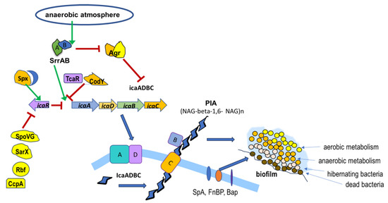
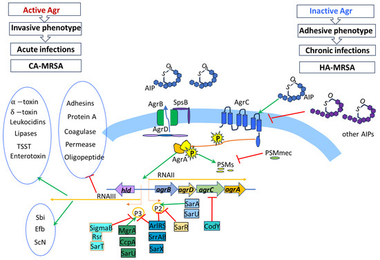
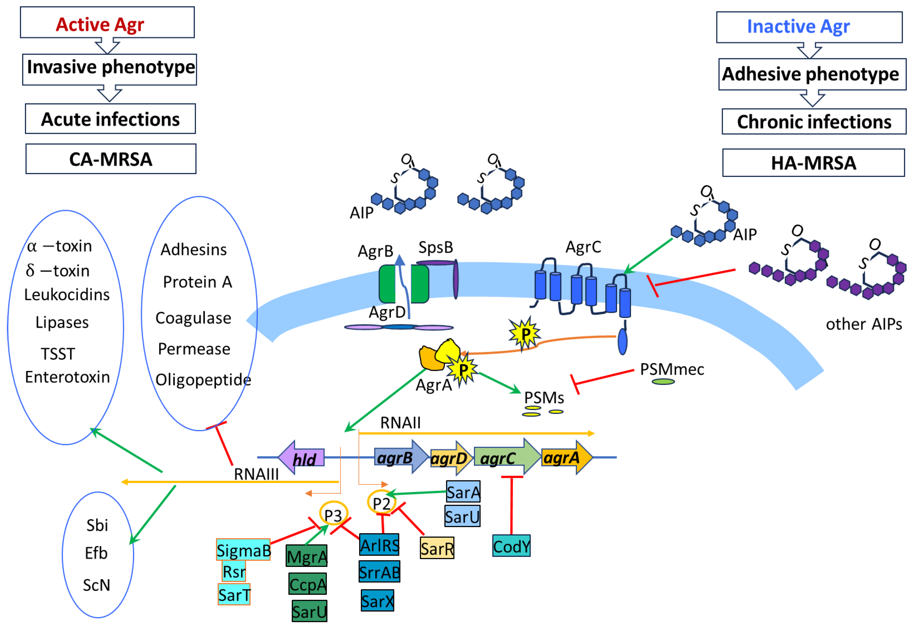

Disclaimer/Publisher’s Note: The statements, opinions and data contained in all publications are solely those of the individual author(s) and contributor(s) and not of MDPI and/or the editor(s). MDPI and/or the editor(s) disclaim responsibility for any injury to people or property resulting from any ideas, methods, instructions or products referred to in the content. |
© 2025 by the authors. Licensee MDPI, Basel, Switzerland. This article is an open access article distributed under the terms and conditions of the Creative Commons Attribution (CC BY) license (https://creativecommons.org/licenses/by/4.0/).