


Abstract
Diabetic sensorimotor neuropathy (DSPN) is strongly associated with the extent of cellular oxidative stress and endothelial dysfunction in type 2 diabetes (T2DM). Alpha-lipoic acid (ALA) attenuates the progression of DSPN through its antioxidant and vasculoprotective effects. Kallistatin has antioxidant and anti-inflammatory properties. We aimed to evaluate changes in kallistatin levels and markers of endothelial dysfunction in patients with T2DM and DSPN following six months of treatment with 600 mg/day of ALA. A total of 54 patients with T2DM and DSPN and 24 control patients with T2DM but without neuropathy participated in this study. The serum concentrations of kallistatin, ICAM-1, VCAM-1, oxLDL, VEGF, ADMA, and TNF-alpha were measured by an ELISA. Peripheral sensory neuropathy was assessed with neuropathy symptom questionnaires and determination of the current perception threshold. After ALA treatment, the level of kallistatin significantly decreased, as well as the levels of TNF-alpha and ADMA. Changes in kallistatin levels were positively correlated with changes in oxLDL. The improvement in DSPN symptoms following ALA treatment showed a positive correlation with changes in kallistatin, VEGF, oxLDL, and ADMA levels. Based on our results, kallistatin could represent a potential new biomarker for assessing therapeutic response during ALA treatment in patients with DSPN.
1. Introduction
Diabetes mellitus is among the most prevalent and rapidly increasing diseases, affecting more than 500 million adults worldwide. Diabetic sensorimotor polyneuropathy (DSPN) manifests as a symmetrical, distal polyneuropathy affecting both the lower and upper extremities, significantly worsening the quality of life of patients with type 2 diabetes (T2DM). This microvascular complication in T2DM is attributed to metabolic changes resulting from chronic hyperglycemia and is closely associated with endothelial and mitochondrial dysfunction, along with elevated reactive oxygen species (ROS) production [1,2]. In the early stages of DSPN, the loss of small nerve fibers is strongly linked to endoneurial microangiopathy [3]. As a microvascular complication of T2DM, elevated oxidative stress and a reduced antioxidant capacity lead to greater expression of adhesion molecules such as intercellular adhesion molecule-1 (ICAM-1) and vascular cell adhesion molecule-1 (VCAM-1), which impairs endoneurial blood flow and leads to progressive damage to peripheral axons [4,5,6]. Experimental analyses have demonstrated that alterations in neural microcirculation may be mediated by changes in nitric oxide (NO) metabolism and endothelial dysfunction, which are closely correlated with changes in tumor necrosis factor-alpha (TNF-α) and oxidized LDL (oxLDL) levels [7,8]. Overproduction of superoxide anions in DSPN leads to the binding of this free oxygen radical to NO and the formation of highly reactive peroxynitrite, which inactivates endothelial NO synthase (eNOS) and induces endothelial cell apoptosis [9,10]. Inhibition of eNOS by N-nitro-L-arginine resulted in reduced neural blood flow, and this effect could be reversed by L-arginine. Asymmetric dimethylarginine (ADMA), a naturally occurring inhibitor of eNOS, is significantly elevated in T2DM and is thought to contribute to the development of accelerated atherosclerosis [11]. Our group and others have demonstrated that changes in ADMA levels may impact vascular and endothelial function, as well as the progression of DSPN [12,13].
According to the recommendations of the Diabetic Neuropathy Study Group [14], DSPN may be diagnosed based on symptoms of peripheral neuropathy and instrumental screening tests, such as vibration perception threshold testing and quantitative sensory testing. Furthermore, self-reported questionnaires such as the Neuropathy Total Symptom Score-6 (NTSS-6) and the Douleur Neuropathique en 4 (DN4) questionnaire are commonly employed to assess neuropathic pain in patients with T2DM [15,16]. Currently, diabetic neuropathy is considered incurable and can only be managed through interventions aimed at slowing its progression, alleviating the associated pain, and addressing its complications. Clinically, the field moves toward individualized pain management, which necessitates the detection of predictors and biomarkers to identify possible responder populations.
Alpha-lipoic acid (ALA), serving as a cofactor for the activity of pyruvate dehydrogenase and α-ketoglutarate dehydrogenase, plays a crucial role in mitochondrial oxidative metabolism. As a pathogenetically oriented pharmacotherapeutic agent, its antioxidant effects are beneficial in the treatment of DSPN [14]. Experimental evidence indicates that ALA improves nerve conduction velocity and protects peripheral nerves from oxidative stress induced by hyperglycemia in DSPN [17,18]. Moreover, ALA may enhance the redox potential of neurons through various mechanisms, including the augmentation of cellular glutathione synthesis, enhancement of neuronal nitric oxide synthase activity, suppression of NADPH oxidase activity, inhibition of nuclear factor kappa B (NF-κB), and promotion of NO-mediated vasodilation [3,19]. To date, predicting the response to ALA treatment is unresolved.
Kallistatin is an endogenous serine protease inhibitor and a regulator of tissue kallikrein activity, exerting a significant vasodilatory effect [20]. Although kallistatin is primarily synthesized in the liver, it is also expressed in smaller amounts in other tissues, including the kidneys, lungs, and heart. Kallistatin has been shown to inhibit the production of inflammatory cytokines, such as TNF-α and interleukin-6 (IL-6), thereby mitigating inflammatory responses within the vascular system [21]. Its antioxidant properties contribute to a reduction in ROS levels and enhance the function of endothelial cells while preserving their structural integrity [22]. Furthermore, kallistatin may exert an anticoagulant effect and regulate the expression of vascular endothelial growth factor (VEGF), thereby facilitating vascular regeneration [23]. Kallistatin has been shown to protect against vascular injury and oxidative stress-induced endothelial apoptosis by enhancing eNOS activity through activation of the phosphatidylinositol 3′-kinase-Akt signaling pathway [24]. Moreover, kallistatin is recognized as an endogenous angiogenesis inhibitor, acting through inhibition of the NF-κB signaling pathway and the suppression of VEGF and angiogenesis-related genes [25]. Therefore, kallistatin has promising potential to protect against microvascular complications of diabetes, as its anti-inflammatory and antioxidant effects have been observed in diabetic retinopathy [26] and diabetic nephropathy [27]. However, the role of kallistatin in DSPN and in its ALA treatment has not been thoroughly investigated previously.
The objective of our study was to evaluate the association between kallistatin and markers of chronic inflammation and oxidative stress in patients with T2DM and DSPN after six months of ALA treatment. Additionally, we aimed to identify correlations between changes in kallistatin levels and the progression of DSPN following antioxidant ALA therapy.
2. Results
2.1. Clinical and Laboratory Characteristics of DSPN Patients
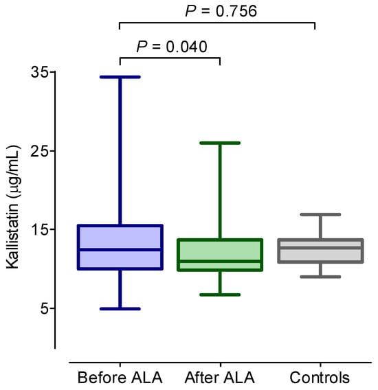
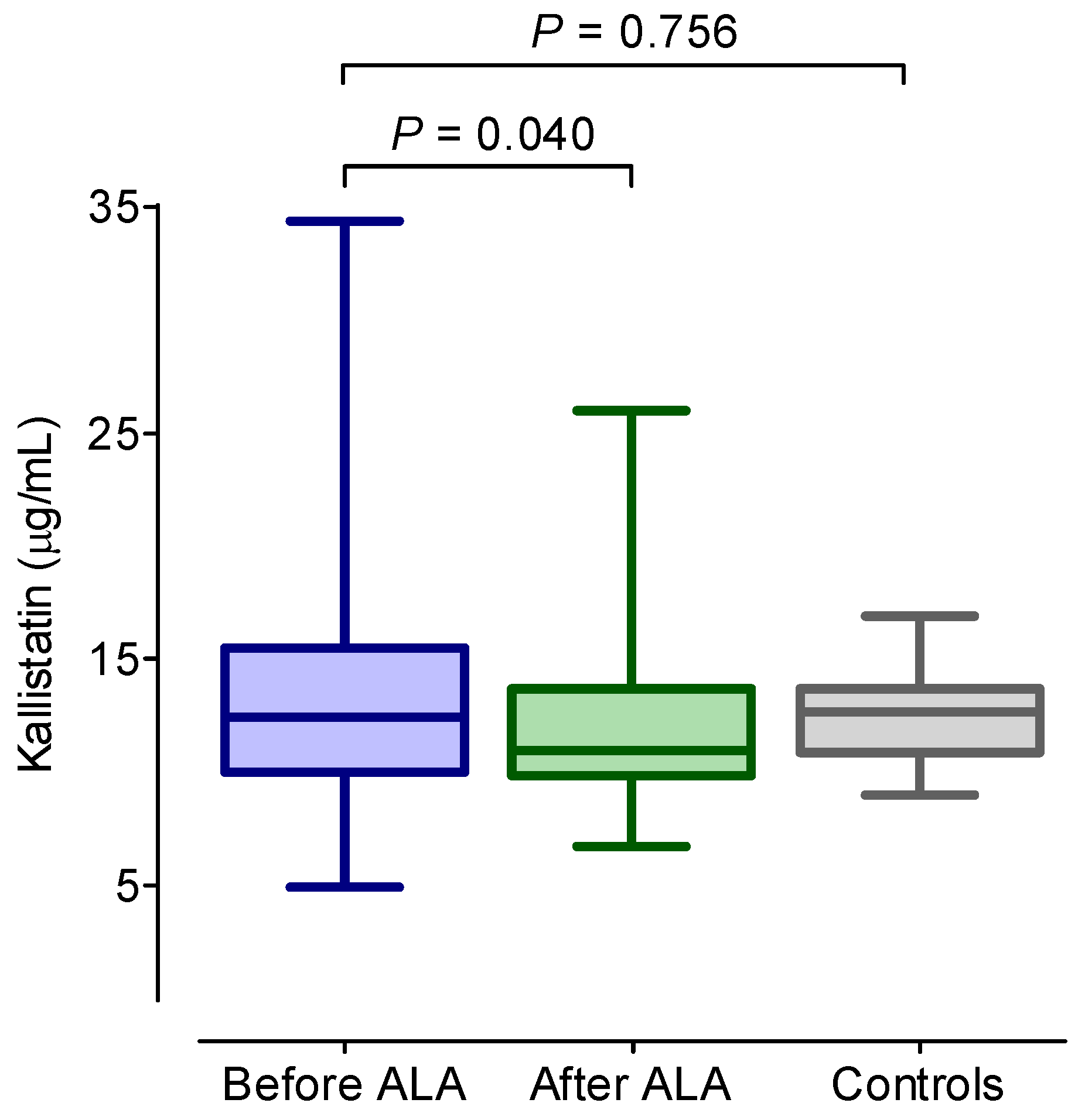
The clinical and laboratory characteristics of DSPN patients before and after 6 months of ALA treatment and data of the control subjects are summarized in Table 1. There was no significant difference observed in the age, body mass index (BMI), duration of diabetes, blood glucose, HbA1c, creatinine, uric acid, hsCRP, VCAM-1, ICAM-1, or oxLDL levels, or lipid parameters between the patient groups both in response to ALA treatment and when compared to the controls. The serum kallistatin concentration was significantly decreased after ALA treatment in the DSPN patients (p < 0.05) (Figure 1, Table 1). ADMA and TNF-α levels also decreased significantly after ALA treatment in the DSPN patients (p < 0.05), while NO levels increased significantly after ALA treatment (p < 0.05). For the pretreatment kallistatin, ADMA and TNF-α levels were significantly higher in the DSPN patients compared with the controls (p < 0.05). The VEGF level did not change significantly as a result of the ALA treatment, but the initial VEGF level was significantly higher in the DSPN patients compared with the controls (p < 0.05). After ALA treatment, the DSPN patients achieved significantly lower scores for the validated neuropathy questionnaires (NTSS-6 and DN4), and there was significant improvement observed in both the current perception threshold values and the composite autonomic score.
Table 1.
Main anthropometric and laboratory parameters of enrolled participants.
Figure 1.
Concentrations of serum kallistatin in diabetic patients with neuropathy before and after 6 months of alpha-lipoic acid (ALA) treatment, as well as in diabetic controls without neuropathy. The boxes represent the interquartile range (25th–75th percentiles), with the 50th percentile (median) indicated by a solid line within the box. The whiskers denote the minimum and maximum values.
2.2. Correlations Between Kallistatin and Main Parameters in DSPN Patients
The correlations of kallistatin with the main anthropometric and laboratory parameters in patients with DSPN before and after 6 months of ALA treatment are summarized in Table 2.
Table 2.
The correlations of kallistatin with the main anthropometric and laboratory parameters in patients with DSPN before and after 6 months of ALA treatment.
The kallistatin levels measured before and after ALA treatment exhibited no correlation with age, BMI, HbA1c, NTSS-6 or DN4 scores, the composite autonomic score, CPT values, or lipid parameters. The kallistatin concentration exhibited a positive correlation with the serum ICAM-1 level (r = 0.31, p = 0.031) and the TNF-α level (r = 0.45, p = 0.001), while a negative correlation was observed between the kallistatin and NO levels (r = −0.4, p = 0.017) after 6 months of ALA treatment in patients with DSPN.
2.3. Spearman’s Correlations Between Changes in Kallistatin Levels and Changes in Different Laboratory Parameters
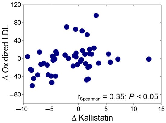
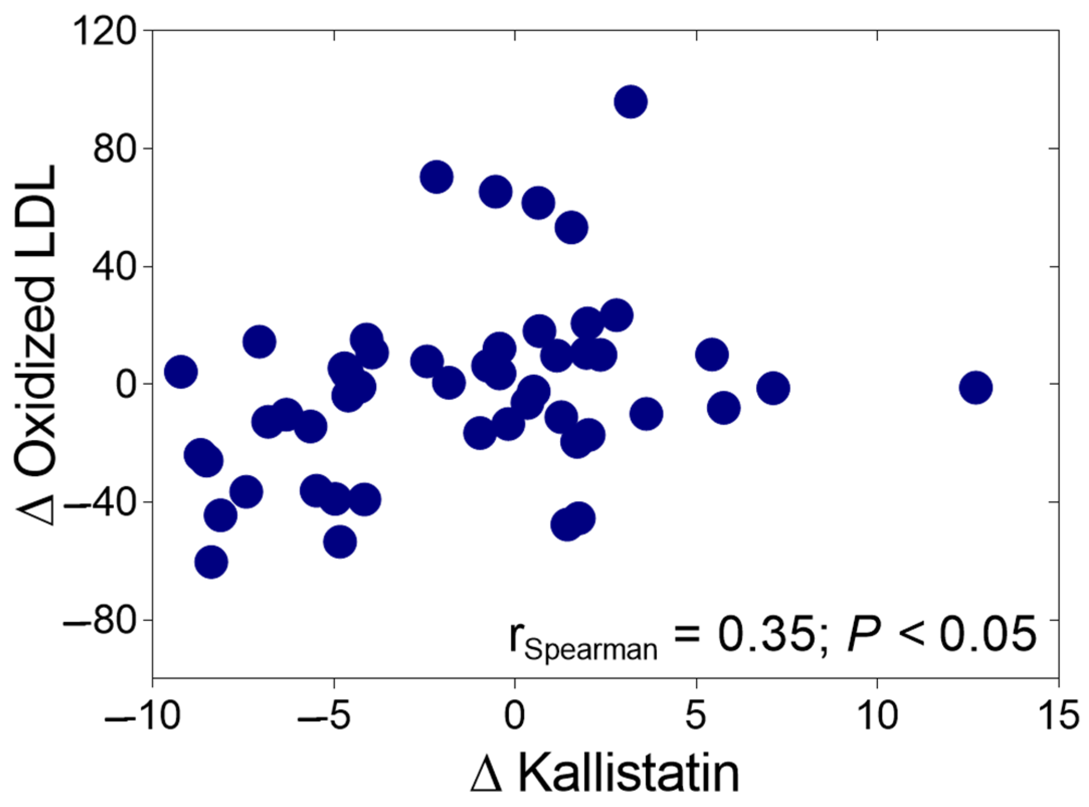
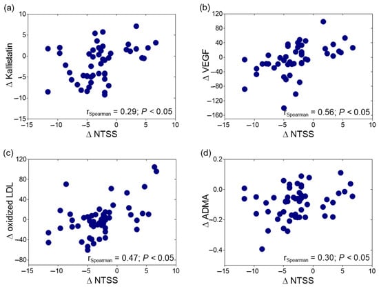
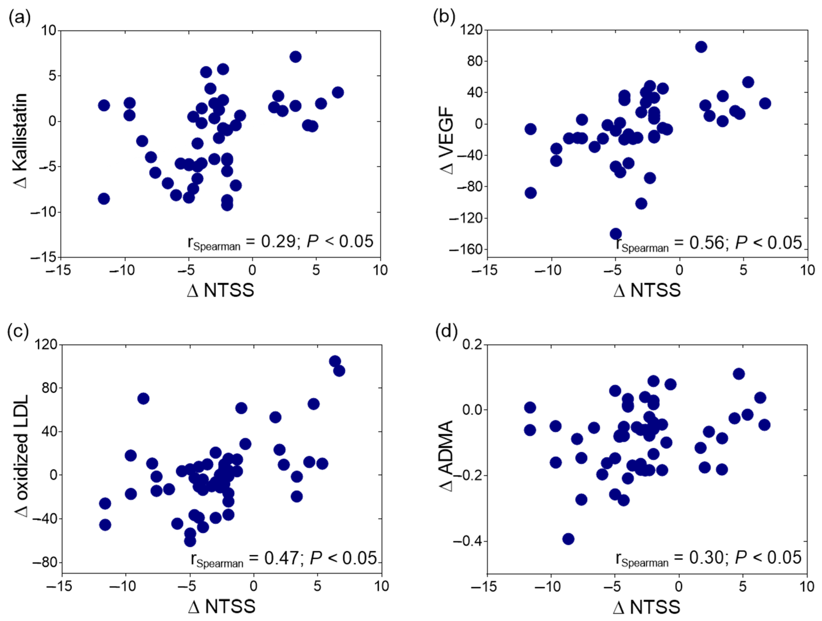
The change in kallistatin levels was correlated positively with the change in oxLDL levels after ALA treatment among the DSPN patients (r = 0.35, p < 0.05, Figure 2). The improvement in NTSS-6 scores following ALA treatment exhibited a positive correlation with alterations in ∆kallistatin (r = 0.29, p < 0.05, Figure 3a), ∆VEGF (r = 0.56, p < 0.05, Figure 3b), ∆oxLDL (r = 0.47, p < 0.05, Figure 3c), and ∆ADMA levels (r = 0.30, p < 0.05, Figure 3d).
Figure 2.
Spearman’s correlation of the change in oxidized low-density lipoprotein (Δ oxidized LDL) levels with the change in kallistatin (Δ kallistatin) in type 2 diabetic patients with neuropathy during 6 months of alpha-lipoic acid (ALA) treatment.
Figure 3.
Spearman’s correlation of the change in the Neuropathy Total Symptom Score (NTSS) with (a) the change in kallistatin (Δ kallistatin); (b) the change in vascular endothelial growth factor (Δ VEGF); (c) the change in oxidized low-density lipoprotein (Δ oxidized LDL) levels; and (d) the change in asymmetric dimethylarginine (Δ ADMA) levels in type 2 diabetic patients with neuropathy during 6 months of alpha-lipoic acid (ALA) treatment.
3. Discussion
This study is the first to explore kallistatin as a potential biomarker in response to chronic inflammatory processes in patients with DSPN. This is, to the best of our knowledge, the first study to investigate the potential role of kallistatin in the progression of DSPN in patients with T2DM. We observed a decrease in the serum kallistatin levels in DSPN patients after six months of ALA treatment, and this reduction was positively correlated with improvements in neuropathic symptoms. Furthermore, the serum kallistatin levels exhibited a positive correlation with the levels of TNF-α. Additionally, changes in kallistatin levels were positively correlated with alterations in oxLDL in the DSPN patients following ALA treatment. These changes may be closely associated with endothelial dysfunction and atherosclerosis in the context of DSPN.
Antioxidant therapy plays a critical role in mitigating oxidative stress in DSPN, a condition characterized by an imbalance between ROS and antioxidant defenses, which leads to cellular damage within the nervous system. Antioxidants, such as ALA, have demonstrated the ability to neutralize free radicals, thereby reducing oxidative stress [17,18]. Kallistatin has demonstrated promise in predicting microvascular complications of T2DM, with its anti-inflammatory and antioxidant effects already established in diabetic retinopathy [26] and diabetic nephropathy [27]. In our study, we assessed the therapeutic effectiveness of ALA, a well-established antioxidant therapy for DSPN, and observed a significant reduction in serum kallistatin levels in individuals with DSPN following ALA treatment. Concomitant with the decrease in kallistatin levels, serum concentrations of ADMA and TNF-α were also significantly decreased, while NO levels were significantly elevated after six months of ALA therapy.
Kallistatin may exert a protective effect against oxidative stress, endothelial dysfunction, and chronic inflammatory processes through multiple signaling pathways. Through its heparin-binding domain, kallistatin inhibits TNF-α-mediated nicotinamide adenine dinucleotide phosphate (NADPH) oxidase activity and ROS generation, thereby downregulating the expression of ICAM-1 and VCAM-1. Moreover, in a mouse model, kallistatin has been demonstrated to activate the SIRT1-eNOS pathway via its active site, resulting in enhanced NO production, which in turn mitigates ROS levels [28]. In our study, the baseline levels of kallistatin, ADMA, and TNF-α were significantly higher in the patients with DSPN compared with the diabetic controls. Our research group previously reported lower serum kallistatin concentrations in a young, healthy cohort (7.09 ± 1.91 µg/mL; n = 49) [29] which were notably lower than the levels observed in our current diabetic cohort (12.45 (10–15.4) µg/mL in DSPN patients prior to ALA treatment and 12.7 (10.9–13.73) µg/mL in diabetic controls). These findings of lower kallistatin levels in healthy individuals are consistent with previous reports [30,31]. However, it is important to acknowledge that direct comparisons of kallistatin values between studies may be challenging due to methodological differences. We hypothesize that under conditions of increased oxidative stress, kallistatin levels rise as part of a compensatory antioxidant defense mechanism. However, effective antioxidant therapies, such as ALA treatment, may reduce kallistatin levels in tandem with decreases in oxidative stress and chronic inflammation in DSPN. The abnormally elevated concentration of ADMA, a competitive inhibitor of eNOS activity, was significantly reduced following ALA therapy, further supporting the beneficial effects of antioxidant treatment [12,13].
Consistent with other studies [21,22,24,32,33,34], it was previously shown that serum kallistatin levels correlate with glucose homeostasis, markers of chronic inflammation, and lipoprotein metabolism in both T2DM and nondiabetic obese patients [29]. Our results also confirmed that kallistatin expression can be induced by glucotoxicity and increased oxidative stress, which is associated with persistent hyperglycemia in T2DM, while its production in non-diabetic patients is primarily related to systemic inflammation. We also investigated the correlations between endothelial dysfunction and NO synthesis following ALA treatment in patients with DSPN. Our recent findings indicate that ALA supplementation enhances endothelial function, as reflected by changes in the serum levels of ADMA and TNF-α in patients with diabetic neuropathy. Moreover, based on our results, changes in serum ADMA levels were identified as potential predictors of the clinical response to ALA treatment in DSPN patients [12].
Previous studies have shown a positive correlation between kallistatin levels and both TNF-α and carotid intima-media thickness in patients with insulin resistance-related conditions, such as polycystic ovary syndrome. Consistent with our current findings, this study also reported no significant correlation between kallistatin levels and fasting or postprandial blood glucose levels, as well as HbA1c levels [35]. Additionally, the administration of recombinant kallistatin has been shown to significantly reduce inflammatory responses in various animal models of myocardial ischemia, hypertension, or chronic inflammatory diseases [22,24,33,36]. Although the exact mechanism is not yet fully understood, kallistatin has been found to inhibit vascular inflammation and apoptosis by suppressing the TNF-α- and high mobility group box protein 1-mediated expression of various inflammatory genes [21,33]. Furthermore, kallistatin has been shown to attenuate inflammatory responses in rheumatoid arthritis via the NF-κB signaling pathway [21]. In obese patients, kallistatin was found to inhibit TNF-α- and lipopolysaccharide-induced inflammation in human adipose tissue by decreasing the expression and secretion of key inflammatory markers [34]. These findings suggest that tissue kallistatin may be involved in the regulation of chronic inflammatory responses. Our results revealed a significant positive correlation between kallistatin and serum TNF-α levels in T2DM patients with DSPN. Additionally, changes in kallistatin levels were positively correlated with changes in oxLDL following ALA treatment. Although VEGF levels did not show significant changes following ALA treatment in the studies, baseline VEGF levels were significantly elevated in the DSPN patients compared with the control group. These findings further support the association between kallistatin levels and the degree of inflammation in DSPN.
By restoring the balance between ROS and antioxidants, antioxidant therapy enhances axonal microcirculation and attenuates chronic inflammation, leading to improved nerve function. Consequently, this approach can alleviate the symptoms of diabetic neuropathy, including pain, numbness, and tingling [18]. The self-reported questionnaires may frequently be used to access the neuropathic pain of T2DM patients [15,16]. Following ALA treatment, our patients with DSPN exhibited significantly reduced scores for validated neuropathy questionnaires (NTSS-6 and DN4). Moreover, significant improvements were observed in both the current perception threshold values and the composite autonomic function score. During the assessment of DSPN-related symptoms in our patient cohort, improvements in NTSS-6 scores demonstrated a positive correlation with changes in the kallistatin, VEGF, oxLDL, and ADMA levels following ALA treatment. These findings suggest that antioxidant treatment with ALA exerts anti-inflammatory and symptom-alleviating effects in T2DM patients with DSPN.
Some limitations of our study should be acknowledged. To further elucidate the potential role of kallistatin in mitigating oxidative stress and regulating chronic inflammatory processes, it will be necessary to assess additional inflammatory markers in the DSPN patient population. While we observed significant associations between the inflammatory markers measured and the serum kallistatin levels, the precise function of kallistatin in regulatory pathways and the pathomechanism of DSPN remains unclear due to the cross-sectional design of this study. Moreover, it is important to note that the sample size may have influenced the significance of our findings. Lastly, the mechanism we hypothesized regarding the role of kallistatin in the increased oxidative stress characteristic of DSPN has previously been described exclusively in an animal model. Despite these limitations, our findings from human samples further support the association between kallistatin levels and the reduction in chronic inflammation and oxidative stress in response to ALA treatment in T2DM patients with DSPN.
4. Materials and Methods
4.1. Study Population
Fifty-four T2DM patients with DSPN (22 men and 32 women; mean age: 64.2 ± 8.7 years; mean known duration of T2DM before the start of our study: 12.4 ± 2.3 years; mean duration of diabetic neuropathy: 6.2 ± 1.4 years) were enrolled in this study. All patients received 600 mg of ALA orally each day for 6 months (WÖRWAG Pharma GmbH, Böblingen, Germany). Additionally, 24 age- and gender-matched T2DM subjects without neuropathy were included as a control group.
All patients were managed with oral antidiabetic agents (metformin, sulfonylureas, or dipeptidyl-peptidase-4 inhibitors), and those on insulin therapy were excluded. Patients with a history of diabetic proliferative retinopathy, diabetic nephropathy (eGFR < 60 mL/min/1.73 m2 or persistent albuminuria), or type 1 diabetes were also excluded. Additionally, subjects with alcoholism, known liver diseases, endocrine or autoimmune diseases, or hematological or neurological disorders associated with peripheral neuropathy were excluded. Patients with prior cardiovascular disease, established coronary artery disease or myocardial infarction or severe congestive heart failure (New York Heart Association class II–IV), smokers, pregnant women, and subjects with malignancy were not enrolled in our study.
All participants were recruited from the Diabetic Neuropathy Center of Debrecen of the Department of Internal Medicine in University of Debrecen’s Faculty of Medicine in Debrecen, Hungary. Written informed consent was obtained from all participants. The study protocol was approved by the local and regional ethics committees and was conducted in accordance with the Declaration of Helsinki. This study protocol was approved by the local and regional ethical committees (protocol code: 5287-2/2019/EKU; date of approval: 7 March 2019), and it was carried out in accordance with the Declaration of Helsinki.
4.2. Blood Sampling
Venous blood samples were collected from all participants in the morning after an overnight fast. Sera and plasma samples were prepared after 45 min of rest following collection. Routine laboratory analyses, including total cholesterol, triglycerides, high-density lipoprotein cholesterol (HDL-C), low-density lipoprotein cholesterol (LDL-C), glucose, hemoglobin A1c (HbA1c), creatinine, and uric acid, were conducted using fresh serum samples. These analyses were performed with a Cobas c501 autoanalyzer (Roche Ltd., Mannheim, Germany) at the Department of Laboratory Medicine of the Faculty of Medicine at the University of Debrecen in Debrecen, Hungary. The reagents were obtained from Roche Ltd., and the tests were carried out according to the manufacturer’s instructions. For enzyme activity measurements and ELISA determinations, samples were stored at −70 °C until analysis.
4.3. Measurement of Serum Kallistatin
Serum kallistatin levels were measured using a commercially available enzyme-linked immunosorbent assay (ELISA) kit (Human Serpin A4/Kallistatin DuoSet ELISA, Cat: DY1669, R&D Systems, Abingdon, UK). Serum samples were diluted 10,000 fold and analyzed in duplicate according to the manufacturer’s instructions. The assay had a range of 125.0–8000 pg/mL.
4.4. Serum ADMA Measurement
Serum ADMA concentrations were measured using a commercially available competitive ELISA kit (ADMA-ELISA; DLD Diagnostika GmbH, Hamburg, Germany). The intra-assay coefficients of variation (CVs) ranged from 5.7% to 6.4%, and the inter-assay CVs ranged from 8.3% to 10.3%. ADMA measurements were performed according to the manufacturer’s instructions, and the results were expressed in micromoles per liter (µmol/L).
4.5. Serum TNF-α Measurement
Serum levels of TNF-α were measured using a commercially available ELISA kit (R&D Systems Europe Ltd., Abington, UK). The measurements were performed according to the manufacturer’s recommendations. The intra-assay CVs ranged from 1.9% to 2.2%, while the inter-assay CVs ranged from 6.2% to 6.7%. The results were expressed in picograms per milliliter (pg/mL).
4.6. Measurement of oxLDL
Serum concentrations of oxLDL were measured using a commercially available sandwich ELISA kit (Mercodia AB, Uppsala, Sweden). This assay utilizes a direct sandwich technique where two monoclonal antibodies target distinct antigenic determinants on the oxidized apolipoprotein B molecule. The intra-assay and inter-assay CVs were 5.5% to 7.3% and 4% to 6.2%, respectively, with a sensitivity of less than 1 milliunit per liter (mU/L).
4.7. Measurement of sICAM-1, sVCAM-1, and VEGF
The levels of sICAM-1 and sVCAM-1 in serum were measured using human soluble sICAM-1 and sVCAM-1 sandwich ELISA kits (R&D Systems Europe Ltd., Abingdon, UK). The ELISA procedures were performed according to the manufacturer’s instructions. For sICAM-1, the intra-assay CVs ranged from 3.7% to 5.2%, and the inter-assay CVs ranged from 4.4% to 6.7%. For sVCAM-1, the intra-assay CVs ranged from 2.3% to 3.6%, while the inter-assay CVs ranged from 5.5% to 7.8%. The results were expressed in nanograms per milliliter (ng/mL). VEGF levels were also measured by an ELISA provided by R&D (R&D Systems Europe Ltd., Abingdon, UK). The intra- and inter-assay CVs were 4.5–6.7% and 6.2–8.8%, respectively. The values were presented in nanograms per liter (ng/mL).
4.8. Assay for Nitric Oxide Concentration
The nitrite concentration was measured as an indicator of NO production using the Griess reaction [37] as described previously [12]. The results were expressed in micromoles per liter (µmol/L).
4.9. Assessment of Autonomic and Peripheral Nerve Function
All participants underwent a comprehensive assessment of peripheral neuropathy, which included the use of the Neuropathy Total Symptom Score-6 (NTSS-6) and the Douleur Neuropathique en 4 (DN4) questionnaires for screening neuropathic pain syndrome [15,16], vibration perception threshold testing, and quantitative sensory testing. Consequently, there was typically no need for additional confirmatory tests, such as electrophysiological assessments evaluating nerve conduction velocity (electroneurography) [14,38]. Previous studies have demonstrated that screening with questionnaires such as the NTSS-6 or DN4 may provide a valid assessment of sensory symptoms of DSPN and can serve as evaluable endpoints in clinical trials [15,16].
Peripheral sensory nerve function was assessed using current perception threshold (CPT) testing with a Neurometer® (Neurotron Inc., Baltimore, MD, USA, 2002). This neurodiagnostic device can detect peripheral sensory neuropathy in various diseases, including diabetes mellitus [39]. Autonomic function was assessed using Ewing’s five standard cardiovascular reflex tests. These tests include changes in heart rate during deep inspiration and expiration, heart rate responses to standing (30/15 ratio), the Valsalva maneuver, systolic blood pressure response to standing, and changes in diastolic pressure during a sustained handgrip. A score was developed to quantify the severity of autonomic neuropathy based on the results of five tests (normal = 0, borderline = 1, and abnormal = 2). The composite autonomic score ranged from 0 to 10, with a score of 0–1 classified as normal, 2–3 classified as mild, 4–6 classified as moderate, and 7–10 classified as severe autonomic neuropathy [40].
4.10. Statistical Methods
Statistical analyses were conducted using Statistica 13.5.0.17 software (TIBCO Software Inc., Palo Alto, CA, USA). Graphs were created using GraphPad 8.0 software. The normality of distribution was assessed using the Kolmogorov–Smirnov test. For variables with normal distributions, differences between variables in the diabetic control and neuropathic patient groups were calculated using a Student’s paired t-test. These data are presented as the mean ± standard deviation (SD). In case of a skewed distribution, the difference was calculated with a Mann–Whitney U test, and data were presented as medians (upper-lower quartiles and 95% confidence intervals).
To investigate the effect of ALA treatment, a Student’s paired test (normal distribution) and Wilcoxon matched-pairs test (skewed distribution) were conducted with the variables before and after ALA treatment. Spearman’s rank order correlation analysis was employed to examine the relationships between the selected variables. During the statistical analysis, changes in specific parameters were presented as the difference between the post-exposure (after alpha-lipoic acid treatment) and pre-exposure values for each individual. A p value ≤ 0.05 was considered statistically significant.
5. Conclusions
In conclusion, this study provides novel insights into the role of kallistatin as a potential biomarker for chronic inflammation and oxidative stress in T2DM patients with DSPN. Our findings suggest that kallistatin levels are significantly correlated with improvements in neuropathic symptoms following ALA treatment, indicating a potential protective effect against oxidative stress and endothelial dysfunction. Additionally, the observed correlations between kallistatin and the inflammatory marker TNF-α further emphasize its involvement in the inflammatory processes linked to DSPN. While the exact mechanisms remain to be elucidated, the results highlight the importance of antioxidant therapy in modulating kallistatin levels and reducing chronic inflammation. Ultimately, these insights underscore the potential of kallistatin as a therapeutic target and biomarker in managing complications associated with diabetic neuropathy.
Author Contributions
Conceptualization, M.H. (Mariann Harangi) and F.S.; methodology, S.C. and H.L.; investigation, Á.M.; data curation, M.H. (Marcell Hernyák) and L.I.T.; writing—original draft preparation, M.H. (Marcell Hernyák), H.L. and F.S.; writing—review and editing, M.H. (Mariann Harangi) and G.P.; visualization, M.H. (Marcell Hernyák); supervision, F.S.; funding acquisition, M.H. (Mariann Harangi). All authors have read and agreed to the published version of the manuscript.
Funding
This research was funded by the National Research, Development and Innovation Office (NKFIH) (grant number: K142273). TKP2021-EGA-18 was implemented with support provided by the Ministry of Innovation and Technology of Hungary from the National Research, Development and Innovation Fund, financed under the TKP2021-EGA funding scheme. The research group was supported by the Eötvös Loránd Research Network (11003).
Institutional Review Board Statement
This study was conducted in accordance with the Declaration of Helsinki and approved by the institutional review board (or ethics committee) of the University of Debrecen (protocol code: 5287-2/2019/EKU; date of approval: 7 March 2019).
Informed Consent Statement
Informed consent was obtained from all subjects involved in this study.
Data Availability Statement
All data generated or analyzed during this study are included in this published article. All data generated or analyzed during the current study are available from the corresponding author upon reasonable request.
Acknowledgments
We are indebted to the subjects of this study for their cooperation. The authors thank the members of the Lipid Research Laboratory for their excellent technical support.
Conflicts of Interest
The authors declare no conflicts of interest.
Abbreviations
| ADMA | Asymmetric dimethylarginine |
| ALA | Alpha-lipoic acid |
| CPT | Current perception threshold |
| CVs | Coefficients of variation |
| DN4 | Douleur Neuropathique 4 |
| DSPN | Diabetic sensorimotor neuropathy |
| ELISA | Enzyme-linked immunosorbent assay |
| eNOS | Endothelial NO synthase |
| HbA1c | Hemoglobin A1c |
| HDL-C | High-density lipoprotein cholesterol |
| ICAM-1 | Intercellular adhesion molecule-1 |
| IL-6 | Interleukin-6 |
| LDL-C | Low-density lipoprotein cholesterol |
| NADPH | Nicotinamide adenine dinucleotide phosphate |
| NF-κB | Nuclear factor kappa B |
| NO | Nitric oxide |
| NTSS-6 | Neuropathy Total Symptom Score-6 |
| oxLDL | Oxidized LDL |
| ROS | Reactive oxygen species |
| T2DM | Type 2 diabetes |
| TNF-α | Tumor necrosis factor-alpha |
| VCAM-1 | Vascular cell adhesion molecule-1 |
| VEGF | Vascular endothelial growth factor |
References
- Callaghan, B.C.; Price, R.S.; Chen, K.S.; Feldman, E.L. The Importance of Rare Subtypes in Diagnosis and Treatment of Peripheral Neuropathy: A Review. JAMA Neurol. 2015, 72, 1510–1518. [Google Scholar] [CrossRef] [Scilit] [PubMed]
- Ziegler, D.; Tesfaye, S.; Spallone, V.; Gurieva, I.; Al Kaabi, J.; Mankovsky, B.; Martinka, E.; Radulian, G.; Nguyen, K.T.; Stirban, A.O.; et al. Screening, diagnosis and management of diabetic sensorimotor polyneuropathy in clinical practice: International expert consensus recommendations. Diabetes Res. Clin. Pract. 2022, 186, 109063. [Google Scholar] [CrossRef] [Scilit]
- Coppey, L.J.; Gellett, J.S.; Davidson, E.P.; Dunlap, J.A.; Lund, D.D.; Yorek, M.A. Effect of antioxidant treatment of streptozotocin-induced diabetic rats on endoneurial blood flow, motor nerve conduction velocity, and vascular reactivity of epineurial arterioles of the sciatic nerve. Diabetes 2001, 50, 1927–1937. [Google Scholar] [CrossRef] [Scilit]
- Goldberg, R.B. Cytokine and cytokine-like inflammation markers, endothelial dysfunction, and imbalanced coagulation in development of diabetes and its complications. J. Clin. Endocrinol. Metab. 2009, 94, 3171–3182. [Google Scholar] [CrossRef] [Scilit] [PubMed]
- Maiuolo, J.; Gliozzi, M.; Musolino, V.; Carresi, C.; Nucera, S.; Macri, R.; Scicchitano, M.; Bosco, F.; Scarano, F.; Ruga, S.; et al. The Role of Endothelial Dysfunction in Peripheral Blood Nerve Barrier: Molecular Mechanisms and Pathophysiological Implications. Int. J. Mol. Sci. 2019, 20, 3022. [Google Scholar] [CrossRef] [Scilit] [PubMed]
- Caturano, A.; D’Angelo, M.; Mormone, A.; Russo, V.; Mollica, M.P.; Salvatore, T.; Galiero, R.; Rinaldi, L.; Vetrano, E.; Marfella, R.; et al. Oxidative Stress in Type 2 Diabetes: Impacts from Pathogenesis to Lifestyle Modifications. Curr. Issues Mol. Biol. 2023, 45, 6651–6666. [Google Scholar] [CrossRef] [Scilit]
- Vincent, A.M.; Callaghan, B.C.; Smith, A.L.; Feldman, E.L. Diabetic neuropathy: Cellular mechanisms as therapeutic targets. Nat. Rev. Neurol. 2011, 7, 573–583. [Google Scholar] [CrossRef] [Scilit]
- Rask-Madsen, C.; King, G.L. Vascular complications of diabetes: Mechanisms of injury and protective factors. Cell Metab. 2013, 17, 20–33. [Google Scholar] [CrossRef] [Scilit]
- Ostergaard, L.; Finnerup, N.B.; Terkelsen, A.J.; Olesen, R.A.; Drasbek, K.R.; Knudsen, L.; Jespersen, S.N.; Frystyk, J.; Charles, M.; Thomsen, R.W.; et al. The effects of capillary dysfunction on oxygen and glucose extraction in diabetic neuropathy. Diabetologia 2015, 58, 666–677. [Google Scholar] [CrossRef] [Scilit]
- Du, M.R.; Ju, G.X.; Li, N.S.; Jiang, J.L. Role of Asymmetrical Dimethylarginine in Diabetic Microvascular Complications. J. Cardiovasc. Pharmacol. 2016, 68, 322–326. [Google Scholar] [CrossRef] [Scilit]
- Abbasi, F.; Asagmi, T.; Cooke, J.P.; Lamendola, C.; McLaughlin, T.; Reaven, G.M.; Stuehlinger, M.; Tsao, P.S. Plasma concentrations of asymmetric dimethylarginine are increased in patients with type 2 diabetes mellitus. Am. J. Cardiol. 2001, 88, 1201–1203. [Google Scholar] [CrossRef] [Scilit] [PubMed]
- Sztanek, F.; Lőrincz, H.; Molnár, Á.; Szentpéteri, A.; Zöld, E.; Seres, I.; Páll, D.; Harangi, M.; Kempler, P.; Paragh, G. The effect of α-lipoic acid treatment on plasma asymmetric dimethylarginine, a biomarker of endothelial dysfunction in diabetic neuropathy. Arch. Med. Sci. 2020. [Google Scholar] [CrossRef] [Scilit]
- Yasar, H.; Senol, M.G.; Kendirli, T.; Onem, Y.; Ozdag, F.; Saracoglu, M. Serum asymmetric dimethylarginine levels in diabetic patients with neuropathy. Diabetes Res. Clin. Pract. 2011, 92, 223–227. [Google Scholar] [CrossRef] [Scilit] [PubMed]
- Tesfaye, S.; Boulton, A.J.; Dyck, P.J.; Freeman, R.; Horowitz, M.; Kempler, P.; Lauria, G.; Malik, R.A.; Spallone, V.; Vinik, A.; et al. Diabetic neuropathies: Update on definitions, diagnostic criteria, estimation of severity, and treatments. Diabetes Care 2010, 33, 2285–2293. [Google Scholar] [CrossRef] [Scilit]
- Cornblath, D.R.; Chaudhry, V.; Carter, K.; Lee, D.; Seysedadr, M.; Miernicki, M.; Joh, T. Total neuropathy score: Validation and reliability study. Neurology 1999, 53, 1660–1664. [Google Scholar] [CrossRef] [Scilit]
- Bastyr, E.J.; Price, K.L.; Bril, V.; Group, M.S. Development and validity testing of the neuropathy total symptom score-6: Questionnaire for the study of sensory symptoms of diabetic peripheral neuropathy. Clin. Ther. 2005, 27, 1278–1294. [Google Scholar] [CrossRef] [Scilit]
- Tibullo, D.; Li Volti, G.; Giallongo, C.; Grasso, S.; Tomassoni, D.; Anfuso, C.D.; Lupo, G.; Amenta, F.; Avola, R.; Bramanti, V. Biochemical and clinical relevance of alpha lipoic acid: Antioxidant and anti-inflammatory activity, molecular pathways and therapeutic potential. Inflamm. Res. 2017, 66, 947–959. [Google Scholar] [CrossRef] [Scilit]
- Papanas, N.; Ziegler, D. Efficacy of α-lipoic acid in diabetic neuropathy. Expert. Opin. Pharmacother. 2014, 15, 2721–2731. [Google Scholar] [CrossRef] [Scilit]
- Maciejczyk, M.; Żebrowska, E.; Nesterowicz, M.; Żendzian-Piotrowska, M.; Zalewska, A. α-Lipoic Acid Strengthens the Antioxidant Barrier and Reduces Oxidative, Nitrosative, and Glycative Damage, as well as Inhibits Inflammation and Apoptosis in the Hypothalamus but Not in the Cerebral Cortex of Insulin-Resistant Rats. Oxid. Med. Cell. Longev. 2022, 2022, 7450514. [Google Scholar] [CrossRef] [Scilit]
- Chao, J.; Stallone, J.N.; Liang, Y.M.; Chen, L.M.; Wang, D.Z.; Chao, L. Kallistatin is a potent new vasodilator. J. Clin. Investig. 1997, 100, 11–17. [Google Scholar] [CrossRef] [Scilit]
- Yin, H.; Gao, L.; Shen, B.; Chao, L.; Chao, J. Kallistatin inhibits vascular inflammation by antagonizing tumor necrosis factor-alpha-induced nuclear factor kappaB activation. Hypertension 2010, 56, 260–267. [Google Scholar] [CrossRef] [Scilit] [PubMed]
- Chao, J.; Yin, H.; Yao, Y.Y.; Shen, B.; Smith, R.S.; Chao, L. Novel role of kallistatin in protection against myocardial ischemia-reperfusion injury by preventing apoptosis and inflammation. Hum. Gene Ther. 2006, 17, 1201–1213. [Google Scholar] [CrossRef] [Scilit] [PubMed]
- Huang, K.F.; Yang, H.Y.; Xing, Y.M.; Lin, J.S.; Diao, Y. Recombinant human kallistatin inhibits angiogenesis by blocking VEGF signaling pathway. J. Cell. Biochem. 2014, 115, 575–584. [Google Scholar] [CrossRef] [Scilit] [PubMed]
- Shen, B.; Gao, L.; Hsu, Y.T.; Bledsoe, G.; Hagiwara, M.; Chao, L.; Chao, J. Kallistatin attenuates endothelial apoptosis through inhibition of oxidative stress and activation of Akt-eNOS signaling. Am. J. Physiol. Heart Circ. Physiol. 2010, 299, H1419–H1427. [Google Scholar] [CrossRef] [Scilit] [PubMed]
- Huang, K.F.; Huang, X.P.; Xiao, G.Q.; Yang, H.Y.; Lin, J.S.; Diao, Y. Kallistatin, a novel anti-angiogenesis agent, inhibits angiogenesis via inhibition of the NF-κB signaling pathway. Biomed. Pharmacother. 2014, 68, 455–461. [Google Scholar] [CrossRef] [Scilit]
- Xing, Q.; Zhang, G.; Kang, L.; Wu, J.; Chen, H.; Liu, G.; Zhu, R.; Guan, H.; Lu, P. The Suppression of Kallistatin on High-Glucose-Induced Proliferation of Retinal Endothelial Cells in Diabetic Retinopathy. Ophthalmic Res. 2017, 57, 141–149. [Google Scholar] [CrossRef] [Scilit]
- Yiu, W.H.; Wong, D.W.; Wu, H.J.; Li, R.X.; Yam, I.; Chan, L.Y.; Leung, J.C.; Lan, H.Y.; Lai, K.N.; Tang, S.C. Kallistatin protects against diabetic nephropathy in db/db mice by suppressing AGE-RAGE-induced oxidative stress. Kidney Int. 2016, 89, 386–398. [Google Scholar] [CrossRef] [Scilit]
- Chao, J.; Guo, Y.; Chao, L. Protective Role of Endogenous Kallistatin in Vascular Injury and Senescence by Inhibiting Oxidative Stress and Inflammation. Oxid. Med. Cell. Longev. 2018, 2018, 4138560. [Google Scholar] [CrossRef] [Scilit]
- Lorincz, H.; Csiha, S.; Ratku, B.; Somodi, S.; Sztanek, F.; Paragh, G.; Harangi, M. Associations between Serum Kallistatin Levels and Markers of Glucose Homeostasis, Inflammation, and Lipoprotein Metabolism in Patients with Type 2 Diabetes and Nondiabetic Obesity. Int. J. Mol. Sci. 2024, 25, 6264. [Google Scholar] [CrossRef] [Scilit]
- Fang, Z.; Shen, G.; Wang, Y.; Hong, F.; Tang, X.; Zeng, Y.; Zhang, T.; Liu, H.; Li, Y.; Wang, J.; et al. Elevated Kallistatin promotes the occurrence and progression of non-alcoholic fatty liver disease. Signal Transduct. Target. Ther. 2024, 9, 66. [Google Scholar] [CrossRef] [Scilit]
- Gateva, A.; Assyov, Y.; Velikova, T.; Kamenov, Z. Increased kallistatin levels in patients with obesity and prediabetes compared to normal glucose tolerance. Endocr. Res. 2017, 42, 163–168. [Google Scholar] [CrossRef] [Scilit] [PubMed]
- Shen, B.; Hagiwara, M.; Yao, Y.Y.; Chao, L.; Chao, J. Salutary effect of kallistatin in salt-induced renal injury, inflammation, and fibrosis via antioxidative stress. Hypertension 2008, 51, 1358–1365. [Google Scholar] [CrossRef] [Scilit] [PubMed]
- Li, P.; Bledsoe, G.; Yang, Z.R.; Fan, H.; Chao, L.; Chao, J. Human kallistatin administration reduces organ injury and improves survival in a mouse model of polymicrobial sepsis. Immunology 2014, 142, 216–226. [Google Scholar] [CrossRef] [Scilit] [PubMed]
- Fruhbeck, G.; Gomez-Ambrosi, J.; Rodriguez, A.; Ramirez, B.; Valenti, V.; Moncada, R.; Becerril, S.; Unamuno, X.; Silva, C.; Salvador, J.; et al. Novel protective role of kallistatin in obesity by limiting adipose tissue low grade inflammation and oxidative stress. Metabolism 2018, 87, 123–135. [Google Scholar] [CrossRef] [Scilit]
- Calan, M.; Guler, A.; Unal Kocabas, G.; Alarslan, P.; Bicer, M.; Imamoglu, C.; Yuksel, A.; Bozkaya, G.; Bilgir, O. Association of kallistatin with carotid intima-media thickness in women with polycystic ovary syndrome. Minerva Endocrinol. 2018, 43, 236–245. [Google Scholar] [CrossRef] [Scilit]
- Gao, L.; Li, P.; Zhang, J.; Hagiwara, M.; Shen, B.; Bledsoe, G.; Chang, E.; Chao, L.; Chao, J. Novel role of kallistatin in vascular repair by promoting mobility, viability, and function of endothelial progenitor cells. J. Am. Heart Assoc. 2014, 3, e001194. [Google Scholar] [CrossRef] [Scilit]
- Green, L.C.; Wagner, D.A.; Glogowski, J.; Skipper, P.L.; Wishnok, J.S.; Tannenbaum, S.R. Analysis of nitrate, nitrite, and [15N]nitrate in biological fluids. Anal. Biochem. 1982, 126, 131–138. [Google Scholar] [CrossRef] [Scilit]
- Pop-Busui, R.; Boulton, A.J.; Feldman, E.L.; Bril, V.; Freeman, R.; Malik, R.A.; Sosenko, J.M.; Ziegler, D. Diabetic Neuropathy: A Position Statement by the American Diabetes Association. Diabetes Care 2017, 40, 136–154. [Google Scholar] [CrossRef] [Scilit]
- Matsutomo, R.; Takebayashi, K.; Aso, Y. Assessment of peripheral neuropathy using measurement of the current perception threshold with the neurometer in patients with type 2 diabetes mellitus. J. Int. Med. Res. 2005, 33, 442–453. [Google Scholar] [CrossRef] [Scilit]
- Ewing, D.J.; Clarke, B.F. Diagnosis and management of diabetic autonomic neuropathy. Br. Med. J. (Clin. Res. Ed.) 1982, 285, 916–918. [Google Scholar] [CrossRef] [Scilit]

Disclaimer/Publisher’s Note: The statements, opinions and data contained in all publications are solely those of the individual author(s) and contributor(s) and not of MDPI and/or the editor(s). MDPI and/or the editor(s) disclaim responsibility for any injury to people or property resulting from any ideas, methods, instructions or products referred to in the content. |
© 2024 by the authors. Licensee MDPI, Basel, Switzerland. This article is an open access article distributed under the terms and conditions of the Creative Commons Attribution (CC BY) license (https://creativecommons.org/licenses/by/4.0/).