


Abstract
Metabolic changes that facilitate tumor growth are one of the hallmarks of cancer. These changes are not specific to tumors but also take place during the physiological growth of tissues. Indeed, the cellular and tissue mechanisms present in the tumor have their physiological counterpart in the repair of tissue lesions and wound healing. These molecular mechanisms have been acquired during metazoan evolution, first to eliminate the infection of the tissue injury, then to enter an effective regenerative phase. Cancer itself could be considered a phenomenon of antagonistic pleiotropy of the genes involved in effective tissue repair. Cancer and tissue repair are complex traits that share many intermediate phenotypes at the molecular, cellular, and tissue levels, and all of these are integrated within a Systems Biology structure. Complex traits are influenced by a multitude of common genes, each with a weak effect. This polygenic component of complex traits is mainly unknown and so makes up part of the missing heritability. Here, we try to integrate these different perspectives from the point of view of the metabolic changes observed in cancer.
1. Introduction
Cancer is associated with a series of metabolic changes initially thought to result from tumor behavior. However, they were subsequently shown to contribute to its pathogenesis. Indeed, the metabolic changes observed in the tumor are now considered one of the hallmarks of cancer [1]. These metabolic changes result from molecular and cellular interactions between the tumor cells and the stromal compartments and allow tumor growth [2]. The de novo metabolic activity in the stroma is triggered by the tumor cells after activating oncogenes and the loss of suppressor genes. This signaling causes the stroma to be functionally activated, thereby allowing tumor growth [3].
We may wonder how these molecular and cellular metabolic mechanisms that permit tumor growth originated. A possible explanation comes from the fact that these cellular and molecular metabolic changes are usually already present in the organism’s physiology. Indeed, all of them have their physiological correlate in the process of physiological tissue growth and, more specifically, in the wound repair phase [4]. Certainly, Dvorak had considered cancer a wound whose healing process never finished [5]. In short, a tumor is a chronic reparative phase, and metastasis is the maintenance of that same chronic reparative phase at distant locations. Moreover, globally, the exact molecular and cellular metabolic processes driving tumor growth also occur in the chronic reparative phase of normal tissues [4]. The presence of pretty similar metabolic interactions in tumor growth and in the tissue repair process sheds light on how these metabolic interactions must have been constructed and improved over time. Every biological process must be justified from an evolutionary perspective. The explanation of diseases and their prevalence cannot escape the ramifications of this brilliant intuition ascribed to Dobzhansky [6].
Indeed, these molecular and cellular metabolic interactions might be formed, each with their own peculiarities, during the evolution of species in each phylum. Since the appearance of complex life forms, there has been strong selection pressure to meet the need to regenerate tissues after environmental aggressions. This environmental selection pressure must have generated the molecular processes and cellular metabolic interactions that allow tissue repair, which eventually enable cancer to grow.
These molecular and cellular metabolic mechanisms permit wound repair and tumor growth. The growth of normal and pathological tissues are complex traits, which are the consequence of many intermediate molecular and cellular phenotypes controlled by many genes [7]. However, one of the characteristics of complex traits is that the phenotypic variation due to genetics (known as heritability) is only a tiny percentage of what is estimated (known as expected heritability) because the genes responsible for it are not found. This lack of localization of the genes is known as missing heritability [8]. We have proposed that missing heritability originates in many genetic variants that contribute to intermediate phenotypes. It is possible to find such genetic variants associated with intermediate phenotypes of a complex trait. However, their contribution to the central complex phenotype is not strong enough to be detected in association with it [7].
This review explores the similarities of these molecular and cellular metabolic interactions with those in the tissue repair process. We explain how these metabolic interactions must have been assembled throughout evolution to achieve efficient tissue repair. Finally, we discuss how intermediate metabolic phenotypes contribute to phenotypic variation between individuals in tissue repair and cancer development. We conclude by considering what implications this architecture of metabolic interactions has for locating genetic variants that explain tumor susceptibility
3. Towards an Evolutionary Integration: Susceptibility and Cancer Progression as a Consequence of Antagonistic Pleiotropy of Genes That Participate in the Repair Phase of Tissue Injuries
As Theodosius Dobzhansky famously proclaimed, “Nothing in Biology makes sense except in the light of Evolution” [6]. In the present context, therefore, we should expect to find that the molecular and cellular mechanisms involved in repairing lesions have evolved towards greater efficacy during their evolution. The molecular and cellular mechanisms of the inflammatory and regenerative phases were subjected to selection pressure; in the former case because, among other reasons, the regenerative phase does not begin until the inflammatory phase has finished removing microorganisms from the wounds. Thus, the evolution of these cellular and molecular processes to fight infections is probably one of the main factors involved in their architecture. In short, the molecular and cellular mechanisms for fighting infection and repairing injuries have improved and co-evolved with infectious microorganisms that contaminate lesions and may eventually progress to septicemia. Tumors use similar repair mechanisms to ensure their growth. Antagonistic pleiotropy is determined by genes associated with advantageous traits for the development of the species, usually during the reproductive period; however, it has harmful effects in later stages of life [80,81].
Therefore, it is possible to consider cancer as a process derived from the antagonistic pleiotropy of genes whose function has been selected during evolution to participate in the efficient elimination of microorganisms and tissue repair.
As already mentioned, microorganisms must be eradicated during the inflammatory phase to ensure that the regenerative phase of wound healing begins. Therefore, there must have been significant selection pressure to remove microbes and prevent chronic wounds effectively [82]. Indeed, host genetics influences the elimination and colonization of microorganisms in wounds [83]. There are genetic loci associated with having or not having skin infections that encompass candidate genes associated with the innate immune system [84,85]. Host genetics influence the bacterial flora of wounds and the degree of difficulty in achieving wound healing. For example, they determine the bacterial composition of chronic ulcers and normal tissues, such as Staphylococcus aureus colonization in normal individuals [86,87]. Also, immune responses for the same microorganisms differ between individuals [88].
However, as we will see, a more complex scenario emerges from evidence suggesting that the TH1/M1 inflammatory phase is also necessary for proper subsequent tissue regeneration, even in the absence of infection. Moreover, there is also evidence that the TH2 response, which is involved in the regenerative phase, reacts to infections by helminths and from insect bites, which have been crucial in mammalian evolution.
3.1. The Inflammatory Phase TH1/M1 Influences Correct Tissue Injury Repair
Acute inflammation is triggered by molecules derived from tissue damage or DAMPs, such as ATP, uric acid, fibrin, HMGB1 (high mobility group box 1) protein, and heat shock proteins. Inflammation can also be activated by molecules derived from pathogens, such as lipopolysaccharides, lipoteichoic acid, mannose from the bacterial wall, and viral nucleic acids, which are collectively referred to as PAMPs (pathogen-associated molecular patterns) [89,90,91]. These molecules are picked out by pattern-recognizing receptors (PRRs), which are present in immune (and other) cells, thereby activating inflammation. The PRRs can be soluble, membrane-bound, or located within the cytoplasm.
The inflammasome is a protein complex formed by: (i) a cytosolic sensor, the PRR; (ii) an adapter molecule called ASC (Apoptosis-associated Speck-like protein containing a Caspase recruitment domain), which has a domain that interacts with the PRR, and, as indicated, another domain that recruits and activates (iii) caspase 1, which activates IL-18 and IL-1β, and gasdermin D. The latter polymerizes, generating pores in the membrane causing pyroptosis, a caspase 1-dependent cell death accompanied by the release of the indicated cytokines, IL-1 β and IL-18 [92]. One of the best-characterized inflammasomes encompasses the NLRP3 (NLR family pyrin domain containing 3) receptor, previously called NALP3 (NACHT, LRR, and PYD domains-containing protein 3) [93,94,95,96].
Despite their preferentially destructive activity, the inflammation phase and the inflammasome contribute to the successful regeneration of tissue lesions. Mice deficient in NLRP3 and caspase 1 not only have a less potent inflammatory phase (producing smaller quantities of cytokines, such as IL-1β and TNFα (Tumor Necrosis Factor α), and accumulating fewer inflammatory cells, such as neutrophils and macrophages) but also experience a delay in wounding repair that is alleviated by treatment with IL-1β [97]. Furthermore, NLPR3 and ASC knockout mice both exhibit weaker inflammatory activity and delayed wound repair, although these improve when ATP, a ligand of NLPR3, is added [98]. Nlpr3 knockout mice also have defective liver regeneration [99], making the inflammasome necessary for both the inflammatory and regenerative processes. These effects could arise because inflammatory cytokines stabilize factors with pro-regenerative activity, such as bFGF (Fibroblast Growth Factor)/FGF2, which are essential for wound repair and cytoprotection [100].
Since the primary function of the inflammatory phase is to eliminate microorganisms, mainly bacteria and fungi, from the wounds, it is not surprising that the inflammatory phase co-evolved with the ability of microorganisms to invade the body. In this way, more molecular tools of increasing effectiveness emerged to repel the microbial attack, and bacteria also co-evolved to infect hosts, similarly to how microbes respond to antibiotics.
3.2. The TH2 and M2 Responses Also Control Some Infections, Such as Those Arising from Helminths and Insect Bites
Helminths and insect bites are two major inducers of the TH2 response [101,102]. The most significant effect of helminth attacks is tissue destruction once they invade the body. It has been proposed that the response to helminth infection is an adaptation of the tissue to being damaged [103] and that the best strategy for the body is to tolerate them to a degree and regenerate the tissue [104,105]. This has been described in other species, even those that are phylogenetically very distinct from humans, as is the case with Salmonidae [106].
At the same time, the TH2 pathway facilitates other means of attacking helminths, such as IgE production and eosinophilia. Remarkably, the response to helminths may have evolved because a TH1 response to helminths, which are relatively large animals, would produce excessive tissue damage [103]. Nevertheless, coevolution with helminth infections may shape the TH2 response. This would have significant implications for the physiological repair of tissue damage and cancer, given the role of the TH2 response in tumor development.
3.3. Cancer May Be a Phenomenon of Antagonistic Pleiotropy by Genes Participating in the Local Immune Response and Wound Healing
In biological or genuine pleiotropy, one or more genetic variants are associated with two phenotypes that are not pathogenically related to each other. In other words, one phenotype is not causally related to another and so is not an intermediate phenotype [107,108]. By contrast, the concept of mediated pleiotropy involves genetic variants, each of which is linked to two phenotypes because one of them is causally related (i.e., it is an intermediate phenotype) to the second (which can be a complex phenotype) [109]. Antagonistic pleiotropy involves genes that are associated with favorable phenotypes for the development of the species in particular environmental conditions and that allow its evolution, usually in reproductive phases, although it has deleterious effects in later stages of life [80,81]. It is likely that genetic variants related to metabolism and the changes they induce in immune activity are related to more effective wound repair, but in later stages in life also bestow greater tumor susceptibility or more efficient tumor growth. For example, Zbp-1, essential for the response to viruses, has been related to a higher susceptibility to breast cancer in a mouse model [110].
For all the above reasons, cancer can be considered a process derived from the antagonistic pleiotropy of genes whose function has been selected during evolution to participate in efficient tissue repair. This implies that there is selection pressure favoring an efficient regenerative phase and TH2/M2 activity. However, there will also be selection pressure on the inflammatory phase and TH1/M1 function for the aforementioned reasons. First, the regenerative phase does not begin until the contaminant microorganisms have been eliminated, which implies coevolution between microbes and their host’s ability to eliminate them. Thus, an effective inflammatory TH1/M1 response would have evolved to destroy microorganisms. Second, the inflammatory phase participates in a subsequent efficient regenerative phase. Apart from this, it is also possible that there has been additional selection pressure on the regenerative stage, or, more specifically, on the TH2 and M2 immune response, through their action against helminth and insect bite-borne infections.
In conclusion, these molecular and cellular interactions have developed during metazoan evolution to enable the correct repair of tissue lesions. An inflammatory phase precedes tissue repair to eliminate the infection by microorganisms. The inflammatory phase ends once the wound has been sterilized, after which the reparative phase begins. There must have been selection pressure on both processes for an effective inflammatory phase to have evolved, followed by an efficient regenerative stage. Selection in the inflammatory phase required the coevolution of the mechanisms of host inflammation with those of the infection of the different microbes in their attempt to evade the host defense mechanisms. However, the molecular and cellular processes responsible for correct tissue repair also operate in effective tumor growth. On the other hand, genetic variants possibly associated with tumor susceptibility and development may simultaneously participate in properly eliminating microbes that contaminate the lesions and correct tissue repair. Therefore, cancer could be considered a phenomenon of antagonistic pleiotropy involving the genes that help restore tissue lesions. It is possible that the networks of genes involved in a more efficient regenerative phase also determine a “more efficient” endless regeneration phase during the tumor process. Indeed, many genetic variants involved in metabolism, immune response, and inflammation have also been identified as tumor-susceptibility genes (Table 1).
Table 1.
Summary of genes implicated in cancer and metabolism, infection, or the immune system.
4. Parallels between Embryogenesis and Cancer
We have seen the molecular and cellular metabolic changes that occur in the local growth of the tumors and their similitude with wound healing. However, two essential characteristics of cancer would find their physiological correlate in embryonic development. On the one hand, in embryos, there is a proliferation of immature cells. Similarly, in tumors, there is a continuous proliferation of the tumor parenchyma, characteristically accompanied by a variable blockade of cell differentiation and even an aberrant differentiation [143]. Also, tumor cells can migrate and spread at a distance, a process that marks the prognosis of the disease. Although cell migration is present in the phase of reepithelialization of wounds, migration is already evident in the embryonic stage of gastrulation, and its mechanisms are shared with those of tumor dissemination [144,145].
The resemblance between embryogenesis and tumor development was already indicated by Virchow in 1859 [146] and then by Lobstein and Récamier in 1892 [147]. Today the resemblance between carcinogenesis and embryogenesis is broadly accepted. Thus, the state of high proliferation and the mechanisms of cell cycle regulation of early embryonic cells and tumor cells would be similar. Tumor stem cells and embryonic stem cells share many characteristics, so tumors, especially those with a poor degree of differentiation (such as triple-negative basal breast cancer or glioblastoma), express similar gene signatures to those of embryonic stem cells [143]. In addition, tumors with an expression of gene signatures similar to embryonic ones have worse evolution and prognosis [148]. Embryonic carcinoma cells and embryonic stem cells express similar levels of the same groups of genes [149].
The origin of an endless proliferation of the tumor has its origin in mutations that affect protooncogenes that also participate in tissue proliferation and repair. These protooncogenes often participate in embryogenesis and induce epigenetic and transcription programs that mimic those present in the embryo [150,151,152]. This explains that many genes expressed in embryonic cells, necessary for their proliferation and survival, would also be expressed in tumor cells.
There are also similarities between embryonic and tumor metabolisms [153], which are also present in wound repair. So the pressure of evolutionary selection might have also been exerted at the embryonic level and improved the metabolic changes that allow tissue growth. Certainly, aerobic glycolysis is not only a characteristic of tumors but of proliferating tissues [154], and it is already present in the embryo at the preimplantation stage [155]. Similar to what is seen in tumor growth and wound repair, an increase in glucose consumption is observed in embryos [156,157,158,159]. In addition, the blastocyst also releases large amounts of lactate, even in the presence of oxygen [156,160,161]. In both early embryos and tumors, the pentose phosphate pathway would be enhanced, which would generate large amounts of ribose necessary for the synthesis of nucleic acids and NADPH [154].
The need for glutamine is also increased in the embryo [162,163,164,165]. In embryos from different species studied, glutamine disappears rapidly from the culture medium [166,167,168]. The behavior concerning other amino acids would also be similar between embryos and tumors [169]. In short, tissue growth processes, both pathological, such as cancer, and physiological, such as wound repair, activate metabolic pathways typical of tissues with high proliferation already present in the early stages of the embryo. Therefore, in such early stages, a selection pressure could have been exerted that maintains the embryo’s viability as a proliferating tissue. Subsequently, these mechanisms would be reactivated and present in the adult’s tissue growth processes.
Therefore, possibly part of the cellular and molecular metabolic mechanisms involved in tumor growth and wound repair would have their evolutionary origins in phases as early as the embryo’s preimplantation stage. However, local tumor growth involves stromal activation with the participation of inflammatory cells, which are also present in the repair of adult tissues. In the embryo, the repair of wounds is different from that of the adult because it is not accompanied by inflammation [170,171], as it happens in postnatal life in tissue repair and tumor growth. Therefore, the metabolic, proliferation, and differentiation pathways accompanying tumor growth (and tissue repair) have already been subject to selection pressure from embryonic stages. However, the postnatal stage has outlined stromal activation with the inflammation accompanying tissue repair, whose mechanisms are also used in tumor growth.
A partly different context is the fetal period, where inflammatory cells are already present. In viviparous vertebrates, the fetus is the developmental stage between the embryonic phase and the moment of birth. In humans, the embryonic phase lasts eight weeks from the moment of fertilization, and from the beginning of the ninth week begins the fetal stage that lasts until birth. During the fetal period, new organs and tissues are no longer formed, but the maturation of existing ones occurs.
In the fetal liver, activated stromal cells similar to those that appear in tumors and wound repair have been identified. Therefore, it has been proposed that there would be a fetal reprogramming of the stroma in adult tissue growth [172]. Thus, in the fetal liver have been identified macrophages comparable to tumor-associated macrophages (TAMs), specific endothelial cells, and myofibroblasts similar to CAFs. These same cells, with their specific markers, have also been detected in hepatocarcinoma and the cirrhotic liver, where regeneration is activated [172]. These activated cells in the fetal liver probably reflect this organ’s enormous regeneration capacity, which is unique even in adults [173,174]. Moreover, major extramedullary hematopoiesis occurs in the fetal liver [175]. Thus, these macrophages and fibroblast cells in the fetal liver likely represent the stromal activation in any growing tissue, whether normal or pathological.
Overall, the behavior of wound repair in early gestation and the fetal stage is somewhat different from that of the adult [176]. Wound repair is carried out in the embryo and the early fetal stages without forming a fibrous scar [177], which has been confirmed in fetuses of various animals and humans [176,178]. Moreover, the ability to heal without fibrosis in mammalian fetuses depends on age [179,180]. Indeed, in skin wounds in human fetuses, fibrous scar takes place from the 24th week of gestation and in mice from day 18.5 (the average gestation period for mice is 20 days) [181,182,183], although it may depend on the size of the tissue lesion [180].
The lower fibrosis observed in fetal wounds is due to poorer inflammatory response. The inflammatory reaction involves the recruitment of immune cells that produce and release mediators of inflammation, such as growth factors and cytokines that recruit more inflammatory cells. Platelets are the first cells involved in the inflammatory reaction. These are activated and aggregated and form the hemostatic plug. Platelets release PDGF, IL1 and 6, and TGF-β into the microenvironment of the lesion. Interestingly, it has been proven that fetal platelets aggregate with more difficulty, which is attributed to the high levels of hyaluronic acid in the fetal matrix. This reduces neutrophils, macrophages, and lymphocyte infiltration into wounds early in gestation [184]. A lower number of macrophages and T lymphocytes is also observed, as well as cytokine activity. Indeed, in fetal skin, there is a reduction of chemoattractant cytokines such as CCL17 (Chemokine (C-C motif) Ligand 17), CCL21, and CCL27 [185]. Furthermore, significant TGF-β activity has been detected with its potent anti-inflammatory properties that, however, do not translate into an increase in fibrosis in this context [186].
The extensive stromal fibrosis in many tumor types would indicate that their tissue repair response more closely resembles a postnatal one. However, probably the pressure of evolutionary selection on tissue growth mechanisms already began in the embryonic and fetal periods because all those processes allow individuals to reach the reproductive stage. Indeed, the selection pressure is exerted on the embryo to reach the fetal phase and, in the latter, to be born and reach the postnatal stage. However, in the postnatal stage, intense environmental pressure must be overcome to reach the reproductive stage. Within this pressure selection to survive, there would be to develop correct wound repair mechanisms to avoid infections associated with them, to permit reaching the reproductive period. At this stage, postnatal tissue repairing generates much more fibrosis with stromal activation, which is common with tumor growth [187].
6. Concluding Remarks
Cancer is the consequence of many molecular and cellular intermediate phenotypes whose physiological correlate is found in the equivalent processes involved in tissue repair. These molecular and cellular processes have arisen during the biological evolution of organisms under the pressure of selection to overcome external aggressions, mainly accidents and microorganisms. Therefore, some of the differences in susceptibility and tumor evolution between individuals may be explained by differences in the behavior of these intermediate phenotypes. On the other hand, this difference in the behavior of the intermediate molecular and cellular phenotypes is, in turn, determined by the genetic variation between individuals. However, it is difficult to detect any significant association of these genetic variants with tumor phenotypic variation because their small effect does not significantly affect the level of the main complex trait and is part of its missing heritability [7]. Therefore, we may be at the limit of the ability of strategies such as genome-wide association studies (GWAS) to identify genes responsible for the susceptibility of complex traits such as cancer. New RNA-Seq strategies can help identify an additional part of missing heritability [235], especially concerning splicing variants, noncoding regulatory zones, and lncRNAs (long non-coding RNAs). However, we will always be limited by the small individual effect these genetic variants have on the complex trait that will not permit their localization. For this reason, it would probably not be easy to go further, even with the application of new techniques, such as new-generation RNA-seq.
Like any complex trait, cancer is composed of more simple intermediate phenotypes at the molecular and cellular levels. However, these intermediate molecular and cellular phenotypes directly influence other intermediate phenotypes that are complex traits to a greater or lesser extent. For example, in the case of cancer, intermediate phenotypes, which in part are complex traits, are angiogenesis, apoptosis, proliferation, or dissemination. It is difficult to dissect a complex trait’s molecular and cellular architecture thoroughly and identify the genetic variants involved in it. However, it is possible to use intermediate phenotypes as surrogates of the complex trait, and it is possible to identify genetic determinants that form part of the missing heritability but could significantly contribute to the variability of the complex trait. In doing so, including the effect of different genetic variants in mathematical models featuring those genetic markers directly associated with the main trait could help reveal the effect of genetic determinants on complex trait variability. Combining multiple genetic variants that explain complex intermediate phenotypes could also help explain tumor susceptibility and bring us closer to providing more individualized medicine.
Author Contributions
J.P.-L. and S.C.-L.: conceptualization. J.P.-L., S.C.-L., R.C.-C., N.G.-S. and M.M.-E.: writing the original draft. R.C.-C. and N.G.-S.: design and production of the figures. A.B.-G., A.J.-N. and M.J.P.-B.: literature search and review. A.G.-V., A.J.-N., M.J.P.-B., M.H.-M., J.C. and J.-H.M.: writing and editing. All authors have read and agreed to the published version of the manuscript.
Funding
This work was supported in JPL’s lab by Grant PID2020-118527RB-I00 funded by MCIN/AEI/10.13039/501100011039; Grant PDC2021-121735-I00 funded by MCIN/AEI/10.13039/501100011039 and by the “European Union Next Generation EU/PRTR.”, the Regional Government of Castile and León (CSI234P18 and CSI144P20). SCLl was the recipient of a Ramón y Cajal research contract from the Spanish Ministry of Economy and Competitiveness and was supported by grant RTI2018-094130-B-100 funded by MCIN/AEI/10.13039/501100011039 and by “ERDF A way of making Europe.” RCC and AJN are funded by fellowships from the Spanish Regional Government of Castile and León. NGS is a recipient of an FPU fellowship (MINECO/FEDER). MJPB is funded by grant PID2020-118527RB-I00 funded by MCIN/AEI/10.13039/501100011039. J.C. is partially supported by grant GRS2139/A/20 (Gerencia Regional de Salud de Castilla y León) and by the Instituto de Salud Carlos III (PI18/00587 and PI21/01207), co-financed by FEDER funds, and by the “Programa de Intensificación” of the ISCIII, grant number INT20/00074. We thank Phil Mason for English language support.
Data Availability Statement
Not applicable.
Conflicts of Interest
The authors declare no conflict of interest.
References
- Hanahan, D.; Weinberg, R.A. Hallmarks of cancer: The next generation. Cell 2011, 144, 646–674. [Google Scholar] [CrossRef] [Scilit] [PubMed]
- Corchado-Cobos, R.; García-Sancha, N.; Mendiburu-Eliçabe, M.; Gómez-Vecino, A.; Jiménez-Navas, A.; Pérez-Baena, M.J.; Holgado-Madruga, M.; Mao, J.-H.; Cañueto, J.; Castillo-Lluva, S.; et al. Pathophysiological Integration of Metabolic Reprogramming in Breast Cancer. Cancers 2022, 14, 322. [Google Scholar] [CrossRef] [Scilit] [PubMed]
- Schwörer, S.; Vardhana, S.A.; Thompson, C.B. Cancer Metabolism Drives a Stromal Regenerative Response. Cell Metab. 2019, 29, 576–591. [Google Scholar] [CrossRef] [Scilit] [PubMed]
- MacCarthy-Morrogh, L.; Martin, P. The hallmarks of cancer are also the hallmarks of wound healing. Sci. Signal. 2020, 13, 648. [Google Scholar] [CrossRef] [Scilit]
- Dvorak, H.F. Tumors: Wounds that do not heal. Similarities between tumor stroma generation and wound healing. N. Engl. J. Med. 1986, 315, 1650–1659. [Google Scholar] [CrossRef] [Scilit]
- Dobzhansky, T. Nothing in Biology Makes Sense except in the Light of Evolution. Am. Biol. Teach. 1973, 35, 125–129. [Google Scholar] [CrossRef] [Scilit]
- Blanco-Gómez, A.; Castillo-Lluva, S.; Sáez-Freire, M.D.M.; Hontecillas-Prieto, L.; Mao, J.H.; Castellanos-Martín, A.; Pérez-Losada, J. Missing heritability of complex diseases: Enlightenment by genetic variants from intermediate phenotypes. BioEssays 2016, 38, 664–673. [Google Scholar] [CrossRef] [Scilit]
- Manolio, T.A.; Collins, F.S.; Cox, N.J.; Goldstein, D.B.; Hindorff, L.A.; Hunter, D.J.; McCarthy, M.I.; Ramos, E.M.; Cardon, L.R.; Chakravarti, A.; et al. Finding the missing heritability of complex diseases. Nature 2009, 461, 747–753. [Google Scholar] [CrossRef] [Scilit]
- Hagemann, T.; Lawrence, T.; McNeish, I.; Charles, K.A.; Kulbe, H.; Thompson, R.G.; Robinson, S.C.; Balkwill, F. “Re-educating” tumor-associated macrophages by targeting NF-κB. J. Exp. Med. 2008, 205, 1261–1268. [Google Scholar] [CrossRef] [Scilit]
- Dvorak, H.F. Tumors: Wounds That Do Not Heal—Redux. Cancer Immunol. Res. 2015, 3, 1–11. [Google Scholar] [CrossRef] [Scilit]
- Oh, D.Y.; Talukdar, S.; Bae, E.J.; Imamura, T.; Morinaga, H.; Fan, W.Q.; Li, P.; Lu, W.J.; Watkins, S.M.; Olefsky, J.M. GPR120 Is an Omega-3 Fatty Acid Receptor Mediating Potent Anti-inflammatory and Insulin-Sensitizing Effects. Cell 2010, 142, 687–698. [Google Scholar] [CrossRef] [Scilit]
- Suganami, T.; Nishida, J.; Ogawa, Y. A Paracrine Loop Between Adipocytes and Macrophages Aggravates Inflammatory Changes: Role of free fatty acids and tumor necrosis factor α. Arter. Thromb. Vasc. Biol. 2005, 25, 2062–2068. [Google Scholar] [CrossRef] [Scilit]
- Moncada, S.; Gryglewski, R.; Bunting, S.; Vane, J.R. An enzyme isolated from arteries transforms prostaglandin endoperoxides to an unstable substance that inhibits platelet aggregation. Nature 1976, 263, 663–665. [Google Scholar] [CrossRef] [Scilit]
- Ignarro, L.J.; Byrns, R.E.; Buga, G.M.; Wood, K.S.; Chaudhuri, G. Pharmacological evidence that endothelium-derived relaxing factor is nitric oxide: Use of pyrogallol and superoxide dismutase to study endothelium-dependent and nitric oxide-elicited vascular smooth muscle relaxation. J. Pharmacol. Exp. Ther. 1988, 244, 181–189. [Google Scholar]
- Coussens, L.M.; Werb, Z. Inflammation and cancer. Nature 2002, 420, 860–867. [Google Scholar] [CrossRef] [Scilit]
- Ishii, K.; Suzuki, K.; Coban, C.; Takeshita, F.; Itoh, Y.; Matoba, H.; Kohn, L.D.; Klinman, D.M. Genomic DNA Released by Dying Cells Induces the Maturation of APCs. J. Immunol. 2001, 167, 2602–2607. [Google Scholar] [CrossRef] [Scilit]
- Matzinger, P. The Danger Model: A Renewed Sense of Self. Science 2002, 296, 301–305. [Google Scholar] [CrossRef] [Scilit]
- Shi, Y.; Evans, J.E.; Rock, K.L. Molecular identification of a danger signal that alerts the immune system to dying cells. Nature 2003, 425, 516–521. [Google Scholar] [CrossRef] [Scilit]
- Bianchi, R.; Adami, C.; Giambanco, I.; Donato, R. S100B binding to RAGE in microglia stimulates COX-2 expression. J. Leukoc. Biol. 2006, 81, 108–118. [Google Scholar] [CrossRef] [Scilit]
- Arnold, L.; Henry, A.; Poron, F.; Baba-Amer, Y.; Van Rooijen, N.; Plonquet, A.; Gherardi, R.K.; Chazaud, B. Inflammatory monocytes recruited after skeletal muscle injury switch into antiinflammatory macrophages to support myogenesis. J. Exp. Med. 2007, 204, 1057–1069. [Google Scholar] [CrossRef] [Scilit]
- Dai, X.; Ryan, G.R.; Hapel, A.J.; Dominguez, M.G.; Russell, R.G.; Kapp, S.; Sylvestre, V.; Stanley, E.R. Targeted disruption of the mouse colony-stimulating factor 1 receptor gene results in osteopetrosis, mononuclear phagocyte deficiency, increased primitive progenitor cell frequencies, and reproductive defects. Blood 2002, 99, 111–120. [Google Scholar] [CrossRef] [Scilit]
- Nahrendorf, M.; Swirski, F.K.; Aikawa, E.; Stangenberg, L.; Wurdinger, T.; Figueiredo, J.-L.; Libby, P.; Weissleder, R.; Pittet, M.J. The healing myocardium sequentially mobilizes two monocyte subsets with divergent and complementary functions. J. Exp. Med. 2007, 204, 3037–3047. [Google Scholar] [CrossRef] [Scilit]
- Perdiguero, E.; Sousa-Victor, P.; Ruiz-Bonilla, V.; Jardí, M.; Caelles, C.; Serrano, A.L.; Muñoz-Cánoves, P. p38/MKP-1–regulated AKT coordinates macrophage transitions and resolution of inflammation during tissue repair. J. Cell Biol. 2011, 195, 307–322. [Google Scholar] [CrossRef] [Scilit]
- Seno, H.; Miyoshi, H.; Brown, S.L.; Geske, M.J.; Colonna, M.; Stappenbeck, T.S. Efficient colonic mucosal wound repair requires Trem2 signaling. Proc. Natl. Acad. Sci. USA 2009, 106, 256–261. [Google Scholar] [CrossRef] [Scilit]
- Mahoney, E.; Reichner, J.; Bostom, L.R.; Mastrofrancesco, B.; Henry, W.; Albina, J. Bacterial Colonization and the Expression of Inducible Nitric Oxide Synthase in Murine Wounds. Am. J. Pathol. 2002, 161, 2143–2152. [Google Scholar] [CrossRef] [Scilit]
- Ashcroft, G.S.; Mills, S.; Lei, K.; Gibbons, L.; Jeong, M.-J.; Taniguchi, M.; Burow, M.; Horan, M.A.; Wahl, S.M.; Nakayama, T. Estrogen modulates cutaneous wound healing by downregulating macrophage migration inhibitory factor. J. Clin. Investig. 2003, 111, 1309–1318. [Google Scholar] [CrossRef]
- Eming, S.A.; Krieg, T.; Davidson, J.M. Inflammation in Wound Repair: Molecular and Cellular Mechanisms. J. Investig. Dermatol. 2007, 127, 514–525. [Google Scholar] [CrossRef] [Scilit]
- Burzyn, D.; Kuswanto, W.; Kolodin, D.; Shadrach, J.L.; Cerletti, M.; Jang, Y.; Sefik, E.; Tan, T.G.; Wagers, A.J.; Benoist, C.; et al. A Special Population of Regulatory T Cells Potentiates Muscle Repair. Cell 2013, 155, 1282–1295. [Google Scholar] [CrossRef] [Scilit] [PubMed]
- Kuswanto, W.; Burzyn, D.; Panduro, M.; Wang, K.K.; Jang, Y.C.; Wagers, A.J.; Benoist, C.; Mathis, D. Poor Repair of Skeletal Muscle in Aging Mice Reflects a Defect in Local, Interleukin-33-Dependent Accumulation of Regulatory T Cells. Immunity 2016, 44, 355–367. [Google Scholar] [CrossRef] [Scilit] [PubMed]
- Balkwill, F. Cancer and the chemokine network. Nat. Cancer 2004, 4, 540–550. [Google Scholar] [CrossRef] [Scilit] [PubMed]
- Eming, S.A.; Martin, P.; Tomic-Canic, M. Wound repair and regeneration: Mechanisms, signaling, and translation. Sci. Transl. Med. 2014, 6, 265sr6. [Google Scholar] [CrossRef] [Scilit]
- Gabbiani, G.; Ryan, G.B.; Majno, G. Presence of modified fibroblasts in granulation tissue and their possible role in wound contraction. Experientia 1971, 27, 549–550. [Google Scholar] [CrossRef] [Scilit]
- Hinz, B.; Celetta, G.; Tomasek, J.J.; Gabbiani, G.; Chaponnier, C. Alpha-Smooth Muscle Actin Expression Upregulates Fibroblast Contractile Activity. Mol. Biol. Cell 2001, 12, 2730–2741. [Google Scholar] [CrossRef] [Scilit]
- Calvo, F.; Ege, N.; Grande-Garcia, A.; Hooper, S.; Jenkins, R.P.; Chaudhry, S.I.; Harrington, K.; Williamson, P.; Moeendarbary, E.; Charras, G.; et al. Mechanotransduction and YAP-dependent matrix remodelling is required for the generation and maintenance of cancer-associated fibroblasts. Nat. Cell Biol. 2013, 15, 637–646. [Google Scholar] [CrossRef] [Scilit]
- Ronnov-Jessen, L.; Petersen, O.W. Induction of alpha-smooth muscle actin by transforming growth factor-beta 1 in quiescent human breast gland fibroblasts. Implications for myofibroblast generation in breast neoplasia. Lab. Investig. J. Tech. Methods Pathol. 1993, 68, 696–707. [Google Scholar]
- Toullec, A.; Gerald, D.; Despouy, G.; Bourachot, B.; Cardon, M.; Lefort, S.; Richardson, M.; Rigaill, G.; Parrini, M.; Lucchesi, C.; et al. Oxidative stress promotes myofibroblast differentiation and tumour spreading. EMBO Mol. Med. 2010, 2, 211–230. [Google Scholar] [CrossRef] [Scilit]
- Desmoulière, A.; Geinoz, A.; Gabbiani, F. Transforming growth factor-beta 1 induces alpha-smooth muscle actin expression in granulation tissue myofibroblasts and in quiescent and growing cultured fibroblasts. J. Cell Biol. 1993, 122, 103–111. [Google Scholar] [CrossRef] [Scilit]
- Corzo, C.A.; Condamine, T.; Lu, L.; Cotter, M.J.; Youn, J.-I.; Cheng, P.; Cho, H.-I.; Celis, E.; Quiceno, D.G.; Padhya, T.; et al. HIF-1α regulates function and differentiation of myeloid-derived suppressor cells in the tumor microenvironment. J. Exp. Med. 2010, 207, 2439–2453. [Google Scholar] [CrossRef] [Scilit]
- Liyanage, U.K.; Moore, T.T.; Joo, H.-G.; Tanaka, Y.; Herrmann, V.; Doherty, G.; Drebin, J.A.; Strasberg, S.M.; Eberlein, T.J.; Goedegebuure, P.S.; et al. Prevalence of Regulatory T Cells Is Increased in Peripheral Blood and Tumor Microenvironment of Patients with Pancreas or Breast Adenocarcinoma. J. Immunol. 2002, 169, 2756–2761. [Google Scholar] [CrossRef] [Scilit]
- De Palma, M.; Lewis, C.E. Macrophage Regulation of Tumor Responses to Anticancer Therapies. Cancer Cell 2013, 23, 277–286. [Google Scholar] [CrossRef] [Scilit]
- Solinas, G.; Germano, G.; Mantovani, A.; Allavena, P. Tumor-associated macrophages (TAM) as major players of the cancer-related inflammation. J. Leukoc. Biol. 2009, 86, 1065–1073. [Google Scholar] [CrossRef] [Scilit]
- Kühn, H.; O’Donnell, V.B. Inflammation and immune regulation by 12/15-lipoxygenases. Prog. Lipid Res. 2006, 45, 334–356. [Google Scholar] [CrossRef] [Scilit]
- Turnbull, I.R.; Gilfillan, S.; Cella, M.; Aoshi, T.; Miller, M.; Piccio, L.; Hernandez, M.; Colonna, M. Cutting Edge: TREM-2 Attenuates Macrophage Activation. J. Immunol. 2006, 177, 3520–3524. [Google Scholar] [CrossRef] [Scilit]
- Wynn, T.A. Fibrotic disease and the TH1/TH2 paradigm. Nat. Rev. Immunol. 2004, 4, 583–594. [Google Scholar] [CrossRef] [Scilit]
- Wynn, T.; Barron, L. Macrophages: Master Regulators of Inflammation and Fibrosis. Semin. Liver Dis. 2010, 30, 245–257. [Google Scholar] [CrossRef] [Scilit]
- Schornack, P.A.; Gillies, R.J. Contributions of Cell Metabolism and H+ Diffusion to the Acidic pH of Tumors. Neoplasia 2003, 5, 135–145. [Google Scholar] [CrossRef] [Scilit]
- Faubert, B.; Li, K.Y.; Cai, L.; Hensley, C.T.; Kim, J.; Zacharias, L.G.; Yang, C.; Do, Q.N.; Doucette, S.; Burguete, D.; et al. Lactate Metabolism in Human Lung Tumors. Cell 2017, 171, 358–371.e9. [Google Scholar] [CrossRef] [Scilit]
- Hosios, A.M.; Hecht, V.C.; Danai, L.V.; Johnson, M.O.; Rathmell, J.C.; Steinhauser, M.L.; Manalis, S.R.; Vander Heiden, M.G. Amino Acids Rather than Glucose Account for the Majority of Cell Mass in Proliferating Mammalian Cells. Dev. Cell 2016, 36, 540–549. [Google Scholar] [CrossRef] [Scilit]
- Currie, E.; Schulze, A.; Zechner, R.; Walther, T.C.; Farese, R.V., Jr. Cellular Fatty Acid Metabolism and Cancer. Cell Metab. 2013, 18, 153–161. [Google Scholar] [CrossRef] [Scilit]
- Gandhi, N.; Das, G. Metabolic Reprogramming in Breast Cancer and Its Therapeutic Implications. Cells 2019, 8, 89. [Google Scholar] [CrossRef] [Scilit]
- Schiliro, C.; Firestein, B.L. Mechanisms of Metabolic Reprogramming in Cancer Cells Supporting Enhanced Growth and Proliferation. Cells 2021, 10, 1056. [Google Scholar] [CrossRef] [Scilit] [PubMed]
- Hardie, D.G. 100 years of the Warburg effect: A historical perspective. Endocrine-Relat. Cancer 2022, 29, T1–T13. [Google Scholar] [CrossRef] [Scilit] [PubMed]
- Warburg, O.; Wind, F.; Negelein, E. The metabolism of tumors in the body. J. Gen. Physiol. 1927, 8, 519–530. [Google Scholar] [CrossRef] [Scilit] [PubMed]
- Pavlides, S.; Vera, I.; Gandara, R.; Sneddon, S.; Pestell, R.G.; Mercier, I.; Martinez-Outschoorn, U.E.; Whitaker-Menezes, D.; Howell, A.; Sotgia, F.; et al. Warburg Meets Autophagy: Cancer-Associated Fibroblasts Accelerate Tumor Growth and Metastasis via Oxidative Stress, Mitophagy, and Aerobic Glycolysis. Antioxid. Redox Signal. 2012, 16, 1264–1284. [Google Scholar] [CrossRef] [Scilit]
- Trabold, O.; Wagner, S.; Wicke, C.; Scheuenstuhl, H.; Hussain, M.Z.; Rosen, N.; Seremetiev, A.; Becker, H.D.; Hunt, T.K. Lactate and oxygen constitute a fundamental regulatory mechanism in wound healing. Wound Repair Regen. 2003, 11, 504–509. [Google Scholar] [CrossRef] [Scilit]
- Ardawi, M.S.M. Skeletal muscle glutamine production in thermally injured rats. Clin. Sci. 1988, 74, 165–172. [Google Scholar] [CrossRef] [Scilit]
- Caldwell, M.D. Local glutamine metabolism in wounds and inflammation. Metabolism 1989, 38, 34–39. [Google Scholar] [CrossRef] [Scilit]
- Gurtner, G.C.; Werner, S.; Barrandon, Y.; Longaker, M.T. Wound repair and regeneration. Nature 2008, 453, 314–321. [Google Scholar] [CrossRef] [Scilit]
- Angelin, A.; Gil-De-Gómez, L.; Dahiya, S.; Jiao, J.; Guo, L.; Levine, M.H.; Wang, Z.; Quinn, W.J., III; Kopinski, P.K.; Wang, L.; et al. Foxp3 Reprograms T Cell Metabolism to Function in Low-Glucose, High-Lactate Environments. Cell Metab. 2017, 25, 1282–1293.e7. [Google Scholar] [CrossRef] [Scilit]
- Mounier, R.; Théret, M.; Arnold, L.; Cuvellier, S.; Bultot, L.; Göransson, O.; Sanz, N.; Ferry, A.; Sakamoto, K.; Foretz, M.; et al. AMPKα1 Regulates Macrophage Skewing at the Time of Resolution of Inflammation during Skeletal Muscle Regeneration. Cell Metab. 2013, 18, 251–264. [Google Scholar] [CrossRef] [Scilit]
- Andrianifahanana, M.; Hernandez, D.M.; Yin, X.; Kang, J.; Jung, M.; Wang, Y.; Yi, E.S.; Roden, A.C.; Limper, A.H.; Leof, E.B. Profibrotic up-regulation of glucose transporter 1 by TGF-β involves activation of MEK and mammalian target of rapamycin complex 2 pathways. FASEB J. 2016, 30, 3733–3744. [Google Scholar] [CrossRef] [Scilit]
- Bernard, K.; Logsdon, N.J.; Ravi, S.; Xie, N.; Persons, B.P.; Rangarajan, S.; Zmijewski, J.W.; Mitra, K.; Liu, G.; Darley-Usmar, V.M.; et al. Metabolic Reprogramming Is Required for Myofibroblast Contractility and Differentiation. J. Biol. Chem. 2015, 290, 25427–25438. [Google Scholar] [CrossRef] [Scilit]
- Shyh-Chang, N.; Zhu, H.; DE Soysa, T.; Shinoda, G.; Seligson, M.T.; Tsanov, K.M.; Nguyen, L.; Asara, J.M.; Cantley, L.; Daley, G.Q. Lin28 Enhances Tissue Repair by Reprogramming Cellular Metabolism. Cell 2013, 155, 778–792. [Google Scholar] [CrossRef] [Scilit]
- Gladden, L.B. Lactate metabolism: A new paradigm for the third millennium. J. Physiol. 2004, 558, 5–30. [Google Scholar] [CrossRef] [Scilit]
- Rattigan, Y.I.; Patel, B.B.; Ackerstaff, E.; Sukenick, G.; Koutcher, J.A.; Glod, J.W.; Banerjee, D. Lactate is a mediator of metabolic cooperation between stromal carcinoma associated fibroblasts and glycolytic tumor cells in the tumor microenvironment. Exp. Cell Res. 2012, 318, 326–335. [Google Scholar] [CrossRef] [Scilit]
- Savolainen, E.-R.; Leo, M.A.; Timpl, R.; Lieber, C.S. Acetaldehyde and lactate stimulate collagen synthesis of cultured baboon liver myofibroblasts. Gastroenterology 1984, 87, 777–787. [Google Scholar] [CrossRef] [Scilit]
- Hunt, T.K.; Aslam, R.S.; Beckert, S.; Wagner, S.; Ghani, Q.P.; Hussain, M.Z.; Roy, S.; Sen, C.K. Aerobically Derived Lactate Stimulates Revascularization and Tissue Repair via Redox Mechanisms. Antioxid. Redox Signal. 2007, 9, 1115–1124. [Google Scholar] [CrossRef] [Scilit]
- Porporato, P.E.; Payen, V.L.; De Saedeleer, C.J.; Préat, V.; Thissen, J.-P.; Feron, O.; Sonveaux, P. Lactate stimulates angiogenesis and accelerates the healing of superficial and ischemic wounds in mice. Angiogenesis 2012, 15, 581–592. [Google Scholar] [CrossRef] [Scilit]
- Constant, J.S.; Feng, J.J.; Zabel, D.D.; Yuan, H.; Suh, D.Y.; Scheuenstuhl, H.; Hunt, T.K.; Hussain, M.Z. Lactate elicits vascular endothelial growth factor from macrophages: A possible alternative to hypoxia. Wound Repair Regen. 2000, 8, 353–360. [Google Scholar] [CrossRef]
- Hunt, T.K.; Conolly, W.; Aronson, S.B.; Goldstein, P. Anaerobic metabolism and wound healing: An hypothesis for the initiation and cessation of collagen synthesis in wounds. Am. J. Surg. 1978, 135, 328–332. [Google Scholar] [CrossRef] [Scilit]
- Fu, Y.; Zheng, S.; Lin, J.; Ryerse, J.; Chen, A. Curcumin Protects the Rat Liver from CCl4-Caused Injury and Fibrogenesis by Attenuating Oxidative Stress and Suppressing Inflammation. Mol. Pharmacol. 2007, 73, 399–409. [Google Scholar] [CrossRef] [Scilit]
- Jain, M.; Rivera, S.; Monclus, E.A.; Synenki, L.; Zirk, A.; Eisenbart, J.; Feghali-Bostwick, C.; Mutlu, G.M.; Budinger, G.R.S.; Chandel, N.S. Mitochondrial Reactive Oxygen Species Regulate Transforming Growth Factor-β Signaling. J. Biol. Chem. 2013, 288, 770–777. [Google Scholar] [CrossRef] [Scilit]
- Ono, A.; Utsugi, M.; Masubuchi, K.; Ishizuka, T.; Kawata, T.; Shimizu, Y.; Hisada, T.; Hamuro, J.; Mori, M.; Dobashi, K. Glutathione redox regulates TGF-β-induced fibrogenic effects through Smad3 activation. FEBS Lett. 2008, 583, 357–362. [Google Scholar] [CrossRef] [Scilit]
- Fukuzumi, M.; Shinomiya, H.; Shimizu, Y.; Ohishi, K.; Utsumi, S. Endotoxin-induced enhancement of glucose influx into murine peritoneal macrophages via GLUT1. Infect. Immun. 1996, 64, 108–112. [Google Scholar] [CrossRef] [Scilit]
- Rodríguez-Prados, J.-C.; Través, P.G.; Cuenca, J.; Rico, D.; Aragonés, J.; Martín-Sanz, P.; Cascante, M.; Boscá, L. Substrate Fate in Activated Macrophages: A Comparison between Innate, Classic, and Alternative Activation. J. Immunol. 2010, 185, 605–614. [Google Scholar] [CrossRef] [Scilit] [PubMed]
- Freemerman, A.J.; Johnson, A.R.; Sacks, G.N.; Milner, J.J.; Kirk, E.L.; Troester, M.A.; Macintyre, A.N.; Goraksha-Hicks, P.; Rathmell, J.C.; Makowski, L. Metabolic reprogramming of macrophages: Glucose transporter 1 (GLUT1)-mediated glucose metabolism drives a proinflammatory phenotype. J. Biol. Chem. 2014, 289, 7884–7896. [Google Scholar] [CrossRef] [Scilit] [PubMed]
- Tan, Z.; Xie, N.; Cui, H.; Moellering, D.; Abraham, E.; Thannickal, V.J.; Liu, G. Pyruvate Dehydrogenase Kinase 1 Participates in Macrophage Polarization via Regulating Glucose Metabolism. J. Immunol. 2015, 194, 6082–6089. [Google Scholar] [CrossRef] [Scilit] [PubMed]
- Curran, T.-A.; Jalili, R.B.; Farrokhi, A.; Ghahary, A. IDO expressing fibroblasts promote the expansion of antigen specific regulatory T cells. Immunobiology 2014, 219, 17–24. [Google Scholar] [CrossRef] [Scilit]
- Opitz, C.A.; Litzenburger, U.M.; Sahm, F.; Ott, M.; Tritschler, I.; Trump, S.; Schumacher, T.; Jestaedt, L.; Schrenk, D.; Weller, M.; et al. An endogenous tumour-promoting ligand of the human aryl hydrocarbon receptor. Nature 2011, 478, 197–203. [Google Scholar] [CrossRef] [Scilit]
- Byars, S.G.; Voskarides, K. Antagonistic Pleiotropy in Human Disease. J. Mol. Evol. 2019, 88, 12–25. [Google Scholar] [CrossRef] [Scilit]
- Elena, S.F.; Sanjuán, R. Climb Every Mountain? Science 2003, 302, 2074–2075. [Google Scholar] [CrossRef] [Scilit]
- Tipton, C.D.; Wolcott, R.D.; Sanford, N.E.; Miller, C.; Pathak, G.; Silzer, T.K.; Sun, J.; Fleming, D.; Rumbaugh, K.P.; Little, T.D.; et al. Patient genetics is linked to chronic wound microbiome composition and healing. PLoS Pathog. 2020, 16, e1008511. [Google Scholar] [CrossRef] [Scilit]
- Goodrich, J.K.; Davenport, E.R.; Clark, A.G.; Ley, R.E. The Relationship between the Human Genome and Microbiome Comes into View. Annu. Rev. Genet. 2017, 51, 413–433. [Google Scholar] [CrossRef] [Scilit]
- Stappers, M.H.T.; Thys, Y.; Oosting, M.; Plantinga, T.S.; Ioana, M.; Reimnitz, P.; Mouton, J.W.; Netea, M.G.; Joosten, L.A.B.; Gyssens, I.C. TLR1, TLR2, and TLR6 Gene Polymorphisms Are Associated with Increased Susceptibility to Complicated Skin and Skin Structure Infections. J. Infect. Dis. 2014, 210, 311–318. [Google Scholar] [CrossRef] [Scilit]
- Meng, W.; Veluchamy, A.; Hébert, H.; Campbell, A.; Colhoun, H.; Palmer, C. A genome-wide association study suggests thatMAPK14is associated with diabetic foot ulcers. Br. J. Dermatol. 2017, 177, 1664–1670. [Google Scholar] [CrossRef] [Scilit]
- Grice, E.A.; Segre, J.A. The skin microbiome. Nat. Rev. Microbiol. 2011, 9, 244–253. [Google Scholar] [CrossRef] [Scilit]
- Brown, E.L.; Below, J.; Fischer, R.; Essigmann, H.T.; Hu, H.; Huff, C.; Robinson, D.A.; Petty, L.E.; Aguilar, D.; Bell, G.I.; et al. Genome-Wide Association Study of Staphylococcus aureus Carriage in a Community-Based Sample of Mexican-Americans in Starr County, Texas. PLoS ONE 2015, 10, e0142130. [Google Scholar] [CrossRef] [Scilit]
- Deusenbery, C.B.; Kalan, L.; Meisel, J.S.; Gardner, S.E.; Grice, E.A.; Spiller, K.L. Human macrophage response to microbial supernatants from diabetic foot ulcers. Wound Repair Regen. 2019, 27, 598–608. [Google Scholar] [CrossRef] [Scilit]
- Elliott, M.; Chekeni, F.B.; Trampont, P.C.; Lazarowski, E.R.; Kadl, A.; Walk, S.F.; Park, D.; Woodson, R.I.; Ostankovitch, M.; Sharma, P.; et al. Nucleotides released by apoptotic cells act as a find-me signal to promote phagocytic clearance. Nature 2009, 461, 282–286. [Google Scholar] [CrossRef] [Scilit]
- De Oliveira, S.; López-Muñoz, A.; Candel, S.; Pelegrín, P.; Calado, Â.; Mulero, V. ATP Modulates Acute Inflammation In Vivo through Dual Oxidase 1–Derived H2O2Production and NF-κB Activation. J. Immunol. 2014, 192, 5710–5719. [Google Scholar] [CrossRef] [Scilit]
- Pelegrín, P. Many ways to dilate the P2X7 receptor pore. J. Cereb. Blood Flow Metab. 2011, 163, 908–911. [Google Scholar] [CrossRef] [Scilit]
- Von Moltke, J.; Ayres, J.S.; Kofoed, E.M.; Chavarría-Smith, J.; Vance, R.E. Recognition of Bacteria by Inflammasomes. Annu. Rev. Immunol. 2013, 31, 73–106. [Google Scholar] [CrossRef] [Scilit]
- Jha, S.; Brickey, W.J.; Ting, J.P.-Y. Inflammasomes in Myeloid Cells: Warriors within. Microbiol. Spectr. 2017, 5, 305–324. [Google Scholar] [CrossRef] [Scilit]
- Davis, B.K.; Wen, H.; Ting, J.P.-Y. The Inflammasome NLRs in Immunity, Inflammation, and Associated Diseases. Annu. Rev. Immunol. 2011, 29, 707–735. [Google Scholar] [CrossRef] [Scilit]
- Schroder, K.; Tschopp, J. The Inflammasomes. Cell 2010, 140, 821–832. [Google Scholar] [CrossRef] [Scilit]
- Ogura, Y.; Sutterwala, F.S.; Flavell, R.A. The Inflammasome: First Line of the Immune Response to Cell Stress. Cell 2006, 126, 659–662. [Google Scholar] [CrossRef] [Scilit]
- Weinheimer-Haus, E.M.; Mirza, R.E.; Koh, T.J. Nod-Like Receptor Protein-3 Inflammasome Plays an Important Role during Early Stages of Wound Healing. PLoS ONE 2015, 10, e0119106. [Google Scholar] [CrossRef] [Scilit]
- Ito, H.; Kanbe, A.; Sakai, H.; Seishima, M. Activation of NLRP3 signalling accelerates skin wound healing. Exp. Dermatol. 2017, 27, 80–86. [Google Scholar] [CrossRef] [Scilit]
- Ando, T.; Ito, H.; Kanbe, A.; Hara, A.; Seishima, M. Deficiency of NALP3 Signaling Impairs Liver Regeneration after Partial Hepatectomy. Inflammation 2017, 40, 1717–1725. [Google Scholar] [CrossRef] [Scilit] [PubMed]
- Keller, M.; Rüegg, A.; Werner, S.; Beer, H.-D. Active Caspase-1 Is a Regulator of Unconventional Protein Secretion. Cell 2008, 132, 818–831. [Google Scholar] [CrossRef] [Scilit] [PubMed]
- Brossard, M.; Wikel, S.K. Tick immunobiology. Parasitology 2004, 129, S161–S176. [Google Scholar] [CrossRef] [Scilit] [PubMed]
- Boppana, V.; Thangamani, S.; Adler, A.; Wikel, S. SAAG-4 is a novel mosquito salivary protein that programmes host CD4+T cells to express IL-4. Parasite Immunol. 2009, 31, 287–295. [Google Scholar] [CrossRef] [Scilit]
- Allen, J.E.; Wynn, T.A. Evolution of Th2 Immunity: A Rapid Repair Response to Tissue Destructive Pathogens. PLoS Pathog. 2011, 7, e1002003. [Google Scholar] [CrossRef] [Scilit]
- Schneider, D.S.; Ayres, J.S. Two ways to survive infection: What resistance and tolerance can teach us about treating infectious diseases. Nat. Rev. Immunol. 2008, 8, 889–895. [Google Scholar] [CrossRef] [Scilit]
- Graham, A.L.; Allen, J.E.; Read, A.F. Evolutionary Causes and Consequences of Immunopathology. Annu. Rev. Ecol. Evol. Syst. 2005, 36, 373–397. [Google Scholar] [CrossRef] [Scilit]
- Skugor, S.; Glover, K.A.; Nilsen, F.; Krasnov, A. Local and systemic gene expression responses of Atlantic salmon (Salmo salar L.) to infection with the salmon louse (Lepeophtheirus salmonis). BMC Genom. 2008, 9, 498. [Google Scholar] [CrossRef] [Scilit]
- Stearns, F.W. One Hundred Years of Pleiotropy: A Retrospective. Genetics 2010, 186, 767–773. [Google Scholar] [CrossRef] [Scilit]
- Wagner, G.P.; Zhang, J. The pleiotropic structure of the genotype–phenotype map: The evolvability of complex organisms. Nat. Rev. Genet. 2011, 12, 204–213. [Google Scholar] [CrossRef] [Scilit]
- Solovieff, N.; Cotsapas, C.; Lee, P.H.; Purcell, S.M.; Smoller, J.W. Pleiotropy in complex traits: Challenges and strategies. Nat. Rev. Genet. 2013, 14, 483–495. [Google Scholar] [CrossRef] [Scilit]
- Sáez-Freire, M.D.M.; Blanco-Gómez, A.; Castillo-Lluva, S.; Gómez-Vecino, A.; Galvis-Jiménez, J.M.; Martín-Seisdedos, C.; Isidoro-García, M.; Hontecillas-Prieto, L.; García-Cenador, M.B.; García-Criado, F.J.; et al. The biological age linked to oxidative stress modifies breast cancer aggressiveness. Free Radic. Biol. Med. 2018, 120, 133–146. [Google Scholar] [CrossRef] [Scilit]
- McKay, J.D.; SpiroMeta Consortium; Hung, R.J.; Han, Y.; Zong, X.; Carreras-Torres, R.; Christiani, D.C.; Caporaso, N.E.; Johansson, M.; Xiao, X.; et al. Large-scale association analysis identifies new lung cancer susceptibility loci and heterogeneity in genetic susceptibility across histological subtypes. Nat. Genet. 2017, 49, 1126–1132. [Google Scholar] [CrossRef] [Scilit]
- Sun, Q.; Wang, Y.; Fan, J.; Li, Z.; Zhang, J.; Wang, L.; Fan, X.; Ji, M.; Zhu, M.; Dai, J.; et al. Association of expression quantitative trait loci for long noncoding RNAs with lung cancer risk in Asians. Mol. Carcinog. 2019, 58, 1303–1313. [Google Scholar] [CrossRef] [Scilit]
- Wang, Q.; Gu, J.; Wang, L.; Chang, D.W.; Ye, Y.; Huang, M.; Roth, J.A.; Wu, X. Genetic associations of T cell cancer immune response-related genes with T cell phenotypes and clinical outcomes of early-stage lung cancer. J. Immunother. Cancer 2020, 8, e000336. [Google Scholar] [CrossRef] [Scilit] [PubMed]
- Liang, S.-K.; Chien, L.-H.; Chang, G.-C.; Tsai, Y.-H.; Su, W.-C.; Chen, Y.-M.; Huang, M.-S.; Lin, H.-C.; Fang, W.-T.; Hung, H.-H.; et al. Programmed Death Ligand 2 Gene Polymorphisms Are Associated with Lung Adenocarcinoma Risk in Female Never-Smokers. Front. Oncol. 2021, 11, 753788. [Google Scholar] [CrossRef] [Scilit] [PubMed]
- Hu, N.; Wang, Z.; Song, X.; Wei, L.; Kim, B.S.; Freedman, N.D.; Baek, J.; Burdette, L.; Chang, J.; Chung, C.; et al. Genome-wide association study of gastric adenocarcinoma in Asia: A comparison of associations between cardia and non-cardia tumours. Gut 2015, 65, 1611–1618. [Google Scholar] [CrossRef] [Scilit]
- Tanikawa, C.; Kamatani, Y.; Toyoshima, O.; Sakamoto, H.; Ito, H.; Takahashi, A.; Momozawa, Y.; Hirata, M.; Fuse, N.; Takai-Igarashi, T.; et al. Genome-wide association study identifies gastric cancer susceptibility loci at 12q24.11-12 and 20q11.21. Cancer Sci. 2018, 109, 4015–4024. [Google Scholar] [CrossRef] [Scilit]
- Chang, J.; Zhong, R.; Tian, J.; Li, J.; Zhai, K.; Ke, J.; Lou, J.; Chen, W.; Zhu, B.; Shen, N.; et al. Exome-wide analyses identify low-frequency variant in CYP26B1 and additional coding variants associated with esophageal squamous cell carcinoma. Nat. Genet. 2018, 50, 338–343. [Google Scholar] [CrossRef] [Scilit]
- Wu, C.; Wang, Z.; Song, X.; Feng, X.-S.; Abnet, C.C.; He, J.; Hu, N.; Zuo, X.-B.; Tan, W.; Zhan, Q.; et al. Joint analysis of three genome-wide association studies of esophageal squamous cell carcinoma in Chinese populations. Nat. Genet. 2014, 46, 1001–1006. [Google Scholar] [CrossRef] [Scilit]
- Childs, E.J.; Chaffee, K.G.; Gallinger, S.; Syngal, S.; Schwartz, A.G.; Cote, M.L.; Bondy, M.L.; Hruban, R.H.; Chanock, S.J.; Hoover, R.N.; et al. Association of Common Susceptibility Variants of Pancreatic Cancer in Higher-Risk Patients: A PACGENE Study. Cancer Epidemiol. Biomark. Prev. 2016, 25, 1185–1191. [Google Scholar] [CrossRef] [Scilit]
- Wolpin, B.M.; Rizzato, C.; Kraft, P.; Kooperberg, C.; Petersen, G.M.; Wang, Z.; Arslan, A.A.; Beane-Freeman, L.; Bracci, P.M.; Buring, J.; et al. Genome-wide association study identifies multiple susceptibility loci for pancreatic cancer. Nat. Genet. 2014, 46, 994–1000. [Google Scholar] [CrossRef] [Scilit]
- Baxter, J.S.; Leavy, O.; Dryden, N.H.; Maguire, S.; Johnson, N.; Fedele, V.; Simigdala, N.; Martin, L.-A.; Andrews, S.; Wingett, S.; et al. Capture Hi-C identifies putative target genes at 33 breast cancer risk loci. Nat. Commun. 2018, 9, 1028. [Google Scholar] [CrossRef] [Scilit]
- Fehringer, G.; Kraft, P.; Pharoah, P.D.; Eeles, R.A.; Chatterjee, N.; Schumacher, F.R.; Schildkraut, J.M.; Lindström, S.; Brennan, P.; Bickeböller, H.; et al. Cross-Cancer Genome-Wide Analysis of Lung, Ovary, Breast, Prostate, and Colorectal Cancer Reveals Novel Pleiotropic Associations. Cancer Res. 2016, 76, 5103–5114. [Google Scholar] [CrossRef] [Scilit]
- Zhang, H.; kConFab Investigators; Ahearn, T.U.; Lecarpentier, J.; Barnes, D.; Beesley, J.; Qi, G.; Jiang, X.; O’Mara, T.A.; Zhao, N.; et al. Genome-wide association study identifies 32 novel breast cancer susceptibility loci from overall and subtype-specific analyses. Nat. Genet. 2020, 52, 572–581. [Google Scholar] [CrossRef] [Scilit]
- Kelemen, L.E.; Lawrenson, K.; Tyrer, J.; Li, Q.; Lee, J.M.; Seo, J.H.; Phelan, C.M.; Beesley, J.; Chen, X.; Spindler, T.J.; et al. Genome-wide significant risk associations for mucinous ovarian carcinoma. Nat. Genet. 2015, 47, 888–897. [Google Scholar] [CrossRef] [Scilit]
- Cheng, T.H.T.; National Study of Endometrial Cancer Genetics Group (NSECG); Thompson, D.J.; O’Mara, T.; Painter, J.N.; Glubb, D.M.; Flach, S.; Lewis, A.; French, J.D.; Freeman-Mills, L.; et al. Five endometrial cancer risk loci identified through genome-wide association analysis. Nat. Genet. 2016, 48, 667–674. [Google Scholar] [CrossRef] [Scilit]
- Wei, Q.; Yu, D.; Liu, M.; Wang, M.; Zhao, M.; Liu, M.; Jia, W.; Ma, H.; Fang, J.; Xu, W.; et al. Genome-wide association study identifies three susceptibility loci for laryngeal squamous cell carcinoma in the Chinese population. Nat. Genet. 2014, 46, 1110–1114. [Google Scholar] [CrossRef] [Scilit]
- Cerhan, J.R.; Berndt, S.I.; Vijai, J.; Ghesquières, H.; McKay, J.; Wang, S.S.; Wang, Z.; Yeager, M.; Conde, L.; Bakker, P.I.W.D.; et al. Genome-wide association study identifies multiple susceptibility loci for diffuse large B cell lymphoma. Nat. Genet. 2014, 46, 1233–1238. [Google Scholar] [CrossRef] [Scilit]
- Wang, M.; Liu, F.; Hsing, A.W.; Wang, X.; Shao, Q.; Qi, J.; Ye, Y.; Wang, Z.; Chen, H.; Gao, X.; et al. Replication and cumulative effects of GWAS-identified genetic variations for prostate cancer in Asians: A case–control study in the ChinaPCa consortium. Carcinogenesis 2011, 33, 356–360. [Google Scholar] [CrossRef] [Scilit]
- Penney, M.E.; Parfrey, P.S.; Savas, S.; Yilmaz, Y.E. Associations of single nucleotide polymorphisms with mucinous colorectal cancer: Genome-wide common variant and gene-based rare variant analyses. Biomark. Res. 2018, 6, 17. [Google Scholar] [CrossRef] [Scilit]
- Zhou, W.; Nielsen, J.B.; Fritsche, L.G.; Dey, R.; Gabrielsen, M.E.; Wolford, B.N.; LeFaive, J.; VandeHaar, P.; Gagliano, S.A.; Gifford, A.; et al. Efficiently controlling for case-control imbalance and sample relatedness in large-scale genetic association studies. Nat. Genet. 2018, 50, 1335–1341. [Google Scholar] [CrossRef] [Scilit] [PubMed]
- Schmit, S.L.; Schumacher, F.; Edlund, C.K.; Conti, D.V.; Ihenacho, U.; Wan, P.; Berg, D.V.D.; Casey, G.; Fortini, B.; Lenz, H.-J.; et al. Genome-wide association study of colorectal cancer in Hispanics. Carcinogenesis 2016, 37, 547–556. [Google Scholar] [CrossRef] [Scilit]
- Turnbull, C.; Perdeaux, E.R.; Pernet, D.; Naranjo, A.; Renwick, A.; Seal, S.; Munoz-Xicola, R.M.; Hanks, S.; Slade, I.; Zachariou, A.; et al. A genome-wide association study identifies susceptibility loci for Wilms tumor. Nat. Genet. 2012, 44, 681–684. [Google Scholar] [CrossRef] [Scilit]
- Gudmundsson, J.; Thorleifsson, G.; Sigurdsson, J.K.; Stefansdottir, L.; Jonasson, J.G.; Gudjonsson, S.A.; Gudbjartsson, D.F.; Masson, G.; Johannsdottir, H.; Halldorsson, G.H.; et al. A genome-wide association study yields five novel thyroid cancer risk loci. Nat. Commun. 2017, 8, 14517. [Google Scholar] [CrossRef] [Scilit]
- Jacobs, L.C.; Hamer, M.A.; Gunn, D.A.; Deelen, J.; Lall, J.S.; van Heemst, D.; Uh, H.-W.; Hofman, A.; Uitterlinden, A.G.; Griffiths, C.E.; et al. A Genome-Wide Association Study Identifies the Skin Color Genes IRF4, MC1R, ASIP, and BNC2 Influencing Facial Pigmented Spots. J. Investig. Dermatol. 2015, 135, 1735–1742. [Google Scholar] [CrossRef] [Scilit]
- Asgari, M.M.; Wang, W.; Ioannidis, N.; Itnyre, J.; Hoffmann, T.; Jorgenson, E.; Whittemore, A.S. Identification of Susceptibility Loci for Cutaneous Squamous Cell Carcinoma. J. Investig. Dermatol. 2016, 136, 930–937. [Google Scholar] [CrossRef] [Scilit]
- Chung, C.C.; Kanetsky, P.A.; Wang, Z.; Hildebrandt, M.A.T.; Koster, R.; Skotheim, R.I.; Kratz, C.P.; Turnbull, C.; Cortessis, V.K.; Bakken, A.C.; et al. Meta-analysis identifies four new loci associated with testicular germ cell tumor. Nat. Genet. 2013, 45, 680–685. [Google Scholar] [CrossRef] [Scilit]
- Qu, L.-S.; Jin, F.; Guo, Y.-M.; Liu, T.-T.; Xue, R.-Y.; Huang, X.-W.; Xu, M.; Chen, T.-Y.; Ni, Z.-P.; Shen, X.-Z. Nine susceptibility loci for hepatitis B virus-related hepatocellular carcinoma identified by a pilot two-stage genome-wide association study. Oncol. Lett. 2015, 11, 624–632. [Google Scholar] [CrossRef] [Scilit]
- Sawai, H.; Nishida, N.; Khor, S.-S.; Honda, M.; Sugiyama, M.; Baba, N.; Yamada, K.; Sawada, N.; Tsugane, S.; Koike, K.; et al. Genome-wide association study identified new susceptible genetic variants in HLA class I region for hepatitis B virus-related hepatocellular carcinoma. Sci. Rep. 2018, 8, 7958. [Google Scholar] [CrossRef] [Scilit]
- Ishigaki, K.; Akiyama, M.; Kanai, M.; Takahashi, A.; Kawakami, E.; Sugishita, H.; Sakaue, S.; Matoba, N.; Low, S.-K.; Okada, Y.; et al. Large-scale genome-wide association study in a Japanese population identifies novel susceptibility loci across different diseases. Nat. Genet. 2020, 52, 669–679. [Google Scholar] [CrossRef] [Scilit]
- Liao, X.; Yu, L.; Liu, X.; Han, C.; Yu, T.; Qin, W.; Yang, C.; Zhu, G.; Su, H.; Peng, T. Genome-wide association pathway analysis to identify candidate single nucleotide polymorphisms and molecular pathways associated with TP53 expression status in HBV-related hepatocellular carcinoma. Cancer Manag. Res. 2018, 10, 953–967. [Google Scholar] [CrossRef] [Scilit]
- Melin, B.S.; Barnholtz-Sloan, J.S.; Wrensch, M.R.; Johansen, C.; Il’Yasova, D.; Kinnersley, B.; Ostrom, Q.T.; LaBreche, K.; Chen, Y.; Armstrong, G.; et al. Genome-wide association study of glioma subtypes identifies specific differences in genetic susceptibility to glioblastoma and non-glioblastoma tumors. Nat. Genet. 2017, 49, 789–794. [Google Scholar] [CrossRef] [Scilit] [PubMed]
- Kiemeney, L.A.; Thorlacius, S.; Sulem, P.; Geller, F.; Aben, K.K.H.; Stacey, S.N.; Gudmundsson, J.; Jakobsdottir, M.; Bergthorsson, J.T.; Sigurdsson, A.; et al. Sequence variant on 8q24 confers susceptibility to urinary bladder cancer. Nat. Genet. 2008, 40, 1307–1312. [Google Scholar] [CrossRef] [Scilit] [PubMed]
- Ben-Porath, I.; Thomson, M.W.; Carey, V.J.; Ge, R.; Bell, G.W.; Regev, A.; Weinberg, R.A. An embryonic stem cell–like gene expression signature in poorly differentiated aggressive human tumors. Nat. Genet. 2008, 40, 499–507. [Google Scholar] [CrossRef] [Scilit] [PubMed]
- Moustakas, A.; Heldin, C.-H. Signaling networks guiding epithelial?mesenchymal transitions during embryogenesis and cancer progression. Cancer Sci. 2007, 98, 1512–1520. [Google Scholar] [CrossRef] [Scilit]
- Nieto, M.A.; Huang, R.Y.-J.; Jackson, R.A.; Thiery, J.P. EMT: 2016. Cell 2016, 166, 21–45. [Google Scholar] [CrossRef] [Scilit]
- Virchow, R.L. Cellular Pathology, 1859 Special ed.; John Churchill: London, UK, 1978; pp. 204–207. [Google Scholar]
- Krebs, E.T. Cancer and the Embryonal Hypothesis. Calif. Med. 1947, 66, 270–271. [Google Scholar]
- Riester, M.; Wu, H.-J.; Zehir, A.; Gönen, M.; Moreira, A.L.; Downey, R.J.; Michor, F. Distance in cancer gene expression from stem cells predicts patient survival. PLoS ONE 2017, 12, e0173589. [Google Scholar] [CrossRef] [Scilit]
- Sperger, J.M.; Chen, X.; Draper, J.S.; Antosiewicz, J.E.; Chon, C.H.; Jones, S.B.; Brooks, J.D.; Andrews, P.W.; Brown, P.O.; Thomson, J.A. Gene expression patterns in human embryonic stem cells and human pluripotent germ cell tumors. Proc. Natl. Acad. Sci. USA 2003, 100, 13350–13355. [Google Scholar] [CrossRef] [Scilit]
- Larson, J.L.; Yuan, G.-C. Chromatin States Accurately Classify Cell Differentiation Stages. PLoS ONE 2012, 7, e31414. [Google Scholar] [CrossRef] [Scilit]
- Chiang, J.-H.; Cheng, W.-S.; Hood, L.; Tian, Q. An Epigenetic Biomarker Panel for Glioblastoma Multiforme Personalized Medicine through DNA Methylation Analysis of Human Embryonic Stem Cell-like Signature. OMICS J. Integr. Biol. 2014, 18, 310–323. [Google Scholar] [CrossRef] [Scilit]
- Dobersch, S.; Rubio, K.; Barreto, G. Pioneer Factors and Architectural Proteins Mediating Embryonic Expression Signatures in Cancer. Trends Mol. Med. 2019, 25, 287–302. [Google Scholar] [CrossRef] [Scilit]
- Smith, D.G.; Sturmey, R.G. Parallels between embryo and cancer cell metabolism. Biochem. Soc. Trans. 2013, 41, 664–669. [Google Scholar] [CrossRef] [Scilit]
- Vander Heiden, M.G.; Cantley, L.C.; Thompson, C.B. Understanding the Warburg Effect: The Metabolic Requirements of Cell Proliferation. Science 2009, 324, 1029–1033. [Google Scholar] [CrossRef] [Scilit]
- Fridhandler, L. Pathways of glucose metabolism in fertilized rabbit ova at various pre-implantation stages. Exp. Cell Res. 1961, 22, 303–316. [Google Scholar] [CrossRef] [Scilit]
- Houghton, F.D.; Thompson, J.G.; Kennedy, C.J.; Leese, H.J. Oxygen consumption and energy metabolism of the early mouse embryo. Mol. Rep. Dev. 1996, 44, 476–485. [Google Scholar] [CrossRef]
- Martin, K.L.; Leese, H.J. Role of glucose in mouse preimplantation embryo development. Mol. Reprod. Dev. 1995, 40, 436–443. [Google Scholar] [CrossRef] [Scilit]
- Martin, K.L.; Leese, H.J. Role of developmental factors in the switch from pyruvate to glucose as the major exogenous energy substrate in the preimplantation mouse embryo. Reprod. Fertil. Dev. 1999, 11, 425–433. [Google Scholar] [CrossRef] [Scilit]
- Gardner, D.K.; Wale, P.L.; Collins, R.; Lane, M. Glucose consumption of single post-compaction human embryos is predictive of embryo sex and live birth outcome. Hum. Reprod. 2011, 26, 1981–1986. [Google Scholar] [CrossRef] [Scilit]
- Thompson, J.G.; Partridge, R.J.; Houghton, F.; Cox, C.I.; Leese, H.J. Oxygen uptake and carbohydrate metabolism by in vitro derived bovine embryos. Reproduction 1996, 106, 299–306. [Google Scholar] [CrossRef] [Scilit]
- Swain, J.; Bormann, C.; Clark, S.; Walters, E.; Wheeler, M.; Krisher, R. Use of energy substrates by various stage preimplantation pig embryos produced in vivo and in vitro. Reproduction 2002, 123, 253–260. [Google Scholar] [CrossRef] [Scilit]
- Carney, E.W.; Bavister, B.D. Stimulatory and inhibitory effects of amino acids on the development of hamster eight-cell embryos in vitro. J. Assist. Reprod. Genet. 1987, 4, 162–167. [Google Scholar] [CrossRef] [Scilit]
- Chatot, C.L.; Ziomek, C.A.; Bavister, B.D.; Lewis, J.L.; Torres, I. An improved culture medium supports development of random-bred 1-cell mouse embryos in vitro. Reproduction 1989, 86, 679–688. [Google Scholar] [CrossRef] [Scilit]
- Petters, R.M.; Johnson, B.H.; Reed, M.L.; Archibong, A.E. Glucose, glutamine and inorganic phosphate in early development of the pig embryo in vitro. Reproduction 1990, 89, 269–275. [Google Scholar] [CrossRef] [Scilit]
- Devreker, F.; Winston, R.M.; Hardy, K. Glutamine improves human preimplantation development in vitro. Fertil. Steril. 1998, 69, 293–299. [Google Scholar] [CrossRef] [Scilit]
- Houghton, F.D.; Hawkhead, J.A.; Humpherson, P.G.; Hogg, J.E.; Balen, A.H.; Rutherford, A.J.; Leese, H.J. Non-invasive amino acid turnover predicts human embryo developmental capacity. Hum. Reprod. 2002, 17, 999–1005. [Google Scholar] [CrossRef] [Scilit]
- Humpherson, P.; Leese, H.; Sturmey, R. Amino acid metabolism of the porcine blastocyst. Theriogenology 2005, 64, 1852–1866. [Google Scholar] [CrossRef] [Scilit] [PubMed]
- Sturmey, R.G.; Hawkhead, J.A.; Barker, E.A.; Leese, H.J. DNA damage and metabolic activity in the preimplantation embryo. Hum. Reprod. 2008, 24, 81–91. [Google Scholar] [CrossRef] [Scilit] [PubMed]
- Jain, M.; Nilsson, R.; Sharma, S.; Madhusudhan, N.; Kitami, T.; Souza, A.L.; Kafri, R.; Kirschner, M.W.; Clish, C.B.; Mootha, V.K. Metabolite Profiling Identifies a Key Role for Glycine in Rapid Cancer Cell Proliferation. Science 2012, 336, 1040–1044. [Google Scholar] [CrossRef] [Scilit] [PubMed]
- Hopkinson-Woolley, J.; Hughes, D.; Gordon, S.; Martin, P. Macrophage recruitment during limb development and wound healing in the embryonic and foetal mouse. J. Cell Sci. 1994, 107, 1159–1167. [Google Scholar] [CrossRef] [Scilit] [PubMed]
- Fantin, A.; Vieira, J.M.; Gestri, G.; Denti, L.; Schwarz, Q.; Prykhozhij, S.; Peri, F.; Wilson, S.W.; Ruhrberg, C. Tissue macrophages act as cellular chaperones for vascular anastomosis downstream of VEGF-mediated endothelial tip cell induction. Blood 2010, 116, 829–840. [Google Scholar] [CrossRef] [Scilit] [PubMed]
- Sharma, A.; Blériot, C.; Currenti, J.; Ginhoux, F. Oncofetal reprogramming in tumour development and progression. Nat. Cancer 2022, 22, 593–602. [Google Scholar] [CrossRef] [Scilit]
- Fausto, N.; Campbell, J.S.; Riehle, K.J. Liver regeneration. Hepatology 2006, 43, S45–S53. [Google Scholar] [CrossRef] [Scilit]
- Yagi, S.; Hirata, M.; Miyachi, Y.; Uemoto, S. Liver Regeneration after Hepatectomy and Partial Liver Transplantation. Int. J. Mol. Sci. 2020, 21, 8414. [Google Scholar] [CrossRef] [Scilit]
- Belyavsky, A.; Petinati, N.; Drize, N. Hematopoiesis during Ontogenesis, Adult Life, and Aging. Int. J. Mol. Sci. 2021, 22, 9231. [Google Scholar] [CrossRef] [Scilit]
- Larson, B.J.; Longaker, M.T.; Lorenz, H.P. Scarless Fetal Wound Healing: A Basic Science Review. Plast. Reconstr. Surg. 2010, 126, 1172–1180. [Google Scholar] [CrossRef] [Scilit]
- Rowlatt, U. Intrauterine wound healing in a 20 week human fetus. Virchows Arch. 1979, 381, 353–361. [Google Scholar] [CrossRef] [Scilit]
- Colwell, A.S.; Longaker, M.T.; Lorenz, H.P. Mammalian Fetal Organ Regeneration. Regen. Med. 2005, 93, 83–100. [Google Scholar] [CrossRef] [Scilit]
- Somasundaram, K.; Prathap, K. Intra-uterine healing of skin wounds in rabbit foetuses. J. Pathol. 1970, 100, 81–86. [Google Scholar] [CrossRef] [Scilit]
- Cass, D.; Bullard, K.M.; Sylvester, K.G.; Yang, E.Y.; Longaker, M.T.; Adzick, N. Wound size and gestational age modulate scar formation in fetal wound repair. J. Pediatr. Surg. 1997, 32, 411–415. [Google Scholar] [CrossRef] [Scilit]
- Lorenz, H.P.; Whitby, D.J.; Longaker, M.T.; Adzick, N.S. Fetal Wound Healing the Ontogeny of Scar Formation in the Non-Human Primate. Ann. Surg. 1993, 217, 391–396. [Google Scholar] [CrossRef] [Scilit]
- Colwell, A.S.; Krummel, T.M.; Longaker, M.T.; Lorenz, H.P. An In Vivo Mouse Excisional Wound Model of Scarless Healing. Plast. Reconstr. Surg. 2006, 117, 2292–2296. [Google Scholar] [CrossRef] [Scilit]
- Lorenz, H.P.; Lin, R.Y.; Longaker, M.T.; Whitby, D.J.; Adzick, N.S.; Martin, P. Discussion: The Fetal Fibroblast: The Effector Cell of Scarless Fetal Skin Repair. Plast. Reconstr. Surg. 1995, 96, 1260–1261. [Google Scholar] [CrossRef] [Scilit]
- Wilgus, T.A. Regenerative healing in fetal skin: A review of the literature. Ostomy Wound Manag. 2007, 53, 16. [Google Scholar]
- Walraven, M.; Talhout, W.; Beelen, R.; Van Egmond, M.; Ulrich, M. Healthy human second-trimester fetal skin is deficient in leukocytes and associated homing chemokines. Wound Repair Regen. 2016, 24, 533–541. [Google Scholar] [CrossRef] [Scilit]
- Walraven, M.; Beelen, R.; Ulrich, M. Transforming growth factor-β (TGF-β) signaling in healthy human fetal skin: A descriptive study. J. Dermatol. Sci. 2015, 78, 117–124. [Google Scholar] [CrossRef] [Scilit]
- Piersma, B.; Hayward, M.-K.; Weaver, V.M. Fibrosis and cancer: A strained relationship. Biochim. Biophys. Acta 2020, 1873, 188356. [Google Scholar] [CrossRef] [Scilit]
- Balmain, A. Cancer as a Complex Genetic Trait: Tumor Susceptibility in Humans and Mouse Models. Cell 2002, 108, 145–152. [Google Scholar] [CrossRef] [Scilit]
- Rai, M.F.; Schmidt, E.J.; McAlinden, A.; Cheverud, J.M.; Sandell, L.J. Molecular Insight into the Association between Cartilage Regeneration and Ear Wound Healing in Genetic Mouse Models: Targeting New Genes in Regeneration. G3 Genes Genomes Genet. 2013, 3, 1881–1891. [Google Scholar] [CrossRef] [Scilit]
- Nguyen, A.; Yoshida, M.; Goodarzi, H.; Tavazoie, S.F. Highly variable cancer subpopulations that exhibit enhanced transcriptome variability and metastatic fitness. Nat. Commun. 2016, 7, 11246. [Google Scholar] [CrossRef] [Scilit] [PubMed]
- Pérez-Losada, J.; Castellanos-Martín, A.; Mao, J.-H. Cancer evolution and individual susceptibility. Integr. Biol. 2011, 3, 316–328. [Google Scholar] [CrossRef] [Scilit] [PubMed]
- Guo, S.; DiPietro, L.A. Factors Affecting Wound Healing. J. Dent. Res. 2010, 89, 219–229. [Google Scholar] [CrossRef] [Scilit] [PubMed]
- Pomerantz, M.M.; Freedman, M.L. The Genetics of Cancer Risk. Cancer J. 2011, 17, 416–422. [Google Scholar] [CrossRef] [Scilit] [PubMed]
- Leal, S.M. Genetics and Analysis of Quantitative Traits. Am. J. Hum. Genet. 2001, 68, 548–549. [Google Scholar] [CrossRef] [Scilit]
- Falconer, D.S. Introduction to Quantitative Genetics, 4th ed.; Addison Wesley Longman: Harlow, UK, 1996. [Google Scholar]
- Marian, A.J. Challenges in Medical Applications of Whole Exome/Genome Sequencing Discoveries. Trends Cardiovasc. Med. 2012, 22, 219–223. [Google Scholar] [CrossRef] [Scilit]
- Maher, B. Personal genomes: The case of the missing heritability. Nature 2008, 456, 18–21. [Google Scholar] [CrossRef] [Scilit]
- Masinde, G.L.; Li, X.; Gu, W.; Davidson, H.; Mohan, S.; Baylink, D.J. Identification of Wound Healing/Regeneration Quantitative Trait Loci (QTL) at Multiple Time Points that Explain Seventy Percent of Variance in (MRL/MpJ and SJL/J) Mice F2 Population. Genome Res. 2001, 11, 2027–2033. [Google Scholar] [CrossRef] [Scilit]
- Masinde, G.L.; Li, R.; Nguyen, B.; Yu, H.; Srivastava, A.K.; Edderkaoui, B.; Wergedal, J.E.; Baylink, D.J.; Mohan, S. New quantitative trait loci that regulate wound healing in an intercross progeny from DBA/1J and 129×1/SvJ inbred strains of mice. Funct. Integr. Genom. 2005, 6, 157–163. [Google Scholar] [CrossRef] [Scilit]
- Hunt, K.A.; Mistry, V.; Bockett, N.; Ahmad, T.; Ban, M.; Barker, J.; Barrett, J.; Blackburn, H.; Brand, O.; Burren, O.; et al. Negligible impact of rare autoimmune-locus coding-region variants on missing heritability. Nature 2013, 498, 232–235. [Google Scholar] [CrossRef] [Scilit]
- Bloom, J.S.; Ehrenreich, I.M.; Loo, W.T.; Lite, T.-L.V.; Kruglyak, L. Finding the sources of missing heritability in a yeast cross. Nature 2013, 494, 234–237. [Google Scholar] [CrossRef] [Scilit]
- Hitchins, M.P. Constitutional epimutation as a mechanism for cancer causality and heritability? Nat. Cancer 2015, 15, 625–634. [Google Scholar] [CrossRef] [Scilit]
- Lee, D.-S.; Park, J.; Kay, K.A.; Christakis, N.A.; Oltvai, Z.N.; Barabási, A.-L. The implications of human metabolic network topology for disease comorbidity. Proc. Natl. Acad. Sci. USA 2008, 105, 9880–9885. [Google Scholar] [CrossRef] [Scilit]
- Barabási, A.-L.; Gulbahce, N.; Loscalzo, J. Network medicine: A network-based approach to human disease. Nat. Rev. Genet. 2010, 12, 56–68. [Google Scholar] [CrossRef] [Scilit]
- Marian, A.J. Elements of ‘missing heritability’. Curr. Opin. Cardiol. 2012, 27, 197–201. [Google Scholar] [CrossRef] [Scilit]
- Castellanos-Martín, A.; Castillo-Lluva, S.; Sáez-Freire, M.D.M.; Blanco-Gómez, A.; Hontecillas-Prieto, L.; Patino-Alonso, C.; Galindo-Villardon, P.; Del Villar, L.P.; Martín-Seisdedos, C.; Isidoro-Garcia, M.; et al. Unraveling heterogeneous susceptibility and the evolution of breast cancer using a systems biology approach. Genome Biol. 2015, 16, 40. [Google Scholar] [CrossRef] [Scilit]
- Yang, J.; Benyamin, B.; McEvoy, B.P.; Gordon, S.; Henders, A.; Nyholt, D.; Madden, P.A.; Heath, A.C.; Martin, N.; Montgomery, G.; et al. Common SNPs explain a large proportion of the heritability for human height. Nat. Genet. 2010, 42, 565–569. [Google Scholar] [CrossRef] [Scilit]
- Loh, P.-R.; Schizophrenia Working Group of the Psychiatric Genomics Consortium; Bhatia, G.; Gusev, A.; Finucane, H.K.; Bulik-Sullivan, B.K.; Pollack, S.J.; De Candia, T.R.; Lee, S.H.; Wray, N.R.; et al. Contrasting genetic architectures of schizophrenia and other complex diseases using fast variance-components analysis. Nat. Genet. 2015, 47, 1385–1392. [Google Scholar] [CrossRef] [Scilit]
- Shi, H.; Kichaev, G.; Pasaniuc, B. Contrasting the Genetic Architecture of 30 Complex Traits from Summary Association Data. Am. J. Hum. Genet. 2016, 99, 139–153. [Google Scholar] [CrossRef] [Scilit]
- The International Schizophrenia Consortium. Common polygenic variation contributes to risk of schizophrenia and bipolar disorder. Nature 2009, 460, 748–752. [Google Scholar] [CrossRef] [Scilit]
- Võsa, U.; Claringbould, A.; Westra, H.-J.; Bonder, M.J.; Deelen, P.; Zeng, B.; Kirsten, H.; Saha, A.; Kreuzhuber, R.; Yazar, S.; et al. Large-scale cis- and trans-eQTL analyses identify thousands of genetic loci and polygenic scores that regulate blood gene expression. Nat. Genet. 2021, 53, 1300–1310. [Google Scholar] [CrossRef] [Scilit]
- Khera, A.V.; Chaffin, M.; Aragam, K.G.; Haas, M.E.; Roselli, C.; Choi, S.H.; Natarajan, P.; Lander, E.S.; Lubitz, S.A.; Ellinor, P.T.; et al. Genome-wide polygenic scores for common diseases identify individuals with risk equivalent to monogenic mutations. Nat. Genet. 2018, 50, 1219–1224. [Google Scholar] [CrossRef] [Scilit]
- Boyle, E.A.; Li, Y.I.; Pritchard, J.K. An Expanded View of Complex Traits: From Polygenic to Omnigenic. Cell 2017, 169, 1177–1186. [Google Scholar] [CrossRef] [Scilit]
- Liu, X.; Li, Y.I.; Pritchard, J.K. Trans Effects on Gene Expression Can Drive Omnigenic Inheritance. Cell 2019, 177, 1022–1034.e6. [Google Scholar] [CrossRef] [Scilit]
- Al-Rakan, M.A.; Colak, D.; Hendrayani, S.; Al-Bakheet, A.; Al-Mohanna, F.H.; Kaya, N.; Al-Malik, O.; Aboussekhra, A. Breast stromal fibroblasts from histologically normal surgical margins are pro-carcinogenic. J. Pathol. 2013, 231, 457–465. [Google Scholar] [CrossRef] [Scilit] [PubMed]
- Yin, M.; Li, X.; Tan, S.; Zhou, H.J.; Ji, W.; Bellone, S.; Xu, X.; Zhang, H.; Santin, A.D.; Lou, G.; et al. Tumor-associated macrophages drive spheroid formation during early transcoelomic metastasis of ovarian cancer. J. Clin. Investig. 2016, 126, 4157–4173. [Google Scholar] [CrossRef] [Scilit] [PubMed]
- Pollard, J.W. Tumour-educated macrophages promote tumour progression and metastasis. Nat. Cancer 2004, 4, 71–78. [Google Scholar] [CrossRef] [Scilit] [PubMed]
- Knipper, J.A.; Willenborg, S.; Brinckmann, J.; Bloch, W.; Maaß, T.; Wagener, R.; Krieg, T.; Sutherland, T.; Munitz, A.; Rothenberg, M.E.; et al. Interleukin-4 Receptor α Signaling in Myeloid Cells Controls Collagen Fibril Assembly in Skin Repair. Immunity 2015, 43, 803–816. [Google Scholar] [CrossRef] [Scilit]
- Castillo-Lluva, S.; Hontecillas-Prieto, L.; Blanco-Gómez, A.; Sáez-Freire, M.D.M.; García-Cenador, B.; García-Criado, J.; Pérez-Andrés, M.; Orfao, A.; Cañamero, M.; Mao, J.H.; et al. Erratum: A new role of SNAI2 in postlactational involution of the mammary gland links it to luminal breast cancer development. Oncogene 2015, 34, 4797–4798. [Google Scholar] [CrossRef] [Scilit]
- Blanco-Gómez, A.; Hontecillas-Prieto, L.; Corchado-Cobos, R.; García-Sancha, N.; Salvador, N.; Castellanos-Martín, A.; Sáez-Freire, M.D.M.; Mendiburu-Eliçabe, M.; Alonso-López, D.; Rivas, J.D.L.; et al. Stromal SNAI2 Is Required for ERBB2 Breast Cancer Progression. Cancer Res. 2020, 80, 5216–5230. [Google Scholar] [CrossRef] [Scilit]
- Savagner, P.; Kusewitt, D.F.; Carver, E.A.; Magnino, F.; Choi, C.; Gridley, T.; Hudson, L.G. Developmental transcription factor slug is required for effective re-epithelialization by adult keratinocytes. J. Cell. Physiol. 2004, 202, 858–866. [Google Scholar] [CrossRef] [Scilit]
- Arnoux, V.; Nassour, M.; L’Helgoualc’h, A.; Hipskind, R.A.; Savagner, P. Erk5 Controls Slug Expression and Keratinocyte Activation during Wound Healing. Mol. Biol. Cell 2008, 19, 4738–4749. [Google Scholar] [CrossRef] [Scilit]
- Shirley, S.H.; Hudson, L.G.; He, J.; Kusewitt, D.F. The skinny on slug. Mol. Carcinog. 2010, 49, 851–861. [Google Scholar] [CrossRef] [Scilit]
- Gottesman, I.I.; Gould, T.D. The Endophenotype Concept in Psychiatry: Etymology and Strategic Intentions. Am. J. Psychiatry 2003, 160, 636–645. [Google Scholar] [CrossRef] [Scilit]
- Gottesman, I.I.; Shields, J. A polygenic theory of schizophrenia. Proc. Natl. Acad. Sci. USA 1967, 58, 199–205. [Google Scholar] [CrossRef] [Scilit]
- Keating, M.; Atkinson, D.; Dunn, C.; Timothy, K.; Vincent, G.; Leppert, M. Linkage of a cardiac arrhythmia, the long QT syndrome, and the Harvey ras-1 gene. Science 1991, 252, 704–706. [Google Scholar] [CrossRef] [Scilit]
- Vincent, G.M.; Timothy, K.W.; Leppert, M.; Keating, M. The Spectrum of Symptoms and QT Intervals in Carriers of the Gene for the Long-QT Syndrome. N. Engl. J. Med. 1992, 327, 846–852. [Google Scholar] [CrossRef] [Scilit]
- Silva, C.T.; Kors, J.A.; Amin, N.; Dehghan, A.; Witteman, J.C.M.; Willemsen, R.; Oostra, B.A.; Van Duijn, C.M.; Isaacs, A. Heritabilities, proportions of heritabilities explained by GWAS findings, and implications of cross-phenotype effects on PR interval. Qual. Life Res. 2015, 134, 1211–1219. [Google Scholar] [CrossRef] [Scilit]
- Lalouel, J.M.; Le Mignon, L.; Simon, M.; Fauchet, R.; Bourel, M.; Rao, D.C.; Morton, N.E. Genetic analysis of idiopathic hemochromatosis using both qualitative (disease status) and quantitative (serum iron) information. Am. J. Hum. Gen. 1985, 37, 700–718. [Google Scholar]
- Leppert, M.; Burt, R.; Gardner, E.; Lalouel, J.-M.; White, R.; Hughes, J.; Samowitz, W.; Nakamura, Y.; Woodward, S. Genetic Analysis of an Inherited Predisposition to Colon Cancer in a Family with a Variable Number of Adenomatous Polyps. N. Engl. J. Med. 1990, 322, 904–908. [Google Scholar] [CrossRef] [Scilit]
- Kariuki, S.N.; Franek, B.S.; Kumar, A.A.; Arrington, J.; Mikolaitis, R.A.; Utset, T.O.; Jolly, M.; Crow, M.K.; Skol, A.D.; Niewold, T.B. Trait-stratified genome-wide association study identifies novel and diverse genetic associations with serologic and cytokine phenotypes in systemic lupus erythematosus. Arthritis Res. Ther. 2010, 12, R151. [Google Scholar] [CrossRef] [Scilit]
- Kariuki, S.N.; Ghodke-Puranik, Y.; Dorschner, J.M.; Chrabot, B.S.; Kelly, J.A.; Tsao, B.P.; Kimberly, R.P.; Alarcón-Riquelme, M.E.; Jacob, C.O.; Criswell, L.A.; et al. Genetic analysis of the pathogenic molecular sub-phenotype interferon-alpha identifies multiple novel loci involved in systemic lupus erythematosus. Genes Immun. 2014, 16, 15–23. [Google Scholar] [CrossRef] [Scilit]
- Li, X.; Zhu, X. Cross-Phenotype Association Analysis Using Summary Statistics from GWAS. Stat. Hum. Genet. 2017, 1666, 455–467. [Google Scholar] [CrossRef] [Scilit]
- Kiemer, A.K.; Takeuchi, K.; Quinlan, M.P. Identification of genes involved in epithelial-mesenchymal transition and tumor progression. Oncogene 2001, 20, 6679–6688. [Google Scholar] [CrossRef] [Scilit]
- Ouwens, K.G.; BIOS Consortium; Jansen, R.; Nivard, M.G.; van Dongen, J.; Frieser, M.; Hottenga, J.-J.; Arindrarto, W.; Claringbould, A.; van Iterson, M.; et al. A characterization of cis- and trans-heritability of RNA-Seq-based gene expression. Eur. J. Hum. Genet. 2019, 28, 253–263. [Google Scholar] [CrossRef] [Scilit]
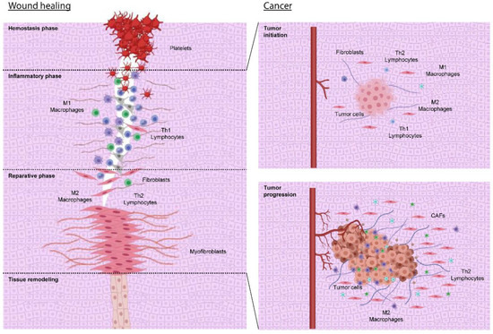
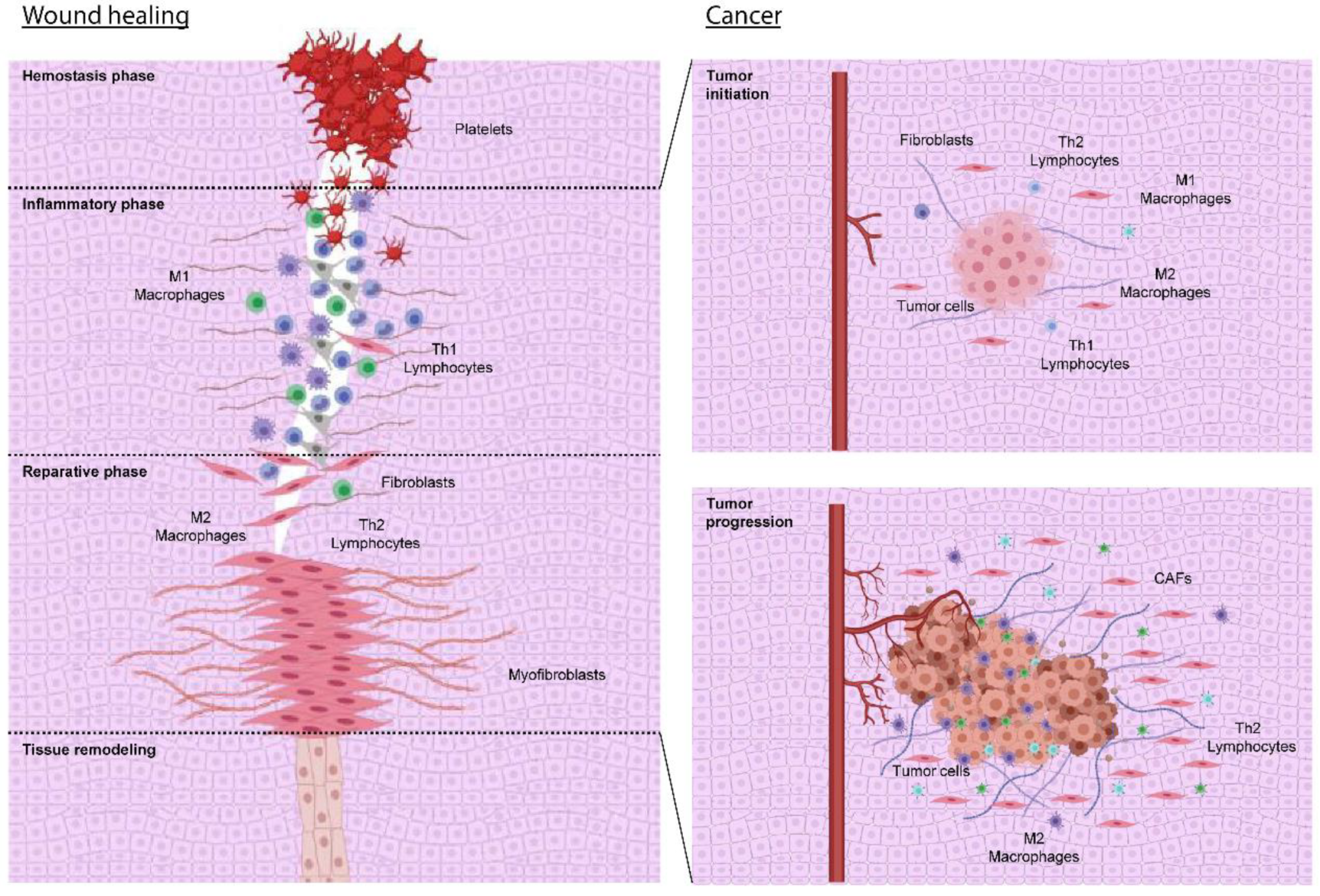
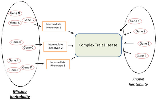
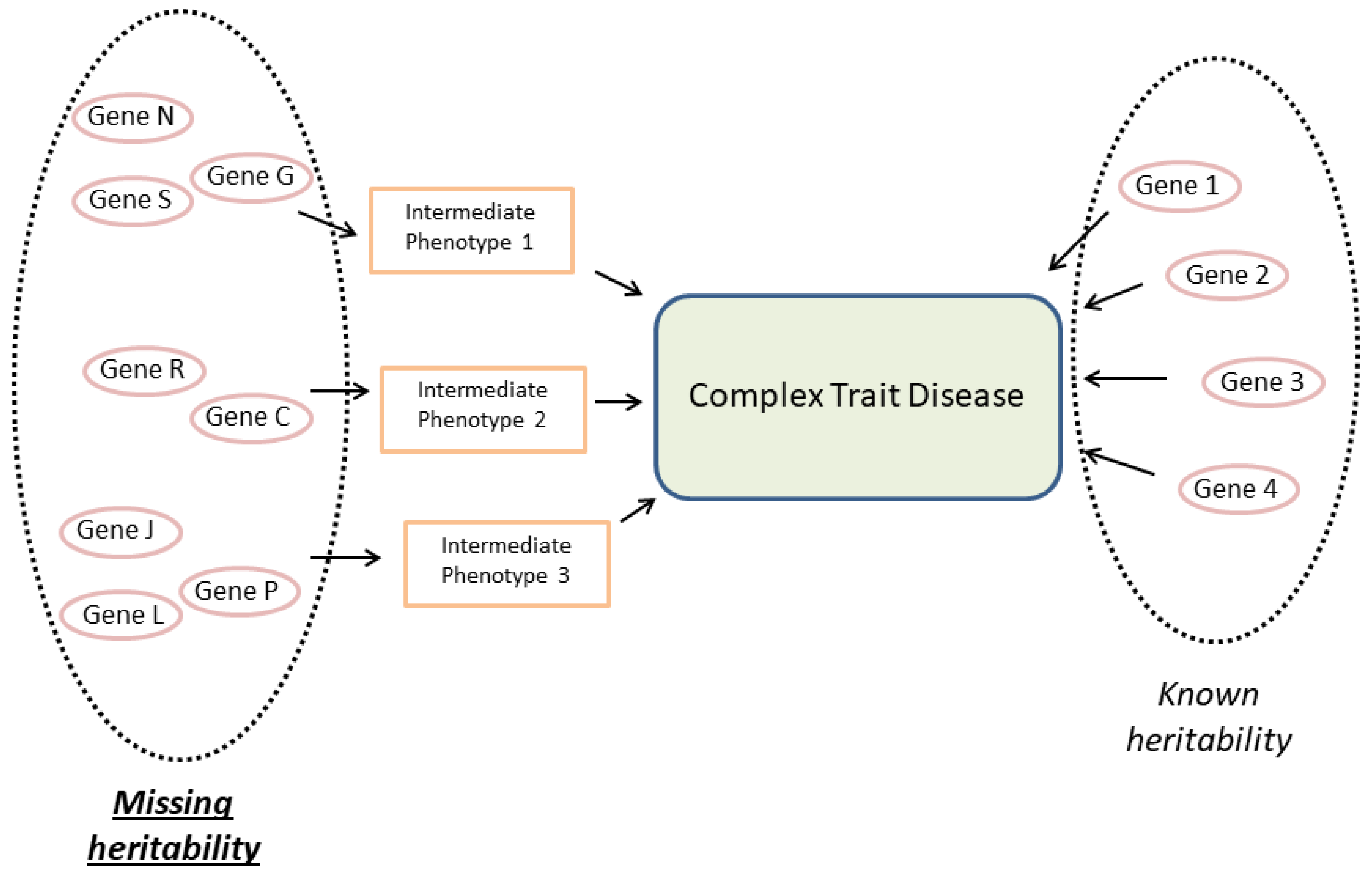

Publisher’s Note: MDPI stays neutral with regard to jurisdictional claims in published maps and institutional affiliations. |
© 2022 by the authors. Licensee MDPI, Basel, Switzerland. This article is an open access article distributed under the terms and conditions of the Creative Commons Attribution (CC BY) license (https://creativecommons.org/licenses/by/4.0/).