


Abstract
PYY, also known as peptide tyrosine tyrosine, is a 36-amino acid peptide that is highly conserved in vertebrate species. The role of PYY has long been established as a satiety hormone released by “L-cells,” a subset of enteroendocrine cells of the gastrointestinal tract, especially in the ileum and colon. In the mammalian intestine, full-length PYY (PYY1–36) is cleaved by DPP-IV to form endocrine PYY3–36, after which PYY3–36 is released into the bloodstream. Recently, we reported that PYY1–36 is also expressed by Paneth cells (PCs) of the gut. Previously underappreciated and never before demonstrated, full-length PYY1–36 has antifungal activity and regulates fungal commensalism in the gut. In this review, we summarize the current understanding of PYY—its mode of action and regulation of this unique innate antimicrobial peptide that specifically kills the virulent hyphae form, but not the commensal yeast form, of Candida albicans. Finally, we speculate on the potential roles of PYY in regulating obesity and in the pathogenesis of Crohn’s disease.
1. Introduction
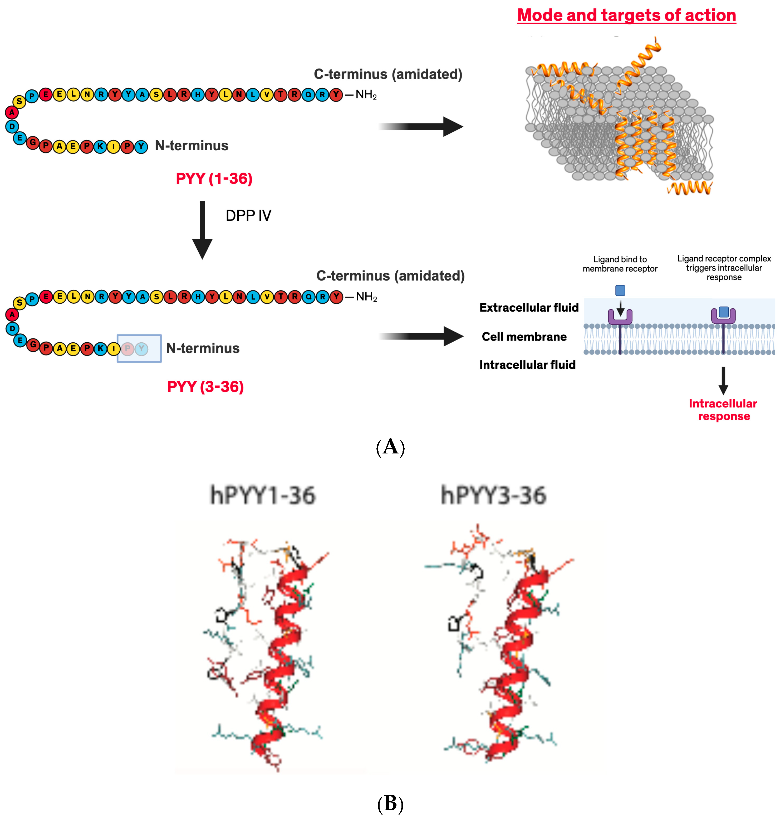
PYY (peptide tyrosine tyrosine) is a well-described satiety hormone which was named after the presence of tyrosine (Y) residues on its C- and N-terminals [1]. This 36-amino acid peptide is highly conserved in vertebrate species, and its structure is virtually unchanged between cartilaginous fish and humans. PYY was first isolated from porcine intestine and brain stem tissue in 1982 [2]. Since then, diverse regulatory roles of PYY have been identified, mostly involving the regulation of energy homeostasis and food intake, digestion, circadian rhythms, and other endocrine sympathetic vascular tone and autonomic functions [1]. PYY peptide exists in two endogenous biologically active forms, full-length PYY1–36 and the truncated PYY3–36 form [3,4,5] (Figure 1). PYY3–36 is the predominant circulatory form with the first two N-terminal amino acids of PYY1–36 cleaved by dipeptidyl peptidase IV (DPP-IV), a ubiquitous serine protease that cleaves at the canonical penultimate proline to form PYY3–36, a satiety hormone. Cleavage of the N-terminal tyrosine–proline by DPP-IV changes the tertiary structure of PYY [6,7]. While PYY1–36 has the capacity to bind to the Y1, Y2, Y4, or Y5 receptors, PYY3–36 has exclusive specificity towards only the Y2 receptor [8], thus altering its biological effects. Diverse regulatory roles of PYY3–36 have been identified, mostly involving the regulation of energy homeostasis and food intake, digestion, circadian rhythms, and other endocrine sympathetic vascular tone and autonomic functions [1].
Until Pierre et al. [9] reported the antifungal activity of PYY1–36, PYY was known mainly as a satiety hormone. In the lumen of the gut, PYY1–36 released by secretory granules of murine and human Paneth cells (PCs) is protected from DPP-IV cleavage by mucus [9]. While PC-PYY1–36 has marginal antibacterial activity, its specificity towards only the virulent hyphal form of Candida, and not the commensal yeast form, makes it even more distinctive [9]. As such, PYY joins a growing list of gut hormones and enteric neuropeptides that possess antimicrobial activity [10,11,12,13,14]. In this review, we summarize the previously known facts about PYY including the structure and satiety function and highlight its newly discovered role as an antimicrobial peptide.
Figure 1.
PYY exists in two forms PYY1–36 and PYY3–36. (A) PYY1–36 has specific antimicrobial activity against hyphae form of C. albicans (shown later) and penetrates the phospholipid membrane of hyphae to induce killing. Cleavage of the first two amino acids from the N-terminus of PYY by DPP-IV changes the structure and therefore function of the peptide. PYY3–36 acts as a satiety hormone binding to Y2 receptors in the brain to regulate feeding. (B) PEP-FOLD4 peptide models of human PYY1–36 and PYY3–36 [15,16,17,18]. Figure in (A) was made with Biorender.
2. Structure of PYY
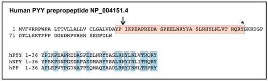
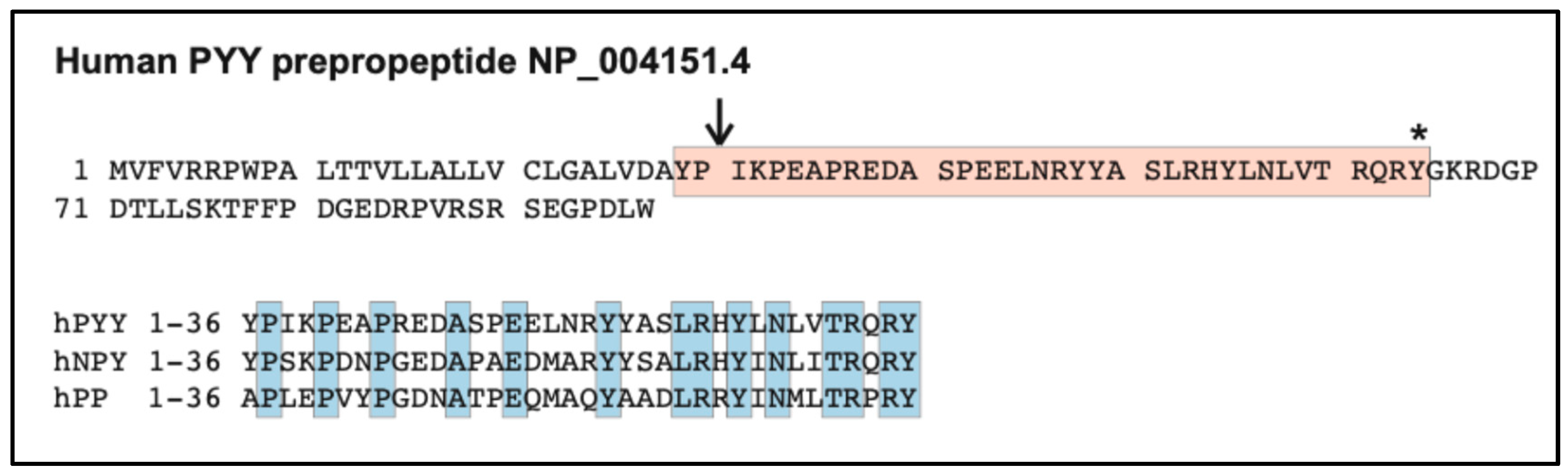
PYY belongs to the Neuropeptide Y (NPY) family of peptides, also known as the Pancreatic Peptide (PP)-Fold family of peptides, by sharing 14 amino acids at the same positions as that of pancreatic peptide (Figure 2). This family also includes peptides NPY and pancreatic polypeptide (PP), characterized by varying levels of sequence conservation. NPY is the most prominent neuropeptide in neuronal tissue of the central and peripheral nervous systems and is orexigenic, whereas PYY and PP are endocrine peptides of the gastrointestinal tract that act as satiety-regulating hormones [19]. The first NMR structure of PYY was studied in 2000 which showed a monomer with a well-ordered backfolded N-terminus and a kinked helix [6]. Mutational studies indicated that the preference for different receptors by PYY was determined both by C- and N-terminal composition and length which implied a joint between both C- and N-terminus that provided a surface for the binding of different Y receptors [6].
Figure 2.
(Top panel), peptide sequence of human prepropeptide PYY (NCBI Reference Sequence NP_004151.4). PYY1–36 is shaded in orange. Cleavage site by DPP-IV to create PYY3–36 is indicated by the arrow. PYY Y36 is amidated (*). (Bottom panel), sequence alignment of human PYY and related peptides NPY and PP. Shared sequences are shaded in blue.
PYY is encoded by the PYY gene in humans (Figure 2) and is found on chromosome 17 at position 17q21.31 (https://www.ncbi.nlm.nih.gov/datasets/gene/5697/, accessed on 15 November 2026) [20]. This gene is located 10 kilobases away from the PP-encoding gene and has four exons and three introns. The gene for NPY is located on chromosome 7. These three genes represent a multiple gene duplication event that resulted in these peptides having the same tertiary structure of the PP-fold, a U-shaped peptide composed of an alpha-helix and polyproline helix II connected by a beta-turn (Figure 1B) [21].
The PP-fold that is formed by the incorporation of Pro2, Pro5, Pro8, Gly9, Tyr20, and Tyr27 protects the peptide against enzymatic activity and produces an energy-reducing hydrophobic pocket [22]. The C-terminal alpha−helix is important for the structural integrity of PYY. The alpha−helix of PYY is amphipathic, where one side of the helix is comprised of hydrophilic amino acids while the other side is made up of hydrophobic residues. In a partitioning assay that tested the proclivity of PYY to an aqueous buffer or the same buffer containing 2.5% mucus, PYY1–36 preferentially partitioned into mucus [9], likely through electrostatic charge interactions, hence explaining its retention on the mucosal surface. In contrast, PYY3–36 was not detected in the overlying mucus. Therefore, it was hypothesized that mucus inhibited DPP-IV activity. DPP-IV and PYY1–36 were incubated with or without mucin in an in vitro assay. PYY3–36 was generated in the absence of mucin (detected by Liquid Chromatography-Mass Spectrometry) [9], but not when mucin was present, suggesting the proteolytic protection of PYY1–36 within mucus layers. Thus, mucus protects the host from fungal hyphae by inhibiting cleavage by DPP-IV, retaining the full-length PYY peptide, and, in turn, allowing PYY1–36’s antimicrobial functions.
3. Regulation and Secretion of PYY as a Satiety Hormone
PYY1–36 release into the lumen by L-cells is induced following a meal by intraluminal nutrients like salts, lipids, amino acids, short-chain fatty acids, bile salts, and glucose [23]. The levels of PYY depend on the meal size and composition. Protein, fat and fiber-rich diets are known to induce high levels of PYY [24]. In addition, the production of short-chain fatty acids from the digestion of dietary fibers by gut microbes is also a stimulant for L-cells to produce PYY [25]. The neural reflex involving the vagus nerve and other regulatory peptides including CCK (cholecystokinin), gastrin, VIP (vasoactive intestinal polypeptide), and GLP-1 (glucagon-like peptide-1) also regulates PYY release. Notably, GLP-1 is co-secreted with PYY from L-cells and may act synergistically [26]. PYY3–36 binds to the Y2 receptor following the cleavage by DPP-IV and thus inhibits the secretion of fluid and electrolytes, slows down gastrointestinal transit, and nutrient absorption, and produces the systemic feeling of fullness. PYY is also a strong enterogastrone and inhibitor of pancreatic secretion. PYY-knockout mice show an increase in body weight and fat mass and lower glucose tolerance [27]. Moreover, obese individuals secrete less PYY than those who are non-obese [28].
Full-length PYY1–36 is a precursor for the satiety hormone, PYY3–36. The two N-terminal amino acids of PYY1–36 are cleaved by dipeptidyl peptidase IV (DPP-IV), a ubiquitous serine protease that cleaves at the canonical penultimate proline to form PYY3–36. Whereas PYY1–36 binds to Y receptor subtypes Y1, Y2, Y4, and Y5 with a low affinity, PYY3–36 shows a particular high affinity for the Y2-receptor subtype and a very low affinity for the Y1-receptor and Y5-receptor subtypes [1]. Cleavage of the N-terminal tyrosine–proline by DPP-IV changes the tertiary structure of PYY (Figure 1), thus changing its receptor specificity and biological effects [29]. Due to the difference in receptor affinities, PYY3–36 binding to the Y2 receptor mediates satiety and weight loss as well as decreases appetite, whereas PYY1–36 increases appetite and stimulates weight gain via the other Y receptors [30].
The specific binding of PYY3–36 to the Y2 receptor is thought to be mediated by the C-terminus end of PYY in the hippocampus of the brain and sympathetic and parasympathetic nerve fibers. The Y2 receptor is a seven-transmembrane-spanning, G-protein-coupled receptor consisting of 381 amino acids. This receptor is found in humans throughout the central nervous system and is localized in the hypothalamic regions of the arcuate nucleus (ARC), dorsomedial hypothalamus, and the preoptic nucleus. Agonists of the Y2 receptor suppress the release of NPY whereas Y2 receptor antagonists stimulate NPY release. In direct opposition, PYY has an agonistic effect on Y2 receptors, therefore the binding of PYY leads to the inhibition of food intake [31]. Mice with a deletion of the Y2 receptor are unable to inhibit food intake upon the administration of PYY [32].
Recently, PYY3–36 has also been associated with anorexigenic pro-opiomelanocortin (POMC) neurons. The binding of agonists such as PYY3–36 to Y2 receptors expressed on POMC neurons results in the release of the alpha-melanocyte stimulation hormone (α-MSH) from POMC neurons, further suppressing food intake [33]. The inhibition of NPY secretion by PYY3–36 indirectly leads to an increase in the stimulation of POMC neurons, which further helps the satiety effect of PYY3–36. There are some contradicting reports regarding the stimulation of these neurons by PYY3–36. One study by Acuna-Goycolea et al. reported that PYY3–36 inhibited both NPY and POMC neurons in a dose-dependent manner [34]. In this case, the inhibition of NPY release by PYY3–36 may dominate over its inhibiting effect on POMC neurons. Another report further suggested that POMC might not be the primary regulator of satiety induced by PYY3–36, as there was reduced food intake in the POMC knockout mice upon the administration of PYY3–36 [35]. Thus, a decrease in NPY release appears to be the primary mechanism of the induction of the satiety effect by PYY3–36.
4. Regulation and Secretion of PYY by Paneth Cells
First discovered by Gustav Schwalbe in the 19th century, Paneth cells (PCs) are pyramidal-shaped, columnar, secretory epithelial cells found in small clusters at base of the crypts of the small intestine [36]. Although Paneth cells were first discovered in humans, they are found in other vertebrates like primates, rodents, horse, sheep and chicken. They have extensive endoplasmic reticula (ER), a well-developed Golgi network and apically oriented secretory granules. The large secretory granules in Paneth cells accumulate a wide variety of proteins that contain antimicrobial peptides (AMPs), enzymes and growth factors, many of which are crucial in host defense [37]. The antimicrobial-rich granules are discharged into the lumen of crypts, releasing AMPs to protect the host from microbial invasion. As AMPs also kill resident microbes, PCs help control the composition of the enteric microbiota and maintain gut homeostasis. Alterations in PCs or PC dysfunction contribute to several diseases, for example, graft-versus-host disease (GVHD) and inflammatory bowel disease (IBD), including Crohn’s disease (CD) and ulcerative colitis [38].
Recently, Pierre, et al. found that PYY is also secreted by Paneth cells [9]. This was a breakthrough as it led to the discovery of the antifungal activity of PYY. While it is not yet known whether PYY gene expression in Paneth cells is regulated the same way, PYY is transcribed and translated in L-cells as the full-length PYY1–36 peptide [24], then processed into PYY3–36 by DPP-IV. Whereas L-cells release PYY into the bloodstream post-prandially, Paneth cells secrete AMPs into the gut lumen when triggered by microbial products. To provide adequate defense, Paneth cells must be poised for immediate release upon the sensing of microbes, therefore PYY must already be transcribed, translated, and sorted into secretory granules for immediate release upon stimulation. Studies have shown that PYY and another AMP, lysozyme, were found in separate granules by high-resolution stimulated emission depletion (STED) microscopy [9]. Since lysozyme is known to have antibacterial activity [39], what regulates the selective release of lysozyme or PYY or other AMPs? The compartmentalization of different AMPs secreted by Paneth cells suggests regulated secretion mechanisms for different pathogens, and that Paneth cells may have the ability to selectively release specific AMPs depending on the target.
In the case of PYY, secretion is regulated by and targeted specifically towards the hyphae of Candida species. To study the regulation of PC-PYY1–36, Pierre, et al. [9] used small bowel (ileal) ligated loops created through in vivo non-survival surgery that maintains vascular flow until tissue harvest. Loops were filled with solutions containing C. albicans hyphae, yeast or lipopolysaccharide (LPS). The secretion of PC-PYY1–36 into mucus was found to be specifically stimulated by only live C. albicans hyphae and not yeast or LPS [9]. There was an accumulation of PYY1–36, not PYY3–36, in the mucus but not in the lumen, as determined by mass spectrometry [9]. One possibility is that PYY1–36 is subjected to other peptidases in the lumen and leads to the significant degradation of PYY1–36. Another plausible explanation is that the mucus retains PYY1–36, preventing its translocation to the lumen. This may be why the endocrine form PC-PYY1–36 was not found in luminal fluids and had more affinity towards the mucus layer. Further studies are required to test the regulation of PYY secretion, and to investigate the regulation of selective release of AMPs that target specific pathogens.
5. PYY as Host Defense Peptide
The mechanism by which the host senses and controls the fungi of the gut microbiome has been inadequately explored, except in cases of extreme overgrowth. C. albicans is a polymorphic commensal yeast found in 70% of humans. C. albicans and other Candida spp. are among the most abundant fungal species of the healthy human gut, where they live harmoniously with the host and other microbiota. When this relationship is disturbed, such as during dysbiosis, commensal fungi of the gut microbiome like C. albicans can become pathogenic to cause mucosal inflammation and injury. It can transform into virulent hyphae under stress and activating conditions to cause or contribute to disease in immunocompromised individuals and can cause some severe and life-threatening systemic infections. This ability to change reversibly between yeast and hyphal morphologies is a key determinant of its interactions with the host. C. albicans propagates primarily as single-celled, oval yeasts during the commensal colonization of the skin and oral mucosa; however, during invasive infections of the blood and deep tissues, a multicellular, filamentous hyphal form predominates [40].
As Paneth cells play a crucial role in maintaining gut microbiota by secreting AMPs, the expression of PYY in secretory granules of human and mouse Paneth cells raised the possibility that PYY may also function as an antimicrobial peptide. The alpha-helical amphipathic structure of PYY is different from the defensin peptides produced by Paneth cells. However, the C-terminal half of PYY1–36 resembles other peptides with antifungal activity. This similarity led to further investigation of the undiscovered roles of Paneth cell-derived PYY (PC-PYY).
Antimicrobial studies confirmed that PYY1–36 had modest antibacterial activity [9]. In growth curve studies, the presence of PYY1–36 had little effect on the growth of four distinct bacterial species found in the gut, Enterococcus faecalis, Lacticaseibacillus rhamnosus GG, Listeria monocytogenes, and Escherichia coli, with minimal inhibitory concentrations (MICs) of approximately 10 μM. By comparison, magainin-2, an AMP expressed in the skin of Xenopus laevis, demonstrated an MIC of 1 μM against the same bacteria. Interestingly, PYY3–36, the truncated peptide had no antimicrobial activity. Based on the predicted structure (Figure 1) of PYY1–36 and PYY3–36, the removal of the first two amino acids of PYY would not alter the alpha-helical structure. However, NPY, a closely related peptide and also a member of the PP family, has been shown to self-associate and form dimers in an antiparallel fashion [41]. Therefore, one possible explanation for the lack of antimicrobial activity by PYY3–36 is that the absence of the Tyr and Pro may affect PYY’s ability to multimerize, weakening these complexes and preventing its ability to penetrate target membranes.
Strikingly, PYY1–36 exhibited significant and specific antifungal activity, specifically against the hyphal or virulent form of C. albicans. When C. albicans yeast and hyphae were challenged with PYY1–36, the growth of only hyphae (as measured by dried mass) was maximally inhibited at a concentration of 30 μM. Notably, this activity was bimodal. At concentrations above 30 μM, PYY1–36 lost its ability to inhibit hyphal growth. It was speculated that the high concentrations of PYY1–36 led to multimerization prior to targeting the membrane, which prevented the effective killing of hyphae. A similar effect was observed in hyphal biofilm assays. In fact, the addition of mucus in the assay further enhanced the ability of PYY1–36 to block the biofilm formation of hyphae, even at concentrations of up to 100 μM. Mucus likely prevented the multimerization of PYY1–36 peptides under these conditions. In contrast, PYY1–36 had no effect on the growth of C. albicans yeast. Interestingly, PYY3–36, the truncated peptide had no antifungal activity [9]. When other fungal species were tested, PYY1–36 showed activity against C. tropicalis and C. dubliniensis, but not Aspergillus fumigatus. Thus, PYY1–36 may be specific against Candida species.
Pierre et al. further investigated the mechanism behind the selective killing of hyphal forms by PYY [9]. The molecular basis for the discrimination remains obscure, but interaction with the morphotype-specific cell wall or plasma membrane components seems likely. The reversible transition of C. albicans between yeast and hyphae morphotypes involves a highly regulated genetic program where the organism undergoes major structural and functional changes. Cell wall polysaccharide analysis showed that yeast cell walls contained significantly higher amounts of mannan but lower amounts chitin and glucan (which can affect the net charge) compared to that of hyphal cell walls [42]. In addition, studies from Kriznik, et al. suggest that yeast and hyphae cell walls have different surface hydrophobicities, presumably due to specific hydrophobic proteins inside the most external layers of the fungal wall [43]. Thus, it was hypothesized, based on the amphipathic structure of PYY, that specific electrostatic and biophysical interactions could be a mechanism for the selectivity of C. albicans hyphae.
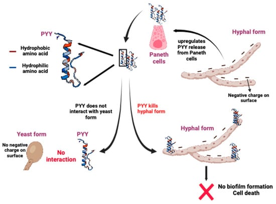
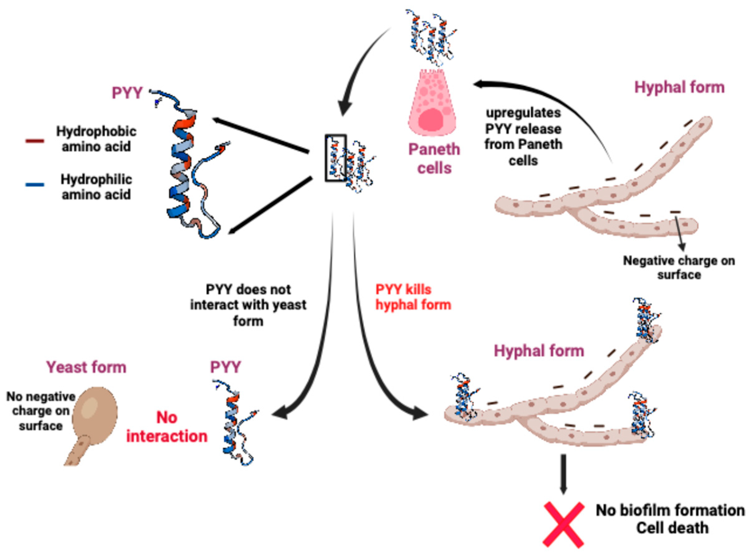
To test this, a cationic ferritin probe was used to demonstrate that the cell walls of the hyphal and yeast forms of C. albicans are differentially charged. Strikingly, the positively charged ferritin probe was bound only along the negatively charged phospholipid membrane of C. albicans hyphae. By contrast, no accumulation of the ferritin probe on the yeast membrane was observed, indicating that the yeast membrane is not negatively charged (Figure 3). Further microscopic studies revealed that PYY1–36 peptide causes membrane disruption particularly at the growing tip of hyphae, impairment of biofilm formation, and ultimate death. Pierre et al. also challenged wildtype and PYY KO mice [9 with C. albicans. Because C. albicans is not native to the mouse gut, both wildtype and PYY KO mice fell severely ill [9]. However, more wildtype mice cleared C. albicans and the survival rate was better than PYY KO mice. The conversion of C. albicans to hyphae in the gut of PYY KO mice was observed, but not in wildtype mice. This provides evidence that PYY is critical for the protection of mice from pathogenic C. albicans. PC-PYY1–36 differs from other PC-AMPs by selectively targeting and killing the virulent (hyphal), but not commensal (yeast) forms of polymorphic gut fungi like C. albicans, thereby playing an important role in maintaining the fungal commensalism of the gut.
Figure 3.
PC-PYY selectively kills the hyphae form, not the yeast form of C. albicans. During dysbiosis, increased virulent hyphae of C. albicans upregulate the secretion of PYY from Paneth cells. PYY binds to the hyphal form due to the presence of negative charges on these hyphae, whereas there is no interaction between yeast and PYY, due to the lack of negative charge on the surface of yeast. PYY mostly binds to the growing end of hyphae, inhibiting biofilm production and leading to hyphal cell death. Figure made with Biorender.
The dual use of PYY by the host as an appetite-regulating hormone and antifungal peptide is a model in which PYY is encoded by a single gene, but proteolytic cleavage by DPP-IV in the brush border of the intestine lend different functions to the resulting peptide products. In addition, the activity of PYY is affected by the milieu. PYY shows a biphasic dose response in aqueous buffer, likely due to concentration-dependent multimerization or self-aggregation due to its amphipathic nature (see below), whereas, in the presence of mucus, it shows a sigmoidal curve and monophasic activity because mucus prevents the self-aggregation [9]. Therefore, PYY1–36 has higher bioactivity and kills C. albicans more efficiently in mucus than in aqueous solution. A decrease in the binding of C. albicans hyphae to intestinal epithelial cells was observed following treatment with PYY1–36 during in vitro studies in the presence of mucus [9]. In conclusion, the mucus layer in the intestine serves to (1) prevent self-aggregation of PYY and thereby retaining its bioactivity, (2) attract PYY where it retains the peptide and increases its local concentration, and (3) inactivate DPP-IV to prevent cleavage to the hormonal form PYY3–36.
6. Evolutionary Perspectives on the Functions of PYY
The expression of Pyy can be found in all vertebrate animals [44]. PYY evolved from a common ancestor with Neuropeptide Y (NPY) early in vertebrate evolution, likely arising from a gene duplication event before jawed vertebrates appeared. A second duplication later generated pancreatic polypeptide (PP) from a PYY precursor in early tetrapods [44]. PYY is a highly conserved gastrointestinal hormone across vertebrates, and it shows more variability than NPY, especially in mammals. In bony and cartilaginous fishes, PYY and NPY have only five to six amino acid differences, indicating a high functional overlap. In tetrapods, PYY and NPY differ by 9–12 positions [45] (Figure 2). While generally known as a gut hormone, PYY’s ancestral role may have involved both gut regulation and neurotransmission, especially in fish. It has been studied primarily for its endocrine role and only recently has it been more extensively studied for its antimicrobial properties.
PYY and the PP-fold family are found in vertebrates. While invertebrates express Neuropeptide F (NPF), it shares a high structural homology to vertebrate PYY. PYY3–36 seems to have appeared in more highly-evolved organisms that required a finer regulation of food intake and energy conservation. The cleavage of PYY is mediated by a serine protease DPP-IV, which changes PYY’s affinity for Y2 from low-binding to high-affinity [8]. Four receptor subtypes, Y1, Y2, Y4, and Y5, are known to bind PYY1–36, whereas the PYY3–36 selectively binds to Y2 receptors, which inhibit feeding [46]. Dipeptidyl cleavage thus transforms an unselective Y agonist into a highly selective Y2 agonist, PYY3–36. Therefore, in higher vertebrates, these organisms may have developed the need to signal through Y receptors for food intake regulation.
The evolutionary link between the antimicrobial function and hormonal function of PYY is not yet understood. We speculate that the antimicrobial function of PYY came before its hormonal function, and, perhaps, this feeding regulation was necessitated by the evolution of intestinal tracts. Invertebrates mostly depend on the ingestion of unicellular organisms such as bacteria, microalgae, and protists [47]. Cationic antimicrobial peptides have been found in lower invertebrates, whereas neurosensory systems evolved later and are found in higher invertebrates [47]. It is thought that complex nervous systems first appeared during the Ediacaran period [48]. Lower invertebrates do not have through-gut intestines and, when more complex vertebrates evolved intestinal tracts, the need to protect the intestinal epithelium remained important, along with the need to regulate feeding. The comparative analysis of PYY across vertebrates and invertebrates in future studies will help in understanding the evolutionary diversification of the function of PYY.
7. Other AMPs Secreted by Paneth Cells
Apart from PYY, Paneth cells produce a variety of other antimicrobial peptides. These antimicrobial peptides (AMPs) are small host defense peptides (less than 100 amino acids) that act as crucial components of the innate defense system of the intestine, providing a first line of protection against pathogens [49]. Some of these AMPs like PYY can be very specific in their killing mechanism [9,49,50,51,52], while others have a broader range of targets. Most of the AMPs are positively charged (cationic) and their mode of action is different from traditional antibiotics. Due to their positive charge, AMPs bind to anionic bacterial cell membranes, inducing damage and the death of these microbes. Thus, these AMPs have huge potential for the treatment of drug-resistant microbes [53]. In addition to antibacterial activity, AMPs also show antifungal and antiviral activities. Some of the most common peptides by Paneth cells are discussed below.
- (A)
- α-Defensins (Human Defensin-5 and Human Defensin-6) (HD-5 and HD-6)—These are the most abundant Paneth cell-produced antimicrobial peptides in humans. Their activities are distinct, but, together, they synergize to enhance bacteriocidal activity. [54]. HD-5 also displays antiviral activity by interacting with viral proteins and blocking viral cellular attachment or endosomal escape [55,56].
- (B)
- Cathelicidin (LL-37)—LL-37 contributes to antimicrobial defense by membrane disruption, protein and nucleic acid binding, and the disintegration of biofilms. Its net positive charge allows it to bind to negatively charged bacterial membranes, which then permits penetration into the cell membrane to interact with intracellular targets [57].
- (C)
- Regenerating islet-derived protein 3 alpha (REG3A) (human; REGIIIγ in mice)—Another hallmark secretory product of Paneth cells responsible for innate mucosal defense, RegIIIγ is particularly active against Gram-positive bacteria. Similar to PYY’s actions, it is thought to be a link between antimicrobial activity and metabolic regulation, though the mechanism by which this occurs is still being debated [58].
- (D)
- Lysozyme—Lysozyme is a highly abundant protein produced by Paneth cells that hydrolyzes the beta-1,4-glycosidic bonds between N-acetylmuramic acid and N-acetylglucosamide in bacterial peptidoglycans of Gram-positive bacteria cell walls [59].
8. Other Gut Neuropeptides That Possess Antimicrobial Activity
As a gut hormone, PYY is not unique in its ability to target microbes. The closely related NPY has also been shown to target Cryptococcus neoformans, Arthrodema semii, E. coli, E. faecalis, and Pseudomonas aeruginosa [10,14]. Vouldoukis, et al. even showed that NPY inhibited the growth of C. albicans with an MIC of 25 μg/mL, though it was unclear whether NPY had specificity towards the yeast or hyphal form [10]. Interestingly, one group studying porcine NPY showed that the MIC of C. albicans changed from 10–12 μM to 1.5–2.0 μM when the first 12 amino acids at the N-terminus was truncated (NPY13–36) [60]. Ghrelin, a hormone secreted by the stomach and pancreas that signals hunger, inhibits the growth of Gram-negative bacteria such as E. coli and P. aeruginosa [12].
Neuropeptides produced by the enteric nervous system have also demonstrated antimicrobial activity. An 11-amino acid peptide regulator of gut motility, Substance P (SP), targets Staphylococcus aureus, E. coli, E. faecalis, Proteus vulgaris, P. aeruginosa, and C. albicans [14]. In in vitro radial diffusion assays, SP, compared to NPY, was highly effective against Lactobacillus acidophilus (MIC 74.1 vs. 283.7 μg/mL), E. coli (MIC 5.7 vs. 46.5 μg/mL), P. aeruginosa (MIC 15.7 vs. 134.3 μg/mL), and C. albicans (MIC 8.1 vs 243.2 μg/mL). SP showed little activity against E. faecalis and S. aureus. There are other neuropeptides that have been shown in vitro to have antibacterial activity and some also target C. albicans, namely, the alpha-melanocyte stimulating hormone (alpha-MSH), vasoactive intestinal peptide (VIP), calcitonin gene-related peptide (CGRP), and adrenomedullin, to name a few [13].
Together, the fact that many gut hormones and neuropeptides display antimicrobial activity is a display of nature’s versatility. A common theme among these gut hormones and neuropeptides is a characteristic amphipathic alpha-helix and a net negative charge that attracts these peptides through electrostatic interactions to the surface of their targets [13,60]. Differences in the net charge of these peptides and the cationicity of the target membrane surface (from phosphomannans or other glucans) may account for the diversity and broad-spectrum activity across commensal and pathogenic microbial species. What makes PYY unique is its specificity for the hyphal but not yeast form of C. albicans. Studies showing antifungal activity against C. albicans should be performed comparing the yeast versus hyphae forms. Based on PYY’s selectivity for the anionic membrane of hyphae, we predict that the activity of other neuropeptides against C. albicans hyphae may be even greater than what has been previously reported.
9. Clinical Implications of PYY
There is no approved therapeutic use of PYY yet, but PYY could be a potential target for obesity and weight management, as it is a satiety hormone released alongside glucagon-like peptide-1 (GLP-1), the subject of much attention lately due to the development of GLP-1 receptor agonists for inducing weight loss. Over recent years, a major role in diabetes remission after surgery has been attributed to GLP-1 analogues (exenatide, liraglutide, dulaglutide, and lixisenatide). These are already listed among current antidiabetic treatments [61]. In addition to GLP-1, the role of PYY is now increasingly recognized in the surgical control of diabetes extending beyond its classical effect on appetite regulation. The subcutaneous infusion of three hormones, GLP-1, PYY, and oxyntomodulin, over a 4-week period improved glucose tolerance in diabetic patients by an effect superior to RYGB and caloric restriction [62].
For IBD, the serological biomarker anti-Saccharomyces cerevisiae antibodies (ASCAs) is a routine clinical test for patients who have been diagnosed with Crohn’s disease (CD) or ulcerative colitis. Often used to distinguish between these two types of IBD, patients with CD exhibit high levels of ASCAs and ulcerative colitis patients have lower levels [63]. The levels of ASCAs appeared to correlate with disease progression and the need for surgical intervention. Some reported that high levels of ASCAs was associated with the ileal or ileocolonic involvement of CD [64]. Today, ASCAs are considered a reliable and robust diagnostic for managing patients with Crohn’s disease.
Recently, Sendid et al. [65] reviewed and updated new developments since ASCAs became a preferred clinical tool. Studies showed that ASCAs have been shown to include not just antibodies against the oligomannan components of S. cerevisiae cell walls, but antibodies against proteins from other types of fungi, including C. albicans [65]. They argue that a main source of ASCAs may not be S. cerevisiae, but, rather, C. albicans. Indeed, ASCAs immunopurified from CD patients used to probe C. albicans hyphae in tissue sections from biopsies of patients with systemic C. albicans infection showed strong reactivity [66]. Moreover, they showed that C. albicans proteins are mannosylated and share similar epitopes with other mannoproteins. They further posit that antibodies against proteins from several other fungal species (Candida spp., Malassezia spp.) could also make up the repertoire of ASCAs.
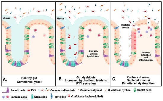
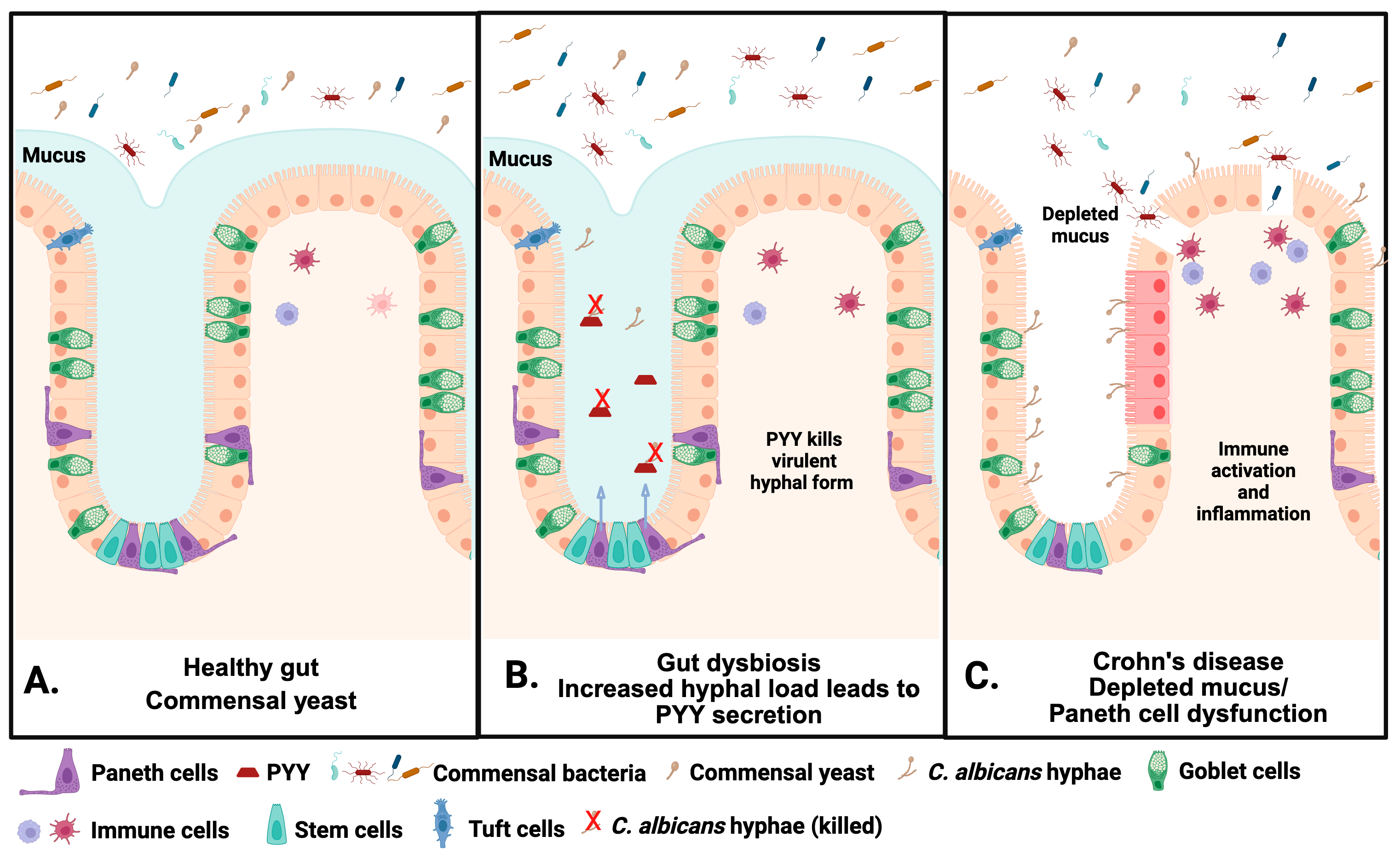
Based on the discovery that PC-PYY regulates fungal commensalism in the ileum by targeting the hyphal morphotype of C. albicans, we hypothesize that defects in PYY function could increase the risk of developing IBD. The impairment of PC-PYY may have dire negative consequences relevant to the etiopathogenesis of IBD, particularly Crohn’s disease involving the terminal ileum (iCD) where Paneth cells are found. Several studies have implicated dysfunctional Paneth cells as a cause underlying this subset of CD. PC-PYY dysfunction could therefore lead to the loss of fungal commensalism and an increased opportunity for fungal overgrowth that triggers and promotes chronic disease (Figure 4). Given that PYY1–36 is a potent AMP against the virulent form of C. albicans and is secreted by ileal Paneth cells [9], and C. albicans is the most abundant fungal species in the human gut, we speculate that some CD patients, especially those with ileal or ileocolonic involvement, may have reduced production or activity of PYY1–36 from Paneth cells. We further hypothesize that the lack of ability to keep C. albicans in check could contribute to the onset or progression of disease. The C. albicans-derived signal that triggers ileal Paneth cells to secrete PYY is still unclear. The lack of PYY1–36 could also arise from mutations in the PYY gene that diminish the potency of the PYY1–36-induced killing of C. albicans hyphae. The genetic screening of CD patients for PYY and determining PYY levels in Paneth cell granules could be done to test this hypothesis.
Figure 4.
Regulation of fungal commensalism in the healthy and diseased intestine. (A) In a healthy gut, fungi such as C. albicans are held in check, with the commensal yeast form dominating. (B) Under conditions of gut dysbiosis, hyphal loads increase and stimulate secretion of PYY by Paneth cells to prevent the rise of pathogenic hyphae. (C) During disease states such as Crohn’s Disease, thinned mucus leads to a breach in the intestinal epithelial barrier and a rise in pathogenic C. albicans. These, combined with a Paneth cell dysfunction, could result in disease. Figure made with Biorender.
10. Conclusions and Future Directions
PYY is an example of the wisdom of nature. A molecule that may have started off as an AMP became repurposed as an endocrine peptide to regulate satiety through receptor-mediated interactions with the brain. As an AMP, PC-PYY has an essential role in maintaining fungal commensalism.
The co-evolution or co-speciation of metazoans and digestive tract microbes over millions of years has led to dynamic and mutually beneficial relationships, but this necessitated the need to develop defense systems to counter breaches of intestinal homeostasis. For the Bacteria kingdom, many host mechanisms have been described that permit engraftment and colonization by specific gut bacteria, yet respond in the event virulence is encountered. However, much less is known about other microbial kingdoms such as Fungi that also colonize this space and are believed to also affect host health and disease. The fact that they are part of the “healthy” gut microbiome suggests they provide benefits, but what keeps them in check when perturbations are encountered remains poorly understood.
While PC-PYY has limited antibacterial activity, it targets virulent but not commensal C. albicans, an action that is due to its cationic properties that promote interaction with the anionic surface charge found in C. albicans hyphae, but not in yeast. This is the first example of an antimicrobial peptide that targets a particular morphotype within a species. The ability to target and clear virulent hyphal forms of C. albicans while sparing the commensal Candida yeast form is an attractive therapeutic target. This property arguably plays a role in maintaining the gut mycobiome in a state of commensalism. While C. albicans was used as a representative fungal organism in these studies, it remains to be determined whether other gut-resident fungal species (or pathogenic forms thereof) are susceptible to PYY.
The discovery of this novel and apparently selective properties of PYY as a PC antimicrobial peptide raises further questions about the need for specific types of AMPs to maintain the gut microbiota. Despite the seemingly non-specific, biophysical actions on gut microbiota, PC-PYY’s antimicrobial properties are uniquely selective, targeting virulent hyphal C. albicans and little activity against bacteria and commensal C. albicans yeast. This must also be the case for all other PC-AMPs. Otherwise, why would there be so many types of PC-AMPs if they all shared the same mechanisms of action? Moreover, the finding that PC-PYY is separately packaged in secretory granules of other PC-AMPs (e.g., lysozyme) suggests that its role, regulation, response to agonists, and functions are called upon only under specific circumstances and not part of an indiscriminate shotgun blast of PC-AMPs. When dysbiosis occurs in the gut, are only select granules containing the specific AMPs secreted to combat the right microbes? Or are granules non-specifically released all at once? These are intriguing questions that will reveal more about the antimicrobial mechanisms of the gut. Future studies are needed to elucidate this new mechanism of microbial commensalism.
Author Contributions
Conceptualization, E.B.C. and C.M.C.; writing—original draft preparation, D.K. and C.M.C.; writing—review and editing, E.B.C. and C.M.C.; funding acquisition, E.B.C. All authors have read and agreed to the published version of the manuscript.
Funding
We acknowledge support from NIH R01 DK113788, the Host-Microbe, Integrative and Clinical Biospecimens, and the Tissue Engineering and Cell Models Cores of the Center for Interdisciplinary Studies of Intestinal Inflammatory Diseases (NIH DDRCC P30 DK042086), and the University of Chicago Gastrointestinal Research Foundation.
Institutional Review Board Statement
Not applicable.
Informed Consent Statement
Not applicable.
Data Availability Statement
The original contributions presented in this study are included in the article. Further inquiries can be directed to the corresponding author.
Acknowledgments
The authors would like to thank Joseph Pierre and other members of the Chang lab who were involved in the discovery and characterization of PC-PYY.
Conflicts of Interest
The authors declare no conflicts of interest. The funders had no role in the design of the study; in the collection, analyses, or interpretation of the data; in the writing of the manuscript; or in the decision to publish the results.
Abbreviations
The following abbreviations are used in this manuscript:
| PYY | Peptide tyrosine tyrosine |
| PC | Paneth cell |
| AMP | Antimicrobial peptide |
| DPP-IV | Dipeptidyl peptidase IV |
| CD | Crohn’s Disease |
| NPY | Neuropeptide Y |
| PP | Pancreatic Peptide |
| GLP-1 | Glucagon-like Peptide-1 |
| IBDs | Inflammatory bowel diseases |
References
- Karra, E.; Chandarana, K.; Batterham, R.L. The Role of Peptide YY in Appetite Regulation and Obesity. J. Physiol. 2009, 587, 19–25. [Google Scholar] [CrossRef] [Scilit] [PubMed]
- Tatemoto, K.; Mutt, V. Isolation of Two Novel Candidate Hormones Using a Chemical Method for Finding Naturally Occurring Polypeptides. Nature 1980, 285, 417–418. [Google Scholar] [CrossRef] [Scilit] [PubMed]
- Grandt, D.; Schimiczek, M.; Beglinger, C.; Layer, P.; Goebell, H.; Eysselein, V.E.; Reeve, J.R. Two Molecular Forms of Peptide YY (PYY) Are Abundant in Human Blood: Characterization of a Radioimmunoassay Recognizing PYY 1-36 and PYY 3-36. Regul. Pept. 1994, 51, 151–159. [Google Scholar] [CrossRef] [Scilit] [PubMed]
- Batterham, R.L.; Heffron, H.; Kapoor, S.; Chivers, J.E.; Chandarana, K.; Herzog, H.; Le Roux, C.W.; Thomas, E.L.; Bell, J.D.; Withers, D.J. Critical Role for Peptide YY in Protein-Mediated Satiation and Body-Weight Regulation. Cell Metab. 2006, 4, 223–233. [Google Scholar] [CrossRef] [Scilit] [PubMed]
- Korner, J.; Inabnet, W.; Conwell, I.M.; Taveras, C.; Daud, A.; Olivero-Rivera, L.; Restuccia, N.L.; Bessler, M. Differential Effects of Gastric Bypass and Banding on Circulating Gut Hormone and Leptin Levels. Obesity 2006, 14, 1553–1561. [Google Scholar] [CrossRef] [Scilit] [PubMed]
- Keire, D.A.; Mannon, P.; Kobayashi, M.; Walsh, J.H.; Solomon, T.E.; Reeve, J.R. Primary Structures of PYY, [Pro34]PYY, and PYY-(3–36) Confer Different Conformations and Receptor Selectivity. Am. J. Physiol.-Gastrointest. Liver Physiol. 2000, 279, G126–G131. [Google Scholar] [CrossRef] [Scilit] [PubMed]
- Grandt, D.; Schimiczek, M.; Struk, K.; Shively, J.; Eysselein, V.E.; Goebell, H.; Reeve, J.R. Characterization of Two Forms of Peptide YY, PYY(1–36) and PYY(3–36), in the Rabbit. Peptides 1994, 15, 815–820. [Google Scholar] [CrossRef] [Scilit] [PubMed]
- Grandt, D.; Dahms, P.; Schimiczek, M.; Eysselein, V.E.; Reeve, J.R.; Mentlein, R. Proteolytic processing by dipeptidyl aminopeptidase IV generates receptor selectivity for peptide YY (PYY). Med. Klin. 1993, 88, 143–145. [Google Scholar]
- Pierre, J.F.; Peters, B.M.; La Torre, D.; Sidebottom, A.M.; Tao, Y.; Zhu, X.; Cham, C.M.; Wang, L.; Kambal, A.; Harris, K.G.; et al. Peptide YY: A Paneth Cell Antimicrobial Peptide That Maintains Candida Gut Commensalism. Science 2023, 381, 502–508. [Google Scholar] [CrossRef] [Scilit] [PubMed]
- Vouldoukis, I.; Shai, Y.; Nicolas, P.; Mor, A. Broad Spectrum Antibiotic Activity of skin-PYY. FEBS Lett. 1996, 380, 237–240. [Google Scholar] [CrossRef] [Scilit] [PubMed]
- Chiba, M.; Sanada, Y.; Kawano, S.; Murofushi, M.; Okada, I.; Yoshizawa, Y.; Gomi, A.; Yatsuzuka, M.; Toki, A.; Hirai, Y. Glicentin Inhibits Internalization of Enteric Bacteria by Cultured INT-407 Enterocytes. Pediatr. Surg. Int. 2007, 23, 551–554. [Google Scholar] [CrossRef] [Scilit] [PubMed]
- Min, C.; Ohta, K.; Kajiya, M.; Zhu, T.; Sharma, K.; Shin, J.; Mawardi, H.; Howait, M.; Hirschfeld, J.; Bahammam, L.; et al. The Antimicrobial Activity of the Appetite Peptide Hormone Ghrelin. Peptides 2012, 36, 151–156. [Google Scholar] [CrossRef] [Scilit] [PubMed]
- Aresti Sanz, J.; El Aidy, S. Microbiota and Gut Neuropeptides: A Dual Action of Antimicrobial Activity and Neuroimmune Response. Psychopharmacology 2019, 236, 1597–1609. [Google Scholar] [CrossRef] [Scilit] [PubMed]
- El Karim, I.A.; Linden, G.J.; Orr, D.F.; Lundy, F.T. Antimicrobial Activity of Neuropeptides against a Range of Micro-Organisms from Skin, Oral, Respiratory and Gastrointestinal Tract Sites. J. Neuroimmunol. 2008, 200, 11–16. [Google Scholar] [CrossRef] [Scilit] [PubMed]
- Lamiable, A.; Thévenet, P.; Rey, J.; Vavrusa, M.; Derreumaux, P.; Tufféry, P. PEP-FOLD3: Faster de Novo Structure Prediction for Linear Peptides in Solution and in Complex. Nucleic Acids Res. 2016, 44, W449–W454. [Google Scholar] [CrossRef] [Scilit] [PubMed]
- Binette, V.; Mousseau, N.; Tuffery, P. A Generalized Attraction–Repulsion Potential and Revisited Fragment Library Improves PEP-FOLD Peptide Structure Prediction. J. Chem. Theory Comput. 2022, 18, 2720–2736. [Google Scholar] [CrossRef] [Scilit] [PubMed]
- Tufféry, P.; Derreumaux, P. A Refined pH-Dependent Coarse-Grained Model for Peptide Structure Prediction in Aqueous Solution. Front. Bioinforma. 2023, 3, 1113928. [Google Scholar] [CrossRef] [Scilit] [PubMed]
- Rey, J.; Murail, S.; de Vries, S.; Derreumaux, P.; Tuffery, P. PEP-FOLD4: A pH-Dependent Force Field for Peptide Structure Prediction in Aqueous Solution. Nucleic Acids Res. 2023, 51, W432–W437. [Google Scholar] [CrossRef] [Scilit] [PubMed]
- Holzer, P.; Reichmann, F.; Farzi, A. Neuropeptide Y, Peptide YY and Pancreatic Polypeptide in the Gut–Brain Axis. Neuropeptides 2012, 46, 261–274. [Google Scholar] [CrossRef] [Scilit] [PubMed]
- National Center for Biotechnology Information. PYY Peptide YY [Homo sapiens (Human)]; National Library of Medicine: Bethesda, MD, USA, 2026. [Google Scholar]
- Berglund, M.M.; Hipskind, P.A.; Gehlert, D.R. Recent Developments in Our Understanding of the Physiological Role of PP-Fold Peptide Receptor Subtypes. Exp. Biol. Med. 2003, 228, 217–244. [Google Scholar] [CrossRef] [Scilit] [PubMed]
- Hutchinson, J.A.; Burholt, S.; Hamley, I.W. Peptide Hormones and Lipopeptides: From Self-assembly to Therapeutic Applications. J. Pept. Sci. 2017, 23, 82–94. [Google Scholar] [CrossRef] [Scilit] [PubMed]
- Adrian, T.E.; Ferri, G.-L.; Bacarese-Hamilton, A.J.; Fuessl, H.S.; Polak, J.M.; Bloom, S.R. Human Distribution and Release of a Putative New Gut Hormone, Peptide YY. Gastroenterology 1985, 89, 1070–1077. [Google Scholar] [CrossRef] [Scilit] [PubMed]
- Batterham, R.L.; Bloom, S.R. The Gut Hormone Peptide YY Regulates Appetite. Ann. N. Y. Acad. Sci. 2003, 994, 162–168. [Google Scholar] [CrossRef] [Scilit] [PubMed]
- Larraufie, P.; Martin-Gallausiaux, C.; Lapaque, N.; Dore, J.; Gribble, F.M.; Reimann, F.; Blottiere, H.M. SCFAs Strongly Stimulate PYY Production in Human Enteroendocrine Cells. Sci. Rep. 2018, 8, 74. [Google Scholar] [CrossRef] [Scilit] [PubMed]
- Nguyen, N.Q.; Fraser, R.J.; Bryant, L.K.; Chapman, M.J.; Wishart, J.; Holloway, R.H.; Butler, R.; Horowitz, M. The Relationship between Gastric Emptying, Plasma Cholecystokinin, and Peptide YY in Critically Ill Patients. Crit. Care 2007, 11, R132. [Google Scholar] [CrossRef] [Scilit] [PubMed]
- Boey, D.; Lin, S.; Karl, T.; Baldock, P.; Lee, N.; Enriquez, R.; Couzens, M.; Slack, K.; Dallmann, R.; Sainsbury, A.; et al. Peptide YY Ablation in Mice Leads to the Development of Hyperinsulinaemia and Obesity. Diabetologia 2006, 49, 1360–1370. [Google Scholar] [CrossRef] [Scilit] [PubMed]
- Alvarez Bartolomé, M.; Borque, M.; Martinez-Sarmiento, J.; Aparicio, E.; Hernández, C.; Cabrerizo, L.; Fernández-Represa, J.A. Peptide YY Secretion in Morbidly Obese Patients before and after Vertical Banded Gastroplasty. Obes. Surg. 2002, 12, 324–327. [Google Scholar] [CrossRef] [Scilit] [PubMed]
- Østergaard, S.; Jessen, C.; Paulsson, J.F.; Kasimova, M.A.; Conde-Frieboes, K.W.; Straarup, E.M.; Skyggebjerg, R.B.; Ynddal, L.; Sanfridson, A.; Wulff, B.S.; et al. Variant Screening of PYY3–36 Leads to Potent Long-Acting PYY Analogs with Superior Y2 Receptor Selectivity. Sci. Transl. Med. 2025, 17, eadq6392. [Google Scholar] [CrossRef] [Scilit] [PubMed]
- Gomez, G.A.; Englander, E.W.; Greeley, G.H. Postpyloric Gastrointestinal Peptides. In Physiology of the Gastrointestinal Tract; Elsevier: Amsterdam, The Netherlands, 2012; pp. 155–198. [Google Scholar]
- Suzuki, K.; Jayasena, C.N.; Bloom, S.R. Obesity and Appetite Control. Exp. Diabetes Res. 2012, 2012, 824305. [Google Scholar] [CrossRef] [Scilit] [PubMed]
- Tovar, S.A.; Seoane, L.M.; Caminos, J.E.; Nogueiras, R.; Casanueva, F.F.; Diéguez, C. Regulation of Peptide YY Levels by Age, Hormonal, and Nutritional Status. Obes. Res. 2004, 12, 1944–1950. [Google Scholar] [CrossRef] [Scilit] [PubMed]
- Batterham, R.L.; Cowley, M.A.; Small, C.J.; Herzog, H.; Cohen, M.A.; Dakin, C.L.; Wren, A.M.; Brynes, A.E.; Low, M.J.; Ghatei, M.A.; et al. Gut Hormone PYY(3-36) Physiologically Inhibits Food Intake. Nature 2002, 418, 650–654. [Google Scholar] [CrossRef] [Scilit] [PubMed]
- Acuna-Goycolea, C.; van den Pol, A.N. Peptide YY(3-36) Inhibits Both Anorexigenic Proopiomelanocortin and Orexigenic Neuropeptide Y Neurons: Implications for Hypothalamic Regulation of Energy Homeostasis. J. Neurosci. Off. J. Soc. Neurosci. 2005, 25, 10510–10519. [Google Scholar] [CrossRef] [Scilit] [PubMed]
- Challis, B.G.; Coll, A.P.; Yeo, G.S.H.; Pinnock, S.B.; Dickson, S.L.; Thresher, R.R.; Dixon, J.; Zahn, D.; Rochford, J.J.; White, A.; et al. Mice Lacking Pro-Opiomelanocortin Are Sensitive to High-Fat Feeding but Respond Normally to the Acute Anorectic Effects of Peptide-YY(3-36). Proc. Natl. Acad. Sci. USA 2004, 101, 4695–4700. [Google Scholar] [CrossRef] [Scilit] [PubMed]
- Lueschow, S.R.; McElroy, S.J. The Paneth Cell: The Curator and Defender of the Immature Small Intestine. Front. Immunol. 2020, 11, 587. [Google Scholar] [CrossRef] [Scilit] [PubMed]
- Ayabe, T.; Satchell, D.P.; Wilson, C.L.; Parks, W.C.; Selsted, M.E.; Ouellette, A.J. Secretion of Microbicidal α-Defensins by Intestinal Paneth Cells in Response to Bacteria. Nat. Immunol. 2000, 1, 113–118. [Google Scholar] [CrossRef] [Scilit] [PubMed]
- Levine, J.E.; Huber, E.; Hammer, S.T.G.; Harris, A.C.; Greenson, J.K.; Braun, T.M.; Ferrara, J.L.M.; Holler, E. Low Paneth Cell Numbers at Onset of Gastrointestinal Graft-versus-Host Disease Identify Patients at High Risk for Nonrelapse Mortality. Blood 2013, 122, 1505–1509. [Google Scholar] [CrossRef] [Scilit] [PubMed]
- Nawaz, N.; Wen, S.; Wang, F.; Nawaz, S.; Raza, J.; Iftikhar, M.; Usman, M. Lysozyme and Its Application as Antibacterial Agent in Food Industry. Molecules 2022, 27, 6305. [Google Scholar] [CrossRef] [Scilit] [PubMed]
- Lemberg, C.; Martinez de San Vicente, K.; Fróis-Martins, R.; Altmeier, S.; Tran, V.D.T.; Mertens, S.; Amorim-Vaz, S.; Rai, L.S.; d’Enfert, C.; Pagni, M.; et al. Candida Albicans Commensalism in the Oral Mucosa Is Favoured by Limited Virulence and Metabolic Adaptation. PLoS Pathog. 2022, 18, e1010012. [Google Scholar] [CrossRef] [Scilit] [PubMed]
- Monks, S.A.; Karagianis, G.; Howlett, G.J.; Norton, R.S. Solution Structure of Human Neuropeptide Y. J. Biomol. NMR 1996, 8, 379–390. [Google Scholar] [CrossRef] [Scilit] [PubMed]
- Mukaremera, L.; Lee, K.K.; Mora-Montes, H.M.; Gow, N.A.R. Candida Albicans Yeast, Pseudohyphal, and Hyphal Morphogenesis Differentially Affects Immune Recognition. Front. Immunol. 2017, 8, 629. [Google Scholar] [CrossRef] [Scilit] [PubMed]
- Kriznik, A.; Bouillot, M.; Coulon, J.; Gaboriaud, F. Morphological Specificity of Yeast and Filamentous Candida Albicans Forms on Surface Properties. C. R. Biol. 2005, 328, 928–935. [Google Scholar] [CrossRef] [Scilit] [PubMed]
- Larhammar, D. Evolution of Neuropeptide Y, Peptide YY and Pancreatic Polypeptide. Regul. Pept. 1996, 62, 1–11. [Google Scholar] [CrossRef] [Scilit] [PubMed]
- Larhammar, D.; Blomqvist, A.G.; Söderberg, C. Evolution of Neuropeptide Y and Its Related Peptides. Comp. Biochem. Physiol. C Pharmacol. Toxicol. Endocrinol. 1993, 106, 743–752. [Google Scholar] [CrossRef] [Scilit] [PubMed]
- Tang, T.; Tan, Q.; Han, S.; Diemar, A.; Löbner, K.; Wang, H.; Schüß, C.; Behr, V.; Mörl, K.; Wang, M.; et al. Receptor-Specific Recognition of NPY Peptides Revealed by Structures of NPY Receptors. Sci. Adv. 2022, 8, eabm1232. [Google Scholar] [CrossRef] [Scilit] [PubMed]
- Hartenstein, V.; Martinez, P. Structure, Development and Evolution of the Digestive System. Cell Tissue Res. 2019, 377, 289–292. [Google Scholar] [CrossRef] [Scilit] [PubMed]
- Budd, G.E. Early Animal Evolution and the Origins of Nervous Systems. Philos. Trans. R. Soc. B Biol. Sci. 2015, 370, 20150037. [Google Scholar] [CrossRef] [Scilit] [PubMed]
- Kim, S.H.; Min, Y.-H.; Park, M.C. Antimicrobial Peptides: Current Status, Mechanisms of Action, and Strategies to Overcome Therapeutic Limitations. Microorganisms 2025, 13, 2574. [Google Scholar] [CrossRef] [Scilit] [PubMed]
- Wallaeys, C.; Garcia-Gonzalez, N.; Libert, C. Paneth Cells as the Cornerstones of Intestinal and Organismal Health: A Primer. EMBO Mol. Med. 2023, 15, EMMM202216427. [Google Scholar] [CrossRef] [Scilit] [PubMed]
- Salzman, N.H.; Underwood, M.A.; Bevins, C.L. Paneth Cells, Defensins, and the Commensal Microbiota: A Hypothesis on Intimate Interplay at the Intestinal Mucosa. Semin. Immunol. 2007, 19, 70–83. [Google Scholar] [CrossRef] [Scilit] [PubMed]
- Wang, S.; Fang, Q.; Lu, Z.; Gao, Y.; Trembleau, L.; Ebel, R.; Andersen, J.H.; Philips, C.; Law, S.; Deng, H. Discovery and Biosynthetic Investigation of a New Antibacterial Dehydrated Non-Ribosomal Tripeptide. Angew. Chem. Int. Ed. 2021, 60, 3229–3237. [Google Scholar] [CrossRef] [Scilit] [PubMed]
- Lei, J.; Sun, L.; Huang, S.; Zhu, C.; Li, P.; He, J.; Mackey, V.; Coy, D.H.; He, Q. The Antimicrobial Peptides and Their Potential Clinical Applications. Am. J. Transl. Res. 2019, 11, 3919–3931. [Google Scholar] [CrossRef] [Scilit] [PubMed]
- Zhao, A.; Lu, W.; De Leeuw, E. Functional Synergism of Human Defensin 5 and Human Defensin 6. Biochem. Biophys. Res. Commun. 2015, 467, 967–972. [Google Scholar] [CrossRef] [Scilit] [PubMed]
- Gounder, A.P.; Wiens, M.E.; Wilson, S.S.; Lu, W.; Smith, J.G. Critical Determinants of Human α-Defensin 5 Activity against Non-Enveloped Viruses. J. Biol. Chem. 2012, 287, 24554–24562. [Google Scholar] [CrossRef] [Scilit] [PubMed]
- Ouellette, A.J.; Selsted, M.E. HD6 Defensin Nanonets. Science 2012, 337, 420–421. [Google Scholar] [CrossRef] [Scilit] [PubMed]
- Kahlenberg, J.M.; Kaplan, M.J. Little Peptide, Big Effects: The Role of LL-37 in Inflammation and Autoimmune Disease. J. Immunol. 2013, 191, 4895–4901. [Google Scholar] [CrossRef] [Scilit] [PubMed]
- Shin, J.H.; Bozadjieva-Kramer, N.; Seeley, R.J. Reg3γ: Current Understanding and Future Therapeutic Opportunities in Metabolic Disease. Exp. Mol. Med. 2023, 55, 1672–1677. [Google Scholar] [CrossRef] [Scilit] [PubMed]
- Ferraboschi, P.; Ciceri, S.; Grisenti, P. Applications of Lysozyme, an Innate Immune Defense Factor, as an Alternative Antibiotic. Antibiotics 2021, 10, 1534. [Google Scholar] [CrossRef] [Scilit] [PubMed]
- Shimizu, M.; Shigeri, Y.; Tatsu, Y.; Yoshikawa, S.; Yumoto, N. Enhancement of Antimicrobial Activity of Neuropeptide Y by N-Terminal Truncation. Antimicrob. Agents Chemother. 1998, 42, 2745–2746. [Google Scholar] [CrossRef] [Scilit] [PubMed]
- Nauck, M.A.; Quast, D.R.; Wefers, J.; Meier, J.J. GLP-1 Receptor Agonists in the Treatment of Type 2 Diabetes—State-of-the-Art. Mol. Metab. 2021, 46, 101102. [Google Scholar] [CrossRef] [Scilit] [PubMed]
- Behary, P.; Tharakan, G.; Alexiadou, K.; Johnson, N.; Wewer Albrechtsen, N.J.; Kenkre, J.; Cuenco, J.; Hope, D.; Anyiam, O.; Choudhury, S.; et al. Combined GLP-1, Oxyntomodulin, and Peptide YY Improves Body Weight and Glycemia in Obesity and Prediabetes/Type 2 Diabetes: A Randomized, Single-Blinded, Placebo-Controlled Study. Diabetes Care 2019, 42, 1446–1453. [Google Scholar] [CrossRef] [Scilit] [PubMed]
- Walker, L.J.; Aldhous, M.C.; Drummond, H.E.; Smith, B.R.K.; Nimmo, E.R.; Arnott, I.D.R.; Satsangi, J. Anti- Saccharomyces Cerevisiae Antibodies (ASCA) in Crohn’s Disease Are Associated with Disease Severity but Not NOD2/CARD15 Mutations. Clin. Exp. Immunol. 2004, 135, 490–496. [Google Scholar] [CrossRef] [Scilit] [PubMed]
- Kaul, A.; Hutfless, S.; Liu, L.; Bayless, T.M.; Marohn, M.R.; Li, X. Serum Anti-Glycan Antibody Biomarkers for Inflammatory Bowel Disease Diagnosis and Progression: A Systematic Review and Meta-Analysis. Inflamm. Bowel Dis. 2012, 18, 1872–1884. [Google Scholar] [CrossRef] [Scilit] [PubMed]
- Sendid, B.; Cornu, M.; Cordier, C.; Bouckaert, J.; Colombel, J.F.; Poulain, D. From ASCA Breakthrough in Crohn’s Disease and Candida Albicans Research to Thirty Years of Investigations about Their Meaning in Human Health. Autoimmun. Rev. 2024, 23, 103486. [Google Scholar] [CrossRef] [Scilit] [PubMed]
- Standaert–Vitse, A.; Jouault, T.; Vandewalle, P.; Mille, C.; Seddik, M.; Sendid, B.; Mallet, J.; Colombel, J.; Poulain, D. Candida Albicans Is an Immunogen for Anti–Saccharomyces Cerevisiae Antibody Markers of Crohn’s Disease. Gastroenterology 2006, 130, 1764–1775. [Google Scholar] [CrossRef] [Scilit] [PubMed]
Disclaimer/Publisher’s Note: The statements, opinions and data contained in all publications are solely those of the individual author(s) and contributor(s) and not of MDPI and/or the editor(s). MDPI and/or the editor(s) disclaim responsibility for any injury to people or property resulting from any ideas, methods, instructions or products referred to in the content. |
© 2026 by the authors. Licensee MDPI, Basel, Switzerland. This article is an open access article distributed under the terms and conditions of the Creative Commons Attribution (CC BY) license.