


Abstract
Heavy metal pollution poses a severe threat to ecosystems and human health. Therefore, it is important to develop rapid, sensitive, and reliable detection methods. Fluorescent probes with near-infrared (NIR, 650–900 nm) emission have emerged as powerful analytical tools owing to their distinct advantages, including low background interference, deep-tissue penetration, minimal photodamage, and high signal-to-noise ratios, which make them particularly attractive for both environmental monitoring and bioimaging applications. This review systematically summarizes recent progress in NIR organic small-molecule fluorescent probes for the detection of heavy metal ions, covering the period from 2022. Representative examples are categorized and discussed, with a focus on key metal ions such as Cu2+, Hg2+, Cd2+, Zn2+, Pb2+, Fe2+ and Fe3+. The molecular design strategies, sensing performance, and practical applications in environmental samples, food samples and biological imaging are critically discussed. Furthermore, current challenges and future opportunities in this rapidly advancing field are highlighted.
1. Introduction
With the rapid development of modern industrialization and urbanization, heavy metal pollution has become one of the most severe global environmental problems. Heavy metal ions such as Hg2+, Pb2+, Cd2+, Fe2+/Fe3+, and Cu2+/Cu+ are released into the environment through various pathways, including mineral mining, metal smelting, electronic waste disposal, industrial wastewater discharge, and pesticide use, posing serious threats to ecosystems and human health. Owing to their characteristics of high toxicity, non-biodegradability and bioaccumulation, even at trace levels, heavy metal ions can accumulate in living organisms through the food chain, ultimately leading to various severe effects to human beings [1,2]. For example, excessive Cu2+ in the human body can induce oxidative stress, leading to gastrointestinal dysfunction and neurological damage. Chronic copper overload may affect the liver, brain, and kidneys, producing clinical manifestations resembling Wilson disease, including cirrhosis, tremor, and neuropsychiatric symptoms [3,4]. Hg2+ overload can cause irreversible damage to multiple organs and systems, including the kidneys, heart, central nervous system, cardiovascular system, and reproductive system. In particular, methylmercury (CH3Hg+) readily crosses the blood–brain barrier and poses a particular threat to fetal and childhood neurodevelopment [5,6]. Zinc overload can result in multisystem toxicity, primarily manifesting as gastrointestinal disturbances, immune dysfunction, neurological damage, and dyslipidemia, thereby increasing the risk of atherosclerosis; it may also disrupt cardiac electrophysiological activity and contribute to arrhythmias [7]. Following entry into the human body, Cd2+ predominantly accumulates in the kidneys and bones. Excessive cadmium exposure can cause renal tubular injury, disturbances in calcium and phosphorus metabolism, osteoporosis, and osteomalacia, and in severe cases may progress to Itai-Itai disease. Chronic exposure is also associated with a significantly increased risk of malignancies, including lung and prostate cancers [8]. Lead poisoning can cause severe multisystem damage, primarily affecting the hematopoietic, nervous, digestive, urinary, cardiovascular, and immune systems [9]. Excessive iron intake can irritate the gastrointestinal tract, increase the risk of cardiovascular diseases, cause liver injury, and induce tumorigenesis [10,11]. Thus, the World Health Organization (WHO) and the United States Environmental Protection Agency (EPA) have set strict limits on the concentration of various heavy metal ions in drinking water. For example, the WHO has established a maximum allowable concentration of 0.5 μg/L for Hg2+ [12], 5 μg/L for Cd2+ [13], and 10–15 mg/L for Pb2+ [14]. Thus, the development of rapid, sensitive and reliable technology to detect heavy metal ions has significant social and economic significance for environmental monitoring, food safety and public health protection.
Traditional detection methods for monitoring heavy metal ions include atomic absorption spectrometry (AAS), atomic emission spectrometry (AES), inductively coupled plasma mass spectrometry (ICP-MS) and electrochemical analysis [15,16,17,18]. Although these methods have high sensitivity and accuracy, they have inherent limitations including high instrument costs, complex pre-processing procedures, time-consuming operations, and the inability to be utilized for in situ visualization of biological imaging, which limit their application in a wide range of scenarios [19,20].
The field of fluorescent probes began with the pioneering work of Minta et al., who reported the first Cu2+-selective fluorescent probe in 1989. Since then, thousands of fluorescent probes have been developed [21,22,23,24,25,26,27]. Compared with traditional methods, fluorescent probes are characterized by their high selectivity and sensitivity, real-time detection, low cost and non-invasive imaging, making them attractive tools in life sciences, environmental monitoring, and medical diagnostics, particularly for ion detection, bioimaging, and drug delivery [28,29,30,31]. Most fluorescent probes emit in the visible region (λem < 600 nm), making them susceptible to background fluorescence, tissue absorption and strong light scattering during application. In comparison, near-infrared fluorescent probes (NIR, 650–900 nm) offer the benefits of minimal background fluorescence, lower light scattering, deep-tissue imaging capability, and high imaging contrast [32,33,34,35,36]. Commonly used NIR fluorophores include BODIPYs [37,38,39,40], hemicyanines [41,42,43,44], cyanines [45], dicyanoisophorone analogues [46,47,48,49], DCM derivatives [50,51,52], rhodamines [53,54], etc. (Figure 1). These dyes generally exhibit large Stokes shifts, tunable structures, and pronounced spectral changes upon interaction with analytes. Owing to these advantages, NIR fluorescence imaging has gained considerable attention over the past decade, with numerous NIR-emitting materials being applied to detect intracellular active species, analyze tumor biomarkers, monitor disease progression, evaluate drug efficacy in vivo, and more recently, to address challenges in environmental and food safety monitoring.
Figure 1.
General structures of commonly used NIR fluorophores.
In recent years, there have been several reviews covering the developments in NIR fluorescent probes, focusing primarily on the sensing and bioimaging of small molecules (e.g., biothiols, H2S) [55,56] and biomacromolecules (e.g., proteins, enzymes, nucleic acids) [57,58,59], as well as their applications in cancer targeting and disease diagnosis (e.g., Alzheimer’s disease, kidney injury, urological disorders) [60,61,62,63]. For example, Yu, Cong and co-workers (2025) systematically outlined advances in NIR small-molecule probes for responsive biomarkers from 2018 to 2025, emphasizing their roles in cancer research and therapy [64]. Wang et al. (2025) comprehensively reviewed NIR fluorescent probes for detecting Alzheimer’s disease biomarkers, categorizing them by dye structure, for instance curcumin, BODIPY, quinoline, and donor acceptor systems, and discussing their spectral characteristics, in vitro detection, and in vivo imaging performance [65]. Additionally, many reviews have addressed design strategies based on specific fluorophore platforms (e.g., heptamethine cyanines, coumarins, rhodamines, silicon-substituted xanthenes) and their sensing and bioimaging applications [66,67,68,69,70]. Pu et al. (2021) systematically summarized recent advances in NIR probes built upon a hemicyanine skeleton for disease imaging and diagnosis [71], while Guo et al. (2025) highlighted hemicyanine-based NIR probes for detecting key disease-related biomarkers and their utility in diagnosis and treatment [72].
Despite these contributions, a comprehensive and dedicated review that systematically described the construction strategies, analytical performance, response mechanisms, and bioimaging utility of NIR fluorescent probes for heavy metal ions is still lacking. Over the past few years, remarkable progress has been made in the detection and bioimaging of ions such as Cu2+, Hg2+, Cd2+, Zn2+, Pb2+ and Fe2+/Fe3+ using NIR probes. Hence, a comprehensive and timely survey is urgently required to guide future developments in this vibrant research domain.
Here, we comprehensively reviewed the research progress on NIR fluorescent probes for heavy metal ions reported since 2022. This review introduces the common sensing mechanisms employed by these probes. Then, the probes are classified by the target metal ions, focusing on molecular design strategies, sensing behaviors, recognition mechanisms, and practical applications. Finally, based on an in-depth analysis of the sensing performance and application scope of the reported NIR probes, we identify the existing challenges and future directions in this rapidly evolving field. This review is expected to provide valuable guidance for the rational design of next-generation NIR fluorescent probes for the detection of heavy metal ions in complex environmental and biological systems.
2. Sensing Mechanism of Fluorescent Probes for Heavy Metal Ions
There are various mechanisms by which fluorescent probes detect heavy metal ions, including photo-induced electron transfer (PET), intramolecular charge transfer (ICT), twisted intramolecular charge transfer (TICT), fluorescence resonance energy transfer (FRET), excited-state intramolecular proton transfer (ESIPT), chelation-enhanced fluorescence (CHEF) or quenching (CHEQ), aggregation-induced emission (AIE), ligand-to-metal charge transfer (LMCT), metal-to-ligand charge transfer (MLCT), through-bond energy transfer (TBET), and so on. The following sections will detail the mechanisms most frequently employed in near-infrared fluorescent probes for heavy metal ion sensing, which are also extensively discussed in key review articles [73,74].
2.1. Photo-Induced Electron Transfer (PET)
Fluorescent probes based on the PET mechanism typically consist of three key components: a fluorophore, a linker, and a recognition group. In such a system, the fluorophore serves as an electron acceptor, while the recognition unit functions as an electron donor. A critical feature of this design is that the highest occupied molecular orbital (HOMO) energy level of the recognition unit (electron donor) lies between the HOMO and the lowest unoccupied molecular orbital (LUMO) energy levels of the fluorophore (electron acceptor) (Figure 2). Upon photoexcitation of the fluorophore, an electron is promoted from its HOMO to its LUMO, creating a vacancy in the HOMO of the fluorophore that is subsequently filled by an electron from the HOMO of the receptor. This inter-component electron transfer process, namely the PET process, effectively quenches the fluorescence of the fluorophore. When a target cation binds to the recognition unit, the HOMO energy level of the donor is lowered. It reaches a lower level than that of the fluorophore, thereby suppressing the PET pathway and restoring the fluorescence emission. This is a characteristic signal “turn-on” response.
Figure 2.
Mechanism of photo-induced electron transfer (PET).
2.2. Intramolecular Charge Transfer (ICT)
Typically, ICT-based fluorescent probes are composed of an electron-donating moiety and an electron-withdrawing moiety linked via a conjugated bridge, thereby creating a “push–pull” electronic system (Figure 3). Upon binding of the recognition unit to the target analyte, the electron-donating or electron-withdrawing nature of the system is modulated, leading to readjustment of the charge distribution throughout the entire conjugated system. Consequently, the “push–pull” effect is either enhanced or weakened, producing a spectral shift (either red or blue) in absorption or fluorescence. This mechanism is often used to design ratiometric fluorescence probes.
Figure 3.
The mechanism of intramolecular charge transfer (ICT).
2.3. Fluorescence Resonance Energy Transfer (FRET)
FRET is another frequently employed mechanism for constructing ratiometric fluorescent probes. Such probes typically consist of two fluorophores, serving as the energy donor–acceptor pair. The core principle of FRET relies on sufficient spectral overlap between donor emission and acceptor absorption, with their separation generally falling within 10–100 Å (Figure 4). In the absence of energy transfer, donor emission is predominantly observed. Upon target recognition, the FRET pathway is activated, causing an increase in acceptor emission and a corresponding decline in donor emission. Conversely, if the probe is initially in a FRET-active state, fluorescence from the acceptor predominates. Interaction with the analyte can inhibit the FRET progress, causing recovery of donor fluorescence and attenuation of acceptor emission.
Figure 4.
The mechanism of fluorescence resonance energy transfer (FRET). D: donor; A: acceptor; hn: energy.
2.4. Chelation-Enhanced Fluorescence (CHEF)
CHEF serves as a crucial photophysical pathway for fluorescence sensing. In such probes, the fluorophore or receptor unit typically incorporates heteroatoms with lone-pair electrons (such as, N, O, and S) (Figure 5). Upon excitation, the probes undergo non-radiative decay through molecular vibrations, rotations, or electronic energy transfer, leading to fluorescence quenching or very weak emission. Upon specific coordination with the target metal ion (Mn+), the lone-pair electrons become engaged in bond formation, which rigidifies the local conformation of the recognition site. Consequently, the radiative transition pathway is restored or significantly enhanced, generating an intense fluorescence signal (“turn-on” response).
Figure 5.
The mechanism of chelation-enhanced fluorescence (CHEF).
3. NIR Fluorescent Probes for Heavy Metal Ions
3.1. NIR Fluorescent Probes for Cu2+
Copper (Cu2+) plays dual roles as both an essential trace element and a significant environmental pollutant. Anthropogenic activities, such as industrial manufacturing and agriculture, release substantial amounts of copper into the ecosystems. Cu2+ accumulates in water and soil and is subsequently enriched via the food chain, thereby constituting a significant hazard to public health [75]. Biologically, Cu2+ is an indispensable co-factor in numerous enzymes critical for hematopoiesis, neural function, development, and antioxidant defense [76]. Consequently, both deficiency and overload disrupt cellular homeostasis, leading to severe disorders, including Menkes and Wilson’s diseases, neurodegeneration, and oxidative damage, underscoring the critical importance of maintaining copper homeostasis [77]. Given its environmental prevalence and profound biological impact, developing reliable monitoring tools is essential. Leveraging their deep penetration into tissues and minimal background signals, NIR fluorescent probes offer a promising approach for the real-time detection of Cu2+ in complicated biological and environmental specimens. During the period of 2022–2026, diverse NIR fluorescent probes were successfully developed for Cu2+ sensing. Based on dyes including rhodamine, isophorone, heptamethine cyanine, and so on, these probes demonstrate outstanding sensing capabilities. The photophysical properties of representative probes are summarized in Table 1.
Table 1.
The photophysical properties of NIR fluorescent probes for Cu2+.
3.1.1. NIR Fluorescent Probes Derived from Dicyanoisohporone Derivatives
Dicyanoisophorone derivatives, owing to their unique D-π-A structure, exhibit excellent fluorescence properties in the NIR region, such as tunable emission wavelengths, large Stokes shifts, and good photostability. Thus, they have emerged as an ideal platform for designing Cu2+ fluorescent probes. To date, numerous NIR fluorescent probes (Figure 6) have been fabricated by introducing different specific recognition groups for Cu2+ to the dicyanoisophorone skeleton [78,79,80,81,82,83].
Figure 6.
Structures of NIR fluorescent probes for Cu2+ based on dicyanoisohporone platform.
In 2022, Zhou et al. introduced a 2-pyridinecarbonyl group as a recognition unit and initial fluorescence quencher into the dicyanoisophorone platform to construct the probe DDP-Cu [78]. Owing to the interrupted ICT process, the probe was initially in a fluorescence-off state. Upon Cu2+-induced hydrolysis of the picolinate ester, DDP-Cu was converted to the NIR fluorophore with restored ICT, producing a strong emission at 665 nm (LOD = 36 nM). DDP-Cu enabled the imaging and quantification of Cu2+ in live cells, zebrafish, and brain tissues of AD model mice. Fluorescence imaging revealed that Cu2+ levels in AD brains increased 3.5-fold (mice) and 4-fold (Drosophila) and further uncovered the connections between elevated Cu2+, oxidative stress, and Aβ aggregation, making it a promising tool for studying Cu2+-related AD pathology.
In 2023, Zhang et al. incorporated a chlorine atom into the picolinate recognition moiety, obtaining the probe DCM-CuCl [79]. The introduction of a chloride substituent effectively accelerated the Cu2+-catalyzed hydrolysis reaction, shortening the response time to 2.5 min, while maintaining NIR emission at 668 nm. DCM-CuCl was effectively used for Cu2+ visualization in HeLa cells and for the assay of Cu2+ in real water samples, demonstrating the positive effect of chlorine modification on enhancing the reaction kinetics.
As research progressed, the strategic incorporation of appropriate recognition moieties onto the dicyanoisophorone framework led to the construction of multifunctional fluorescent probes that can simultaneously sense Cu2+ and other biologically or environmentally relevant analytes. In 2025, Li’s group created a coumarin–dicyanoisophorone-based probe CL(Cu2+), capable of simultaneously responding to lysine (Lys), Cu2+, Fe3+, and biothiols [80]. The coordination of Cu2+ involved the phenolic oxygen and imine nitrogen of CL(Cu2+), blocking ICT and causing fluorescence quenching at 704 nm (LOD = 1.70 mM). The resulting CL(Cu2+)-Cu2+ complex could further undergo competitive coordination with biothiols like glutathione (GSH), restoring fluorescence and enabling the secondary detection of GSH. CL(Cu2+) exhibited good biocompatibility in live-cell imaging.
Dong’s team reported a probe QA, featuring aggregation-induced emission (AIE) characteristics [81]. The oxime and phenolic hydroxyl groups in its structure coordinated with Cu2+ in a 2:1 stoichiometry, causing a fluorescence turn-off at 688 nm. Upon subsequent addition, GSH exhibited a stronger binding affinity for Cu2+ than QA, thereby displacing the metal ion from the QA-Cu2+ complex and liberating the free probe, which led to fluorescence recovery. The QA-Cu2+ complex demonstrated high selectivity for GSH, with an LOD of 0.347 μM. QA exhibited excellent selectivity, high sensitivity, strong anti-interference capability, and rapid response toward Cu2+ and GSH. The probe has been successfully applied to the quantitative determination of Cu2+ in tap water and lake water samples with satisfactory spiked recoveries. Furthermore, in combination with RGB color analysis and a smartphone platform, the probe enabled convenient on-site quantitative detection of Cu2+ with an LOD of 40.4 mM. In addition, QA was effectively applied to visualize Cu2+ and GSH in HepG-2 cells, C. elegans, and mice, indicating the advantages of AIE probes in biological research.
While the aforementioned isophorone-based probes have primarily been employed for Cu2+ detection under controlled laboratory conditions, recent efforts have shifted toward the development of practical tools that enable rapid, on-site monitoring of Cu2+ in real-world settings. In 2025, Li et al. synthesized the probe DCPO-Cu using 2-aminonicotinic acid as the responsive group for Cu2+ [82]. Cu2+ exclusively triggered fluorescence quenching at 655 nm, whereas other common metal ions and anions had negligible effects. Moreover, the coexistence of these ions did not compromise the probe’s response to Cu2+, highlighting its superior selectivity and anti-interference performance. DCPO-Cu not only enabled sensitive detection of Cu2+ in water samples, food (rice, mung beans) and plant tissues (onion roots), but was also further adapted into convenient test strips for field use. Additionally, combined with a smartphone color recognition APP, it achieved on-site quantitative analysis of Cu2+ in soil samples. However, the fluorescence quenching mechanism restricted its use for live-cell imaging, underscoring the need for further development of probes tailored to biomedical applications.
Zhang et al. constructed the probe VIC using diethanolamine as the recognition unit [83]. The N and O atoms in diethanolamine, along with the phenolic hydroxyl group, coordinated with Cu2+, blocking ICT and causing fluorescence quenching at 675 nm. VIC demonstrated excellent sensing performance for Cu2+, including fast response (30 s), ultra-high sensitivity (LOD = 7 nM), excellent selectivity, and wide pH adaptability (5.0–12.0), enabling the rapid assessment of Cu2+ in soil samples. Furthermore, the probe has been successfully fabricated into a smartphone-assisted portable test kit for rapid, on-site detection of Cu2+ residues on the surface of traditional Chinese medicines (senna alexandrina mill) and vegetables (reed leaves, Chinese chives and garlic bolts) with satisfactory recoveries (89.5–108.7%). With the advantages of low cytotoxicity and good biocompatibility, VIC can image exogenous Cu2+ in the endoplasmic reticulum of MCF-7 cells, as well as in live zebrafish and nude mice. Notably, VIC enabled three-dimensional fluorescence imaging of AD mouse brains, revealing that Cu2+ levels progressively increased with disease progression, highlighting its potential for studying Cu2+-related neurodegenerative diseases.
3.1.2. NIR Fluorescent Probes Derived from Rhodamine Derivatives
Owing to their unique spirolactam structure, high fluorescence quantum yields, excellent photostability and large molar extinction coefficients, rhodamine derivatives have become an ideal platform for designing “turn-on” fluorescent probes. Classical rhodamines are typically colorless and have a non-fluorescent spirocyclic form. Upon binding to specific analytes (such as metal ions), the spirolactam ring opens, leading to significant fluorescence enhancement and color change. This mechanism has been widely exploited for the highly selective detection of Cu2+. However, conventional rhodamine-based probes generally emit in the visible region (550–590 nm) and have small Stokes shifts (20–40 nm), which limits their utility in deep-tissue imaging. To overcome this limitation, a series of novel rhodamine-based Cu2+ probes (Figure 7) with emission wavelengths red-shifted to the NIR region and large Stokes shifts have been developed through strategies such as extending the π-conjugation system, introducing strong electron-donating groups, or constructing a hybrid scaffold [84,85,86,87,88,89].
Figure 7.
Structures of NIR fluorescent probes for Cu2+ based on rhodamine skeleton.
For example, based on the mechanism mentioned above, Guo, Xie and co-workers strategically constructed an NIR colorimetric and fluorometric probe 1(Cu2+), featuring a bis-spiro rhodamine B framework, in 2023 [84]. The probe alone showed negligible color and fluorescence. Upon interaction with Cu2+, irreversible spirolactam ring opening was induced, generating a dominant absorption band at 700 nm with intensity linearly dependent on Cu2+ concentration. Concurrently, two emission bands emerged at 580 nm (λex = 520 nm) and 770 nm (λex = 650 nm). The probe showed high sensitivity for Cu2+ with LODs of 0.57 μM (colorimetric method), 9 nM (fluorometric at 580 nm), and 28 nM (fluorometric at 770 nm). Mechanistic studies revealed that the Cu2+-coordinated spiro-ring opening was followed by hydrolysis and acylation reactions, generating a new compound with NIR absorption/emission characteristics. The practical utility of 1(Cu2+) was demonstrated by the successful fluorescence imaging of Cu2+ in HepG2 cells and in the mitochondria of paulownia sprout root tip cells.
Later, Yuan’s group established a distinctive rhodamine–hemicyanine platform in which Cu2+ coordination opened the spirolactam ring and suppressed PET, thereby switching on both color and NIR fluorescence [85,86,87,88]. In 2024, the group combined a hemicyanine dye with a rhodamine framework to synthesize the probe NRh-NP [85]. Upon coordination with Cu2+ in a 1:1 stoichiometry, the probe produced a pronounced “turn-on” fluorescence response at 750 nm (λex = 660 nm) with a yellow-to-green color transition. NRh-NP performed effectively at pH 6.5–8.0 and showed negligible cytotoxicity (cell viability >98%), enabling imaging of exogenous/endogenous Cu2+ in MCF-7 cells and in living mice. Later that year, they further optimized the structure to obtain NRh6G-FA [86]. It enabled highly selective recognition of Cu2+ with strong interference resistance, rapid response (<60 s) and an LOD of 0.225 μM. Beyond cellular imaging, NRh6G-FA was successfully used for spiked recovery experiments and naked-eye detection of Cu2+ in Chinese herbal medicines (Astragalus and Paeonia lactiflora). Subsequently, NRK-BD was constructed on an extended π-conjugated xanthene–quinoline core using 2-pyridinecarboxaldehyde as the recognition unit. Coordination through the carbonyl O, imine N, and pyridine N atoms produced a strong emission at 730 nm, an LOD of 81 nM, and an ultrafast response of 1.5 s, while retaining EDTA-reversible behavior. NRK-BD also enabled test strip detection of Cu2+ on vegetable surfaces and smartphone-assisted quantitative analysis in real water samples with B/G values correlating linearly with Cu2+ (0–60 μM, LOD = 1.55 μM). With negligible cytotoxicity (cell viability > 96%), NRK-BD also achieved quantitative detection of intracellular Cu2+ in 4T1 cells, and fluorescence imaging of Cu2+ in living mice, with no significant attenuation of the fluorescence signal over time.
More recently, the same group obtained the probe NRTB by introducing ethyl phenothiazine group as a strong electron donor into the rhodamine framework. This modification effectively disrupted the electronic symmetry and bathochromically shifted the emission to 765 nm (λex = 610 nm) [88]. Cu2+ recognition triggered a 268-fold fluorescence enhancement and a new absorption band at 600 nm, accompanied by a colorless-to-blue transition. NRTB demonstrated high selectivity towards Cu2+, with LODs of 22.6 nM by fluorescence and 43.28 nM by UV-Vis absorption. NRTB was successfully applied to smartphone-assisted visual quantification (LOD = 1.52 μM) and test strip-based rapid screening of Cu2+ residues on the surfaces of fresh produce. NRTB also demonstrated negligible cytotoxicity, with cell viability greater than 90%, enabling the effective imaging of Cu2+ in 4T1 cells as well as in mice, with an 80-fold signal enhancement in vivo.
Among these four probes, NRh-NP, NRh6G-FA, and NRK-BD are reversible and applicable over a certain pH range, and they could rapidly respond to Cu2+ in aqueous media (<60 s). In contrast, the original report on NRTB did not explicitly address its reversibility or pH tolerance. NRTB was used for Cu2+ detection in organic solvents and exhibited slower response kinetics, with a response time of 300 s. This slower response may be associated with its extended π-conjugated system and strong electron-donating phenothiazine moiety. Despite this, NRTB exhibited higher sensitivity. Collectively, these four probes illustrate a clear evolution from reversible, fast-responsive rhodamine–hemicyanine probes to a π-extended phenothiazine–rhodamine analogue with red-shifted emission and improved sensitivity, while also revealing a trade-off between extended conjugation and response kinetics.
Given the complexity of the pathological mechanisms underlying AD, the design of bifunctional probes capable of sequentially monitoring multiple related biomarkers is crucial for improving diagnostic accuracy. In 2025, Yan and colleagues rationally designed a panel of NIR fluorescent probes with a rhodamine spirolactam structure for sequentially detecting Cu2+ and Aβ42 aggregates [89]. LDMD-N exhibited an ultra-low LOD of 0.0543 μM for Cu2+ with rapid response (5 min), significantly outperforming the other candidates in the LDM series. Upon reaction with Cu2+, the ring opened to generate the compound LDMD, which could subsequently bind to Aβ42 aggregates, inducing an enhanced emission at 750 nm. LDMD-N was effectively used to monitor Cu2+ fluctuations in living cells and revealed that Cu2+ promoted Aβ aggregation in a zebrafish model. In vitro brain section staining and in vivo imaging experiments confirmed that LDMD-N could clearly distinguish AD model mice from wild-type controls, providing a powerful and versatile tool for elucidating the complex interplay between Cu2+ and Aβ aggregation in the pathogenesis of AD with potential applications in early diagnosis and therapeutic development.
3.1.3. NIR Fluorescent Probes Derived from Hemicyanine/Cyanine Derivatives
Hemicyanine/cyanine dyes have emerged as outstanding scaffolds for the development of NIR fluorescent probes for Cu2+ due to their excellent near-infrared optical properties, including high molar extinction coefficients, high fluorescence quantum yields, emission tunability with 650–900 nm, and good biocompatibility. These probes primarily respond to Cu2+ through two mechanisms: (1) fluorescence quenching caused by the paramagnetic nature of Cu2+ (off–on response); and (2) Cu2+-triggered chemical transformations (e.g., hydrolysis) that alter the probe structure, yielding pronounced spectral shifts suitable for ratiometric detection. More recently, researchers have reported various high-performance Cu2+ probes (Figure 8) by introducing various recognition groups into the hemicyanine/cyanine framework, demonstrating broad application prospects in subcellular structure imaging, in vivo imaging, and disease model studies [90,91,92,93,94,95].
Figure 8.
Structures of NIR fluorescent probes for Cu2+ based on rhodamine skeleton.
On the common NIR fluorophore platform of cyanine, replacing the Cu2+ recognition unit allows for systematic modulation of the probe’s response mechanism and imaging performance. For example, in 2022, Wang’s group introduced a sulfhydryl group (-SH) into a cyanine scaffold and exploited sulfur donor-triggered PET to construct a turn-on NIR probe, CySH (λex/em = 694/784 nm) [90]. Coordination of Cu2+ with –SH suppressed PET, causing a significant concentration-dependent increase in fluorescence. The 2:1 binding mode was identified by Job’s plot and HR-MS. CySH demonstrated favorable cell permeability, low cytotoxicity and mitochondria-targeting ability, enabling the fluctuation of labile copper in the mitochondria of HepG-2 cells to be monitored. Under copper-overload conditions, HepG-2 cells pretreated with NaVO3 and then exposed to CuCl2 exhibited a higher fluorescence intensity than cells exposed to CuCl2 alone, indicating that CySH enables monitoring of mitochondrial copper accumulation. Subsequently, Guo’s team reported a probe IRPhen, constructed by incorporating the classic Cu2+ ligand 1,10-phenanthroline into a cyanine framework via a Schiff base linker [91]. IRPhen absorbed strongly at 771 nm and emitted at 808 nm (λex = 750 nm). Coordination with Cu2+ triggered rapid fluorescence quenching, driven by the paramagnetic nature of Cu2+ and the PET effect. IRPhen displayed an LOD of 0.286 μM, a 1:1 complexation stoichiometry with a high affinity (Kₐ = 1.3 × 10⁶ M−1), excellent selectivity, and good stability at physiological pH. Importantly, the probe was cell-permeable and enabled confocal imaging of dynamic Cu2+ in WS1 fibroblasts. These studies demonstrated that coordination-based recognition could achieve either turn-on or reversible turn-off responses, although turn-off modes may still suffer from background fluorescence interference.
To circumvent paramagnetic quenching by Cu2+ and improve quantification, subsequent studies shifted toward reaction-based recognition and ratiometric signal output. New et al. [92] exploited the Cu2+-catalyzed hydrolysis of 2-picolinate esters to construct an NIR ratiometric probe, CyCu1, by modifying a cyanine scaffold (derived from IR-780). Upon reaction with Cu2+, CyCu1 underwent hydrolysis followed by tautomerization to afford the ketone product CyK1, causing a dramatic hypsochromic shift exceeding 200 nm in both absorption and fluorescence emission, enabling ratiometric detection (I600/I810) with high sensitivity (LOD = 53 nM). CyCu1 not only imaged exogenous Cu2+ in SH-SY5Y cells and neuroblastoma tumor tissues, but also effectively distinguished chelator-treated tumor tissues from controls, crucially offering the first detection of endogenous labile Cu2+ in neuroblastoma tumors. Most importantly, CyCu1, for the first time, revealed a significant decrease in Cu2+ levels in the cytoplasm, neurons, and extraneuronal space of the substantia nigra in patients with dementia with Lewy bodies (DLB) (Figure 9). By attaching a Schiff base structure bearing phenolic hydroxyl and C=N groups to the same cyanine fluorophore as that in CyCu1 via an ester bond, a FRET-based fluorescent probe, NUST-Cy-1, was crafted [93]. Upon excitation at 450 nm, NUST-Cy-1 displayed dual emission peaks at 593 nm and 825 nm, corresponding to the donor and the acceptor, respectively. However, Cu2+ induced the hydrolytic cleavage of the Schiff base unit and interrupted the FRET process, decreasing the 825 nm signal and generating a new peak at 790 nm, allowing for ratiometric detection (I825/I790) with an LOD of 1.4 μM. The bioimaging results demonstrated the successful application of NUST-Cy-1 for ratiometric Cu2+ imaging in HeLa cells and living mice.
Figure 9.
Representative fluorescence images of CyCu1 for Cu2+ in the SN of human brain tissues from DLB patients and the control group. (a–d) Representative images of control SN. (e–h) Representative images of DLB SN. (i) Ratio of quantified fluorescence intensity in CH1 (580–650 nm) and CH2(750–800 nm) in nuclei, cytoplasm, whole cell, an extraneuronal space of control SN and DLB SN. The error bar represents SEM. * p < 0.05. Yellow arrow: nucleus; red arrow: cytoplasm. Reproduced with permission from Ref. [93]. Copyright (2024) American Chemical Society.
Overall, these four probes share the same cyanine fluorophore but employ different recognition units (SH, 1,10-phenanthroline, 2-picolinate ester, and Schiff base) to achieve PET turn-on, reversible turn-off, reaction-based ratiometric detection, and FRET ratiometric detection, respectively, extending Cu2+ imaging from the cellular level to mitochondria, tumor tissues, brain tissues, and living animals.
During the same period, exploiting the synthetic versatility of hemicyanine scaffolds, researchers conjugated hemicyanine to xanthene dyes via a C=C bridge to construct NIR fluorophores. The subsequent introduction of different Cu2+-specific recognition units afforded a series of high-performance NIR fluorescent probes. For instance, in 2025, a Schiff base moiety was introduced into a hemicyanine–xanthene scaffold as the Cu2+ recognition unit, yielding the Cu2+-responsive NIR fluorescent probe HX [94]. Among all the tested metal ions, only Cu2+ induced over 90% fluorescence quenching at 810 nm (λex = 730 nm) within 60 s. HX showed a linear concentration-dependent fluorescence decrease upon Cu2+ binding, achieving an LOD of 0.54 μM. Taking advantage of its superior optical performance, HX was successfully used to visualize Cu2+ in H22 tumor-bearing mice. The results indicated that HX not only enabled visualization of Cu2+ accumulation in the liver for hepatotoxicity assessment but also allowed for the monitoring of tumor Cu2+ uptake to evaluate copper transporter (Ctr) function, offering a novel potential strategy for predicting tumor sensitivity to platinum-based drugs.
Recently, Li and colleagues incorporated a tris(2-pyridylmethyl)amine (TPA) unit into a hemicyanine–xanthene fluorophore analogous to that of HX and designed a reaction-based NIR fluorescent probe HC-TPA, achieving the cascade sensing of Cu2+ and GSH in complex food and biological systems, [95]. Upon rapid coordination with Cu2+, the fluorescence of HC-TPA at 668 nm was sharply quenched within 1 min. Subsequently, GSH triggered Cu2+-mediated oxidative cleavage, releasing the strongly NIR-emitting fluorophore HC, which restored the fluorescence at 696 nm within a response time of 20 min. DFT calculations revealed that HC-TPA exhibited weak intrinsic fluorescence due to the presence of a “dark” S1 state; Cu2+ coordination further suppressed the fluorescence via a CHEQ mechanism, while GSH achieved a fluorescence “turn-on” response through reductive oxidative cleavage. The excellent analytical performance, including linear detection ranges of 100–800 nM for Cu2+ (LOD = 48 nM) and 6–30 μM for GSH (LOD = 0.56 μM), rapid response kinetics, and exceptional selectivity over competing metal ions, biogenic amines, and other thiol-containing biomolecules such as Cys and Hcy, enables spiked recovery tests of Cu2+ and GSH in food matrices (e.g., apples, grapes, and tomatoes), as well as fluorescence imaging of Cu2+ and GSH in HEK293T cells. Herein, the probe offers a promising platform for food safety monitoring and bioanalytical applications involving interconnected metal–thiol systems, leveraged by a mechanistically driven tandem sensing strategy.
3.1.4. NIR Fluorescent Probes Derived from Other NIR Fluorophores
In addition to the aforementioned common near-infrared (NIR) fluorophores, researchers have also explored other types of NIR dyes for the construction of fluorescent probes for Cu2+, as shown in Figure 10 [96,97,98]. Chen et al. introduced a picolinate moiety as the reaction site for Cu2+ into the NIR fluorophore 3-Dichloro-7-hydroxy-9,9-dimethylacridin-2(9H)-one (DDAO) to develop DDAO-Cu [96]. DDAO-Cu showed weak fluorescence owing to the inhibition of the ICT effect by the 2-picolinyl group. However, upon Cu2+-triggered hydrolysis of the picolinate ester, the hydroxyl group was released and the ICT effect was restored, causing a turn-on fluorescence response at 672 nm. DDAO-Cu exhibited ultra-high sensitivity (LOD = 1.8 nM) and selectivity towards Cu2+ with rapid response (<3 min) and favorable tissue penetration. DDAO-Cu was successfully used to image Cu2+ in cellular and zebrafish models of Parkinson’s disease (PD). Notably, DDAO-Cu demonstrated good blood–brain barrier permeability, achieving for the first time in situ imaging and differential analysis of Cu2+ levels in different brain regions (cortex, hippocampus, striatum, and substantia nigra) of Parkinson’s disease mice, revealing the complexity of copper distribution in brain regions under disease conditions.
Figure 10.
Mechanisms of Cu2+ sensing by DDAO-Cu (A), DCX-Cu (B) and NS-5 (C).
Based on an identical recognition mechanism, Zhang’s group constructed DCX-Cu by introducing a 2-pyridinecarbonyl group into a benzopyranonitrile–xanthene scaffold [97]. Upon the addition of Cu2+, a strong fluorescence turn-on signal at 746 nm was observed for DCX-Cu, displaying a linear correlation with Cu2+ concentration (0–50 mM). Owing to its superior anti-interference performance and outstanding sensitivity (LOD = 19.47 nM), DCX-Cu enabled reliable Cu2+ detection in real water samples, with recoveries of 98–103%, and imaging of exogenous Cu2+ in HepG2 cells.
In 2025, Fang’s group introduced NS-5, an NIR probe for Cu+, featuring a tetradentate thioether ligand [98]. The PET-induced weak fluorescence was restored upon 1:1 Cu+ coordination, enabling rapid (>13-fold) enhancement at 720 nm with an LOD of 32 nM. NS-5 demonstrated good photostability, functioned effectively in aqueous media, and specifically localized to the mitochondria with strong colocalization with Mito-Tracker Green (PCC = 0.90). Cell imaging studies confirmed that the probe could monitor both endogenous and exogenous Cu+ fluctuations in HepG2 cells and differentiate Cu+ levels between hepatocellular carcinoma (HepG2) and normal liver (L02) cells. Moreover, NS-5 was effectively applied to in vivo imaging of labile Cu+ pools in living mice, successfully visualizing elevated Cu+ levels upon Cu(NO3)2 supplementation and reduced signals upon treatment with the copper chelator ATN-224.
3.2. NIR Fluorescent Probes for Hg2+
Among the most hazardous heavy metal pollutants, mercury ions (Hg2+) originate predominantly from anthropogenic sources, including fossil fuel combustion, mining, and industrial discharge [99]. Hg2+ exhibits strong bioaccumulation and persistence, with methylmercury (CH3Hg+) capable of biomagnifying millions-fold through the food chain [100]. Owing to its strong affinity for thiol groups present in biomolecules, even exposure to trace amounts of Hg2+ are sufficient to cause irreversible damage to the neurological, renal, and immune systems [101]. Consequently, the development of highly sensitive and selective methods for detecting Hg2+ in environmental and biological samples is of critical importance. Recently, various NIR fluorescent probes based on rhodamine, isophorone, DCM, heptamethine cyanine scaffolds, etc., have been developed to detect Hg2+. The photophysical properties of representative probes are summarized in Table 2.
Table 2.
The photophysical properties of NIR fluorescent probes for Hg2+.
3.2.1. NIR Fluorescent Probes Derived from Dicyanoisophorone Derivatives
Leveraging the specific chemical reactivity of Hg2+, notably its ability to promote thioacetal/thioketal desulfurization and thiocarbonate hydrolysis, as well as principles of hard and soft acid–base (HSAB) theory, researchers have introduced various specific recognition groups (e.g., thioacetals/thioketals, thiocarbonates, and N/O/S-containing ligands) into the readily modifiable isophorone skeleton, which possesses desirable NIR luminescent properties. As illustrated in Figure 11, this strategy has enabled the creation of numerous superior fluorescent probes for Hg2+ detection [102,103,104,105,106,107,108,109,110].
Figure 11.
Structures of NIR fluorescent probes for Hg2+ based on dicyanoisohporone platform.
Exploiting the high thiophilicity of Hg2+ to induce thiocarbonate hydrolysis, thereby triggering a fluorescence “turn-on” response, is a classic design strategy for Hg2+ probes. In 2022, Zhang and Zhou’s group presented the probe HgS by introducing a thiocarbonate moiety as the specific Hg2+ recognition unit into a dicyanoisophorone scaffold [102]. HgS exhibited fluorescence due to the PET effect. Upon specific reaction with Hg2+, the thiobenzoate moiety was cleaved and the ICT process was restored, triggering a pronounced emission increase at 645 nm (lex = 580 nm). HgS exhibited excellent selectivity and anti-interference, high sensitivity (LOD = 64 nM) towards Hg2+, and good biocompatibility, enabling successfully fluorescence imaging of exogenous Hg2+ in HeLa cells and living mice. Furthermore, HgS was effectively applied to the quantitative determination of Hg2+ in real water samples with satisfactory recoveries (83.4–108.6%).
Later, using the same mechanism mentioned above, Jiang et al. obtained the NIR fluorescent probe CL(Hg2+) by covalently conjugating a coumarin fluorophore with a dicyanoisophorone unit via a C=C bond to construct the NIR emissive skeleton, while introducing a thiocarbonate moiety as the responsive site for Hg2+/Ag+ [103]. Under excitation at 379 nm, CL(Hg2+) was weakly emissive at 695 nm. Upon addition of Hg2+ or Ag+, a strongly emissive coumarin–dicyanoisophorone fluorophore was released, which simultaneously triggered a remarkable emission enhancement and a visible color transition. CL(Hg2+) offers an exceptionally large Stokes shift (316 nm), outstanding selectivity, and good sensitivity, with LODs of 0.83 μM for Hg2+ and 8.8 μM for Ag+. Moreover, CL(Hg2+) features a rapid response to Hg2+ (<3 min), a wide pH tolerance (pH 2–12), and favorable biocompatibility (cell viability >90% even at 80 μM), enabling its successful application for in vitro fluorescence imaging of Hg2+ in U87MG cells, where the fluorescence intensity exhibits a concentration-dependent relationship with Hg2+.
Following a similar design strategy, in 2024, Li et al. constructed the probe N-FP [104]. Upon addition of Hg2+/Ag+ to N-FP solution, a distinct a fluorescence turn-on at 661 nm was observed, along with a bathochromic shift and a color change. N-FP exhibited LODs of 0.72 μM for Hg2+ and 1.1 μM for Ag+, rapid response (1–3 min), excellent selectivity, and efficient performance across an exceptionally wide pH range of 1–10. These features enabled its successful application for colorimetric and fluorimetric detection of Ag+ and Hg2+. N-FP was also investigated in parallel by another group [105]. Interestingly, in this report, the probe was developed for Hg2+-selective detection, featuring strong anti-interference performance, and an ultra-low LOD of 6.57 nM, enabling successful application in water and food sample analysis with satisfactory recoveries (93.79–101.80%). Benefiting from its low cytotoxicity and good photostability, the probe was further extended to visualize Hg2+ in HeLa cells and zebrafish via fluorescence imaging. After incubation with Hg2+, the intracellular red fluorescence was remarkably attenuated.
Based on a synergistic ICT-ESIPT mechanism, in 2025, Zhu and co-workers constructed NIRPC-Hg by integrating a benzothiazole ESIPT unit, a dicyanoisophorone ICT unit, and a thiocarbonate moiety as the Hg2+-specific recognition group for ratiometric fluorescence detection of Hg2+ [106]. The probe was initially emitted at 535 nm. However, upon Hg2+-promoted hydrolysis, the phenol was released, restoring the ICT-ESIPT pathway and shifting the emission to 665 nm, enabling ratiometric detection with an ultra-low LOD (9.21 nM). Using NIRPC-Hg, Hg2+ was successfully detected in environmental water samples and exogenous Hg2+ was imaged ratiometrically in HepG2 cells and zebrafish.
Beyond Hg2+-induced thiocarbonate hydrolysis, thioacetal deprotection is another key strategy for probe design. Guided by this mechanism, Chen’s group (2023) constructed a dual-channel colorimetric and NIR fluorescent probe YF-Hg by introducing a thioacetal moiety (a specific reaction site for Hg2+) into the malononitrile isophorone backbone [107]. After the addition of Hg2+, YF-Hg underwent a desulfurization reaction to generate the aldehyde precursor YF-CHO, causing a 12-fold fluorescence turn-on at 659 nm. Meanwhile, the probe exhibited a blue shift in absorption along with a distinct violet-to-red color variation. Selectivity experiments demonstrated that, apart from Hg2+, other common ions, bioactive molecules, H2S, and H2O2 induced no significant spectral changes in YF-Hg. Moreover, their coexistence with Hg2+ did not interfere with Hg2+ detection by YF-Hg. In addition, YF-Hg exhibited a large Stokes shift (191 nm), favorable membrane permeability, and low cytotoxicity (mouse fibroblast viability >80% at 100 μM), enabling successful fluorescence imaging of Hg2+ in mouse fibroblasts, where intense red fluorescence signals were observed in the group treated with Hg2+. Furthermore, dual-colorimetric test strips were developed based on its color response, allowing for naked-eye, semi-quantitative visual detection of Hg2+.
Han and colleagues (2023) rationally designed W-2a by incorporating a phenylboronic acid pinacol ester as an ONOO− recognition unit into a dithioacetal-based Hg2+ probe for the sequential and discriminative detection of Hg2+ and peroxynitrite (ONOO−) [108]. W-2a exhibited weak fluorescence due to hindered ICT. Hg2+ triggered dithioacetal deprotection, producing fluorescence at 653 nm, while subsequent ONOO− cleavage of the phenylboronate ester further intensified the emission at 656 nm, facilitating stepwise detection at 656 nm of Hg2+ and ONOO−. The detection limits were 1.46 μM for Hg2+ and 97 nM for ONOO− (Hg2+ first) or 7.25 nM and 0.12 μM (ONOO− first). W-2a effectively enabled multi-channel fluorescence imaging of EMT6 cells, allowing us to monitor intracellular Hg2+ and ONOO− levels. Furthermore, W-2a was utilized for imaging endogenous ONOO− in an LPS-induced mouse model of inflammation. Compared with normal tissue in the right leg, the inflammatory region in the left leg exhibited significantly enhanced fluorescence signals. Coupled with a smartphone-based portable device and a WeChat mini-program (“Colorimetric acquisition”), the probe exhibited a good linear relationship between the R/B ratio and Hg2+ concentration (R2 > 0.9) in both fluorescence and UV–vis dual modes, thereby providing an intelligent, on-site analytical platform for rapid qualitative and quantitative analysis of Hg2+ in the environment. Overall, W-2a serves as a versatile tool for studying Hg2+-induced oxidative stress and its downstream peroxynitrite signaling in living systems.
Apart from utilizing specific chemical reactions, Hg2+ fluorescent probes can also be developed through the coordination of Hg2+ with nitrogen, oxygen, and sulfur atoms, as guided by Pearson’s HSAB theory. Based on this approach, in 2022, Erdemir et al. synthesized an NIR probe THI derived from a thiazoline–isophorone framework [109]. A significant fluorescence turn-on response at 645 nm was elicited upon Hg2+ coordination with the thiazoline N/S atoms, which disrupted hydrogen bonding between water molecules and the diethylamino group, restored the ICT pathway, and resulted in an ~11-fold increase in quantum yield (from 0.0106 to 0.1177), a slight blue shift, and an LOD of 7.22 μM. The recognition process was reversible, as demonstrated by S2−-induced fluorescence quenching. Its practical utility has been validated through portable TLC test strip assays for naked-eye Hg2+ detection and successful application in real water sample analysis. Additionally, THI showed good permeability and low cytotoxicity. Cytotoxicity assays showed no obvious toxic effects on either HeLa or Hep2 cells, and the probe was applied to fluorescence imaging of Hg2+ in living HeLa cells, where bright red fluorescence signals were observed.
In 2024, Yan’s group constructed the fluorescent probe 1(Hg2+) via the condensation reaction of dicyanoisophorone aldehyde with 2-pyridinecarbohydrazide [110]. Upon coordination with Hg2+ in a 1:1 stoichiometry, the PET process was inhibited, generating a turn-on NIR signal at 660 nm together with a linear detection range of 0–7 μM. Mechanistic studies revealed that coordination occurred exclusively through the pyridine nitrogen, while the hydroxyl and amide groups were not involved. Within the physiological pH range (7.5–9.5), Probe 1 offered remarkable selectivity (Ka = 1.63 × 10⁴ M−1), excellent sensitivity (LOD = 5.6 nM), a 220 nm Stokes shift and a rapid equilibration time (30 s). The successful imaging of Hg2+ in HepG2 cells and reliable recovery tests in real water samples further confirmed its practical applicability.
3.2.2. NIR Fluorescent Probes Derived from Rhodamine Derivatives
Rhodamine dyes, characterized by their unique spirocyclic lactam structure, high fluorescence quantum yields, and excellent photostability, have emerged as an ideal platform for constructing “turn-on” fluorescent probes. In recent years, researchers have successfully extended the emission wavelengths of rhodamine derivatives into the NIR region by expanding the π-conjugation system or introducing heterocyclic structures, thereby significantly enhancing their application potential in bioimaging [111,112,113,114,115,116,117,118,119,120,121,122], as shown in Figure 12.
Figure 12.
Structures of NIR fluorescent probes for Hg2+ based on rhodamine derivatives.
In 2022, utilizing a coumarin–rhodamine fused dye as the fluorophore, Kan’s group reported a novel fluorescent probe RBLY, via acyl chlorination, phenylhydrazine coupling, and Lawesson’s reagent sulfurization [111]. The probe exhibited negligible emission due to the dominant spirolactam conformation. Upon exposure to Hg2+, the spirocycle was selectively cleaved, triggering a pronounced turn-on response at 695 nm, with a detection limit of 0.34 μM. The 1:1 binding mode was confirmed by Job’s plot analysis and mass spectrometry and the binding constant was determined to be 1.63 × 10⁵ M−1. The recognition of Hg2+ by RBLY was reversible (quenchable by S2−), and the system reached equilibrium within approximately 120 s. RBLY was capable of detecting Hg2+ in the actual environmental water samples. More importantly, the successful visualization of Hg2+ in MCF-7 cells, soybean root tissue, and zebrafish was achieved by using RBLY, underscoring its wide applicability in both plant and animal systems.
Subsequently, Kan’s group reported another structurally similar NIR fluorescent probe RBLS [112]. Complexation with Hg2+ in a 2:1 stoichiometric ratio triggered a prominent emission turn-on at 685 nm, yielding an LOD of 0.26 μM. Similar to RBLY, the recognition of Hg2+ by RBLS was reversible, and the probe proved effective for quantifying trace Hg2+ in environmental water samples and for fluorescence imaging in biological models. Notably, leveraging the distinct colorimetric response of RBLS, the research team further developed a test strip for naked-eye visualization of Hg2+, which exhibited a color change from slight yellow to blue as the Hg2+ concentration increased. More importantly, by integrating the test strip with smartphone photography and RGB color analysis, quantitative detection of Hg2+ was achieved with an LOD of 1.87 μM, thereby offering a practical tool for rapid on-site monitoring of Hg2+.
Liang et al. introduced a sulfur atom into chromenylium-cyanine dye via sodium sulfide treatment to yield CCS-Hg, which retained the classical “off–on” response characteristics of traditional rhodamines while offering the advantages of near-infrared excitation and emission (λex/em = 680/780 nm) [113]. Upon coordination with Hg2+ in a 2:1 stoichiometric ratio, CCS-Hg displayed a strong NIR emission at 780 nm, along with a marked increase in absorbance at 680 nm and 740 nm, thereby achieving both fluorometric and colorimetric detection. Spectroscopic investigations revealed that CCS-Hg exhibited excellent selectivity and high sensitivity for Hg2+ detection, achieving detection limits of 73 nM (fluorometric mode) and 12 nM (colorimetric mode), and quick response (<30 s), under conditions at pH 5–9. Benefiting from these advantages, CCS-Hg was successfully employed for fluorescence imaging of Hg2+ distribution in living A549 cells.
Similar to CCS-Hg, Yuan’s group successfully engineered Rho-Hg by introducing a thiolactone moiety as the Hg2+-responsive switch into the YLF dye platform [114]. Rho-Hg showed a dramatic 14-fold fluorescence surge at 665 nm upon Hg2+ addition, with a low LOD of 17.5 nM, a rapid equilibration time (<1 min), and excellent pH tolerance spanning from 2 to 10. Colocalization experiments revealed that the red fluorescence signal was predominantly localized within mitochondria and exhibited substantial overlap with the green fluorescence of Mito-Tracker Green (Pearson’s correlation coefficient = 0.8), indicating that Rho-Hg possessed mitochondrial targeting capability. Owing to these merits, Rho-Hg was successfully utilized not only for imaging mitochondrial Hg2+ in HeLa cells but also for dynamically tracking Hg2+ accumulation and migration during zebrafish development. Fluorescence imaging revealed that as zebrafish grew to maturity, the red fluorescence signal gradually translocated from the embryonic chorion to the liver and intestine of the larvae, indicating the progressive transfer of Hg2+ to these organs.
More recently, Zhou’s team presented a fluorescent probe JZ-1, structurally analogous to CCS-Hg [115]. The probe JZ-1 featured dual NIR analytical wavelengths, with both excitation and emission maxima exceeding 700 nm (λex = 700 nm, λem = 728 nm), which minimized background autofluorescence and enhances tissue penetration. Upon reaction with Hg2+, the solution of JZ-1 displayed a pronounced turn-on fluorescence signal at 780 nm (>200-fold), accompanied by significantly enhanced absorption bands at 663 nm and 722 nm. JZ-1 demonstrated high selectivity towards Hg2+, a low detection limit of 33 nM, and stable performance across a broad pH range (3.7–10.0) and temperature range (25–45 °C). Cell imaging studies showed that JZ-1 selectively accumulated in the mitochondria and effectively visualized exogenous Hg2+ in HeLa cells, while zebrafish experiments further validated its capacity for monitoring Hg2+ accumulation in vivo. Notably, the probe was effectively applied to image gastrointestinal damage in an acute mercury poisoning mouse model, intuitively revealing Hg2+ accumulation in the gastrointestinal tract. Additionally, H&E staining confirmed the mitigating effect of sodium selenite on mercury-induced toxicity.
Moreover, based on the chromenylium-cyanine platform, Ismail Yilmaz’s group systematically constructed a family of structurally analogous NIR fluorescent probes for sensing Hg2+, with progressive optimization through different recognition units (methionine, mercaptopropanehydrazide, thiourea, selenolactone), achieving improved sensitivity, response speed, and versatility for environmental and biological applications [116,117,118,119,120]. In 2023, they reported CSME, a methionine-modified probe that responded to Hg2+ via ratiometric and turn-on modes [116]. Upon Hg2+ addition, CSME exhibited fluorescence enhancement at 740 nm (linear range: 9.49 × 10⁻⁹–1.13 × 10⁻⁵ M) and a ratiometric absorbance change (A711/A385) over 6.29 × 10⁻⁸–1.86 × 10⁻⁴ M, together with a distinct colorimetric change from pale yellow to green. The sensing system reached equilibrium in just 90 s, offering LOD values of 58.9 nM (UV-Vis) and 4.93 nM (fluorescence). The practical applicability of CSME was further demonstrated through ratiometric/turn-on assays in real water, smartphone-assisted colorimetric analysis, and NIR fluorescence imaging of 3T3 cells.
In 2024, they developed CIME, a structurally similar probe with faster response (30 s) and higher sensitivity (LOD = 0.65 nM), enabling dual-mode detection in water samples, smartphone analysis, and imaging in MCF-7 and 3T3 cells [117]. In the same year, they reported NIR8, featuring a mercaptopropanehydrazide recognition group, which exhibited the fastest response (15 s) and detection limits of 49.3 nM (UV-Vis) and 11.3 nM (fluorescence) [118]. NIR8 was applied to water sample analysis, smartphone RGB detection, and imaging in MCF-7 and NIH/3T3 cells. In 2025, they introduced NIR9, a dual chemosensor capable of independently detecting Hg2+ (turn-on fluorescence at 750 nm, LOD = 20 nM) and Cu2+ (turn-off UV-Vis at 380 nm, LOD = 10 nM). The Hg2+ response involves desulfurization-induced spirolactam ring opening, while Cu2+ simply coordinates to the thiourea moiety without ring opening [119]. NIR9 has been successfully applied in environmental water, food samples, and A549 cell imaging. Most recently (2026), they designed NIR-2, a selenolactone-based probe that displayed strong turn-on fluorescence at 755 nm (LOD = 4.54 nM) and a ratiometric UV-Vis response (LOD = 0.377 nM) to Hg2+ [120]. NIR-2 featured an ultra-fast response (10 s), the widest linear range (1–10 nM), and the most sensitive smartphone-based detection (linear range 1.66 nM-7.50 μM). The researchers successfully employed NIR-2 for Hg2+ detection in water, food, and legume samples, and also utilized it for fluorescence imaging in A549 and H1299 cancer cells. All probes operated via the Hg2+-induced spiro ring opening mechanism, offering NIR emission, high sensitivity, fast response, dual-functional detection, and cell imaging capabilities, making them effective tools for convenient Hg2+ detection in environmental and biological samples.
A quinoline–rhodamine-based ratiometric NIR probe QR was engineered by Wang’s team, featuring a phenylthio ester group for selective Hg2+ recognition [121]. QR itself emitted visible fluorescence at 465 nm. Only Hg2+ promoted a spirolactam ring opening reaction, which induced significant expansion of molecular conjugation and generated a 1,3,4-oxadiazole moiety. This structural transformation triggered a ratiometric response (I725/I465) with a remarkable 354-fold enhancement. The probe possessed exceptional selectivity, fast response (<1 min), a remarkable Stokes shift, and a low LOD of 23 nM for sensing Hg2+. Leveraging its excellent optical properties, QR was effectively utilized to monitor Hg2+ in tap water with satisfactory recoveries. Moreover, QR demonstrated negligible cytotoxicity (cell viability >90% at 10 mM) and excellent cell-penetrating ability, facilitating the dual-channel ratiometric fluorescence visualization of Hg2+ in live HeLa cells, where the red channel fluorescence signal increased greatly while the green channel signal decreased upon the addition of Hg2+.
Recently, Wang and co-workers pioneered the development of an NIR probe Se-SiRH, featuring a selenophene-functionalized silicon–rhodamine B scaffold, leveraging the excellent chelation ability of the selenium atom toward Hg2+ [122]. The probe itself was colorless and non-fluorescent. Upon coordination with Hg2+ in a 2:1 stoichiometric ratio, Se-SiRH underwent spirocyclic opening, producing a >75-fold fluorescence turn-on at 672 nm and a colorless-to-pink color change. The sensing mechanism, involving Hg2+-triggered spirolactone opening facilitated by coordination via the carbonyl oxygen and selenophene selenium atom, was further validated by DFT calculations, which revealed a reduced HOMO-LUMO energy gap upon complexation. Owing to its superior selectivity and ultra-high sensitivity (LOD = 2.1 nM), the applicability of Se-SiRH was further extended to Hg2+ detection in environmental water matrices and confocal imaging in HepG2 cells.
3.2.3. NIR Fluorescent Probes Derived from Hemicyanine/Cyanine Dyes
NIR fluorescent probes constructed from a hemicyanine/cyanine scaffold have attracted considerable attention for detecting Hg2+ owing to superior photophysical properties. By exploiting the high thiophilicity of Hg2+, a series of novel NIR fluorescent probes (Figure 13) were developed by introducing specific recognition groups (such as carbonothioate, phosphinothioate, etc.) into hemicyanine or cyanine platforms to effectively modulate the ICT process, enabling turn-on, ratiometric, or quenching fluorescence responses toward Hg2+ [123,124,125,126,127].
Figure 13.
Structures of NIR fluorescent probes for Hg2+ based on hemicyanine/cyanine dyes.
In 2022, Zhang’s group developed an “off–on” NIR probe CyP, built on a hemicyanine skeleton, for selective detection and imaging of Hg2+ in cells and animals [123]. The probe was composed of a xanthene unit, a trimethylindoline moiety, and a diphenylphosphinothioic chloride group. The fluorescence was initially quenched via covalent modification of the phenolic hydroxyl group with a diphenylphosphinothioate moiety, which suppressed the ICT process. Upon reaction with Hg2+, CyP underwent hydrolysis to product the fluorophore CyOH, restoring the ICT pathway and producing a strong NIR emission at 710 nm. The probe exhibited remarkable selectivity for Hg2+, accompanied by a binding constant of (1.45 ± 0.12) × 105 M−1, an LOD of 54.8 nM, and a rapid equilibration time (10 min). In addition, CyP worked well across a broad pH window (4.0–9.0) and showed low cytotoxicity, enabling successful fluorescence imaging of exogenous Hg2+ in SHSY-5Y neuroblastoma cells and zebrafish with good concentration-dependent signal enhancement (Figure 14). Furthermore, the probe demonstrated excellent tissue penetration (up to 800 μm) and was successfully applied for subcutaneous imaging in Balb/c mice, underscoring its potential as a versatile NIR tool for monitoring Hg2+-associated pathological conditions in complex biological environments.
Figure 14.
(A) (a) Fluorescence imaging of different concentrations of Hg2+ in SHSY-5Y cells; (b) The corresponding flow cytometric analysis. Letters a–e represent Hg2+ concentrations of 0, 20, 40, 80, and 100 μM, respectively; (c) The corresponding mean fluorescence intensities in cell imaging. (d) The corresponding mean fluorescence intensities of SHSY-5Y cells determined by flow cytometry; (B) Fluorescence imaging of Hg2+ in zebrafish. (a) Diagram of the zebrafish imaging experiment process; (b) Fluorescence imaging of different concentrations of Hg2+ in zebrafish; (c) The corresponding mean fluorescence intensities in zebrafish imaging; (d) Plot of the linear relationship between Hg2+ concentration and fluorescence intensity in zebrafish. Reproduced with permission from Ref. [123]. Copyright (2022) Royal Society of Chemistry.
Based on the similar mechanism mentioned above, Shang and Fang et al., in 2024, reported two hemicyanine-bassed probes CyHg and CyHg-Cl, employing a carbonothioate group as the Hg2+ recognition unit [124]. Notably, higher reactivity was observed for CyHgCl compared to CyHg, attributed to the electron-withdrawing effect of the chlorine substituent. The probe responded to Hg2+ via cleavage of the carbonothioate ester, releasing the chloro-substituted hemicyanine fluorophore that emitted strongly at 725 nm in the NIR region. Moreover, under identical experimental conditions, other common heavy metal ions such as Pb2+, Ag+, Cu2+, Cd2+, and Zn2+ caused no appreciable changes in the fluorescence spectrum of Cy-Hg, whereas Hg2+ did, demonstrating the good selectivity of Cy-Hg for Hg2+. CyHg-Cl was also capable of sensing Hg2+ in living HeLa cells and authentic water specimens. MTT assays demonstrated the low cytotoxicity of CyHg-Cl in HeLa cells (cell viability > 90% at 20 μM, IC50 = 72 μM), and confocal imaging experiments confirmed its excellent membrane permeability and ability to visualize intracellular Hg2+. Furthermore, CyHg-Cl was successfully utilized to detect Hg2+ in real water samples.
Subsequently, Liu and colleagues fabricated HCYS, an NIR probe based on a hemicyanine framework featuring a thiomorpholine moiety as the Hg2+ recognition unit, for Hg2+ detection in environmental and biological systems via the ICT mechanism [125]. Among the common metal ions tested, only Hg2+ induced a rapid decrease in the fluorescence intensity of HCYS at 674 nm within 1 min, owing to its spin–orbit coupling and heavy atom effects. Furthermore, the fluorescence intensity exhibited a good linear relationship with Hg2+ concentration over the range of 0–25 μM, with a detection limit of 1.41 μM. The probe enabled imaging of Hg2+ in various cell lines, such as SH-SY5Y, HepG2 cells and HeLa cells, where the intracellular red fluorescence gradually diminished with prolonged incubation with Hg2+ and was recovered upon the addition of EDTA. It also allowed for dynamic monitoring of Hg2+ in living zebrafish. Moreover, HCYS can dynamically monitor Hg2+ in living zebrafish. Importantly, HCYS was capable of imaging Hg2+ in both longitudinal and transverse root slices of pea sprouts, confirming its ability to track mercury contamination in plant tissues.
In 2025, Liu et al. designed and synthesized HDS-Hg, a thiahemicyanine-based probe structurally similar to CyHg but featuring a monothiocarbonate group [126]. The probe itself exhibited weak fluorescence due to the electron-withdrawing thiocarbonate group that suppressed ICT [126]. Upon selective cleavage of the carbon–sulfur bond triggered by Hg2+, the NIR fluorophore HDS was released, restoring ICT and producing a pronounced turn-on signal at 765 nm. HDS-Hg demonstrated the advantages of a rapid response, high sensitivity (LOD = 89 nM), wide pH tolerance, superior selectivity, and favorable mitochondrial localization ability (Pearson’s coefficient = 0.94). The probe enabled not only the quantitative detection of Hg2+ in environmental water samples, herbal medicines, and seafood, but also fluorescence imaging of Hg2+ in HeLa cells, notably in tobacco seedling leaves, which was a rarely explored application for Hg2+ probes. Furthermore, owing to its NIR excitation and emission properties, HDS-Hg enabled in situ imaging in a mouse model of chronic mercury poisoning. The imaging results revealed that the red fluorescence signal was predominantly localized in the liver and exhibited a time-dependent increase with prolonged mercury exposure, demonstrating the potential of HDS-Hg for real-time monitoring and early diagnosis of Hg2+-induced liver injury.
Erdemir et al. constructed HTC-S for ratiometric and colorimetric sensing of Hg2+, by integrating a carbonothioate recognition group into a cyanine scaffold [127]. Upon specific reaction with Hg2+, HTC-S underwent hydrolysis, disrupting its extended π-conjugated system and causing a hypsochromic shift of fluorescence from 816 nm to 625 nm, a reduction in absorbance at 714 nm and 780 nm and the emergence of a new absorption band at 502 nm. HTC-S exhibited a wide linear range (0–30.0 μM), a low LOD of 0.10 μM, and a quick response (40 s). The probe was shown to be effective for monitoring Hg2+ residues on the surfaces of diverse food matrices and soil samples, as well as in water samples, through pronounced colorimetric and fluorescent changes. HTC-S was further loaded onto TLC plates and combined with a smartphone for quantitative analysis, revealing good linear relationships between the R/G and R/B ratios and Hg2+ concentrations in the range of 0–20 μM, thus providing a rapid on-site quantitative analysis method for Hg2+ detection. Cellular experiments confirmed that HTC-S exhibited low cytotoxicity (cell viability >90% at 10 mM) and mainly localized in mitochondria, as verified by the strong colocalization with Mito-Tracker Green (Pearson’s coefficient = 0.96). On this basis, HTC-S successfully achieved visual detection of Hg2+ in mitochondria of living cells: compared with the control group, after the addition of Hg2+, the red fluorescence signal in the NIR channel decreased, while the green fluorescence signal in channel 1 increased.
3.2.4. NIR Fluorescent Probes Derived from Other Organic Dyes
Apart from NIR probes constructed on rhodamine, cyanine, and isophorone frameworks, significant advances have also been made in the development of Hg2+ sensors using other NIR fluorophore platforms that operate via desulfurization-based reaction mechanisms, as illustrated in Figure 15 [128,129,130,131,132].
Figure 15.
Structures and mechanisms of Hg2+ sensing by CB-Hg (A), DCM-Hg (B), TM-ND-Hg (C), TP-Hg (D) and BBN-Hg (E).
Lv’s group constructed a coumarin–benzopyrylium hybrid NIR probe CB-Hg for colorimetric and ratiometric Hg2+ sensing [128]. In aqueous solution, without Hg2+, the probe absorbed at 430 nm and emitted at 495 nm. Adding Hg2+ caused the absorption to red shift to 670 nm (Δλ = 240 nm), with a yellow-to-green color change, and produced a new NIR emission peak at 720 nm, while the 495 nm band diminished, enabling dual-channel ratiometric detection. CB-Hg showed extremely high sensitivity (LOD = 6.94 nM), excellent selectivity, a rapid response (30 s), and a 225 nm emission shift. The probe was further validated in tap water, mineral water, white rice, and live zebrafish, highlighting its practicality for Hg2+ monitoring in foodstuffs and biological specimens.
Zhou and colleagues constructed an NIR turn-on probe DCM-Hg, comprising a DCM fluorophore and a thiocarbonate recognition moiety [129]. Upon Hg2+-triggered desulfurization, the fluorophore DCM-OH was released, accompanied by prominent emission enhancement at 695 nm (LOD = 3.35 μM). With a Stokes shift of 145 nm that minimized self-absorption, DCM-Hg proved effective for fluorescence imaging of exogenous Hg2+ in HepG2 cells.
Utilizing a Hg2+-triggered thioacetal deprotection reaction, Zhang’s group designed an NIR probe TP-Hg, featuring a thiopyran–tetracarbonitrile core and a thioacetal-responsive unit [130]. Upon interaction with Hg2+, thioacetal deprotection triggered a marked turn-on NIR emission at 770 nm, with an LOD of 0.13 μM and an impressive 207 nm Stokes shift. Furthermore, TP-Hg possessed robust anti-interference capacity and naked-eye visual detection properties. Its practical versatility was demonstrated via successful fluorescence imaging of A549 cells and the fabrication of portable test strips for on-site Hg2+ monitoring, highlighting its potential for both biological imaging and environmental surveillance.
To further enhance detection accuracy, the group further designed TM-ND-Hg, a ratiometric fluorescent probe that integrated a benzothianone platform with a thioacetal group for detecting Hg2+ [131]. Interaction with Hg2+ caused a reduction in fluorescence at 516 nm and a concurrent enhancement at 794 nm, thereby achieving a ratiometric response with an LOD of 0.39 μM and an exceptionally large Stokes shift (358 nm). More importantly, TM-ND-Hg, in combination with smartphone-based RGB color recognition software, facilitated the portable on-site quantitative detection of Hg2+. Over the concentration range of 0–70 μM, both the R and B values demonstrated excellent linear relationships with Hg2+ concentration. In addition, the applicability of TM-ND-Hg was validated not only through the detection of Hg2+ in water samples, but also by fluorescence imaging of Hg2+ in living cells and organisms (zebrafish): as the Hg2+ concentration increased, the red-channel fluorescence signal in A549 cells and zebrafish was progressively enhanced, whereas the blue-channel fluorescence signal was gradually attenuated. These results suggest that TM-ND-Hg holds promise as an intelligent tool for rapid Hg2+ screening in complex matrices.
Employing a thiocarbonate recognition moiety similar to that of DCM-Hg, Fu’s group synthesized an NIR probe BBN-Hg, derived from an anthocyanin derivative, for visualizing Hg2+ [132]. Upon Hg2+-triggered cleavage, the probe released the fluorophore BBN-OH and the ICT effect was activated, producing a strong emission at 650 nm. BBN-Hg was employed for fluorescence imaging of Hg2+ in MCF-7 cells and zebrafish, with the red fluorescence signal displaying a concentration-dependent enhancement upon Hg2+ treatment. Remarkably, the probe was further applied to three-dimensional (3D) fluorescence imaging of zebrafish, thereby permitting direct visualization of the spatial distribution of Hg2+. Moreover, BBN-Hg proved useful not only for quantifying Hg2+ in real water samples (lake water, tea water, herbal extract) but also for the qualitative and quantitative analysis of Hg2+ in diverse soil types, while also enabling visualization of Hg2+ distribution in plant roots (Salvia miltiorrhiza and rice seedlings). Additionally, after immobilization on test strips, BBN-Hg allowed for qualitative recognition of Hg2+ through dual-modal colorimetric and fluorescent signals and could be coupled with smartphone-assisted RGB color analysis for on-site quantification. Taken together, BBN-Hg can provide a versatile tool for sensing and monitoring Hg2+ in both ecological and biological systems.
3.3. NIR Fluorescent Probes for Zn2+
As essential micronutrients, Zn2+ is critically involved in diverse biological processes, including enzymatic function, gene expression, and immune regulation [133]. However, imbalances in Zn2+ levels, whether deficiency or excess, are linked to serious health disorders, including developmental delays, neurodegenerative diseases, and gastrointestinal illness [134]. Beyond physiology, Zn2+ is widely used in industrial applications such as galvanization, battery production, and as a food additive. The environmental release of Zn2+ through industrial waste poses lasting ecological and health threats owing to its persistence and non-biodegradability [135]. Thus, the development of sensitive and selective monitoring methods for Zn2+ is crucial for understanding its biological roles and mitigating the related health and environmental impacts. Recently, large-scale NIR fluorescent probes with superior sensing performance have been developed to detect Zn2+. The photophysical properties of representative probes are summarized in Table 3.
Table 3.
The photophysical properties of representative NIR fluorescent probes for Zn2+.
3.3.1. NIR Fluorescent Probes Derived from Dicyanoisophorone Derivatives
Dicyanoisophorone derivatives, which are characterized by their large planar conjugated structures, synthetic accessibility, and modifiable frameworks, have been widely exploited as NIR fluorophores. Based on the PET mechanism, numerous NIR fluorescent probes (Figure 16) have been developed for the selective detection of Zn2+ by introducing various recognition units into the dicyanoisophorone backbone, which exhibited superior sensing performance and hold significant promise for practical applications spanning environmental monitoring, food safety analysis and bioimaging [136,137,138,139,140,141,142,143,144,145].
Figure 16.
Structures of NIR fluorescent probes for Zn2+ based on dicyanoisophorone derivatives.
In 2022, Yan, Wu and colleagues incorporated a furan-2-carbohydrazide moiety into the isophorone scaffold to afford Probe 2 [136]. The probe initially displayed negligible fluorescence, which was attributed to the PET process from the C=N bond to the isophorone unit. Upon coordination with Zn2+ via the hydroxyl oxygen, acylamino oxygen, and imine nitrogen, the PET effect was effectively suppressed, causing a fold increase in emission at 670 nm. A good linear correlation (R2= 0.9911) was established between fluorescence intensity and Zn2+ concentration, and the LOD was calculated to be 2.7 nM. Probe 2 featured high selectivity, reversibility, and successful application in real water sample analysis. Based on the same sensing mechanism, the group later reported ISOHP, which was synthesized via condensation of dicyanoisophorone with 2-hydrazinopyridine [137]. Structurally similar to Probe 2, ISOHP displayed a rapid turn-on response at 668 nm (within 15 s) upon 1:1 coordination with Zn2+ and Cd2+, with LODs of 0.21 μM and 0.31 μM, respectively. In contrast to Probe 2, ISOHP was further employed for fluorescence imaging in A549 cells and for visual detection using test strips.
To develop an NIR fluorescent probe with high selectivity for Zn2+ and suitability for bioimaging, in 2024, the team conjugated an o-phenylenediamine recognition group to a dicyanoisophorone scaffold, constructing the NIR probe NIR-Zn [138]. Binding Zn2+ in a 1:1 ratio suppressed the PET process, generating a sharp emission rise at 660 nm. NIR-Zn exhibited much higher selectivity for Zn2+ than for other metal ions, including Cd2+, along with a rapid response (<30 s), remarkable sensitivity (LOD = 4.8 nM), and a 179 nm Stokes shift. However, the coexistence of Cu2+, Co2+, and Ni2+ severely interfered with the detection signal of NIR-Zn toward Zn2+. Despite this interference, the probe was still effectively used for the quantitative detection of Zn2+ in pond water with excellent recovery and for confocal imaging of Zn2+ in A549 cells.
Similarly, in 2024, Miao’s team constructed AXS from the same dicyanoisophorone platform, functionalized with a furan-2-carbohydrazide moiety [139]. After specific binding to Zn2+, AXS displayed a prominent turn-on fluorescence response at 688.4 nm with outstanding sensing performance, including an exceptionally low detection limit (69.24 pM), a large Stokes shift (180 nm), good reversibility, high photostability, and pH insensitivity. AXS enabled both the qualitative and quantitative determination of Zn2+ in diverse real samples, including vegetables, fruits, and zinc gluconate oral solutions. Moreover, AXS was not only successfully used to fabricate portable test strips for visual on-site screening of Zn2+, but was also was coupled with a smartphone-assisted RGB analysis platform for accurate Zn2+ quantification with excellent sensitivity (LOD = 3.07 nM). Additionally, AXS was successfully employed for bioimaging of Zn2+ in Caenorhabditis elegans and mouse tissues (heart, liver, and kidney).
Erdemir et al. reported the NIR probe IQ for rapidly and selectively recognizing Zn2+ in environmental and biological specimens [140]. IQ exhibited weak intrinsic fluorescence, attributed to a PET state. Among various metal ions including Cd2+, Cu2+, and Hg2+, only Zn2+ significantly inhibited PET, increasing the fluorescence quantum yield of IQ from 0.056 to 0.40, which demonstrated good selectivity towards Zn2+. IQ was successfully loaded onto test strips, cotton swabs, and polysulfone capsules for convenient visual monitoring of Zn2+. With increasing Zn2+ concentration, a yellow-to-pink transition was observed under daylight, accompanied by a gradual enhancement in red fluorescence under UV light. Furthermore, when combined with smartphone-assisted RGB analysis, IQ enabled real-time quantitative detection in both colorimetric and fluorescent modes, with limits of detection of 0.30 μM and 0.21 μM, respectively. Its utility was further validated by Zn2+ analysis in water, food, and soil matrices. Finally, IQ successfully achieved the visual detection of Zn2+ in living cells, leveraging its NIR emission, large Stokes shift, and low cytotoxicity.
Recently, Qiao and Miao et al. constructed the probe BTDM, which possessed the same fluorophore scaffold as IQ, featuring a benzothiazole–hydrazine moiety as the Zn2+-specific recognition unit, for Zn2+ detection [141]. Only Zn2+ coordinated with BTDM in a 1:2 stoichiometric ratio via the imine nitrogen along with the benzothiazole nitrogen and sulfur atoms, triggering a pronounced turn-on emission at 683 nm. The probe was characterized by pH insensitivity (5–10), a remarkably large Stokes shift (248 nm) and detection limits of 34 nM (UV-Vis) and 18 nM (fluorescence). BTDM was subsequently fabricated into a multifunctional sensing toolkit, enabling Zn2+ detection through smartphones, test strips and electrospinning. These diverse platforms enabled BTDM to effectively quantify Zn2+ in actual foodstuff samples, such as apples, oranges, bananas, garlic and pears. Additionally, BTDM-functionalized electrospun fibers served as solid-state sensors for visual Zn2+ detection. More importantly, BTDM exhibited superior bioimaging capability and was employed for in vivo fluorescence imaging of Zn2+ in C. elegans and mice.
Beyond the pursuit of highly selective single-analyte probes, the integration of multifunctional recognition motifs into the isophorone platform has facilitated the development of NIR fluorescent sensors capable of simultaneous detection of Zn2+ and other anionic species. A representative example was reported in 2022 by Lei’s group, who constructed Probe 1 by introducing a methyl hydrazate group onto a dicyanoisophorone skeleton via Schiff base reaction [142]. The probe initially displayed weak emission, arising from the combined PET effect of the C=N bond and inefficient ICT. Coordination with Zn2+ via N and O atoms inhibited both quenching pathways, yielding a rapid fluorescence turn-on at 653 nm (within 20 s), with an LOD of 15.3 nM. In contrast, HClO reacted with the C=N bond via addition, inhibiting PET while allowing for ICT, which induced an emission enhancement with a bathochromic shift from 670 nm to 705 nm (response time: 4 min; LOD: 1.39 μM). Probe 1 was further validated by fluorescence imaging of Zn2+ and HClO in HepG2 cells and by recovery studies in environmental waters, highlighting its potential for multi-analyte applications in biological and environmental fields.
Yang’s group constructed an NIR fluorescent probe NIRF by incorporating semicarbazide hydrochloride into the dicyanoisophorone platform, enabling sequential detection of Zn2+ and CN- [143]. Only Zn2+ could form a stable complex with NIRF (Kb = 2.06 × 10⁵), thereby causing significant fluorescence enhancement at 658 nm. Subsequent addition of CN− triggered a ligand exchange reaction, displacing Zn2+ to form [Zn(CN)x] complexes and releasing free NIRF, resulting in distinct fluorescence quenching with an LOD of 7.9 × 10⁻⁷ M for CN−. Benefiting from its excellent sensing performance and low cytotoxicity, NIRF was successfully applied to the visual detection of Zn2+ and CN− in living macrophages and mice via fluorescence imaging, as well as for the detection of both ions in real samples, highlighting its potential for dual-analyte sensing in complex systems.
Based on the FRET mechanism, in 2024, Yang’s group presented an NIR fluorescent probe DR6G for sequentially monitoring of Zn2+ and pyrophosphate (PPi) [144]. The probe consisted of rhodamine 6G hydrazide (donor) and a dicyanoisophorone derivative (acceptor). Zn2+ induced spiro-ring opening, producing a remarkable fluorescence emission at 646 nm with high sensitivity (LOD = 20.2 nM). The resulting DR6G–Zn2+ complex enabled PPi detection via fluorescence quenching (LOD = 20.6 nM), attributed to the strong binding between Zn2+ and PPi. DR6G was successfully used to visualize Zn2+ and PPi in living cells, mice, and Staphylococcus aureus, and to quantify Zn2+ in oral liquids and PPi in toothpaste.
Recently, Yu’s group developed DHP, a dicyanoisophorone-based NIR probe bearing a di(thiophen-2-yl)ethyl amide [145]. Owing to the intrinsic PET effect, DHP initially exhibited weak fluorescence. Upon 1:1 coordination with Zn2+ via the phenolic oxygen, imine nitrogen, and carbonyl oxygen atoms, the PET pathway was effectively suppressed and the CHEF effect was activated, giving rise to a pronounced NIR turn-on response at 660 nm. The probe featured an ultrafast response (4.7 s), a large Stokes shift (160 nm), and a low LOD of 0.13 μM for Zn2+, along with excellent selectivity and robust anti-interference capability. Notably, the DHP-Zn2+ complex could be further employed for the sequential detection of pyrophosphate (PPi) via competitive displacement of Zn2+, achieving an LOD of 0.39 μM and rapid response (6.8 s), thereby endowing the probe with dual-analyte sensing functionality. Beyond solution-based assays, DHP was successfully integrated into smartphone-assisted RGB analysis and portable swab tests for visual and on-site Zn2+ detection. Bioimaging experiments demonstrated that, in addition to the sequential imaging of exogenous Zn2+ and PPi in living cells (MCF-7 and HepG2), DHP also enabled the detection of Zn2+ in plant tissue (mung bean sprouts), highlighting its low background interference in complex biological tissues.
3.3.2. NIR Fluorescent Probes Derived from Other Organic Dyes
DCM derivatives are commonly employed as NIR fluorophores due to their favorable photophysical properties. Zhang’s group reported TZn (Figure 17), constructed on a DCM scaffold, by using a dipicolylamine (DPA) moiety as a specific recognition motif for Zn2+ [146]. The PET pathway of the probe TZn was effectively inhibited upon 1:1 chelation with Zn2+ via the DPA nitrogen and the phenolic oxygen, resulting in marked NIR fluorescence enhancement at 652 nm and a concomitant 30 nm red shift in the absorption band. TZn demonstrated outstanding selectivity for Zn2+ against various competing metal ions, achieving a detection limit of 0.31 μM and a fast response time of 40 s. Furthermore, the probe possessed favorable photostability, low cytotoxicity (cell viability >95% at 10 mM) and good biocompatibility, allowing for successful fluorescence imaging of Zn2+ in living HeLa cells and reliable quantification of Zn2+ in diverse food matrices (milk, water, red wine, and apple juice) with recoveries in the range of 92–103%.
Figure 17.
Structure and mechanism of TZn responses to Zn2+.
To quantitatively analyze Zn2+ in mitochondria, Huang and co-workers presented a ratiometric fluorescent probe Mito-Zn (Figure 18A) for mitochondrial Zn2+ detection [147]. The probe featured a cyanine-derived NIR fluorophore, a lipophilic cation for mitochondrial targeting, a pyridine-based Zn2+ recognition unit, and sulfo-groups for water solubility. Upon Zn2+ addition, Mito-Zn exhibited a distinct ratiometric response, characterized by a decrease in emission at 807 nm and a concomitant increase at 653 nm. The probe demonstrated high sensitivity (LOD = 26.7 nM), remarkable selectivity and low cytotoxicity (cell viability >92.4% at 60 mM). In addition, Mito-Zn mainly localized to mitochondria, as evidenced by strong colocalization with Mito-Tracker Green (PCC =0.88), enabling quantitative detection of exogenous Zn2+ in mitochondria. With increasing Zn2+ concentrations, the red-channel emission decreased gradually and the green-channel emission increased concomitantly, with the Fgreen/Fred ratio rising from 0.0425 to 0.8039. Additionally, it successfully visualized Zn2+ fluctuations in living cells under oxidative stress (Figure 18B) and notably revealed elevated Zn2+ levels in the brains of mice with depression.
Figure 18.
(a) Structure and mechanism of Mito-Zn responses to Zn2+; (b) fluorescence imaging of endogenous Zn2+ in HeLa cells. (A–E) Fluorescence images from the green channel (λem = 610–690 nm). (F–J) Fluorescence images from the red channel (λem = 760–840 nm). (K–O) Pseudo images obtained from the green and red channels. (P) The corresponding Fgreen/Fred. Scale bar: 20 μm. Reproduced with permission from Ref. [147]. Copyright (2024) Royal Society of Chemistry.
3.4. NIR Fluorescent Probes for Pb2+
Lead ions (Pb2+), as a typical heavy metal pollutant, are extensively utilized in daily materials including gasoline, batteries, pigments, cosmetics, food additives, and toys, and can be accumulated in organisms through the environment and the food chain [148]. Given its high toxicity, persistence, and strong bioaccumulative nature, even trace Pb2+ exposure can cause irreversible damage to the nervous system, kidneys, and other vital organs [149]. In response, the WHO has set a maximum limit of 10 μM for Pb2+ in drinking water [14]. Therefore, developing simple, cost-effective, and rapid detection methods for Pb2+ in complex samples is of great significance. Recently, researchers have successively reported several Pb2+ detection systems based on NIR fluorescent probes (Figure 19), which demonstrate promising application potential in environmental monitoring and bioimaging owing to their superior photophysical properties and sensitive response mechanisms, as shown in Table 4 [150,151,152,153].
Figure 19.
Structures of NIR fluorescent probes for Pb2+.
Table 4.
The photophysical properties of representative NIR fluorescent probes for Pb2+.
In 2024, Miao and colleagues reported the first fluorescent probe NDHN featuring both NIR emission and ratiometric fluorescence properties for sensing Pb2+ [150]. The probe consisted of a dicyanoisophorone scaffold, with a pyridine amide moiety serving as the Pb2+ recognition unit [150]. Coordination with Pb2+ inhibited the PET effect and C=N isomerization while enhancing the ICT effect, resulting in a red shift of the maximum absorption wavelength from 423 to 466 nm and a red shift of the fluorescence emission peak from 584 to 677 nm. NDHN exhibited high sensitivity and selectivity toward Pb2+, with minimal interference from competing ions. Leveraging these optical properties, a fluorescence/colorimetric/smartphone triple-mode sensing platform was constructed and successfully applied for Pb2+ detection in foods, including milk, milk powder and white wine. In smartphone mode, color intensity showed a good linear relationship with Pb2+ concentration over the range of 1–10 μM, with a detection limit of 130 nM. NDHN could be used for subcellular localization in HeLa cells and fluorescence imaging of exogenous Pb2+. The intracellular red fluorescence increased with increasing Pb2+ concentration and highly overlapped with the blue fluorescence of DAPI (PCC = 0.81). Notably, NDHN could be used for imaging Pb2+ in mouse organ cells, and NDHN-Pb2+ exhibited excellent tumor-targeting ability in tumor-bearing mice, with the fluorescence intensity in tumor tissue being 5.1 times higher than that in the liver. Thus, NDHN offers a promising molecular tool for diagnosing Pb2+-related diseases.
Similarly, Zhao’s group successfully constructed a multifunctional NIR probe DHB based on the isophorone fluorescent scaffold, using benzohydrazide as the Pb2+ recognition group [151]. DHB exhibited an “off–on–off” fluorescence response, enabling sequential detection of Pb2+ ions and glyphosate (Glyp). DHB initially showed weak orange–red emission. Upon binding with Pb2+, the ICT effect was significantly enhanced, leading to a substantial increase in fluorescence intensity and a 70 nm red shift of the emission wavelength. Concurrently, the maximum absorption increased, accompanied by a 47 nm red-shift. Subsequent addition of glyphosate (Glyp) competitively removed Pb2+ from the DHB-Pb2+ ensemble, restoring the initial weak fluorescence. This reversible response enabled at least five sequential cycles. The probe showed high selectivity, excellent anti-interference capability, and high sensitivity for Pb2+ and Gly, with reported LODs of 35.8 mM and 36.5 mM, respectively, and a large Stokes shift of 220 nm. DHB was further utilized to quantitatively determine Pb2+ and Glyp in diverse real samples, including environmental water, soil extracts, and food matrices, with acceptable recoveries of 97.2–113.2% for Pb2+ and 90.1–102.8% for Glyp. Both DHB and the DHB-Pb2+ complex showed low cytotoxicity, with >80% cell viability at 25 μM. In HeLa cells, DHB alone gave weak fluorescence, whereas Pb2+ induced concentration-dependent red fluorescence. Subsequent Glyp addition caused concentration-dependent fluorescence quenching. Similar phenomena were observed in rice root tips, zebrafish, and mouse models. Organ imaging suggested preferential hepatic accumulation of DHB-Pb2+, with stronger fluorescence in the liver than in other organs. These findings indicated that DHB may serve as a visual and potentially semi-quantitative probe for Pb2+ and Glyp in complex biological matrices.
In 2025, Meng’s group reported an NIR probe LCE1 derived from a TCF platform functionalized with an azacrown ether as the Pb2+ recognition group [152]. Pb2+ coordinated with the N/O donor atoms of the azacrown ether and the oxygen atom of the phenolic, forming a 1:1 complex with LCE1, thereby suppressing the ICT process and resulting in fluorescence quenching at 670 nm. Fluorescence titration showed good linearity over the Pb2+ concentration range of 0–6 μM (R2 = 0.9917), with an LOD of 0.34 μM. LCE1 could stably respond to Pb2+ over the pH range of 4.5–10.0, with a response time of 30 s, and exhibited strong anti-interference capability in the presence of coexisting ions such as Cd2+, Ag+, Cu2+, Hg2+, and so on. The probe enabled the quantitative determination of Pb2+ in real samples, including tap water, river water, lake water, black tea, and milk, with favorable recoveries (95.2–108.2%). In addition, LCE1-based test strips were used not only for semi-quantitative detection of Pb2+, as indicated by a color change from deep pink to pale orange with increasing Pb2+ concentration, but also, in combination with smartphone-based RGB analysis, for on-site rapid relative quantification of Pb2+ in water samples. The R/(G + B) ratio showed a linear correlation with Pb2+ concentration over the range of 0–300 μM (R2 = 0.9717). Furthermore, LCE1 exhibited low cytotoxicity (cell viability >85% at 30 μM) and was successfully applied for fluorescence imaging of Pb2+ in MCF-7 cells, zebrafish, nude mice, and Pb-accumulating plants (Chrysopogon zizanioides (L.) Roberty and Medicago sativa L.), where Pb2+-dependent fluorescence attenuation was observed. This study suggests that LCE1 holds promise for understanding the physiological roles of Pb2+, evaluating food safety, and screening plants for the phytoremediation of heavy metal-contaminated soils.
In addition, Liu and Xu et al. reported YSQ, an NIR probe built on an isophorone-isoquinoline scaffold [153]. In its free state, the probe showed weak emission owing to C=N isomerization and PET. However, coordination with Pb2+ effectively suppressed these quenching pathways and activated the CHEF effect, leading to a dramatic fluorescence enhancement at 678 nm. YSQ offered advantages of excellent selectivity, high sensitivity (LOD = 18 nM), fast response (1 min), and good reversibility. Based on these outstanding properties, YSQ was effectively employed to quantitatively detect Pb2+ in real water samples, imaging Pb2+ in living PK-15 cells, and visually sensing Pb2+ using test strips and SiO2 powder. Furthermore, with the aid of smartphone-assisted RGB color analysis, the probe enabled on-site visual screening of Pb2+.
3.5. NIR Fluorescent Probes for Cd2+
Cadmium (Cd2+) is a widely used industrial metal, employed in electroplating, smelting, battery manufacturing, and phosphate fertilizers. Its extensive application has led to the release of cadmium-containing wastewater, exhaust gases, and waste residues into the environment, contaminating soil and water through atmospheric deposition and irrigation [154]. Unlike other heavy metals, Cd2+ is readily absorbed by crops, such as rice, and undergoes significant bioaccumulation in plants due to its non-biodegradability. Through contaminated food, water, and air, Cd2+ enters the human body, posing serious health risks, including cardiovascular diseases, Alzheimer’s disease, kidney dysfunction, calcium metabolism disorders and Parkinson’s disease [155]. A typical example is Itai-Itai disease, caused by long-term consumption of cadmium-contaminated rice, which remains the primary route of human Cd2+ exposure [156]. The WHO has set a stringent limit of 3 ppb for Cd2+ in drinking water. Therefore, developing portable, efficient, and sensitive methods for Cd2+ detection is of great importance.
However, there have been few reports on NIR fluorescence probes for Cd2+ detection in recent years, primarily due to the following reasons. First, the chemical similarity between Cd2+ and Zn2+ poses a fundamental obstacle to highly selective recognition. Moreover, as an essential trace element for the human body, Zn2+ is ubiquitous in biological systems and far more abundant than Cd2+, making it difficult for probes to accurately discriminate Cd2+ in the presence of Zn2+. Second, the heavy-atom effect of Cd2+ itself drives probe design toward “turn-off” modes with weak signal changes. This intrinsic tendency makes the design of probes based on conventional “turn-on” mechanisms extremely challenging. Existing strategies are largely limited to “turn-off” responses or require ratiometric probes with higher synthetic difficulty to compensate for insufficient signal changes, thereby significantly restricting detection sensitivity and practical value. Third, Cd2+ is primarily regarded as an environmental pollutant, and its direct pathophysiological relevance is less clear than that of ions such as Zn2+ or Cu2+, resulting in relatively low research attention. Fourth, red-shifting the probe emission wavelength into the NIR region (650–900 nm) is itself a complex molecular engineering task, and on this basis, designing a suitable recognition group for Cd2+ requires simultaneously balancing the electronic structure of the fluorophore and the coordination ability of the recognition unit, leaving a narrow design window.
Given the complexity of environmental pollutants and the interconnectivity of metabolites within organisms, the development of multifunctional probes capable of the continuous detection of multiple analytes has become a research hotspot. In 2025, Zu and co-workers constructed the probe XL (Figure 20) by linking a dicyanodiphenyl isophorone derivative with a quinoline ring via a hydrazone bond, achieving continuous detection of Cd2+ and glutathione (GSH) [157]. The free probe emitted fluorescence at 565 nm. Upon binding with Cd2+ in a 1:2 molar ratio, the ICT process was activated, leading to a ratiometric fluorescence change, with the fluorescence intensity at 650 nm gradually increasing and progressively decreasing at 565 nm. When GSH was added to the XL-Cd2+ complex, the strong coordination ability of GSH competitively displaced Cd2+, releasing free XL and restoring fluorescence to 565 nm, while the solution color changed from purplish-red to yellow, enabling the detection of GSH with a detection limit of 0.784 μM. The probe demonstrates rapid response times, completing detection of both Cd2+ and GSH within 3 s. Based on its distinct color and fluorescence changes, the research team developed a smartphone-assisted visual detection platform, successfully applied for quantitative analysis of Cd2+ in water samples and GSH in garlic samples, providing a novel tool for rapid on-site detection.
Figure 20.
Structure and mechanism of XL responses to Cd2+ and GSH.
3.6. NIR Fluorescent Probes for Fe2+/Fe3+
Iron (Fe2+/Fe3+) is an essential trace element involved in oxygen transport, DNA synthesis, metabolism, and immune defense. Intracellular iron exists mainly as redox-active Fe2+ in the labile iron pool [158]. Precise maintenance of iron homeostasis is critical for normal cellular function, and dysregulation of the Fe2+/Fe3+ balance can lead to serious health problems. Iron deficiency may cause iron-deficiency anemia and immune dysfunction, while iron overload induces oxidative damage via the Fenton reaction, leading to liver injury, neurodegeneration, cardiovascular disease, and cancer [159,160]. Moreover, excess Fe2+ drives ferroptosis, a key process in drug-induced liver injury and neurodegenerative diseases [161]. Thus, developing sensitive and selective methods for Fe2+/Fe3+ detection is crucial for investigating disease mechanisms and early diagnosis. NIR fluorescent probes, with their high spatiotemporal resolution and biocompatibility, have become powerful tools for real-time monitoring of iron dynamics in biological systems. The photophysical properties of representative probes are summarized in Table 5.
Table 5.
The photophysical properties of representative NIR fluorescent probes for Fe2+/Fe3+.
3.6.1. NIR Fluorescent Probes Derived from Dicyanoisophorone Derivatives
In recent years, leveraging the readily modifiable nature of the isophorone skeleton, researchers have successfully developed a variety of high-performance NIR fluorescent probes for Fe2+ detection and imaging. These probes, which operate through distinct response mechanisms, were constructed by structurally tuning the isophorone scaffold and introducing specific recognition moieties for Fe2+ (Figure 21).
Figure 21.
Structures and mechanisms of Fe2+ sensing by NRF-Fe2+ (A), MDJ-O (B) and DICO-Fe(II) (C).
To enable tracking of Fe2+ in iron-associated diseases in vivo, Feng’s group described a FRET-based ratiometric NIR probe NRF-Fe2+ [162]. It consisted of a coumarin moiety as the energy donor and a dicyanoisophorone derivative as the acceptor, with an N-oxide group functioning as the Fe2+ recognition unit. Reduction of N-oxide by Fe2+ induced a marked emission shift from 460 nm to 675 nm, enabling ratiometric response with an LOD of 72 nM. The probe exhibited outstanding selectivity for Fe2+ over metal ions, ROS, RSS, saccharides, and amino acids, with negligible interference even in 10 mM GSH and operated over pH 2–8. NRF-Fe2+ showed low cytotoxicity (A549 viability >82% at 0–50 μM) and enabled ratiometric imaging of exogenous and endogenous Fe2+ in living cells, where blue fluorescence gradually decreased with prolonged Fe2+ exposure, while red fluorescence was enhanced. DFO (deferoxamine mesylate, a Fe2+ chelator) reversed this signal. Moreover, similar phenomena were also observed when NRF-Fe2+ was employed to monitor Fe2+ dynamics during Erastin-induced ferroptosis. Notably, NRF-Fe2+ also detected elevated Fe2+ in acetaminophen (APAP)-induced liver injury, with fluorescence intensity approximately twice that of controls. As one of the few NIR ratiometric probes for tracking Fe2+ in ferroptosis and DILI, NRF-Fe2+ holds promise for Fe2+-related disease research, although it has a slow response (~30 min) and requires multi-step synthesis.
To address the slow response of many Fe2+ probes, in 2024, Xiao et al. introduced MDJ-O, an NIR probe featuring an isophorone-based fluorophore and an N-oxide unit on the julolidine ring as the Fe2+-responsive moiety [163]. Upon exposure to Fe2+, MDJ-O was converted to the strongly emissive MDJ, yielding a 49-fold increase in fluorescence intensity at 720 nm (lex = 535 nm) and a red shift of absorption peak from 390 nm to 550 nm within 5 min. The probe enabled both colorimetric and fluorometric detection of Fe2+ in nearly 100% aqueous medium. MDJ-O exhibited high selectivity for Fe2+ over other analytes, without interference except for Cu2+, and showed a pronounced response over pH 4–9. It had low cytotoxicity (cell viability > 90% at 10–30 μM), good intracellular photostability, and was mainly localized in lysosomes (PCC = 0.88, significantly higher than 0.56 for mitochondria). The successful fluorescence imaging of exogenous and endogenous Fe2+ in cells and imaging of Fe2+ in zebrafish in a concentration-dependent manner were achieved by using MDJ-O. More importantly, MDJ-O could detect both exogenous and endogenous Fe2+ in mice and visualize elevated Fe2+ levels in APAP-induced liver and kidney injury models (Figure 22), where fluorescence in the liver and kidneys was markedly stronger than in other organs. 2,2′-bipyridine (Bpy) pretreatment significantly suppressed this enhancement, confirming that the signal originated from Fe2+. Therefore, MDJ-O offers a promising tool for investigating drug-induced organ damage and iron-related diseases, although its capacity for Fe2+ sensing is still compromised by Cu2+ interference and a relatively high detection limit (3.09 mM).
Figure 22.
(A) Fluorescence imaging of endogenous Fe2+ in zebrafish; (B) fluorescence imaging of exogenous Fe2+ in living mice; (C) fluorescence imaging of main organs of APAP-induced mice model. Reproduced with permission from Ref. [163]. Copyright (2024) Royal Society of Chemistry.
Later, to enable specific and sensitive monitoring of Fe2+ in food and biological systems, Zhu and Sheng et al. rationally engineered an NIR fluorescent probe DICO-Fe(II), incorporating a carbamoyl oxime recognition moiety and an isophorone-based fluorophore [164]. Unlike conventional N-oxide-based probes, the carbamoyl oxime group underwent Fe2+-mediated N–O bond cleavage, restoring the ICT effect and generating a turn-on emission at 666 nm within 8 min. DICO-Fe(II) exhibited high selectivity for Fe2+ over common cations, anions, reactive oxygen species, and biothiols, with negligible interference. Fluorescence titration showed excellent linearity over the Fe2+ concentration range of 0–50 μM (R2 = 0.9917), with an LOD of 47 nM. Concurrently, the solution changed from colorless to tawny, enabling on-site visual detection via smartphone-assisted RGB analysis. Benefiting from its excellent sensitivity and selectivity, DICO-Fe(II) was successfully applied to the accurate determination of Fe2+ in food samples (spinach, rape, bean sprouts, and chicken) with acceptable recoveries of 89.87–110.22%. Moreover, the probe exhibited negligible cytotoxicity and excellent cell membrane permeability, enabling fluorescence imaging of Fe2+ in cells, where fluorescence intensity increased with Fe2+ concentration and was significantly suppressed by Bpy. DICO-Fe(II) also enabled visualization of Fe2+ in living zebrafish in a concentration-dependent manner. Notably, it allowed for fluorescent imaging of elevated Fe2+ levels in a mouse model of APAP-induced liver injury, whereas NAC treatment markedly reduced the signal, suggesting its potential as an effective tool for studying Fe2+ dynamics in drug-induced liver injury.
3.6.2. NIR Fluorescent Probes Derived from Other Organic Dyes
Taking advantage of the reducing property of Fe2+ and the strong coordination ability of Fe3+ with nitrogen and oxygen atoms, the researchers introduced specific recognition groups such as N-oxide and carbamoyl oxime into near-infrared fluorophores with large π-conjugated structures and successfully constructed a range of iron ion NIR fluorescent probes with excellent performance (Figure 23) [165,166,167,168,169,170].
Figure 23.
Structures of NIR fluorescent probes for Fe2+/Fe3+.
In 2022, Tang’s group obtained DCA-Mln, a dual-channel colorimetric and NIR probe with a D-π-A framework, by coupling a dicyanovinyl group with a DCA fluorophore via the Knoevenagel condensation reaction [165]. DCA-MIn emitted strongly at 670 nm. While the fluorescence was efficiently quenched by Fe3+. Concurrently, a distinct color change from purple to peach pink was observed, allowing for naked-eye detection, and the solution color changed from purple to peach pink. The sensing mechanism was based on a 2:1 binding stoichiometry between DCA-Mln and Fe3+, which suppressed the ICT effect and triggered the PET process. DCA-Mln demonstrated excellent selectivity, a fast response (within 15 s) and a low LOD of 19.5 mM. Its applicability was further demonstrated in real water sample analysis, fluorescence imaging of Fe3+ in living MCF-7 cells, and anti-counterfeiting ink preparation.
To improve the sensitivity of NIR fluorescent probes for Fe2+ detection, He’s group designed an NIR probe DHX-Fe in 2025 [166]. The probe employed a quinoline–xanthene scaffold as the NIR emissive unit and a nitroxide radical as the responsive group for Fe2+. In the absence of Fe2+, the nitroxide radical quenched the fluorescence of DHX-Fe through electron transfer. Upon reaction with Fe2+, the nitroxide radical was reduced to hydroxylamine, triggering a 9.4-fold enhancement at 690 nm (λex = 610 nm). Apart from Fe2+, DHX-Fe showed a negligible response to common metal ions, biological reductants, reactive oxygen species, and CYP450, and their coexistence does not interfere with Fe2+ detection. Furthermore, the probe displayed a fast response (<5 min) and high sensitivity (LOD = 115 nM) towards Fe2+ and operated effectively at a wide pH range (3–8). In addition, DHX-Fe exhibited low cytotoxicity (cell viability >80% at 50 μM) and excellent mitochondrial targeting capability (PCC = 0.89), enabling concentration-dependent fluorescence imaging of exogenous Fe2+ in A2780 and SKOV3 cells, with the fluorescence signal significantly reduced upon addition of Bpy or DFO. In cisplatin-resistant ovarian cancer cells (A2780/DPP), the probe was used to evaluate Fe2+ changes induced by various ferroptosis inducers, revealing that Brusatol markedly elevated mitochondrial Fe2+, which was reversed by Fer-1. Mechanistic studies showed that Brusatol induced ferroptosis by increasing Fe2+, ROS, and lipid peroxidation (LPO), decreasing GPX4, and increasing ACSL4. Furthermore, in A2780/DPP tumor-bearing nude mice, DHX-Fe enabled in vivo imaging of exogenous Fe2+ and dynamically monitored the sustained elevation of intratumoral Fe2+ during Brusatol treatment (days 7–14). Pharmacokinetic studies indicated hepatic and renal metabolism, and H&E staining confirmed no obvious toxicity in major organs. Therefore, as the first mitochondria-targeted NIR Fe2+ probe for evaluating ferroptosis inducers in chemoresistant ovarian cancer, DHX-Fe provides a powerful visual tool for overcoming tumor drug resistance.
To further improve the response speed and detection accuracy, based on the nitroxide radical reduction mechanism, Dutta et al. described BDNO, featuring a BODIPY scaffold modified with an N-oxide recognition unit for Fe2+ [167]. The ICT pathway of BDNO was suppressed by the N-oxide group, resulting in yellow fluorescence emission at 570 nm (λex = 540 nm). Upon addition of Fe2+, the original emission decreased while new emission at 715 nm (λex = 570 nm) increased, enabling self-calibrating ratiometric detection. BDNO offered an ultrafast response (<5 s), excellent selectivity, and high sensitivity (LOD = 41 nM), and responded effectively to Fe2+ over pH 4–10. Its practical utility was confirmed by the quantitative detection of Fe2+ in diverse food samples and environmental water samples with satisfactory recoveries. Furthermore, BDNO-functionalized paper strips and cotton buds allowed for on-site visual detection with the naked eye via both colorimetric and fluorescent modes without the need for specialized equipment. In addition, a smartphone-integrated test strip platform enabled quantitative Fe2+ detection with recoveries of 91.5–96.28%. These results suggest that BDNO holds promise for food safety monitoring and environmental water analysis. However, the reduced product BDN suffered from poor solubility in high-water-content systems, which may limit its NIR imaging applications in pure aqueous environments and biological systems.
Later, Jiang and colleagues reported a chromenylium–cyanine probe FTP for Fe3+ sensing via a spirolactam ring opening mechanism [168]. FTP was synthesized by conjugating a modified rhodamine scaffold with a diphenylphosphine moiety through a hydrazine bridge. In the solution of EtOH/PBS (v/v, 3/7), coordination with Fe3+ induced spirolactam ring opening, causing a sharp increase in fluorescence intensity at 743 nm and a distinct color change from pale yellow to green, enabling both fluorometric and colorimetric detection with LODs of 24.0 nM (fluorescence) and 79.7 nM (UV-Vis). In addition, FTP showed excellent selectivity and reversibility, a rapid response (<2 min), outstanding anti-interference, and favorable stability at pH of 4–10, enabling quantitative analysis of Fe3+ in water samples with good recoveries, as well as visual qualitative detection on filter paper and visual semi-quantitative detection on cotton swabs. Its applicability was further confirmed by fluorescence imaging of living cells and mice. Moreover, compared with the aforementioned probes, FTP exhibited improved aqueous solubility and sensitivity, but remains inadequate for bioimaging applications.
Recently, Fang’s group successfully constructed a water-soluble carbamoyl oxime-based NIR probe, Cy-LIP, utilizing an Fe2+-mediated reductive N–O bond cleavage mechanism [169]. In PBS solution, upon Fe2+ activation, Cy-LIP released a hemicyanine fluorophore, producing an 18-fold fluorescence increase at 705 nm. Cy-LIP showed high selectivity, rapid response kinetics (<20 min), high sensitivity (LOD = 51 nM), and a broad pH range (5–9). It successfully detected both exogenous and endogenous Fe2+ in cells and monitored Fe2+ dynamics during erastin/RSL3-induced ferroptosis, with malondialdehyde levels paralleling the Fe2+ signal, further confirming ferroptosis. Cy-LIP was mainly localized in mitochondria, showing a high PCC of 0.91 with Mito-Tracker Green, much higher than that with lysosomes (PCC = 0.43) or the endoplasmic reticulum (PCC = 0.51). Importantly, Cy-LIP detected elevated Fe2+ levels in murine models of rheumatoid arthritis (RA) and DILI. In the RA model, fluorescence increased progressively over time. In the DILI model, the APAP-treated group showed significantly higher fluorescence than the control, while NAC treatment markedly reduced the signal. Thus, Cy-LIP offers a versatile tool for studying iron-related disorders.
To simultaneously visualize changes in Fe2+ and intracellular viscosity during pathophysiological processes of hepatocellular carcinoma (HCC), in 2025, Hong, Qiu and Feng et al. synthesized a dual-detection probe QM-FV, utilizing an N-oxide moiety for Fe2+ recognition and a molecular rotor for viscosity sensing [170]. Upon Fe2+ addition, QM-FV exhibited marked fluorescence turn-on at 740 nm within 150 s, with a 30-fold fluorescence enhancement. The probe was effective over pH 3–8 and showed high selectivity toward Fe2+ over common metal ions, anions, amino acids, biothiols, and ROS/RNS. Meanwhile, elevated viscosity triggered a distinct orange–red emission enhancement at 587 nm, thereby enabling dual-channel independent responses to Fe2+ and viscosity. Owing to its high sensitivity (LOD = 25 nM), large stroke shift (290 nm), NIR emission, dual response and low cycotoxity, QM-FV enabled imaging of exogenous and endogenous Fe2+ in living cells and monitored the simultaneous elevation of intracellular Fe2+ and viscosity induced by monensin and nystatin. Moreover, the probe effectively discriminated cancer cells from normal counterparts, with both green and red channels showing markedly stronger fluorescence in cancer cells. Importantly, QM-FV was successfully applied to image Fe2+ and viscosity changes during erastin-induced ferroptosis and cisplatin-induced apoptosis, as well as in a mouse model of HCC, revealing a progressive increase in both parameters during disease progression. As the first dual-responsive probe capable of simultaneously monitoring Fe2+ and viscosity, QM-FV provides a powerful tool for investigating Fe2+- and viscosity-related pathological processes in HCC.
3.7. Cross-Comparison Among Different Metal Ions
In the above discussion of research progress on heavy metal ions, we first classified the probes by metal ion species and then summarized the research progress on target ions based on different NIR fluorescent platforms. To help readers better understand the latest advances and remaining challenges in this field, we cross-compared different metal ions from the following aspects and discussed the challenges they share.
In terms of fluorophore selection, NIR fluorescent probes for Cu2+, Hg2+, Zn2+, Pb2+, Cd2+ and Fe2+/Fe3+ show a certain preference. NIR fluorescent probes for Cu2+ and Hg2+ are often based on rhodamine, dicyanoisophorone, and hemicyanine/cyanine scaffolds; Zn2+ probes commonly employ isophorone and cyanine scaffolds. Pb2+ probes are frequently constructed on a dicyanoisophorone scaffold. Fe2+/Fe3+ probes often combine reactive recognition groups such as N-oxide or carbamoyl oxide with isophorone or xanthene scaffolds. Overall, rhodamine, dicyanoisophorone, hemicyanine and cyanine are the four most commonly used scaffolds for constructing NIR heavy metal ion probes.
From the perspective of recognition mechanisms, reversible coordination-based probes and irreversible reaction-based probes each have advantages and limitations. Zn2+, Pb2+, and some Cu2+ and Hg2+ probes mostly adopt reversible coordination mechanisms, which offer the advantages of reusability, a relatively fast response, and suitability for real-time imaging, but they may suffer from interference by metal ions with similar chemical properties. For example, the probe CL(Hg2+) can respond to both Hg2+ and Ag+. Fe2+ probes, some Cu2+ probes, and some Hg2+ probes employ irreversible reaction-based mechanisms. For instance, Cu2+ can induce picolinate ester hydrolysis or spirolactam ring opening of rhodamine. Hg2+ can induce thioacetal desulfurization, spirolactam ring opening of rhodamine, or thioester hydrolysis. Fe2+ can reduce N-oxide or hydrolyze carbamoyl oxide. Irreversible reaction-based probes offer advantages such as high selectivity and irreversible signal accumulation, but they may suffer from a relatively slow response, lack of reusability, and more complex evaluation of in vivo metabolism and toxicity.
In terms of interfering ions, Cd2+/Zn2+ and Hg2+/Ag+ are two of the most challenging ion pairs. Zn2+ and Cd2+ have similar ionic radii and coordination chemistry, and some Zn2+ probes are susceptible to Cd2+ interference, and vice versa. Hg2+ and Ag+ also share similar thiophilicity, and some Hg2+ probes produce false positive signals in the presence of Ag+. In addition, Fe2+ probes are sometimes interfered with by Cu2+ (e.g., probe MDJ-O). Therefore, high selectivity remains one of the core challenges in the design of NIR heavy metal ion probes.
For practical applications, the development of probes for different metal ions is uneven. NIR probes for Cu2+, Hg2+, and Zn2+ have been reported more extensively and have been widely used in water samples, food, cells, and in vivo imaging, whereas NIR probes for Cd2+, Pb2+, and Fe2+/Fe3+ are relatively scarce. Most probes still rely on organic cosolvents (e.g., DMSO, DMF, EtOH and acetonitrile) and have limited water solubility, which restricts their application in environmental water samples and on-site detection.
In bioimaging, most probes remain at the level of cytotoxicity evaluation, and systematic studies on biocompatibility, biodistribution, metabolism, long-term toxicity, clearance pathways, and subcellular targeting are lacking. Although some probes have been applied to cell, zebrafish, mouse, and plant imaging, cases that truly achieve clinical translation or environmental on-site application remain rare. In addition, the emission wavelengths of most probes are confined to the NIR-I region (650–900 nm), while NIR-II (1000–1700 nm) probes remain very rare, limiting the resolution and penetration depth of deep-tissue imaging.
4. Comparative Analysis of Fluorophore Platforms
Although the preceding sections have systematically reviewed the research progress of NIR fluorescent probes categorized by metal ions, it is evident that the same fluorophore platforms (e.g., isophorone, rhodamine, hemicyanie and cyanine) have been repeatedly employed for the detection of different ions. Therefore, an in-depth comparison of the intrinsic merits and limitations of these platforms is crucial for guiding the rational design of future probes.
Dicyanoisophorone (DCI)-based fluorophores are highly favored owing to their well-defined D–π–A architecture, which confers excellent ICT characteristics. Consequently, DCI-based probes generally possess large Stokes shifts (typically exceeding 150 nm, with some exceeding 300 nm), effectively avoiding excitation light scattering and self-absorption interference while enhancing detection sensitivity. Moreover, their core structure is readily amenable to modification through π-conjugation extension or heteroatom substitution, enabling precise tuning of the emission wavelength into the NIR region. Despite these advantages, DCI-based fluorophores also have limitations. The most prominent issue is their poor water solubility, which to some extent restricts their direct application in pure aqueous systems and compromises detection efficiency in biological environments. In addition, DCI-based probes often exhibit relatively low fluorescence quantum yields, which adversely affects the signal-to-noise ratio and imaging contrast. In terms of sensing mechanisms, many DCI-based probes rely on irreversible chemical reactions (e.g., Cu2+-induced hydrolysis or Hg2+-triggered desulfurization), thereby limiting their ability to monitor dynamic fluctuations of target ions in real time. Furthermore, although the emission wavelengths of DCI platforms have entered the NIR region, the emission peaks of most probes remain below 700 nm. Thus, compared with the NIR-II window (1000–1700 nm), there is still room for improvement in tissue penetration depth.
Rhodamine derivatives, owing to their unique spirolactam structure, serve as an ideal platform for constructing “turn-on” fluorescent probes. In the free state, rhodamine-based probes exist in a colorless and non-fluorescent spirocyclic form with extremely low background signals. Upon binding to specific analytes (such as metal ions), the spirolactam ring opens, accompanied by pronounced fluorescence enhancement and naked-eye color changes, thereby providing a natural “switch-on” signal for highly selective detection. Rhodamine dyes also have high fluorescence quantum yields, large molar extinction coefficients, and excellent photostability, endowing them with high sensitivity and good reproducibility in analytical detection. In recent years, by extending the π-conjugated system, introducing strong electron-donating groups, or constructing hybrid scaffolds (for example replacing the oxygen atom in the xanthene core with silicon or carbon), researchers have successfully red-shifted the emission wavelengths of rhodamine derivatives into the NIR region (>650 nm) while retaining their spirocycle ring opening response mechanism, significantly expanding their potential for bioimaging. However, rhodamine dyes generally suffer from small Stokes shifts, leading to severe self-absorption, low signal-to-noise ratios, and insufficient deep-tissue penetration, which seriously hampers their application in complex systems such as food detection, environmental monitoring, and bioimaging. In addition, the spirocycle ring opening process of many rhodamine-based probes is irreversible, preventing probe reuse and making it difficult to continuously monitor dynamic changes in target ion concentrations. Finally, rhodamine-based probes in complex biological matrices may also be constrained by environmental factors (e.g., acidic conditions) and background interference.
Cyanine dyes are among the most widely studied and applied NIR fluorophores. Their principal advantages include extremely high molar extinction coefficients (typically >105 L·mol−1·cm−1), good fluorescence quantum yields, and facile tuning of absorption and emission into the NIR region (650–900 nm) through structural modification of terminal heterocycles and meso-substituents on the methine chain. They also generally show good biocompatibility, and certain dyes, such as IR-780, even exhibit intrinsic tumor-targeting ability. However, cyanine dyes have inherent drawbacks. Insufficient photostability is the most critical bottleneck. Under illumination, they readily undergo photooxidative degradation, mechanistically involving regioselective C–C cleavage of the methine chain by singlet oxygen to form a dioxetane intermediate that subsequently decomposes, leading to fluorescence signal decay and deteriorated probe performance, particularly in long-term imaging experiments. Poor water solubility is another key issue limiting their biological applications. Basic cyanine dyes are highly lipophilic and require modifications such as sulfonation to prepare water-soluble derivatives (e.g., Sulfo-Cy7) suitable for aqueous systems. At high concentrations, cyanine dyes also tend to self-aggregate, causing aggregation-caused quenching (ACQ) and restricting their use in scenarios requiring high probe concentrations. In terms of chemical stability, cyanine dyes are sensitive to reactive oxygen and nitrogen species and may undergo nonspecific degradation in complex biological environments. Many cyanine-based probes also rely on irreversible chemical reactions induced by target ions (e.g., carbothioate hydrolysis or desulfurization), which similarly limits reversibility.
Hemicyanine scaffolds, as asymmetric derivatives of cyanine dyes, combine NIR emission with greater molecular design flexibility. Their core advantage also stems from a unique D–π–A structure. By introducing different recognition groups, the ICT process can be effectively modulated to achieve “turn-on,” ratiometric, or quenching-type fluorescence responses toward target ions. Hemicyanines generally exhibit larger Stokes shifts (some exceeding 200 nm), effectively avoiding self-absorption and excitation light scattering interference and thereby improving detection sensitivity. In addition, hemicyanine dyes generally show good mitochondrial targeting ability, conferring unique advantages for organelle-level imaging. In terms of reversibility, some hemicyanine probes have demonstrated good reversible response capability, enabling multiple detection cycles while maintaining stable performance. However, the emission wavelengths of hemicyanine-based fluorophores are generally shorter than those of cyanines, with maximum absorption mostly limited to 650–720 nm and emission to 680–750 nm, resulting in relatively limited tissue penetration depth. Additionally, although the D–π–A structure of hemicyanines offers flexible design space, some probes still suffer from relatively low fluorescence quantum yields, especially in the long-wavelength emission region, which may affect imaging contrast. Traditional hemicyanine dyes also exhibit relatively insufficient photostability and pronounced solvatochromic effects, which limit their application in cellular microenvironment detection to some extent.
Overall, these four representative fluorophore platforms each have distinct characteristics, and their performance features show a clear correspondence with suitable application scenarios. DCI-based probes, with their large Stokes shifts and tunable emission wavelengths, are suitable for applications extremely sensitive to self-absorption and background interference, but water solubility and quantum yield remain their shortcomings. Rhodamine-based probes offer low-background advantages unmatched by other platforms through their spirocycle switch mechanism, performing excellently in high-SNR detection. However, irreversibility and relatively small Stokes shifts limit their dynamic monitoring capability and deep-tissue imaging applications. Cyanine-based probes, with long excitation/emission wavelengths and high molar extinction coefficients, occupy an irreplaceable position in deep-tissue imaging and photodynamic therapy, but their photostability and water solubility require systematic optimization. Hemicyanine platforms are molecularly flexible, and their D–π–A structure makes them ideal templates for constructing activatable probes, with intrinsic mitochondrial targeting ability, although their wavelength and quantum yield still need improvement.
Rather than pursuing a single “perfect” platform, future development of fluorophore platforms can be advanced in four complementary directions. First, hybrid fluorophores should be designed. This can be achieved, for example, by combining the D–π–A architecture of DCI with the long-wavelength emission of cyanines while exploiting the spirocycle ring opening mechanism of rhodamines to achieve highly selective responses. Second, water solubility should be optimized. The aqueous compatibility of DCI and cyanine platforms can be improved by introducing hydrophilic groups such as sulfonate, quaternary ammonium, or PEG chains. Third, reversibly responsive NIR fluorescent probes should be developed. For instance, probes based on competitive coordination or redox-reversible reactions can be designed to enable real-time tracking of dynamic target ion fluctuations. Fourth, the emission wavelength should be extended into the NIR-II region. Emission beyond 1000 nm can be achieved by further extending π-conjugation and introducing strong electron-donating groups, thereby meeting the demands of deeper tissue imaging. The synergistic implementation of these strategies is expected to advance NIR fluorescent probes for heavy metal ion detection from “capable of detection” to “precise, dynamic, and field-deployable detection.”
5. Summary and Outlook
Excessive accumulation of heavy metal ions in the environment poses a critical hazard to both ecological balance and public health. Consequently, achieving accurate quantification of these pollutants and elucidating their correlation with disease mechanisms carries considerable scientific significance and societal benefits. In recent years, NIR small-molecule fluorescent probes have become powerful tools for heavy metal ion monitoring, with notable strides made since 2022. This review provides a comprehensive and updated overview of NIR organic fluorogenic probes developed for the detection of major heavy metals, specifically Cu2+, Hg2+, Zn2+, Pb2+, Cd2+, Fe2+, and Fe3+. We focus particularly on the molecular design strategies, response modes, analytical performance, and biological imaging utilities of representative sensors for these seven ions. A range of high-performance NIR probes have been successfully engineered by leveraging fluorophore cores such as isophorone, rhodamine, heptamethine cyanine, and DCM derivatives, coupled with the incorporation of target-specific recognition units. The sophistication of probe design continues to advance, with sensing mechanisms encompassing a variety of photophysical pathways, including PET, ICT, FRET, CHEF, and ESIPT. The emission wavelengths of these probes generally exceed 650 nm, and in certain cases, Stokes shifts exceeding 200 nm have been achieved, substantially mitigating autofluorescence interference and markedly improving the signal-to-noise ratio in imaging applications.
From the current perspective, the development of NIR fluorescent probes exhibits the following trends. (1) The function of the probe has evolved from detecting a single target to simultaneously identifying multiple analytes. Researchers have developed multifunctional probes capable of sequentially detecting multiple targets, providing powerful tools for multi-factor correlation analysis in complex systems. For example, the NIR probe QA presented by Dong’s group enabled the sequential detection of Cu2+ and GSH; W-2a developed by Wang et al. responded to Hg2+ and ONOO− with dual fluorescence signals, serving as an effective tool for studying the correlation mechanism between intracellular Hg2+ and ONOO−; DNB constructed by Zhao and colleagues achieved the sequential detection of Pb2+ and the pesticide glyphosate both in living cells and in vivo. (2) The application of probes has expanded from laboratory analyses to on-site rapid detection. By integrating probes with portable technologies such as smartphone-based RGB recognition, visual test strips, and SiO2 powder-based detection platforms, NIR fluorescent probes have been successfully applied for on-site quantitative screening of heavy metal ions in complex matrices like real water samples, food, soil, and residues on the surface of traditional Chinese medicines, significantly enhancing detection convenience and practicality. (3) The application of fluorescent probes in imaging has progressively advanced from the cellular level to multi-level in vivo imaging, including plant tissues (e.g., roots of soybean, rice, vetiver, alfalfa), model organisms (e.g., zebrafish, Caenorhabditis elegans), and mammalian in vivo imaging (e.g., mice, nude mice). Some probes have even achieved 3D fluorescence imaging and tumor-targeted imaging. For instance, Zhang’s team utilized VIC to perform, for the first time, 3D fluorescence imaging analysis of the relationship between Cu2+ accumulation and disease progression in the brain tissue of an AD mouse model, providing a visual tool for studying the distribution, migration, and toxicity mechanisms of heavy metal ions in living organisms. (4) The practical application of probes has progressively extended from fundamental research to the exploration of disease mechanisms. Some probes have been successfully applied for the dynamic monitoring of relevant metal ions (Cu2+, Zn2+) in disease models such as Alzheimer’s disease, Parkinson’s disease, and depression, revealing the correlation between ion homeostasis imbalance and disease progression, thus opening new avenues for early disease diagnosis and mechanistic studies.
Despite the fruitful achievements in NIR fluorescent probes for heavy metal ions, several challenges in their development and translation into practical applications remain. First, the ability to achieve high-specificity recognition and anti-interference towards chemically similar ion pairs, such as Cd2+/Zn2+ and Hg2+/Ag+, remains insufficient. Second, most probes remain limited to cytotoxicity evaluation, and systematic studies on biocompatibility, biodistribution, metabolism, long-term toxicity, and subcellular targeting are still lacking. Third, matrix effects in complex samples, such as soil extracts, food matrices, and traditional Chinese medicine surfaces, can compromise detection accuracy, and robust signal correction strategies are urgently needed. Fourth, photostability, storage stability, water solubility, reproducibility, and cost remain important factors limiting on-site detection and commercialization. Fifth, emission wavelengths of most probes are confined to the NIR-I region, while NIR-II region (1000–1700 nm) probes remain scarce. Sixth, since 2022, no NIR fluorescent probes for As detection have been reported, and only one NIR probe for Cr3+ detection has been documented, indicating that the coverage of target analytes remains imbalanced. Finally, integrating probes into test strips or smartphone devices for portable on-site detection also requires further optimization of probe loading, signal stability, and readout methods.
Therefore, the following issues should be given more considerations in future: (1) Designing and synthesizing new fluorophores with emission wavelengths exceeding 1000 nm using strategies like expanding conjugated systems and introducing strong electron-donating groups to achieve high-resolution deep-tissue imaging. (2) Constructing smart responsive probes integrated machine learning-assisted design that can respond to specific ion and possess signal self-calibration or logic operations capabilities. (3) Developing multi-mode imaging probes that combine fluorescence imaging with photoacoustic imaging, magnetic resonance imaging and other modalities to achieve comprehensive visualization from the molecular to the whole-body level. (4) Promoting the development of standardized detection kits and portable detection devices for practical needs such as environmental pollution monitoring, food safety screening, and occupational disease prevention. (5) Deepening the application of probes in mechanism studies of complex diseases like neurodegenerative and metabolic disorders to systematically investigate the dynamic changes and regulatory mechanisms of heavy metal ions, providing new targets and strategies for disease diagnosis and treatment. (6) Expanding the scope of target metal ions by developing highly selective and sensitive NIR fluorescent probes for highly toxic metal ions such as arsenic and chromium. It is also necessary to simultaneously improve probe stability, water solubility, biocompatibility, and targeting ability and to construct standardized portable detection platforms.
In summary, NIR fluorescent probes demonstrate immense application potential and promising development prospects in the field of heavy metal ion detection. With the interdisciplinary integration of materials science, chemical biology, artificial intelligence, and other fields, it is believed that in the future, an increasing number of NIR fluorescent probes for heavy metal ions with superior performance, enhanced functionality, and broader applications will continue to emerge, providing powerful molecular tools for environmental monitoring, food safety, disease diagnosis, and life science research.
Author Contributions
Resources, funding acquisition, writing–original draft, review and editing, and supervision, L.Z.; data curation and writing—original draft, J.Z.; investigation, X.D.; supervision and investigation, X.X.; conceptualization, supervision, and writing—review and editing, D.Q. All authors have read and agreed to the published version of the manuscript.
Funding
This research was funded by the Scientific and Technological Project of Henan Province (No. 262102321059) and the Cultivation Fund of Nanyang Normal University (No. 2025PY017).
Data Availability Statement
No new data were created or analyzed in this study. Data sharing is not applicable to this article.
Conflicts of Interest
The authors declare no conflicts of interest.
References
- Wu, X.Y.; Cobbina, S.J.; Mao, G.H.; Xu, H.; Zhang, Z.; Yang, L.Q. A review of toxicity and mechanisms of individual and mixtures of heavy metals in the environment. Environ. Sci. Pollut. Res. 2016, 23, 8244–8259. [Google Scholar] [CrossRef] [Scilit] [PubMed]
- Ciupa, A. Hydrazone fluorescent sensors for the monitoring of toxic metals involved in human health from 2014–2024. RSC Adv. 2025, 15, 3465–3473. [Google Scholar] [CrossRef] [Scilit] [PubMed]
- Tardito, S.; Bassanetti, I.; Bignardi, C.; Elviri, L.; Tegoni, M.; Mucchino, C.; Bussolati, O.; Franchi-Gazzola, R.; Marchiò, L. Copper binding agents acting as copper ionophores lead to caspase inhibition and paraptotic cell death in human cancer cells. J. Am. Chem. Soc. 2011, 133, 6235–6242. [Google Scholar] [CrossRef] [Scilit] [PubMed]
- Qing, Z.H.; Mao, Z.G.; Qing, T.P.; He, X.X.; Zou, Z.; He, D.G.; Shi, H.; Huang, J.; Liu, J.B.; Wang, K.M. Visual and portable strategy for copper(II) detection based on a striplike poly(thymine)-caged and microwell-printed hydrogel. Anal. Chem. 2014, 86, 11263–11268. [Google Scholar] [CrossRef] [Scilit] [PubMed]
- Zhang, W.; Zhang, X.; Tian, Y.L.; Zhu, Y.; Tong, Y.D.; Li, Y.; Wang, X.J. Risk assessment of total mercury and methylmercury in aquatic products from offshore farms in China. J. Hazard Mater. 2018, 354, 198–205. [Google Scholar] [CrossRef] [Scilit] [PubMed]
- Li, Y.B.; Yuan, M.; Khana, A.J.; Wang, L.P.; Zhang, F. Peptide-gold nanocluster synthesis and intracellular Hg2+ sensing. Colloids Surf. A Physicochem. Eng. Asp. 2019, 579, 123666–123673. [Google Scholar] [CrossRef] [Scilit]
- Fukada, T.; Yamasaki, S.; Nishida, K.; Murakami, M.; Hirano, T. Zinc homeostasis and signaling in health and diseases. J. Biol. Inorg. Chem. 2011, 16, 1123–1134. [Google Scholar] [CrossRef] [Scilit] [PubMed]
- Liu, H.L.; Cui, S.Q.; Shi, F.; Pu, S.Z. A diarylethene based multifunctional sensor for fluorescent detection of Cd2+ and colorimetric detection of Cu2+. Dyes Pigm. 2019, 161, 34–43. [Google Scholar] [CrossRef] [Scilit]
- Jiang, C.Y.; Yang, L.; Li, P.; Liu, Y.T.; Li, S.J.; Fu, Y.; Ye, F. A simple and rapid fluorescent approach for Pb2+ determination and application in water samples and living cells. Spectrochim. Acta A Mol. Biomol. Spectrosc. 2021, 263, 120168–120176. [Google Scholar] [CrossRef] [Scilit] [PubMed]
- Xie, Y.; Hou, W.; Song, X.; Yu, Y.; Huang, J.; Sun, X.; Kang, R.; Tang, D. Ferroptosis: Process and function. Cell Death Differ. 2016, 23, 369–379. [Google Scholar] [CrossRef] [Scilit] [PubMed]
- Toyokuni, S. Role of iron in carcinogenesis: Cancer as a ferrotoxic disease. Cancer Sci. 2009, 100, 9–16. [Google Scholar] [CrossRef] [Scilit] [PubMed]
- Kumar, M.; Puri, A. A review of permissible limits of drinking water. Indian J. Occup. Environ. Med. 2012, 16, 40–44. [Google Scholar] [CrossRef] [Scilit] [PubMed]
- Berhanu, A.L.; Mohiuddin, I.; Malik, A.K.; Aulakh, J.S.; Kumar, V.; Kim, K.H. A review of the applications of Schiff bases as optical chemical sensors. TrAC Trends Anal. Chem. 2019, 116, 74–91. [Google Scholar] [CrossRef] [Scilit]
- Singh, H.; Bamrah, A.; Bhardwaj, S.K.; Deep, A.; Khatri, M.; Kim, K.H.; Bhardwaj, N. Nanomaterial-based fluorescent sensors for the detection of lead ions. J. Hazard. Mater. 2021, 407, 124379–124399. [Google Scholar] [CrossRef] [Scilit] [PubMed]
- Erdemir, E.; Suna, G.; Gunduz, S.; Şahin, M.; Eğlence-Bakır, S.; Karakuş, E. Rapid, ultrasensitive, highly selective detection of toxic Hg (II) ions in seabass, swordfish and water samples. Food Chem. 2022, 371, 131309–131315. [Google Scholar] [CrossRef] [Scilit] [PubMed]
- Si, L.; Wu, Q.; Jin, Y.; Wang, Z. Research progress in the detection of trace heavy metal ions in food samples. Front. Chem. 2024, 12, 142366–142372. [Google Scholar] [CrossRef] [Scilit] [PubMed]
- Li, K.; Yang, H.; Yuan, X.; Zhang, M. Recent developments of heavy metals detection in traditional chinese medicine by atomic spectrometry. Microchem. J. 2021, 160, 105726–125736. [Google Scholar] [CrossRef] [Scilit]
- Ustabasi, G.S.; Ozcan, M.; Yilmaz, I. Review-voltammetric determination of heavy metals with carbon-based electrodes. J. Electrochem. Soc. 2021, 168, 097508–097519. [Google Scholar] [CrossRef] [Scilit]
- Malik, L.A.; Bashir, A.; Qureashi, A.; Pandith, A.H. Detection and removal of heavy metal ions: A review. Environ. Chem. Lett. 2019, 17, 1495–1521. [Google Scholar] [CrossRef] [Scilit]
- Yu, L.H.; Pang, Y.H.; Mo, Z.L.; Huang, Y.Y.; Shen, X.F. Coordination array for accurate colorimetric sensing of multiple heavy metal ions. Talanta 2021, 231, 122357–122364. [Google Scholar] [CrossRef] [Scilit] [PubMed]
- Yao, S.-Y.; Cui, H.-W.; Jiang, Z.-B.; Wang, H.; Li, J.-Y.; Wang, Y.-Y.; Shen, F.-Q.; Zhao, L.-X.; Liu, Y.-T.; Zou, Y.-L. A NIR isophorone-based Zinc coordination fluorescence probe for the detection of glyphosate and application in environment, food and living systems. J. Fluoresc. 2025, 35, 12757–12766. [Google Scholar] [CrossRef] [Scilit] [PubMed]
- Dai, M.C.; Yang, Y.J.; Sarkara, S.; Ahn, K.H. Strategies to convert organic fluorophores into red/near-infrared emitting analogues and their utilization in bioimaging probes. Chem. Soc. Rev. 2023, 52, 6344–6358. [Google Scholar] [CrossRef] [Scilit] [PubMed]
- Li, G.F.; Ge, E.X.; Zheng, H.; Lin, W.Y. Advancements and perspectives on organelle-targeted fluorescent probes for super-resolution SIM imaging. Chem. Sci. 2025, 16, 16433–16482. [Google Scholar] [CrossRef] [Scilit] [PubMed]
- Ye, Y.X.; Pan, J.-C.; Wang, H.-C.; Zhang, X.-T.; Zhu, H.-L.; Liu, X.-H. Advances in small-molecule fluorescent probes for the study of apoptosis. Chem. Soc. Rev. 2024, 53, 9133–9189. [Google Scholar] [CrossRef] [Scilit] [PubMed]
- He, J.R.; Han, X.W.; Yue, Y.K. Recent progress of dual-responsive fluorescent probes for polarity and analytes. Analyst 2025, 150, 3045–3070. [Google Scholar] [CrossRef] [Scilit] [PubMed]
- Wang, S.; Guo, W.; Wan, B.-W.; Wang, K.-M.; Dong, B.L.; Jiang, C.-S.; Zhang, J. Recent advances in Golgi-targeted fluorescent probes in ferroptosis research. Analyst 2026, 151, 665–696. [Google Scholar] [CrossRef] [Scilit] [PubMed]
- He, L.; Li, H.; Tang, Y.; Ren, T.-B.; Yuan, L. Recent advances in fluorescent probes for fatty liver imaging by detecting lipid droplets. J. Mater. Chem. B 2024, 12, 10149–10162. [Google Scholar] [CrossRef] [Scilit] [PubMed]
- Wang, K.Y.; Zhang, Z.X.; Guo, X.F.; Zhang, G.Y.; Jin, Y.L.; Wang, Z. Recent advances in organic molecule fluorescent probes for microbial imaging. J. Mater. Chem. B 2025, 13, 10700–10714. [Google Scholar] [CrossRef] [Scilit] [PubMed]
- Wang, J.D.; Yin, C.X.; Huo, F.J. Recent advances in glutathione fluorescent probes based on small organic molecules and their bioimaging. Analyst 2025, 150, 220–239. [Google Scholar] [CrossRef] [Scilit] [PubMed]
- Xiao, W.W.; Chen, S.; Liu, M.X.; Yu, Y.L. Fluorescent probes for lysosomes, mitochondria, and lipid droplets: Precision design, dynamic microenvironment monitoring, and heterogeneity exploration. Chem. Commun. 2025, 61, 7929–7944. [Google Scholar] [CrossRef] [Scilit] [PubMed]
- Geng, Y.J.; Wang, Z.; Zhou, J.Y.; Zhu, M.G.; Liu, J.; James, T.D. Recent progress in the development of fluorescent probes for imaging pathological oxidative stress. Chem. Soc. Rev. 2023, 52, 3873–3926. [Google Scholar] [CrossRef] [Scilit] [PubMed]
- Zhang, J.J.; Zhao, J.Y.; Lu, H.H.; Sayman, H.B.; An, F.F.; Ting, R.; Qu, W. Turn-on near-infrared fluorescence/positron emission tomography dual-modal probe for intracranial hemorrhage diagnosis. J. Mater. Chem. B 2026, 14, 916–926. [Google Scholar] [CrossRef] [Scilit] [PubMed]
- Murugan, P.; Praburaman, L.; Singaravel, V.; Moovendhan, M.; Peng, Y.-L.; Yang, F.; Liu, S.-Y. A comprehensive review on recent developments in near-infrared organic fluorophores for breast cancer: From molecular design to diagnostic applications. J. Mater. Chem. B 2025, 13, 15247–15272. [Google Scholar] [CrossRef] [Scilit] [PubMed]
- Su, L.N.; Wang, J.D.; Liu, B.W.; Liu, H.; Chen, Q.X.; Liu, J.; Li, S.L.; Yuan, L.; An, L.H.; Lin, H.; et al. Construction of a near-infrared fluorescent probe for dynamic monitoring and early diagnosis of heart failure. ACS Sens. 2024, 9, 3075–3084. [Google Scholar] [CrossRef] [Scilit] [PubMed]
- Biswas, B.; Surbhi Dogra, S.; Sen, A.; Murugan, N.A.; Dhingra, P.; Jaswal, K.; Mondal, P.; Ghosh, S. NIR-I emissive cyanine derived molecular probe for selective monitoring of hepatic albumin levels during hyperglycemia. J. Mater. Chem. B 2024, 12, 4441–4450. [Google Scholar] [CrossRef] [Scilit] [PubMed]
- Biswas, B.; Dogra, S.; Dey, G.; Murugan, N.A.; Mondal, P.; Ghosh, S. Near-infrared emissive cyanine probes for selective visualization of the physiological and pathophysiological modulation of albumin levels. J. Mater. Chem. B 2022, 10, 3657–3666. [Google Scholar] [CrossRef] [Scilit] [PubMed]
- Li, Y.J.; Jiang, M.L.; Yan, M.M.; Ye, J.T.; Li, Y.; Dehaen, W.; Yin, S.C. Near-infrared boron-dipyrrin (BODIPY) nanomaterials: Molecular design and anti-tumor therapeutics. Coord. Chem. Rev. 2024, 506, 215718–215737. [Google Scholar] [CrossRef] [Scilit]
- Liang, Y.Y.; Xu, T.T.; Xu, S.; Xu, X.; Wang, L.P.; Wang, Z.L.; Wang, S.F. A HClO-activated BODIPY based ratiometric fluorescent probe with dual near-infrared channels for differentiating cancerous cells from normal cells and surgical guidance of tumor resection. Biosens. Bioelectron. 2025, 275, 117247–117254. [Google Scholar] [CrossRef] [Scilit] [PubMed]
- Zhao, J.Y.; Ren, L.-Q.; Lan, D.R.; Zhan, B.Q.; Zhang, J.Y.; Mu, J.; He, C. Streamlined construction of boron-stereogenic BODIPY library for near-infrared bioimaging. Nat. Commun. 2025, 16, 9719–9728. [Google Scholar] [CrossRef] [Scilit] [PubMed]
- Mei, Y.; Hai, Z.J.; Li, Z.Y.; Rong, K.R.; Tang, W.J.; Song, Q.-H. Dual-responsive near-infrared BODIPY-based fluorescent probe for the detection of F- and HClO in organisms. Anal. Chem. 2024, 96, 3802–3809. [Google Scholar] [CrossRef] [Scilit] [PubMed]
- Sun, Y.; Zhang, Y.X.; Zhou, C.L.; Yan, L.Q. Research progress on design strategies of hemicyanine-based fluorescent probes and their applications in environmental and food analysis. Analyst 2025, 150, 4233–4251. [Google Scholar] [CrossRef] [Scilit] [PubMed]
- Liu, J.; Zhu, M.-S.; Zhang, G.; Sun, R.; Xu, Y.-J.; Ge, J.-F. Viscosity probes towards different organelles with red emission based on an identical hemicyanine structure. Analyst 2023, 148, 4463–4469. [Google Scholar] [CrossRef] [Scilit] [PubMed]
- Sun, Q.L.; He, D.M.; Zhang, L.S.; Li, Z.H.; Qu, L.B.; Sun, Y.Q. 2023 Coumarin-hemicyanine-based far-red to near-infrared fluorescent probes: A new generation of fluorescent probe design platform. TrAC Trends Anal. Chem. 2023, 167, 117272–117305. [Google Scholar] [CrossRef] [Scilit]
- Liu, Q.N.; Li, Z.Y.; Huang, Y.J.; Lin, Z.N.; Xing-Can Shen, X.-C.; Chen, H. Deep-NIR to NIR-II hemicyanine fluorophore scaffolds with dual optically tunable sites for in vivo multiplexed imaging. Chem. Sci. 2025, 16, 23394–23404. [Google Scholar] [CrossRef] [Scilit] [PubMed]
- Xuan, J.G.; Yu, J.J.; Huang, C.S. Research progress of cyanine-based near-infrared fluorescent probes for biological application. ChemBioChem 2024, 25, e202400467–e202400478. [Google Scholar] [CrossRef] [Scilit] [PubMed]
- Kong, F.; Liu, H.Q.; Huang, J.; Qin, J.C. Imaging of ONOO- fluctuations during drug-induced liver/kidney injury in vitro and in vivo via a dicyanoisophorone-based NIR fluorescent probe with a large Stokes shift. J. Mater. Chem. B 2024, 12, 10004–10011. [Google Scholar] [CrossRef] [Scilit] [PubMed]
- Zhang, L.; Yao, X.; Peng, M.; Ma, Y.; Peng, Y.; Wang, Y.-W. An HPQ-based near-infrared dye for the detection of ClO- and accurate imaging of cells and mouse tumor sites in situ. Org. Biomol. Chem. 2025, 23, 721–727. [Google Scholar] [CrossRef] [Scilit] [PubMed]
- Wang, X.; Wang, N.W.; Ren, Y.K.; Wang, J.C.; Bai, J.; Hua, H.M.; Li, D.H. A near-infrared fluorescent probe selectively recognizing cysteine to release H2S and its applications. Analyst 2025, 150, 2251–2258. [Google Scholar] [CrossRef] [Scilit] [PubMed]
- Lin, B.; Li, S.T.; Han, Y.F. The dicyanoisophorone scaffold: A versatile platform for near-infrared, large stokes shift fluorescent probes in sensing and bioimaging. J. Mol. Struct. 2026, 1351, 144298–144335. [Google Scholar] [CrossRef] [Scilit]
- Li, H.D.; Wang, J.Y.; Kim, H.; Peng, X.J. Activatable near-infrared versatile fluorescent and chemiluminescent dyes based on the dicyanomethylene-4H-pyran scaffold: From design to imaging and theranostics. Angew. Chem. Int. Ed. 2024, 63, e202311764–e2023117679. [Google Scholar] [CrossRef] [Scilit] [PubMed]
- Liu, Q.; Sun, C.Y.; Dai, R.L.; Yan, C.X.; Zhang, Y.T.; Zhu, W.-H.; Guo, Z.Q. Engineering high-performance dicyanomethylene-4H-pyran fluorophores for biosensing and phototherapy. Coord. Chem. Rev. 2024, 503, 215652–215666. [Google Scholar] [CrossRef] [Scilit]
- Zhou, Z.L.; Fang, C.; Yu, F.J.; Shen, Y.M.; Xu, H.; Li, H.T.; Zhang, Y.Y. Visualization of cysteine in AD mouse with a high-quantum yield NIR fluorescent probe. Talanta 2024, 278, 126482–126487. [Google Scholar] [CrossRef] [Scilit] [PubMed]
- Ohno, H.; Sasaki, E.; Yamada, S.; Hanaoka, K. Recent advances in Si-rhodamine-based fluorescent probes for live-cell imaging. Org. Biomol. Chem. 2024, 22, 3099–3108. [Google Scholar] [CrossRef] [Scilit] [PubMed]
- Zeng, S.; Liu, X.S.; Kafuti, Y.S.; Kim, H.; Wang, J.Y.; Peng, X.J.; Li, H.D.; Yoon, J. Fluorescent dyes based on rhodamine derivatives for bioimaging and therapeutics: Recent progress, challenges, and prospects. Chem. Soc. Rev. 2023, 52, 5607–5651. [Google Scholar] [CrossRef] [Scilit] [PubMed]
- Jin, Y.L.; Liu, R.; Zhan, Z.X.; Lv, Y. Fast response near-infrared fluorescent probe for hydrogen sulfide in natural waters. Talanta 2019, 202, 159–164. [Google Scholar] [CrossRef] [Scilit] [PubMed]
- Lu, W.Y.; Li, H.-J.; Wu, Y.-C. Assessment of environmental and biological stress using mitochondria-targeted red-emitting and near-infrared fluorescent probes for biothiol analysis: A review. Environ. Chem. Lett. 2024, 22, 3135–3169. [Google Scholar] [CrossRef] [Scilit]
- Jeong, H.; Wu, X.F.; Lee, J.-S.; Yoon, J. Recent advances in enzyme-activated NIR fluorescent probes for biological applications. TRAC Trends. Anal. Chem. 2023, 168, 117335–117358. [Google Scholar] [CrossRef] [Scilit]
- Zhao, J.T.; Ma, T.; Chang, B.B.; Fang, J.G. Recent progress on NIR fluorescent probes for enzymes. Molecules 2022, 27, 5922–5942. [Google Scholar] [CrossRef] [Scilit] [PubMed]
- Reja, S.I.; Minoshima, M.; Hori, Y.; Kikuchi, K. Near-infrared fluorescent probes: A next generation tool for protein-labeling applications. Chem. Sci. 2022, 12, 3437–3447. [Google Scholar] [CrossRef] [Scilit] [PubMed]
- Wang, J.; Sheng, Z.J.; Guo, J.R.; Wang, H.-Y.; Sun, X.L.; Liu, Y. Near-infrared fluorescence probes for monitoring and diagnosing nephron-urological diseases. Coord. Chem. Rev. 2023, 486, 215137–215158. [Google Scholar] [CrossRef] [Scilit]
- Li, Y.; Zhuang, D.P.; Wang, J.J.; Huang, H.Y.; Li, R.C.; Wu, C.Y.; Deng, Y.F.; Hu, G.W.; Guo, B. Recent advances in small molecular near-infrared fluorescence probes for a targeted diagnosis of the Alzheimer disease. Analyst 2022, 147, 4701–4723. [Google Scholar] [CrossRef] [Scilit] [PubMed]
- Bajad, N.G.; Kumar, A.; Dingh, S.K. Recent advances in the development of near-infrared fluorescent probes for the in Vivo brain imaging of Amyloid-β species in Alzheimer’s disease. ACS Chem. Neurosci. 2023, 14, 2955–2967. [Google Scholar] [CrossRef] [Scilit] [PubMed]
- Nandanwar, S.; Nguyen, V.K.T.; Tran, D.L.; Pham, T.C.; Lee, S. Small organic molecule-based next-generation NIR fluorescent probes for cancer theranostics. Coord. Chem. Rev. 2026, 548, 217165–217193. [Google Scholar] [CrossRef] [Scilit]
- Ye, J.S.; Wang, D.Y.; Wang, Z.Y.; Cao, M.Y.; Shen, Y.Q.; Yu, B.; Cong, H.L. Recent advances in NIR small molecule activable probes for imaging and therapy. Coord. Chem. Rev. 2025, 543, 216904–216938. [Google Scholar] [CrossRef] [Scilit]
- Liu, Y.; Sun, Y.N.; Zhao, P.; Liu, Z.L.; Wang, Y.L.; Wang, Y.L.; Zhao, R.T.; Wang, C.N.; Wang, S.H. Near-infrared fluorescent probe for the early diagnosis of Alzheimer’s disease. Med. Res. Rev. 2025, 46, 5–39. [Google Scholar] [CrossRef] [Scilit] [PubMed]
- Yan, L.Q.; Tang, L.T.; Wu, X.Z.; Li, L. Recent advances in organic small-molecule fluorescent probes based on dicyanoisophorone derivatives. Crit. Rev. Anal. Chem. 2024, 55, 1340–1367. [Google Scholar] [CrossRef] [Scilit] [PubMed]
- Zhao, M.; Guo, Y.-S.; Xu, W.-N.; Zhao, Y.-F.; Xie, H.-Y.; Li, H.-J.; Chen, X.-F.; Zhao, R.-S.; Guo, D.-S. Far-red to near-infrared fluorescent probes based on silicon-substituted xanthene dyes for sensing and imaging. TRAC Trends. Anal. Chem. 2020, 122, 115704–115727. [Google Scholar] [CrossRef] [Scilit]
- Feng, L.; Chen, W.J.; Ma, X.X.; Liu, S.H.; Yin, J. Near-infrared heptamethine cyanines (Cy7): From structure, property to application. Org. Biomol. Chem. 2020, 18, 9385–9397. [Google Scholar] [CrossRef] [Scilit] [PubMed]
- Li, M.; Li, Y.T.; Wang, X.J.; Cui, X.Y.; Wang, T. Synthesis and application of near-infrared substituted rhodamines. Chin. Chem. Lett. 2019, 30, 1682–1688. [Google Scholar] [CrossRef] [Scilit]
- Li, H.D.; Kim, H.; Xu, F.; Han, J.J.; Wang, J.Y.; Pu, K.Y.; Peng, X.J.; Yoon, J. Activity-based NIR fluorescent probes based on the versatile hemicyanine scaffold: Design strategy, biomedical applications, and outlook. Chem. Soc. Rev. 2022, 51, 1795–1835. [Google Scholar] [CrossRef] [Scilit] [PubMed]
- Zeng, Z.L.; Liew, S.S.; Wei, X.; Pu, K.Y. Hemicyanine-based near-infrared activatable probes for imaging and diagnosis of diseases. Angew. Chem. Int. Ed. 2021, 60, 26454–26475. [Google Scholar] [CrossRef] [Scilit] [PubMed]
- Muhammad, S.; Ahmad, H.; Yan, Y.Q.; Chen, X.; Muhammad, S.; Maridevaru, M.C.; Roy, S.; Wang, Z.; Zhang, Y.H.; Guo, B. Hemicyanine-based fluorescent probes: Advancements in biomedical sensing and activity-based detection. Coord. Chem. Rev. 2025, 534, 216602–216639. [Google Scholar] [CrossRef] [Scilit]
- Routray, S.; Acharya, S.; Nayak, L.; Pattnaik, S.; Satapathy, R. Advances in mercury ion sensing using BODIPY based compounds: A sexennial update. RSC Adv. 2025, 15, 9910–9951. [Google Scholar] [CrossRef] [Scilit] [PubMed]
- Bumagina, N.A.; Antina, E.V. Review of advances in development of fluorescent BODIPY probes (chemosensors and chemodosimeters) for cation recognition. Coord. Chem. Rev. 2024, 505, 215688–215730. [Google Scholar] [CrossRef] [Scilit]
- Zhao, X.; Li, X.; Zhang, H.B.; Chen, X.; Xu, J.; Yang, J.; Zhang, H.C.; Hu, G.Z. Atomic-dispersed copper simultaneously achieve high-efficiency removal and high-value-added conversion to ammonia of nitrate in sewage. J. Hazard. Mater. 2022, 424, 127319–127330. [Google Scholar] [CrossRef] [Scilit] [PubMed]
- Jia, T.; Fu, M.; Zhang, M.; Qiu, J.; Zhu, H.; Gao, Y. A novel cholesterol conjugated fluorescence probe for Cu2+ detection and bioimaging in living cells. Spectrochim. Acta A Mol. Biomol. Spectrosc. 2022, 227, 117530–117535. [Google Scholar]
- Qiu, S.Y.; Wei, Y.H.; Tu, T.H.; Xiang, J.J.; Zhang, D.W.; Chen, Q.L.; Luo, L.G.; Lin, Z.Y. Triazole-stabilized fluorescence sensor for highly selective detection of copper in tea and animal feed. Food Chem. 2020, 317, 126434–126439. [Google Scholar] [CrossRef] [Scilit] [PubMed]
- Zhou, Z.L.; Chen, S.Y.; Huang, Y.H.; Gu, B.; Li, J.Q.; Wu, C.Y.; Yin, P.; Zhang, Y.Y.; Li, H.T. Simultaneous visualization and quantification of copper (II) ions in Alzheimer’s disease by a near-infrared fluorescence probe. Biosens. Bioelectron. 2022, 198, 113858–113864. [Google Scholar] [CrossRef] [Scilit] [PubMed]
- Zhang, Y.B.; Miu, J.; Wang, B.L.; Rong, X.Q.; Liu, J.; Tang, C.; Wang, C.; Gao, W.X.; Gui, Y.R.; Wang, H.P.; et al. A novel near-infrared fluorescent probe based on the dicyanoisophorone for the selective detection of Cu2+ in real water samples. J. Mol. Struct. 2023, 1286, 135632–135637. [Google Scholar] [CrossRef] [Scilit]
- Shi, Y.; Yu, J.R.; Song, Y.X.; Fan, J.; Wang, X.W.; Li, S.J.; Li, H.Q. Multifunctional near-infrared fluorescent probe for sensing of lysine and Cu2+/Fe3+ and relay detection of biothiols. Talanta 2025, 281, 126944–126955. [Google Scholar] [CrossRef] [Scilit] [PubMed]
- Dong, Y.J.; Qiao, R.; Song, Q.X.; Chen, M.Y.; Wang, S.Z.; Hu, J.D.; Wang, X.; Xue, W.H.; Zhang, Y.Q.; Bai, C.B.; et al. Two birds with one stone: A near-infrared AIE fluorescent probe for highly selective recognition of Cu2+/GSH and bioimaging. J. Photochem. Photobiol. A Chem. 2025, 462, 116263–126271. [Google Scholar] [CrossRef] [Scilit]
- Li, P.J.; Yang, J.J.; Yan, L.Q. A near-infrared fluorescent probe for detecting Cu2+ and its versatile applications. Anal. Methods 2025, 17, 4679–4685. [Google Scholar] [CrossRef] [Scilit] [PubMed]
- Zhang, Y.Y.; Zhai, H.S.; Shang, Z.Y.; Wang, Y.S.; He, M.M.; Zhang, Z.Q.; Zhang, R.; Meng, Q.T. Near-infrared copper ions fluorescent probe for assessing Alzheimer’s disease progression and its application in real samples by constructing a portable test kit. Sens. Actuators B Chem. 2025, 444, 138442–138451. [Google Scholar] [CrossRef] [Scilit]
- Gao, G.Q.; Hao, C.G.; Ding, C.X.; Zheng, X.; Liu, L.J.; Xie, P.H.; Guo, F.Q. A near-infrared colorimetric and fluorometric chemodosimeter for Cu2+ based on a bis-spirocyclic rhodamine and its application in imagings. Res. Chem. Intermediat. 2023, 49, 1771–1783. [Google Scholar] [CrossRef] [Scilit]
- Wu, J.-P.; Xing, A.-P.; Yuan, Y.-Y.; Hao, Y.-T.; Pan, P.; He, S.-N.; Yuan, J.; Zeng, D. A near-infrared hemicyanine-based colorimetric and fluorescent chemosensor for highly sensitive detection of Cu2+ and imaging in living cells and in vivo. Dyes Pigm. 2024, 221, 111797–111807. [Google Scholar] [CrossRef] [Scilit]
- Yuan, Y.-Y.; Hao, Y.-T.; Zeng, D.; Pan, P.; Lu, J.-X.; Zhang, B.; He, S.-N.; Xing, A.-P.; Chen, S.-Q.; Yuan, J. A near-infrared fluorescent probe for the detection of Cu2+ in Chinese herbal medicine and imaging in living cells. Spectrochim. Acta A Mol. Biomol. Spectrosc. 2024, 317, 124407–124415. [Google Scholar] [CrossRef] [Scilit] [PubMed]
- Zhu, Y.-J.; Pan, P.; Zhang, B.; Lu, J.-X.; Wang, J.; Shen, A.; Li, X.-M.; Xing, A.-P.; Zeng, D.; Yuan, J. A highly sensitive near-infrared fluorescent probe for intelligent visual detection of Cu2+ in water/vegetables and bioimaging in vitro and in vivo. Dyes Pigm. 2025, 243, 113033–113043. [Google Scholar] [CrossRef] [Scilit]
- Zhang, B.; Zeng, D.; Zhang, Y.-X.; Pan, P.; Wang, J.; Shen, A.; Lu, J.-X.; Zhu, Y.-J.; Xing, A.-P.; Yuan, J. A highly sensitive and selective rectilinearly π-extended NIR fluorescent rhodamine probe for Cu2+ detection in real food samples and fluorescence bioimaging in living cells and mice. Spectrochim. Acta A Mol. Biomol. Spectrosc. 2025, 340, 126310–126319. [Google Scholar] [CrossRef] [Scilit] [PubMed]
- Wang, B.B.; Shi, J.Z.; Zhai, W.B.; Jiang, L.; Ma, Y.M.; Zhang, Z.K.; Zhao, F.Q.; Wu, X.T.; Wu, J.; Wang, J.F.; et al. Construction of a bifunctional near-infrared fluorescent probe for visualization of copper (II) ions and amyloid-β aggregates in Alzheimer’s disease. Sens. Actuators B Chem. 2025, 423, 136767–136774. [Google Scholar] [CrossRef] [Scilit]
- Guo, T.Y.; Tian, R.W.; Qu, W.B.; Yang, B.; Geng, Z.R.; Wang, Z.L. A near-infrared turn-on fluorescent sensor for the determination of copper in mitochondria. Dyes Pigm. 2022, 205, 110483–110490. [Google Scholar] [CrossRef] [Scilit]
- Aydin, Z.; Yan, B.; Guo, M.L. A heptamethine cyanine-based near-infrared optical sensor for copper(II) detection in aqueous solutions and living cells. Sensors 2026, 26, 130–146. [Google Scholar] [CrossRef] [Scilit] [PubMed]
- Zhu, J.P.; Graziotto, M.E.; Cottam, V.; Hawtrey, T.; Adair, L.D.; Trist, B.G.; Pham, N.T.H.; Rouaen, J.R.C.; Ohno, C.; Heisler, M.; et al. Near-infrared ratiometric fluorescent probe for detecting endogenous Cu2+ in the brain. ACS Sens. 2024, 9, 2858–2868. [Google Scholar] [CrossRef] [Scilit] [PubMed]
- Chang, Z.J.; Li, S.M.; Ye, J.-H.; Lin, F.Y.; Chen, Y.C.; Guo, Z.J.; He, W.J. A dual-response ratiometric near-infrared fluorescence probe based on cyanine platform for Cu2+ detection and its imaging in vitro and vivo. Spectrochim. Acta A Mol. Biomol. Spectrosc. 2025, 325, 125115–125123. [Google Scholar] [CrossRef] [Scilit] [PubMed]
- Zhang, T.; Chi, H.R.; Guo, J.J.; Lu, X.M.; Li, G.L. Construction of a Cu2+-responsive NIR fluorescent probe and the preliminary evaluation of its multifunctional application. J. Fluoresc. 2025, 35, 1505–1513. [Google Scholar] [CrossRef] [Scilit] [PubMed]
- Zhou, F.J.; Xu, Q.R.; Li, R.X.; Leng, L.H.; Yang, M.F.; Zhuang, X.Y.; Yang, H.; Yang, F.; Wen, X.; Zou, M.J.; et al. A reaction-activated near-infrared hemicyanine probe for tandem detection of copper(II) and glutathione in food systems and living cells. Microchem. J. 2026, 224, 117818–117830. [Google Scholar] [CrossRef] [Scilit]
- Chen, J.M.; Luo, R.Q.; Li, S.; Shao, J.P.; Wang, T.; Xie, S.M.; Xu, L.; You, Q.Y.; Feng, S.M.; Feng, G.Q. A novel NIR fluorescent probe for copper(II) imaging in Parkinson’s disease mouse brain. Chem. Sci. 2024, 15, 13082–13089. [Google Scholar] [CrossRef] [Scilit] [PubMed]
- Zhou, H.; Wang, S.; Jin, Y.; Pang, X.-F.; Zhao, Q.; Zhang, T.; Zhang, J. A near-infrared “turn-on” fluorescent probe for selective detection of copper(II) ions in aqueous media and its application in cell imaging. Spectrochim. Acta A Mol. Biomol. Spectrosc. 2025, 336, 126036–126044. [Google Scholar] [CrossRef] [Scilit] [PubMed]
- Yi, M.R.; Zhao, J.T.; Shen, R.P.; Ma, T.; Tu, J.W.; Li, X.M.; Zhang, B.X.; Fang, J.G. A near-infrared fluorescent probe for sensing cuprous ions. Sens. Actuators B Chem. 2025, 425, 136982–136987. [Google Scholar] [CrossRef] [Scilit]
- Al-Sulaiti, M.M.; Soubra, L.; Al-Ghouti, M.A. The causes and effects of mercury and methylmercury contamination in the marine environment: A review. Curr. Pollut. Rep. 2022, 8, 249–272. [Google Scholar] [CrossRef] [Scilit]
- Yin, P.; Zou, T.; Yao, G.; Li, S.; He, Y.; Li, G.; Li, D.; Tan, W.; Yang, M. In situ microwave-assisted preparation of NS-codoped carbon dots stabilized silver nanoparticles as an off-on fluorescent probe for trace Hg2+ detection. Chemosphere 2023, 338, 13945–139457. [Google Scholar] [CrossRef] [Scilit] [PubMed]
- Piyanuch, P.; Aryamueang, S.; Khrootkaew, T.; Mahingsadet, K.; Chansaenpak, K.; Kamkaew, A. Development of a new series of thioacetal based fluorescence chemosensors for highly sensitive determination of Hg2+ in environmental samples and cell imaging. Spectrochim. Acta A 2024, 317, 124429–124439. [Google Scholar] [CrossRef] [Scilit] [PubMed]
- Zhang, Y.B.; Yan, Q.; Cheng, Y.T.; Wang, B.L.; Rong, X.Q.; Kuang, Y.H.; Qiu, X.Y.; Sun, L.; Zhou, Y.Y. A novel “Off–On” NIR fluorescent probe for detecting Hg2+ based on dicyanoisophorone and its application in bio-imaging and real water samples. Food Anal. Method. 2022, 15, 3490–3497. [Google Scholar] [CrossRef] [Scilit]
- Jiang, L.; Zheng, T.; Xu, Z.X.; Li, J.Y.; Li, H.Q.; Tang, J.J.; Liu, S.C.; Wang, Y.Y. New NIR spectroscopic probe with a large Stokes shift for Hg2+ and Ag+ detection and living cells imaging. Spectrochim. Acta A Mol. Biomol. Spectrosc. 2022, 271, 120916–120924. [Google Scholar] [CrossRef] [Scilit] [PubMed]
- Min, Y.; Han, X.; Qi, Y.; Jiang, L.; Song, Y.; Ma, Y.; Zhang, J.; Li, H. A novel near-infrared fluorescent and colorimetric probe for selective detection of Ag+ and Hg2+. Color. Technol. 2024, 140, 30–41. [Google Scholar] [CrossRef] [Scilit]
- Ma, M.S.; Wang, S.S.; Yu, S.H.; Zeng, X.D.; Liu, Z.G. A highly selective NIR fluorescent probe for Hg2+ and its multiple application in biological and environmental systems. J. Mol. Struct. 2025, 1346, 143214–143223. [Google Scholar] [CrossRef] [Scilit]
- Fang, Y.K.; Zhang, Y.; Liu, C.Y.; Zhu, H.C.; Wang, K.; Ma, L.X.; Li, X.K.; Rong, X.D.; Li, W.Z.; Sheng, W.L.; et al. Development of a novel ICT-ESIPT-based NIR ratiometric fluorescent probe for specific detection of Hg2+ in the environment and living organisms. Spectrochim. Acta A Mol. Biomol. Spectrosc. 2025, 330, 125634–125640. [Google Scholar] [CrossRef] [Scilit] [PubMed]
- Pei, S.-C.; Li, C.-Z.; Pei, X.-Y.; Zeng, W.-H.; Zhang, Y.-Y.; Jiaing, K.-X.; Huang, X.; Liao, X.-L.; Chen, J. Dual colorimetric and near-infrared fluorescence probe for Hg2+ detection and cell imaging. Spectrochim. Acta A Mol. Biomol. Spectrosc. 2023, 285, 121886–121893. [Google Scholar] [CrossRef] [Scilit] [PubMed]
- Wang, X.; Wang, X.C.; Han, Q.X. Intelligent detection strategy and bioimaging application of dual-responsive Hg2+ and ONOO-using near-infrared probes. Anal. Chim. Acta 2023, 1266, 341358–341367. [Google Scholar] [CrossRef] [Scilit] [PubMed]
- Erdemir, S.; Oguz, M.; Malkondu, S. A NIR fluorescent sensor based on thiazoline-isophorone with low cytotoxicity in living cells for Hg2+ detection through ICT associated hydrogen bonding effect. Anal. Chim. Acta 2022, 1192, 339353–339360. [Google Scholar] [CrossRef] [Scilit] [PubMed]
- Xie, F.L.; Yang, H.; Lu, D.Q.; Wu, X.Z.; Yan, L.Q. A dicyanoisoflurone-based near-infrared fluorescence probe for highly sensitive detection of Hg2+. J. Fluoresc. 2024, 34, 1821–1828. [Google Scholar] [CrossRef] [Scilit] [PubMed]
- Wu, L.Y.; Wang, X.; Huang, J.; Wei, H.Y.; Kan, C. Switch-type near-infrared fluorescent probes for Hg2+ based on rhodamines. J. Photochem. Photobiol. A Chem. 2022, 429, 113936–113943. [Google Scholar] [CrossRef] [Scilit]
- Huang, J.; Liu, K.Y.; Tian, J.X.; Wei, H.Y.; Kan, C. A rhodamine NIR probe for naked eye detection of mercury ions and its application. Spectrochim. Acta A Mol. Biomol. Spectrosc. 2024, 306, 123553–123561. [Google Scholar] [CrossRef] [Scilit] [PubMed]
- Liang, F.H.; Xu, L.B.; Jin, D.H.; Dong, L.D.; Lin, S.R.; Huang, R.; Song, D.Q.; Ma, P.Y. A novel near-infrared fluorescence probe for detecting and imaging Hg2+ in living cells. Luminescence 2022, 37, 161–169. [Google Scholar] [CrossRef] [Scilit] [PubMed]
- Patthana, P.; Zhong, H.-C.; Wu, Q.; Ren, T.-B.; Yuan, L. Engineering a far-red fluorescent probe for rapid detection of Hg(II) ions in both cells and zebrafish. Spectrochim. Acta A Mol. Biomol. Spectrosc. 2024, 318, 124469–124475. [Google Scholar] [CrossRef] [Scilit] [PubMed]
- Zhou, J.J.; Li, J.C.; Xu, X.T.; Long, S.; Cui, N.N.; Zhang, Y.; Shi, L.H.; Zhou, J. Imaging gastrointestinal damage due to acute mercury poisoning using a mitochondria-targeted dual near-infrared fluorescent probe. J. Hazard. Mater. 2024, 470, 134269–134276. [Google Scholar] [CrossRef] [Scilit] [PubMed]
- Alcay, Y.; Ozdemir, E.; Yildirim, M.S.; Ertugral, U.; Yavuz, O.; Aribuga, H.; Ozkilic, Y.; Tuzun, N.Ş.; Sert, A.B.O.; Kok, F.N.; et al. A methionine biomolecule-modified chromenylium-cyanine fluorescent probe for the analysis of Hg2+ in the environment and living cells. Talanta 2023, 259, 124471–124480. [Google Scholar] [CrossRef] [Scilit] [PubMed]
- Yildirim, M.S.; Alcay, Y.; Ozdemir, E.; Ertugral, U.; Yavuz, O.; Aribuga, H.; Kaya, K.; Ozkilic, Y.; Tuzun, N.Ş.; Sert, A.B.O.; et al. Molecular architecture of a novel indoline-fused chromenylium-cyanine probe carrying methionine biomolecule for ultrasensitive analyzing Hg2+ ion in real samples. J. Environ. Chem. Eng. 2024, 12, 114388–114399. [Google Scholar] [CrossRef] [Scilit]
- Basan, V.; Alcay, Y.; Ozdemir, E.; Gunduz, M.I.; Kura, B.; Tuzun, N.Ş.; Cil, C.; Kilic, A.; Yilmaz, I. Architecture of a novel NIR and switch-on fluorescent chemosensor based on mercapto propanehydrazide-functionalized chromenylium-cyanine for the quantification of mercury(II) in environment and living cells. Spectrochim. Acta A Mol. Biomol. Spectrosc. 2025, 329, 125589–125600. [Google Scholar] [CrossRef] [Scilit] [PubMed]
- Gunduz, M.I.; Ozdemir, E.; Alcay, Y.; Yilmaz, E.; Yavuz, O.; Ejder, N.; Kura, B.; Tuzun, N.Ş.; Yilmaz, I. Innovative molecular architecture of a new Hg2+/Cu2+-dual chemosensor based on functionalizable chromenylium-cyanine framework. Microchem. J. 2026, 220, 116340–116352. [Google Scholar] [CrossRef] [Scilit]
- Cetin, D.; Ozdemir, E.; Yavuz, O.; Alcay, Y.; Aribuga, H.; Ertugral, U.; Ejder, N.; Yilmaz, I. A novel near-infrared-emitting selenolactone-cyanine chemosensor platform for ultra-trace mercury (II) detection in environmental, food and living cell samples. Talanta 2026, 306, 129679–129689. [Google Scholar] [CrossRef] [Scilit] [PubMed]
- Lai, W.P.; Lin, Y.F.; Ye, T.Q.; Yu, Y.T.; Zhou, H.W.; Li, L.; Mao, G.L.; Wang, J.B. A novel near-infrared ratiometric fluorescence probe for Hg2+ based on quinoline-fused Rhodamine dye. J. Fluoresc. 2023, 33, 1413–1419. [Google Scholar] [CrossRef] [Scilit] [PubMed]
- Wang, X.C.; Yu, H.F.; Tan, F. Selenophene-containing silicon-rhodamine: A novel near-infrared fluorescent probe for Hg2+ detection and its application in cell imaging. Spectrochim. Acta A Mol. Biomol. Spectrosc. 2025, 328, 125480–125487. [Google Scholar] [CrossRef] [Scilit] [PubMed]
- Li, M.; Li, R.Y.; Chen, X.F.; Liu, J.; Shao, Y.; Xu, Z.A.; Zhang, W. An activatable near-infrared hemicyanine-based probe for selective detection and imaging of Hg2+ in living cells and animals. Analyst 2022, 147, 3065–3071. [Google Scholar] [CrossRef] [Scilit] [PubMed]
- Zhang, Y.B.; Ge, H.J.; Sun, L.; Cheng, Y.T.; Xu, Z.H.; Gao, W.; Wang, B.L.; Rong, X.Q.; Qiu, X.Y.; Li, J.J.; et al. Novel design of near-infrared fluorescent sensors for the detection of Hg2+ in living cells and real water samples. Spectrochim. Acta A Mol. Biomol. Spectrosc. 2024, 304, 123258–123263. [Google Scholar] [CrossRef] [Scilit] [PubMed]
- Fang, S.J.; Zhang, L.W.; Zhao, Y.; Zhang, X.; Zhang, L.; Chen, L.X.; Yoon, J.; Liu, S.D. Insight into mercury ion detection in environmental samples and imaging in living systems by a near-infrared fluorescent probe. Sens. Actuators B Chem. 2024, 411, 135768–135775. [Google Scholar] [CrossRef] [Scilit]
- Liu, F.Y.; Cui, J.W.; Hu, H.Y.; Ren, S.M.; Chen, H.L.; Deng, Y.; Huang, Z.; Peng, C.; Dehaen, W.; Lu, J.; et al. Real-time tracking and early diagnosis of mercury(II) exposure via a novel NIR-based fluorescent probe based on thiahemicyanine dye. J. Hazard. Mater. 2025, 496, 139462–139472. [Google Scholar] [CrossRef] [Scilit] [PubMed]
- Erdemir, S.; Malkondu, S.; Oguz, M.; Kocak, A. Monitoring Hg2+ ions in food and environmental matrices using a novel ratiometric NIR fluorescent sensor via carbonothioate-deprotection reaction. Environ. Pollut. 2024, 348, 123859–123867. [Google Scholar] [CrossRef] [Scilit] [PubMed]
- Lv, H.S.; Sun, H.Y. A novel coumarin-benzopyrylium based near-infrared fluorescent probe for Hg2+ and its practical applications. Spectrochim. Acta A Mol. Biomol. Spectrosc. 2022, 267, 120527–120534. [Google Scholar] [CrossRef] [Scilit] [PubMed]
- Zhou, H.; Jin, Y.; Liu, Y.K.; Wang, Y.X.; Pang, X.F.; Zhao, Q.; Bu, M. A near-infrared “turn-on” fluorescent probe based on dicyanomethylene-4H-pyran for the selective detection of Hg2+ in living cells. J. Photochem. Photobiol. A Chem. 2024, 447, 115294–115301. [Google Scholar] [CrossRef] [Scilit]
- Zhang, H.; Cong, L.M.; Sun, C.; Zhang, J.W.; Guo, S.; Zhu, S.H.; Guo, B.Z.; Zhou, J.Q.; Zhang, Y.L. A novel NIR fluorescent probe with large stokes shift for the detection of Hg2+ and its imaging in living cells and zebrafish. J. Fluoresc. 2025, 36, 2267–2275. [Google Scholar] [CrossRef] [Scilit] [PubMed]
- Zhang, H.; Wu, H.Y.; Cong, L.M.; Dai, A.J.; Qu, N.; Guo, S.; Zhu, S.H.; Gou, X.B.; Zhang, Y.L. Construction and application of a NIR benzothianone-based ratiometric fluorescent probe for visual detection of Hg2+ in biological systems and water samples. Anal. Chim. Acta 2025, 1362, 344176–344186. [Google Scholar] [CrossRef] [Scilit] [PubMed]
- Liu, X.-Y.; Lin, Y.-M.; Hua, F.-F.; Fu, Y.-L. Near-infrared fluorescent probe visual detection of Hg2+ and its application in biological system and ecological system. Spectrochim. Acta A Mol. Biomol. Spectrosc. 2025, 324, 124983–124991. [Google Scholar] [CrossRef] [Scilit] [PubMed]
- Tang, L.; Dong, Y.; Cui, M.; Yao, N.; Wang, Y. A triphenylamine based fluorescent probe for Zn2+ detection and its applicability in live cell imaging. Polyhedron 2024, 248, 116759. [Google Scholar] [CrossRef] [Scilit]
- Wu, Q.; Feng, L.; Chao, J.B.; Wang, Y.; Shaung, S. Ratiometric sensing of Zn(II) with a new benzothiazole-based fluorescent sensor and living cell imaging. Analyst 2021, 146, 4348–4356. [Google Scholar] [CrossRef] [Scilit] [PubMed]
- Shafique, A. Removal of toxic pollutants from aqueous medium through adsorption: A review. Desalin. Water Treat. 2021, 234, 38–57. [Google Scholar] [CrossRef] [Scilit]
- Li, J.; Zhou, C.P.; Yang, H.; Wu, X.Z.; Yan, L.Q. Two near-infrared fluorescent probes based on dicyanoisfluorone for rapid monitoring of Zn2+ and Pb2+. Methods Appl. Fluoresc. 2022, 10, 035010–035017. [Google Scholar] [CrossRef] [Scilit] [PubMed]
- Yan, L.Q.; Lu, D.Q.; Yang, H.; Wu, X.Z. A dicyanisophorone-based probe for dual sensing Zn2+ and Cd2+ by near-infrared fluorescence. Spectrochim. Acta A Mol. Biomol. Spectrosc. 2023, 290, 122243–122248. [Google Scholar] [CrossRef] [Scilit] [PubMed]
- Yan, L.Q.; Zhou, C.P.; Li, J.; Yang, H.; Wu, X.Z.; Li, L. A near-infrared fluorescent probe based on dicyanisophorone for the detection of Zinc Ions (Zn2+) in water and living cells. J. Fluoresc. 2023, 33, 201–207. [Google Scholar] [CrossRef] [Scilit] [PubMed]
- Song, Q.X.; Bai, C.B.; Dong, Y.J.; Chen, M.Y.; Wang, S.Z.; Hu, J.D.; Qiao, X.; Chen, J.; Li, S.Y.; Liu, X.Y.; et al. Highly selective Zn2+ near-infrared fluorescent probe and its application in biological imaging. Spectrochim. Acta A Mol. Biomol. Spectrosc. 2024, 322, 124828–124837. [Google Scholar] [CrossRef] [Scilit] [PubMed]
- Aydin, D.; Oguz, M.; Alici, O.; Erdemir, S. Reversible NIR emitting fluorescent sensor with biocompatible and large Stokes shift for the Zn2+ in environmental samples and living cells. Measurement 2024, 235, 115011–115019. [Google Scholar] [CrossRef] [Scilit]
- Wang, S.Z.; Wang, X.; Xue, W.H.; Huo, Y.; Chen, M.Y.; Bai, C.B.; Song, Q.X.; Liu, C.X.; Zhang, L.; Qu, C.Q.; et al. Qualitative and quantitative detection of Zn2+ in food: Design, synthesis and characterization of a near-infrared probe with large Stokes shift. Food Chem. 2025, 492, 145599–145608. [Google Scholar] [CrossRef] [Scilit] [PubMed]
- Yan, L.Q.; Yang, H.; Li, J.; Zhou, C.P.; Li, L.; Wu, X.Z.; Lei, C.H. A near infrared fluorescent probe for detection and bioimaging of zinc ions and hypochlorous acid. Anal. Chim. Acta 2022, 1206, 339750. [Google Scholar] [CrossRef] [Scilit] [PubMed]
- Wu, L.Q.; Liu, Y.; Wu, X.D.; Li, Y.P.; Du, J.S.; Qi, S.L.; Yang, Q.B.; Xu, H.; Li, Y.X. A novel Near-Infrared fluorescent probe for Zn2+ and CN- double detection based on dicyanoisfluorone derivatives with highly sensitive and selective, and its application in Bioimaging. Spectrochim. Acta A Mol. Biomol. Spectrosc. 2022, 267, 120621–120632. [Google Scholar] [CrossRef] [Scilit] [PubMed]
- Yao, Y.X.; Zeng, F.D.; Ma, J.L.; Wu, L.Q.; Xing, S.; Yang, H.Q.; Li, Y.P.; Du, J.S.; Yang, Q.B.; Li, Y.X. An innovative near-infrared fluorescent probe with FRET effect for the continuous detection of Zn2+ and PPi with high sensitivity and selectivity, and its application in bioimaging. Spectrochim. Acta A Mol. Biomol. Spectrosc. 2024, 309, 123837–123850. [Google Scholar] [CrossRef] [Scilit] [PubMed]
- Liu, L.; Luo, P.W.; Guo, X.S.; Yang, Y.H.; Chen, H.; Chen, X.X.; Feng, Y.T.; Hu, B.; Xie, Y.; Yu, M.H. A multifunctional dicyanoisophorone-based fluorescent probe for Zn2+ and pyrophosphate detection: Practical applications in water, living sprout, and cell imaging. Spectrochim. Acta A Mol. Biomol. Spectrosc. 2026, 344, 126699–126706. [Google Scholar] [CrossRef] [Scilit] [PubMed]
- Zhang, Y.B.; Wang, B.L.; Rong, X.Q.; Liu, J.; Qiu, X.Y.; Sun, L.; Cheng, X.T. Development of a novel near-infrared fluorescent probe for selective detection of zinc ions in environmental and food samples. Tetrahedron Lett. 2023, 129, 15471–15477. [Google Scholar] [CrossRef] [Scilit]
- Jin, H.B.; Ju, C.; Duan, C.X.; Zhang, N.W.; Cao, Y.Y.; Xia, Q.E.; Zhou, J.; Gao, S.M.; Wang, Y.G.; Huang, H. Revealing the elevation of Zn2+ in the brain of depressed mice by a ratiometric fluorescent probe with dual near-infrared emissions. Chem. Commun. 2024, 60, 1100–1103. [Google Scholar] [CrossRef] [Scilit] [PubMed]
- Li, Y.; Li, H.; Fang, W.; Liu, D.; Liu, M.; Zheng, M.; Zhang, L.; Yu, H.; Tang, H. Amplification of the fluorescence signal with clustered regularly interspaced short palindromic repeats-cas12a based on Au nanoparticle-DNAzyme probe and on-site detection of Pb2+ via the photonic crystal chip. ACS Sens. 2022, 7, 1572–1580. [Google Scholar] [CrossRef] [Scilit] [PubMed]
- Lee, J.Y.; Mehta, P.K.; Sumita Subedi, S.; Keun-Hyeung Lee, K.-H. Development of ratiometric fluorescent probes based on peptides for sensing Pb2+ in aquatic environments and human serum. Spectrochim. Acta A Mol. Biomol. Spectrosc. 2023, 294, 122502–122511. [Google Scholar] [CrossRef] [Scilit] [PubMed]
- Bai, C.B.; Yao, J.X.; Meng, Q.; Dong, Y.J.; Chen, M.Y.; Liu, X.Y.; Wang, X.Y.; Qiao, R.; Huang, H.N.; Wei, B.; et al. A near-infrared fluorescent ratiometric probe with large Stokes shift for multi-mode sensing of Pb2+ and bioimaging. J. Hazard. Mater. 2024, 469, 133968–133979. [Google Scholar] [CrossRef] [Scilit] [PubMed]
- Liu, Y.-T.; Zhang, Q.-Q.; Yao, S.-Y.; Zhao, K.-Y.; Cui, H.-W.; Zhao, H.-Y.; Li, J.-Y.; Zou, Y.-L.; Zhao, L.-X. A near-infrared multifunctional fluorescent bio-probe with large stokes shift and high quantum yield for effective determination of heavy metal lead and pesticide glyphosate in vitro and vivo. J. Hazard. Mater. 2025, 486, 137017–137030. [Google Scholar] [CrossRef] [Scilit] [PubMed]
- Zhang, Y.Y.; Wei, H.C.; Li, Y.; Shang, Z.Y.; Zhang, R.; Zhang, Z.Q.; Meng, Q.T. An azacrown ether-based near-infrared fluorescent probe for the detection of Pb2+ and its applications in food, environmental water, plant and animal samples. Anal. Chim. Acta 2025, 1351, 343882–343891. [Google Scholar] [CrossRef] [Scilit] [PubMed]
- Xiao, Y.L.; Zhu, J.Y.; Xu, H.Y.; Liu, Y.-G. Development of a novel isophorone-derived near-infrared fluorescent probe for specific and visual detection of Pb2+: Theoretical, practical and bioimaging investigations. J. Photochem. Photobiol. A Chem. 2025, 458, 115965–115975. [Google Scholar] [CrossRef] [Scilit]
- Fan, X.M.; Qin, L.; Wang, J.X.; Zhan, F.D.; He, Y.M.; Zu, Y.Q. A review of glutathione metabolism and cadmium tolerance in plants. J. West China Sci. 2019, 48, 50–56. [Google Scholar]
- Suhani, I.; Sahab, S.; Srivastava, V.; Singh, R.P. Impact of cadmium pollution on food safety and human health. Curr. Opin. Toxicol. 2021, 27, 1–7. [Google Scholar] [CrossRef] [Scilit]
- Tian, G.; Han, Y.Z.; Yang, Q. A colorimetric and ratiometric fluorescence probe for Zn2+ and Cd2+ with 1, 10-phenanthroline derivative. J. Mol. Struct. 2023, 1273, 134341–134349. [Google Scholar] [CrossRef] [Scilit]
- Zu, P.R.; Yu, Y.C.; Li, B.L.; You, J.; Wu, W.J.; Wu, M.Y. Visual detection of Cadmium and Glutathione by dicyanoisophorone fluorescent probe and its portable application. J. Fluoresc. 2025, 35, 11989–11998. [Google Scholar] [CrossRef] [Scilit] [PubMed]
- Dutt, S.; Hamza, I.; Bartnikas, T.B. Molecular mechanisms of Iron and heme metabolism. Annu. Rev. Nutr. 2022, 42, 311–335. [Google Scholar] [CrossRef] [Scilit] [PubMed]
- Higashida, K.; Inoue, S.; Nakai, N. Iron deficiency attenuates protein synthesisstimulated by branched-chain amino acids and insulin in myotubes. Biochem. Bioph. Res. Co. 2020, 531, 112–117. [Google Scholar] [CrossRef] [Scilit] [PubMed]
- Liu, C.Y.; Rong, X.D.; Wang, Y.; Yan, T.Y.; Fu, T.T.; Sheng, W.L.; Zhu, B.C. Rational construction of a novel fluorescent probe for imaging peroxynitrite in the endoplasmic reticulum during drug-induced liver injury. Sens. Actuators B Chem. 2024, 417, 136114–136120. [Google Scholar] [CrossRef] [Scilit]
- Zhu, X.Y.; Lu, H.Y.; Jia, H.N.; Wei, X.M.; Xue, J.W.; Li, W.J.; Zhang, J.; Wang, Y.L.; Yan, J.Y.; Sun, H.Y.; et al. Ferrostatin-1 reduces the inflammatory response of rheumatoid arthritis by decreasing the antigen presenting function of fibroblast-like synoviocytes. J. Transl. Med. 2025, 23, 280. [Google Scholar] [CrossRef] [Scilit] [PubMed]
- Feng, S.M.; Zheng, J.R.; Zhang, J.Z.; Gui, Z.S.; Feng, G.Q. Fe2+ imaging in ferroptosis and drug-induced liver injury with a ratiometric near-infrared fluorescent probe. Sens. Actuators B Chem. 2022, 371, 132512–132520. [Google Scholar] [CrossRef] [Scilit]
- Xiang, H.Y.; Song, Y.J.; Wang, Y.L.; Fu, W.Z.; Xiao, N. A novel NIR fluorescent probe for in situ visualizing Fe(II) and its application in drug-induced liver/kidney injury. Mater. Adv. 2024, 5, 5624–5631. [Google Scholar] [CrossRef] [Scilit]
- Liu, C.Y.; Yan, T.Y.; Fu, T.T.; Wang, K.; Rong, X.D.; Liu, X.T.; Wang, Y.; Cai, X.Y.; Sheng, W.L.; Zhu, B.C. A NIR fluorescent probe based on carbamoyl oxime with high specificity for detecting ferrous ions in food and in vivo. Food Res. Int. 2025, 201, 115560–115566. [Google Scholar] [CrossRef] [Scilit] [PubMed]
- He, Y.Q.; Sun, X.F.; Yan, X.M.; Li, Y.; Zhong, K.L.; Tang, L.J. A colorimetric, NIR, ultrafast fluorescent probe for ferric iron detection based on the PET mechanism and its multiple applications. J. Mater. Chem. C 2022, 10, 9009–9016. [Google Scholar] [CrossRef] [Scilit]
- Huang, Y.R.; Shen, J.C.; Yang, L.; Zhao, G.; Tian, Q.Q.; Ji, N.; Yuan, J.L.; Wang, X.S.; He, W. A radical near-infrared fluorescent probe for imaging mitochondrial labile iron pool and evaluating ferroptosis inducers in ovarian cancer. Sens. Actuators B Chem. 2025, 444, 138526–138535. [Google Scholar] [CrossRef] [Scilit]
- Mondal, A.; Mondal, S.; Majee, A.; Dutta, S. An N-oxide-BODIPY-based ratiometric NIR fluorescent probe for the selective and sensitive detection of ferrous ions in real samples. New J. Chem. 2025, 49, 9166–9176. [Google Scholar] [CrossRef] [Scilit]
- Luo, P.W.; Chen, H.; Cheng, X.X.; Liu, L.; Yang, Y.H.; Wen, H.Y.; Hu, B.; Zhou, D.; Jiang, H.L. Colorimetric and fluorimetric detection of Fe(III) using a novel chromenylium-cyanine near-infrared fluorescence probe: Photophysical properties, DFT studies and cell imaging. Spectrochim. Acta A Mol. Biomol. Spectrosc. 2025, 343, 126593–126605. [Google Scholar] [CrossRef] [Scilit] [PubMed]
- Yi, M.R.; Wang, Y.T.; Shen, R.P.; Ma, T.; Zhao, J.T.; Han, F.; Liu, J.R.; Li, X.M.; Zhang, B.X.; Fang, J.G. A NIR fluorescent probe for imaging labile iron(II) pools in ferroptosis-related disorders. Sens. Actuators B Chem. 2026, 446, 138726–138734. [Google Scholar] [CrossRef] [Scilit]
- Luo, R.Q.; Xu, L.; Chen, J.M.; Zhang, W.X.; Feng, S.M.; Qiu, Z.P.; Hong, Y.; Feng, G.Q. Near-infrared fluorescent probe for simultaneously imaging Ferrous ions and viscosity in a mouse model of hepatocellular carcinoma. Anal. Chem. 2025, 97, 1719–1728. [Google Scholar] [CrossRef] [Scilit] [PubMed]
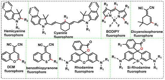
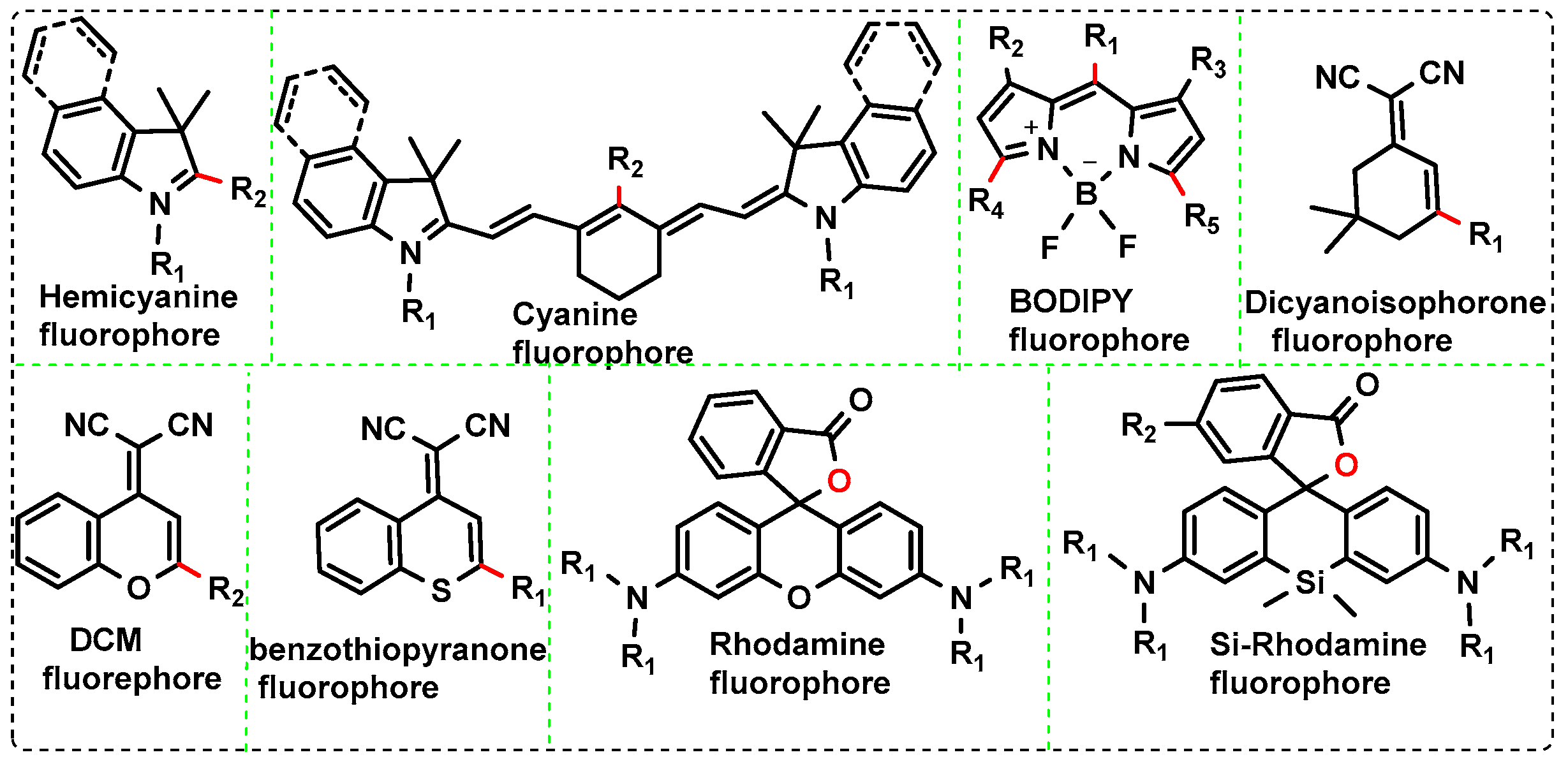
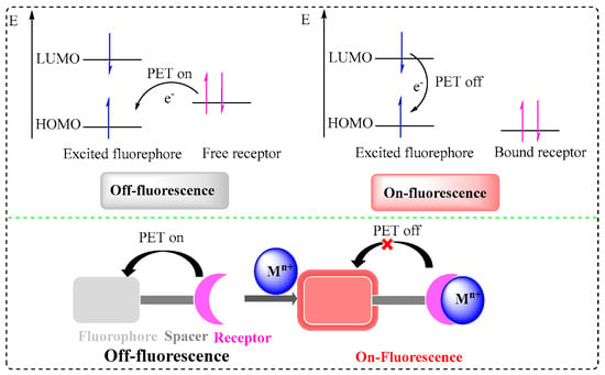
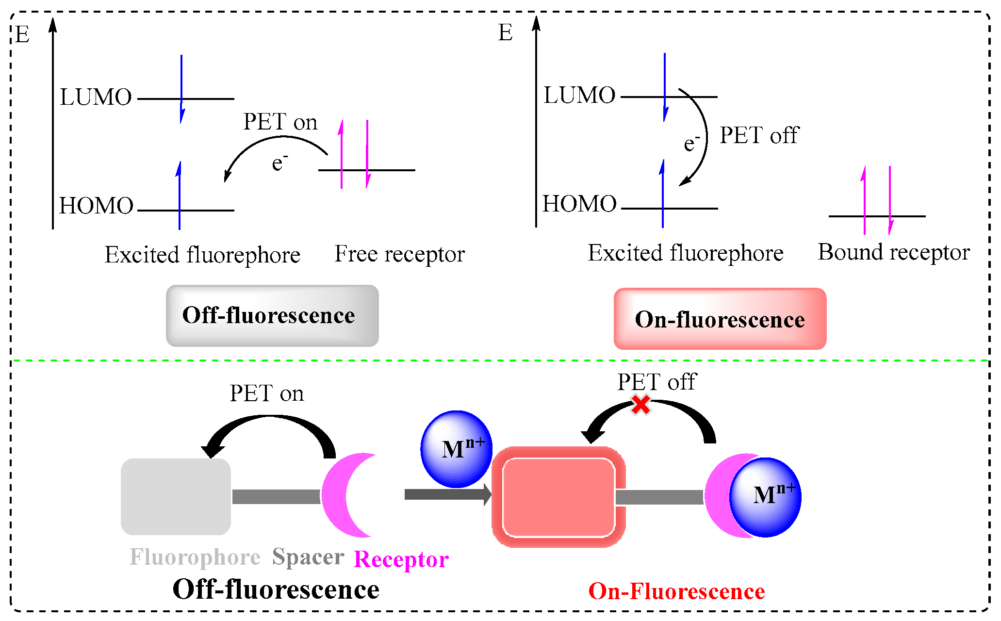
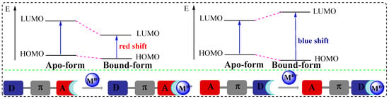
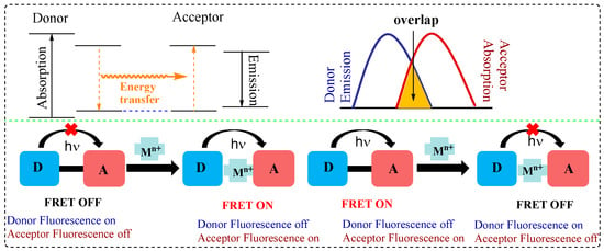
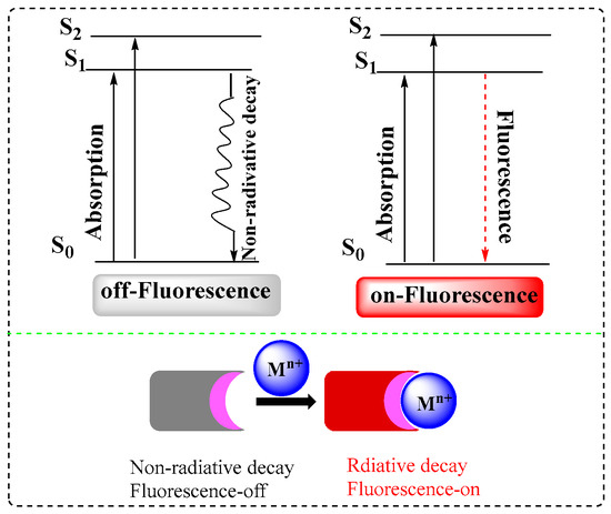
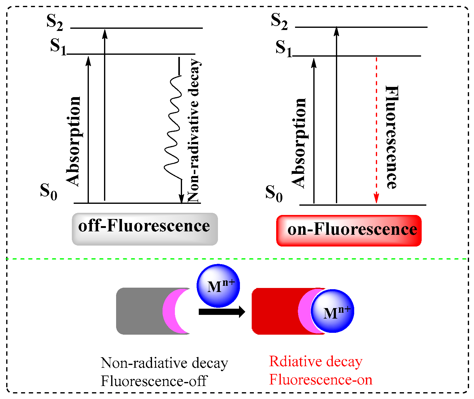
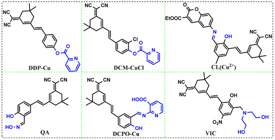
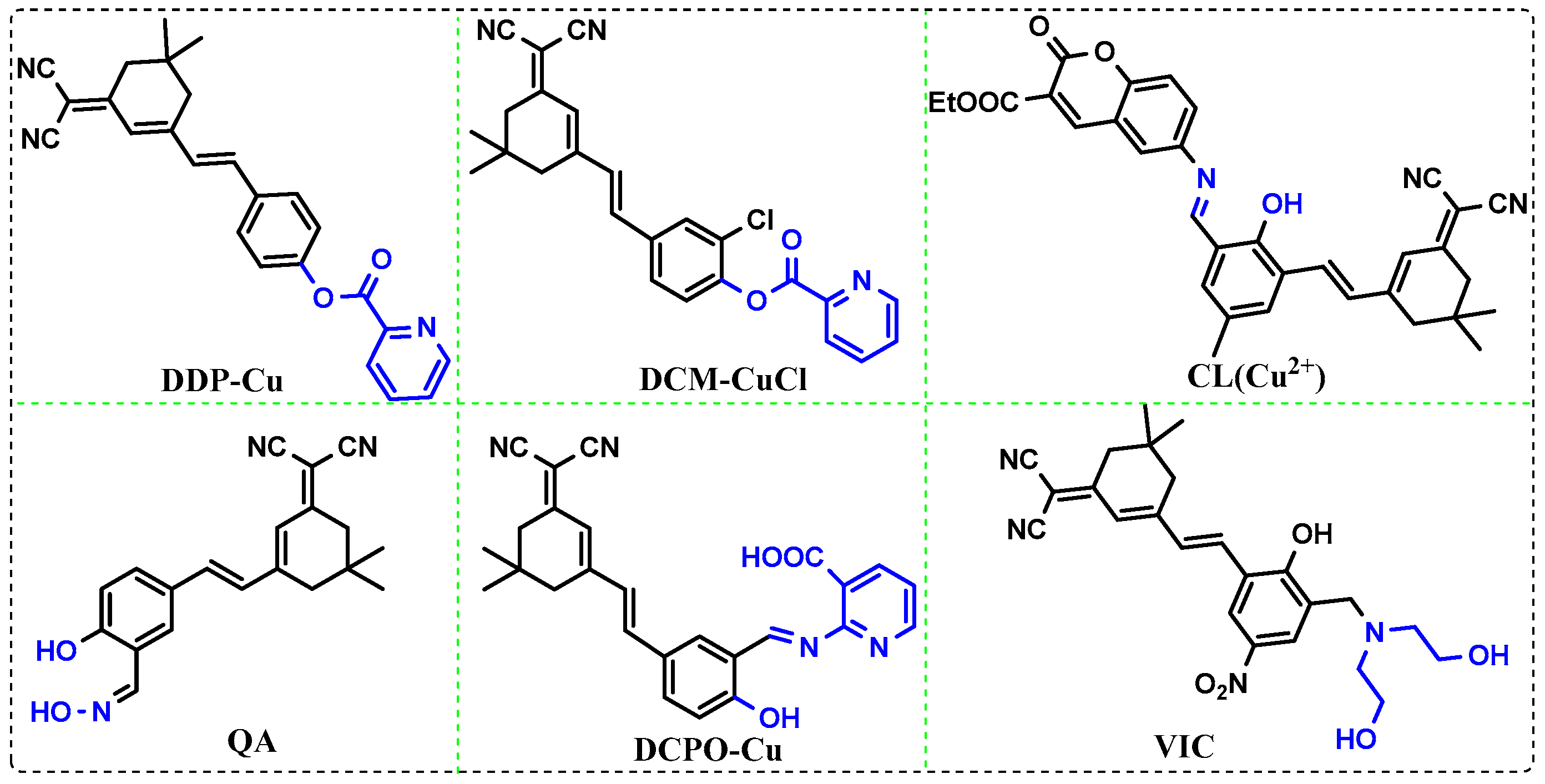
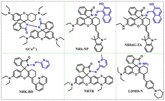
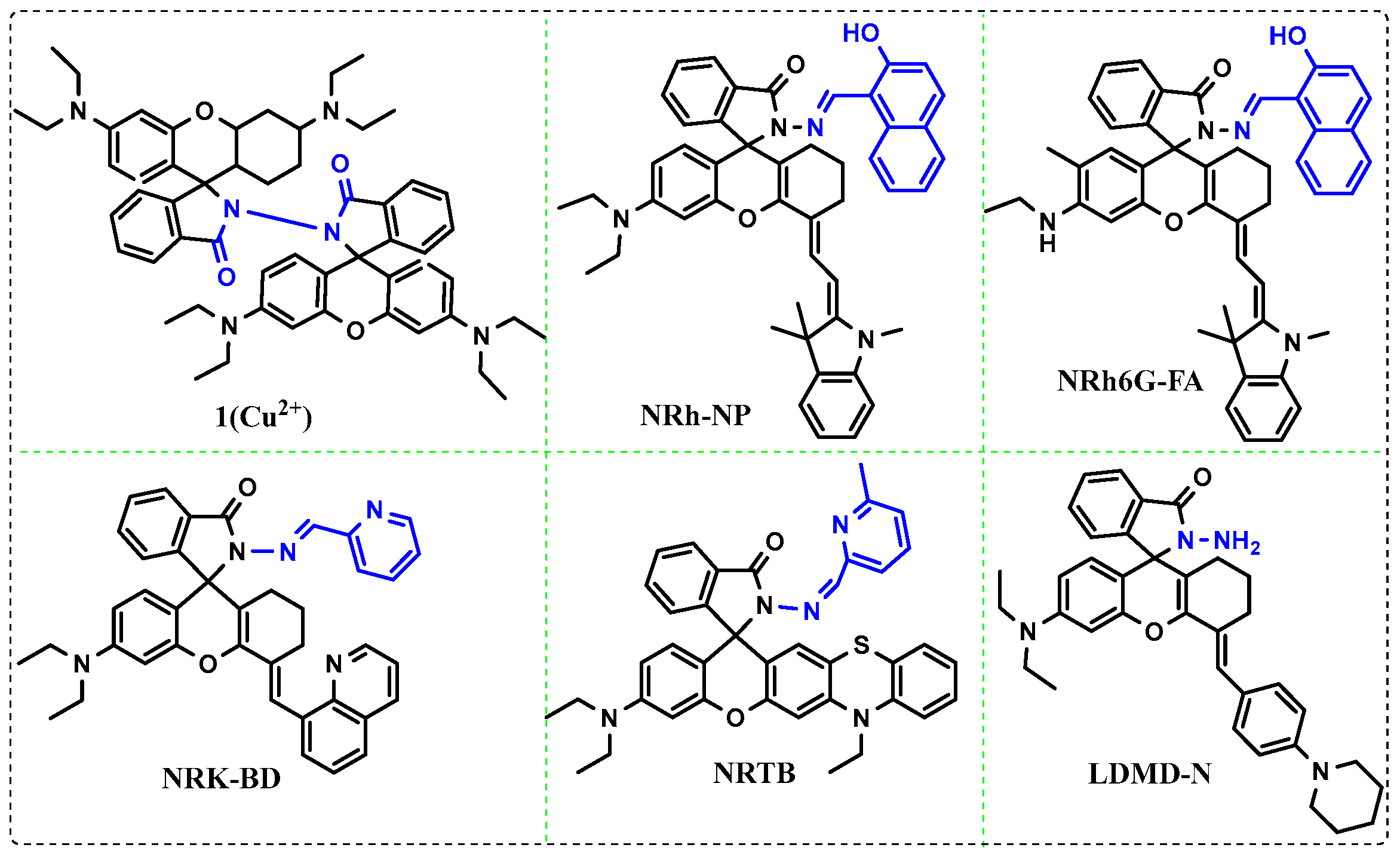
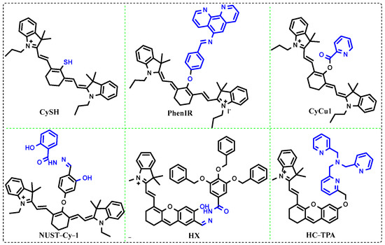
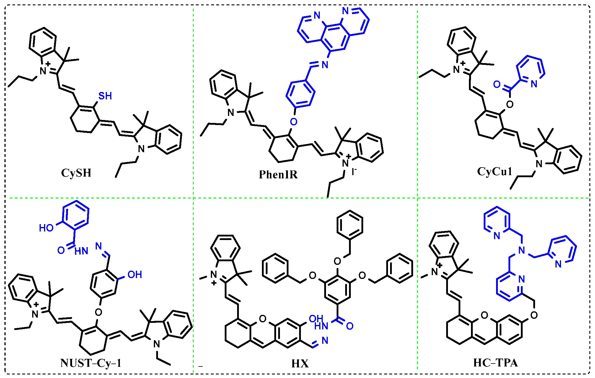
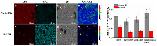
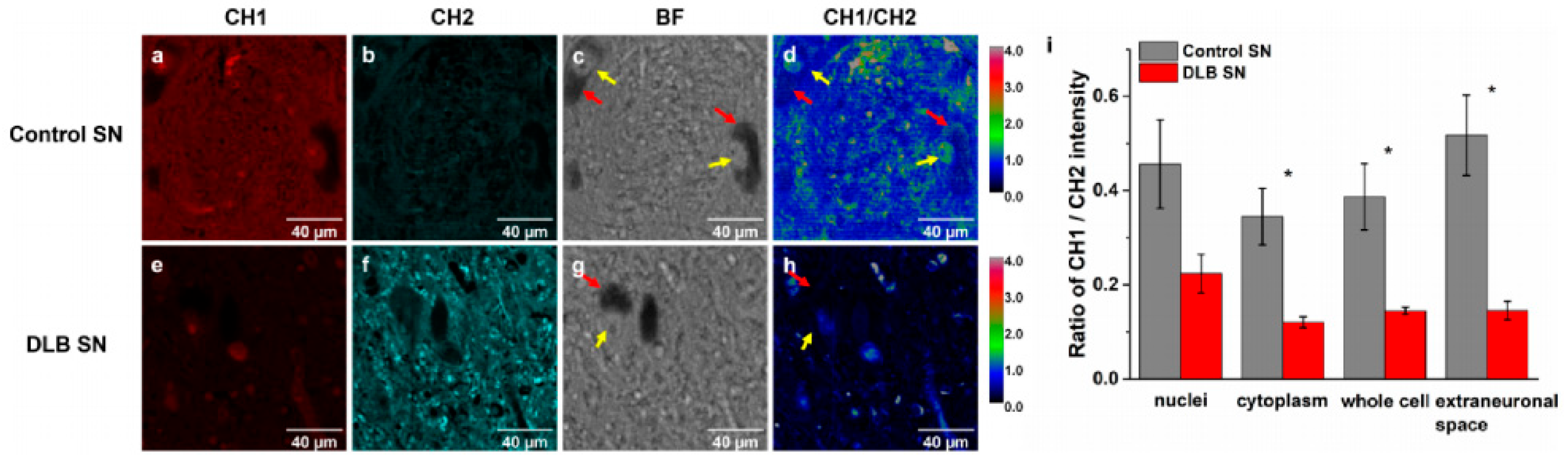
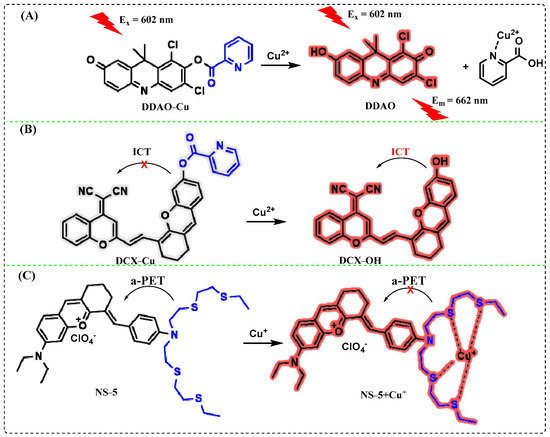
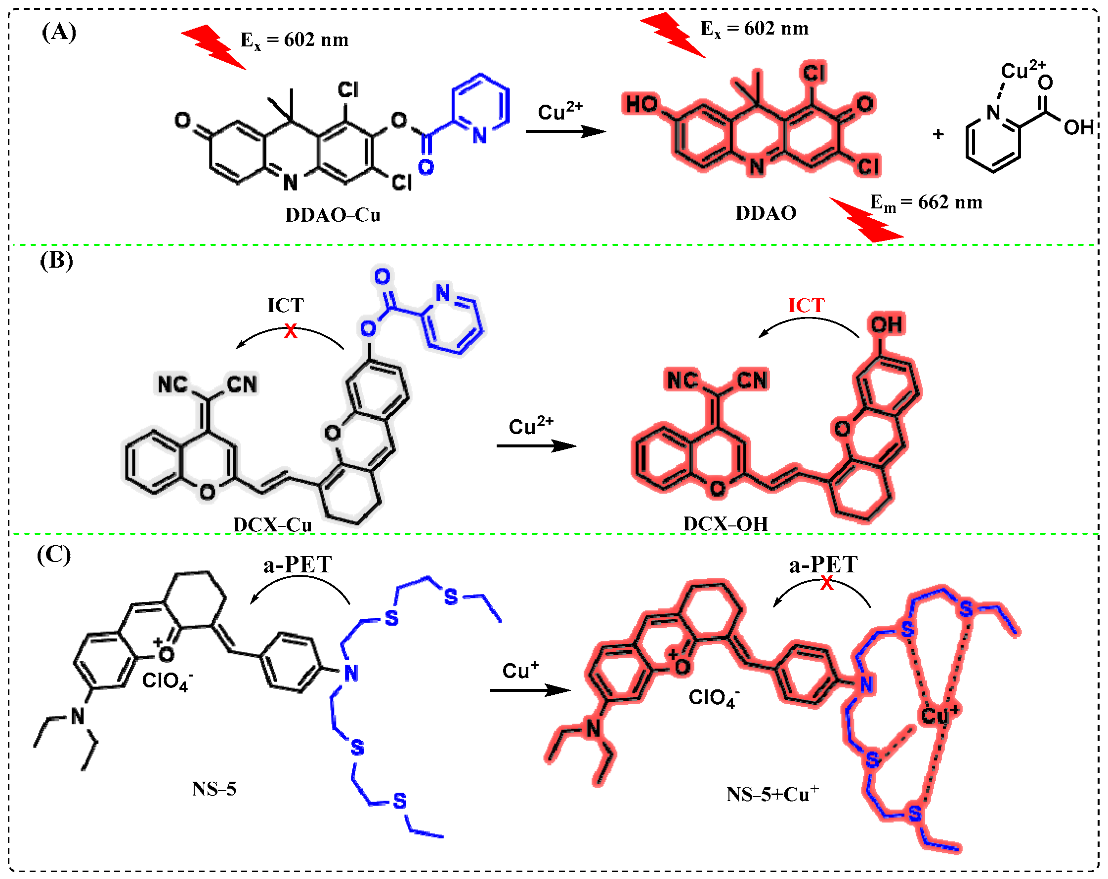
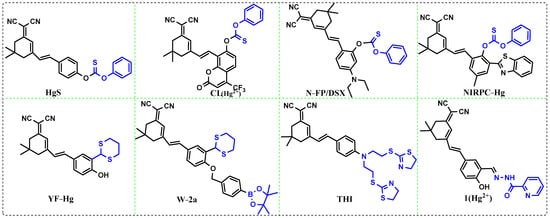
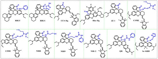
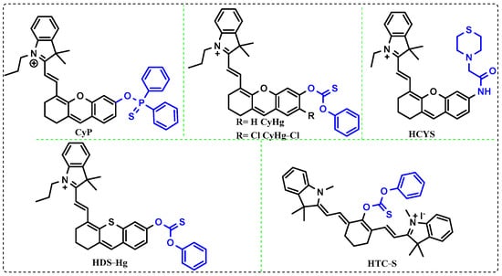
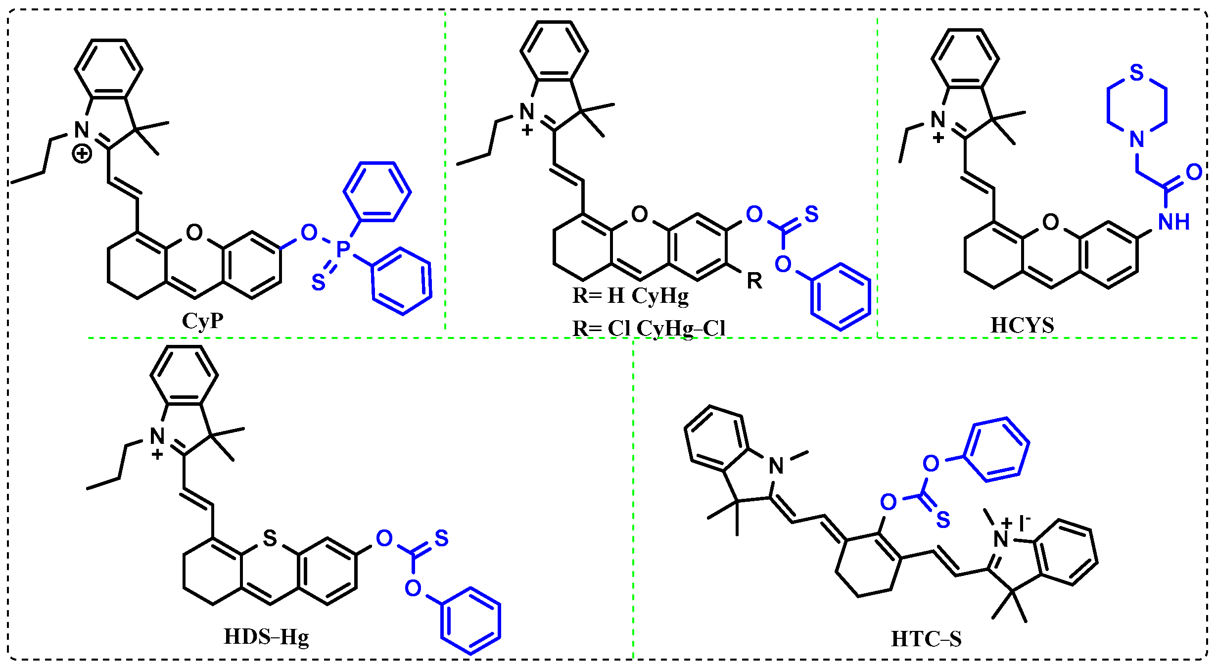
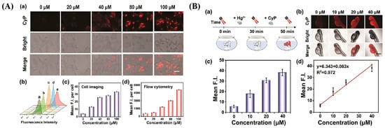
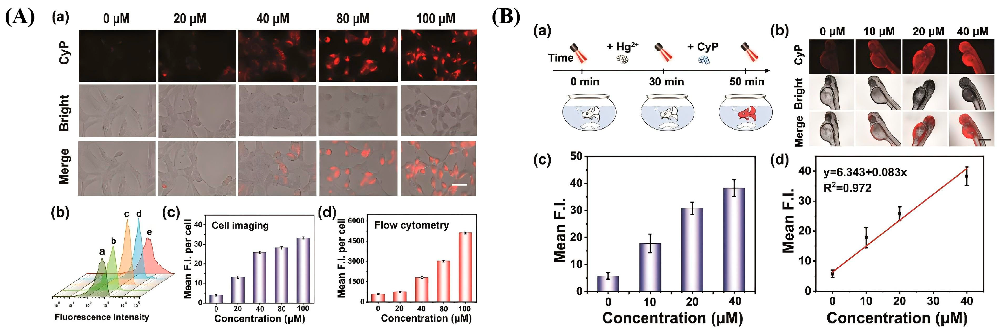
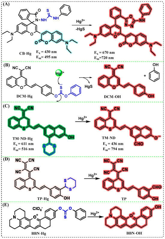
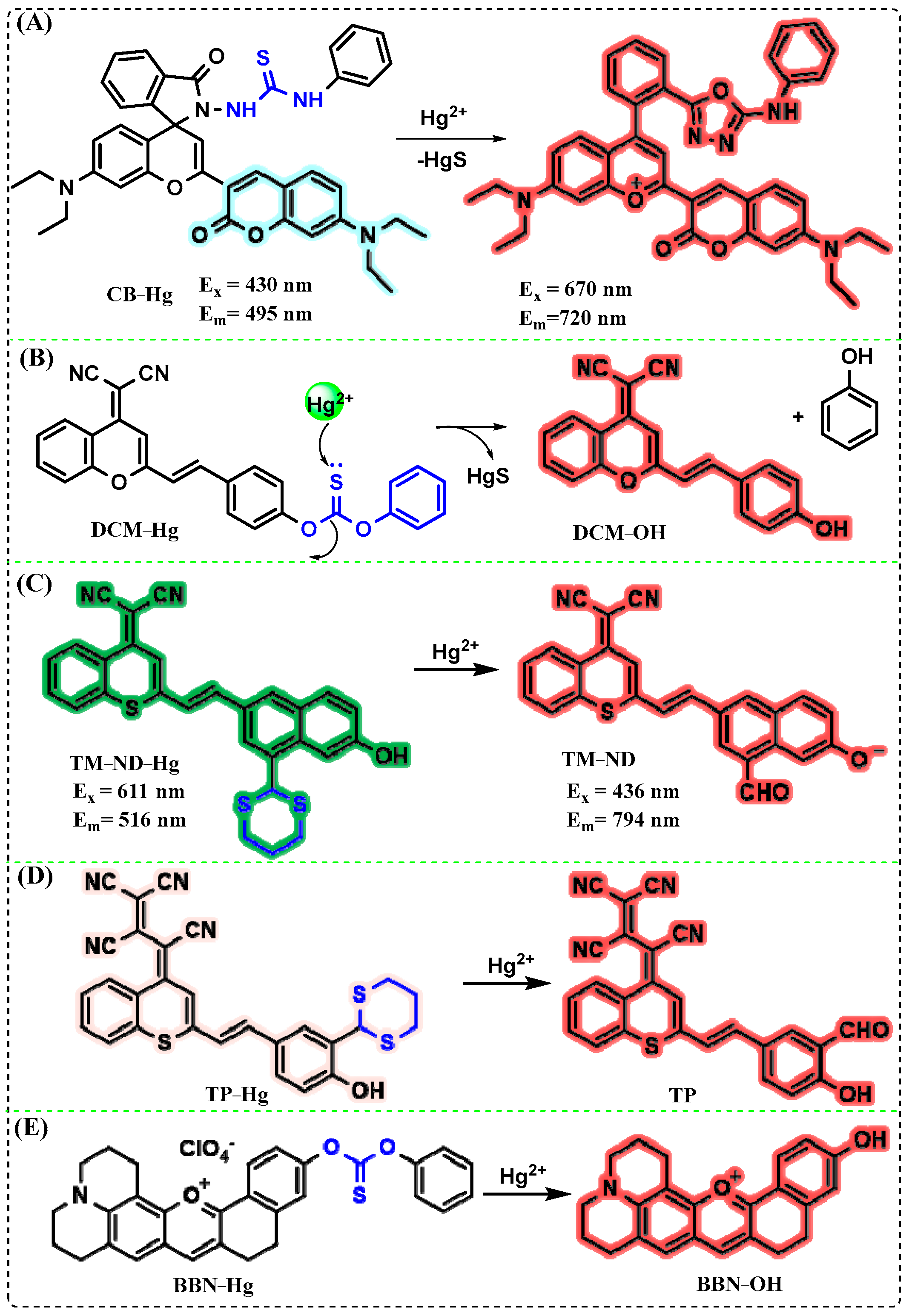
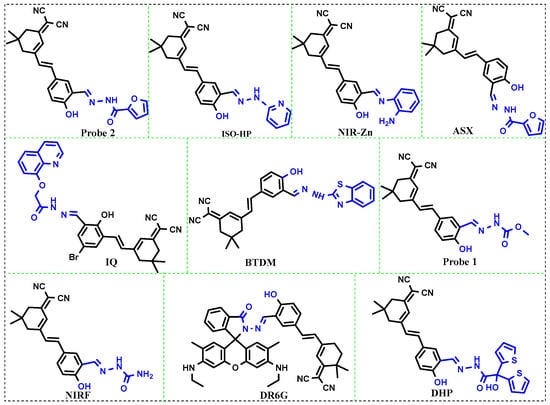
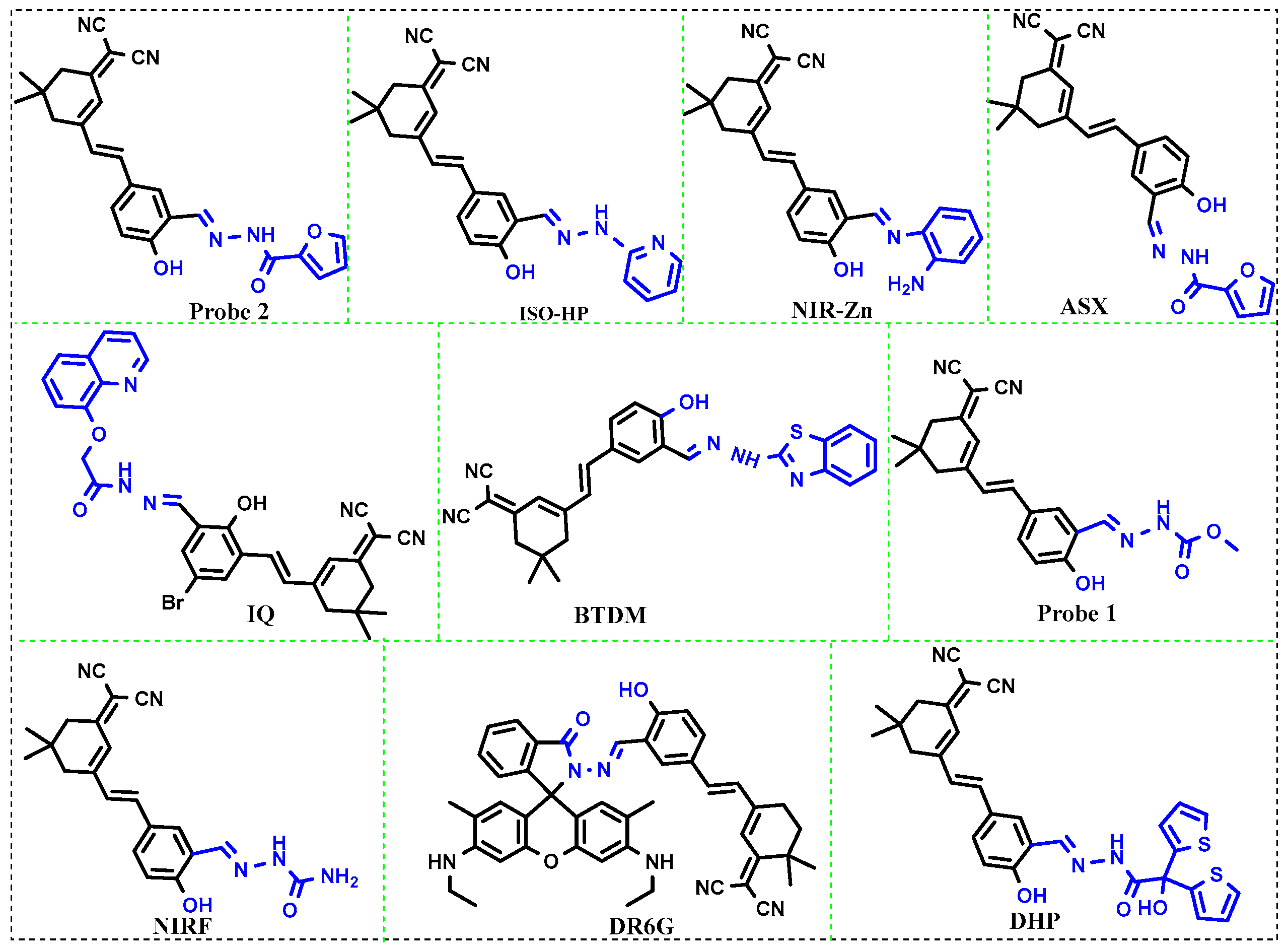
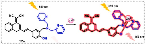
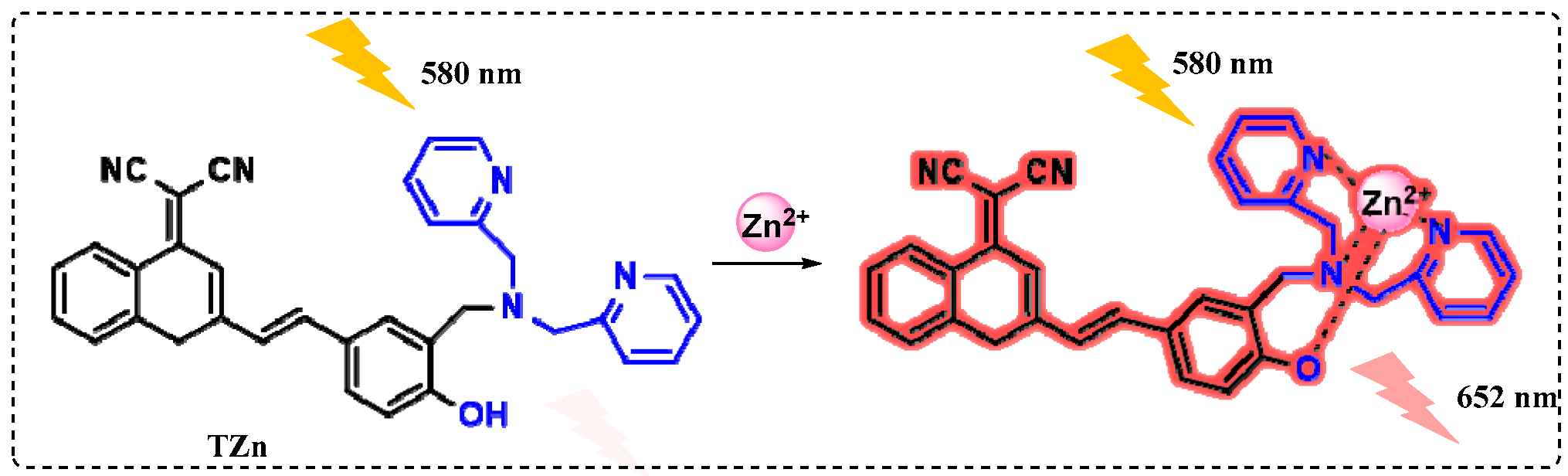
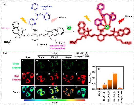
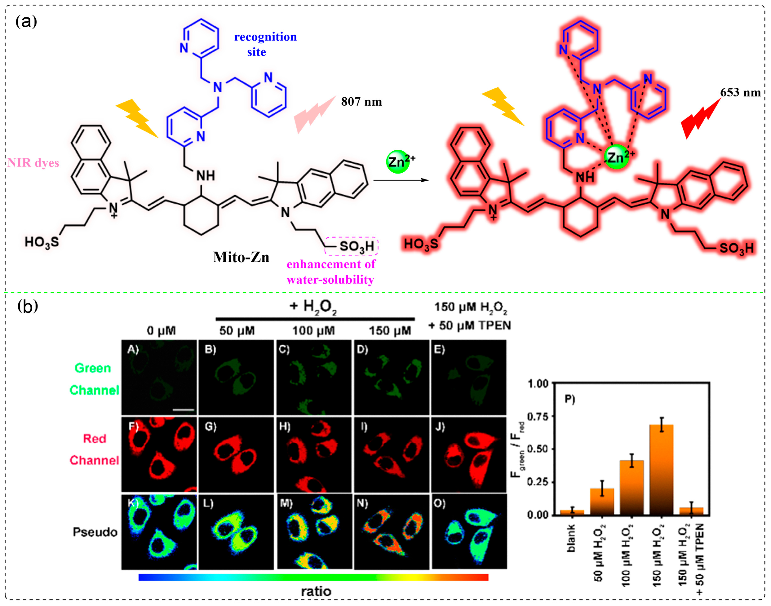
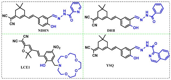
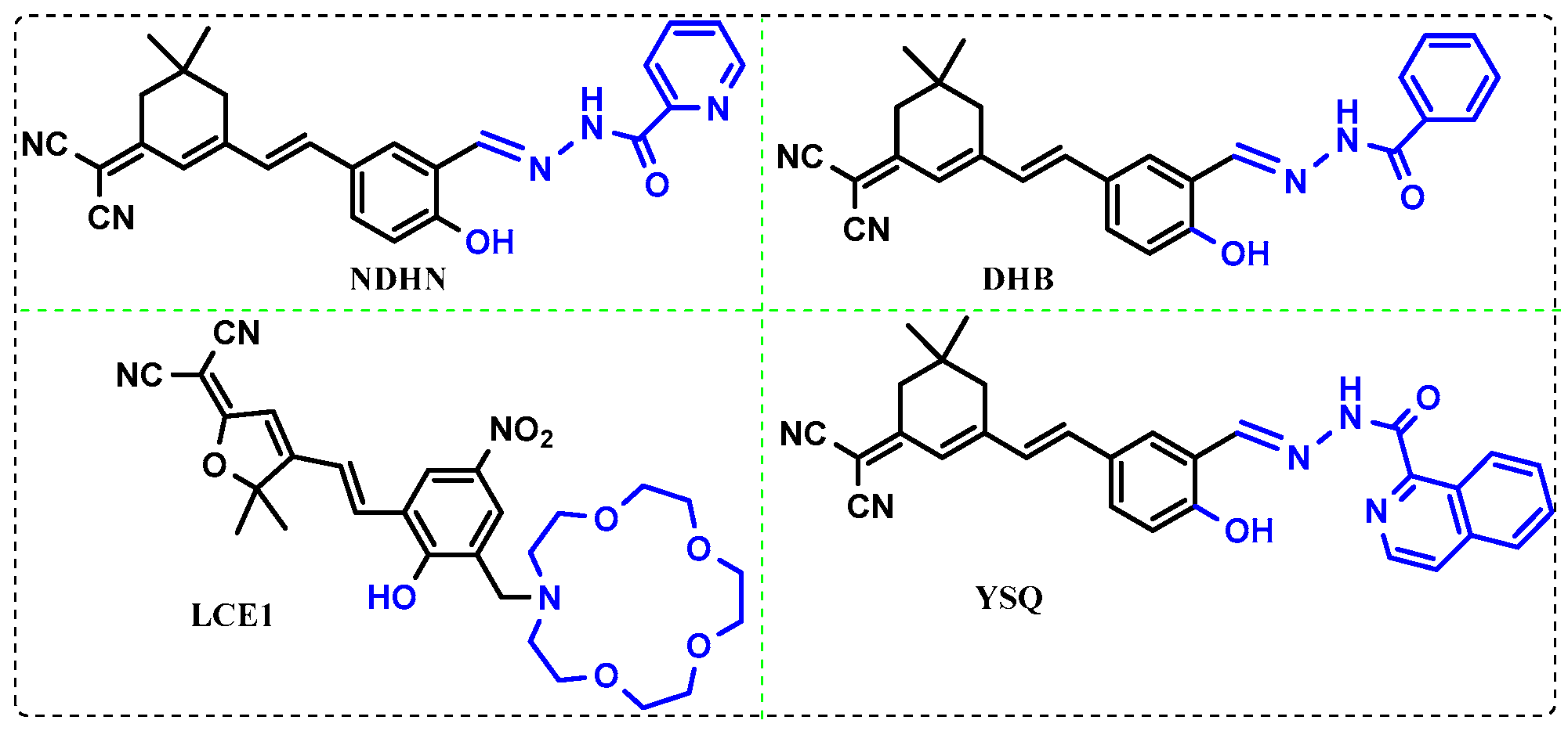
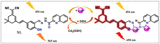
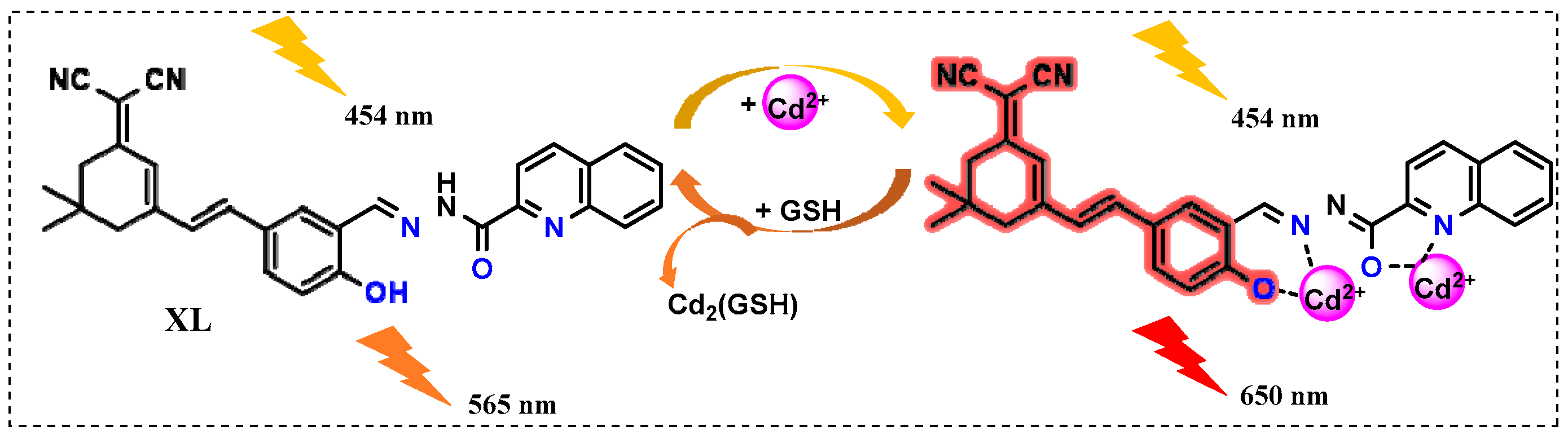
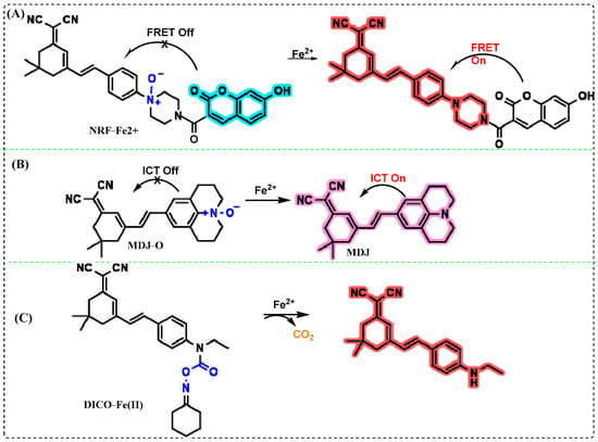
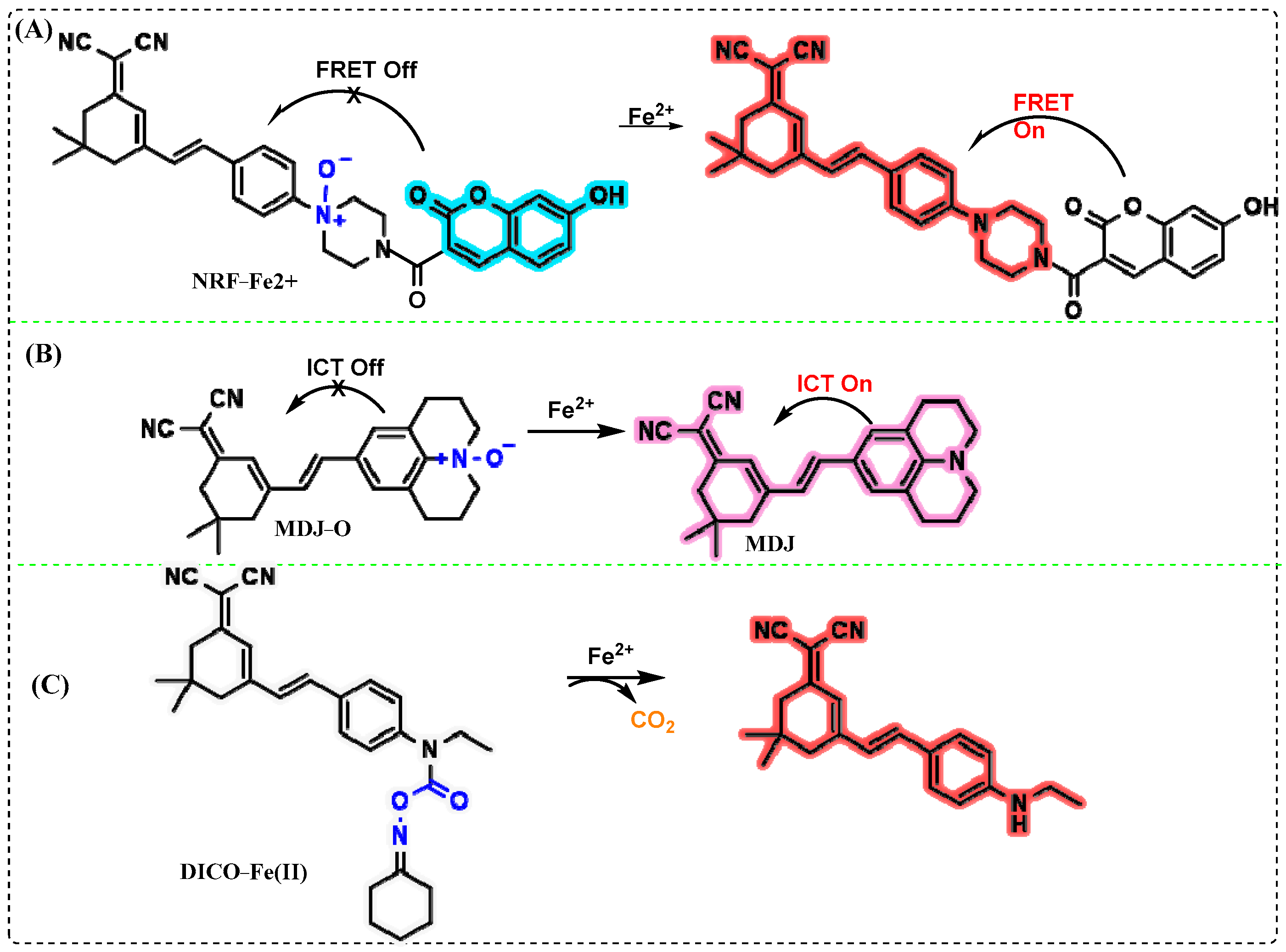
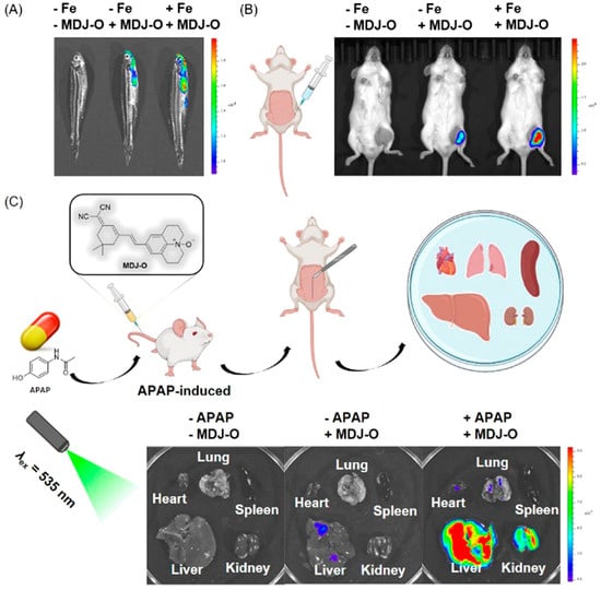
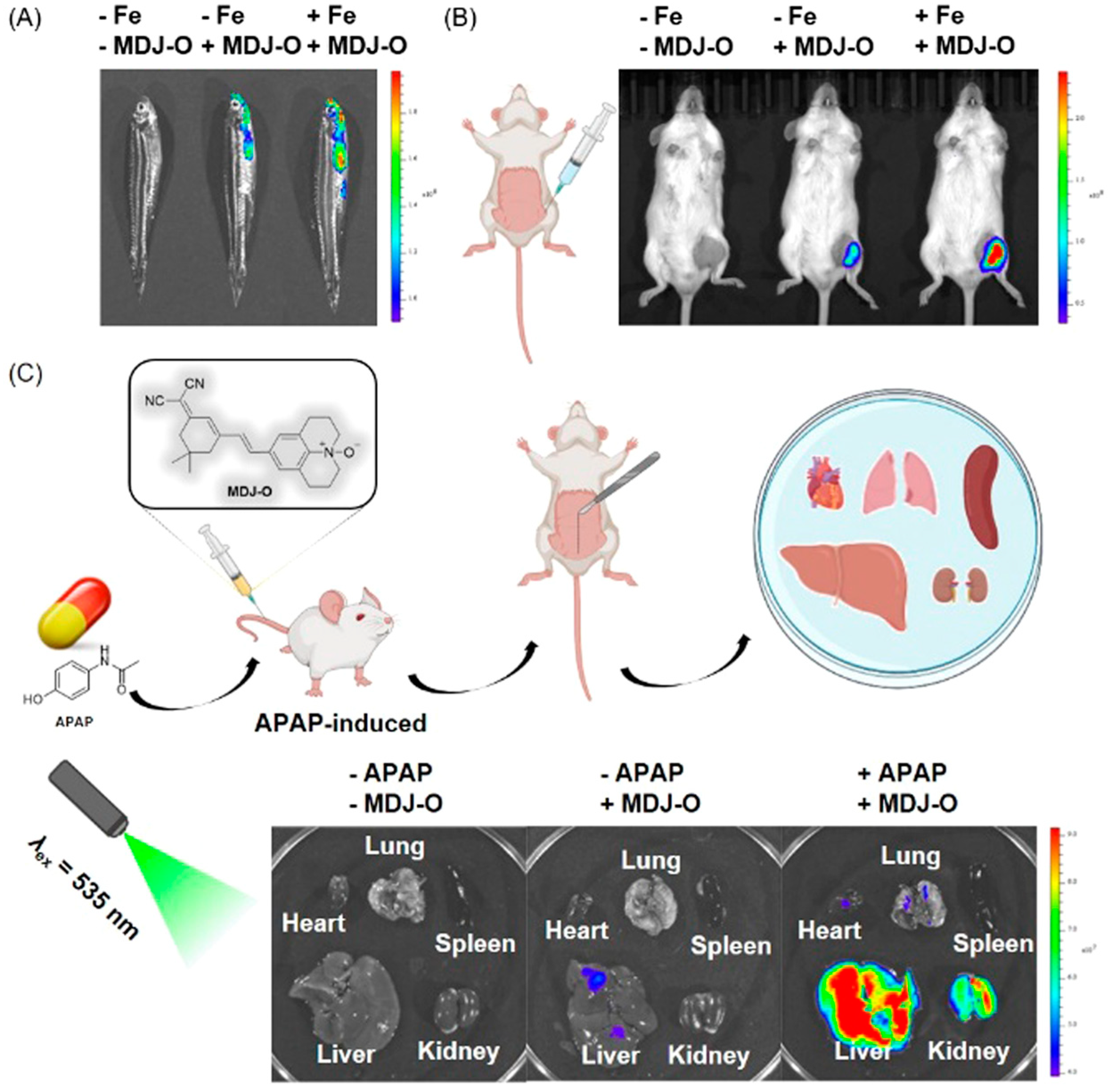
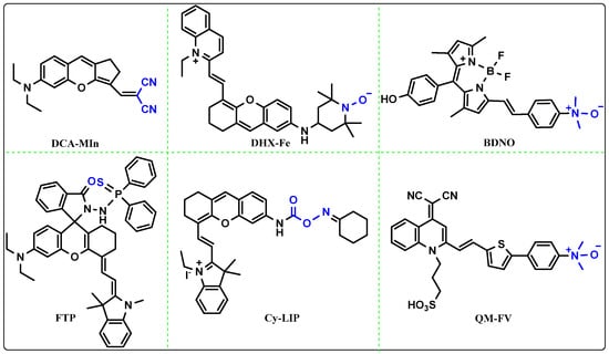
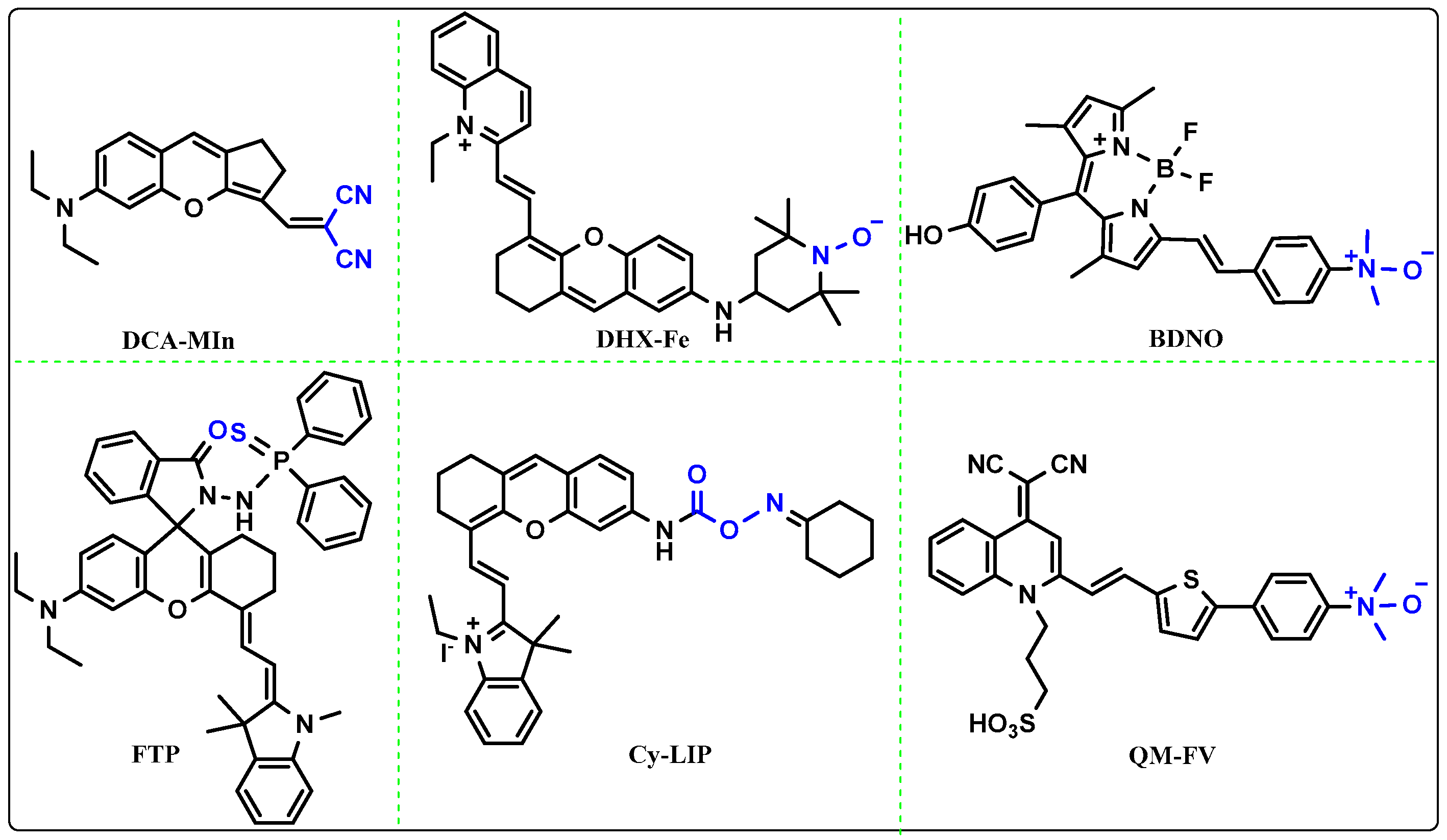

Disclaimer/Publisher’s Note: The statements, opinions and data contained in all publications are solely those of the individual author(s) and contributor(s) and not of MDPI and/or the editor(s). MDPI and/or the editor(s) disclaim responsibility for any injury to people or property resulting from any ideas, methods, instructions or products referred to in the content. |
© 2026 by the authors. Licensee MDPI, Basel, Switzerland. This article is an open access article distributed under the terms and conditions of the Creative Commons Attribution (CC BY) license.