


Abstract
Nanoparticle-based drug delivery systems have become an important component of modern nanomedicine, enabling improved drug protection, controlled release, targeted delivery, and the modulation of pharmacokinetic behavior. Their therapeutic performance is governed by physicochemical properties such as size, shape, surface chemistry, and material composition, which influence biological interactions, biodistribution, cellular uptake, and clearance. This review examines major nanoparticle platforms, including polymeric, lipid-based, inorganic, carbon-based, and hybrid systems, together with passive and active targeting and endogenous and externally triggered release strategies. Current and emerging applications in oncology, infectious diseases, central nervous system disorders, gene therapy, and vaccines are discussed alongside theranostic and combination-delivery approaches. Particular emphasis is placed on computational modeling, artificial intelligence, and digital twins for formulation optimization and personalized nanomedicine. Key barriers to clinical translation, including manufacturing scalability, biological variability, limitations of EPR-mediated targeting, regulatory standardization, and long-term safety, are critically evaluated. Finally, emerging directions in sustainable nanomanufacturing and biomimetic delivery are discussed. By integrating biological mechanisms with computational, manufacturing, regulatory, and clinical considerations, this review provides a translational perspective on advancing nanoparticle drug-delivery systems from laboratory development toward clinical implementation.
1. Introduction
1.1. From Conventional Systems to Nanomedicine: Needs and Opportunities
The development of nanomedicine has been driven by the accumulating limitations of conventional drug delivery approaches, such as oral tablets, capsules, and parenteral injections, which were adequate for small-molecule pharmaceuticals but are proving increasingly insufficient for the complex, fragile, and highly potent therapeutic agents that dominate modern pipelines. Standard dosage forms suffer from poor aqueous solubility, rapid systemic clearance, nonspecific biodistribution, and resulting off-target toxicity, which reduces therapeutic index and treatment tolerability [1]. In oncology, cardiovascular diseases, and neurological disorders, these limitations are particularly consequential. Drugs must reach a localized disease site at therapeutic concentrations while sparing surrounding healthy tissue, a spatial discrimination that conventional formulations cannot achieve. The progressive increase in the molecular complexity of biologics, nucleic acid therapeutics, and precision oncology agents has further widened the gap between drug potency and delivery capability, as many of these agents are inherently unstable in biological fluids, membrane-impermeable, or rapidly degraded before reaching the target cells [2,3].
Compared with conventional pharmaceutical formulations, nanoparticle-based delivery systems can provide additional control over drug solubility, protection from premature degradation, pharmacokinetics, biodistribution, and site-specific or stimulus-responsive release. These advantages are particularly relevant for poorly soluble drugs and biologics such as proteins and nucleic acids; however, they are accompanied by greater formulation complexity, manufacturing and characterization requirements, potential nanomaterial-specific safety concerns, and additional regulatory challenges [4,5].
Nanomedicine broadly refers to the application of nanoscale materials and technologies to the diagnosis, prevention, monitoring, and treatment of disease, including their use as engineered systems for therapeutic delivery. Within this field, nanoparticles are generally defined as materials or structures with dimensions in the nanoscale range, although pharmaceutical nanocarriers may extend beyond the conventional 1–100 nm definition depending on their composition and intended biomedical function [6]. In drug delivery, these systems are engineered to encapsulate, protect, transport, and release therapeutic agents while modifying their pharmacokinetic and biodistribution profiles [7]. Nanoparticles below approximately 200 nm exhibit preferential accumulation in tumor vasculature through the EPR effect, the passive extravasation of nanocarriers through fenestrated tumor blood vessels, and their retention due to impaired lymphatic drainage [3]. Beyond this passive mechanism, nanoparticles can be equipped with active targeting ligands, antibodies, peptides, and aptamers that selectively bind disease-associated receptors, enabling discrimination between diseased and healthy tissue at the molecular level [2]. The resulting combination of extended circulation, site-specific accumulation, and controlled release within the therapeutic target constitutes the central value proposition of nanomedicine over conventional dosage forms.
1.2. Core Advantages and Inherent Challenges
The advantages of nanoparticle-based drug delivery extend across multiple dimensions of therapeutic performance. Protection of labile cargo (nucleic acids, proteins, and unstable small molecules) from enzymatic degradation during systemic circulation is one of the most clinically significant benefits, enabling the delivery of therapeutic classes that were previously considered undruggable due to stability limitations [8]. Controlled and sustained drug release over hours to weeks reduces peak plasma concentration fluctuations, maintains drug levels within the therapeutic window for prolonged periods, and decreases dosing frequency, all of which improve both therapeutic consistency and patient adherence [9,10]. The multifunctionality of modern nanoparticle platforms, their ability to co-load multiple therapeutic agents at defined molar ratios, incorporate imaging reporters, and carry targeting moieties simultaneously, enables combination therapies, theranostics, and sequential drug release strategies that are structurally impossible with conventional single-molecule formulations [11,12].
Despite these advantages, several fundamental challenges continue to limit the broader clinical translation of nanomedicine. The complexity of nanoparticle systems introduces variability in synthesis, characterization, and batch reproducibility that is difficult to control at the manufacturing scale [13]. Protein corona formation (the rapid adsorption of serum proteins onto nanoparticle surfaces upon systemic injection) alters targeting ligand accessibility, accelerates immunological recognition, and modifies pharmacokinetic behavior in ways that are poorly predicted by in vitro testing [14,15]. Long-term biodistribution and organ accumulation of non-biodegradable materials, particularly inorganic nanoparticles, remain incompletely characterized, raising safety questions that regulatory agencies require extensive preclinical evidence to address [16,17]. Regulatory frameworks continue to evolve in response to the technological diversity of nanomedicines, and the absence of globally harmonized standards for nanoparticle characterization and safety assessment creates friction in the approval process [18]. Overcoming these barriers requires not only advances in materials science and formulation engineering, but also progress in manufacturing technology, computational design, and regulatory science.
Unlike conventional reviews that primarily classify nanoparticle platforms or summarize their biological applications, this review integrates therapeutic design with an engineering and translational perspective. Particular emphasis is placed on the convergence of AI-guided formulation design and digital twins, scalable and sustainable nanomanufacturing, and the regulatory and production challenges governing clinical translation. By connecting nanoparticle design, biological performance, computational optimization, manufacturing, and clinical implementation within a unified framework, this review aims to bridge the gap between laboratory-scale innovation and clinically deployable nanomedicines.
1.3. Scope and Organization of the Review
This review critically examines nanoparticle-based drug delivery from physicochemical design and biological interactions to therapeutic applications and clinical translation. Unlike reviews focused primarily on nanoparticle classification or biological applications, this review emphasizes the integration of AI-guided formulation design and digital twins, scalable and sustainable nanomanufacturing, and the engineering, safety, and regulatory considerations governing clinical translation. By connecting nanoparticle design, biological performance, computational optimization, manufacturing, and clinical implementation, the review aims to bridge the gap between laboratory-scale innovation and clinically deployable nanomedicines.
2. Fundamentals of Nanoparticle Design and Biological Interactions
2.1. Physicochemical Properties: Size, Shape, Surface Chemistry, and Drug Loading
The biological behavior of drug-delivery nanoparticles is governed by the interplay between their material composition, structural architecture, and physicochemical properties. The systems considered in this review include polymeric nanoparticles and micelles, lipid-based nanoparticles and liposomes, inorganic nanoparticles such as silica, gold, and iron oxide systems, carbon-based nanomaterials, and hybrid or biomimetic carriers. These platforms differ structurally, ranging from solid or porous matrices and inorganic cores to micellar assemblies and lipid bilayer vesicles, and these differences influence drug loading and release, protein adsorption, cellular interactions, biodistribution, and clearance [19]. Among their physicochemical characteristics, particle size is a particularly important determinant of circulation, tissue distribution, cellular uptake, and elimination. In general, larger nanoparticles (>200 nm) are more susceptible to recognition and clearance by the mononuclear phagocyte system (MPS), whereas nanoparticles with hydrodynamic diameters below approximately 5.5 nm, particularly small inorganic systems, may undergo efficient renal filtration and urinary excretion [7]. The optimal size window for passive tumor targeting via the EPR effect is generally cited as 10–200 nm, though the actual effective range varies with tumor type, vascularization density, and disease stage. Within this range, smaller particles penetrate deeper into tumor tissue, while larger particles may accumulate at the periphery [20]. Figure 1 summarizes the influence of nanoparticle size on biological fate and pharmacokinetic behavior in vivo.
Figure 1.
Effect of nanoparticle size on circulation behavior, biodistribution, tissue penetration, and clearance mechanisms in vivo, illustrating the size-dependent transition between renal elimination, prolonged systemic circulation, and uptake by the mononuclear phagocyte system. Reprinted from Ref. [21], under the terms of the Creative Commons CC BY license.
Shape is an equally important but often underappreciated design variable. Spherical nanoparticles are the most widely studied geometry and are favored for their predictable behavior and efficient endocytosis, but non-spherical morphologies confer distinct biological advantages. Rod-shaped particles exhibit prolonged circulation times relative to spheres of equivalent volume, attributed to their reduced phagocytic uptake due to asymmetric contact with macrophage membranes [20,22]. Worm-like filomicelles exhibit shape- and length-dependent biodistribution; early studies demonstrated markedly prolonged circulation compared with spherical micelles, whereas subsequent studies indicate that tumor accumulation and cellular uptake depend strongly on filament length and formulation characteristics [23]. Disk-shaped and platelet-mimicking particles have attracted interest for their ability to adhere to vascular walls and avoid margination from blood flow. The implications for drug delivery design are clear, where the shape must be selected in conjunction with the intended delivery route, target tissue, and cellular uptake pathway rather than defaulting to spherical geometry. Figure 2 illustrates representative examples of non-spherical nanoparticle geometries employed in drug delivery applications, including filomicelles, bottlebrush structures, worm-like vesicles, nanorods, prisms, cylinders, disks, and ellipsoids. These morphologies exhibit distinct hydrodynamic behavior, circulation profiles, and cellular interactions, further highlighting the importance of shape as a critical parameter in the rational design of nanocarriers [24].
Figure 2.
Representative non-spherical nanoparticle geometries used in drug delivery applications. Reprinted from Ref. [24], under the terms of the Creative Commons CC BY license.
Surface charge, quantified by zeta potential, governs colloidal stability, protein adsorption, and cellular interaction. Cationic surfaces facilitate electrostatic interaction with the negatively charged cell membrane, improving uptake efficiency for nucleic acid delivery, but also increase non-specific protein adsorption and MPS recognition in vivo. Anionic and neutral surfaces generally offer better systemic circulation but lower intrinsic cellular internalization [25]. Surface functionalization with hydrophilic polymers such as polyethylene glycol (PEG) is commonly used to generate a ‘stealth’ coating. PEG chains form a hydrated steric barrier around the nanoparticle surface that reduces nonspecific protein adsorption and recognition by the mononuclear phagocyte system, thereby prolonging systemic circulation [14]. Liposomes are vesicular nanocarriers composed of phospholipid bilayers surrounding an aqueous core, enabling the encapsulation of therapeutic compounds within the aqueous compartment or lipid bilayer [26]. A clinically established example is PEGylated liposomal doxorubicin, in which doxorubicin is encapsulated within PEG-coated phospholipid liposomes. This formulation illustrates how surface PEGylation and liposomal encapsulation can alter drug pharmacokinetics and reduce systemic toxicity compared with free doxorubicin [27]. Drug loading efficiency depends on the physicochemical compatibility between the drug and the carrier matrix, and on whether passive (encapsulation during self-assembly) or active (post-formation loading driven by pH or ion gradients) loading strategies are employed. Active loading generally achieves higher efficiency and more controlled loading for ionizable drugs [28].
2.2. Pharmacokinetics, Biodistribution, and Navigating Biological Barriers
The pharmacokinetics of nanoparticle-based therapeutics differ fundamentally from those of free drugs, and understanding these differences is essential for rational dosing and the prediction of therapeutic outcomes. Following systemic administration, nanoparticles encounter a sequential series of biological barriers, each of which can attenuate the fraction of the administered dose that ultimately reaches the therapeutic target [21]. The first barrier is the blood compartment, where protein corona formation occurs within seconds of injection, altering the particle’s identity as perceived by the immune system and potentially redirecting biodistribution to MPS organs regardless of the engineered targeting strategy [15]. Stealth coatings mitigate but do not eliminate this effect, and anti-PEG antibodies generated after repeated dosing can accelerate clearance in a subset of patients, the accelerated blood clearance (ABC) phenomenon.
Vascular extravasation into solid tumors is typically achieved through the EPR effect for nanoparticles in the appropriate size range, but the magnitude of EPR-mediated accumulation is highly variable and has been called into question as a reliable basis for clinical performance [29]. Heterogeneous tumor vascularization, variable pericyte coverage, and inter-patient differences in lymphatic function mean that EPR-mediated targeting can differ by an order of magnitude across tumor types and patients, explaining in part why nanoparticle systems that show compelling efficacy in murine tumor models frequently underperform in human clinical trials [2]. Transcytosis, receptor-mediated endocytosis, and active targeting using ligands that bind disease-specific surface receptors provide more predictable targeting mechanisms that do not rely on the anatomical features of the tumor vasculature [2].
Cellular barriers constitute the final stage of delivery. Following uptake via endocytosis, nanoparticles enter the endo-lysosomal pathway, where progressively acidifying compartments (from pH 7.4 in early endosomes to pH 4.5–5.0 in lysosomes) can degrade acid-sensitive cargo and expose lysosomal nucleases that degrade unprotected nucleic acids [30]. Endosomal escape is therefore a critical design objective for the intracellular delivery of biologics and gene therapies. Ionizable lipids that undergo protonation and membrane-disruptive conformational change at low pH, proton sponge polymers that buffer acidification and induce osmotic swelling, and photosensitizer-mediated membrane disruption are among the strategies employed to achieve cytoplasmic delivery after endosomal uptake [31,32]. Subcellular targeting (delivery to the nucleus for gene therapy, to the mitochondria for metabolic interventions, or to specific organelles for precision pharmacological action) represents the next frontier in intracellular targeting and requires additional targeting sequences beyond those sufficient for cell-level selectivity [22].
3. Material Systems, Synthesis, and Functionalization
3.1. Key Nanoparticle Platforms: Polymeric, Lipid-Based, Inorganic, Carbon-Based, and Hybrid
The choice of material platform is the most fundamental decision in nanoparticle design because it determines the physicochemical properties, drug loading capacity, degradation behavior, and biological interactions of the resulting system. Table 1 provides a systematic comparison of the principal nanoparticle platforms across these dimensions. Polymeric nanoparticles (PNPs) are among the most extensively studied platforms due to their compositional versatility, tunable degradation rates, and the wide range of drugs and biologics they can encapsulate. Polyesters such as poly(lactic-co-glycolic acid) (PLGA), polylactic acid (PLA), and polycaprolactone (PCL) degrade hydrolytically to non-toxic metabolites and are the basis of the majority of clinically progressed polymeric systems [10,33]. The polymer molecular weight, copolymer composition, and end-group chemistry collectively determine the degradation rate and drug release kinetics, providing extensive tunability for sustained-release applications ranging from days to months.
Table 1.
Comparative overview of the principal nanoparticle drug delivery platforms: composition, size range, key advantages, primary limitations, and representative therapeutic applications.
Beyond their structural and compositional differences, modern nanoparticle platforms are increasingly designed as multifunctional systems capable of simultaneously integrating therapeutic, diagnostic, and targeting functionalities. Figure 3 illustrates representative nanocarrier architectures and the diverse range of payloads and functional moieties that can be incorporated into these systems [40].
Figure 3.
Representative nanoparticle platforms used in drug delivery and their multifunctional capabilities, including the incorporation of drugs, nucleic acids, proteins, imaging agents, and targeting ligands within a single nanocarrier system. Reprinted from Ref. [40], under the terms of the Creative Commons CC BY license.
Lipid-based nanoparticles, including liposomes and lipid nanoparticles (LNPs), represent the most clinically successful nanoparticle class to date, encompassing several FDA-approved products and the transformative COVID-19 mRNA vaccines [13,31]. Their biocompatibility, biodegradability, and the structural amphiphilicity that allows for the simultaneous encapsulation of hydrophilic cargo in the aqueous core and hydrophobic drugs in the lipid bilayer are key advantages. The development of ionizable lipids (lipids with pKa values in the range of 6–7 that are neutral at physiological pH but positively charged in acidic endosomal compartments) was a critical advance that enabled efficient siRNA and mRNA delivery by facilitating endosomal membrane disruption and cargo release [31]. Mesoporous silica nanoparticles (MSNs) offer a distinctive combination of high internal surface area (>1000 m2 g−1), tunable and uniform pore dimensions (2–50 nm), and chemical stability that makes them exceptionally capable drug reservoirs [35]. The pores can be functionalized with gating molecules (molecular machines such as nanovalves, pseudorotaxanes, and DNA duplexes) that open in response to specific stimuli, transforming the MSN into a precisely controlled drug release device [34].
Inorganic nanoparticles, including gold, iron oxide, and quantum dot systems, derive their clinical utility primarily from unique physical properties unavailable to organic carriers. Gold nanoparticles exhibit strong and size-tunable surface plasmon resonance, enabling both highly sensitive optical imaging and photothermal conversion, while superparamagnetic iron oxide nanoparticles (SPIONs) provide MRI contrast enhancement and are responsive to external magnetic fields for guided delivery and magnetic hyperthermia [36,37]. Hybrid nanoparticles that combine a polymeric core with a lipid shell (the lipid-polymer hybrid nanoparticle) represent an attempt to capture the stability and drug-loading advantages of polymers with the biocompatibility and surface versatility of lipids within a single architecture [7]. Carbon dots, a recently emerged class of photoluminescent carbon-based nanomaterials smaller than 10 nm, combine biocompatibility, tunable emission, and surface functionalizability with inherent EPR-mediated tumor accumulation, making them attractive candidates for simultaneous cancer imaging and drug/gene co-delivery [38]. Sustainable biopolymer-derived nanocarriers, including cellulose nanofibers and nanocellulose-based composites, are also emerging as promising platforms due to their biocompatibility, biodegradability, high surface area, and ease of surface functionalization for drug delivery and biomedical applications. Exosomes and red blood cell membrane-coated nanoparticles represent the biomimetic frontier, exploiting naturally derived lipid bilayers to achieve stealth circulation, and in the case of exosomes, inherent tissue targeting capabilities derived from their cell of origin [15,39].
3.2. Fabrication, Encapsulation Methods, and Manufacturing Scalability
The transition from materials design to a manufacturable product requires fabrication processes that reliably produce nanoparticles meeting defined critical quality attributes (CQAs), including specifications for size, polydispersity, surface charge, drug loading, and encapsulation efficiency, which are predictive of clinical performance [28]. Historically, liposomes and polymeric nanoparticles were formed by bulk mixing methods such as thin-film hydration, nanoprecipitation, and solvent emulsification, which produce acceptable laboratory-scale particles but suffer from poor process control, high batch-to-batch variability, and difficulty scaling to commercial volumes without reoptimization [28]. The introduction of microfluidic fabrication technologies represented a paradigm shift. By confining mixing to channels of defined geometry at controlled flow rates, microfluidic systems achieve rapid, reproducible nanoprecipitation with size control as fine as ±5 nm, and the laminar-to-turbulent mixing transition can be precisely tuned to optimize nanoparticle formation kinetics [41]. More recently, impingement jet mixing (IJM) technology has enabled the scale-up of LNP manufacturing to commercial volumes while preserving the mixing precision achieved by microfluidics at smaller scales, with multi-jet configurations allowing proportional increases in throughput without altering the critical hydrodynamic mixing parameters [42]. Active loading strategies (post-formation drug loading driven by transmembrane pH or ammonium sulfate gradients) achieve significantly higher encapsulation efficiencies for ionizable drugs than passive co-encapsulation during particle formation, and are the basis of the remote loading strategy used in Doxil® and related pegylated liposomal products [28,43].
3.3. Surface Engineering: Targeting Ligands, Stealth Coatings, and Stimuli-Responsive Designs
Surface engineering is the process by which a nanoparticle with appropriate bulk properties is transformed into a precisely programmed biological agent, one that knows where to go, when to release its cargo, and how to avoid immune detection along the way. Stealth coatings are the most fundamental surface modification, and polyethylene glycol (PEG) has been the dominant strategy for more than three decades. PEGylation creates a hydrophilic, flexible polymer brush that sterically prevents protein adsorption, reduces MPS recognition, and extends circulation half-life from minutes to hours or days [14,44]. However, the clinical adoption of PEGylated systems has revealed a limitation. Repeated dosing in a subset of patients generates anti-PEG antibodies that accelerate clearance and can trigger hypersensitivity reactions. Polyzwitterionic coatings and naturally derived polymer coatings such as polysarcosine are being investigated as PEG alternatives that preserve stealth properties without immunogenic liability [45].
Active targeting is accomplished by conjugating targeting ligands (small molecules, peptides, aptamers, or antibodies) to the nanoparticle surface at densities that allow multivalent binding to disease-associated receptors while preserving the steric properties of the stealth layer. The folate receptor, transferrin receptor, ErbB2/HER2, EGFR, and PSMA are among the most intensively targeted receptors in cancer nanomedicine, while CXCR4 and VCAM-1 are studied in cardiovascular and inflammatory applications [2,46]. Folic acid-targeted carbon quantum dot systems have demonstrated dual siRNA delivery and selective cytotoxicity in folate receptor-overexpressing osteosarcoma cells, illustrating how ligand–receptor specificity can be combined with dual cargo delivery in a single platform [47]. Stimuli-responsive surface designs add a further level of programmability: pH-sensitive bonds that expose targeting ligands only in the acidic tumor microenvironment, enzyme-cleavable coatings that are stripped by tumor-associated proteases to reveal the active targeting surface, and thermo-responsive polymers that undergo conformational transitions at specified temperatures to release cargo or alter surface accessibility [10,48]. The convergence of stealth coatings, active targeting, and environmental responsiveness in hybrid surface architectures represents the current frontier of nanoparticle surface engineering.
4. Release Mechanisms and Advanced Delivery Strategies
4.1. Controlled Release via Diffusion, Degradation, and Endogenous Triggers
Controlled drug release from nanoparticles is rarely governed by a single mechanism. Rather, it typically reflects the simultaneous and coupled operation of diffusion, matrix degradation, and erosion, with their relative contributions determined by the specific polymer composition, drug physicochemical properties, and environmental conditions at the delivery site [10,49]. In biodegradable polymeric systems based on PLGA and PCL, drug molecules traverse the matrix through water-filled pores that develop as hydrolysis proceeds, and the rate of hydrolytic bond cleavage, which sets the degradation rate, is tunable through the adjustment of copolymer composition, molecular weight, and end-group chemistry [10]. As described by Kamaly et al. [10], drug release from biodegradable polymeric systems commonly results from the combined contributions of diffusion, degradation, and erosion rather than from a single mechanism. Understanding and controlling these interconnected processes through rational polymer design can enable the precise spatial and temporal regulation of drug release.
Endogenous chemical triggers exploit pathological features of disease sites to achieve selective drug release. pH-responsive systems exploit the acidic microenvironment of tumors (extracellular pH 6.5–7.0) and the even more acidic lysosomal compartment (pH 4.5–5.0) to activate acid-labile bonds or protonate ionizable lipids that then disrupt endosomal membranes, enabling cytoplasmic cargo escape [31,32]. Redox-responsive systems exploit the approximately 1000-fold difference in glutathione (GSH) concentration between the reducing cytoplasm of cancer cells (2–10 mM) and the oxidizing extracellular space (~2 µM), using disulfide crosslinks that are stable in circulation but cleaved intracellularly to trigger cargo release with high selectivity for cancerous over healthy tissue [12,50]. Enzyme-responsive designs use cleavable substrates for cathepsin B, matrix metalloproteinases (MMP-2/9), or other enzymes overexpressed in the tumor microenvironment as gatekeeping elements that remain intact in healthy tissue but are cleaved at the target site [34,51]. MSNs are particularly well-suited to enzyme-gated delivery because their pores can be sealed with supramolecular caps (pseudorotaxanes, DNA duplexes, protein gatekeepers) that are displaced or cleaved by specific biological signals, providing digital on/off control over drug release rather than the analog, diffusion-governed release of polymer systems [34]. Table 2 provides a systematic overview of the principal release mechanisms, their molecular bases, and their most relevant clinical application contexts.
Table 2.
Systematic overview of nanoparticle drug release mechanisms: trigger type, representative carrier systems, molecular basis, and primary clinical application context.
4.2. Externally Triggered Systems: Light, Magnetic Field, and Ultrasound
Externally triggered release systems give clinicians active spatiotemporal control over drug liberation by applying a physical stimulus that is absent during circulation and storage but applied precisely to the therapeutic target. Photothermal systems based on gold nanoparticles and polymer-embedded photothermal agents leverage the surface plasmon resonance of metal nanostructures (or the strong near-infrared (NIR) absorption of organic dye-loaded polymer matrices) to convert incident light energy into localized heat with high efficiency [37,52]. In photothermal cancer therapy, this heat selectively destroys tumor cells through protein denaturation and membrane disruption while simultaneously triggering the release of thermosensitive drug payloads. Gold nanoparticles offer shape-dependent tunability of the plasmon resonance. Nanorods can be tuned to absorb maximally in the tissue-transparent NIR window (650–950 nm), nanostars exhibit a “lightning rod” concentration of electromagnetic field intensity at their sharp tips, enabling deeper tissue penetration, and nanoshells provide a structurally robust option for clinical translation [37]. For photodynamic therapy, photosensitizer-loaded nanoparticles generate reactive oxygen species upon light irradiation, providing an alternative cytotoxic mechanism that is independent of thermal effects and suitable for oxygen-replete tumor microenvironments [54].
Magnetically triggered systems use superparamagnetic iron oxide nanoparticles (SPIONs) that can be simultaneously guided to the target site by an external magnetic field and activated by an alternating magnetic field (AMF) to generate local hyperthermia through Néel and Brownian relaxation [36]. The combination of spatial guidance and thermal activation provides two independent levels of targeting control that synergize to improve tumor-to-background drug accumulation while limiting systemic exposure. Functionalization with targeting ligands and enzyme-responsive drug linkers further extends the specificity of this platform [51]. Ultrasound-mediated drug delivery exploits acoustic cavitation (the oscillation and collapse of microbubbles in an ultrasonic field) to create transient pores in cell membranes (sonoporation) that dramatically increase the uptake of co-administered nanoparticles and drugs [53]. Focused ultrasound systems allow this effect to be spatially confined to a tissue volume of millimeter-scale precision, enabling delivery through the blood–brain barrier by transient BBB opening. This approach has progressed to Phase I/II clinical evaluation, including studies in patients with recurrent glioblastoma (e.g., NCT03744026) [55].
4.3. Combination, Sequential, and Theranostic Delivery
The conceptual unification of diagnosis and therapy in theranostic nanoparticle systems, and the combination of multiple therapeutic modalities in a single carrier, represent logical extensions of the nanoparticle multifunctionality that distinguishes nanomedicine from conventional drug formulation. Co-delivery of cytotoxic drug combinations at defined molar ratios has demonstrated synergistic antiproliferative effects in cell culture and animal models, and the clinical success of Vyxeos® (CPX-351) (a liposomal formulation of daunorubicin and cytarabine at a fixed 5:1 molar ratio) provides proof-of-concept validation that nanoparticle-enforced drug ratio maintenance can translate into meaningful survival improvements in patients [13]. Sequential release strategies deliver multiple drugs in a defined temporal sequence from the same carrier, exploiting the different sensitivity of the target cells to different treatments at different stages. Amphiphilic Janus nanoparticles with distinct hydrophilic and hydrophobic domains enable the simultaneous loading of two drugs in separate compartments and their sequential release through independent triggers (for example, pH-responsive release of one agent in the endosome followed by NIR-triggered release of the second agent in the cytoplasm), generating synergistic effects not achievable through simultaneous co-delivery [11,12].
Photo-triggered theranostic nanoparticle systems combine NIR-light-activated drug release with photoacoustic or fluorescence imaging signals generated by the same chromophore, enabling the real-time monitoring of drug distribution and tumor response during treatment [54]. MSN-based theranostic systems incorporate imaging agents (MRI contrast agents, photoacoustic dyes, or radioactive tracers) directly into the silica framework or pore channels, allowing simultaneous CT, MR, and optical imaging to guide drug delivery in real-time [11,35]. The convergence of therapeutic action and diagnostic feedback in a single injectable agent eliminates the temporal and spatial uncertainty inherent in sequentially administering separate diagnostic and therapeutic agents and represents a fundamental advance in treatment monitoring capability. Polymeric nanoparticle platforms further extend this concept through the incorporation of “smart” stimuli-responsive elements that respond to the specific biochemical environment of the tumor, enabling both activation of the therapeutic payload and the generation of a detectable signal that confirms correct localization before drug release commences [33,47].
5. Therapeutic Applications and Preclinical to Clinical Translation
5.1. Major Disease Targets: Oncology, Infectious Diseases, and CNS/CVD Disorders
Oncology has been and remains the dominant application domain for nanoparticle-based drug delivery, driven by the unmet need for treatments that can improve the therapeutic index of highly toxic chemotherapeutic agents and the growing portfolio of molecularly targeted therapies that require intracellular delivery [2,56]. Nanoparticles accumulate preferentially in tumor tissue through EPR-mediated passive targeting, and the tumor microenvironment provides multiple endogenous stimuli (acidic pH, elevated GSH, hypoxia, overexpressed proteases) for triggered drug release [12,50]. Clinical translation is exemplified by PEGylated liposomal doxorubicin, which maintains antitumor activity while reducing cardiotoxicity compared with conventional doxorubicin, demonstrating how nanoparticle encapsulation can improve the therapeutic index of established chemotherapeutic agents [57]. Carbon dots have emerged as a functionally versatile nanoplatform for simultaneous cancer imaging and drug/gene delivery, exploiting their photoluminescent properties for tumor visualization while serving as carriers for therapeutic payloads including chemotherapeutic drugs, siRNAs, and targeted gene silencing agents [38]. Folic acid-functionalized carbon quantum dot systems incorporating macrophage cell membrane coatings demonstrated the simultaneous delivery of two siRNAs targeting osteosarcoma-relevant pathways (with in vivo results showing significant tumor regression and suppression of lung metastasis without systemic toxicity), representing the integration of biomimetic stealth, active targeting, and multi-cargo delivery in a single platform [47]. Gold nanoparticles functionalized for photothermal applications have shown significant preclinical efficacy in breast cancer models, with simulation studies confirming thermal confinement within tumor tissue without damage to surrounding normal tissue [37,58].
Polysaccharide-based nanoparticle platforms have demonstrated clinically relevant performance across multiple disease targets, including cancer, cardiovascular disease, and neurodegenerative disorders, by combining biocompatibility with the chemical versatility that polysaccharides offer for surface functionalization and stimuli-responsive design [12]. In glioblastoma treatment, silica nanoparticle systems and nanoparticle-stem cell combination approaches have shown preclinical promise by protecting therapeutic payloads, enabling sustained local drug delivery and improving gene/siRNA delivery within the immunosuppressive brain tumor microenvironment [32]. For cardiovascular applications, nanoparticle-based delivery of anti-inflammatory and antifibrotic agents to sites of vascular injury and myocardial infarction exploits the selectivity of targeted nanocarriers for activated endothelium and macrophage-rich plaques, with enzyme-responsive magnetic nanoparticles combining MRI-guided localization with spatially precise drug activation [51]. Clinical translation of CNS delivery is also being explored using focused ultrasound-mediated blood–brain barrier opening. Early-phase clinical evaluation, including a Phase I/II study in recurrent glioblastoma (NCT03744026), has demonstrated the feasibility of transient BBB opening to facilitate therapeutic delivery [55]. Infectious disease applications include both direct antimicrobial delivery, where nanoparticle-encapsulated antibiotics achieve sustained local concentrations at infection sites, and vaccine delivery, where nanoparticles serve as antigen carriers and adjuvant platforms that improve immunogenicity and reduce dosing frequency [59,60].
5.2. Gene Delivery and Vaccines
Gene therapy and RNA interference represent therapeutic modalities that fundamentally require nanoparticle delivery due to the inherent pharmacological limitations of nucleic acids as free agents. They are rapidly degraded by serum nucleases, do not cross cell membranes passively due to their large size and negative charge, and in the case of plasmid DNA, must reach the nucleus for transcription [30]. Lipid nanoparticles with ionizable lipid components are the leading platform for nucleic acid delivery, exemplified by the clinical success of patisiran, the first FDA-approved siRNA therapeutic, which uses LNPs to achieve efficient hepatocyte-targeted gene silencing in hereditary transthyretin amyloidosis, and by the COVID-19 mRNA vaccines, which established the largest clinical validation of LNP-mediated nucleic acid delivery in history [8,31]. In the pivotal Phase III APOLLO trial, patisiran significantly improved polyneuropathy and quality-of-life measures compared with the placebo, providing direct clinical validation of systemic LNP-mediated siRNA delivery [61]. Similarly, pivotal Phase III trials of LNP-formulated mRNA COVID-19 vaccines demonstrated 95.0% efficacy for BNT162b2 and 94.1% efficacy for mRNA-1273 against symptomatic COVID-19, providing large-scale clinical validation of LNP-mediated mRNA delivery [62,63,64]. The key mechanistic challenge addressed by ionizable LNPs is endosomal escape. At lysosomal pH, the ionizable lipid becomes protonated, assumes a positive charge, and disrupts the endosomal membrane through charge-driven lipid mixing, releasing siRNA or mRNA into the cytoplasm, where it can access the RNA interference machinery or ribosomes [31].
Carbon dots and polymeric carriers have demonstrated capacity for multi-cargo nucleic acid delivery in preclinical cancer models, with folic acid-targeted platforms simultaneously delivering two siRNAs targeting complementary oncogenic pathways at the same tumor cell, demonstrating that nucleic acid cargo multiplicity is achievable without compromising targeting or cellular uptake efficiency [47]. Exosome-based delivery systems represent a biomimetic alternative to synthetic nucleic acid carriers that exploits the natural intracellular communication function of exosomes. Their inherent membrane stability, natural targeting capabilities encoded by surface proteins from their cell of origin, and reduced immunogenicity relative to synthetic liposomes are advantageous for gene therapy applications where repeated dosing may be required [39]. Vaccine delivery using nanoparticles extends beyond mRNA vaccines to encompass protein antigen delivery with polysaccharide-based systems that achieve sustained antigen release over three or more weeks, enabling single-dose immunization protocols that are particularly valuable for programs targeting vaccine-hesitant or geographically dispersed populations [20,59]. Hydrogel nanoparticle systems for intranasal vaccine delivery have demonstrated improved mucosal immune responses in animal models compared to conventional intranasal formulations, attributable to the improved mucoadhesion and prolonged antigen residence time at the nasal-associated lymphoid tissue [59].
5.3. Evaluation Pipeline: In Vitro Models, In Vivo Studies, and Clinical Trials
The evaluation pipeline for nanoparticle drug delivery systems follows a progression from in vitro cellular models through in vivo preclinical studies to clinical trials, each stage providing increasingly complex and predictive biological data at progressively higher cost and regulatory scrutiny. In vitro studies provide initial evidence of therapeutic activity, cellular uptake, and cytotoxicity selectivity at low cost and high throughput, with two-dimensional cell cultures being supplemented by more predictive three-dimensional tumor spheroids, organoids, and organ-on-chip systems that better replicate the spatial heterogeneity and mass transport barriers of solid tumors [47,65]. Assessment of in vitro drug release kinetics under physiologically relevant conditions (including buffer pH, enzyme concentration, and temperature matching the intended delivery environment) provides a mechanistic understanding of release behavior that informs in vivo dose prediction and study design. For folic acid-targeted carbon quantum dot systems, in vitro studies demonstrated the inhibition of osteosarcoma cell proliferation and invasion, dose-dependent cytotoxicity, and pH-responsive drug release profiles that provided the mechanistic rationale for in vivo testing [47].
In vivo studies in rodent xenograft and syngeneic tumor models provide evidence for systemic pharmacokinetics, biodistribution, tumor accumulation efficiency, and therapeutic efficacy in a living organism with intact vasculature and partially functional immune response [65]. Significant tumor regression and the suppression of distant metastasis without systemic toxicity in in vivo models represent the evidence threshold typically required before clinical investigation begins, and has been demonstrated for multiple carbon dot, polysaccharide, and MSN-based systems in preclinical oncology work [47,59]. However, the predictive value of murine preclinical data for human clinical outcomes has been consistently questioned, particularly for tumor targeting. This limitation has been demonstrated experimentally for lipid nanoparticle (LNP)-mediated mRNA delivery. In a comparative study evaluating 89 chemically distinct LNPs across murine, non-human-primate-derived, and human hepatocytes in vivo, delivery to non-human primate and human hepatocytes showed a strong correlation (R2 = 0.83), whereas the correlation between murine and human hepatocytes was substantially lower (R2 = 0.53). These findings demonstrate that nanoparticle delivery performance can be species-dependent and highlight the importance of selecting physiologically relevant preclinical models before clinical translation [66]. The EPR effect is more pronounced and reproducible in subcutaneous xenograft models than in orthotopic, spontaneous, or patient-derived xenograft (PDX) models that better reflect the vascular and immunological complexity of human tumors [29]. This disconnect between preclinical promise and clinical performance is reflected in the substantial attrition of nanoparticle-based oncology therapeutics during clinical development, highlighting the limited predictive value of many conventional preclinical models for human outcomes [13]. Approved nanoparticle products such as Doxil®, Abraxane®, Onpattro®, and the mRNA vaccines confirm that clinical success is achievable, but that the pathway from compelling preclinical data to approved therapeutic is long, expensive, and uncertain [13]. Table 3 provides an overview of key approved and late-stage clinical nanoparticle products.
Table 3.
Representative clinically approved and late-stage nanoparticle drug delivery products: platform type, active ingredient, indication, approval status, and key clinical benefit.
6. Computational Design, Artificial Intelligence, and Machine Learning
6.1. AI-Driven Design of Nanoparticle Formulations and Properties
The integration of artificial intelligence and machine learning into nanoparticle design represents a paradigm shift from the iterative empirical approach, namely synthesize, characterize, and modify, that has governed the field since its inception. The fundamental limitation of empirical optimization is that nanoparticle performance depends on the simultaneous interaction of dozens of formulation variables (lipid composition, molar ratios, polymer molecular weight, surface ligand density, drug loading method) and process variables (mixing conditions, temperature, flow rate), creating a parameter space too vast for exhaustive experimental exploration [3,67]. Machine learning models trained on existing experimental datasets can identify multivariate structure–property relationships across this space and predict optimal formulation orders of magnitude faster than laboratory synthesis, making them invaluable for initial formulation screening before physical experiments are undertaken.
Tunable Nanoparticles using Artificial Intelligence (TuNa-AI) is a hybrid machine-learning framework developed to guide nanoparticle formulation by learning relationships between formulation composition and experimentally measured performance. The approach integrates formulation data with predictive modeling to identify optimized nanoparticle compositions while reducing unnecessary formulation components and experimental iterations [67]. Notably, in a trametinib formulation, the AI-guided approach reduced excipient usage by 75% while preserving in vitro efficacy and in vivo pharmacokinetics relative to the standard formulation, demonstrating the potential of machine learning to simplify nanoparticle compositions while maintaining therapeutic performance [67]. The development of science-specific language models such as LightGBM for gradient-boosted decision tree learning, combined with microfluidic synthesis platforms that generate high-quality training data rapidly and reproducibly, is creating a closed-loop computational-experimental design cycle in which AI predictions are tested, results are fed back as new training data, and model accuracy improves iteratively [41,67]. For mRNA-LNP manufacturing, AI-coupled microfluidic systems have been demonstrated to achieve the precise control of particle size and morphology, parameters that critically influence mRNA delivery efficiency and immunogenicity, within a quality-by-design framework that is directly compatible with pharmaceutical manufacturing standards [41,42].
6.2. Modeling Drug Release, Pharmacokinetics, and Targeting Efficacy
Computational pharmacokinetic modeling occupies a critical role in the nanoparticle development pipeline by providing quantitative predictions of in vivo behavior that inform dose selection, schedule optimization, and the design of clinical studies before first-in-human testing. Multi-view learning neural networks trained on physicochemical nanoparticle descriptors and animal pharmacokinetic data have been demonstrated to predict key PK parameters (area under the curve (AUC), maximum plasma concentration, and clearance rate) across multiple nanoparticle classes with predictive accuracy sufficient for dose range selection in preclinical studies [68]. These predictive models accelerate the transition from formulation design to in vivo testing by reducing the number of pharmacokinetic studies required to characterize a new formulation, and their integration with population PK modeling approaches is enabling patient-specific dose personalization based on individual physiological parameters [68,69].
AI-based protein design tools, including RFdiffusion, enable de novo generation of proteins with specified structural and functional properties and may provide a foundation for the rational development of targeting ligands for nanoparticle delivery systems [70]. The ANIDS AI system has been proposed as a computational framework for optimizing nanoparticle formulations and illustrates the potential of AI-guided approaches for simultaneously considering targeting and drug-encapsulation parameters [22]. However, independent experimental validation across diverse nanoparticle systems is still required before the predictive performance and generalizability of such approaches can be established. Table 4 summarizes the principal AI and machine learning approaches being applied to nanoparticle research, their specific applications, and the key outcomes demonstrated.
Table 4.
AI and machine learning approaches in nanoparticle drug delivery research: specific application, representative system, and key outcomes.
6.3. Predictive Toxicology and Personalized Nanomedicine
The application of AI to predictive nanotoxicology addresses one of the most time-consuming and resource-intensive aspects of nanoparticle development such as the characterization of safety profiles for materials with large compositional spaces. Traditional nanotoxicology testing evaluates one variable at a time (nanoparticle size, then surface charge, then composition, in sequence), generating incomplete safety pictures that cannot capture the interaction effects between multiple variables that commonly govern real biological responses [71]. Random forest ensemble models trained on multi-dimensional nanotoxicology datasets can simultaneously evaluate the size, surface chemistry, shape, and composition as predictors of cytotoxicity, genotoxicity, and inflammatory response, identifying non-obvious safety signals that univariate testing would miss while dramatically reducing the animal experiment burden required to establish preliminary safety profiles [71]. These predictive toxicology tools are now being integrated into the early formulation design process, enabling the identification of potentially toxic compositions before laboratory synthesis, a true safety-by-design paradigm for nanomedicine.
Recent studies provide practical examples of this potential. TuNa-AI integrates automated high-throughput nanoparticle synthesis with machine learning to optimize both material selection and formulation composition. Using a dataset of 1275 formulations, the platform increased successful nanoparticle formation by 42.9% through composition optimization, and in a trametinib case study, reduced excipient use by 75% while maintaining in vitro efficacy and in vivo pharmacokinetics [67]. Such approaches illustrate how AI can move beyond retrospective data analysis toward closed-loop formulation design, in which computational predictions guide experimental synthesis and validation. Looking forward, integration with pharmacokinetic modeling and patient-specific digital twins could further support individualized formulation selection and dosing, although these applications still require substantial clinical validation.
The most transformative near-term prospect for AI in nanomedicine is the development of truly personalized nanoparticle therapeutics in which drug formulation, dose, and administration schedule are individually optimized for each patient based on their genomic, proteomic, and clinical data [13,73]. In silico digital twin (IST) frameworks implement this concept by creating a patient-specific computational replica (parameterized with the individual’s age, genetic polymorphisms, organ function indices, and disease characteristics) that simulates the pharmacokinetics, tissue distribution, and pharmacodynamics of candidate nanoparticle formulations before any drug is administered [72]. By running thousands of simulated dosing scenarios on the patient’s digital twin, the optimal formulation and schedule can be identified, and predicted adverse effects can be anticipated and mitigated before clinical exposure. This approach is particularly valuable for patient populations with variable metabolism (pediatric, elderly, renally impaired, and genetically polymorphic individuals) who are systematically underrepresented in the clinical trials that inform standard dosing recommendations [72].
7. Safety, Regulation, and Societal Impact
7.1. Biocompatibility, Long-Term Toxicity, and Immunogenicity
The assessment of nanoparticle biocompatibility demands a more comprehensive evaluation framework than is applied to conventional small-molecule drugs because nanoparticles interact with biological systems as physical objects through their geometry, surface area, and charge, in addition to their chemical properties, and because their behavior at the cellular and subcellular levels cannot be predicted from bulk material properties alone [17]. Cytotoxicity assessment in relevant cell lines provides an initial indication of cellular safety, but must be distinguished from systemic biocompatibility in multi-tissue organisms, where compensatory mechanisms, organ-level accumulation, and chronic exposure effects introduce complexities absent from cell culture models [17]. Robust safety evaluation therefore requires in vitro panels covering hepatic, renal, pulmonary, and immune cells, followed by in vivo studies assessing systemic toxicity, genotoxicity, carcinogenicity, and reproductive toxicity through standard toxicological protocols adapted for nanoparticle-specific considerations [17].
The immunogenicity of nanoparticle systems presents a particularly complex safety challenge because the immune system responds to nanoparticles through multiple overlapping mechanisms (complement activation, macrophage phagocytosis, T-cell activation, and antibody-mediated recognition), and these responses can be either beneficial (adjuvant effects in vaccines) or detrimental (inflammatory reactions, anti-drug antibody formation, accelerated blood clearance), depending on the specific nanoparticle composition and application [18]. The formation of anti-PEG antibodies following repeated dosing with PEGylated nanoparticles is a clinically established example of immunogenicity-driven pharmacokinetic variability that was not predicted by preclinical studies [15]. Long-term organ accumulation represents a critical safety concern for inorganic nanoparticles and carbon-based systems that are not fully biodegradable. The liver and spleen are primary sites of nanoparticle sequestration by resident macrophages, and the long-term consequences of progressive nanoparticle accumulation, including their degradation products’ effects on hepatic and splenic function, require extensive longitudinal study before clinical approval [16]. As Kutner et al. [16] have emphasized, even biodegradable nanoparticle systems depend on the safety of their degradation products, since accumulated byproducts may themselves provoke inflammation or foreign body responses distinct from those caused by the intact carrier.
7.2. Regulatory Frameworks and Standardization Pathways
The regulatory classification of nanomedicine products is a fundamental determinant of the development pathway, approval timeline, and post-market surveillance requirements a product faces. The U.S. FDA, the European Medicines Agency (EMA), and the Japanese PMDA classify nanoproducts as drugs, biologics, or medical devices based on their primary mode of action, with each classification imposing different Chemistry, Manufacturing, and Controls (CMC) requirements, clinical trial designs, and safety evidence thresholds [17,18]. The complexity of nanoparticle systems (which can simultaneously exhibit drug-like pharmacological activity, device-like physical function, and biologic-like immune modulation) creates regulatory ambiguity that has been a persistent source of friction in the approval process. Nucleic acid nanoparticles represent the most acute example of regulatory boundary cases. The traditional pharmaceutical regulatory framework distinguishes between active pharmaceutical ingredients (APIs) and excipients, but a nucleic acid nanoparticle in which the lipid carrier is both the therapeutic enabler and the structural component challenges this binary classification in ways that require entirely new regulatory thinking [74].
The urgent need for standardized characterization and testing protocols for nanomedicines is recognized by regulatory agencies, standards organizations, and the academic community alike. Without agreed standards for measuring nanoparticle size distribution, surface charge, protein corona composition, and drug release profiles under physiologically relevant conditions, it is impossible to make meaningful cross-study comparisons, establish the material characterization requirements that a regulatory submission must meet, or define the performance specifications that a generic nanoparticle product must satisfy to demonstrate equivalence to an innovator product [15,17]. Organizations including ISO, ASTM, and the OECD are actively developing nanoparticle-specific standards and test guidelines to fill this gap, and the FDA and EMA have published nanotechnology-specific guidance documents that provide important initial frameworks, though global harmonization remains incomplete [17].
7.3. Ethical, Societal, and Cost-Accessibility Considerations
The integration of nanotechnology into clinical medicine raises ethical dimensions that extend beyond conventional pharmaceutical ethics to encompass questions specific to the novel capabilities and uncertainties of nanoscale systems. Informed consent for clinical nanoparticle trials, and eventually, approved nanoparticle therapies, require that patients receive meaningful information about nanoparticle compositions, potential long-term biodistribution, the possibility of multi-generational biological effects from persistent non-biodegradable materials, and the inherent uncertainty surrounding safety profiles that have not yet been characterized over full human lifetimes [17]. The asymmetry between the sophisticated technical understanding required to evaluate nanoparticle safety data and the lay understanding available to most patients creates a consent quality gap that is larger for nanomedicines than for conventional drugs.
Accessibility and cost equity arguably present the most consequential societal dimension of nanomedicine. The development and clinical translation of nanoparticle therapeutics can involve substantial costs associated with formulation optimization, scale-up, physicochemical characterization, preclinical evaluation, regulatory compliance, and clinical testing. These requirements may ultimately contribute to high product costs and create accessibility challenges, particularly for patients in low- and middle-income countries [75]. This economic structure risks creating a “two-tier medicine” in which the most powerful therapeutic tools available are preferentially accessible to the wealthy, widening rather than closing global health inequities. Extended-release and long-acting drug delivery formulations may improve treatment adherence by reducing dosing frequency and simplifying therapeutic regimens [75]. Sustainable manufacturing approaches using biopolymer-based and green chemistry synthesis routes directly address the cost and environmental burden dimensions of this equity challenge.
8. Challenges and Critical Barriers to Translation
Despite extensive preclinical research demonstrating nanoparticle efficacy across multiple disease areas, the vast majority of nanoparticle systems that show compelling laboratory performance do not reach clinical approval, reflecting a complex set of scientific, manufacturing, regulatory, and biological barriers that are distinct from those confronting conventional small-molecule drug development. Table 5 provides a structured overview of the principal barriers, their root causes, current mitigation strategies, and development status.
Table 5.
Critical barriers to the clinical translation of nanoparticle drug delivery systems: root cause, current mitigation approaches, and development status.
8.1. Reproducibility, Scalability, and Manufacturing Hurdles
Reproducibility at the batch level (the ability to produce nanoparticles with consistent size, loading, surface properties, and biological activity across independent manufacturing runs) is a fundamental requirement for clinical use that many laboratory-scale nanoparticle synthesis methods cannot reliably meet [28]. The sensitivity of nanoparticle formation to mixing conditions, reagent lot-to-lot variability, and minor process parameter deviations means that a synthesis protocol that produces excellent material in one laboratory may yield unacceptably variable material in another, or in the same laboratory at different times [77]. This reproducibility gap is not merely a regulatory inconvenience, it is a scientific problem that limits the ability to draw meaningful conclusions from clinical trial data when the nanoparticle formulation itself may vary between patients or even between treatment cycles within the same patient. The adoption of microfluidic and IJM manufacturing approaches with in-process analytical monitoring, combined with design of experiments (DoE) frameworks that systematically map the design space and identify process parameters critical to product quality, represents the current industrial approach to resolving this challenge [28,42].
Scalability from laboratory to commercial manufacturing is a distinct and equally challenging problem because the physical processes that govern nanoparticle formation (fluid mixing, interfacial energy minimization, nucleation and growth kinetics) do not scale linearly with volume. Increasing batch size without changing mixing conditions alters the effective mixing time and shear rate that the formulation experiences, which can change particle size distribution, drug loading, and surface properties in ways that require complete reoptimization of the manufacturing process at each scale step [41]. Green-synthesized nanoparticles, which use biologically derived reducing agents and natural extract-mediated synthesis routes, offer a potentially more environmentally sustainable manufacturing pathway, but currently lack the process control and characterization data required for regulatory filing, representing an important research agenda for sustainable nanomedicine [78,79].
8.2. Biological Complexity, Patient Variability, and the EPR Debate
The enhanced permeability and retention (EPR) effect has served as the foundational rationale for passive nanoparticle targeting in oncology, and it underlies the design logic of virtually every passively targeted nanoparticle oncology product [3]. However, a growing body of clinical and meta-analytic evidence has called into question the reliability of EPR-mediated tumor accumulation as a predictable clinical phenomenon. A widely cited meta-analysis by Wilhelm et al. reported that the median delivery of systemically administered nanoparticles to solid tumors was approximately 0.7% of the injected dose, highlighting the substantial biological barriers that limit nanoparticle tumor accumulation. However, subsequent analyses have emphasized that estimates of nanoparticle delivery efficiency depend on the pharmacokinetic metrics and analytical approaches used [80]. The factors governing this variability include tumor vascular density and permeability, pericyte coverage and vessel maturation, interstitial fluid pressure, and lymphatic drainage function, all of which differ substantially between the transplanted subcutaneous xenografts used in most preclinical studies and the spontaneous, stromal-rich tumors encountered in clinical oncology. This EPR controversy does not negate the value of nanoparticle delivery (the improved tolerability and pharmacokinetics of Doxil® relative to free doxorubicin are real clinical benefits regardless of whether EPR is the mechanism). Still, it fundamentally undermines the assumption that passive targeting alone is sufficient for clinical efficacy and strengthens the case for active targeting strategies.
8.3. Standardization and Long-Term Safety Data
The absence of standardized characterization, testing, and reporting frameworks for nanoparticle drug delivery is a structural barrier that impedes progress at every stage from basic research to regulatory approval. Without agreed protocols for measuring nanoparticle size distribution by appropriate techniques (DLS, NTA, AF4-MALS), reporting protein corona composition under physiologically relevant conditions, and characterizing drug release under validated in vitro conditions that correlate with in vivo performance, the results of different research groups studying apparently similar systems cannot be meaningfully compared or synthesized into collective knowledge [15,17]. This fragmentation means that the global community of nanoparticle researchers is, to a significant degree, producing data that cannot be aggregated into the large, high-quality datasets needed to train the machine learning models and establish the mechanistic understanding required for reliable clinical translation prediction.
Long-term safety data gaps are particularly acute for nanoparticle systems containing non-biodegradable materials (gold, silica, iron oxide) that may persist in organs for months to years after administration. Preclinical toxicology studies conducted over weeks or months cannot capture the effects of years of organ accumulation that will occur in patients who receive repeated treatment cycles for chronic conditions. Similarly, the emergence of anti-PEG immune responses in clinical populations (not predicted by preclinical studies) illustrates that nanoparticle-specific immunological effects may only become apparent after years of clinical use. The nanotoxicology community, in partnership with regulatory agencies and ISO/OECD standards bodies, is actively developing standardized in vitro and in vivo test batteries specifically designed for nanoparticle safety assessment, and the establishment of these standards is recognized as a prerequisite for the accelerated development of the next generation of nanomedicines [17,18].
9. Emerging Trends and Future Directions
9.1. Personalized and Biomimetic Nanomedicine
The convergence of personalized medicine and nanomedicine is producing a new generation of nanoparticle therapeutics whose composition, surface functionalization, and dosing are tailored to the molecular and physiological profile of individual patients. AI-powered in silico digital twin frameworks represent the most comprehensive implementation of this concept, enabling patient-specific simulation of nanoparticle pharmacokinetics and pharmacodynamics before clinical exposure and the optimization of formulation and dosing schedules for each individual [72]. The clinical utility of this approach is greatest for patient populations with high inter-individual variability in drug metabolism and tissue distribution, including pediatric, elderly, and genetically polymorphic populations, where standard population-average dosing is least reliable. Personalized surface functionalization, where targeting ligands are selected to match the specific receptor expression profile of a patient’s tumor as determined by biopsy proteomics or liquid biopsy, represents a related frontier that is becoming technically feasible as next-generation protein design tools mature [46].
Biomimetic nanoparticle strategies exploit biological structures and materials (cell membranes, extracellular vesicles, protein cages, and naturally derived biopolymers) to construct delivery systems with inherent biological compatibility and targeting capabilities that synthetic materials cannot replicate. Red blood cell membrane-coated nanoparticles acquire the self-recognition “CD47” and “do not eat me” signals of erythrocytes, enabling immune evasion and prolonged circulation without PEGylation, while exosomes derived from tumor-homing immune cells, such as macrophages and NK cells, carry natural targeting ligands for tumor-associated antigens [15,39]. Wound healing and tissue regeneration applications have recently exploited nanocellulose-based scaffolds and nanomaterial-functionalized wound dressings that modulate inflammation, provide mechanical support for tissue reconstruction, and deliver therapeutic factors with spatial precision unavailable to conventional wound care formulations. These biomimetic platforms illustrate the broadening scope of nanomedicine beyond drug delivery into tissue engineering and regenerative medicine.
9.2. Sustainable Nanomanufacturing and Green Chemistry Approaches
The environmental footprint of nanoparticle synthesis is an increasingly important consideration as nanomedicine moves from small-scale laboratory production to industrial manufacturing. Conventional nanoparticle synthesis routes frequently employ non-biodegradable polymers, toxic organic solvents, and hazardous reducing agents whose waste streams require specialized treatment and generate significant environmental burden [78,79]. Green chemistry approaches for drug-delivery nanoparticles seek to replace hazardous solvents and reagents with safer alternatives and adopt resource-efficient manufacturing processes while preserving the physicochemical properties, biocompatibility, drug-loading capacity, and reproducibility required for therapeutic use. Table 6 summarizes representative sustainable manufacturing strategies directly relevant to nanoparticle drug-delivery systems.
Table 6.
Green and sustainable manufacturing strategies for nanoparticle drug-delivery systems: approach, sustainability advantage, therapeutic relevance, and key findings.
For drug-delivery applications, green nanomanufacturing should focus not only on reducing environmental impact but also on preserving the physicochemical properties, biocompatibility, drug-loading capability, and reproducibility required for therapeutic use. Recent studies demonstrate that greener solvent systems can be employed for PLGA nanoparticle production while maintaining particle characteristics and cellular uptake comparable to conventional formulations [81]. Process-intensified approaches such as microfluidic synthesis may further reduce reagent and energy consumption while improving control over nanoparticle size and reproducibility [83]. Nevertheless, regulatory translation requires the rigorous characterization of residual solvents, batch consistency, stability, and biological performance before such sustainable manufacturing strategies can be adopted clinically.
9.3. Next-Generation Clinical Translation and Global Health Integration
The most impactful near-term opportunity for expanding nanomedicine’s global health contribution lies in closing the substantial translational gap between the large number of nanoparticle systems reported in preclinical research and the comparatively limited number that have progressed to clinical approval [13]. The primary causes of this translational attrition, poor predictive validity of standard murine models, EPR effect variability in human tumors, unresolved long-term safety concerns, and manufacturing scalability barriers, are well-characterized, and targeted research investments addressing each barrier are likely to yield proportionate improvements in clinical trial success rates. Patient-derived tumor models, including organoids and humanized mouse models with reconstituted human immune systems, provide more predictive platforms for efficacy and safety assessment that may improve the pre-clinical-to-clinical translation rate [32]. Standardized regulatory pathways with pre-competitive collaborative development of characterization standards (modeled on the ICH guidelines that have harmonized small-molecule drug development across major regulatory jurisdictions) would reduce the per-product regulatory burden and accelerate approval timelines for nanoparticle therapeutics globally.
Nanotechnology education in medical training programs represents an underappreciated systemic barrier to clinical translation. Medical practitioners who will prescribe, administer, and monitor nanoparticle-based therapeutics must understand the fundamental properties of these systems well enough to select appropriate patients, recognize unique adverse effects, and interpret performance deviations. Díaz-López et al. [84] documented significant gaps in nanotechnology awareness among postgraduate medical students and identified structural curricula limitations, including limited dedicated course time and the insufficient integration of nanotechnology into existing pharmacology and clinical oncology training, as root causes that require systemic educational reform rather than individual study. Addressing this knowledge gap is as important for clinical translation as the technical challenges, because therapeutic benefit can only be realized if clinicians possess the understanding needed to use nanomedicines correctly and safely.
10. Conclusions and Outlook
Nanoparticle-based drug delivery systems have moved from concept to clinical standard over the past three decades, with FDA-approved products spanning liposomal chemotherapeutics, albumin-bound nanoparticles, siRNA-LNP gene therapy, and mRNA vaccines that have collectively benefited hundreds of millions of patients. The fundamental scientific principles are well-established. The size, shape, surface charge, and composition jointly determine pharmacokinetic behavior and cellular fate; the EPR effect enables passive tumor accumulation, though with high patient-to-patient variability; stimuli-responsive mechanisms can achieve controlled, site-specific drug release with spatial and temporal precision; and multifunctional architectures integrating drug delivery with imaging and diagnostic functionality are now technically feasible across multiple material platforms.
The central challenge of the present moment is not discovery; enough nanoparticle platforms, surface engineering strategies, and targeting approaches have been characterized to fill an enormous clinical portfolio, but translation: reliably converting compelling laboratory demonstrations into manufacturable, safe, and clinically effective products that reach patients at an accessible cost. This translation challenge has multiple interacting dimensions. Manufacturing scalability requires engineering solutions that maintain nanoparticle quality attributes during commercial-scale production. Biological predictability requires improved preclinical models that faithfully replicate the immunological, vascular, and cellular complexity of human disease. Regulatory efficiency requires harmonized characterization standards and approval pathways designed for the unique properties of nanoparticle systems. Clinical utility requires patient-specific approaches that account for the inter-individual biological variability that causes population-average nanoparticle performance to diverge from the performance needed for each patient.
The emerging integration of artificial intelligence, machine learning, and digital twin frameworks into nanomedicine design is the most powerful accelerant currently available for addressing these challenges. By enabling multi-variable formulation optimization, predictive pharmacokinetics, high-throughput toxicity screening, and patient-specific treatment planning, AI tools compress the empirical iteration cycles that currently consume most of the time and cost of nanomedicine development. Sustainable green chemistry approaches address the cost and environmental barriers that limit the accessibility of nanomedicine in low-resource settings. Biomimetic platforms that exploit natural biological components for immune evasion and targeting address the immunogenicity and protein corona challenges that limit the in vivo performance of synthetic carriers. Together, these converging advances suggest that the third decade of clinical nanomedicine will be defined not by the technical achievement of ultralow friction or single-particle drug delivery precision, but by the achievement of manufacturing reliability, clinical predictability, regulatory clarity, and global accessibility that transforms nanoparticle platforms from specialized research tools into routine elements of personalized medical care.
Author Contributions
Conceptualization, S.A.J. and P.L.M.; methodology, S.A.J., B.C., M.C., E.C. and I.D.; formal analysis, S.A.J., B.C., M.C., E.C. and I.D.; writing—original draft preparation, S.A.J., B.C., M.C., E.C. and I.D.; writing—review and editing, S.A.J., B.C., M.C., E.C., I.D. and P.L.M.; supervision, P.L.M. All authors have read and agreed to the published version of the manuscript.
Funding
This research received no external funding.
Data Availability Statement
No new data were created or analyzed in this study. Data sharing is not applicable to this article.
Conflicts of Interest
The authors declare no conflicts of interest.
References
- Patra, J.K.; Das, G.; Fraceto, L.F.; Campos, E.V.R.; Rodriguez-Torres, M.D.P.; Acosta-Torres, L.S.; Diaz-Torres, L.A.; Grillo, R.; Swamy, M.K.; Sharma, S.; et al. Nano Based Drug Delivery Systems: Recent Developments and Future Prospects. J. Nanobiotechnol. 2018, 16, 71. [Google Scholar] [CrossRef] [Scilit] [PubMed]
- Li, M.; Zhou, S.; Zhang, Y.; Li, J.; Zhang, K. Advancements in Tumor-Targeted Nanoparticles: Design Strategies and Multifunctional Therapeutic Approaches. Nanomaterials 2025, 15, 1262. [Google Scholar] [CrossRef] [Scilit] [PubMed]
- Kantesaria, R.; Panda, H.S. A Review on AI-Based Data-Driven Models for Optimization of Nanocarriers as Drug Delivery Systems. ACS Biomater. Sci. Eng. 2026, 12, 1397–1418. [Google Scholar] [CrossRef] [Scilit] [PubMed]
- Srivastava, A.; Ahmad, A.; Siddiqui, S.; Islam, A. Innovations in Targeted Drug Delivery: From Nanotechnology to Clinical Applications. Next Nanotechnol. 2026, 9, 100336. [Google Scholar] [CrossRef] [Scilit]
- Liu, Y.; Liang, Y.; Yuhong, J.; Xin, P.; Han, J.L.; Du, Y.; Yu, X.; Zhu, R.; Zhang, M.; Chen, W.; et al. Advances in Nanotechnology for Enhancing the Solubility and Bioavailability of Poorly Soluble Drugs. Drug Des. Dev. Ther. 2024, 18, 1469–1495. [Google Scholar] [CrossRef] [Scilit] [PubMed]
- Kurul, F.; Turkmen, H.; Cetin, A.E.; Topkaya, S.N. Nanomedicine: How Nanomaterials Are Transforming Drug Delivery, Bio-Imaging, and Diagnosis. Next Nanotechnol. 2025, 7, 100129. [Google Scholar] [CrossRef] [Scilit]
- Jacob, S.; Varkey, N.R.; Boddu, S.H.S.; Gorain, B.; Rao, R.; Nair, A.B. Advances in Lipid-Polymer Hybrid Nanoparticles: Design Strategies, Functionalization, Oncological and Non-Oncological Clinical Prospects. Pharmaceuticals 2025, 18, 1772. [Google Scholar] [CrossRef] [Scilit] [PubMed]
- Lu, M.; Xing, H.; Zheng, A.; Huang, Y.; Liang, X.-J. Overcoming Pharmaceutical Bottlenecks for Nucleic Acid Drug Development. Acc. Chem. Res. 2023, 56, 224–236. [Google Scholar] [CrossRef] [Scilit] [PubMed]
- Perrigue, P.M.; Murray, R.A.; Mielcarek, A.; Henschke, A.; Moya, S.E. Degradation of Drug Delivery Nanocarriers and Payload Release: A Review of Physical Methods for Tracing Nanocarrier Biological Fate. Pharmaceutics 2021, 13, 770. [Google Scholar] [CrossRef] [Scilit] [PubMed]
- Kamaly, N.; Yameen, B.; Wu, J.; Farokhzad, O.C. Degradable Controlled-Release Polymers and Polymeric Nanoparticles: Mechanisms of Controlling Drug Release. Chem. Rev. 2016, 116, 2602–2663. [Google Scholar] [CrossRef] [Scilit] [PubMed]
- Zhang, L.; Zhang, M.; Zhou, L.; Han, Q.; Chen, X.; Li, S.; Li, L.; Su, Z.; Wang, C. Dual Drug Delivery and Sequential Release by Amphiphilic Janus Nanoparticles for Liver Cancer Theranostics. Biomaterials 2018, 181, 113–125. [Google Scholar] [CrossRef] [Scilit] [PubMed]
- Sánchez-Orozco, J.L.; Aguirre-Loredo, R.; García-Uriostegui, L.; Meléndez-Ortiz, H.I. Drug Delivery Systems Based on Polysaccharides and Inorganic Nanoparticles: A Comprehensive Review on Their Therapeutic Applications and Recent Advances. Next Mater. 2026, 11, 101707. [Google Scholar] [CrossRef] [Scilit]
- Desai, N.; Rana, D.; Patel, M.; Bajwa, N.; Prasad, R.; Vora, L.K. Nanoparticle Therapeutics in Clinical Perspective: Classification, Marketed Products, and Regulatory Landscape. Small 2025, 21, 2502315. [Google Scholar] [CrossRef] [Scilit] [PubMed]
- Fam, S.Y.; Chee, C.F.; Yong, C.Y.; Ho, K.L.; Mariatulqabtiah, A.R.; Tan, W.S. Stealth Coating of Nanoparticles in Drug-Delivery Systems. Nanomaterials 2020, 10, 787. [Google Scholar] [CrossRef] [Scilit] [PubMed]
- Hao, X.; Zhang, H.; Liu, R.; Che, J.; Zhang, D.; Liang, J.; Sun, L. Red Blood Cell Membrane Functionalized Biomimetic Nanoparticles for Systemic Lupus Erythematosus Treatment. Mater. Today Adv. 2022, 16, 100294. [Google Scholar] [CrossRef] [Scilit]
- Kutner, N.; Kunduru, K.R.; Rizik, L.; Farah, S. Recent Advances for Improving Functionality, Biocompatibility, and Longevity of Implantable Medical Devices and Deliverable Drug Delivery Systems. Adv. Funct. Mater. 2021, 31, 2010929. [Google Scholar] [CrossRef] [Scilit]
- Yusuf, A.; Almotairy, A.R.Z.; Henidi, H.; Alshehri, O.Y.; Aldughaim, M.S. Nanoparticles as Drug Delivery Systems: A Review of the Implication of Nanoparticles’ Physicochemical Properties on Responses in Biological Systems. Polymers 2023, 15, 1596. [Google Scholar] [CrossRef] [Scilit] [PubMed]
- Rodríguez-Gómez, F.D.; Monferrer, D.; Penon, O.; Rivera-Gil, P. Regulatory Pathways and Guidelines for Nanotechnology-Enabled Health Products: A Comparative Review of EU and US Frameworks. Front. Med. 2025, 12, 1544393. [Google Scholar] [CrossRef] [Scilit] [PubMed]
- Akomolafe, O.A.; Akinsiku, A.A. Advances in Nanoparticles as Drug Delivery Systems: A Review. Sci. Afr. 2025, 30, e03101. [Google Scholar] [CrossRef] [Scilit]
- Kaga, S.; Truong, N.P.; Esser, L.; Senyschyn, D.; Sanyal, A.; Sanyal, R.; Quinn, J.F.; Davis, T.P.; Kaminskas, L.M.; Whittaker, M.R. Influence of Size and Shape on the Biodistribution of Nanoparticles Prepared by Polymerization-Induced Self-Assembly. Biomacromolecules 2017, 18, 3963–3970. [Google Scholar] [CrossRef] [Scilit] [PubMed]
- Haripriyaa, M.; Suthindhiran, K. Pharmacokinetics of Nanoparticles: Current Knowledge, Future Directions and Its Implications in Drug Delivery. Future J. Pharm. Sci. 2023, 9, 113. [Google Scholar] [CrossRef] [Scilit]
- Mustahkimovna, J.; Khikmatuloyevna, M.; Harale, A. AI-Based Smart Drug Delivery Systems: Integrating Artificial Intelligence and Nanotechnology. Int. J. Drug Deliv. Technol. 2026, 16, 522–528. [Google Scholar] [CrossRef] [Scilit]
- Geng, Y.; Dalhaimer, P.; Cai, S.; Tsai, R.; Tewari, M.; Minko, T.; Discher, D.E. Shape Effects of Filaments versus Spherical Particles in Flow and Drug Delivery. Nat. Nanotechnol. 2007, 2, 249–255. [Google Scholar] [CrossRef] [Scilit] [PubMed]
- Lagarrigue, P.; Moncalvo, F.; Cellesi, F. Non-Spherical Polymeric Nanocarriers for Therapeutics: The Effect of Shape on Biological Systems and Drug Delivery Properties. Pharmaceutics 2023, 15, 32. [Google Scholar] [CrossRef] [Scilit] [PubMed]
- Patil, S.; Sandberg, A.; Heckert, E.; Self, W.; Seal, S. Protein Adsorption and Cellular Uptake of Cerium Oxide Nanoparticles as a Function of Zeta Potential. Biomaterials 2007, 28, 4600–4607. [Google Scholar] [CrossRef] [Scilit] [PubMed]
- Chelliah, R.; Rubab, M.; Vijayalakshmi, S.; Karuvelan, M.; Barathikannan, K.; Oh, D.-H. Liposomes for Drug Delivery: Classification, Therapeutic Applications, and Limitations. Next Nanotechnol. 2025, 8, 100209. [Google Scholar] [CrossRef] [Scilit]
- Gabizon, A.A. Pegylated Liposomal Doxorubicin: Metamorphosis of an Old Drug into a New Form of Chemotherapy. Cancer Investig. 2001, 19, 424–436. [Google Scholar] [CrossRef] [Scilit] [PubMed]
- Herdiana, Y. Bridging the Gap: The Role of Advanced Formulation Strategies in the Clinical Translation of Nanoparticle-Based Drug Delivery Systems. Int. J. Nanomed. 2025, 20, 13039–13053. [Google Scholar] [CrossRef] [Scilit] [PubMed]
- Florence, A.T. Reductionism and Complexity in Nanoparticle-Vectored Drug Targeting. J. Control. Release 2012, 161, 399–402. [Google Scholar] [CrossRef] [Scilit] [PubMed]
- Zhang, H.; Vandesompele, J.; Braeckmans, K.; De Smedt, S.C.; Remaut, K. Nucleic Acid Degradation as Barrier to Gene Delivery: A Guide to Understand and Overcome Nuclease Activity. Chem. Soc. Rev. 2024, 53, 317–360. [Google Scholar] [CrossRef] [Scilit] [PubMed]
- Hu, M.; Li, X.; You, Z.; Cai, R.; Chen, C. Physiological Barriers and Strategies of Lipid-Based Nanoparticles for Nucleic Acid Drug Delivery. Adv. Mater. 2024, 36, 2303266. [Google Scholar] [CrossRef] [Scilit] [PubMed]
- Munasir; Maulana, S.A.; Nurlaili, R.A.; Taufiq, A.; Faaizatunnisa, N.; Ariesta, M.N.; Suaebah, E.; Putri, N.P.; Sunaryono; Astuti, S.D.; et al. Engineering Silica Nanoparticle-Based Drug Delivery Systems for Glioblastoma Therapy. J. Drug Deliv. Sci. Technol. 2026, 119, 108178. [Google Scholar] [CrossRef] [Scilit]
- Eltaib, L. Polymeric Nanoparticles in Targeted Drug Delivery: Unveiling the Impact of Polymer Characterization and Fabrication. Polymers 2025, 17, 833. [Google Scholar] [CrossRef] [Scilit] [PubMed]
- Cheng, C.-A.; Deng, T.; Lin, F.-C.; Cai, Y.; Zink, J.I. Supramolecular Nanomachines as Stimuli-Responsive Gatekeepers on Mesoporous Silica Nanoparticles for Antibiotic and Cancer Drug Delivery. Theranostics 2019, 9, 3341–3364. [Google Scholar] [CrossRef] [Scilit] [PubMed]
- Djayanti, K.; Maharjan, P.; Cho, K.H.; Jeong, S.; Kim, M.S.; Shin, M.C.; Min, K.A. Mesoporous Silica Nanoparticles as a Potential Nanoplatform: Therapeutic Applications and Considerations. Int. J. Mol. Sci. 2023, 24, 6349. [Google Scholar] [CrossRef] [Scilit] [PubMed]
- Stueber, D.D.; Villanova, J.; Aponte, I.; Xiao, Z.; Colvin, V.L. Magnetic Nanoparticles in Biology and Medicine: Past, Present, and Future Trends. Pharmaceutics 2021, 13, 943. [Google Scholar] [CrossRef] [Scilit] [PubMed]
- Almajidi, Y.Q.; Al-hakeem, M.A.; Abdulsahib, W.K.; Mustafa, W.W.; Jyothi, S.R.; Nayak, P.P.; Janney, J.B.; Singh, G.; Sinha, A.; Polatova, D. Engineering Plasmonic Gold Nanoparticles for Synergistic Photothermal Cancer Therapy. Photonics Nanostruct.-Fundam. Appl. 2026, 70, 101520. [Google Scholar] [CrossRef] [Scilit]
- Kanagaraj, K.; Nangan, S.; Thirumalaivasan, N. Carbon Dots and Their Role in Cancer Immunotherapy: Targeted Drug Delivery and Tumor Microenvironment Modulation-a Narrative Review. Next Mater. 2026, 11, 101711. [Google Scholar] [CrossRef] [Scilit]
- Saka, O.M.; Dora, D.D.; Kibar, G.; Tevlek, A. Expanding the Role of Exosomes in Drug, Biomolecule, and Nanoparticle Delivery. Life Sci. 2025, 368, 123499. [Google Scholar] [CrossRef] [Scilit] [PubMed]
- Siafaka, P.I.; Üstündağ Okur, N.; Karavas, E.; Bikiaris, D.N. Surface Modified Multifunctional and Stimuli Responsive Nanoparticles for Drug Targeting: Current Status and Uses. Int. J. Mol. Sci. 2016, 17, 1440. [Google Scholar] [CrossRef] [Scilit] [PubMed]
- Zhang, H.; Yang, J.; Sun, R.; Han, S.; Yang, Z.; Teng, L. Microfluidics for Nano-Drug Delivery Systems: From Fundamentals to Industrialization. Acta Pharm. Sin. B 2023, 13, 3277–3299. [Google Scholar] [CrossRef] [Scilit] [PubMed]
- Devos, C.; Udepurkar, A.; Sagmeister, P.; Hodlewsky, A.S.; Chen, J.; Hatas, A.; Ostrovsky, N.; Al-Jazrawe, M.; Ren, J.I.; Liu, A.Y.; et al. Manufacturing mRNA-Loaded Lipid Nanoparticles with Precise Size and Morphology Control. ACS Nano 2025, 19, 33991–34002. [Google Scholar] [CrossRef] [Scilit] [PubMed]
- Tan, Y.F.; Lao, L.L.; Xiong, G.M.; Venkatraman, S. Controlled-Release Nanotherapeutics: State of Translation. J. Control. Release 2018, 284, 39–48. [Google Scholar] [CrossRef] [Scilit] [PubMed]
- Mukthavaram, R.; Tanis, S.P.; Sagi, A.; Parirokh, P.; Davis, M.; Hong, H.; Huynh, H.P.; Nguyen, M.; Lee, R.; Guo, Y.; et al. Peptide Substitutes of PEG: Biological Outcomes on Prospective Lipid Nanoparticle Shielding Materials. Adv. Healthc. Mater. 2026, 15, e05392. [Google Scholar] [CrossRef] [Scilit] [PubMed]
- Kabil, M.F.; Azzazy, H.M.E.-S.; Nasr, M. Recent Progress on polySarcosine as an Alternative to PEGylation: Synthesis and Biomedical Applications. Int. J. Pharm. 2024, 653, 123871. [Google Scholar] [CrossRef] [Scilit] [PubMed]
- Zhao, L.; Liu, X.; Deng, X. AI-Engineered Multifunctional Nanoplatforms: Synergistically Bridging Precision Diagnosis and Intelligent Therapy in next-Generation Oncology. J. Nanobiotechnol. 2025, 24, 73. [Google Scholar] [CrossRef] [Scilit] [PubMed]
- Yao, E.; Li, Y.; Deng, K.; He, W.; Ashrafizadeh, M.; Conde, J.; Yang, L. Folic Acid-Targeted Macrophage Membrane-Coated Carbon Quantum Dot Nanoplatform for Dual Drug and Gene Delivery in Metastatic Osteosarcoma Therapy. Chem. Eng. J. 2025, 515, 163227. [Google Scholar] [CrossRef] [Scilit]
- Huang, W.; Hsu, J.C.; Zhou, J.; Kwon, G.S.; Kang, L.; Hua, C.; Cai, W. Stimuli-Responsive Nanomaterials for Targeted Drug Delivery in Inflammatory Bowel Disease: Advances and Emerging Directions. Matter 2025, 8, 102249. [Google Scholar] [CrossRef] [Scilit] [PubMed]
- Sivasubramanian, P.D.; Unnikrishnan, G.; Kolanthai, E.; Muthuswamy, S. Engineered Nanoparticle Systems: A Review on Emerging Strategies for Enhanced Cancer Therapeutics. Next Mater. 2025, 6, 100405. [Google Scholar] [CrossRef] [Scilit]
- Liu, J.; Liu, J.; Mu, W.; Ma, Q.; Zhai, X.; Jin, B.; Liu, Y.; Zhang, N. Delivery Strategy to Enhance the Therapeutic Efficacy of Liver Fibrosis via Nanoparticle Drug Delivery Systems. ACS Nano 2024, 18, 20861–20885. [Google Scholar] [CrossRef] [Scilit] [PubMed]
- Du, Z.; Saleh, E.A.M.; MMoharam, M.; Rab, S.O.; Ballal, S.; Singh, A.; Sabarivani, A.; Sahoo, S.; Hsu, C.-Y. Enzyme Responsive Magnetic Nanoparticles for Targeted Drug Delivery and Precision Medicine. Drug Dev. Res. 2025, 86, e70185. [Google Scholar] [CrossRef] [Scilit] [PubMed]
- Pierini, F.; Nakielski, P.; Urbanek, O.; Pawłowska, S.; Lanzi, M.; De Sio, L.; Kowalewski, T.A. Polymer-Based Nanomaterials for Photothermal Therapy: From Light-Responsive to Multifunctional Nanoplatforms for Synergistically Combined Technologies. Biomacromolecules 2018, 19, 4147–4167. [Google Scholar] [CrossRef] [Scilit] [PubMed]
- Seip, R.; Chin, C.T.; Hall, C.S.; Raju, B.I.; Ghanem, A.; Tiemann, K. Targeted Ultrasound-Mediated Delivery of Nanoparticles: On the Development of a New HIFU-Based Therapy and Imaging Device. IEEE Trans. Biomed. Eng. 2010, 57, 61–70. [Google Scholar] [CrossRef] [Scilit] [PubMed]
- Abueva, C.D. Photo-Triggered Theranostic Nanoparticles in Cancer Therapy. Med. Lasers 2021, 10, 7–14. [Google Scholar] [CrossRef] [Scilit]
- Carpentier, A.; Stupp, R.; Sonabend, A.M.; Dufour, H.; Chinot, O.; Mathon, B.; Ducray, F.; Guyotat, J.; Baize, N.; Menei, P.; et al. Repeated Blood–Brain Barrier Opening with a Nine-Emitter Implantable Ultrasound Device in Combination with Carboplatin in Recurrent Glioblastoma: A Phase I/II Clinical Trial. Nat. Commun. 2024, 15, 1650. [Google Scholar] [CrossRef] [Scilit] [PubMed]
- Ghazal, H.; Waqar, A.; Yaseen, F.; Shahid, M.; Sultana, M.; Tariq, M.; Bashir, M.K.; Tahseen, H.; Raza, T.; Ahmad, F. Role of Nanoparticles in Enhancing Chemotherapy Efficacy for Cancer Treatment. Next Mater. 2024, 2, 100128. [Google Scholar] [CrossRef] [Scilit]
- Ghahremanloo, A.; Erfani, B.; Asgharzadeh, F.; Mansoori, S.; Gheybi, F.; Hashemy, S.I. Reducing Toxicity and Enhancing Efficacy of Doxorubicin by Liposomal Doxorubicin and Aprepitant in Breast Cancer. Sci. Rep. 2025, 15, 9798. [Google Scholar] [CrossRef] [Scilit] [PubMed]
- Dheyab, M.A.; Aziz, A.A.; Khaniabadi, P.M.; Jameel, M.S.; Oladzadabbasabadi, N.; Rahman, A.A.; Braim, F.S.; Mehrdel, B. Gold Nanoparticles-Based Photothermal Therapy for Breast Cancer. Photodiagn. Photodyn. Ther. 2023, 42, 103312. [Google Scholar] [CrossRef] [Scilit] [PubMed]
- Salatin, S.; Barar, J.; Barzegar-Jalali, M.; Adibkia, K.; Milani, M.A.; Jelvehgari, M. Hydrogel Nanoparticles and Nanocomposites for Nasal Drug/Vaccine Delivery. Arch. Pharm. Res. 2016, 39, 1181–1192. [Google Scholar] [CrossRef] [Scilit] [PubMed]
- Sharma, D.K. Current Advancements in Nanoparticles for Vaccines and Drug Delivery for the Treatment of Tuberculosis. J. Microbiol. Methods 2025, 232–234, 107138. [Google Scholar] [CrossRef] [Scilit] [PubMed]
- Adams, D.; Gonzalez-Duarte, A.; O’Riordan, W.D.; Yang, C.-C.; Ueda, M.; Kristen, A.V.; Tournev, I.; Schmidt, H.H.; Coelho, T.; Berk, J.L.; et al. Patisiran, an RNAi Therapeutic, for Hereditary Transthyretin Amyloidosis. N. Engl. J. Med. 2018, 379, 11–21. [Google Scholar] [CrossRef] [Scilit] [PubMed]
- Parvin, N.; Mandal, T.K.; Joo, S.-W. The Impact of COVID-19 on RNA Therapeutics: A Surge in Lipid Nanoparticles and Alternative Delivery Systems. Pharmaceutics 2024, 16, 1366. [Google Scholar] [CrossRef] [Scilit] [PubMed]
- Baden, L.R.; El Sahly, H.M.; Essink, B.; Kotloff, K.; Frey, S.; Novak, R.; Diemert, D.; Spector, S.A.; Rouphael, N.; Creech, C.B.; et al. Efficacy and Safety of the mRNA-1273 SARS-CoV-2 Vaccine. N. Engl. J. Med. 2021, 384, 403–416. [Google Scholar] [CrossRef] [Scilit] [PubMed]
- Polack, F.P.; Thomas, S.J.; Kitchin, N.; Absalon, J.; Gurtman, A.; Lockhart, S.; Perez, J.L.; Pérez Marc, G.; Moreira, E.D.; Zerbini, C.; et al. Safety and Efficacy of the BNT162b2 mRNA Covid-19 Vaccine. N. Engl. J. Med. 2020, 383, 2603–2615. [Google Scholar] [CrossRef] [Scilit] [PubMed]
- Choi, G.; Rejinold, S.; Huiyan, P.; Choy, J.-H. Inorganic–Inorganic Nanohybrids for Drug Delivery, Imaging and Photo-Therapy: Recent Developments and Future Scope. Chem. Sci. 2021, 12, 5044–5063. [Google Scholar] [CrossRef] [Scilit] [PubMed]
- Hatit, M.Z.C.; Lokugamage, M.P.; Dobrowolski, C.N.; Paunovska, K.; Ni, H.; Zhao, K.; Vanover, D.; Beyersdorf, J.; Peck, H.E.; Loughrey, D.; et al. Species-Dependent in Vivo mRNA Delivery and Cellular Responses to Nanoparticles. Nat. Nanotechnol. 2022, 17, 310–318. [Google Scholar] [CrossRef] [Scilit] [PubMed]
- Zhang, Z.; Xiang, Y.; Laforet, J., Jr.; Spasojevic, I.; Fan, P.; Heffernan, A.; Eyler, C.E.; Wood, K.C.; Hartman, Z.C.; Reker, D. TuNa-AI: A Hybrid Kernel Machine to Design Tunable Nanoparticles for Drug Delivery. ACS Nano 2025, 19, 33288–33296. [Google Scholar] [CrossRef] [Scilit] [PubMed]
- Khakpour, A.; Florescu, L.; Tilley, R.; Jiang, H.; Iyer, K.S.; Carneiro, G. AI-Powered Prediction of Nanoparticle Pharmacokinetics: A Multi-View Learning Approach. Mater. Today Commun. 2025, 49, 113742. [Google Scholar] [CrossRef] [Scilit]
- Kardani, S.L. Nanocarrier-Based Formulations: Regulatory Challenges, Ethical and Safety Considerations in Pharmaceuticals. Asian J. Pharm. 2024, 18, 346–351. [Google Scholar] [CrossRef] [Scilit]
- Watson, J.L.; Juergens, D.; Bennett, N.R.; Trippe, B.L.; Yim, J.; Eisenach, H.E.; Ahern, W.; Borst, A.J.; Ragotte, R.J.; Milles, L.F.; et al. De Novo Design of Protein Structure and Function with RFdiffusion. Nature 2023, 620, 1089–1100. [Google Scholar] [CrossRef] [Scilit] [PubMed]
- Han, Y.; Kim, D.H.; Pack, S.P. Nanomaterials in Drug Delivery: Leveraging Artificial Intelligence and Big Data for Predictive Design. Int. J. Mol. Sci. 2025, 26, 11121. [Google Scholar] [CrossRef] [Scilit] [PubMed]
- Alharthi, S. AI-Powered in Silico Twins: Redefining Precision Medicine through Simulation, Personalization, and Predictive Healthcare. Saudi Pharm. J. 2025, 34, 1. [Google Scholar] [CrossRef] [Scilit] [PubMed]
- Alavi, S.E.; Alavi, S.F.; Koohi, M.; Raza, A.; Ebrahimi Shahmabadi, H. Nanoparticle-Integrated Metal–Organic Frameworks: A Revolution in next-Generation Drug Delivery Systems. J. Pharm. Investig. 2024, 54, 751–783. [Google Scholar] [CrossRef] [Scilit]
- Kozlov, S.; Panigaj, M.; Rebolledo, L.; Bhaskaran, H.; Afonin, K.A. Nucleic Acid Nanoparticles Redefine Traditional Regulatory Terminology: The Blurred Line between Active Pharmaceutical Ingredients and Excipients. ACS Nano Med. 2026, 1, 5–17. [Google Scholar] [CrossRef] [Scilit] [PubMed]
- Li, W.; Tang, J.; Lee, D.; Tice, T.R.; Schwendeman, S.P.; Prausnitz, M.R. Clinical Translation of Long-Acting Drug Delivery Formulations. Nat. Rev. Mater. 2022, 7, 406–420. [Google Scholar] [CrossRef] [Scilit]
- Hammouda, S.; Husien, S.; Salakawy, N.E.; Mamdouh, W. Influence of Polymer Hydrophilicity/Hydrophobicity and Drug Loading Strategy (Free vs. Polycaprolactone/Chitosan Nanoparticle-Encapsulated) on Tamoxifen-Loaded Polyvinyl Alcohol and Polycaprolactone Nanofibers for Breast Cancer Therapy. J. Drug Deliv. Sci. Technol. 2026, 117, 108071. [Google Scholar] [CrossRef] [Scilit]
- Zhang, J.; Liu, R.; Yang, Z.; Luo, C.; Chen, J.; Guo, B.; Weitz, D.A.; Chen, D. Physicochemical and Surface Properties of Nanoparticles: Effects on Cellular Pathway and Uptake. ChemNanoMat 2025, 11, e202500043. [Google Scholar] [CrossRef] [Scilit]
- Abubakar, H.L.; Mustapha, S.; Mustapha, S.I.; Tijani, J.O.; Abdulkareem, A.S. Green Nanoparticles Engineering for Environmental and Biomedical Solutions: A Comprehensive Review of Parameter-Driven Synthesis. Next Mater. 2026, 11, 101750. [Google Scholar] [CrossRef] [Scilit]
- Mobarak, M.H.; Al Mahmud, M.Z.; Hossain, A.; Abir, M.A.H.; Hossain, N.; Chowdhury, M.A. Advances of Green-Synthesized Nanoparticles for Biomedical Applications. BioMed Res. Int. 2026, 2026, 9207147. [Google Scholar] [CrossRef] [Scilit] [PubMed]
- Wilhelm, S.; Tavares, A.J.; Dai, Q.; Ohta, S.; Audet, J.; Dvorak, H.F.; Chan, W.C.W. Analysis of Nanoparticle Delivery to Tumours. Nat. Rev. Mater. 2016, 1, 16014. [Google Scholar] [CrossRef] [Scilit]
- Karmaker, S.; Joshi, R.; Viravalli, A.; Boehnke, N. Evaluating Green Solvents for Sustainable PLGA Nanoparticle Synthesis. Biomater. Sci. 2025, 13, 2883–2890. [Google Scholar] [CrossRef] [Scilit] [PubMed]
- Fischer, D. Sustainability in Drug and Nanoparticle Processing. In Drug Delivery and Targeting; Schäfer-Korting, M., Schubert, U.S., Eds.; Handbook of Experimental Pharmacology; Springer International Publishing: Cham, Switzerland, 2023; Volume 284, pp. 45–68. ISBN 978-3-031-52863-7. [Google Scholar]
- Rezvantalab, S.; Keshavarz Moraveji, M. Microfluidic Assisted Synthesis of PLGA Drug Delivery Systems. RSC Adv. 2019, 9, 2055–2072. [Google Scholar] [CrossRef] [Scilit] [PubMed]
- Díaz-López, M.M.; Bustos, R.-H.; Guevara-Ramírez, J.F.; García, J.-C. Understanding Barriers to Nanotechnology Education in Medical Specialty Programs: A Qualitative Nominal Group Study Among Postgraduate Students. Adv. Med. Educ. Pract. 2025, 16, 1481–1499. [Google Scholar] [CrossRef] [Scilit] [PubMed]
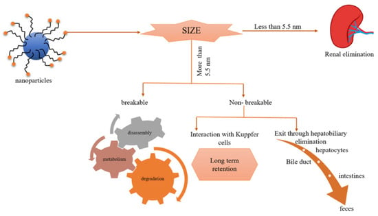
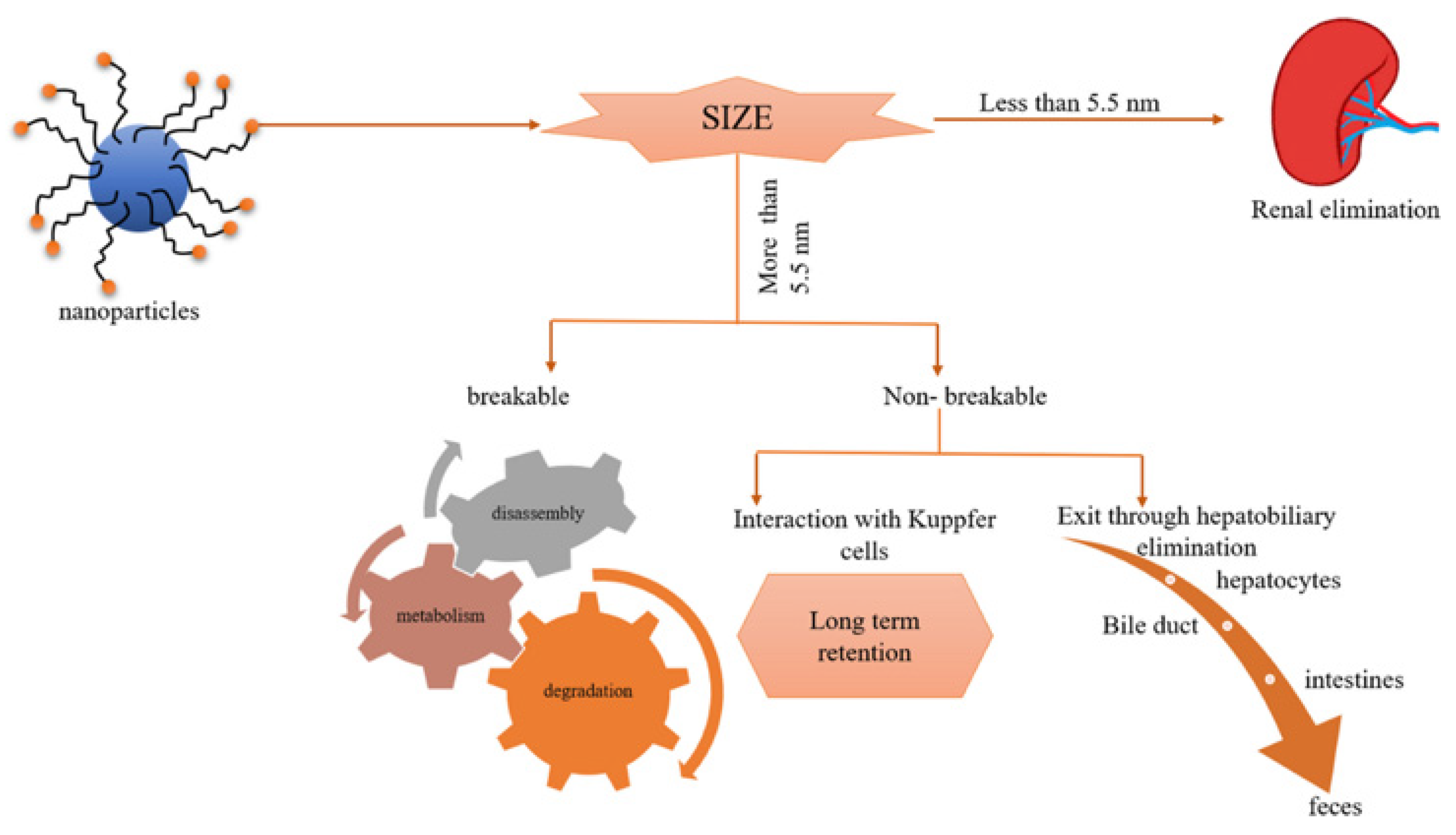
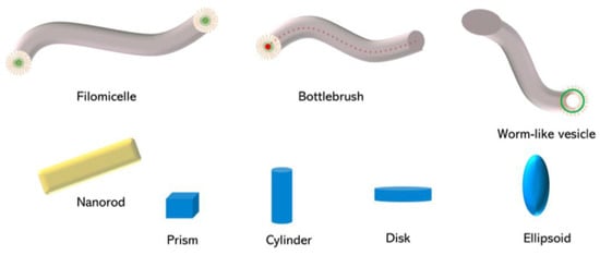
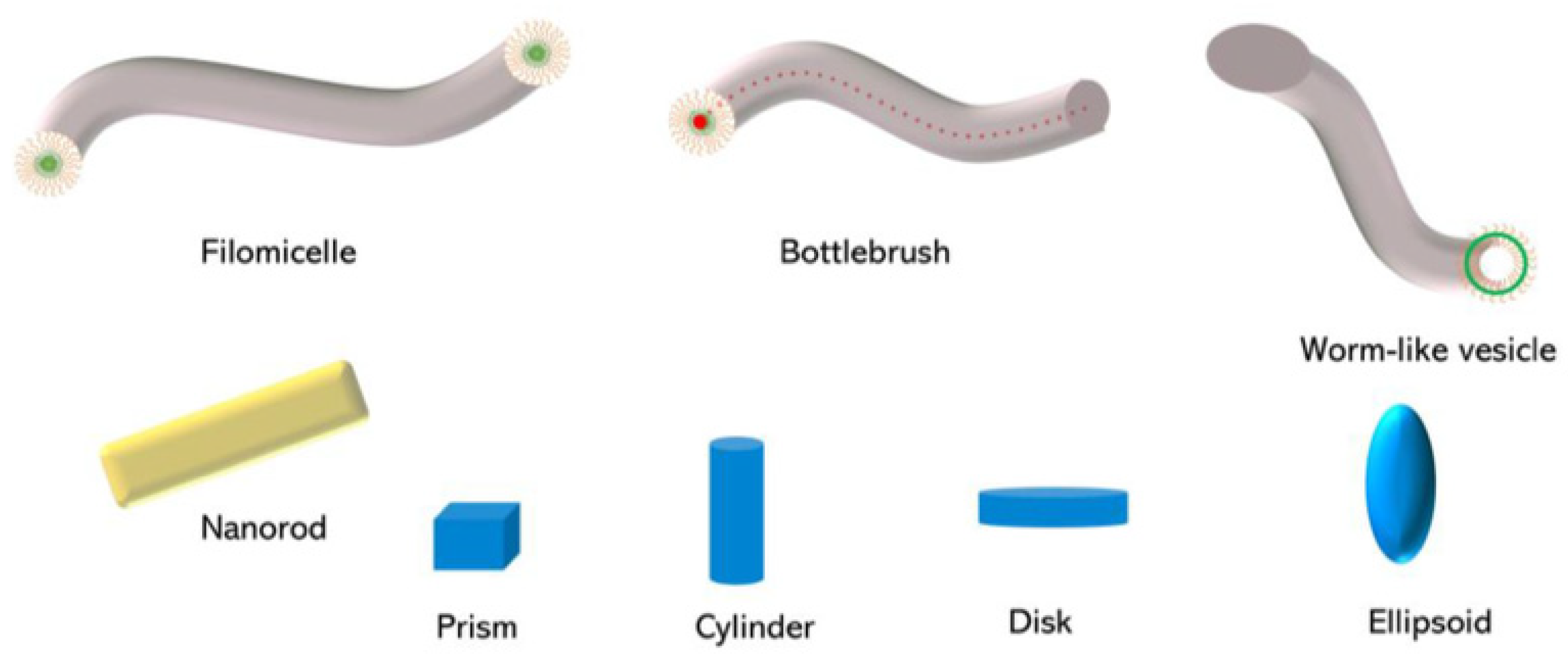
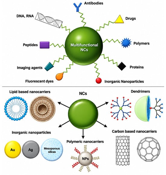
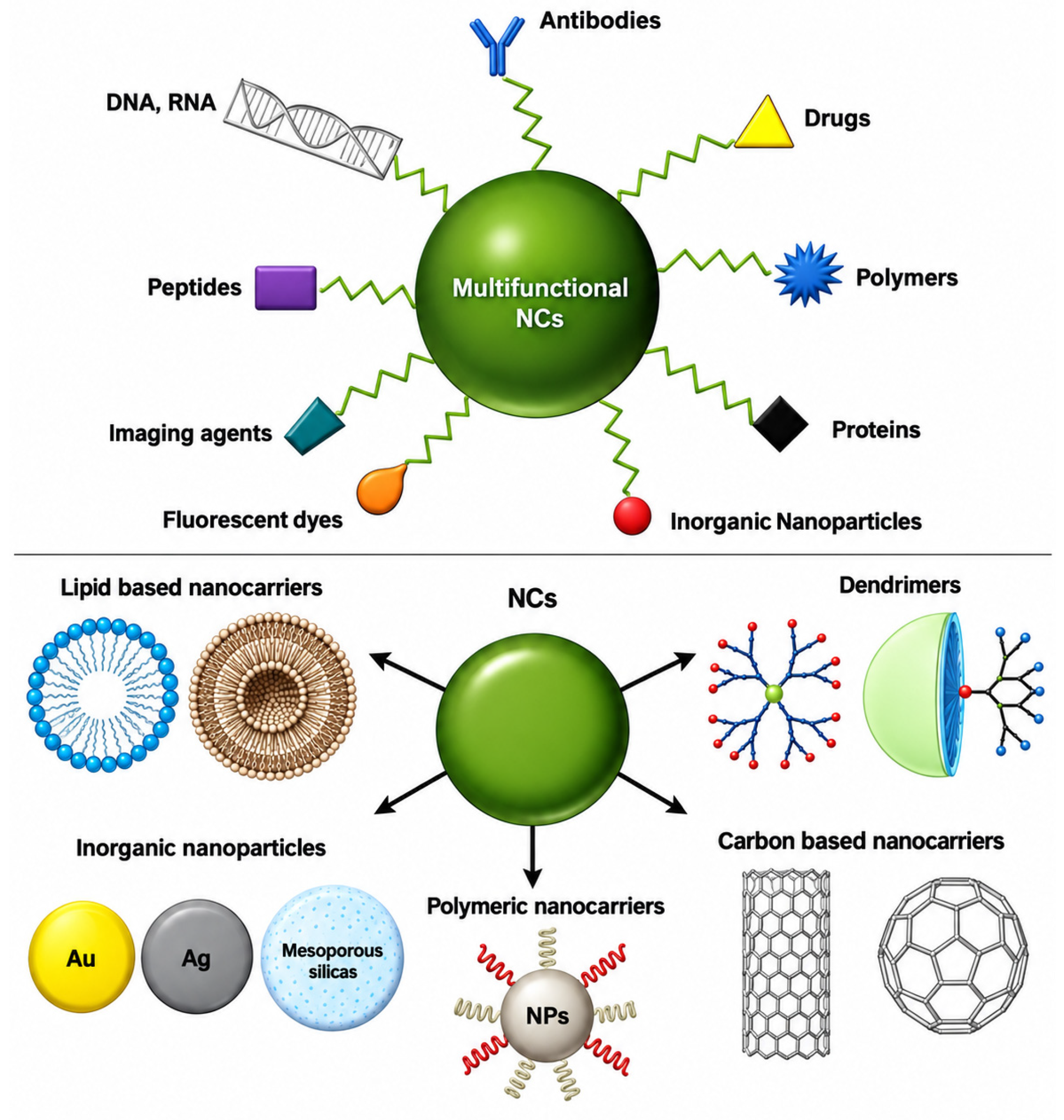

Disclaimer/Publisher’s Note: The statements, opinions and data contained in all publications are solely those of the individual author(s) and contributor(s) and not of MDPI and/or the editor(s). MDPI and/or the editor(s) disclaim responsibility for any injury to people or property resulting from any ideas, methods, instructions or products referred to in the content. |
© 2026 by the authors. Licensee MDPI, Basel, Switzerland. This article is an open access article distributed under the terms and conditions of the Creative Commons Attribution (CC BY) license.