


Abstract
The SARS-CoV-2 pandemic has significantly impacted global health, politics, medicine, finance, and society. Since 2020, various mutations have been reported, leading to drug resistance in current treatments against different SARS-CoV-2 strains and a drastic increase in cases of long-COVID. This situation underscores the urgent need to develop targeted and effective drugs to combat the spread of SARS-CoV-2 strains and their mutants, manage long-COVID symptoms and prepare for future pandemics. Currently, the treatment of SARS-CoV-2 focuses on targeting the virus’s entry and replication mechanisms to disrupt its life cycle. This review examines approved drugs, clinical candidates, and inhibitors under development, along with their bioassay data, while highlighting associated challenges. It illustrates how inhibitors bind to active sites, providing insights and emphasizing the importance of in silico studies, such as molecular docking, molecular dynamics simulation, FEP+, WaterMap, and quantitative structure–activity relationship (QSAR) analyses, and their correlation with experimental studies in expediting the drug discovery process. The review aims to provide researchers with insights into the gaps that need to be addressed concerning mutations affecting viral entry and to prepare for future pandemics.
1. Introduction
The COVID-19 pandemic, caused by SARS-CoV-2, is the most recent global health crisis, marking the seventh human coronavirus [1]. SARS-CoV-2 is the most contagious, with approximately 772.4 million confirmed cases and around 7 million deaths as of December 2023 [1]. Initially identified in Wuhan, China, it rapidly spread globally [2]. Trans- mission occurs through contact with infected individuals via respiratory droplets [3]. The virus enters host cells by binding its spike protein to the ACE2 receptor, with TMPRSS2 facilitating viral entry [4,5]. ACE2 is expressed in various organs, making them susceptible to infection [6,7]. The virus affects all age groups, with a pronounced impact on older adults and individuals with comorbidities [8,9,10].
Adaptive immune responses mediated by T and B cells are critical in defending against the virus during the initial 7–10 days post-infection [7]. COVID-19 is further associated with long-term neurological consequences, exacerbated by the body’s immune system releasing excessive pro-inflammatory cytokines, known as a “cytokine storm”, in an attempt to mitigate neuroinflammation caused by the virus [10]. The persistence of SARS-CoV-2 symptoms, termed “long-COVID”, beyond the acute phase is linked with heart failure, obesity, hypertension, chronic kidney diseases, cardio-metabolic disorders, and atherosclerotic cardiovascular diseases [11]. SARS-CoV-2 was initially designated as a public health emergency of international concern (PHEIC) in January 2020, prompting global mitigation strategies such as lockdowns and social distancing, alongside accelerated therapeutic development [12,13,14,15]. Concurrently, the World Health Organization initiated the “Solidarity” trial, a comprehensive international effort designed to ascertain efficacious treatments for SARS-CoV-2, ultimately resulting in the granting of emergency use authorization (EUA) for several pre-existing antiviral therapies [16]. Computer-aided drug discovery has emerged as one of the most reliable approaches, given the urgent need for novel SARS-CoV-2 treatments. It concurrently reduces the probability of producing toxic drugs and facilitates optimization before experimental validation [17,18]. However, despite significant progress, no comprehensive therapeutic strategy fully addresses emerging variants and long-term complications. Current efforts increasingly focus on viral entry and replication targets. Notably, many antiviral agents incorporate heterocyclic scaffolds, which enhance pharmacokinetic and pharmacodynamic properties and support broad applications in drug development [13,18,19,20].
Unlike previous reviews that primarily provide broad overviews of SARS-CoV-2 biology [20,21,22,23] or summarize therapeutic strategies and clinical outcomes [23,24], as well as those focusing on individual computational techniques or specific targets such as ACE-2 [24], Mpro [25,26], and PLpro [27,28], the present review provides an integrated and critical perspective by systematically evaluating computational approaches used in SARS-CoV-2 drug discovery, including repurposing and de novo design strategies. It analyzes molecular interactions with key viral and host targets involved in infection and replication, while highlighting the strengths and limitations of these methods. Importantly, it emphasizes multi-target strategies such as the Cathepsin L and Mpro dual inhibition to achieve synergistic antiviral effects, offering both methodological insights and a forward-looking framework that goes beyond the predominantly descriptive nature of existing studies.
2. Threats Due to Mutations of Drug Targets Involved in SARS-CoV-2’s Viral Entry and Replication and Long COVID, an Unmet Health Challenge
SARS-CoV-2 has undergone numerous mutations since 2019, leading to variants that complicate global containment efforts. These variants are categorized based on their impact on transmission, disease severity, and intervention effectiveness as variants of concern (VOC), variants of interest, variants being monitored (VBM), and variants of high consequence (VOHC). Variants of concern show increased transmissibility, disease severity, and reduced efficacy of public health interventions [29]. The Omicron variants remain the most mutated strain, with 50 mutations across the viral genome and 26–32 mutations on the spike protein [30]. These mutations occur on the receptor-binding domain (RBD) of the spike protein, including N501Y, E484K, L452R, T478K, and E484Q [31]. The regulation of viral proliferation has been challenging as new variants develop resistance to existing treatments, affecting the effectiveness of potent drugs. The following mutants—S144M/F/A/G/Y, H172Q/F, M165T, E166V/G/A, and Q192T/S/L/A/I/P/H/V/W/C/F—have been identified on the Mpro’s binding site, interrupting the effectiveness of the most reliable SARS-CoV-2 inhibitor so far, “Nirmatrelvir” [32]. PLpro is less susceptible to mutations, with only three points of mutation identified and accounting for >20% of SARS-CoV-2 PLpro’s variant of concern A145D (alpha), K92N (beta), and K232Q (gamma) on the thumb region [28,33,34]. Five more mutations (P247S, Y264H, E263D, T265A, and Y268C) have been identified near the substrate-binding site. These mutations should be carefully considered in future PLpro-targeted drug design to ensure broad-spectrum efficacy and resistance resilience [35].
While most individuals recover from acute COVID-19, 31–69% experience long-term health issues called “long-COVID” [36,37]. The acute phase spans five weeks, with viral load peaking then declining [5,38]. SARS-CoV-2 disrupts host cells, causing impairments and cell death. The immune response triggers systemic hyperinflammation, leading to thrombosis and organ failure [5]. Long-COVID symptoms include fatigue, myalgia, cough, chest discomfort, and breathing difficulties, which may fluctuate [5,36,37]. After three weeks, the viral load becomes undetectable through RT-PCR. Untreated symptoms can permanently damage organs [5]. Complications include heart attacks, cognitive impairment, and organ dysfunction [5,39]. Healthcare centers manage long-COVID through symptom-based approaches [5]. While identifying drugs for SARS-CoV-2, addressing long-COVID mechanisms requires equal attention to viral mutations.
2.1. Small-Molecule Therapeutic Drugs Approved for the Treatment of SARS-CoV-2
Since 2019, researchers have worked to develop or repurpose drugs for SARS-CoV-2 treatments, leading to several drug approvals. Remdesivir, Hydroxychloroquine, and Ivermectin were repurposed from treating Hepatitis C, malaria, and infestations to manage SARS-CoV-2 [40,41]. However, the most recent finding is that although Chloroquine/Hydroxychloroquine (CQ/HCQ) and Ivermectin remain debatable as they may exhibit mechanistic activity against SARS-CoV-2, most phase III clinical trials have failed to demonstrate clinical benefit in COVID-19 patients, and overall evidence from these trials does not support their efficacy for treatment [42]. While Remdesivir proved effective, its intravenous administration requires hospital settings, limiting accessibility; oral formulations are undergoing development [43,44]. The Remdesivir drug affects the respiratory, gastrointestinal, cardiovascular, and reproductive systems [44]. New anti-SARS-CoV-2 drugs include Paxlovid, Xocova, and Molnupiravir [45,46]. China approved four domestic oral drugs: Azvudine, Leritrelvir, Simnotrelvir–Ritonavir, and Mindeudesivir [47] (Table 1, Figure 1). Most therapeutics now show resistance to new SARS-CoV-2 mutants, with efficacy ranging from 88% to below 21% [48]. Drug repurposing, the process of identifying new therapeutic uses for existing drugs, remains the most efficient strategy for the rapid and cost-effective development of broad-spectrum antivirals.
Figure 1.
Approved and repurposed small molecules for SARS-CoV-2 and their molecular targets: Mpro inhibitors—Nirmatrelvir [49,50,51], Ensitrelvir [47,52,53], Simnotrelvir [54,55], Leritrelvir [47], Atilotrelvir [54]; RdRp—Remdesivir [55], Mindeudesivir [30], Azvudine [47], Favipiravir [56]; endosomal entry—baricitinib [57], Chloroquine [58], and Hydroxychloroquine [59].
Remdesivir (see Figure 1) is an intravenously administered nucleoside analog used to inhibit the action of RNA polymerase in the treatment of COVID-19 for non-hospitalized patients with mild-to-moderate symptoms [55]. Its emergency use approval was first awarded in May 2020, and later that year, it was given full approval for the treatment of pediatric patients less than 12 years old [60]. Remdesivir is a prodrug metabolized within the host cells to the therapeutically active triphosphate metabolite that has a reasonable biochemical activity of IC50 = 770 nM and antiviral activity of EC50 = 6.2 µM as a delayed chain terminator blocking viral protein transcription [60,61]. Despite its initial promise, the clinical efficacy of Remdesivir remains debated, and multiple case reports have documented emerging resistance to Remdesivir, particularly in patients with underlying conditions undergoing prolonged treatment [44,62,63,64]. This is supported by Gandhi et al., who reported the de novo emergence of the nsp12 E802D mutation during remdesivir therapy in an immunocompromised patient with persistent COVID-19, resulting in reduced antiviral susceptibility and viral recrudescence [64]. In addition, Luo et al. demonstrated that other mutations, such as V557L, may have contributed to remdesivir resistance through structural mechanisms that affect RdRp function [65]. Baricitinib was the first Food and Drug Administration (FDA)-approved immunomodulatory treatment for COVID-19. On the 19th of November 2020, the FDA issued an Emergency Use Authorization (EUA) for baricitinib to be used in combination with Remdesivir; this decision was reviewed on 28 July 2021, and baricitinib was authorized to be used as a stand-alone treatment [66]. The in silico metabolism prediction of baricitinib suggested that structural alterations to the pyrimidine ring and the pyrrole ring may improve metabolic stability and safety in drug design [67].
Nirmatrelvir is an orally administered 3C-like protease (3CLpro/Mpro) inhibitor prescribed and co-packed with ritonavir to patients suffering from moderate-to-mild COVID-19. Its EUA was originally granted in 2020 by the US FDA before receiving full approval in 2023 for the treatment of adults at high risk of progression to severe COVID-19 symptoms [68]. It was manufactured as Paxlovid by Pfizer and distributed as part of the U.S. Department of Health and Human Services’ response to the COVID-19 pandemic. Nirmatrelvir was selected as an inhibitor due to its potent biochemical activity (Ki = 3.11 nM) and Vero E6 antiviral activity EC50 = 74.5 nM, achieved by its ability to form a reversible covalent thioimidate adduct with Mpro’s Cys145, and stabilizing a single binding mode with its target [69]. Co-administration of nirmatrelvir with subtherapeutic doses of ritonavir extends its clinical efficacy as ritonavir is an inhibitor of the major human hepatic drug-metabolizing enzyme cytochrome P450 3A4 (CYP3A4), responsible for the metabolism of the majority of drugs [70,71]. The inhibition of CYP3A4 boosts the therapeutic concentration of Ritonavir, with an intrinsic clearance value of 25 to 32.8 µL/min/mg [49,51]. Nirmatrelvir maintained a good potency even in the FRET assay and Cell-Based Antiviral Assay (ALI-HBEC), achieving EC50 values of 3.0 nM and 25.3 nM, respectively [50,51]. Nirmatrelvir is the most effective anti-SARS-CoV-2 approved drug, with an EC50 range between 3.0 ± 0.1 nM and 25.3 nM, followed by Molnupiravir, Hydroxychloroquine, and Remdesivir: with EC50 values of 0.4 µM, 0.72 µM, and 0.77 µM, respectively, as shown in Table 1. Despite its proven efficacy in reducing COVID-19 severity, Paxlovid (Nirmatrelvir/Ritonavir) faces several clinical challenges, most notably, drug–drug interactions in patients, primarily due to its co-administration with Ritonavir. Consequently, when it interacts with drugs that also inhibit CYP450 enzymes, it can lead to elevated levels of co-administered drugs, increasing the risk of toxicity or adverse effects, especially in patients taking anticoagulants, immunosuppressants, or certain cardiovascular drugs [72]. Notably, the highest percentage of Mpro inhibitors are covalent inhibitors that bind like Nirmatrelvir and require co-administration to improve their physicochemical properties.
Ensitrelvir, a non-peptidomimetic inhibitor with an IC50 of 0.013 µM, comparable to that of Nirmatrelvir, and moderate potency of 0.37 µM in the Vero cell line, has exhibited improved metabolic stability when compared to Nirmatrelvir [73]. As such, Ensitrelvir is currently approved by the Pharmaceuticals and Medical Devices Agency (PMDA), with an intrinsic clearance of 1.7 mL/min/Kg in rats and a good antiviral activity EC50 of 0.37 µM in a cell-based assay (VeroE6/TMPRSS2) [47,52,53]. However, these results were surpassed by Leritrelvir and Atilotrelvir, with both exhibiting an antiviral activity in the nanomolar range using the same assay [47,54]. Simnotrelvir also showed a good potency in the FRET assay with an EC50 value of 79 nM, comparable to that of Nirmatrelvir [62,63]. Notably, drugs targeting Mpro were more potent than other candidates since their inhibitory activity values were in the nanomolar range, and all covalent Mpro drug candidates have a P4-trifluoroacetyl moiety, facilitating hydrophobic interactions [47,51,52,53,54].
Chloroquine (CQ) and Hydroxychloroquine (HCQ) are two SARS-CoV-2 endosomal entry inhibitors with antiviral activity of 5.47 µM and 0.72 µM in the Vero cell line [59]. The potency of Hydroxychloroquine was improved by the introduction of a hydroxyl group on Chloroquine. This also improved the lipophilicity from a log P value of 4.37 to 3.8. It should be noted that, although LogP is commonly used to assess lipophilicity, oral bioavailability is influenced by multiple physicochemical parameters, including molecular weight, hydrogen bond donors and acceptors, and polar surface area, as described by Lipinski’s Rule of Five and Veber’s criteria [74]. According to the Lipinski Rule of Five, log P should be <5 for oral administration; however, the most acceptable range is 1.35–1.8 for intestinal and oral absorption, and for the central nervous system (CNS), it should be around 2 [75]. Some reports suggest that HCQ’s efficacy may be enhanced when combined with Azithromycin. However, other studies have raised concerns about the long-term and combined use of CQ or HCQ with other drugs, particularly Azithromycin, due to serious cardiac side effects. These include QT interval prolongation, arrhythmogenic risks, QRS widening, diabetes, etc., which can lead to potentially life-threatening heart complications [76,77]. The FDA revoked the emergency use authorization for Chloroquine and Hydroxychloroquine after determining that they are unlikely to be effective against COVID-19 and pose serious cardiac risks. This decision was based on updated scientific data and a request from the Biomedical Advanced Research and Development Authority (BARDA) [78]. The newer drug Molnupiravir received its full approval from the UK Medicines and Healthcare products Regulatory Agency (MHRA) in 2021 before it was given an EUA by the US FDA to treat adults above the age of 18 who tested positive for SARS-CoV-2 due to its impact on bone and cartilage growth [79,80].
Molnupiravir is also restricted for pregnant individuals because of its potential to cause serious fetal abnormalities and increase the risk of miscarriage [81]. Nonetheless, the most clinically relevant drugs for SARS-CoV-2 includes Remdesivir [23], Molnupiravir, and Nirmatrelvir/Ritonavir (Paxlovid), which have demonstrated therapeutic benefit; however, their effectiveness is limited by factors such as modest clinical efficacy in some patient groups, the emergence of viral resistance and variants, adverse side effects, significant drug–drug interactions, and limited accessibility in certain regions [82].
Table 1.
Approved small molecules for SARS-CoV-2 and their pharmacodynamics and physicochemical attributes.
There is a reasonably good correlation between pEC50 and pIC50 values for the approved drugs, as shown in Figure 2; however, Mindeudesivir had negative values, showing poor efficacy. Nirmatrelvir, Ensitrelvir, Simnotrelvir, Atilotrelvir, and Molnupiravir had positive pIC50 and pEC50 values, with Atilotrelvir having the highest inhibitory activity amongst the rest (Table 1). Metabolic stability, log P, and logD are underreported specifically for SARS-CoV-2 since most of these drugs were repurposed. As shown in Table 1, only Nirmatrelvir had data for logD within the range of 1.5–1.9 and an intrinsic clearance of 25–32.8 µL/min/mg in human upon co-administration with Ritonavir, indicating good solubility with moderate clearance [49,51]. Azvudine is administered orally and showed better activity compared to Remdesivir, with minor side effects from the phase III clinical trial, and they both target RdRp. Favipiravir exhibited the lowest pEC50, whereas Monupiravir had the highest pEC50 in the same cell-based assay (Vero E6). However, Mindeudesivir had the highest pEC50 values, although it was conducted in an A549 cell-based assay. RdRp inhibitors mentioned above played a crucial role in reducing SARS-CoV-2 viral replication and mitigating the severity of COVID-19 symptoms [71,85]. However, SARS-CoV-2 is an RNA virus characterized by a high mutation rate due to the low fidelity of its RNA-dependent RNA polymerase [86]. This frequent genetic variability poses a significant challenge, as mutations may alter the structure of viral proteins targeted by these drugs, potentially reducing their effectiveness and leading to antiviral resistance. Mindeudesiver (RdRp) and Hydroxychloroquine (endosomal entry) exhibited good potency in the nanomolar range. Despite its potency, Hydroxychloroquine and Chloroquine were withdrawn for the treatment of SARS-CoV-2 due to the side effects mentioned above.
Figure 2.
Approved small molecules targeting SARS-CoV-2 and their respective pIC50 and pEC50 values.
2.2. Viral Entry Targets for SARS-CoV-2
SARS-CoV-2 enters host cells in two distinct pathways, i.e., the cell surface pathway and the endosomal pathway, as shown in Figure 3. In the surface pathway, the viral spike (S) protein binds to the ACE2 receptor and is activated by the serine protease TMPRSS2, allowing direct fusion with the plasma membrane (Figure 3a) [4,5]. Alternatively, in the endosomal entry, the virus is internalized into endosomes, where Cathepsin L (CTSL), a lysosomal cysteine protease, cleaves and activates the spike protein, facilitating fusion with the endosomal membrane (Figure 3b) [87,88,89].
Figure 3.
SARS-CoV-2 mechanism of entry pathways. (a) The SARS-CoV-2 spike protein interacts with ACE2 (blue), while TMPRSS2 (light blue) and NRP1 (green) facilitate viral attachment and membrane fusion. Furin (orange) cleaves the spike protein at the S1/S2 site, priming the virus for fusion and entry at the cell surface. (b) Endocytic entry pathway. Following binding to ACE2 (dark blue) and alternative receptors, including AXL (cyan), CD147 (light blue), ASGR1 (pink), and KREMEN1 (yellow), SARS-CoV-2 is internalized through endocytosis. Within endosomes, Cathepsin L (green) mediates proteolytic activation of the spike protein, enabling membrane fusion and viral genome release. Both entry routes ultimately lead to viral replication and production of new virions [90]. Reproduced with permission [89]. Copyright 2023 MDPI.
2.3. SARS-CoV2 Spike Protein ACE2: Structure and Function
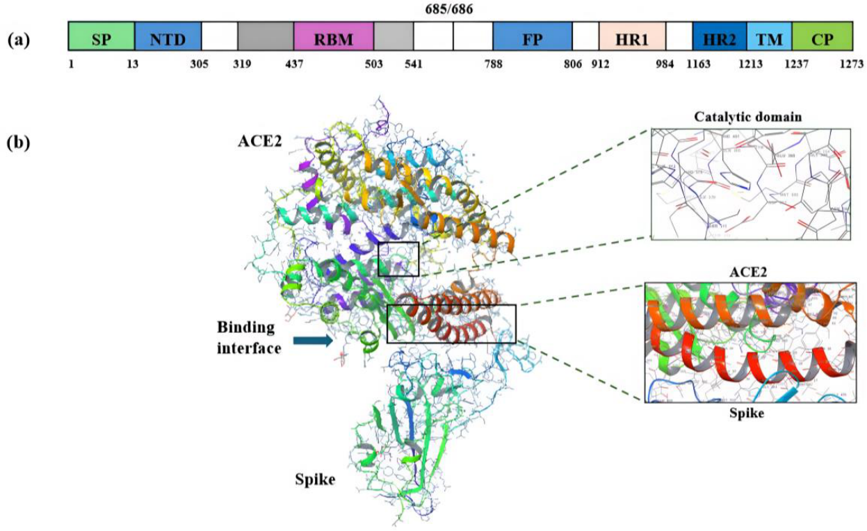
Spike protein is a glycoprotein made up of two subunits, i.e., the S1 subunit, which binds the host cell receptor, and the S2 subunit, responsible for the viral membrane fusion. The S1 subunit, which binds the host cell receptor, and the S2 subunit are responsible for the viral membrane fusion [91]. The S1 sub-unit contains the signal peptide (SP, 1–13 resi-dues), N terminal domain (NTD, 14–305 residues), and receptor binding domain (RBD, 319–541 residues) that bind to human angiotensin-converting enzyme-2 (hACE2); whereas the S2 subunit contains fusion peptide (FP, 788–806 residues), heptad repeats HR1 (912–984 residues) and HR2 (1163–1213 residues), trans-membrane domain (TM, 1213–1237 residues), and cytoplasmic domain (CP, 1237–1273 residues) as shown in Figure 4a [91]. The fusion peptide (FP) plays a crucial role in mediating membrane fusion by disrupting and connecting lipid bilayers of the host cell membrane [46]. SARS-CoV-2 initiates infection by binding to human angiotensin-converting enzyme-2 (hACE2) through the spike protein located at the SARS-CoV-2 virus’s envelope; see Figure 4b [91,92]. The hACE2 is a membrane-bound enzyme highly expressed in various cell types, including lungs, heart, liver, intestine, kidneys, and testis [30]. The spike protein has since been the main target of peptides, monoclonal antibodies, peptidomimetics, antibodies, and small molecules [46,93]. This resulted in a significant number of developed monoclonal antibodies against SARS-CoV-2.
The ACE2 receptor for SARS-CoV-2 targets mainly human organs, including the lungs, heart, liver, intestine, kidneys, and testis in the human body, and if the virus is not monitored, it can lead to cell damage [6]. These two are typical drug targets, especially the ACE2, as it attacks critical organs, resulting in long-COVID symptoms, which may result in permanent damage if untreated. Dysregulation of ACE2 after SARS-CoV-2 infection disrupts the angiotensin system, promoting persistent inflammation, endothelial dysfunction, and multi-organ symptoms [94].
Figure 4.
Structure of the SARS-CoV-2 S protein ACE2. (a) Domain organization of the SARS-CoV-2 spike protein (SP) showing the signal peptide (SP, green), N-terminal domain (NTD, blue), receptor-binding motif (RBM, magenta), fusion peptide (FP, blue), heptad repeats (HR1, peach and HR2, dark blue), transmembrane domain (TM, light blue), and cytoplasmic tail (CP, green), with the furin cleavage site at residues 685/686 (white). Reproduced with permission [91]. Copyright 2020 ACS. (b) ACE2 structure in a complex with the receptor-binding domain (RBD) of the SARS-CoV-2 spike protein (PDB ID: 6M0J). Reproduced with permission [95]. Copyright 2020 Springer Nature.
2.4. Inhibitors Targeting the SARS-CoV-2 Spike Protein ACE2 Protein–Protein Interaction
Several studies have used computational approaches to investigate inhibitors targeting the ACE2–SARS-CoV-2 interaction. Amin et al. demonstrated, using molecular modeling, that ACE2 binds more strongly to SARS-CoV-2 than to SARS-CoV, explaining its higher infectivity [96]. Munawar et al. optimized the ACE2 inhibitor ATN 161, yielding an IC50 of 3.16 µM, and developed a peptide analog, Ac-PHSPN-NH2 (Analog-III), which showed improved antiviral activity (IC50 = 0.6 µM), favorable cytotoxicity, and demonstrated the strongest binding affinities of −17.05 kcal/mol from molecular docking [97]. Bojadzic et al. identified potent spike-ACE2 inhibitors, with DRI C91005 exhibiting nanomolar activity (IC50 = 160 nM) [98]. Similarly, ELISA-based screening by Shin et al. identified small-molecule inhibitors of the ACE2–spike interaction, with SB27012 (cyclopropyl amide) showing an IC50 of 7.7 µM, while SB27016 (lacking R3 substituents) exhibited no activity, indicating structural dependence. Optimization of SB27012 produced SB27041, which demonstrated improved potency with IC50 values of 0.52–3.38 µM (spike–ACE2 binding) and a pseudovirus inhibition IC50 of 5.6 µM [99]. A CADD screening by Bastos et al. using the 6M0J crystal structure identified Ligand_003 with a docking score of −8.645 kcal/mol, outperforming Hydroxychloroquine (−7.595 kcal/mol). Subsequent optimization yielded Ligand_008, which showed improved binding affinity (−7.690 kcal/mol) and a predicted IC50 of 0.49 µM, compared to the experimental IC50 of 0.663 µM for Ligand_003. Lapaillerie et al. identified two SARS-CoV-2 entry inhibitors, AB 00011778 and AB 00047476, that blocked viral entry into human pulmonary cells, with IC50 values of 0.25–2.5 µM. However, despite good antiviral activity, AB 00011778 showed suboptimal cytotoxicity with a CC50 > 30 µM in A549 ACE2 TMPRSS2 cells [100]. Yang et al. reported a naturally occurring compound, Gamabufolatin (Figure 5), with the potential to inhibit SARS-CoV-2 ACE2, with an EC50 value in the nanomolar range; however, Gamabufolatin also showed poor cytotoxicity, with a CC50 value less than 40 µM [101]. Shahhamzehei et al. used molecular docking (PDB ID: 7BNN) to identify nitrile-containing isoquinoline derivatives 9, 14, and 15 as inhibitors of the spike RBD–ACE2 interaction. Among these, compound 15 showed promising activity with a Ki of 4.33 µM and CC50 > 100 µM, indicating good potency and favorable safety, and was proposed as a potential broad-spectrum coronavirus entry inhibitor [102].
Figure 5.
SARS-CoV-2 Spike protein ACE2 experimental inhibitors.
Based on the three reported parameters of IC50, CC50, and Ki values, several compounds have shown strong potential as inhibitors of the ACE2–SARS-CoV-2 interaction. Among them, DRI-C91005 demonstrated the highest potency (IC50 = 160 nM), while Analog-III combined strong activity (IC50 = 0.6 µM) with low toxicity and high binding affinity, followed by compound 15 with a good Ki (4.33 µM) and excellent safety profile (CC50 > 100 µM). Compounds like SB27012, SB27041, and Gamabufolatin showed moderate to variable potency or poor cytotoxicity profiles. Consequently, although several compounds demonstrate promising in silico binding and moderate in vitro activity, discrepancies in reported IC50, CC50, and Ki values, together with incomplete toxicity data, make it difficult to draw definitive conclusions regarding their therapeutic potential. These limitations are further compounded by the reliance on molecular docking as a primary tool, which is constrained by simplified scoring functions, inadequate treatment of protein flexibility and solvent effects, and poor correlation with experimental binding affinities [103]. Therefore, integrated computational–experimental approaches are essential for more reliable validation of entry-target inhibitors.
2.5. SARS-CoV-2 TMPRSS 2: Structure and Function
Transmembrane protease serine 2 (TMPRSS 2), a protease responsible for proteolytically processing the SARS-CoV-2 Spike protein, facilitating virus–host membrane fusion into human cells [4,93]. This protease comprises three subdomains, the LDLR-A, SRCR, and SP, each contributing to its function in viral entry and protein processing (Figure 6a,b). The LDLR-A domain is located around the region of amino acids 106 to 145 and is primarily involved in lipoprotein binding, which may facilitate interactions with cellular membranes. The SRCR domain, located around the region of amino acids 146 to 243, plays a key role in protein–protein interactions, and the serine protease (SP) domain spans the most critical region with amino acids 256 to 492. This domain accommodates the catalytic machinery of the protease enzyme and is responsible for its proteolytic activity (Figure 6a,b) [93]. The TMPRSS2 is particularly important for managing long-COVID symptoms as it is expressed in many epithelial cells, including those in the prostate, kidneys, bronchus, lungs, trachea, small intestine, pancreas, colon, salivary glands, and thymus, with the highest expression levels found in the lungs and airways [104]. The SARS-CoV-2 prefers the TMPRSS2 entry route compared to the alternative ‘endocytosis’ route, which is rather less efficient [105].
Figure 6.
(a) Schematic representation of the amino acid sequence of SARS-CoV-2 TMPRSS2 showing the cytoplasmic region (purple), transmembrane helix (pink), low-density lipoprotein receptor class A-like (LDL-like; grey) domain, scavenger receptor cysteine-rich-like (SRCR-like; green) domain, and serine peptidase (blue) domain. Key residues (P78, Y82, L188, V197, F246, and S265) and the modeled region (aa 187–529) are indicated. Reproduced with permission [106]. Copyright 2021 MDPI. (b) A 3D structural presentation of SARS-CoV-2 S TMPRSS2 7MEQ, highlighting the serine protease (SP) domain, scavenger receptor cysteine-rich (SRCR) domain, and low-density lipoprotein receptor class A (LDLA) domain, and an extended view of the active site with ligand bound. Reproduced with permission [106]. Copyright 2023 ACS.
Inhibitors Targeting SARS-CoV-2 Transmembrane Protease Serine 2
Camostat mesylate is one of the most prominent serine protease inhibitors active against TMPRSS2, with an IC50 of 87 nM; however, Hoffman and colleagues report a 17 times more potent inhibitor, Nafamostat mesylate, with a half-maximal inhibition concentration (IC50) of 5 nM [107]. A molecular modeling study by Shapira et al. using a crystal structure with PDB ID of 6N4T has led to the discovery of a TMPRSS2 inhibitor N-0385 with an EC50 of 2.8 nM against SARS-CoV-2. These compounds were assessed for antiviral efficacy against an ancestral SARS-CoV-2 strain (lineage B, VIDO) and four variants—Alpha (B.1.1.7), Beta (B.1.351), Gamma (P.1), and Delta (B.1.617.2)—in human lung cells, and provided significant therapeutic benefits [108]. Mahoney and colleagues employed rational structure-based drug design (SBDD) combined with substrate specificity screening to identify TMPRSS2 covalent inhibitors containing ketobenzo thiazole scaffolds. Among the identified compounds, MM3122 emerged as the lead candidate, exhibiting an IC50 of 340 pM and EC50 of 430 pM against recombinant full-length TMPRSS2 protein. These inhibitors demonstrated a significantly improved activity compared to existing drugs, such as Camostat and Nafamostat [109]. Wettstein et al. identified compound 2 and compound 5 as potent inhibitors of TMPRSS2, demonstrating a Ki value between 2.5 and 3.8 nM that has 3 times greater activity than the active metabolite FOY-251 with Ki = 9.7 nM. Both compounds effectively prevented cell-mediated proteolysis of a fluorogenic substrate, with IC50 of 12.7 nM for compound 5 and IC50 = 32 nM for compound 2 [110]. Ferková et al. reported a previously modified potential TMPRSS2 inhibitor, N-0385, to improve its pharmacodynamic and pharmacokinetic properties. In addressing some of these challenges, researchers developed a library of peptidomimetic compounds, introducing P3 site modifications to optimize selectivity and pharmacokinetics while maintaining sub-nanomolar potency, leading to the discovery of compound 9 with inhibitory potency of Ki = 0.13 ± 0.03 nM (Figure 7) [111]. Consequently, although several compounds demonstrate promising in silico binding and moderate in vitro activity, discrepancies in reported IC50, CC50, and Ki values, together with incomplete toxicity data, make it difficult to draw definitive conclusions regarding their therapeutic potential. These limitations are further compounded by the reliance on molecular docking as a primary tool, which is constrained by simplified scoring functions, inadequate treatment of protein flexibility and solvent effects, and poor correlation with experimental binding affinities as described in the previous section [103]. Furthermore, the accuracy of the docking score is system dependent and the energy terms in the scoring functions do not account for ligand and receptor’s strain energies.
Figure 7.
Experimental inhibitors targeting transmembrane serine 2 a viral entry target for SARS-CoV-2.
2.6. Cathepsin L (CTSL): Structure and Function
Cathepsin L (CTSL) is a three-dimensional structure that comprises an L domain of α-helix and an R domain of β-sheet. The L-domain comprises three α-helices, with the longest and the vertical helix also known as the central helix that further contains above 30 residues. The R-domain on the other side is made up of a β-barrel with the front strand(s) forming a coiled structure (Figure 8) [89]. These two catalytic residues form the thiolate–imidazolium ion pair, which is essential for the proteolytic activity of the enzymes [87,88,89]. This protease plays a significant role in the endosomal entry pathway of SARS-CoV-2 by facilitating viral entry into human cells through activation of the spike protein [87,88,89]. Cathepsin L protease is found in most human tissues, such as the kidney, bone marrow, respiratory system, sexual tissues, endocrine tissues, urinary bladder, gastrointestinal tract, and lymphoid tissues [89]. CTSL is more highly expressed in human lungs than the angiotensin-converting enzyme 2 (ACE2), transmembrane protease serine 2 (TMPRSS2), and furin [89]. Despite the limited literature on CTSL’s ability to prevent or treat SARS-CoV-2 virus infection, its overexpression in human cell lines boosts SARS-CoV-2 spike-mediated viral entry, making the CTSL one of the most promising targets to block the virus from entering the system [112]. It was believed that the Omicron variant entered the host cell through only Cathepsin L and not via transmembrane protease serine 2, and this claims Cathepsin L as the best target for viral entry; however, recent findings still highlight TMPRSS2 as the main entry pathway, even for Omicron [113].
Figure 8.
(a) A schematic representation of CTSL showing tthe N-terminal domain (NTD) in magenta, receptor-binding domain (RBD) in blue, C-terminal domain (CTD) in green, fusion peptide proximal region (FPPR) in light blue, heptad repeat 1 (HR1) in pink, heptad repeat 2 (HR2) in light green, and transmembrane domain (TMD) in grey, with Cathepsin L (CTSL) and furin cleavage sites indicated with red arrows. Reproduced with permission [114]. Copyright 2022 Springer Nature. (b) The fold of the two-chain form of native Cathepsin L (PDB entry: 8UAC) and an extended view of the active site with ligand bound in both monomers. Reproduced with permission [115]. Copyright 2024 Elsevier.
Inhibitors Targeting SARS-CoV-2 Cathepsin L
Ma et al. designed a dual inhibitor, MPI1, through QSAR modeling with an IC50 of 79 pM against Cathepsin L and an IC50 of 105 nM against Mpro [88]. The SM141, a dual inhibitor for Mpro and Cathepsin L, has shown great potency against Cathepsin L with an antiviral activity in the nanomolar range [116,117]. The K777 is a covalent, irreversible inhibitor that is a candidate in Phase I clinical trials, demonstrating great potency against Cathepsin L with an antiviral activity of 74 nM in the Vero cell line [117]. Meewan et al. discovered triple inhibitors, with RI175, RI173, and RI172 being the most potent inhibitors in an enzymatic assay against Mpro, PLpro, and human Cathepsin L, with IC50 values of 300 nM, 200 nM, and 200 nM, respectively [118]. Sacco et al. investigated dual inhibitors, Calpain inhibitors II and XII, targeting both Mpro and Cathepsin L with inhibition concentrations of 0.41 nM, 1.62 nM for CTL, and 970 nM, 453 nM for Mpro, respectively [119]. Previt et al. conducted a similar study to Ma et al., reporting the use of dual inhibitors targeting both Mpro and human Cathepsin L through QSAR modeling. The most prominent compound 4d, with Ki 0.70 µM and 5.54 µM for Cathepsin L and Mpro, respectively, has antiviral activity of EC50 = 3.50 μM, and CC50 values > 100 μM. This was achieved by varying aliphatic amino acids at the P2 site and fluorine position on the phenyl ring at the P3 site, which significantly influenced their binding affinity [120]. The ability of Mpro inhibitors to also target host proteases like Cathepsin L opens exciting possibilities for drug repurposing and lead optimization. Therefore, by modifying existing Mpro inhibitors, researchers can design dual or multi-target inhibitors, accelerating the discovery of broad-spectrum antivirals that combat SARS-CoV-2 and potentially other viral pathogens (Figure 9). Despite promising results, these dual inhibitors face challenges, including selectivity, toxicity, poor drug-likeness of peptidomimetics, and difficulties translating enzymatic inhibition into effective antiviral activity [121]. In addition, the underlying computational approaches, particularly QSAR and structure-based design, are limited by reliance on training data quality, simplified modeling assumptions, and limited predictive power for selectivity, pharmacokinetics, and efficacy [122].
Figure 9.
SARS-CoV-2 Cathepsin L experimental inhibitors.
2.7. SARS-CoV-2 Viral Replication Targets
Once the virus has entered the host, positive-sense ssRNA transcribes to produce non-structural proteins, including papain-like protease (PLpro) and main protease (Mpro) from large viral polyproteins Pp1a and Pp1ab (Figure 9) [31]. PLpro and Mpro play a massive role in viral replication and transcription as they proteolytically process proteins needed for viral replication [123]. On the other hand, structural proteins, including spike (S), membrane (M), and envelope (E), produced in the endoplasmic reticulum (ER), are transported to the ER–Golgi intermediate compartment (ERGIC) for virion assembly. The nucleocapsid (N) protein is a 419-amino acid, multi-domain RNA-binding protein with two conserved domains, the N-terminal domain (NTD) and the C-terminal domain (CTD), connected by an inherently disordered region called the central linking region (LKR). The N protein stabilizes and protects the viral genome by packaging it into the nucleocapsid, ensuring effective integration of virions during the replication cycle, assembly, and release phases [124,125]. The assembled virions in the ERGIC are then transported through smooth-wall vesicles to the cell membranes to release the mature virus particles (Figure 10) [124].
Figure 10.
Key steps involved in SARS-CoV-2 viral replication. SARS-CoV-2 enters host cells through receptor-mediated attachment (blue), releases its RNA genome (dark blue) for replication and protein synthesis. Genomic and subgenomic RNAs are produced, giving rise to structural proteins including nucleocapsid (blue), spike (green), membrane (purple), and envelope (pink). New virions assemble in the ER–Golgi intermediate compartment (ERGIC) and are released by exocytosis. Image generated with Biorender.
2.7.1. SARS-CoV-2 Main Protease (Mpro): Structure and Function
The main protease (Mpro), well known as 3 Chymotrypsin-like (3CL) or non-structural protein 5 (NSP5), is a three-domain homodimeric cysteine protease that plays a significant role in SARS-CoV-2 viral replication and transcription [45,123]. This protease comprises three domains (Figure 11a) classified as I (residues 8–101) and II (residues 102–184), each consisting of two protomers and six antiparallel β-barrels; and domain III (residues 201–303) consists of five α helices antiparallel globular cluster with active sites (S1′, S1, S2, and S3/S4) located between Domain I and Domain II, as shown in Figure 11b [126]. The S1′ region, located between Domains I and II, features a catalytic dyad, with Cys145 and His41. During peptide bond hydrolysis, His41 acts as a base and deprotonates the nucleophilic -SH group of Cys145. This activation permits the Cys145 residue to attack the peptide bond, leading to its cleavage, and the resulting intermediate is then stabilized by the “oxyanion hole”, formed by the backbones of Gly143 and Cys145. The S1 pocket comprises Phe140, His163, His164, Glu166, and His172, while S1′ has Cys145, S2 contains Met49, with Tyr54, Met165, Pro168, Val186, and, lastly, the S4 pocket contains Gln189, Ala191, Gln192, and Gly251. The Cys145-His41 catalytic dyad is responsible for the cleavage of the two large polyproteins produced by the virus during the invasion of the human host cell (Figure 11c) [49,51]. These sites interact with their respective fragments of inhibitors P1 (warhead containing amino acid), P1′, P2 (contains AA 2), and P3 (N-Cap, or a third AA) [51,127]. After the cell entry into the host, Mpro first auto-cleaves to become a mature enzyme and later cleaves all the 11 remaining downstream non-structural proteins of the polyproteins to polypeptides, which are required for the replication process of the virus [104,123]. The main protease inhibitors inhibit the catalytic cysteine residue (Cys145) through covalent and non-covalent interactions, and many developed inhibitors contain a warhead that reacts directly with Cys145 to form a covalent intermediate [88]. This is the most explored SARS-CoV-2 drug target, sharing 96.1% similarity with SARS-CoV Mpro [49,127,128].
Figure 11.
(a) SARS-CoV-2 Mpro monomer form (PDB: 6LU7) comprising Domain I (red–orange), Domain II (green), and Domain III (blue–purple). (b) Homodimeric structure of SARS-CoV-2 Mpro showing protomers A and B and the catalytic pocket (PDB: 7L13). (c) A close-up view of the catalytic binding site, made up of 4 subsites (S1′, S1, S2, S3, and S4) with a bound ligand. Reproduced with permission [129]. Copyright 2020 Springer Nature.
2.7.2. Inhibitors Targeting SARS-CoV-2 Main Protease
Mpro is the most explored target of all SARS-CoV-2 drug targets, with countless inhibitors in the pipeline under pre-clinical development, some in the clinical stage, and a few approved. Ahmad et al. performed molecular docking, molecular dynamics (MD) simulations, and binding free energy calculations through MM-PBSA for PF-07321332 (Nirmatrelvir), α-ketoamide, lopinavir, and Ritonavir on SARS-CoV-2 Mpro. This report revealed α-ketoamide as the most promising candidate with a binding energy of −102.002 kJ/mol after PF-07321332, with a binding energy of −74.065 kJ/mol. Ritonavir and lopinavir had the lowest binding energies of −43.525 kJ/mol and −33.497 kJ/mol, respectively, as determined by the MM-PBSA [130]. Another study by Hou et al. identified a noncovalent inhibitor, W04, which is active against the Mpro wild-type variant and omicron, with IC50 values of 72 nM and 53 nM, respectively, as determined by docking and free-energy perturbation [53]. An excellent review by Cannalire et al. summarized the results of Computer-Aided Drug Design (CADD) and biophysical and viral cell-based assays of Mpro inhibitors from the literature. The authors reported a ketoamide compound 3 that inhibited Mpro with an IC50 value of 0.67 µM using a biochemical assay with an EC50 of 4 µM in human lung cells (Calu3). The two most promising dipeptides, compounds 5 and 6, were highlighted with IC50 of 53 and 40 nM, and EC50 of 0.53 and 0.72 µM, respectively [15]. Ahmed et al. screened 30 hits from FDA-approved antiviral drugs against SARS-CoV-2 in a cell-based viral replication assay. Only Atovaquone, Ouabain, Dronedarone, Mebendazole, and Entacapone exhibited some level of efficacy, with Atovaquone and Mebendazole being the only candidates with IC50 that fall within their therapeutic potential. They further performed an Mpro assay on the top hits, and only Dronedarone, with an IC50 of 18 μM, Mebendazole, with an IC50 of 19 μM, and entacapone, with an IC50 of 9 μM, exhibited significant Mpro inhibition [131]. Gosh et al. discovered a new Mpro inhibitor, 5e (GRL-190–21), with an IC50 of 44 pM by exploring P1 and P4 in modifying Nirmatrelvir. A high-resolution X-ray crystal structure also confirmed that the ligand-binding properties for 5e in the Mpro active site are comparable with those of Nirmatrelvir [50]. Another study by Dayan Elshan et al. explored SAR across all the SARS-CoV-2 Mpro regions based on knowledge of protease inhibitor designs from the literature, leading to the discovery of the clinical candidate CMX990 with approximately 5 times greater potency compared to Nirmatrelvir. This compound also maintained the correlation between in vitro and viral assay, with an IC50 of 23.4 nM and an EC90 of 10 nM [51]. Huang et al. report an α-ketoamide derivative with nanomolar activity confirmed by an IC50 value of 10.9 nM and an EC50 value of 43.6 nM against Mpro; this was a result of the structure–activity relationship (SAR) of the α-ketoamide compound and biological evaluation [132].
Majrashi and coworkers used pharmacophore-based virtual screening to screen 250 ZINC database chemical substances, which led to the discovery of four hits with acceptable docking scores within the range of −35.98 to −46.44 kcal/mol, respectively. Molecular dynamics (MD) and Molecular Mechanics Poisson–Boltzmann Surface Area (MM-PBSA) calculations have led to the discovery of W1 (ZINC000150656136), a potential Mpro inhibitor with a docking score of −11.1 kcal/mol and binding free energy of −324.7 ± 9.7 kJ/mol [133]. Chihab et al. reported indole derivatives as potential inhibitors for SARS-CoV-2 PLpro and Mpro using in silico ADMET prediction and molecular docking. Those compounds exhibited excellent docking scores with very low predicted inhibition constants (pKi) ranging between micromolar and nanomolar. Compounds 10 and 11 had pKi values of 1.68 μM and 387.71 nM for Mpro, and 402.50 nM and 27.10 nM for PLpro [134]. Wang and colleagues developed a novel approach using DNA-encoded chemical library screening to identify noncovalent Mpro inhibitor 5, which had an IC50 value of 0.079 µM. They further utilized a macrocyclization strategy to stabilize the active conformation, resulting in lactone 12, which showed improved antiviral activity. Further optimization led to the potent lactam 26, with IC50 values of 0.006 µM against SARS-CoV-2, 0.033 µM against OC43, and 0.001–0.003 µM against Pan-CoV, demonstrating efficacy against Nirmatrelvir-resistant variants and various coronaviruses (Table 2 and Table S4) [135]. Carney and coworkers (2024) explored the structure–activity relationship (SAR) utilizing calculated thermodynamic maps of hydration sites, known as WaterMap, to enhance potency by displacing water from high-energy sites by designing thousands of diverse compounds to target these sites and prioritized them for synthesis using physics and structure-based free-energy perturbation (FEP) simulations, which accurately predicted their biochemical potencies leading to the discovery of a new Mpro inhibitor [136]. Compound 7, with an EC50 of 7.1 μM and a ΔG of −3.5 kcal/mol, was selected for generating a co-crystal structure; however, it was not pursued as a lead due to its weak cellular activity. Compound 10 was selected as a lead for further optimization because it showed good potency against SARS-CoV-2 Mpro inhibition and broad-spectrum activity, with better safety than compound 9, which had the highest potency (EC50 = 770 nM) but moderate cytotoxicity (CC50 = 26 µM), as determined in HeLa-ACE2 cells. The FEP+ model prediction showed a good correlation with the experimental values, all within a four-fold difference, highlighting its potential application in drug development.
Several computational approaches, including molecular docking, molecular dynamics (MD) simulations, free-energy perturbation (FEP), MM-GBSA/MM-PBSA, QSAR, and WaterMap analysis, have been widely applied in the design and evaluation of SARS-CoV-2 Mpro inhibitors. These methods have significantly contributed to understanding binding modes, ranking ligand affinities, and guiding lead optimization. However, each approach has inherent limitations. Docking relies on simplified scoring functions and often neglects protein flexibility and solvent effects, resulting in lower accuracy in predicting true binding affinities [137]. MD and MM-GBSA or MM-PBSA/FEP offer more rigorous insights into binding energetics but are computationally demanding and sensitive to force-field parameters and system preparation [138]. In addition, pharmacophore-based and virtual screening approaches may yield false positives or overlook novel scaffolds because they rely on predefined features. Furthermore, while methods such as WaterMap improve the consideration of hydration thermodynamics, they remain computationally intensive and depend on accurate sampling of water networks [137]. This, therefore, highlights the need for integrated, multi-method approaches that combine efficiency with improved physical realism and predictive accuracy in drug discovery, including AI-integrated approaches and machine learning [139,140].
Table 2.
Pharmacological and physicochemical profiles of experimental inhibitors targeting SARS-CoV-2 Mpro.
Ordinarily, the design of compounds and their testing for the SARS-CoV-2 Mpro target needs to test the following parameters: pIC50, pEC50, metabolic stability, and solubility parameters. As such, we have analyzed the data, and a comparative analysis of all the parameters mentioned is illustrated in Figure 12. Some of the compounds lack data for certain metrics; most exhibit pIC50 and pEC50 values within the range of −2 to 2, with fewer outliers demonstrating higher activity with pIC50 ranging from 2 to 8.61 and pEC50 of up to 8.59. Compound 1, listed in the foregoing Table 2, is the most active compound with the highest pIC50 of 8.61, the highest pEC50 of 6.98 in viral assay (Vero cell line), and a reasonable intrinsic clearance of 12.5 μL/min/106 cells in human hepatocytes [141]. Looking at Nirmatrelvir, it is more active in terms of viral activity, with a pEC50 of 8.59; however, in terms of pIC50, it is 3-fold lower, with a value of 2.6, and its intrinsic clearance is also low, as is its logD. However, compounds MAT-POS-e194df51-1, Y180, WU-04, 17(S892216), and NK01-63 have a correlative data of inhibitory activity and viral assay; they are active in terms of the experimental IC50 as well as the pEC50 data with values of 2.43 and 2.19; 2.09 and 1.94; 1.14 and 1.92; 3.18 and 2.61; and 1.8 and 2.22, respectively. Notably, the intrinsic clearance of compound MAT-POS-e194df51-1 and 17(S892216) is low; however, MAT-POS exhibits a reasonable logD value of 2.5. Compound 119 has a positive pIC50 value of 1.8, a good logD value of 4, and a moderate intrinsic clearance of 2011 µL/(min · mg). The compound that showed the most negative results was F01, with a pIC50 value of −1.73 and a pEC50 value of −2.18, which was an outlier. There is a direct correlation between pIC50 and pEC50 values, indicating that as the inhibitory activity of the compound in the cell-based assay increases, its activity in the viral assay also increases. The data in Figure 12 represent a similar trend to the data in Figure S1 of the supporting document, which includes all Mpro inhibitors. Among the covalent inhibitors reported to have high inhibitory activity, areas for improvement are highlighted, particularly in terms of metabolic stability and resistance to other variants. The COVID Moonshot organization reported a novel non-covalent inhibitor with a positive value greater than 1 for both pIC50 and pEC50, indicating high efficacy. Compound 1 is the most active inhibitor, with the most positive pIC50 and pEC50 values; however, it requires co-administration to improve its physicochemical properties, similar to Nirmatrelvir. This is a dual inhibitor as it also inhibits Cathepsin L with a pIC50 value of 7.54. This is a peptidomimetic inhibitor bearing a phenoxymethyl ketone warhead that covalently binds to Cys145 and has a logD of 1.65 [141]. Nirmatrelvir exhibits varying experimental values across different assay types, underscoring the need to validate an assay type for Mpro targets.
Figure 12.
Comparative analysis of bioactivity, viral activity, metabolic stability, and solubility of compounds targeting Mpro reported in the literature.
3. Covalent Inhibitors
Covalent inhibitors have since been the most explored inhibitors for pan coronaviruses, including MERS and SARS-CoV-1, against different proteases, with remarkable results [193]. In addition, electrophilic warhead inhibitors show better efficacy and potency, better pharmacological action and overcome resistance. Mpro is the most conserved target between SARS-CoV and SARS-CoV-2, with about 96.1% similarity; hence, it is the most researched target. The design strategy for Mpro covalent inhibitors for SARS-CoV-2 is peptidomimetics with the electrophilic warhead that binds to Cys145, forming a covalent bond at the P1′ pocket (Figure 13a). The blockage of the viral replication is achieved by the cleavage of the glutamine amino acids and the peptide bond of the adjacent amino acid between the P1 and P1′ pocket (Figure 13b) [194]. Peptide aldehyde-based inhibitors can achieve excellent potency and selectivity by mimicking the interactions between the target protease and its substrate, while the C-terminal aldehyde forms a reversible covalent adduct with the nucleophilic active site cysteine. However, peptides typically exhibit low passive permeability through cellular membranes due to their polarity and excess number of hydrogen bond donors [136]. Covalent inhibitors are further classified into two groups: reversible covalent inhibitors that easily dissociate, unlike non-reversible covalent inhibitors [195,196]. Reversible covalent inhibitors are generally preferred, as their temporary binding allows their effects to wear off over time, reducing the risk of long-term toxicity [196]. Examples of reversible covalent inhibitors include Nirmatrelvir [49], CMX990 [51], Ibuzatrelvir [51,142], and many more. Covalent binding can enable targeting of especially challenging targets, such as the ValUb region in SARS-CoV-2 PLpro, for example, XR8-24 [197]. Irreversible inhibitors typically offer strong antiviral activity, long-lasting binding [195], improved selectivity [198], and targeting shallow binding sites [199]. However, selectivity remains the major concern, as unintended interactions can lead to adverse effects that are difficult to reverse [200]. Compound 17 is a typical example of an irreversible covalent inhibitor for SARS-CoV-2, with an enhanced potency (IC50 of 2.72 µM) compared to its parent compound, ML188, a reversible covalent inhibitor (IC50 of 3.14 mM). This was developed by Zaidman et al. using an automated computational pipeline to design irreversible covalent inhibitors targeting SARS-CoV-2 main protease (Mpro) based on non-covalent binders [201].
Figure 13.
SARS-CoV-2 Mpro covalent inhibition and substrate-binding pockets. (a) Covalent bond formation between the catalytic residue Cys145 and an electrophilic warhead (blue). (b) Schematic representation of the Mpro substrate-binding pockets: S3/S4 (dark blue), S2 (light blue), S1 (green), and S1′ (orange), showing the positions of P3–P1′ substituents and the catalytic cleavage site (purple dashed line). Reproduced with permission [194]. Copyright 2022 ACS.
3.1. Quantitative Structure–Activity Relationship for Mpro Inhibitors
In 2020, Jin et al. made a groundbreaking discovery with N3, the first publicly reported inhibitor targeting SARS-CoV-2 main protease using CADD. Interestingly, N3 binds to the substrate-binding pocket of Mpro, effectively blocking viral replication with an EC50 of 16.77 μM and CC50 > 133 μM, suggesting low cytotoxicity, making it a promising candidate with a favorable safety profile for further development [129]. Following this was the discovery of Nirmatrelvir, the active component of Paxlovid, which is a peptidomimetic inhibitor that binds covalently and reversibly to the catalytic cysteine (Cys145) of Mpro via its nitrile warhead. This forms strong hydrogen bonds with key residues such as Phe140, His163, Glu166, and Cys145, and engages in hydrophobic interactions with His41, contributing to its strong binding affinity and conformational stability, with a docking score of −8.3 kcal/mol [179]. However, as previously stated, Nirmatrelvir is co-administered with Ritonavir to boost its pharmacokinetics [142]. Chen et al. [142] and Allerton et al. [49] reported a second-generation phase III clinical candidate also developed by Pfizer (Ibuzatrelvir), sharing structural similarities with Nirmatrelvir but includes a trifluoromethyl proline at P2, which contributed to the reduction in metabolic stability with good intrinsic clearance (CLint,app), and a methyl carbamate at P4, which contributes positively inbinding affinity with a Ki value of 5 nM [49,142]. Ibizatrelvir (PF-07817883) inhibits Mpro through reversible covalent interaction like Nirmatrelvir and fits precisely into the S2 substrate pocket, forming a stable complex without requiring Ritonavir co-administration, but improved metabolic stability with a rate of 4.7 µL/min/mg using Human Liver Microsomes (HLM) [49,142]. Ibizatrelvir binds with MPro through a covalent inhibition of its nitrile warhead with Cys145, similarly to Nirmatrelvir. Resolved X-ray Crystal structures have revealed that Ibuzatrelvir maintains a strong hydrogen bond network with Glu166, Hie164, and Hie163, and hydrophobic contacts with amino acids such as Ala191, Leu16and Met165 in the active site cavity (Figure 14b). The experimental binding affinity was performed using a FRET-based assay and revealed an IC50 value as low as 19 nM, without any human off-target pharmacological activity in multiple in vitro assay panels (Figure 15) [142].
Figure 14.
Ligand interaction diagram (LID) showing the amino acids involved in hydrogen bonding, π-π, and hydrophobic interactions between Mpro inhibitors with their respective Mpro receptors: (a) Nirmatrelvir (7U29), (b) Ibuzatrelvir (9BPF), (c) PF-00835231 (8J32), (d) MAT-POS e194df51-1 (7GAW).
Figure 15.
SARS-CoV-2 Mpro inhibitors share a similar mechanism of action to Nirmatrelvir.
Boceprevir, originally developed for the hepatitis C virus, has a similar structure to Nirmatrelvir except for an α-ketoamide warhead forming a covalent adduct with Cys145, but is less stable than nitrile-based inhibitors; this is confirmed by a weaker binding affinity of −7.9 kcal/mol compared to Nirmatrelvir [202]. However, the presence of the β-cyclobu-tylalanyl moiety in P4, Dimethylcyclopropylproline (P2), and a N-terminal tert-butylcarbamide in P3 enhances cytotoxicity. Nonetheless, the repulsion with His4 reduces the overall potency [101,202]. Optimization of the P1 and warhead in boceprevir has led to enhanced potency, leading to the development of MPI43, an experimental compound featuring an aldehyde warhead. This structural refinement not only improved binding affinity to SARS-CoV-2 Mpro but also significantly reduced the CC50 value to 34.2 µM, indicating lower cytotoxicity and better therapeutic potential (Figure 15) [101].
The PF-00835231 is an active form of Nirmatrelvir designed for the SARS-CoV outbreak as a covalent inhibitor targeting the SARS-CoV Mpro with a Hydroxymethylketone warhead [203]. Due to the high conservation of the SARS-CoV and SARS-CoV-2 proteases’ active sites, this compound was later adapted for the inhibition of SARS-CoV-2 [203]. This exhibited a very low intrinsic clearance in humans (HLM-CL(int) = 13.9 μL/min/mg), with higher potency (EC50 = 81 nM, IC50 = 101 nM) against SARS-CoV-2 Mpro [51]. PF-00835231 offers a broad spectrum against numerous viruses including SARS-CoV-2 Mpro and its mutants such as G15S, Y54C, M49I, K90R, S46F, P132H, and V186F [204,205]. Compound 1, similar to PF-00835231 but bearing the tetrafluorophenoxymethyl ketone and a fluorobenzene instead of an isopropyl group, was found to be an unstable analog with moderate clearance CLint = 12.5 μL/min/106 cells and a half-life (t1/2 = 55.5 min) in human hepatocytes, without co-administration with Ritonavir, and upon co-administration with Ritonavir, the intrinsic clearance improves to CLint = 6.9 μL/min/106 cells and a half-life (t1/2 = 100.3 min); therefore, co-administration is advisable for this compound as Nirmatrelvir [141]. Replacement of the indole-substituted scaffold and restricted benzyl ring resulted in the loss of potency in compound 10b, containing an aldehyde warhead and GC-376, a bisulfite adduct prodrug of GC-373; their warheads also contributed to their binding affinity (Figure 16).
Figure 16.
SARS-CoV-2 Mpro inhibitors share a similar mechanism of action as PF-00835231.
The CMX990 is a phase III clinical candidate designed with the aim of improving both potency and human microsomal clearance of PF-00835231. CMX990, a trifluoromethoxymethyl ketone warhead, improved potency with an IC50 of 23.4 nM, EC90 of 10 nM, as the warhead plays a crucial role in the potency of covalent inhibitors interacting with Cys145. Further optimization of the P2 and P3 pockets also contributed to the improvement of efficacy, but mainly the intrinsic clearance of HLM-CL(int) = 7.5 μL/min/mg, showing a very good correlation between the assays, with metabolic clearance five times better than that of Nirmatrelvir [51]. It is notable that the second-generation SARS-CoV-2 Mpro inhibitors not only offer improved metabolic stability but also offer less potential for drug–drug interaction and enhanced efficacy without the need for any pharmacokinetic booster. These compounds also contain a warhead for covalent interaction with the thiol of Mpro catalytic cysteine, as Nirmatrelvir, with some perturbation of certain molecules. From the selected experimental compounds inhibiting SARS-CoV-2 Mpro, we were able to conclude that Ibuzatrelvir, CMX990, compound 1, and PF-00835231, covalent inhibitors binding reversibly to Cys 145 and a non-covalent inhibitor from COVID moonshot MAT-POS e194df51-1, were the most potent inhibitors. While acknowledging the drug-like nature of these inhibitors, metabolism remains the main challenge for experimental compounds, especially peptidomimetics.
Compounds that are considered peptidomimetics mimic natural proteins or peptides and retain the ability to interact with biological targets, producing the same biological interaction. Mpro inhibitors are dominated by peptidomimetics and even have an FDA-approved drug under this class of compounds (Nirmatrelvir). The synthetic procedure to prepare CMX990 is an example of the synthesis of peptidomimetic compounds. The method uses the commercially available reagents and forms peptide bonds that are essential to form these pharmacophores [206]. The synthesis for this clinical candidate began with the conversion of an ester to form the novel warhead under cryogenic conditions.
This was followed by the deprotection of Boc with hydrochloric acid to afford the salt (Scheme 1a) [51]. The second linear four-step conversion started by manipulating the protecting group with a maximum yield of 99% and proceeded by coupling D-leucic acid with proline to give SM4 with moderate yield. The coupling product was subjected to basic conditions for the hydrolysis of the ester to carboxylic acid, affording an excellent yield of 93% of SM5. The intermediate obtained was formed through a conventional amide coupling with an amine salt, giving the product P1 as a solid (Scheme 1b) [51].
Scheme 1.
The synthetic procedure adopted for the synthesis of CMX990. (a) Trifluoroacetylation of Boc-protected intermediate SM1 to afford SM2, followed by Boc deprotection using HCl in dioxane to generate the corresponding amine intermediate. (b) SM3 was converted to carbonate intermediate SM4, which was subsequently reacted with (R)-2-methylbutylamine to yield carbamate SM5. Final HATU/DIPEA-mediated amide coupling between the two fragments afforded the target compound P1 [41].
COVID Moonshot, initiated in March 2020, represents a collaborative endeavor aimed at identifying novel molecules capable of inhibiting SARS-CoV-2 viral entry and replication. The primary focus of this initiative was the SARS-CoV-2 main protease (Mpro), utilizing crowdsourced molecular designs. This group employed computational approaches such as high-throughput screening, machine learning, and free-energy perturbation in accelerating the discovery of non-covalent Mpro inhibitors [144]. A resulting hit TRY-UNI-7149760b-6 is a 3-aminopyridine compound, demonstrating an IC50 of 25.0 µM. The exploitation of the P1 pocket through machine learning and free-energy perturbation to prioritize compounds for synthesis revealed a preference for scaffolds possessing an amino pyridine, which increased rigidity to facilitate hydrogen bond interactions. The substitution of pyridine from compound TRY-UNI-714a760b-6 with isoquinoline led to the development of ADA-UCB-6c2cb422-1, which exhibited a 34-fold improvement with an IC50 of 0.73 µM and additional hydrophobic interactions involving Asn142. The introduction of a tetrahydropyran ring into the P2 pocket, which is known to favor hydrophobic moieties, resulted in MAT-POS-b3e365b9-1, exhibiting an IC50 of 0.140 µM. However, this modification resulted in a reduction in antiviral activity in cellular assays, with the EC50 increasing from 4.5 µM to 7.0 µM. The subsequent incorporation of a tetrahydroisoquinoline moiety preserved the compound’s potency, and further substitution with a sulphonamide group enhanced both enzymatic inhibition and antiviral efficacy. This optimization process culminated in the identification of the lead compound MAT-POS e194df51-1, characterized by an IC50 of 0.037 µM and an EC50 of 0.064 µM, as depicted in Figure 17 [144].
Figure 17.
SAR analysis of non-covalent inhibitors targeting SARS-CoV-2 main protease (Mpro) [144].
The synthetic Scheme 2 presented below shows the synthesis of the COVID Moonshot lead inhibitor targeting Mpro. The first step involves the coupling of a carboxylic acid with an amine to form an amide bond under mild reaction conditions. Followed by Boc deprotection under acidic conditions, allowing the secondary amine to functionalize via Buchwald reaction to afford a desired product (P2) (Scheme 2).
Scheme 2.
Synthesis of MAT-POS-e194df51-1 COVID moonshot non-covalent clinical candidate currently in Phase I clinical trials.
3.2. SARS-CoV-2 Papain-like Protease (PLpro): Structure and Function
The papain-like protease (PLpro) is a non-structural protein (~212 kDa) containing 1945 residues responsible for viral replication and transcription; it cleaves the three peptide bonds to produce nonstructural proteins [207,208,209]. Its topological structure is divided (Figure 18) [207,210]. The finger sub-domain contains four conserved cysteines (Cys189 and Cys192 on the loop between b4e5, Cys 224 and Cys 226 on the loop between b6e7), forming a zinc finger which belongs to the “zinc ribbon” fold group [210]. The PLpro can suppress the viral spread and inflammation by deubiquitinating and deISG15ylating (interferon-induced gene 15) in the enzyme [13,210]. However, SARS-CoV PLpro prefers the ubiquitinated substrate, whereas SARS-CoV-2 PLpro prefers the ISGylated proteins [210]. PLpro’s dual functionality prevents virus replication and destroys its role in host immune response evasion, making it one of the most essential anti-SARS-CoV-2 drug targets [13,210]. This is regarded as the most challenging SARS-CoV-2 target, as the binding site contains Asp286, His272, and Cys111, which are located at the interface between the palm and thumb regions. In contrast, non-covalent inhibitors typically bind to nearby sites rather than the active site [211]. PLpro structural features are highly conserved in all coronaviruses, with approximately 82% sequence identity between SARS-CoV and SARS-CoV-2 papain-like proteases [210].
Figure 18.
Structural organization of the SARS-CoV-2 papain-like protease (PLpro). (a) Ribbon representation of PLpro highlighting its subdomains: the ubiquitin-like (Ubl) domain (red/orange), thumb domain (yellow), fingers domain (green), and palm domain (magenta) with the catalytic triad located at the interface of the thumb and palm domains, and forms the enzyme’s active PDB ID: (7CMD). (b) A close-up view of the catalytic binding site with an inhibitor, GRL-0617, in the active site cavity. Reproduced with permission [208]. Copyright 2021 Springer Nature.
The GRL-0617 is a known SARS-CoV PLpro inhibitor with viral assay EC50 of 15 μM in Vero E6 cells, and an in vitro inhibition constant of Ki of 0.49 ± 0.08 µM, without any observed cytotoxicity effects [212]. Furthermore, GRL-0617 has been found to effectively inhibit SARS-CoV-2 PLpro, demonstrating its potential as a promising antiviral agent. Structural studies reveal that GRL-0617 binds to the active site of PLpro, interfering with the enzyme’s ability to cleave viral polyproteins and disrupt host immune responses [210], with an inhibition concentration of 1.39 µM [213] and relative binding free energy (ΔG) of −7.75 kcal/mol [13]. Despite its viral potency of 20 µM for viral replication, not sufficient for it to be a potential drug, GRL-0617, so far, remains the most optimized potential analog for the development of SARS-CoV-2 PLpro inhibitors with stronger binding affinity, selectivity, and better pharmacokinetic properties [13,33,88,214]. Several analogs of GRL-0617 address this issue by optimizing different regions and have managed to enhance the potency from μM to sub-μM and even nanomolar, inhibiting PLpro enzymatic activity (Table 3). Several analogs with moderate enzymatic and cellular activities have been reported. Notable examples with EC50 below 1 µM include CP7 [215], compound 19 [216], PF-07957472 [214], Jun12682 [217], Jun13296 [218], and GZNL-P36 [33]. A few other GRL-0617 analogs, such as XR8-24 [197], compound 2 [219], compound 39 [187], Jun11273 [217], and Jun9-75-4 [220], had EC50 values above 1 µM, hindering them from advancing to clinical trials; however, they are still below 10 µM, so they might serve as backbones for further optimization. Researchers are still exploring different sites of the PLpro, including the newly discovered site Valub; more discoveries are expected. Gumede, 2022 [13] designed GRL-0617 analogs using a Glide-based active learning approach followed by FEP+ to predict their relative binding affinities, with compound 45 being the best candidate, with a binding energy of ΔG = −7.28 ± 0.96 kcal/mol, comparable to that of GRL-0617 (ΔG = −7.75 ± 0.40 kcal/mol) [13]. Klem et al. [221] and Calleja et al. [27] also reported GRL-0617 analogs with IC50 values below 10 µM, rac5c with 0.81 µM, and compound 9 with 0.79 µM, respectively. Furthermore, Fu et al. reported a GRL-0617 analog, such as Compound 6, with an IC50 exceeding 10 µM [211]. Stasiulewicz et al. screened a library of 15 million compounds using pharmacophore modeling, molecular docking, and MM-GB/SA to estimate their binding affinities. Compound 7 yielded the most active compound with 3 times better binding energy of ΔG = −24.54 kcal/mol than GRL-0617 and an acceptable IC50 value of 0.303 µM, also better than that of GRL-0617, making the compound a better backbone for PLpro inhibitors [13,222]. Sousa et al. reported a naphyride non-covalent inhibitor, Compound 85, with an IC50 of 15.06 µM, with fivefold improvement in PLpro inhibition, and enhanced binding interactions compared to their parental compound 1 with an IC50 of 73.61 μM [223]. However, the optimization subsequently affected the metabolic stability; hence, further developments should focus on enhancing both the potency and maintaining or improving metabolic stability and solubility. Barder et al. reported the SAR results of WEHI-P8, a novel PLpro inhibitor aiming to alleviate long-COVID, with an IC50 of 12 nM and an EC50 of 0.36 µM [224]. Ma and co-workers further synthesized 12 analogs of Jun9-13-7 and Jun9-13-9 [88]. Biochemical assays revealed that these analogs inhibited SARS-CoV-2 PLpro at the submicromolar level. The most potent analogs in this series were Jun9-72-2 and Jun9-75-4, with an IC50 of 0.62 μM and 0.67 μM, and EC50 values of 6.62 μM and 7.88 μM, respectively [220]. Another study reported an analog of these compounds with an improved IC50 of 0.56 μM in the FRET assay [225]. While recognizing the important contributions of researchers exploring GRL-0617, it is essential to also highlight the role of drug repurposing, particularly in targeting such a complex system. Notable examples include Neobavasoflavone, which demonstrates a promising docking score of −4.59 kcal/mol against the PLpro target [206]. Other repurposed candidates, such as Tashinone IIA sulfonate, used for anti-inflammatory therapeutics and YM155, initially developed for cancer, exhibit acceptable inhibition concentration of 2.47 ± 0.46 µM and 1.65 ± 0.13 µM, respectively [213,226,227]. Most significantly, Acriflavine, used as an antiseptic or antibacterial agent, has the best nanomolar EC50 value of 0.64 nM and an IC50 of 1.46 µM against SARS-CoV-2 PLpro [228].
SARS-CoV-2 PLpro drug design has explored several computational approaches, including molecular docking, molecular dynamics (MD) simulations, MM-PBSA/MM-GBSA, free-energy perturbation (FEP), pharmacophore modeling, and WaterMap analysis, which have been extensively applied in the design and optimization of PLpro inhibitors such as GRL-0617 and its analogs. These methods have significantly contributed to understanding binding modes, estimating binding affinities, and guiding structure–activity relationship (SAR) optimization, as demonstrated by the identification of several potent submicromolar and nanomolar candidates. However, important limitations remain: docking relies on simplified scoring functions and often neglects protein flexibility and solvent effects [229], while MD and FEP, although more rigorous, are computationally expensive and sensitive to system setup and force-field parameters [138]; as such, FEP+ remains underreported. Additionally, methods such as pharmacophore modeling and virtual screening can introduce bias and may fail to identify novel scaffolds, while discrepancies between predicted binding affinity and experimental activity highlight challenges in accurately translating in silico findings into biological efficacy [229,230]. These limitations underscore the need for integrated, multi-method approaches that combine docking, MD simulations, free energy calculations [138], and data-driven methods such as machine learning [231,232] or AI-based docking [233] to balance computational efficiency with improved physical realism and predictive accuracy in PLpro inhibitor discovery.
Table 3.
Pharmacological and physicochemical profiles of experimental inhibitors targeting SARS-CoV-2 PLpro.
Many GRL-0617 derivatives have improved inhibition activity, including JUN-12162 and JUN-12682, as shown in Figure 19, which follows, even though they demonstrated negative pEC50 values in the viral assay. As expected, there is no correlation between pIC50 and pEC50, which is a hallmark for compounds designed against PLpro. As such, most pIC50 values range between 1 and −1, whereas pEC50 values range between 0 and −1. However, compound WEHI-P8 is an outlier as it is most potent with a pIC50 value of 1.92 and a pEC50 value of 0.44. Notably, WEHI-P8 is a derivative of GNZL-P36, which is also among the most potent, with a pIC50 value of 2.0 and a pEC50 value of 0.95. On the other hand, some derivatives of GRL-0617, such as compound 4 (PF-07957472), compound 6, and compound 85, had their pIC50 values in the negative region. The scatter plot (Figure 19) comparing pEC50 and pIC50 values reveals a distinct difference in the potency profiles of the compounds tested. The pEC50 values are mainly clustered around zero or negative, indicating that most compounds showed poor efficacy in various assays. Only about six compounds (PF-07957472, YM155, JUN9851, GNZL-P36, WEHI-P8, and JUN12682) exhibited positive pEC50 values, suggesting that they had meaningful biological activity in specific assays. In contrast, the pIC50 values are generally more positive, with a wider distribution and a higher median, implying that many compounds demonstrated good inhibitory activity. The data in Figure 19 represent a similar trend to the data in Figure S2 of the supporting document, which includes all PLpro inhibitors. One of the main challenges in improving the effectiveness of PLpro inhibitors is the restricted accessibility of the S2 and S1 substrate-binding pockets, which strongly prefer a Gly–Gly motif, hence, the delays in the advancement of PLpro inhibitors [28].
Figure 19.
Comparative analysis of bioactivity, viral activity, metabolic stability, and solubility of compounds targeting PLpro reported in the literature.
3.3. Quantitative Structure–Activity Relationship (QSAR) of GRL-0617 Analogs
While other PLpro inhibitors feature diverse scaffolds derived from various sources, GRL-0617 stands out as the most studied analog. GRL-0617 remains the most optimized analog for the development of SARS-CoV-2 PLpro inhibitors with stronger binding affinity, selectivity, and better pharmacokinetic properties [3,214]. GRL-0617 forms hydrophobic interactions with residues such as Tyr268, Pro247, and Leu162, forming π–π stacking interactions between the naphthalene ring of GRL-0617 stacks, with Tyr268, stabilizing the binding and hydrogen bonding between the NH group and the carbonyl group of GRL-0617 and the amino acids such as Gln269 and Asp164 for additional stabilization (Table 4) [28]. Retaining the naphthalene moiety and extending the amine side chain of GRL-0617 led to a remarkable improvement in potency, as shown in Figure 20. The inclusion of a withdrawing group within the linker, as seen in compound 6 and CP7, contributed positively to improving the potency; however, cytotoxicity is still a concern. It is important to highlight the importance of linker flexibility and polarity in enhancing biological activity.
Table 4.
Ligand interaction diagram (LID) showing the amino acids involved in hydrogen bonding, π-π, and hydrophobic interactions between PLpro inhibitors with the PLpro receptor.
Figure 20.
Chemical structures and biological activities of GRL-0617 and its selected naphthalene-containing analogs: compound 6, compound 7, and CP7.
Incorporation of a pyridine ring in place of the amine group near the naphthalene scaffold contributed positively to enhancing potency. Notably, covalent peptidomimetic inhibitor compound 2 showed beneficial effects by improving both potency and cytotoxicity. This forms a covalent binding to the catalytic residue Cys111 and promotes interaction with Try268, which accounts for the sub-µM inhibition [219]. Shan et al. reported another piperidine-containing pool of compounds with 19 as a lead. This contains a substituted piperazine group that interacts with the side chains of Tyr269, respectively, mainly through van der Waals forces [216]. The Rac5c compound also contains a bulkier moiety in R1, compound 19, and has a lower IC50 compared to the peptidomimetic moiety inhibitor, compound 2, accounting for the lack of accessibility of certain pockets [237] (Figure 21).
Figure 21.
Chemical structures and biological activities of a piperidine-containing GRL-0617 analogs: compound 2, compound 19, and Rac5c.
The PF-07957472, developed by Pfizer, is a machine learning-designed PLpro inhibitor with strong antiviral potency (EC50 of 147 nM) and oral bioavailability, showing promising results in pre-clinical COVID-19 models. This compound binds to the SARS-CoV-2 PLpro at the BL2 loop pocket, like GRL-0617, but features structural enhancements that improve its potency; its N-methyl pyrazole-quinoline moiety offers better hydrophobic coverage and stronger π-stacking interactions with Tyr268. The protonated piperazinyl group forms a salt bridge interaction with Glu167, distinct from GRL-0617’s distant amine. Additionally, the cyclopropyl group enables CH-π interactions with Tyr264 and contacts polar residues like Thr301 and Asp164, interactions absent in GRL-0617. These optimized scaffolds contributed positively to the superior inhibitory activity offered by this compound [214]. The literature highlights that the lack of potency in PLpro inhibitors is mainly due to the inaccessibility of the P1 and P2 sites recognizing glycine. A high-throughput screening (HTS) and QSAR by Shen et al. was conducted to overcome this challenge, leading to the discovery of XR8-24, a 2-phenylthiophene group substituted with an azetidine ring, enhancing electrostatic stabilization near Glu167, and the benzamide moiety that maintains hydrogen bond interaction with Gln269. A shift in the biaryl ring positions the thiophene into the new active site Valub, promotes van der Waals interaction, while the alicyclic tail forms an additional interaction near Pro248/Pro299 through a water-mediated hydrogen bond with Tyr264. This allows XR8-24 to make extensive contacts with the BL2 groove, accounting for the increased potency to IC50 of 0.56 μM and an EC50 of 1.2 μM [197]. Tan et al. reported a potent PLpro inhibitor Jun12682 from 85 noncovalent PLpro inhibitors binding to the recently explored ubiquitin binding site and the known BL2 groove with an enzymatic activity IC50 of 106.8 nM. This also showed good antiviral activity, with an EC50 ranging between 0.44 and 2.02 μM, against different SARS-CoV-2 variants [217]. Tan et al. achieved this by incorporating aryl substitutions at the third and fifth positions of the phenylethylamine ring to occupy hydrophobic interactions with residues in the two sites.
Amines on the meta-position of the benzoic acid were maintained to engage electrostatic interactions with Glu167, and the 1-phenylethyl benzamide core structure was kept, maintaining the hydrogen bonding and π-π interactions with the BL2 loop [217]. Lu et al. also substituted the naphthalene moiety by introducing a tricyclic (1,2-dihydroacenaphthylene) group, a bulkier substituent that promotes interaction between GZNL-P36 and residue Pro247. This modification shares similarity with the design of Jun12682, which extends the interaction in the BL2 groove with an aryl substitution. GZNL-P36 showed a low nanomolar IC50 of 6.45 nM for PLpro inhibition and broad-spectrum antiviral activity, with EC50 values ranging from 58.2 nM to 2.66 μM against SARS-CoV-2 variants and other human coronaviruses [220].
Jadhav et al. reported quinoline-based inhibitors, including the lead compound Jun13296, similar to the recently reported clinical candidate PF-07957472 by Pfizer, which contains a quinoline scaffold in the R3 position, thereby enhancing binding affinity within specific binding pockets. Notably, the pyrazole substitution on the quinoline ring was tailored to interact with the Val70Ub site, mimicking the natural binding of ubiquitin, while the quinoline core caters for stable hydrogen bonding, contributing to strong and selective binding (Table 4). In addition to potent antiviral activity and efficacy against multiple SARS-CoV-2 variants, the Pfizer compound offers favorable pharmacokinetics, with CLint(liver) = 16.9 mL/min/kg in humans (Figure 22) [218]. GZNL-P36 is one of the most potent compounds compared to PF-07957472; however, its CC50 value is unfavorable, as it is 13-fold higher than that of the Pfizer clinical candidate.
Figure 22.
Chemical structures and biological activities of GRL-0617 analogs optimized at R1 (red), R2 (pink), and R3 (blue) positions.
Ma et al. [220] and Wang [225] did an optimization of GRL-0617 by replacing the carboxamide group with trialkyl ammonium for the design of new analogs such as Jun9-72-2, Jun9-75-4, and Jun9-75-5. The side chain of Asp164 formed a hydrogen bond with the N-H+ group, allowing it to occupy the receptor binding area [220]. Jun9-72-2 contains a benzophenol moiety, forming hydrogen bonding with Leu162. The replacement of benzophenol with an indole ring in Jun9-75-4 increased hydrogen bonding interactions with Gln269. A significant improvement was observed when a methoxy group was introduced into a benzophenol, with IC50 values of 0.67 μM, 0.62 μM, and 0.56 μM, respectively (Figure 23).
Figure 23.
Trialkyl ammonium containing GRL-0617 analogs Jun9-72-2, Jun9-75-4 [219], and Jun9-75-5 [225].
The illustrated synthetic Scheme 3 was reported by Garney et al. for the first time in 2024 [214]. Part A shows the Buchwald coupling from the commercially available methyl-5-bromo-2-methylbenzoate (SM10), facilitated by the palladium catalyst, followed by the hydrolysis of the ester to a carboxylic acid, which affords the first intermediate (INT1). Following part B of the procedure, the insertion of the ester into the ring was achieved under basic conditions, then the cyclization at the alpha carbon, followed by the hydrolysis of the ester under strong basic conditions, giving a carboxylic acid intermediate. An amine insertion, Suzuki coupling, and amide coupling were achieved to afford the desired product (Scheme 3).
Scheme 3.
The synthetic scheme for the multistep synthesis of PF-07957472 and its derivatives, including their reaction conditions. (a) Buchwald-Hartwig amination of methyl 5-bromo-2-methylbenzoate with substituted amines (R1) using RuPhos Pd G3 as catalyst, followed by ester hydrolysis to afford the corresponding benzoic acid intermediate (SM1). (b) Multi-step synthesis of cyclopropyl-substituted quinoline intermediates involving esterification, cyclopropylation, Curtius rearrangement, Suzuki-Miyaura cross-coupling, and final amide coupling with SM1 to furnish the target compounds.
4. Conclusions and Future Prospects
To design more lead compounds targeting Mpro and PLpro, a drug target for SARS-CoV-2, we propose the use of Ligand Designer, a molecular modeling tool that enables the enumeration of R-groups and bioisosteres within the core structure for Multiparameter lead optimization. The idea is to design new compounds with physicochemical properties that fall within the optimal range defined by Lipinski’s Rule of Five. These include a molecular weight < 500 Da, logP between 0 and 5.0, ≤5 HBD, and ≤10 HBA. Optimization strategies may involve structural modifications such as the replacement of sugar moieties, enumeration of R-groups, or alterations to the core scaffold using bioisostere replacement groups. These approaches would enhance drug-likeness while retaining or improving biological activity. Importantly, such modifications should preserve the integrity of the heterocyclic-containing core structure to maintain favorable physicochemical properties. This highlights the importance of Computational Chemistry in drug design, as molecular docking, MD simulations, MM-GB/SA, FEP+, reaction-based enumeration, and, more recently, Ligand Designer, play an essential role in the early stages of drug discovery by enabling the prediction of ligand–target interactions at the atomic level.
We also propose that compounds should be tested for their synergistic effects, as some compounds may offer dual inhibition against different targets. This can be achieved by testing the compounds against an in vitro assay for both viral replication targets and viral entry targets. For example, RI173 is a triple inhibitor for Cathepsin L, Mpro, and PLpro, with IC50 values of 1.7 µM, 0.6 µM, and 0.2 µM, respectively, while MPI8 is a dual inhibitor for Cathepsin L and Mpro, with IC50 values of 1.2 nM and 105 nM, respectively (Table S3). Despite the accuracy and advancement offered by Docking, WaterMap, MM-GB/SA, FEP+, and AI/ML in predicting binding affinities, these approaches have been underreported in SARS-CoV-2 drug design. We suggest that these complementary computer-based techniques should be considered, as they play a huge role in complementing the former approaches. While still acknowledging the advancement of CADD, which has made life much easier for our society, leading to faster, cheaper, and more effective drug development. It is also crucial to balance technological innovation with regulatory and ethical frameworks to ensure that these advancements benefit society at large while also ensuring the synthesizability of newly discovered active compounds by considering retrosynthesis as a guide for synthetic routes. Synthesizability of the enumerated scaffolds is very important, and the enumeration of functional groups with known chemistries is also important. SARS-CoV-2 drug discovery is moving toward fully AI-integrated, mutation-aware, and dynamic computational pipelines that integrate these tools into a single intelligent system to improve and accelerate the reliability of antiviral agent development targeting emerging SARS-CoV-2 variants, long-COVID, and future pandemics. Future progress in SARS-CoV-2 computational drug discovery will depend on integrating complementary methodologies to overcome the limitations of individual approaches. While virtual high-throughput screening (vHTS) and docking remain efficient, their simplified scoring functions limit accuracy, particularly in capturing protein flexibility and solvent effects. Incorporating more rigorous methods, such as absolute binding free energy (ABFE) and MD simulations, is expected to improve thermodynamic accuracy, especially for challenging targets [137,229]. In parallel, hybrid frameworks combining physics-based simulations with machine learning are emerging, aiming to balance the high accuracy of methods like FEP with the scalability of machine learning (ML), although challenges in generalization, data quality, and interpretability remain significant [138,140,231]. Although machine learning scoring functions demonstrate improved accuracy over traditional methods with high-quality crystal structures, their performance on docked poses remains inconsistent and insufficiently validated [139]. Advances in artificial intelligence (AI), including multimodal and physics-informed models, are expected to enhance prediction accuracy and enable efficient exploration of chemical space, particularly through ML-guided docking and active learning strategies [140,244]. Future approaches must also incorporate interaction-aware evaluation metrics (e.g., PLIFs) to ensure biological relevance and improve performance on novel targets [233]. Importantly, next-generation workflows will need to balance molecular novelty with target specificity and integrate computational predictions with automated experimental platforms, ultimately enabling more efficient, accurate, and scalable drug discovery pipelines [244,245,246].
Supplementary Materials
The following supporting information can be downloaded at: https://www.mdpi.com/article/10.3390/molecules31162877/s1, Figure S1: Comparative analysis of the activity, metabolic stability and solubulity parameters of all reported compounds targeting Mpro; Figure S2. Comparative analysis of activity of all reported compounds targeting PLpro; Table S1: Pharmacological and physicochemical profiles for SARS-CoV-2 Spike ACE2 inhibitors; Table S2: Pharmacological and physicochemical profiles of inhibitors targeting SARS-CoV-2 TMPRSS2; Table S3: Pharmacological and physicochemical profiles of inhibitors targeting SARS-CoV-2 Cathepsin L; Table S4: Pharmacological and physicochemical profiles of inhibitors targeting SARS-CoV-2 Mpro; Table S5: Pharmacological and physicochemical profiles of inhibitors targeting SARS-CoV-2 PLpro.
Author Contributions
P.N., P.S.S.M., P.F.T. and N.J.G. contributed equally to this review manuscript. P.F.T. and N.J.G. supervised the whole review writing process. All authors have read and agreed to the published version of the manuscript.
Funding
The Competitive Program for Rated Researchers (CPRR23031081848) through the National Research Foundation (NRF) in South Africa is greatly acknowledged for funding of this project. The content and findings reported/illustrated are the sole deduction, view and responsibility of the authors and do not reflect the official position and sentiments of the NRF.
Data Availability Statement
No new data were created or analyzed in this study. Data sharing is not applicable to this article.
Conflicts of Interest
The authors declare no conflicts of interest.
Abbreviations
The following abbreviations are used in this manuscript:
| SARS-CoV-2 | Severe Acute Respiratory Syndrome Coronavirus-2 |
| SARS-CoV-1 | Severe Acute Respiratory Syndrome Coronavirus-1 |
| SARS-CoV | Severe Acute Respiratory Syndrome Coronavirus |
| FEP+ | Free-Energy Perturbation Plus |
| QSAR | Quantitative Structure–Activity Relationship |
| ACE2 | Angiotensin-Converting Enzyme 2 |
| TMPRSS2 | Transmembrane Protease Serine 2 |
| PHEIC | Public Health Emergency of International Concern |
| EUA | Emergency Use Authorization |
| Mpro | Main Protease |
| PLpro | Papain-Like Protease |
| RdRp | RNA-Dependent RNA Polymerase |
| SAR | Structure–Activity Relationship |
| VOC | Variant of Concern |
| VOI | Variant of Interest |
| VBM | Variant Being Monitored |
| VOH | Variant of High Consequence |
| RBD | Receptor-Binding Domain |
| TR-PCR | Transcription Reverse–Polymerase Chain Reaction |
| RNA | Ribonucleic Acid |
| FDA | Food and Drug Administration |
| 3CLpro | 3-Chymotrypsin-Like Protease |
| US | United States |
| FRET | Förster Resonance Energy Transfer |
| CQ | Chloroquine |
| HCQ | Hydroxychloroquine |
| CNS | Central Nervous System |
| BARDA | Biomedical Advanced Research and Development Authority |
| MHRA | Medicines and Healthcare products Regulatory Agency |
| UK | United Kingdom |
| IC50 | Half-Maximal Inhibitory Concentration |
| CC50 | Half-Maximal Cytotoxic Concentration |
| EC50 | Half-Maximal Effective Concentration |
| Ki | Inhibition Constant |
| ID | Identification |
| CLint | Intrinsic Clearance |
| ND* | Not Determined |
| PMDA | Pharmaceuticals and Medical Devices Agency |
| NMPA | National Medical Products Administration |
| CTSL | Cathepsin L |
| NTD | N-Terminal Domain |
| hACE2 | Human Angiotensin-Converting Enzyme 2 |
| HR | Heptad Repeat |
| TM | Transmembrane Domain |
| FP | Fusion Peptide |
| SP | Serine Protease |
| ACS | American Chemical Society |
| SBDD | Structure-Based Drug Design |
| S | Spike |
| M | Membrane |
| E | Envelope |
| N | Nucleocapsid |
| ER | Endoplasmic Reticulum |
| ERGIC | Endoplasmic Reticulum–Golgi Intermediate Compartment |
| CTD | C-Terminal Domain |
| LKR | Central Linking Region |
| NSP5 | Non-Structural Protein 5 |
| P | |
| S | Site |
| MD | Molecular Dynamics |
| MM-PBSA | Molecular Mechanics Poisson–Boltzmann Surface Area |
| CADD | Computer-Aided Drug Design |
| MM-GBSA | Molecular Mechanics Generalized Born Surface Area |
| ADMET | Absorption, Distribution, Metabolism, Excretion, and Toxicity |
| DNA | Deoxyribonucleic Acid |
| FEP | Free-Energy Perturbation |
| MERS | Middle East Respiratory Syndrome |
| HLM-CLint | Human Liver Microsome Intrinsic Clearance |
| SM | Starting Material |
| P1/P2 | Products |
| INT | Intermediate |
| ΔG | Binding energy |
| HTS | High-Throughput Screening |
| LID | Ligand Interaction Diagram |
| HBD | Hydrogen Bond Donor |
| HBA | Hydrogen Bond Acceptor |
| NRF | National Research Foundation |
| CPRR | Competitive Program for Rated Researchers |
| vHTS | Virtual High Throughput Screening |
| ML | Machine Learning |
| AI | Artificial Intelligence |
References
- Katte, R.H.; Ao, Y.; Xu, W.; Han, Y.; Zhong, G.; Ghimire, D.; Florence, J.; Tucker, T.A.; Lu, M. Differentiating Cell Entry Potentials of SARS-CoV-2 Omicron Subvariants on Human Lung Epithelium Cells. Viruses 2024, 16, 391. [Google Scholar] [CrossRef] [Scilit] [PubMed]
- De Savi, C.; Hughes, D.L.; Kvaerno, L. Quest for a COVID-19 cure by repurposing small-molecule drugs: Mechanism of action, clinical development, synthesis at scale, and outlook for supply. Org. Process Res. Dev. 2020, 24, 940–976. [Google Scholar] [CrossRef] [Scilit] [PubMed]
- Grisard, H.; Schörner, M.A.; Barazzetti, F.H.; Wachter, J.K.; Valmorbida, M.; Wagner, G.; Fongaro, G.; Bazzo, M.L. ACE2 and TMPRSS2 expression in patients before, during, and after SARS-CoV-2 infection. Front. Cell. Infect. Microbiol. 2024, 14, 1355809. [Google Scholar] [CrossRef] [Scilit] [PubMed]
- Hoffmann, M.; Kleine-Weber, H.; Schroeder, S.; Krüger, N.; Herrler, T.; Erichsen, S.; Schiergens, T.S.; Herrler, G.; Wu, N.-H.; Nitsche, A.; et al. SARS-CoV-2 cell entry depends on ACE2 and TMPRSS2 and is blocked by a clinically proven protease inhibitor. Cell 2020, 181, 271–280.e278. [Google Scholar] [CrossRef] [Scilit] [PubMed]
- Koc, H.C.; Xiao, J.; Liu, W.; Li, Y.; Chen, G. Long COVID and its Management. Int. J. Biol. Sci. 2022, 18, 4768. [Google Scholar] [CrossRef] [Scilit] [PubMed]
- Ahmad, I.; Pawara, R.; Surana, S.; Patel, H. The repurposed ACE2 inhibitors: SARS-CoV-2 entry blockers of COVID-19. Top. Curr. Chem. 2021, 379, 40. [Google Scholar] [CrossRef] [Scilit]
- Tat, V.Y.; Drelich, A.K.; Huang, P.; Khanipov, K.; Hsu, J.C.; Widen, S.G.; Tseng, C.-T.K.; Golovko, G. Characterizing temporal and global host innate immune responses against SARS-CoV-1 and -2 infection in pathologically relevant human lung epithelial cells. PLoS ONE 2025, 20, e0317921. [Google Scholar] [CrossRef] [Scilit] [PubMed]
- Tsai, J.C.-H.; Chang, Y.-Y.; Hsu, C.-Y.; Chen, H.-J.; Chan, F.-T.; Shi, Z.-Y. The Epidemiological Analysis of COVID-19 Outbreaks in Nursing Homes during the Period of Omicron Variant Predominance. Healthcare 2023, 11, 2868. [Google Scholar] [CrossRef] [Scilit] [PubMed]
- Liu, T.; Luo, S.; Libby, P.; Shi, G.-P. Cathepsin L-selective inhibitors: A potentially promising treatment for COVID-19 patients. Pharmacol. Ther. 2020, 213, 107587. [Google Scholar] [CrossRef] [Scilit] [PubMed]
- Radhakrishnan, R.K.; Kandasamy, M. SARS-CoV-2-mediated neuropathogenesis, deterioration of hippocampal neurogenesis and dementia. Am. J. Alzheimer’s Dis. Other Dement. 2022, 37, 15333175221078418. [Google Scholar] [CrossRef] [Scilit]
- Man, M.A.; Rosca, D.; Bratosin, F.; Fira-Mladinescu, O.; Ilie, A.C.; Burtic, S.-R.; Fildan, A.P.; Fizedean, C.M.; Jianu, A.M.; Negrean, R.A.; et al. Impact of pre-infection COVID-19 vaccination on the incidence and severity of post-COVID syndrome: A systematic review and meta-analysis. Vaccines 2024, 12, 189. [Google Scholar] [CrossRef] [Scilit] [PubMed]
- Negi, M.; Chawla, P.A.; Faruk, A.; Chawla, V. Role of heterocyclic compounds in SARS and SARS-CoV-2 pandemic. Bioorg. Chem. 2020, 104, 104315. [Google Scholar] [CrossRef] [Scilit] [PubMed]
- Gumede, N.J. Pathfinder-Driven Chemical Space Exploration and Multiparameter Optimization in Tandem with Glide/IFD and QSAR-Based Active learning Approach to Prioritize Design Ideas for FEP+ Calculations of SARS-CoV-2 PLpro Inhibitors. Molecules 2022, 27, 8569. [Google Scholar] [CrossRef] [Scilit] [PubMed]
- Palayew, A.; Norgaard, O.; Safreed-Harmon, K.; Andersen, T.H.; Rasmussen, L.N.; Lazarus, J.V. Pandemic publishing poses a new COVID-19 challenge. Nat. Hum. Behav. 2020, 4, 666–669. [Google Scholar] [CrossRef] [Scilit] [PubMed]
- Cannalire, R.; Cerchia, C.; Beccari, A.R.; Di Leva, F.S.; Summa, V. Targeting SARS-CoV-2 proteases and polymerase for COVID-19 treatment: State of the art and future opportunities. J. Med. Chem. 2020, 65, 2716–2746. [Google Scholar] [CrossRef] [Scilit] [PubMed]
- Alkhalil, E.; Jarad, B.; Saleh, T.; Aldarwish, W.; Joujeh, D. BioNTech/Pfizer (BNT162b2) COVID-19 mRNA vaccine: Manufacturing, immunogenicity, efficacy and safety. Prospect. Pharm. Sci. 2025, 23, 31–46. [Google Scholar] [CrossRef] [Scilit]
- Mahapatra, M.K.; Karuppasamy, M. Fundamental considerations in drug design. In Computer Aided Drug Design (CADD): From Ligand-Based Methods to Structure-Based Approaches; Elsevier: Amsterdam, The Netherlands, 2022; pp. 17–55. [Google Scholar]
- Arora, P.; Arora, V.; Lamba, H.; Wadhwa, D. Importance of heterocyclic chemistry: A review. Int. J. Pharm. Sci. Res. 2012, 3, 2947. [Google Scholar] [CrossRef] [Scilit]
- Qadir, T.; Amin, A.; Sharma, P.K.; Jeelani, I.; Abe, H. A review on medicinally important heterocyclic compounds. Open Med. Chem. J. 2022, 16, e2202280. [Google Scholar] [CrossRef] [Scilit]
- Lotfi, M.; Rezaei, N. SARS-CoV-2: A comprehensive review from pathogenicity of the virus to clinical consequences. J. Med. Virol. 2020, 92, 1864–1874. [Google Scholar] [CrossRef] [Scilit] [PubMed]
- Guo, J.; Bai, Y.; Guo, Y.; Wang, M.; Ji, X.; Wang, Y. Drug Discovery for SARS-CoV-2 Utilizing Computer-Aided Drug Design Approaches. COVID 2025, 5, 32. [Google Scholar] [CrossRef] [Scilit]
- Tu, Y.-F.; Chien, C.-S.; Yarmishyn, A.A.; Lin, Y.-Y.; Luo, Y.-H.; Lin, Y.-T.; Lai, W.-Y.; Yang, D.-M.; Chou, S.-J.; Yang, Y.-P.; et al. A review of SARS-CoV-2 and the ongoing clinical trials. Int. J. Mol. Sci. 2020, 21, 2657. [Google Scholar] [CrossRef] [Scilit] [PubMed]
- Bartoletti, M.; Mozaffari, E.; Amin, A.N.; Doi, Y.; Loubet, P.; Rivera, C.G.; Roshon, M.; Rawal, A.; Kaiser, E.; Vutcovici Nicolae, M. A systematic review and meta-analysis of the effectiveness of remdesivir to treat SARS-CoV-2 in hospitalized patients: Have the guidelines evolved with the evidence? Clin. Infect. Dis. 2025, 81, 20–29. [Google Scholar] [CrossRef] [Scilit] [PubMed]
- Haq, I.U.; Krukiewicz, K.; Tayyab, H.; Khan, I.; Khan, M.; Yahya, G.; Cavalu, S. Molecular understanding of ACE-2 and HLA-conferred differential susceptibility to COVID-19: Host-directed insights opening new windows in COVID-19 therapeutics. J. Clin. Med. 2023, 12, 2645. [Google Scholar] [CrossRef] [Scilit] [PubMed]
- Alagawani, I.; Wang, F. In Silico development of SARS-CoV-2 non-covalent Mpro inhibitors: A review. Appl. Sci. 2025, 15, 6544. [Google Scholar] [CrossRef] [Scilit]
- Katre, S.G.; Asnani, A.J.; Pratyush, K.; Sakharkar, N.G.; Bhope, A.G.; Sawarkar, K.T.; Nimbekar, V.S. Review on development of potential inhibitors of SARS-CoV-2 main protease (MPro). Future J. Pharm. Sci. 2022, 8, 36. [Google Scholar] [CrossRef] [Scilit]
- Calleja, D.J.; Lessene, G.; Komander, D. Inhibitors of sars-cov-2 plpro. Front. Chem. 2022, 10, 876212. [Google Scholar] [CrossRef] [Scilit] [PubMed]
- Kerti, L.; Frecer, V. Design of inhibitors of SARS-CoV-2 papain-like protease deriving from GRL0617: Structure–activity relationships. Bioorg. Med. Chem. 2024, 113, 117909. [Google Scholar] [CrossRef] [Scilit] [PubMed]
- Kalita, P.; Tripathi, T.; Padhi, A.K. Computational protein design for COVID-19 research and emerging therapeutics. ACS Cent. Sci. 2023, 9, 602–613. [Google Scholar] [CrossRef] [Scilit] [PubMed]
- Tian, D.; Sun, Y.; Xu, H.; Ye, Q. The emergence and epidemic characteristics of the highly mutated SARS-CoV-2 Omicron variant. J. Med. Virol. 2022, 94, 2376–2383. [Google Scholar] [CrossRef] [Scilit] [PubMed]
- Wang, P.; Nair, M.S.; Liu, L.; Iketani, S.; Luo, Y.; Guo, Y.; Wang, M.; Yu, J.; Zhang, B.; Kwong, P.D. Antibody resistance of SARS-CoV-2 variants B.1.351 and B.1.1.7. Nature 2021, 593, 130–135. [Google Scholar] [CrossRef] [Scilit] [PubMed]
- Hu, Y.; Lewandowski, E.M.; Tan, H.; Zhang, X.; Morgan, R.T.; Zhang, X.; Jacobs, L.M.; Butler, S.G.; Gongora, M.V.; Choy, J.; et al. Naturally occurring mutations of SARS-CoV-2 main protease confer drug resistance to nirmatrelvir. ACS Cent. Sci. 2023, 9, 1658–1669. [Google Scholar] [CrossRef] [Scilit] [PubMed]
- Lu, Y.; Yang, Q.; Ran, T.; Zhang, G.; Li, W.; Zhou, P.; Tang, J.; Dai, M.; Zhong, J.; Chen, H.; et al. Discovery of orally bioavailable SARS-CoV-2 papain-like protease inhibitor as a potential treatment for COVID-19. Nat. Commun. 2024, 15, 10169. [Google Scholar] [CrossRef] [Scilit] [PubMed]
- Patchett, S.; Lv, Z.; Rut, W.; Bekes, M.; Drag, M.; Olsen, S.K.; Huang, T.T. A molecular sensor determines the ubiquitin substrate specificity of SARS-CoV-2 papain-like protease. Cell Rep. 2021, 36, 109754. [Google Scholar] [CrossRef] [Scilit] [PubMed]
- Perlinska, A.P.; Stasiulewicz, A.; Nguyen, M.L.; Swiderska, K.; Zmudzinski, M.; Maksymiuk, A.W.; Drag, M.; Sulkowska, J.I. Amino acid variants of SARS-CoV-2 papain-like protease have impact on drug binding. PLoS Comput. Biol. 2022, 18, e1010667. [Google Scholar] [CrossRef] [Scilit] [PubMed]
- Carfì, A.; Bernabei, R.; Landi, F. Persistent symptoms in patients after acute COVID-19. JAMA 2020, 324, 603–605. [Google Scholar] [CrossRef] [Scilit] [PubMed]
- Raveendran, A.; Jayadevan, R.; Sashidharan, S. Long COVID: An overview. Diabetes Metab. Syndr. Clin. Res. Rev. 2021, 15, 869–875. [Google Scholar] [CrossRef] [Scilit]
- Altmann, D.M.; Whettlock, E.M.; Liu, S.; Arachchillage, D.J.; Boyton, R.J. The immunology of long COVID. Nat. Rev. Immunol. 2023, 23, 618–634. [Google Scholar] [CrossRef] [Scilit]
- Joshee, S.; Vatti, N.; Chang, C. Long-term effects of COVID-19. Mayo Clin. Proc. 2022, 97, 579–599. [Google Scholar] [CrossRef] [Scilit] [PubMed]
- Cockram, P.E.; Walters, B.T.; Lictao, A.; Shanahan, F.; Wertz, I.E.; Foster, S.A.; Rudolph, J. Allosteric Inhibitors of the SARS-COV-2 Papain-like Protease Domain Induce Proteasomal Degradation of Its Parent Protein NSP3. ACS Chem. Biol. 2023, 19, 22–36. [Google Scholar] [CrossRef] [Scilit] [PubMed]
- Augustin, Y.; Staines, H.M.; Velavan, T.P.; Kamarulzaman, A.; Kremsner, P.G.; Krishna, S. Drug repurposing for COVID-19: Current evidence from randomized controlled adaptive platform trials and living systematic reviews. Br. Med. Bull. 2023, 147, 31–49. [Google Scholar] [CrossRef] [Scilit] [PubMed]
- Sansone, N.M.S.; Boschiero, M.N.; Marson, F.A.L. Efficacy of ivermectin, chloroquine/hydroxychloroquine, and azithromycin in managing COVID-19: A systematic review of phase III clinical trials. Biomedicines 2024, 12, 2206. [Google Scholar] [CrossRef] [Scilit] [PubMed]
- Roche, N.; Crichton, M.L.; Goeminne, P.C.; Cao, B.; Humbert, M.; Shteinberg, M.; Antoniou, K.M.; Ulrik, C.S.; Parks, H.; Wang, C. Update March 2022: Management of hospitalised adults with coronavirus disease-19 (COVID-19): A European Respiratory Society Living Guideline. Eur. Respir. J. 2022, in press. [Google Scholar] [CrossRef] [Scilit] [PubMed]
- Shahrbaf, M.A.; Tabary, M.; Khaheshi, I. Cardiovascular considerations of Remdesivir and Favipiravir in the treatment of COVID-19. Cardiovasc. Haematol. Disord.-Drug Targets 2021, 21, 88–90. [Google Scholar] [CrossRef] [Scilit]
- Gao, K.; Nguyen, D.D.; Chen, J.; Wang, R.; Wei, G.-W. Repositioning of 8565 existing drugs for COVID-19. J. Phys. Chem. Lett. 2020, 11, 5373–5382. [Google Scholar] [CrossRef] [Scilit] [PubMed]
- Huang, Y.; Yang, C.; Xu, X.-F.; Xu, W.; Liu, S.-W. Structural and functional properties of SARS-CoV-2 spike protein: Potential antivirus drug development for COVID-19. Acta Pharmacol. Sin. 2020, 41, 1141–1149. [Google Scholar] [CrossRef] [Scilit] [PubMed]
- Cao, Q.; Ding, Y.; Xu, Y.; Li, M.; Zheng, R.; Cao, Z.; Wang, W.; Bi, Y.; Ning, G.; Xu, Y.; et al. Small-molecule anti-COVID-19 drugs and a focus on China’s homegrown mindeudesivir (VV116). Front. Med. 2023, 17, 1068–1079. [Google Scholar] [CrossRef] [Scilit] [PubMed]
- Asadipooya, K.; Asadipooya, A.; Adatorwovor, R. Combination of spironolactone and DPP-4 inhibitors for treatment of SARS-CoV-2 infection: A literature review. Arch. Virol. 2024, 169, 122. [Google Scholar] [CrossRef] [Scilit] [PubMed]
- Allerton, C.M.; Arcari, J.T.; Aschenbrenner, L.M.; Avery, M.; Bechle, B.M.; Behzadi, M.A.; Boras, B.; Buzon, L.M.; Cardin, R.D.; Catlin, N.R.; et al. A second-generation oral SARS-CoV-2 main protease inhibitor clinical candidate for the treatment of COVID-19. J. Med. Chem. 2024, 67, 13550–13571. [Google Scholar] [CrossRef] [Scilit] [PubMed]
- Ghosh, A.K.; Yadav, M.; Iddum, S.; Ghazi, S.; Lendy, E.K.; Jayashankar, U.; Beechboard, S.N.; Takamatsu, Y.; Hattori, S.I.; Amano, M.; et al. Exploration of P1 and P4 modifications of nirmatrelvir: Design, synthesis, biological evaluation, and X-ray structural studies of SARS-CoV-2 Mpro inhibitors. Eur. J. Med. Chem. 2024, 267, 116132. [Google Scholar] [CrossRef] [Scilit] [PubMed]
- Dayan Elshan, N.; Wolff, K.C.; Riva, L.; Woods, A.K.; Grabovyi, G.; Wilson, K.; Pedroarena, J.; Ghorai, S.; Nazarian, A.; Weiss, F.; et al. Discovery of CMX990: A Potent SARS-CoV-2 3CL Protease Inhibitor Bearing a Novel Warhead. J. Med. Chem. 2024, 67, 2369–2378. [Google Scholar] [CrossRef] [Scilit] [PubMed]
- Lin, M.; Zeng, X.; Duan, Y.; Yang, Z.; Ma, Y.; Yang, H.; Yang, X.; Liu, X. Molecular mechanism of ensitrelvir inhibiting SARS-CoV-2 main protease and its variants. Commun. Biol. 2023, 6, 694. [Google Scholar] [CrossRef] [Scilit] [PubMed]
- Hou, N.; Shuai, L.; Zhang, L.; Xie, X.; Tang, K.; Zhu, Y.; Yu, Y.; Zhang, W.; Tan, Q.; Zhong, G.; et al. Development of highly potent noncovalent inhibitors of SARS-CoV-2 3CLpro. ACS Cent. Sci. 2023, 9, 217–227. [Google Scholar] [CrossRef] [Scilit] [PubMed]
- Guma, S.D.; Song, K.; Sun, H.; Yang, L.; Zhou, Z.; Suo, J.; Bonku, E.M.; Zheng, W.; Tian, G.; Jiang, X.; et al. Design, synthesis, and biological evaluation of thioamide-linked spiropyrrolidine derivatives as novel 3CLpro inhibitors against SARS-CoV-2. Bioorg. Med. Chem. 2025, 128, 118239. [Google Scholar] [CrossRef] [Scilit] [PubMed]
- Eastman, R.T.; Roth, J.S.; Brimacombe, K.R.; Simeonov, A.; Shen, M.; Patnaik, S.; Hall, M.D. Remdesivir: A review of its discovery and development leading to emergency use authorization for treatment of COVID-19. ACS Cent. Sci. 2020, 6, 672–683. [Google Scholar] [CrossRef] [Scilit] [PubMed]
- Zhang, J.-L.; Li, Y.-H.; Wang, L.-L.; Liu, H.-Q.; Lu, S.-Y.; Liu, Y.; Li, K.; Liu, B.; Li, S.-Y.; Shao, F.-M.; et al. Azvudine is a thymus-homing anti-SARS-CoV-2 drug effective in treating COVID-19 patients. Signal Transduct. Target. Ther. 2021, 6, 414. [Google Scholar] [CrossRef] [Scilit] [PubMed]
- Joshi, S.; Parkar, J.; Ansari, A.; Vora, A.; Talwar, D.; Tiwaskar, M.; Patil, S.; Barkate, H. Role of favipiravir in the treatment of COVID-19. Int. J. Infect. Dis. 2021, 102, 501–508. [Google Scholar] [CrossRef] [Scilit] [PubMed]
- Jorgensen, S.C.; Tse, C.L.; Burry, L.; Dresser, L.D. Baricitinib: A review of pharmacology, safety, and emerging clinical experience in COVID-19. Pharmacother. J. Hum. Pharmacol. Drug Ther. 2020, 40, 843–856. [Google Scholar] [CrossRef] [Scilit]
- Smit, C.; Peeters, M.Y.; van den Anker, J.N.; Knibbe, C.A. Chloroquine for SARS-CoV-2: Implications of its unique pharmacokinetic and safety properties. Clin. Pharmacokinet. 2020, 59, 659–669. [Google Scholar] [CrossRef] [Scilit] [PubMed]
- Lamb, Y.N. Remdesivir: First approval. Drugs 2020, 80, 1355–1363. [Google Scholar] [CrossRef] [Scilit] [PubMed]
- Ogando, N.S.; Dalebout, T.J.; Zevenhoven-Dobbe, J.C.; Limpens, R.W.; van der Meer, Y.; Caly, L.; Druce, J.; de Vries, J.J.; Kikkert, M.; Bárcena, M.; et al. SARS-coronavirus-2 replication in Vero E6 cells: Replication kinetics, rapid adaptation and cytopathology. J. Gen. Virol. 2020, 101, 925–940. [Google Scholar] [CrossRef] [Scilit] [PubMed]
- Pan, H.; Peto, R.; Restrepo, A.M.H.; Preziosi, M.-P.; Sathiyamoorthy, V.; Karim, Q.A.; Alejandria, M.; García, C.H.; Kieny, M.-P.; Malekzadeh, R.; et al. Remdesivir and three other drugs for hospitalised patients with COVID-19: Final results of the WHO Solidarity randomised trial and updated meta-analyses. Lancet 2022, 399, 1941–1953. [Google Scholar] [CrossRef] [Scilit]
- Hogan, J.I.; Duerr, R.; Dimartino, D.; Marier, C.; Hochman, S.E.; Mehta, S.; Wang, G.; Heguy, A. Remdesivir resistance in transplant recipients with persistent coronavirus disease 2019. Clin. Infect. Dis. 2023, 76, 342–345. [Google Scholar] [PubMed]
- Gandhi, S.; Klein, J.; Robertson, A.J.; Peña-Hernández, M.A.; Lin, M.J.; Roychoudhury, P.; Lu, P.; Fournier, J.; Ferguson, D.; Mohamed Bakhash, S.A.; et al. De novo emergence of a remdesivir resistance mutation during treatment of persistent SARS-CoV-2 infection in an immunocompromised patient: A case report. Nat. Commun. 2022, 13, 1547. [Google Scholar] [CrossRef] [Scilit] [PubMed]
- Luo, X.; Wang, X.; Yao, Y.; Gao, X.; Zhang, L. Unveiling the “template-dependent” inhibition on the viral transcription of SARS-CoV-2. J. Phys. Chem. Lett. 2022, 13, 7197–7205. [Google Scholar] [CrossRef] [Scilit] [PubMed]
- Rubin, R. Baricitinib is first approved COVID-19 immunomodulatory treatment. JAMA 2022, 327, 2281. [Google Scholar] [CrossRef] [PubMed]
- Attwa, M.W.; Abdelhameed, A.S.; Kadi, A.A. An ultra-fast UPLC-MS/MS approach for the quantification of baricitinib in the HLM matrix: Greenness assessment with application to in vitro and in silico metabolic stability studies. Anal. Methods 2025, 17, 2718–2732. [Google Scholar] [CrossRef] [Scilit] [PubMed]
- US FDA. FDA Approves First Oral Antiviral for Treatment of COVID-19 in Adults. Available online: https://www.fda.gov/news-events/press-announcements/fda-approves-first-oral-antiviral-treatment-covid-19-adults (accessed on 1 September 2025).
- Owen, D.R.; Allerton, C.M.; Anderson, A.S.; Aschenbrenner, L.; Avery, M.; Berritt, S.; Boras, B.; Cardin, R.D.; Carlo, A.; Coffman, K.J.; et al. An oral SARS-CoV-2 Mpro inhibitor clinical candidate for the treatment of COVID-19. Science 2021, 374, 1586–1593. [Google Scholar] [PubMed]
- Focosi, D.; McConnell, S.; Shoham, S.; Casadevall, A.; Maggi, F.; Antonelli, G. Nirmatrelvir and COVID-19: Development, pharmacokinetics, clinical efficacy, resistance, relapse, and pharmacoeconomics. Int. J. Antimicrob. Agents 2023, 61, 106708. [Google Scholar] [CrossRef] [Scilit] [PubMed]
- Hammond, J.; Leister-Tebbe, H.; Gardner, A.; Abreu, P.; Bao, W.; Wisemandle, W.; Baniecki, M.; Hendrick, V.M.; Damle, B.; Simón-Campos, A.; et al. Oral nirmatrelvir for high-risk, nonhospitalized adults with COVID-19. N. Engl. J. Med. 2022, 386, 1397–1408. [Google Scholar] [CrossRef] [Scilit] [PubMed]
- Li, G.-F.; Yu, G. Drug-induced liver injury with ritonavir-boosted nirmatrelvir: Evidence from coronavirus disease 2019 emergency use authorization adverse event reporting system. Gastroenterology 2023, 165, 305–306. [Google Scholar] [CrossRef] [Scilit] [PubMed]
- Tyndall, J.D. S-217622, a 3CL protease inhibitor and clinical candidate for SARS-CoV-2. J. Med. Chem. 2022, 65, 6496–6498. [Google Scholar] [CrossRef] [Scilit] [PubMed]
- Caminero Gomes Soares, A.; Marques Sousa, G.H.; Calil, R.L.; Goulart Trossini, G.H. Absorption matters: A closer look at popular oral bioavailability rules for drug approvals. Mol. Inform. 2023, 42, e202300115. [Google Scholar] [CrossRef] [Scilit] [PubMed]
- Hansch, C.; Björkroth, J.; Leo, A. Hydrophobicity and central nervous system agents: On the principle of minimal hydrophobicity in drug design. J. Pharm. Sci. 1987, 76, 663–687. [Google Scholar] [CrossRef] [Scilit] [PubMed]
- Bolarin, J.A.; Oluwatoyosi, M.A.; Orege, J.I.; Ayeni, E.A.; Ibrahim, Y.A.; Adeyemi, S.B.; Tiamiyu, B.B.; Gbadegesin, L.A.; Akinyemi, T.O.; Odoh, C.K.; et al. Therapeutic drugs for SARS-CoV-2 treatment: Current state and perspective. Int. Immunopharmacol. 2021, 90, 107228. [Google Scholar] [CrossRef] [Scilit] [PubMed]
- Chorin, E.; Dai, M.; Shulman, E.; Wadhwani, L.; Bar-Cohen, R.; Barbhaiya, C.; Aizer, A.; Holmes, D.; Bernstein, S.; Spinelli, M. The QT interval in patients with COVID-19 treated with hydroxychloroquine and azithromycin. Nat. Med. 2020, 26, 808–809. [Google Scholar] [CrossRef] [Scilit] [PubMed]
- Food, U.; Adminiatration, D. FDA New Realease. Coronavirus (COVID-19) Update: FDA Revokes Emergency Use Authorization for Chloroquine and Hydroxychloroquine; FDA: Silver Spring, MD, USA, 2020. [Google Scholar]
- Syed, Y.Y. Molnupiravir: First approval. Drugs 2022, 82, 455–460. [Google Scholar] [CrossRef] [Scilit] [PubMed]
- Coronavirus, F. Update: FDA Authorizes Additional Oral Antiviral for Treatment of COVID-19 in Certain Adults; U.S. Food and Drug Administration: Silver Spring, MD, USA, 2021. [Google Scholar]
- Saravolatz, L.D.; Depcinski, S.; Sharma, M. Molnupiravir and nirmatrelvir-ritonavir: Oral coronavirus disease 2019 antiviral drugs. Clin. Infect. Dis. 2023, 76, 165–171. [Google Scholar] [PubMed]
- Karniadakis, I.; Mazonakis, N.; Tsioutis, C.; Papadakis, M.; Markaki, I.; Spernovasilis, N. Oral molnupiravir and nirmatrelvir/ritonavir for the treatment of COVID-19: A literature review with a focus on real-world evidence. Infect. Dis. Rep. 2023, 15, 662–678. [Google Scholar] [CrossRef] [Scilit] [PubMed]
- Han, B.; Du, C.; Deng, M.; Tang, R.; Dong, J.; Song, X.; Qiao, Y.; Ni, Z.; Yang, W.; Yang, J.; et al. Real-world effectiveness and safety of simnotrelvir/ritonavir for COVID-19: A nationwide, multicenter, prospective, observational cohort study in China. J. Infect. 2024, 89, 106339. [Google Scholar] [CrossRef] [Scilit] [PubMed]
- Jiang, X.; Su, H.; Shang, W.; Zhou, F.; Zhang, Y.; Zhao, W.; Zhang, Q.; Xie, H.; Jiang, L.; Nie, T.; et al. Structure-based development and preclinical evaluation of the SARS-CoV-2 3C-like protease inhibitor simnotrelvir. Nat. Commun. 2023, 14, 6463. [Google Scholar] [CrossRef] [Scilit] [PubMed]
- Jayk Bernal, A.; Gomes da Silva, M.M.; Musungaie, D.B.; Kovalchuk, E.; Gonzalez, A.; Delos Reyes, V.; Martín-Quirós, A.; Caraco, Y.; Williams-Diaz, A.; Brown, M.L.; et al. Molnupiravir for oral treatment of COVID-19 in nonhospitalized patients. N. Engl. J. Med. 2022, 386, 509–520. [Google Scholar] [CrossRef] [Scilit] [PubMed]
- Peacock, T.P.; Sheppard, C.M.; Brown, J.C.; Goonawardane, N.; Zhou, J.; Whiteley, M.; Consortium, P.V.; de Silva, T.I.; Barclay, W.S. The SARS-CoV-2 variants associated with infections in India, B. 1.617, show enhanced spike cleavage by furin. bioRxiv 2021. [Google Scholar] [CrossRef] [Scilit]
- Gomes, C.P.; Fernandes, D.E.; Casimiro, F.; Da Mata, G.F.; Passos, M.T.; Varela, P.; Mastroianni-Kirsztajn, G.; Pesquero, J.B. Cathepsin L in COVID-19: From pharmacological evidences to genetics. Front. Cell. Infect. Microbiol. 2020, 10, 589505. [Google Scholar] [CrossRef] [Scilit] [PubMed]
- Ma, X.R.; Alugubelli, Y.R.; Ma, Y.; Vatansever, E.C.; Scott, D.A.; Qiao, Y.; Yu, G.; Xu, S.; Liu, W.R. MPI8 is potent against SARS-CoV-2 by inhibiting dually and selectively the SARS-CoV-2 main protease and the host cathepsin L. ChemMedChem 2022, 17, e202100456. [Google Scholar] [CrossRef] [Scilit] [PubMed]
- Zhao, M.-M.; Yang, W.-L.; Yang, F.-Y.; Zhang, L.; Huang, W.-J.; Hou, W.; Fan, C.-F.; Jin, R.-H.; Feng, Y.-M.; Wang, Y.-C.; et al. Cathepsin L plays a key role in SARS-CoV-2 infection in humans and humanized mice and is a promising target for new drug development. Signal Transduct. Target. Ther. 2021, 6, 134. [Google Scholar] [CrossRef] [Scilit] [PubMed]
- Barthe, M.; Hertereau, L.; Lamghari, N.; Osman-Ponchet, H.; Braud, V.M. Receptors and cofactors that contribute to SARS-CoV-2 entry: Can skin be an alternative route of entry? Int. J. Mol. Sci. 2023, 24, 6253. [Google Scholar] [CrossRef] [Scilit] [PubMed]
- Woo, H.; Park, S.-J.; Choi, Y.K.; Park, T.; Tanveer, M.; Cao, Y.; Kern, N.R.; Lee, J.; Yeom, M.S.; Croll, T.I.; et al. Developing a fully glycosylated full-length SARS-CoV-2 spike protein model in a viral membrane. J. Phys. Chem. B 2020, 124, 7128–7137. [Google Scholar] [CrossRef] [Scilit] [PubMed]
- Awadasseid, A.; Wu, Y.; Tanaka, Y.; Zhang, W. Current advances in the development of SARS-CoV-2 vaccines. Int. J. Biol. Sci. 2021, 17, 8. [Google Scholar] [CrossRef] [Scilit] [PubMed]
- Fraser, B.J.; Beldar, S.; Seitova, A.; Hutchinson, A.; Mannar, D.; Li, Y.; Kwon, D.; Tan, R.; Wilson, R.P.; Leopold, K. Structure, activity and inhibition of human TMPRSS2, a protease implicated in SARS-CoV-2 activation. bioRxiv 2021. [Google Scholar] [CrossRef] [Scilit]
- Oudit, G.Y.; Wang, K.; Viveiros, A.; Kellner, M.J.; Penninger, J.M. Angiotensin-converting enzyme 2—At the heart of the COVID-19 pandemic. Cell 2023, 186, 906–922. [Google Scholar] [CrossRef] [Scilit] [PubMed]
- Lan, J.; Ge, J.; Yu, J.; Shan, S.; Zhou, H.; Fan, S.; Zhang, Q.; Shi, X.; Wang, Q.; Zhang, L.; et al. Structure of the SARS-CoV-2 spike receptor-binding domain bound to the ACE2 receptor. Nature 2020, 581, 215–220. [Google Scholar] [CrossRef] [Scilit] [PubMed]
- Amin, M.; Sorour, M.K.; Kasry, A. Comparing the binding interactions in the receptor binding domains of SARS-CoV-2 and SARS-CoV. J. Phys. Chem. Lett. 2020, 11, 4897–4900. [Google Scholar] [CrossRef] [Scilit] [PubMed]
- Munawar, M.; Khan, N.-U.-H.; Jabeen, S.; Taj, S.; Bashir, R.; Abbas, W.; Rauf, W.; Iqbal, M. Design and Evaluation of ATN-161 Analogues as Fusion Inhibitors Targeting Integrins in SARS-CoV-2. Int. J. Pept. Res. Ther. 2025, 31, 74. [Google Scholar] [CrossRef] [Scilit]
- Bojadzic, D.; Alcazar, O.; Chen, J.; Chuang, S.-T.; Condor Capcha, J.M.; Shehadeh, L.A.; Buchwald, P. Small-molecule inhibitors of the coronavirus spike: ACE2 protein–protein interaction as blockers of viral attachment and entry for SARS-CoV-2. ACS Infect. Dis. 2021, 7, 1519–1534. [Google Scholar] [CrossRef] [Scilit] [PubMed]
- Shin, Y.H.; Jeong, K.; Lee, J.; Lee, H.J.; Yim, J.; Kim, J.; Kim, S.; Park, S.B. Inhibition of ACE2-Spike Interaction by an ACE2 Binder Suppresses SARS-CoV-2 Entry. Angew. Chem. 2022, 134, e202115695. [Google Scholar] [CrossRef] [Scilit]
- Lapaillerie, D.; Charlier, C.; Guyonnet-Dupérat, V.; Murigneux, E.; Fernandes, H.S.; Martins, F.G.; Magalhães, R.P.; Vieira, T.F.; Richetta, C.; Subra, F.; et al. Selection of bis-indolyl pyridines and triphenylamines as new inhibitors of SARS-CoV-2 cellular entry by modulating the spike protein/ACE2 interfaces. Antimicrob. Agents Chemother. 2022, 66, e00083-00022. [Google Scholar] [CrossRef] [Scilit]
- Alugubelli, Y.R.; Geng, Z.Z.; Yang, K.S.; Shaabani, N.; Khatua, K.; Ma, X.R.; Vatansever, E.C.; Cho, C.-C.; Ma, Y.; Xiao, J.; et al. A systematic exploration of boceprevir-based main protease inhibitors as SARS-CoV-2 antivirals. Eur. J. Med. Chem. 2022, 240, 114596. [Google Scholar] [CrossRef] [Scilit] [PubMed]
- Shahhamzehei, N.; Abdelfatah, S.; Schwarzer-Sperber, H.S.; Sutter, K.; Yücer, R.; Bringmann, G.; Schwarzer, R.; Efferth, T. Identification of nitrile-containing isoquinoline-related natural product derivatives as coronavirus entry inhibitors in silico and in vitro. Biomed. Pharmacother. 2024, 180, 117517. [Google Scholar] [CrossRef] [Scilit] [PubMed]
- Spassov, D.S. Binding affinity determination in drug design: Insights from lock and key, induced fit, conformational selection, and inhibitor trapping models. Int. J. Mol. Sci. 2024, 25, 7124. [Google Scholar] [CrossRef] [Scilit] [PubMed]
- Zhang, C.-H.; Stone, E.A.; Deshmukh, M.; Ippolito, J.A.; Ghahremanpour, M.M.; Tirado-Rives, J.; Spasov, K.A.; Zhang, S.; Takeo, Y.; Kudalkar, S.N.; et al. Potent noncovalent inhibitors of the main protease of SARS-CoV-2 from molecular sculpting of the drug perampanel guided by free energy perturbation calculations. ACS Cent. Sci. 2021, 7, 467–475. [Google Scholar] [CrossRef] [Scilit] [PubMed]
- Teixeira, L.; Temerozo, J.R.; Pereira-Dutra, F.S.; Ferreira, A.C.; Mattos, M.; Gonçalves, B.S.; Sacramento, C.Q.; Palhinha, L.; Cunha-Fernandes, T.; Dias, S.S. Simvastatin downregulates the SARS-CoV-2-induced inflammatory response and impairs viral infection through disruption of lipid rafts. Front. Immunol. 2022, 13, 820131. [Google Scholar] [CrossRef] [Scilit] [PubMed]
- Monticelli, M.; Hay Mele, B.; Benetti, E.; Fallerini, C.; Baldassarri, M.; Furini, S.; Frullanti, E.; Mari, F.; GEN-COVID Multicenter Study; Andreotti, G.; et al. Protective role of a TMPRSS2 variant on severe COVID-19 outcome in young males and elderly women. Genes 2021, 12, 596. [Google Scholar] [CrossRef] [Scilit] [PubMed]
- Hoffmann, M.; Schroeder, S.; Kleine-Weber, H.; Müller, M.A.; Drosten, C.; Pöhlmann, S. Nafamostat mesylate blocks activation of SARS-CoV-2: New treatment option for COVID-19. Antimicrob. Agents Chemother. 2020, 64, e00754-20. [Google Scholar] [CrossRef] [Scilit] [PubMed]
- Shapira, T.; Monreal, I.A.; Dion, S.P.; Buchholz, D.W.; Imbiakha, B.; Olmstead, A.D.; Jager, M.; Désilets, A.; Gao, G.; Martins, M.; et al. A TMPRSS2 inhibitor acts as a pan-SARS-CoV-2 prophylactic and therapeutic. Nature 2022, 605, 340–348. [Google Scholar] [CrossRef] [Scilit] [PubMed]
- Mahoney, M.; Damalanka, V.C.; Tartell, M.A.; Chung, D.H.; Lourenço, A.L.; Pwee, D.; Mayer Bridwell, A.E.; Hoffmann, M.; Voss, J.; Karmakar, P.; et al. A novel class of TMPRSS2 inhibitors potently block SARS-CoV-2 and MERS-CoV viral entry and protect human epithelial lung cells. Proc. Natl. Acad. Sci. USA 2021, 118, e2108728118. [Google Scholar] [CrossRef] [Scilit] [PubMed]
- Wettstein, L.; Knaff, P.M.; Kersten, C.; Müller, P.; Weil, T.; Conzelmann, C.; Müller, J.A.; Brückner, M.; Hoffmann, M.; Pöhlmann, S.; et al. Peptidomimetic inhibitors of TMPRSS2 block SARS-CoV-2 infection in cell culture. Commun. Biol. 2022, 5, 681. [Google Scholar] [CrossRef] [Scilit] [PubMed]
- Ferková, S.; Lepage, M.; Désilets, A.; Assouvie, K.; Lemieux, G.; Brochu, I.; Froehlich, U.; Gravel-Trudeau, A.; Vastra, J.; Jean, F.; et al. Optimizing the Pharmacokinetics and Selectivity of TMPRSS2 Inhibitors. Eur. J. Med. Chem. 2025, 294, 117579. [Google Scholar] [CrossRef] [Scilit] [PubMed]
- Ashhurst, A.S.; Tang, A.H.; Fajtová, P.; Yoon, M.C.; Aggarwal, A.; Bedding, M.J.; Stoye, A.; Beretta, L.; Pwee, D.; Drelich, A.; et al. Potent anti-SARS-CoV-2 activity by the natural product gallinamide A and analogues via inhibition of cathepsin L. J. Med. Chem. 2021, 65, 2956–2970. [Google Scholar] [CrossRef] [Scilit] [PubMed]
- Mykytyn, A.Z.; Breugem, T.I.; Geurts, M.H.; Beumer, J.; Schipper, D.; van Acker, R.; van den Doel, P.B.; van Royen, M.E.; Zhang, J.; Clevers, H.; et al. SARS-CoV-2 Omicron entry is type II transmembrane serine protease-mediated in human airway and intestinal organoid models. J. Virol. 2023, 97, e00851-00823. [Google Scholar] [CrossRef] [Scilit]
- Zhao, M.-M.; Zhu, Y.; Zhang, L.; Zhong, G.; Tai, L.; Liu, S.; Yin, G.; Lu, J.; He, Q.; Li, M.-J.; et al. Novel cleavage sites identified in SARS-CoV-2 spike protein reveal mechanism for cathepsin L-facilitated viral infection and treatment strategies. Cell Discov. 2022, 8, 53. [Google Scholar] [CrossRef] [Scilit] [PubMed]
- Mao, L.; Shaabani, N.; Zhang, X.; Jin, C.; Xu, W.; Argent, C.; Kushnareva, Y.; Powers, C.; Stegman, K.; Liu, J. Olgotrelvir, a dual inhibitor of SARS-CoV-2 Mpro and cathepsin L, as a standalone antiviral oral intervention candidate for COVID-19. Med 2024, 5, 42–61.e23. [Google Scholar] [CrossRef] [Scilit] [PubMed]
- Mondal, S.; Chen, Y.; Lockbaum, G.J.; Sen, S.; Chaudhuri, S.; Reyes, A.C.; Lee, J.M.; Kaur, A.N.; Sultana, N.; Cameron, M.D.; et al. Dual inhibitors of main protease (MPro) and Cathepsin L as potent antivirals against SARS-CoV2. J. Am. Chem. Soc. 2022, 144, 21035–21045. [Google Scholar] [CrossRef] [Scilit] [PubMed]
- Mellott, D.M.; Tseng, C.-T.; Drelich, A.; Fajtová, P.; Chenna, B.C.; Kostomiris, D.H.; Hsu, J.; Zhu, J.; Taylor, Z.W.; Kocurek, K.I.; et al. A clinical-stage cysteine protease inhibitor blocks SARS-CoV-2 infection of human and monkey cells. ACS Chem. Biol. 2021, 16, 642–650. [Google Scholar] [CrossRef] [Scilit] [PubMed]
- Meewan, I.; Kattoula, J.; Kattoula, J.Y.; Skinner, D.; Fajtová, P.; Giardini, M.A.; Woodworth, B.; McKerrow, J.H.; de Siqueira-Neto, J.L.; O’donoghue, A.J.; et al. Discovery of triple inhibitors of both SARS-CoV-2 proteases and human cathepsin L. Pharmaceuticals 2022, 15, 744. [Google Scholar] [CrossRef] [Scilit] [PubMed]
- Sacco, M.D.; Ma, C.; Lagarias, P.; Gao, A.; Townsend, J.A.; Meng, X.; Dube, P.; Zhang, X.; Hu, Y.; Kitamura, N.; et al. Structure and inhibition of the SARS-CoV-2 main protease reveal strategy for developing dual inhibitors against Mpro and cathepsin L. Sci. Adv. 2020, 6, eabe0751. [Google Scholar] [CrossRef] [Scilit] [PubMed]
- Previti, S.; Ettari, R.; Calcaterra, E.; Roggia, M.; Natale, B.; Weldert, A.C.; Müller-Ruttloff, C.; Salisch, F.; Irto, A.; Cigala, R.M.; et al. Identification of Dual Inhibitors Targeting Main Protease (Mpro) and Cathepsin L as Potential Anti-SARS-CoV-2 Agents. ACS Med. Chem. Lett. 2024, 15, 602–609. [Google Scholar] [CrossRef] [Scilit] [PubMed]
- Gao, K.; Wang, R.; Chen, J.; Tepe, J.J.; Huang, F.; Wei, G.-W. Perspectives on SARS-CoV-2 main protease inhibitors. J. Med. Chem. 2021, 64, 16922–16955. [Google Scholar] [CrossRef] [Scilit] [PubMed]
- Manica, A.K. Limitations of QSAR Modeling: Data Bias, Curation, and Predictive Reliability in Computational Drug Discovery. Bioinfo Chem. 2023, 5, 1–13. [Google Scholar]
- Ngo, S.T.; Quynh Anh Pham, N.; Thi Le, L.; Pham, D.-H.; Vu, V.V. Computational determination of potential inhibitors of SARS-CoV-2 main protease. J. Chem. Inf. Model. 2020, 60, 5771–5780. [Google Scholar] [CrossRef] [Scilit] [PubMed]
- Manandhar, S.; Pai, K.S.R.; Krishnamurthy, P.T.; Kiran, A.V.R.; Kumari, G.K. Identification of novel TMPRSS2 inhibitors against SARS-CoV-2 infection: A structure-based virtual screening and molecular dynamics study. Struct. Chem. 2022, 33, 1529–1541. [Google Scholar] [CrossRef] [Scilit] [PubMed]
- Wu, W.; Cheng, Y.; Zhou, H.; Sun, C.; Zhang, S. The SARS-CoV-2 nucleocapsid protein: Its role in the viral life cycle, structure and functions, and use as a potential target in the development of vaccines and diagnostics. Virol. J. 2023, 20, 6. [Google Scholar] [CrossRef] [Scilit] [PubMed]
- Mengist, H.M.; Dilnessa, T.; Jin, T. Structural basis of potential inhibitors targeting SARS-CoV-2 main protease. Front. Chem. 2021, 9, 622898. [Google Scholar] [CrossRef] [Scilit] [PubMed]
- Yang, R.; Timofte, R.; Li, B.; Li, X.; Guo, M.; Zhao, S.; Zhang, L.; Chen, Z.; Zhang, D.; Arora, Y.; et al. NTIRE 2024 challenge on blind enhancement of compressed image: Methods and results. In Proceedings of the IEEE/CVF Conference on Computer Vision and Pattern Recognition, Seattle, WA, USA, 16–22 June 2024; pp. 6524–6535. [Google Scholar]
- Hu, Q.; Xiong, Y.; Zhu, G.H.; Zhang, Y.N.; Zhang, Y.W.; Huang, P.; Ge, G.B. The SARS-CoV-2 main protease (Mpro): Structure, function, and emerging therapies for COVID-19. MedComm 2022, 3, e151. [Google Scholar] [CrossRef] [Scilit] [PubMed]
- Jin, Z.; Du, X.; Xu, Y.; Deng, Y.; Liu, M.; Zhao, Y.; Zhang, B.; Li, X.; Zhang, L.; Peng, C. Structure of Mpro from SARS-CoV-2 and discovery of its inhibitors. Nature 2020, 582, 289–293. [Google Scholar] [CrossRef] [Scilit] [PubMed]
- Ahmad, B.; Batool, M.; Ain, Q.u.; Kim, M.S.; Choi, S. Exploring the binding mechanism of PF-07321332 SARS-CoV-2 protease inhibitor through molecular dynamics and binding free energy simulations. Int. J. Mol. Sci. 2021, 22, 9124. [Google Scholar] [CrossRef] [Scilit] [PubMed]
- Ahmed, M.S.; Farag, A.B.; Boys, I.N.; Wang, P.; Menendez-Montes, I.; Nguyen, N.U.N.; Eitson, J.L.; Ohlson, M.B.; Fan, W.; McDougal, M.B.; et al. FDA approved drugs with antiviral activity against SARS-CoV-2: From structure-based repurposing to host-specific mechanisms. Biomed. Pharmacother. 2023, 162, 114614. [Google Scholar] [CrossRef] [Scilit] [PubMed]
- Huang, C.; Zeng, R.; Qiao, J.; Quan, B.; Luo, R.; Huang, Q.; Guo, N.; Li, Y.; Long, X.; Ma, R.; et al. Discovery and structure-activity relationship studies of novel α-ketoamide derivatives targeting the SARS-CoV-2 main protease. Eur. J. Med. Chem. 2023, 259, 115657. [Google Scholar] [CrossRef] [Scilit] [PubMed]
- Majrashi, T.A.; El Hassab, M.A.; Amin, M.K.A.-H.; Elkaeed, E.B.; Shaldam, M.A.; Al-Karmalawy, A.A.; Eldehna, W.M. Multistep structure-based virtual screening approach toward the identification of potential potent SARS-CoV-2 Mpro inhibitors. J. Biomol. Struct. Dyn. 2024, 44, 1696–1705. [Google Scholar] [CrossRef] [Scilit] [PubMed]
- Chihab, A.; El Brahmi, N.; El Kazzouli, S. Synthesis, characterization, and docking study of a novel indole derivative containing a tosyl moiety as anti-oxidant agent. Molbank 2024, 2024, M1857. [Google Scholar] [CrossRef] [Scilit]
- Wang, X.; Gotchev, D.; Fan, K.Y.; Vega, M.M.; Mani, N.; McGovern-Gooch, K.; Cuconati, A.; Tercero, B.; Wang, X.; Carpino, P.; et al. Rational Design of Macrocyclic Noncovalent Inhibitors of SARS-CoV-2 Mpro from a DNA-Encoded Chemical Library Screening Hit That Demonstrate Potent Inhibition against Pan-Coronavirus Homologues and Nirmatrelvir-Resistant Variants. J. Med. Chem. 2024, 67, 19623–19667. [Google Scholar] [CrossRef] [Scilit] [PubMed]
- Carney, D.W.; Leffler, A.E.; Bell, J.A.; Chandrasinghe, A.S.; Cheng, C.; Chang, E.; Dornford, A.; Dougan, D.R.; Frye, L.L.; Grimes, M.E.; et al. Exploiting high-energy hydration sites for the discovery of potent peptide aldehyde inhibitors of the SARS-CoV-2 main protease with cellular antiviral activity. Bioorg. Med. Chem. 2024, 103, 117577. [Google Scholar] [CrossRef] [Scilit] [PubMed]
- Biswal, J.; Jayaprakash, P.; Rayala, S.K.; Venkatraman, G.; Rangaswamy, R.; Jeyaraman, J. WaterMap and Molecular Dynamic Simulation-Guided Discovery of Potential PAK1 Inhibitors Using Repurposing Approaches. ACS Omega 2021, 6, 26829–26845. [Google Scholar] [CrossRef] [Scilit] [PubMed]
- Cleves, A.E.; Johnson, S.R.; Jain, A.N. Synergy and Complementarity between Focused Machine Learning and Physics-Based Simulation in Affinity Prediction. J. Chem. Inf. Model. 2021, 61, 5948–5966. [Google Scholar] [CrossRef] [Scilit] [PubMed]
- Boyles, F.; Deane, C.M.; Morris, G.M. Learning from Docked Ligands: Ligand-Based Features Rescue Structure-Based Scoring Functions When Trained on Docked Poses. J. Chem. Inf. Model. 2022, 62, 5329–5341. [Google Scholar] [PubMed]
- Abbasi, K.; Razzaghi, P.; Poso, A.; Ghanbari-Ara, S.; Masoudi-Nejad, A. Deep learning in drug target interaction prediction: Current and future perspectives. Curr. Med. Chem. 2021, 28, 2100–2113. [Google Scholar] [CrossRef] [Scilit] [PubMed]
- Porzberg, M.R.; Groenewold, G.M.; Lyoo, H.; Jakob, A.K.; Titulaer, W.H.; Cavina, L.; Poelaert, K.C.; Zwaagstra, M.; Dieteren, C.E.; Lemmers, J.G.; et al. Peptidomimetic Phenoxymethyl Ketone Warheads as Potent Dual-Mode Inhibitors against SARS-CoV-2 Mpro and Cathepsin. J. Med. Chem. 2025, 68, 10953–10969. [Google Scholar] [CrossRef] [Scilit] [PubMed]
- Chen, P.; Van Oers, T.J.; Arutyunova, E.; Fischer, C.; Wang, C.; Lamer, T.; van Belkum, M.J.; Young, H.S.; Vederas, J.C.; Lemieux, M.J. A Structural Comparison of Oral SARS-CoV-2 Drug Candidate Ibuzatrelvir Complexed with the Main Protease (Mpro) of SARS-CoV-2 and MERS-CoV. JACS Au 2024, 4, 3217–3227. [Google Scholar] [CrossRef] [Scilit] [PubMed]
- Lin, Y.; Scalese, G.; Bulman, C.A.; Vinck, R.; Blacque, O.; Paulino, M.; Ballesteros-Casallas, A.; Pérez Díaz, L.; Salinas, G.; Mitreva, M.; et al. Antifungal and antiparasitic activities of metallocene-containing fluconazole derivatives. ACS Infect. Dis. 2024, 10, 938–950. [Google Scholar] [CrossRef] [Scilit] [PubMed]
- Boby, M.L.; Fearon, D.; Ferla, M.; Filep, M.; Koekemoer, L.; Robinson, M.C.; The COVID Moonshot Consortium; Chodera, J.D.; Lee, A.A.; London, N.; et al. Open science discovery of potent noncovalent SARS-CoV-2 main protease inhibitors. Science 2023, 382, eabo7201. [Google Scholar] [CrossRef] [Scilit] [PubMed]
- Hazemann, J.; Kimmerlin, T.; Mac Sweeney, A.; Bourquin, G.; Lange, R.; Ritz, D.; Richard-Bildstein, S.; Regeon, S.; Czodrowski, P. Accelerating the Hit-To-Lead Optimization of a SARS-CoV-2 Mpro Inhibitor Series by Combining High-Throughput Medicinal Chemistry and Computational Simulations. J. Med. Chem. 2025, 68, 8269–8294. [Google Scholar] [CrossRef] [Scilit] [PubMed]
- Yi, Y.; Zhang, M.; Xue, H.; Yu, R.; Bao, Y.-O.; Kuang, Y.; Chai, Y.; Ma, W.; Wang, J.; Shi, X.; et al. Schaftoside inhibits 3CLpro and PLpro of SARS-CoV-2 virus and regulates immune response and inflammation of host cells for the treatment of COVID-19. Acta Pharm. Sin. B 2022, 12, 4154–4164. [Google Scholar] [CrossRef] [Scilit] [PubMed]
- Gao, S.; Sylvester, K.; Song, L.; Claff, T.; Jing, L.; Woodson, M.; Weiße, R.H.; Cheng, Y.; Schäkel, L.; Petry, M.; et al. Discovery and crystallographic studies of trisubstituted piperazine derivatives as non-covalent SARS-CoV-2 main protease inhibitors with high target specificity and low toxicity. J. Med. Chem. 2022, 65, 13343–13364. [Google Scholar] [CrossRef] [Scilit] [PubMed]
- Vatansever, E.C.; Yang, K.S.; Drelich, A.K.; Kratch, K.C.; Cho, C.-C.; Kempaiah, K.R.; Hsu, J.C.; Mellott, D.M.; Xu, S.; Tseng, C.-T.K.; et al. Bepridil is potent against SARS-CoV-2 in vitro. Proc. Natl. Acad. Sci. USA 2021, 118, e2012201118. [Google Scholar] [CrossRef] [Scilit] [PubMed]
- Sun, Q.; Ye, F.; Liang, H.; Liu, H.; Li, C.; Lu, R.; Huang, B.; Zhao, L.; Tan, W.; Lai, L. Bardoxolone and bardoxolone methyl, two Nrf2 activators in clinical trials, inhibit SARS-CoV-2 replication and its 3C-like protease. Signal Transduct. Target. Ther. 2021, 6, 212. [Google Scholar] [CrossRef] [Scilit] [PubMed]
- Pelliccia, S.; Cerchia, C.; Esposito, F.; Cannalire, R.; Corona, A.; Costanzi, E.; Kuzikov, M.; Gribbon, P.; Zaliani, A.; Brindisi, M.; et al. Easy access to α-ketoamides as SARS-CoV-2 and MERS Mpro inhibitors via the PADAM oxidation route. Eur. J. Med. Chem. 2022, 244, 114853. [Google Scholar] [CrossRef] [Scilit] [PubMed]
- Kühl, N.; Lang, J.; Leuthold, M.M.; Klein, C.D. Discovery of potent benzoxaborole inhibitors against SARS-CoV-2 main and dengue virus proteases. Eur. J. Med. Chem. 2022, 240, 114585. [Google Scholar] [CrossRef] [Scilit] [PubMed]
- Samrat, S.K.; Xu, J.; Xie, X.; Gianti, E.; Chen, H.; Zou, J.; Pattis, J.G.; Elokely, K.; Lee, H.; Li, Z.; et al. Allosteric inhibitors of the main protease of SARS-CoV-2. Antivir. Res. 2022, 205, 105381. [Google Scholar] [CrossRef] [Scilit] [PubMed]
- Wang, H.; Pei, R.; Li, X.; Deng, W.; Xing, S.; Zhang, Y.; Zhang, C.; He, S.; Sun, H.; Xiao, S.; et al. The structure-based design of peptidomimetic inhibitors against SARS-CoV-2 3C like protease as Potent anti-viral drug candidate. Eur. J. Med. Chem. 2022, 238, 114458. [Google Scholar] [CrossRef] [Scilit] [PubMed]
- Zhong, B.; Peng, W.; Du, S.; Chen, B.; Feng, Y.; Hu, X.; Lai, Q.; Liu, S.; Zhou, Z.-W.; Fang, P. Oridonin inhibits SARS-CoV-2 by targeting its 3C-Like protease. Small Sci. 2022, 2, 2100124. [Google Scholar] [CrossRef] [Scilit] [PubMed]
- Ma, Y.; Yang, K.S.; Geng, Z.Z.; Alugubelli, Y.R.; Shaabani, N.; Vatansever, E.C.; Ma, X.R.; Cho, C.-C.; Khatua, K.; Xiao, J.; et al. A multi-pronged evaluation of aldehyde-based tripeptidyl main protease inhibitors as SARS-CoV-2 antivirals. Eur. J. Med. Chem. 2022, 240, 114570. [Google Scholar] [CrossRef] [Scilit] [PubMed]
- Dou, X.; Sun, Q.; Xu, G.; Liu, Y.; Zhang, C.; Wang, B.; Lu, Y.; Guo, Z.; Su, L.; Huo, T.; et al. Discovery of 2-(furan-2-ylmethylene) hydrazine-1-carbothioamide derivatives as novel inhibitors of SARS-CoV-2 main protease. Eur. J. Med. Chem. 2022, 238, 114508. [Google Scholar] [CrossRef] [Scilit] [PubMed]
- Luttens, A.; Gullberg, H.; Abdurakhmanov, E.; Vo, D.D.; Akaberi, D.; Talibov, V.O.; Nekhotiaeva, N.; Vangeel, L.; De Jonghe, S.; Jochmans, D.; et al. Ultralarge virtual screening identifies SARS-CoV-2 main protease inhibitors with broad-spectrum activity against coronaviruses. J. Am. Chem. Soc. 2022, 144, 2905–2920. [Google Scholar] [CrossRef] [Scilit] [PubMed]
- Quan, B.-X.; Shuai, H.; Xia, A.-J.; Hou, Y.; Zeng, R.; Liu, X.-L.; Lin, G.-F.; Qiao, J.-X.; Li, W.-P.; Wang, F.-L.; et al. An orally available Mpro inhibitor is effective against wild-type SARS-CoV-2 and variants including Omicron. Nat. Microbiol. 2022, 7, 716–725. [Google Scholar] [PubMed]
- Cantrelle, F.X.; Boll, E.; Brier, L.; Moschidi, D.; Belouzard, S.; Landry, V.; Leroux, F.; Dewitte, F.; Landrieu, I.; Dubuisson, J.; et al. NMR spectroscopy of the main protease of SARS-CoV-2 and fragment-based screening identify three protein hotspots and an antiviral fragment. Angew. Chem. Int. Ed. 2021, 60, 25428–25435. [Google Scholar]
- Dampalla, C.S.; Kim, Y.; Bickmeier, N.; Rathnayake, A.D.; Nguyen, H.N.; Zheng, J.; Kashipathy, M.M.; Baird, M.A.; Battaile, K.P.; Lovell, S.; et al. Structure-guided design of conformationally constrained cyclohexane inhibitors of severe acute respiratory syndrome coronavirus-2 3CL protease. J. Med. Chem. 2021, 64, 10047–10058. [Google Scholar] [CrossRef] [Scilit] [PubMed]
- Wang, Z.; Zhao, Y.; Wang, Q.; Xing, Y.; Feng, L.; Kong, J.; Peng, C.; Zhang, L.; Yang, H.; Lu, M. Identification of proteasome and caspase inhibitors targeting SARS-CoV-2 Mpro. Signal Transduct. Target. Ther. 2021, 6, 214. [Google Scholar] [PubMed]
- Dai, W.; Zhang, B.; Jiang, X.-M.; Su, H.; Li, J.; Zhao, Y.; Xie, X.; Jin, Z.; Peng, J.; Liu, F.; et al. Structure-based design of antiviral drug candidates targeting the SARS-CoV-2 main protease. Science 2020, 368, 1331–1335. [Google Scholar] [CrossRef] [Scilit] [PubMed]
- Qiao, J.; Li, Y.-S.; Zeng, R.; Liu, F.-L.; Luo, R.-H.; Huang, C.; Wang, Y.-F.; Zhang, J.; Quan, B.; Shen, C.; et al. SARS-CoV-2 Mpro inhibitors with antiviral activity in a transgenic mouse model. Science 2021, 371, 1374–1378. [Google Scholar] [CrossRef] [Scilit] [PubMed]
- Han, S.H.; Goins, C.M.; Arya, T.; Shin, W.-J.; Maw, J.; Hooper, A.; Sonawane, D.P.; Porter, M.R.; Bannister, B.E.; Crouch, R.D.; et al. Structure-based optimization of ML300-derived, noncovalent inhibitors targeting the severe acute respiratory syndrome coronavirus 3CL protease (SARS-CoV-2 3CLpro). J. Med. Chem. 2021, 65, 2880–2904. [Google Scholar] [PubMed]
- Stille, J.K.; Tjutrins, J.; Wang, G.; Venegas, F.A.; Hennecker, C.; Rueda, A.M.; Sharon, I.; Blaine, N.; Miron, C.E.; Pinus, S.; et al. Design, synthesis and in vitro evaluation of novel SARS-CoV-2 3CLpro covalent inhibitors. Eur. J. Med. Chem. 2022, 229, 114046. [Google Scholar] [CrossRef] [Scilit] [PubMed]
- Shcherbakov, D.; Baev, D.; Kalinin, M.; Dalinger, A.; Chirkova, V.; Belenkaya, S.; Khvostov, A.; Krut’ko, D.; Medved’ko, A.; Volosnikova, E.; et al. Design and evaluation of bispidine-based SARS-CoV-2 main protease inhibitors. ACS Med. Chem. Lett. 2021, 13, 140–147. [Google Scholar] [CrossRef] [Scilit] [PubMed]
- Kneller, D.W.; Galanie, S.; Phillips, G.; O’Neill, H.M.; Coates, L.; Kovalevsky, A. Malleability of the SARS-CoV-2 3CL Mpro active-site cavity facilitates binding of clinical antivirals. Structure 2020, 28, 1313–1320.e1313. [Google Scholar] [CrossRef] [Scilit] [PubMed]
- Günther, S.; Reinke, P.Y.; Fernández-García, Y.; Lieske, J.; Lane, T.J.; Ginn, H.M.; Koua, F.H.; Ehrt, C.; Ewert, W.; Oberthuer, D.; et al. X-ray screening identifies active site and allosteric inhibitors of SARS-CoV-2 main protease. Science 2021, 372, 642–646. [Google Scholar] [CrossRef] [Scilit] [PubMed]
- Li, Y.; Zhang, J.; Duan, Z.; Wang, N.; Sun, X.; Zhang, Y.; Fu, L.; Liu, K.; Yang, Y.; Pan, S.; et al. High-throughput screening and evaluation of repurposed drugs targeting the SARS-CoV-2 main protease. Signal Transduct. Target. Ther. 2021, 6, 356. [Google Scholar] [CrossRef] [Scilit] [PubMed]
- Li, X.; Lidsky, P.V.; Xiao, Y.; Wu, C.-T.; Garcia-Knight, M.; Yang, J.; Nakayama, T.; Nayak, J.V.; Jackson, P.K.; Andino, R.; et al. Ethacridine inhibits SARS-CoV-2 by inactivating viral particles. PLoS Pathog. 2021, 17, e1009898. [Google Scholar] [PubMed]
- Chamakuri, S.; Lu, S.; Ucisik, M.N.; Bohren, K.M.; Chen, Y.-C.; Du, H.-C.; Faver, J.C.; Jimmidi, R.; Li, F.; Li, J.-Y.; et al. DNA-encoded chemistry technology yields expedient access to SARS-CoV-2 Mpro inhibitors. Proc. Natl. Acad. Sci. USA 2021, 118, e2111172118. [Google Scholar] [CrossRef] [Scilit] [PubMed]
- Du, R.; Cooper, L.; Chen, Z.; Lee, H.; Rong, L.; Cui, Q. Discovery of chebulagic acid and punicalagin as novel allosteric inhibitors of SARS-CoV-2 3CLpro. Antivir. Res. 2021, 190, 105075. [Google Scholar] [CrossRef] [Scilit] [PubMed]
- Iketani, S.; Forouhar, F.; Liu, H.; Hong, S.J.; Lin, F.-Y.; Nair, M.S.; Zask, A.; Huang, Y.; Xing, L.; Stockwell, B.R.; et al. Lead compounds for the development of SARS-CoV-2 3CL protease inhibitors. Nat. Commun. 2021, 12, 2016. [Google Scholar] [CrossRef] [Scilit] [PubMed]
- Rathnayake, A.D.; Zheng, J.; Kim, Y.; Perera, K.D.; Mackin, S.; Meyerholz, D.K.; Kashipathy, M.M.; Battaile, K.P.; Lovell, S.; Perlman, S.; et al. 3C-like protease inhibitors block coronavirus replication in vitro and improve survival in MERS-CoV–infected mice. Sci. Transl. Med. 2020, 12, eabc5332. [Google Scholar] [PubMed]
- Cooper, M.S.; Zhang, L.; Ibrahim, M.; Zhang, K.; Sun, X.; Röske, J.; Göhl, M.; Brönstrup, M.; Cowell, J.K.; Sauerhering, L.; et al. Diastereomeric resolution yields highly potent inhibitor of SARS-CoV-2 main protease. J. Med. Chem. 2022, 65, 13328–13342. [Google Scholar] [PubMed]
- Hattori, S.-i.; Higashi-Kuwata, N.; Hayashi, H.; Allu, S.R.; Raghavaiah, J.; Bulut, H.; Das, D.; Anson, B.J.; Lendy, E.K.; Takamatsu, Y.; et al. A small molecule compound with an indole moiety inhibits the main protease of SARS-CoV-2 and blocks virus replication. Nat. Commun. 2021, 12, 668. [Google Scholar] [CrossRef] [Scilit] [PubMed]
- Gurard-Levin, Z.A.; Liu, C.; Jekle, A.; Jaisinghani, R.; Ren, S.; Vandyck, K.; Jochmans, D.; Leyssen, P.; Neyts, J.; Blatt, L.M.; et al. Evaluation of SARS-CoV-2 3C-like protease inhibitors using self-assembled monolayer desorption ionization mass spectrometry. Antivir. Res. 2020, 182, 104924. [Google Scholar] [CrossRef] [Scilit] [PubMed]
- Vuong, W.; Khan, M.B.; Fischer, C.; Arutyunova, E.; Lamer, T.; Shields, J.; Saffran, H.A.; McKay, R.T.; van Belkum, M.J.; Joyce, M.A.; et al. Feline coronavirus drug inhibits the main protease of SARS-CoV-2 and blocks virus replication. Nat. Commun. 2020, 11, 4282. [Google Scholar] [CrossRef] [Scilit] [PubMed]
- Amporndanai, K.; Meng, X.; Shang, W.; Jin, Z.; Rogers, M.; Zhao, Y.; Rao, Z.; Liu, Z.-J.; Yang, H.; Zhang, L.; et al. Inhibition mechanism of SARS-CoV-2 main protease by ebselen and its derivatives. Nat. Commun. 2021, 12, 3061. [Google Scholar] [CrossRef] [Scilit] [PubMed]
- Drayman, N.; DeMarco, J.K.; Jones, K.A.; Azizi, S.-A.; Froggatt, H.M.; Tan, K.; Maltseva, N.I.; Chen, S.; Nicolaescu, V.; Dvorkin, S.; et al. Masitinib is a broad coronavirus 3CL inhibitor that blocks replication of SARS-CoV-2. Science 2021, 373, 931–936. [Google Scholar] [CrossRef] [Scilit] [PubMed]
- Shang, W.; Dai, W.; Yao, C.; Xu, L.; Tao, X.; Su, H.; Li, J.; Xie, X.; Xu, Y.; Hu, M.; et al. In vitro and in vivo evaluation of the main protease inhibitor FB2001 against SARS-CoV-2. Antivir. Res. 2022, 208, 105450. [Google Scholar] [CrossRef] [Scilit] [PubMed]
- Kneller, D.W.; Li, H.; Phillips, G.; Weiss, K.L.; Zhang, Q.; Arnould, M.A.; Jonsson, C.B.; Surendranathan, S.; Parvathareddy, J.; Blakeley, M.P.; et al. Covalent narlaprevir-and boceprevir-derived hybrid inhibitors of SARS-CoV-2 main protease. Nat. Commun. 2022, 13, 2268. [Google Scholar] [CrossRef] [Scilit] [PubMed]
- Zhang, C.-H.; Spasov, K.A.; Reilly, R.A.; Hollander, K.; Stone, E.A.; Ippolito, J.A.; Liosi, M.-E.; Deshmukh, M.G.; Tirado-Rives, J.; Zhang, S.; et al. Optimization of triarylpyridinone inhibitors of the main protease of SARS-CoV-2 to low-nanomolar antiviral potency. ACS Med. Chem. Lett. 2021, 12, 1325–1332. [Google Scholar] [CrossRef] [Scilit] [PubMed]
- Kitamura, N.; Sacco, M.D.; Ma, C.; Hu, Y.; Townsend, J.A.; Meng, X.; Zhang, F.; Zhang, X.; Ba, M.; Szeto, T.; et al. Expedited approach toward the rational design of noncovalent SARS-CoV-2 main protease inhibitors. J. Med. Chem. 2021, 65, 2848–2865. [Google Scholar] [CrossRef] [Scilit] [PubMed]
- Huff, S.; Kummetha, I.R.; Tiwari, S.K.; Huante, M.B.; Clark, A.E.; Wang, S.; Bray, W.; Smith, D.; Carlin, A.F.; Endsley, M. Discovery and mechanism of SARS-CoV-2 main protease inhibitors. J. Med. Chem. 2021, 65, 2866–2879. [Google Scholar] [CrossRef] [Scilit] [PubMed]
- Malla, T.R.; Brewitz, L.; Muntean, D.-G.; Aslam, H.; Owen, C.D.; Salah, E.; Tumber, A.; Lukacik, P.; Strain-Damerell, C.; Mikolajek, H.; et al. Penicillin derivatives inhibit the SARS-CoV-2 main protease by reaction with its nucleophilic cysteine. J. Med. Chem. 2022, 65, 7682–7696. [Google Scholar] [CrossRef] [Scilit] [PubMed]
- Zheng, M.; Feng, B.; Zhang, Y.; Liu, X.; Zhao, N.; Liu, H.; Xu, Z.; He, X.; Qu, Z.; Chen, S.; et al. Discovery and characterization of novel potent non-covalent small molecule inhibitors targeting papain-like protease from SARS-CoV-2. Acta Pharm. Sin. B 2024, 14, 3286. [Google Scholar] [PubMed]
- Pan, F.; Zhou, Q.; Yan, M.; Yang, S.; Hu, R.; Chen, Y.; Wen, Y.; Chao, Y.; Xie, C.; Ou, W.; et al. Development of pyrimidone derivatives as nonpeptidic and noncovalent 3-chymotrypsin-like protease (3CLpro) inhibitors with anti-coronavirus activities. Bioorg. Chem. 2025, 154, 107988. [Google Scholar] [CrossRef] [Scilit] [PubMed]
- Unoh, Y.; Hirai, K.; Uehara, S.; Kawashima, S.; Nobori, H.; Sato, J.; Shibayama, H.; Hori, A.; Nakahara, K.; Kurahashi, K.; et al. Discovery of The Clinical Candidate S-892216: A Second-Generation of SARS-CoV-2 3CL Protease Inhibitor for Treating COVID-19. J. Med. Chem. 2025, 68, 21099–21119. [Google Scholar] [CrossRef] [Scilit] [PubMed]
- Vankadara, S.; Dawson, M.D.; Fong, J.Y.; Oh, Q.Y.; Ang, Q.A.; Liu, B.; Chang, H.Y.; Koh, J.; Koh, X.; Tan, Q.W.; et al. A warhead substitution study on the coronavirus main protease inhibitor nirmatrelvir. ACS Med. Chem. Lett. 2022, 13, 1345–1350. [Google Scholar] [CrossRef] [Scilit] [PubMed]
- Bai, B.; Arutyunova, E.; Khan, M.B.; Lu, J.; Joyce, M.A.; Saffran, H.A.; Shields, J.A.; Kandadai, A.S.; Belovodskiy, A.; Hena, M.; et al. Peptidomimetic nitrile warheads as SARS-CoV-2 3CL protease inhibitors. RSC Med. Chem. 2021, 12, 1722–1730. [Google Scholar] [CrossRef] [Scilit] [PubMed]
- Liu, H.; Iketani, S.; Zask, A.; Khanizeman, N.; Bednarova, E.; Forouhar, F.; Fowler, B.; Hong, S.J.; Mohri, H.; Nair, M.S.; et al. Development of optimized drug-like small molecule inhibitors of the SARS-CoV-2 3CL protease for treatment of COVID-19. Nat. Commun. 2022, 13, 1891. [Google Scholar] [CrossRef] [Scilit] [PubMed]
- Sharma, A.; Kaliya, K.; Maurya, S.K. Recent advances in the discovery of potent proteases inhibitors targeting the SARS coronaviruses. Curr. Top. Med. Chem. 2021, 21, 307–328. [Google Scholar] [CrossRef] [Scilit] [PubMed]
- La Monica, G.; Bono, A.; Lauria, A.; Martorana, A. Targeting SARS-CoV-2 main protease for treatment of COVID-19: Covalent inhibitors structure–activity relationship insights and evolution perspectives. J. Med. Chem. 2022, 65, 12500–12534. [Google Scholar] [CrossRef] [Scilit] [PubMed]
- Zaidman, D.; Gehrtz, P.; Filep, M.; Fearon, D.; Gabizon, R.; Douangamath, A.; Prilusky, J.; Duberstein, S.; Cohen, G.; Owen, C.D.; et al. An automatic pipeline for the design of irreversible derivatives identifies a potent SARS-CoV-2 Mpro inhibitor. Cell Chem. Biol. 2021, 28, 1795–1806.e1795. [Google Scholar] [CrossRef] [Scilit] [PubMed]
- Bandyopadhyay, A.; Gao, J. Targeting biomolecules with reversible covalent chemistry. Curr. Opin. Chem. Biol. 2016, 34, 110–116. [Google Scholar] [CrossRef] [Scilit] [PubMed]
- Shen, Z.; Ratia, K.; Cooper, L.; Kong, D.; Lee, H.; Kwon, Y.; Li, Y.; Alqarni, S.; Huang, F.; Dubrovskyi, O.; et al. Design of SARS-CoV-2 PLpro inhibitors for COVID-19 antiviral therapy leveraging binding cooperativity. J. Med. Chem. 2021, 65, 2940–2955. [Google Scholar] [CrossRef] [Scilit] [PubMed]
- Ghosh, A.K.; Samanta, I.; Mondal, A.; Liu, W.R. Covalent inhibition in drug discovery. ChemMedChem 2019, 14, 889–906. [Google Scholar] [CrossRef] [Scilit] [PubMed]
- Sutanto, F.; Konstantinidou, M.; Dömling, A. Covalent inhibitors: A rational approach to drug discovery. RSC Med. Chem. 2020, 11, 876–884. [Google Scholar] [CrossRef] [Scilit] [PubMed]
- Johnson, D.S.; Weerapana, E.; Cravatt, B.F. Strategies for discovering and derisking covalent, irreversible enzyme inhibitors. Future Med. Chem. 2010, 2, 949–964. [Google Scholar] [CrossRef] [Scilit] [PubMed]
- Wang, Y.; Zhou, Y.; Khan, F.I. Molecular insights into structural dynamics and binding interactions of selected inhibitors targeting SARS-CoV-2 main protease. Int. J. Mol. Sci. 2024, 25, 13482. [Google Scholar] [CrossRef] [Scilit] [PubMed]
- Wang, Y.; Li, P.; Lavrijsen, M.; Li, Y.; Ma, Z.; Peppelenbosch, M.P.; Baig, M.S.; Pan, Q. Differing pan-coronavirus antiviral potency of boceprevir and GC376 in vitro despite discordant molecular docking predictions. Arch. Virol. 2022, 167, 1125–1130. [Google Scholar] [CrossRef] [Scilit] [PubMed]
- Zakharova, M.Y.; Kuznetsova, A.A.; Uvarova, V.I.; Fomina, A.D.; Kozlovskaya, L.I.; Kaliberda, E.N.; Kurbatskaia, I.N.; Smirnov, I.V.; Bulygin, A.A.; Knorre, V.D.; et al. Pre-steady-state kinetics of the SARS-CoV-2 main protease as a powerful tool for antiviral drug discovery. Front. Pharmacol. 2021, 12, 773198. [Google Scholar] [CrossRef] [Scilit] [PubMed]
- Zhou, X.; Lu, X.; Lin, C.; Zou, X.; Li, W.; Zeng, X.; Wang, J.; Zeng, P.; Wang, W.; Zhang, J. Structural basis for the inhibition of coronaviral main proteases by PF-00835231: Crystal structures of M pro and M pro mutants bound to PF-00835231. Acta Biochim. Biophys. Sin. 2024, 56, 1813. [Google Scholar] [CrossRef] [Scilit] [PubMed]
- Baig, M.H.; Sharma, T.; Ahmad, I.; Abohashrh, M.; Alam, M.M.; Dong, J.-J. Is PF-00835231 a pan-SARS-CoV-2 Mpro inhibitor? A comparative study. Molecules 2021, 26, 1678. [Google Scholar] [CrossRef] [Scilit] [PubMed]
- Vagner, J.; Qu, H.; Hruby, V.J. Peptidomimetics, a synthetic tool of drug discovery. Curr. Opin. Chem. Biol. 2008, 12, 292–296. [Google Scholar] [CrossRef] [Scilit] [PubMed]
- Li, D.; Luan, J.; Zhang, L. Molecular docking of potential SARS-CoV-2 papain-like protease inhibitors. Biochem. Biophys. Res. Commun. 2021, 538, 72–79. [Google Scholar] [CrossRef] [Scilit] [PubMed]
- Osipiuk, J.; Azizi, S.-A.; Dvorkin, S.; Endres, M.; Jedrzejczak, R.; Jones, K.A.; Kang, S.; Kathayat, R.S.; Kim, Y.; Lisnyak, V.G.; et al. Structure of papain-like protease from SARS-CoV-2 and its complexes with non-covalent inhibitors. Nat. Commun. 2021, 12, 743. [Google Scholar] [CrossRef] [Scilit] [PubMed]
- Puhl, A.C.; Godoy, A.S.; Noske, G.D.; Nakamura, A.M.; Gawriljuk, V.O.; Fernandes, R.S.; Oliva, G.; Ekins, S. Discovery of PLpro and Mpro Inhibitors for SARS-CoV-2. ACS Omega 2023, 8, 22603–22612. [Google Scholar] [CrossRef] [Scilit] [PubMed]
- Gao, X.; Qin, B.; Chen, P.; Zhu, K.; Hou, P.; Wojdyla, J.A.; Wang, M.; Cui, S. Crystal structure of SARS-CoV-2 papain-like protease. Acta Pharm. Sin. B 2021, 11, 237–245. [Google Scholar] [CrossRef] [Scilit] [PubMed]
- Fu, Z.; Huang, B.; Tang, J.; Liu, S.; Liu, M.; Ye, Y.; Liu, Z.; Xiong, Y.; Zhu, W.; Cao, D.; et al. The complex structure of GRL0617 and SARS-CoV-2 PLpro reveals a hot spot for antiviral drug discovery. Nat. Commun. 2021, 12, 488. [Google Scholar] [CrossRef] [Scilit] [PubMed]
- Ratia, K.; Pegan, S.; Takayama, J.; Sleeman, K.; Coughlin, M.; Baliji, S.; Chaudhuri, R.; Fu, W.; Prabhakar, B.S.; Johnson, M.E.; et al. A noncovalent class of papain-like protease/deubiquitinase inhibitors blocks SARS virus replication. Proc. Natl. Acad. Sci. USA 2008, 105, 16119–16124. [Google Scholar] [CrossRef] [Scilit] [PubMed]
- Zhao, Y.; Du, X.; Duan, Y.; Pan, X.; Sun, Y.; You, T.; Han, L.; Jin, Z.; Shang, W.; Yu, J.; et al. High-throughput screening identifies established drugs as SARS-CoV-2 PLpro inhibitors. Protein Cell 2021, 12, 877–888. [Google Scholar] [CrossRef] [Scilit] [PubMed]
- Garnsey, M.R.; Robinson, M.C.; Nguyen, L.T.; Cardin, R.; Tillotson, J.; Mashalidis, E.; Yu, A.; Aschenbrenner, L.; Balesano, A.; Behzadi, A.; et al. Discovery of SARS-CoV-2 papain-like protease (PLpro) inhibitors with efficacy in a murine infection model. Sci. Adv. 2024, 10, eado4288. [Google Scholar] [CrossRef] [Scilit] [PubMed]
- Hersi, F.; Sebastian, A.; Tarazi, H.; Srinivasulu, V.; Mostafa, A.; Allayeh, A.K.; Zeng, C.; Hachim, I.Y.; Liu, S.-L.; Abu-Yousef, I.A.; et al. Discovery of novel papain-like protease inhibitors for potential treatment of COVID-19. Eur. J. Med. Chem. 2023, 254, 115380. [Google Scholar] [CrossRef] [Scilit] [PubMed]
- Sanders, B.C.; Pokhrel, S.; Labbe, A.D.; Mathews, I.I.; Cooper, C.J.; Davidson, R.B.; Phillips, G.; Weiss, K.L.; Zhang, Q.; O’Neill, H.; et al. Potent and selective covalent inhibition of the papain-like protease from SARS-CoV-2. Nat. Commun. 2023, 14, 1733. [Google Scholar] [CrossRef] [Scilit] [PubMed]
- Tan, B.; Zhang, X.; Ansari, A.; Jadhav, P.; Tan, H.; Li, K.; Chopra, A.; Ford, A.; Chi, X.; Ruiz, F.X.; et al. Design of a SARS-CoV-2 papain-like protease inhibitor with antiviral efficacy in a mouse model. Science 2024, 383, 1434–1440. [Google Scholar] [CrossRef] [Scilit] [PubMed]
- Kuzikov, M.; Morasso, S.; Reinshagen, J.; Wolf, M.; Monaco, V.; Cozzolino, F.; Grdadolnik, S.G.; Šket, P.; Plavec, J.; Iaconis, D.; et al. Thiol-Reactive or Redox-Active: Revising a Repurposing Screen Led to a New Invalidation Pipeline and Identified a True Noncovalent Inhibitor Against Papain-like Protease from SARS-CoV-2. ACS Pharmacol. Transl. Sci. 2024, 8, 66–77. [Google Scholar] [CrossRef] [Scilit] [PubMed]
- Wang, Q.; Chen, G.; He, J.; Li, J.; Xiong, M.; Su, H.; Li, M.; Hu, H.; Xu, Y. Structure-based design of potent peptidomimetic inhibitors covalently targeting SARS-CoV-2 papain-like protease. Int. J. Mol. Sci. 2023, 24, 8633. [Google Scholar] [CrossRef] [Scilit] [PubMed]
- Ma, C.; Sacco, M.D.; Xia, Z.; Lambrinidis, G.; Townsend, J.A.; Hu, Y.; Meng, X.; Szeto, T.; Ba, M.; Zhang, X.; et al. Discovery of SARS-CoV-2 papain-like protease inhibitors through a combination of high-throughput screening and a FlipGFP-based reporter assay. ACS Cent. Sci. 2021, 7, 1245–1260. [Google Scholar] [CrossRef] [Scilit] [PubMed]
- Klemm, T.; Ebert, G.; Calleja, D.J.; Allison, C.C.; Richardson, L.W.; Bernardini, J.P.; Lu, B.G.; Kuchel, N.W.; Grohmann, C.; Shibata, Y.; et al. Mechanism and inhibition of the papain-like protease, PLpro, of SARS-CoV-2. EMBO J. 2020, 39, e106275. [Google Scholar] [CrossRef] [Scilit] [PubMed]
- Stasiulewicz, A.; Maksymiuk, A.W.; Nguyen, M.L.; Bełza, B.; Sulkowska, J.I. SARS-CoV-2 papain-like protease potential inhibitors—In silico quantitative assessment. Int. J. Mol. Sci. 2021, 22, 3957. [Google Scholar] [CrossRef] [Scilit] [PubMed]
- Sousa, B.K.; Mottin, M.; Seanego, D.; Jurisch, C.D.; Rodrigues, B.S.; da Silva, V.L.; Andrade, M.A.; Morais, G.S.; Boerin, D.F.; Froes, T.Q.; et al. Discovery of Non-Covalent Inhibitors for SARS-CoV-2 PLpro: Integrating Virtual Screening, Synthesis, and Experimental Validation. ACS Med. Chem. Lett. 2024, 15, 2140–2149. [Google Scholar] [CrossRef] [Scilit] [PubMed]
- Bader, S.M.; Calleja, D.J.; Devine, S.M.; Kuchel, N.W.; Lu, B.G.; Wu, X.; Birkinshaw, R.W.; Bhandari, R.; Loi, K.; Volpe, R.; et al. A novel PLpro inhibitor improves outcomes in a pre-clinical model of long COVID. Nat. Commun. 2025, 16, 2900. [Google Scholar] [CrossRef] [Scilit]
- Wang, J. Compositions and Methods for Inhibiting Mpro and PLpro Protease Activity and for Preventing and Treating SARS-CoV-2 Infection. U.S. Patent 11,866,407, 9 January 2024. [Google Scholar]
- Xu, Y.; Chen, K.; Pan, J.; Lei, Y.; Zhang, D.; Fang, L.; Tang, J.; Chen, X.; Ma, Y.; Zheng, Y.; et al. Repurposing clinically approved drugs for COVID-19 treatment targeting SARS-CoV-2 papain-like protease. Int. J. Biol. Macromol. 2021, 188, 137–146. [Google Scholar] [CrossRef] [Scilit] [PubMed]
- Zhou, H.; Zhao, Y.; Peng, W.; Han, W.; Wang, Z.; Ren, X.; Wang, D.; Pan, G.; Lin, Q.; Wang, X. Effect of sodium tanshinone IIA sulfonate injection on blood lipid in patients with coronary heart disease: A systematic review and meta-analysis of randomized clinical trials. Front. Cardiovasc. Med. 2021, 8, 770746. [Google Scholar] [CrossRef] [Scilit] [PubMed]
- Napolitano, V.; Dabrowska, A.; Schorpp, K.; Mourao, A.; Barreto-Duran, E.; Benedyk, M.; Botwina, P.; Brandner, S.; Bostock, M.; Chykunova, Y.; et al. Acriflavine, a clinically approved drug, inhibits SARS-CoV-2 and other betacoronaviruses. Cell Chem. Biol. 2022, 29, 774–784. e778. [Google Scholar] [CrossRef] [Scilit] [PubMed]
- Cournia, Z.; Allen, B.K.; Beuming, T.; Pearlman, D.A.; Radak, B.K.; Sherman, W. Rigorous Free Energy Simulations in Virtual Screening. J. Chem. Inf. Model. 2020, 60, 4153–4169. [Google Scholar] [CrossRef] [Scilit] [PubMed]
- Elsaka, M.Y.; Taha, M.M.; Tayel, A.; Tawfik, H.O.; Ibrahim, M.A.A.; Shoeib, T. Pharmacophore modeling: Advances and pitfalls. Front. Mol. Biosci. 2026, 12, 1760982. [Google Scholar] [CrossRef] [Scilit] [PubMed]
- Valsson, Í.; Warren, M.T.; Deane, C.M.; Magarkar, A.; Morris, G.M.; Biggin, P.C. Narrowing the gap between machine learning scoring functions and free energy perturbation using augmented data. Commun. Chem. 2025, 8, 41. [Google Scholar] [CrossRef] [Scilit] [PubMed]
- Feng, H.; Suwayyid, F.; Zia, M.; Wee, J.; Hozumi, Y.; Chen, C.-L.; Wei, G.-W. CAML: Commutative Algebra Machine Learning─A Case Study on Protein–Ligand Binding Affinity Prediction. J. Chem. Inf. Model. 2025, 65, 6732–6743. [Google Scholar] [CrossRef] [Scilit] [PubMed]
- Errington, D.; Schneider, C.; Bouysset, C.; Dreyer, F.A. Assessing interaction recovery of predicted protein-ligand poses. J. Cheminform. 2025, 17, 76. [Google Scholar] [CrossRef] [Scilit] [PubMed]
- Freitas, B.T.; Durie, I.A.; Murray, J.; Longo, J.E.; Miller, H.C.; Crich, D.; Hogan, R.J.; Tripp, R.A.; Pegan, S.D. Characterization and noncovalent inhibition of the deubiquitinase and deISGylase activity of SARS-CoV-2 papain-like protease. ACS Infect. Dis. 2020, 6, 2099–2109. [Google Scholar] [CrossRef] [Scilit] [PubMed]
- Velma, G.R.; Shen, Z.; Holberg, C.; Fu, J.; Soleymani, F.; Cooper, L.; Ramos, O.L.; Indukuri, D.; Musku, S.R.; Rychetsky, P.; et al. Non-Covalent Inhibitors of SARS-CoV-2 Papain-like Protease (PLpro): In Vitro and In Vivo Antiviral Activity. J. Med. Chem. 2024, 67, 13681–13702. [Google Scholar] [CrossRef] [Scilit] [PubMed]
- Yuan, S.; Gao, X.; Tang, K.; Cai, J.-P.; Hu, M.; Luo, P.; Wen, L.; Ye, Z.-W.; Luo, C.; Tsang, J.O.-L.; et al. Targeting papain-like protease for broad-spectrum coronavirus inhibition. Protein Cell 2022, 13, 940–953. [Google Scholar] [CrossRef] [Scilit] [PubMed]
- Ma, C.; Hu, Y.; Wang, Y.; Choza, J.; Wang, J. Drug-repurposing screening identified tropifexor as a SARS-CoV-2 papain-like protease inhibitor. ACS Infect. Dis. 2022, 8, 1022–1030. [Google Scholar] [CrossRef] [Scilit] [PubMed]
- Kuo, C.-J.; Chao, T.-L.; Kao, H.-C.; Tsai, Y.-M.; Liu, Y.-K.; Wang, L.H.-C.; Hsieh, M.-C.; Chang, S.-Y.; Liang, P.-H. Kinetic characterization and inhibitor screening for the proteases leading to identification of drugs against SARS-CoV-2. Antimicrob. Agents Chemother. 2021, 65, e02577-20. [Google Scholar] [CrossRef] [Scilit] [PubMed]
- Chen, X.; Chen, K.; Zhang, Z.; Wei, P.; Zhang, L.; Xu, Y.; Lun, Q.; Ma, Y.; Wu, F.; Zhang, Y.; et al. Investigating derivatives of tanshinone IIA sulfonate sodium and chloroxine for their inhibition activities against the SARS-CoV-2 papain-like protease. ACS Omega 2022, 7, 48416–48426. [Google Scholar] [CrossRef] [Scilit] [PubMed]
- Weglarz-Tomczak, E.; Tomczak, J.M.; Talma, M.; Burda-Grabowska, M.; Giurg, M.; Brul, S. Identification of ebselen and its analogues as potent covalent inhibitors of papain-like protease from SARS-CoV-2. Sci. Rep. 2021, 11, 3640. [Google Scholar] [CrossRef] [Scilit] [PubMed]
- Shan, H.; Liu, J.; Shen, J.; Dai, J.; Xu, G.; Lu, K.; Han, C.; Wang, Y.; Xu, X.; Tong, Y.; et al. Development of potent and selective inhibitors targeting the papain-like protease of SARS-CoV-2. Cell Chem. Biol. 2021, 28, 855–865. e859. [Google Scholar] [CrossRef] [Scilit] [PubMed]
- Jadhav, P.; Liang, X.; Ansari, A.; Tan, B.; Tan, H.; Li, K.; Chi, X.; Ford, A.; Ruiz, F.X.; Arnold, E.; et al. Design of quinoline SARS-CoV-2 papain-like protease inhibitors as oral antiviral drug candidates. Nat. Commun. 2025, 16, 1604. [Google Scholar] [CrossRef] [Scilit] [PubMed]
- Luttens, A.; Cabeza de Vaca, I.; Sparring, L.; Brea, J.; Martínez, A.L.; Kahlous, N.A.; Radchenko, D.S.; Moroz, Y.S.; Loza, M.I.; Norinder, U.; et al. Rapid traversal of vast chemical space using machine learning-guided docking screens. Nat. Comput. Sci. 2025, 5, 301–312. [Google Scholar] [CrossRef] [Scilit] [PubMed]
- Green, D.V. AI in drug design: Evolution or revolution? Emerg. Top. Life Sci. 2025, 8, 61–71. [Google Scholar] [CrossRef] [Scilit]
- Vost, L.; Ziv, Y.; Deane, C.M. Incorporating targeted protein structure in deep learning methods for molecule generation in computational drug design. Chem. Sci. 2025, 16, 20677–20693. [Google Scholar] [CrossRef] [Scilit] [PubMed]
- Xu, W. Current Status of Computational Approaches for Small Molecule Drug Discovery. J. Med. Chem. 2024, 67, 18633–18636. [Google Scholar] [CrossRef] [Scilit] [PubMed]
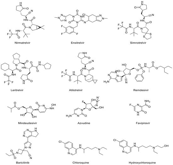
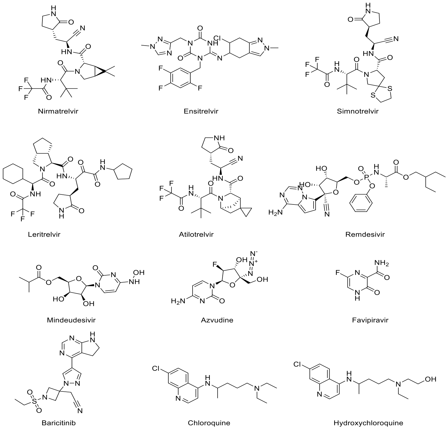
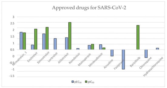
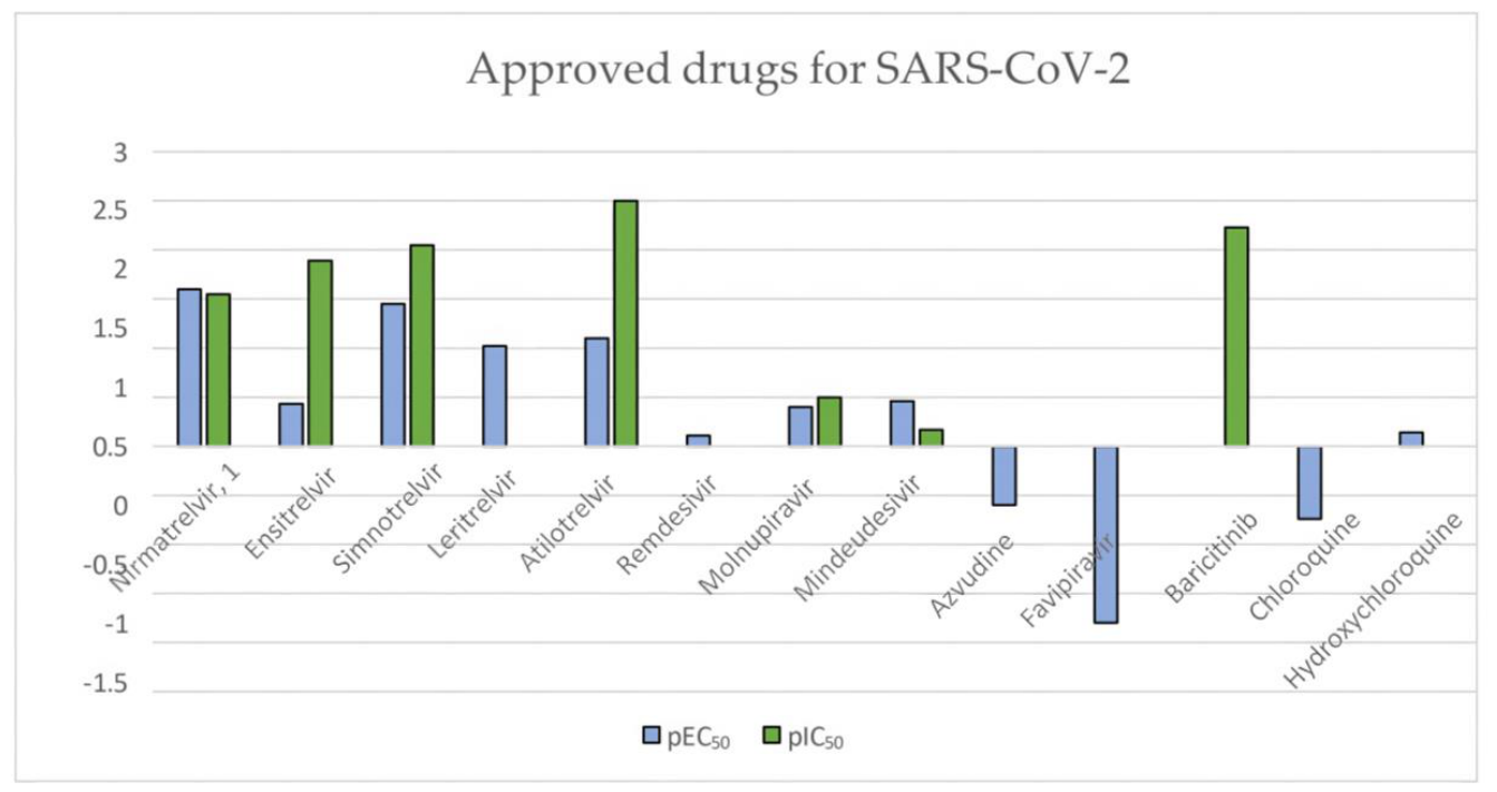
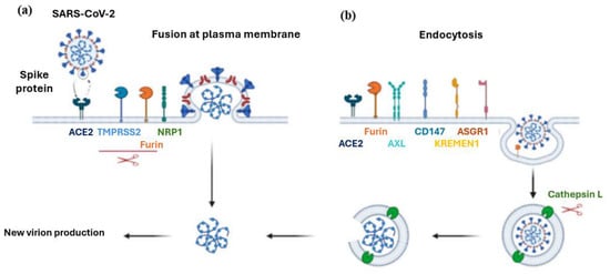
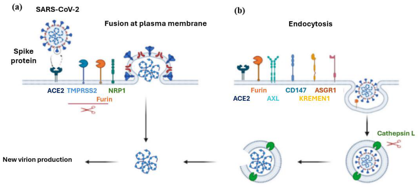
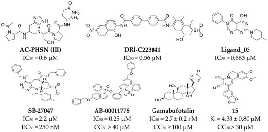
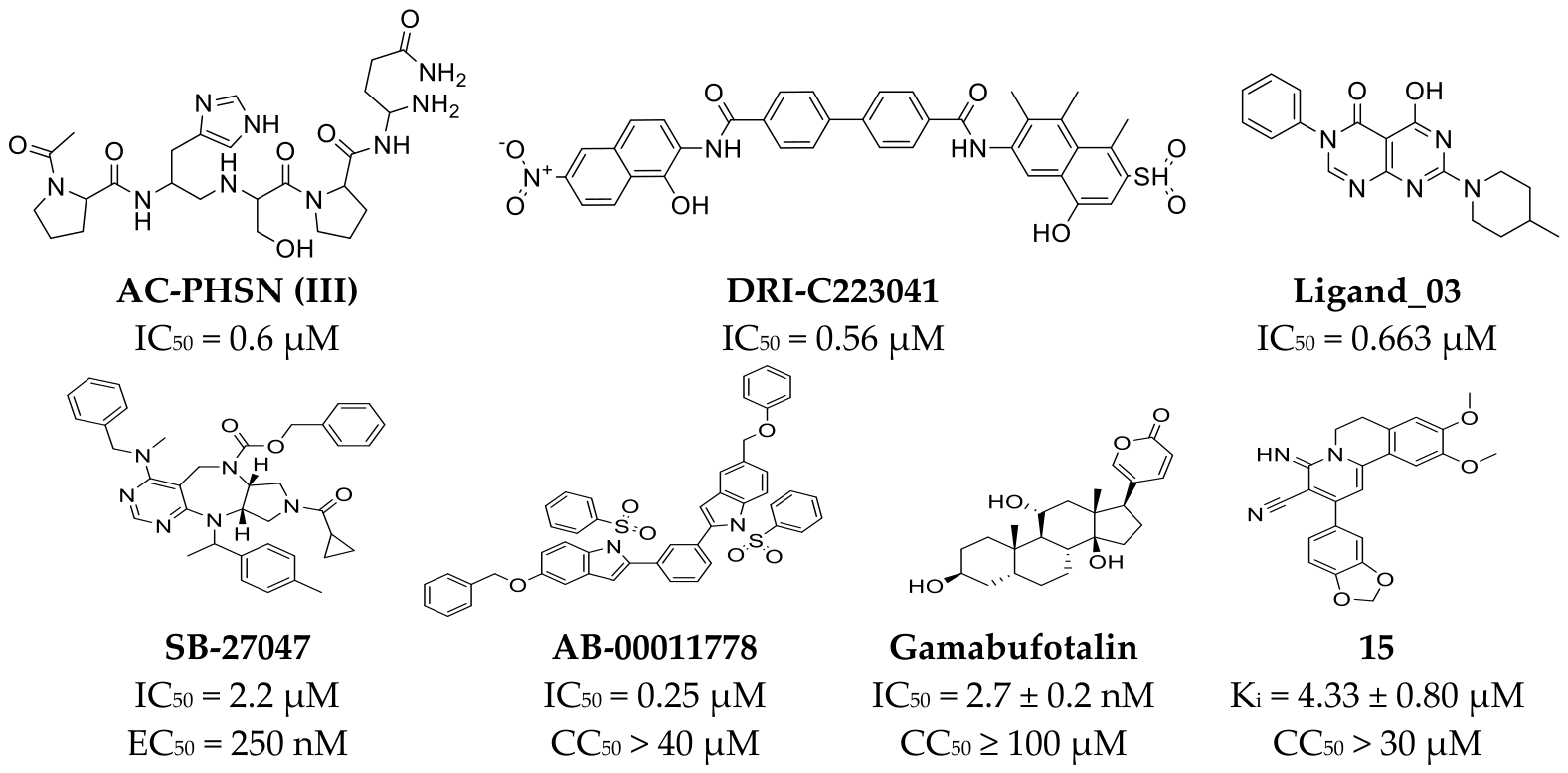
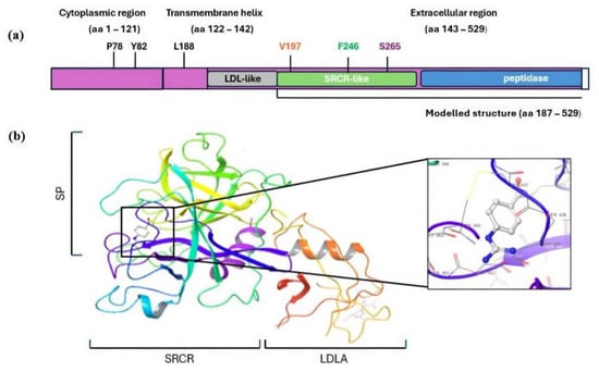
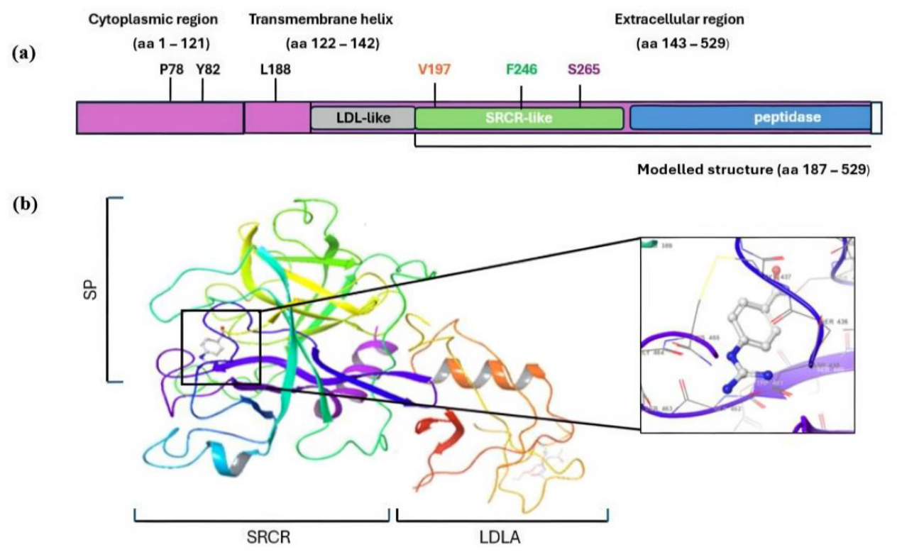
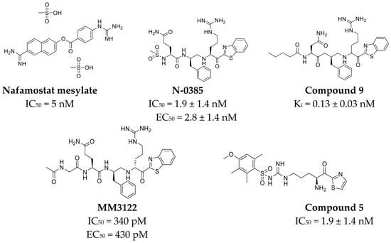
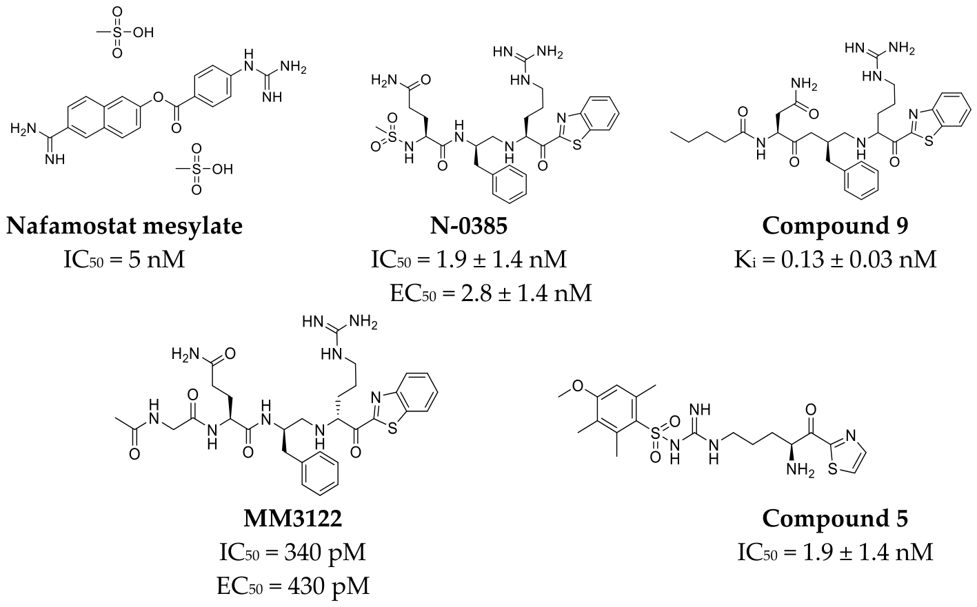
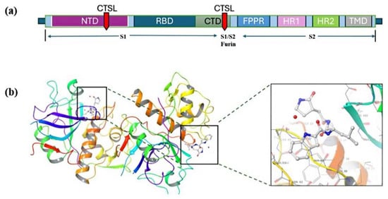
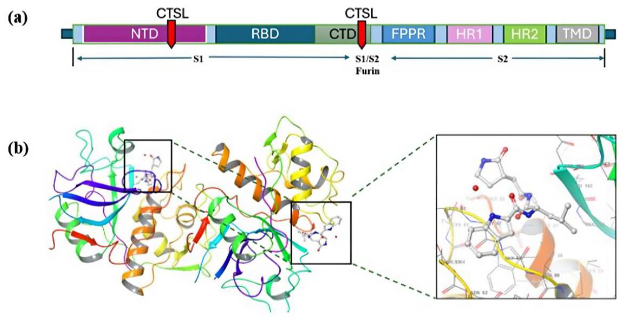
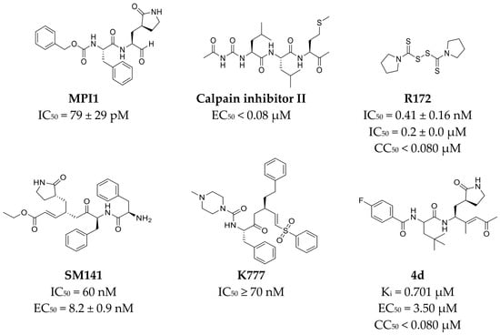
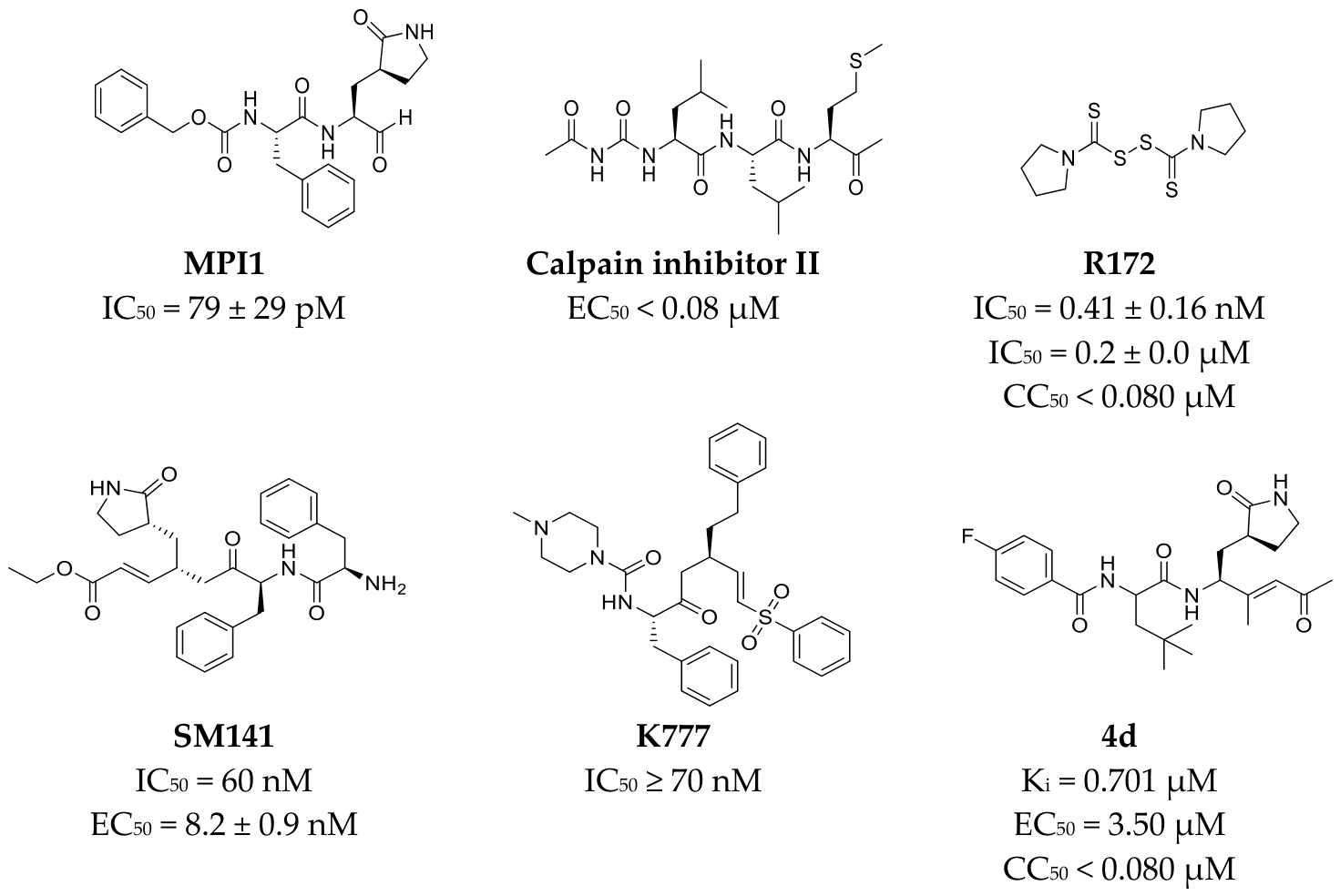
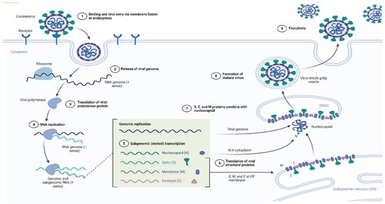
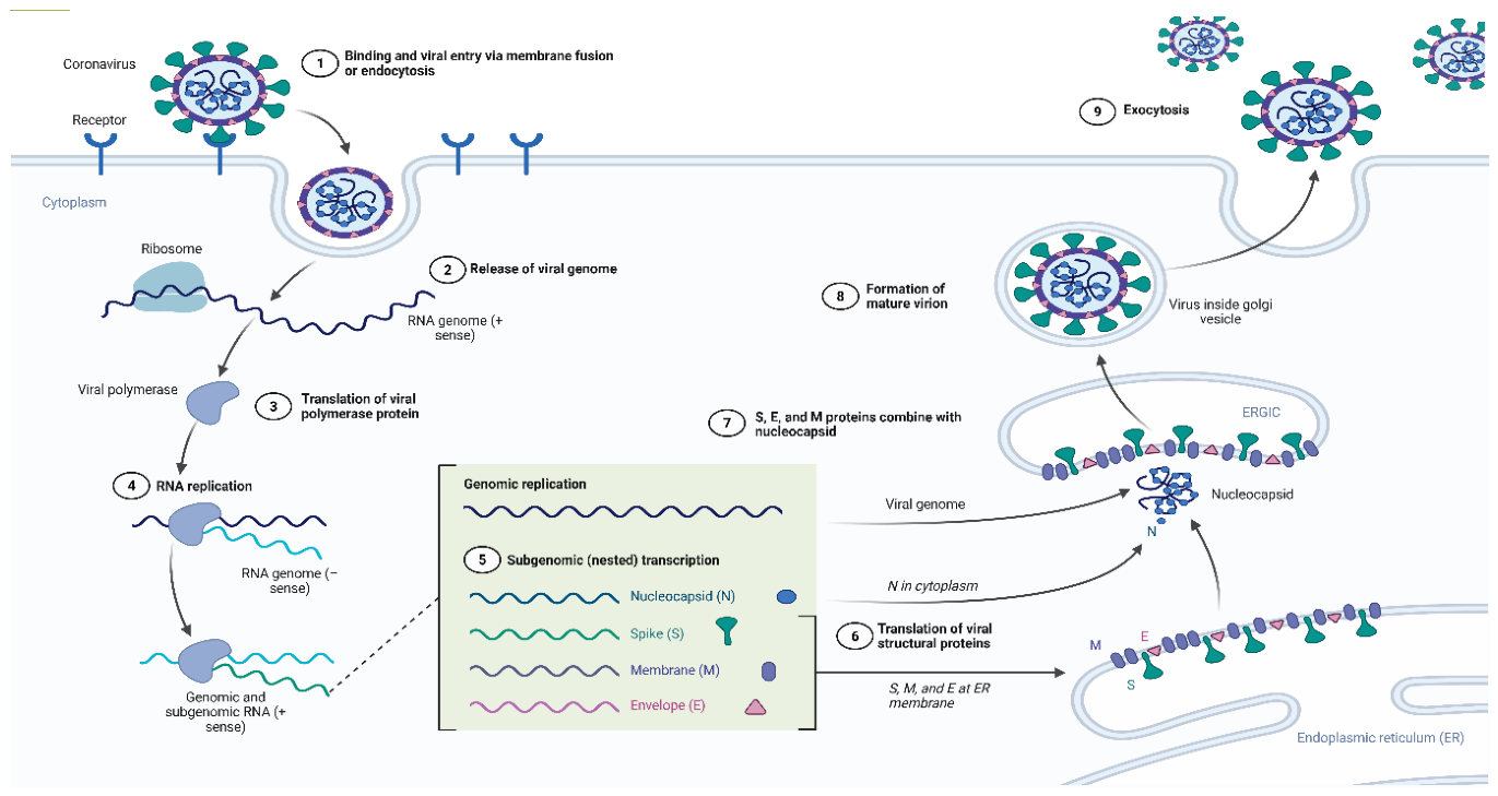
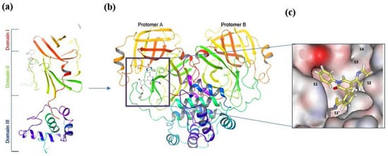
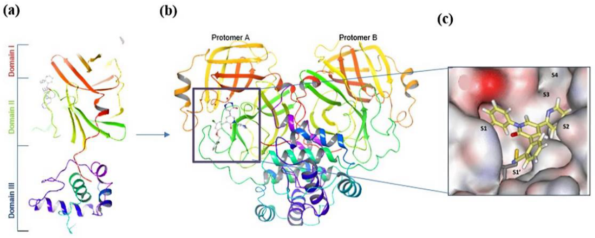
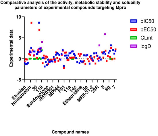
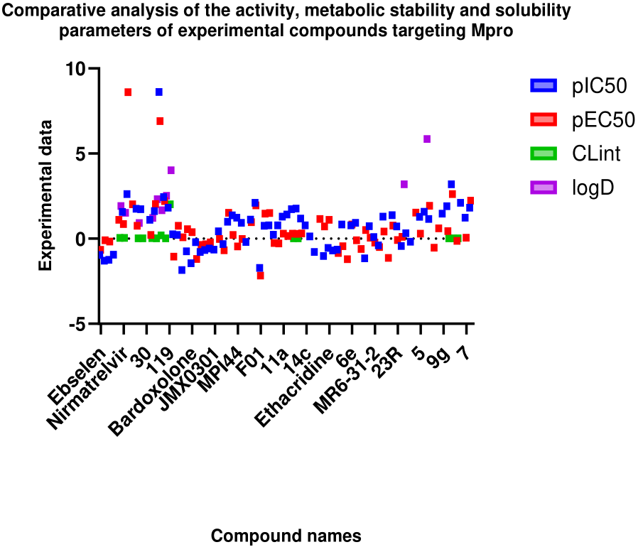
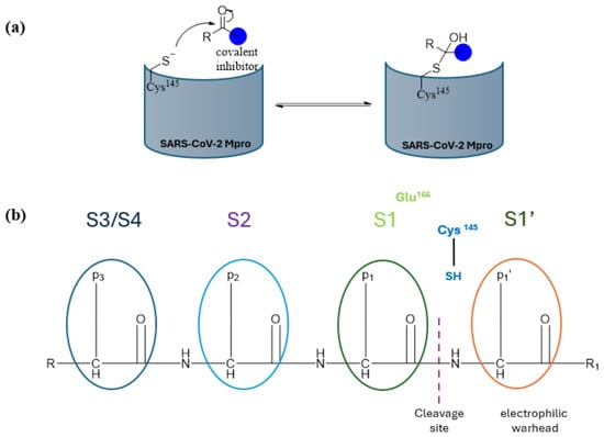
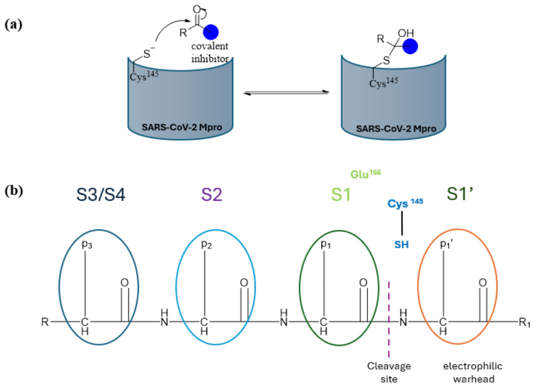
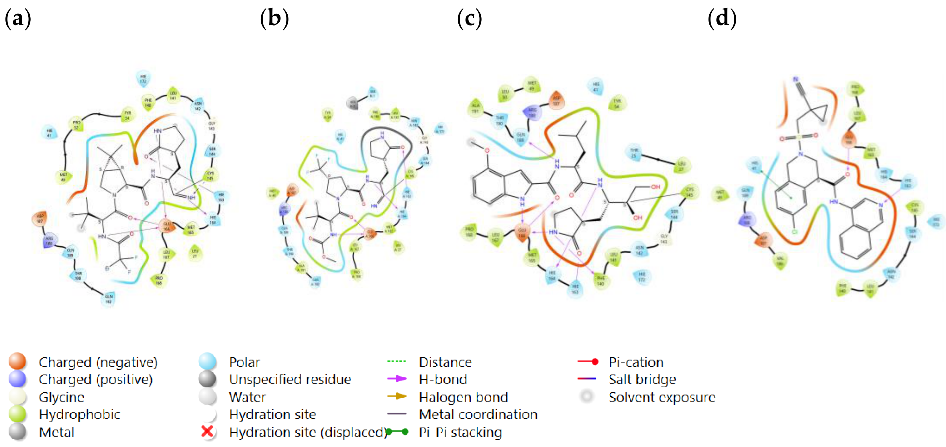
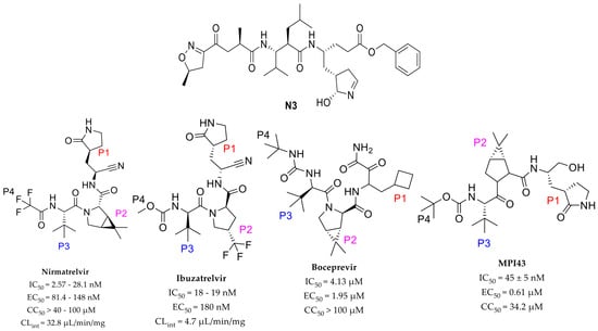
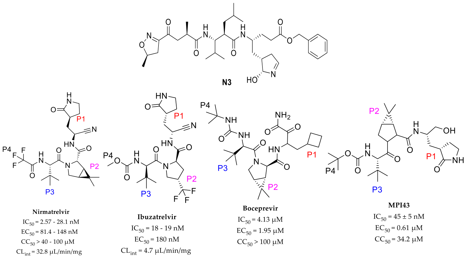
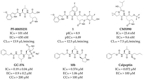
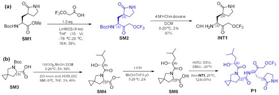
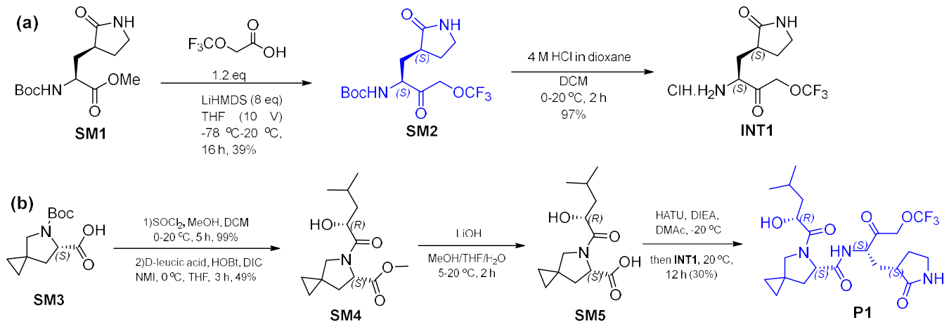
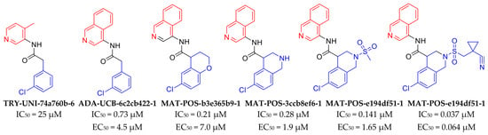
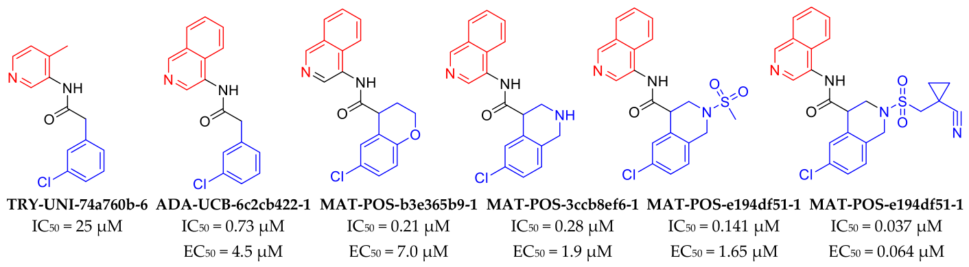
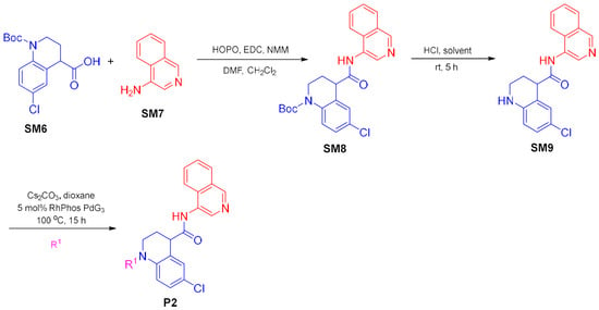
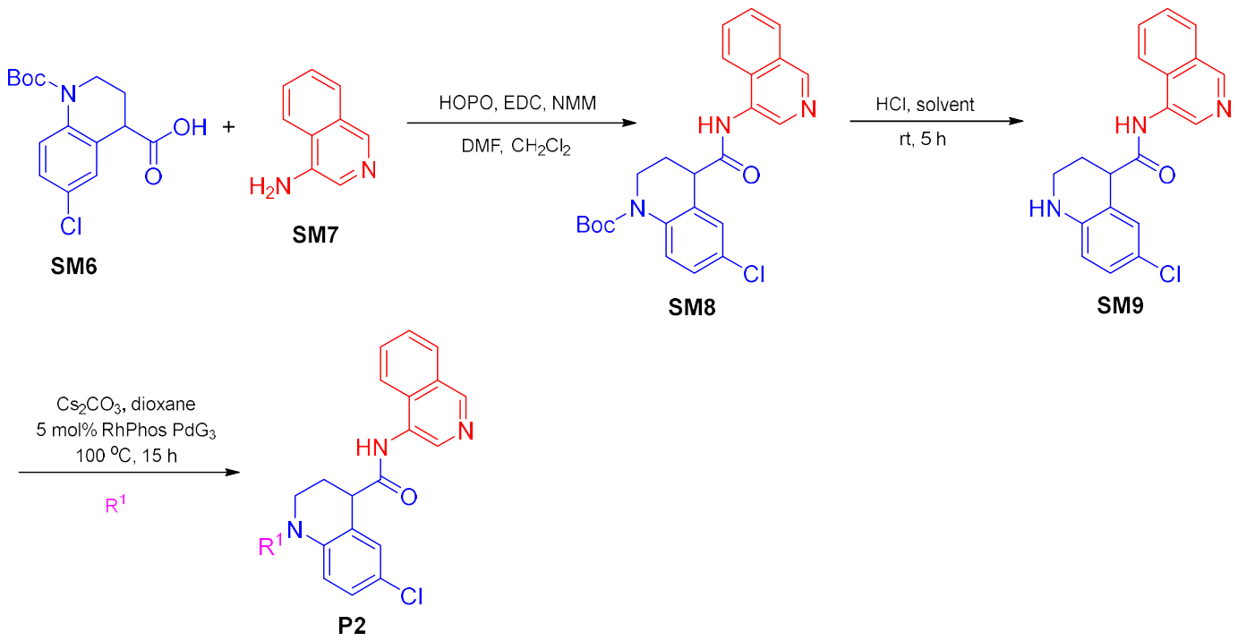
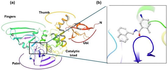
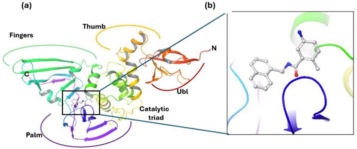
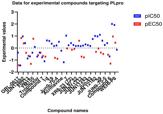
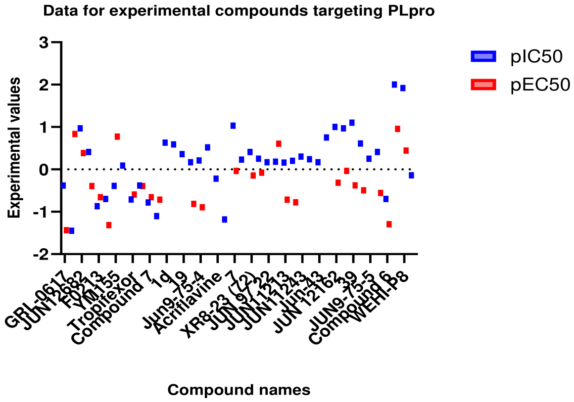
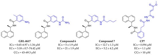
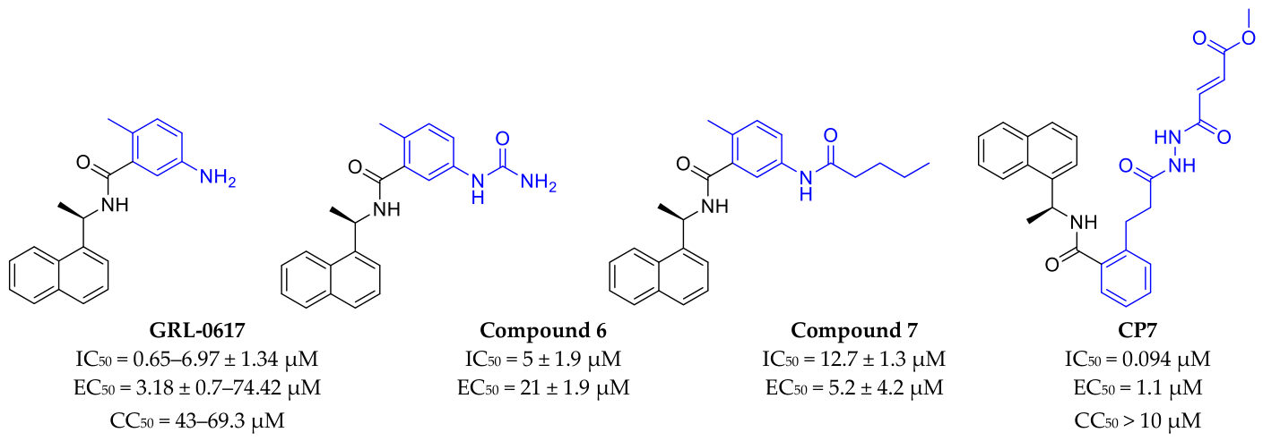
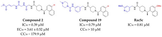
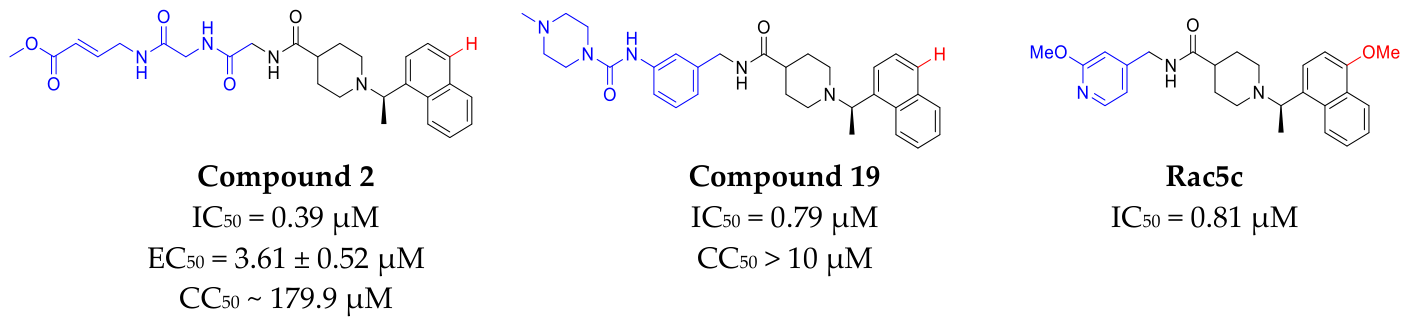
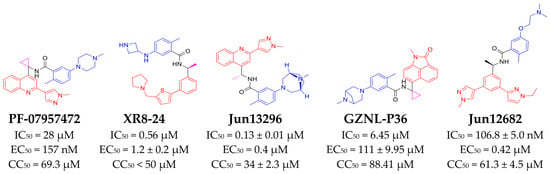
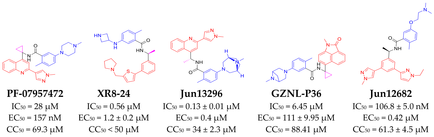
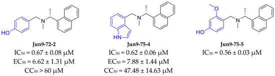
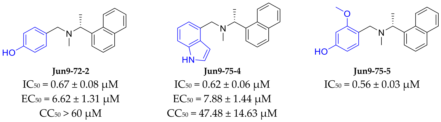
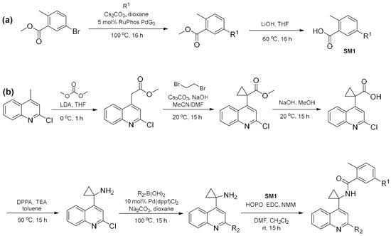
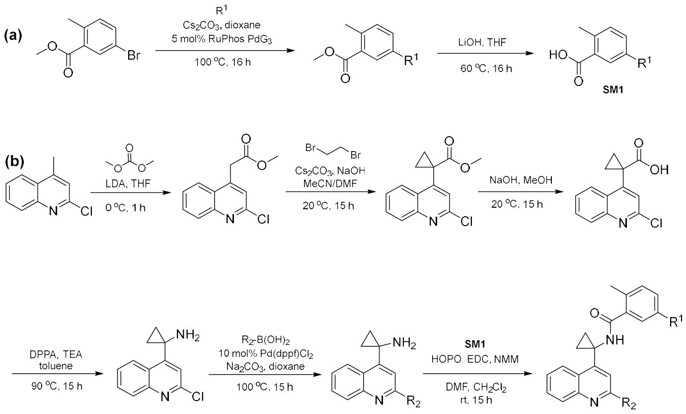

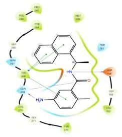
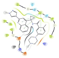
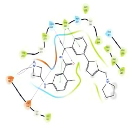
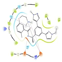
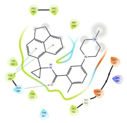
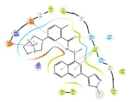
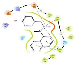
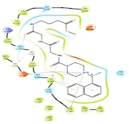
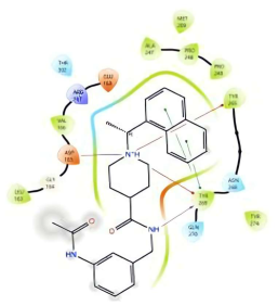

Disclaimer/Publisher’s Note: The statements, opinions and data contained in all publications are solely those of the individual author(s) and contributor(s) and not of MDPI and/or the editor(s). MDPI and/or the editor(s) disclaim responsibility for any injury to people or property resulting from any ideas, methods, instructions or products referred to in the content. |
© 2026 by the authors. Licensee MDPI, Basel, Switzerland. This article is an open access article distributed under the terms and conditions of the Creative Commons Attribution (CC BY) license.