


Abstract
In this study, a new series of (E)-4-((2-(2-(4-amino-3-methyl-5-oxo-4,5-dihydro-1H-1,2,4-triazol-1-yl)acetyl)hydrazono)methyl)phenyl 4-halogenobenzenesulfonates (3a–3d), where 3a = F, 3b = Cl, 3c = Br, and 3d = I, were successfully synthesized via a straightforward synthetic route. The structures of the obtained compounds were fully characterized and confirmed by spectroscopic techniques, including FT-IR, 1H NMR, and 13C NMR, as well as LC-MS/MS analysis. 1,2,4-triazole-based hydrazone derivatives (3a–3d) were investigated using IR and NMR spectroscopy and DFT calculations. Intermolecular interactions, HOMO-LUMO, dipole moment, polarization, first-order hyperpolarizability, and molecular electrostatic potential studies on the molecules were examined. The HOMO and LUMO energy gap study supports the charge transfer probability in the molecules. These were conducted to investigate the reactivity and stability of heterocyclic molecules in bioactivity analysis. Electron density mapping within the molecular electrostatic potential plot and electrostatic potential representation within the iso-surface plot evaluated the concept of charge distribution in the molecule as nucleophilic reactions and electrophilic regions. The predicted nonlinear optical (NLO) properties of the molecules are much greater than those of urea. The results obtained from these investigations collectively provide evidence that the molecules possess nonlinear optical applications. Novel triazole–hydrazone-functionalized aryl sulfonate derivatives (3a–3d) were evaluated for their anticancer potential against a panel of brain and non-brain cancer cell lines. Compound 3b exhibited the most favorable overall biological profile, displaying potent activity against SH-SY5Y neuroblastoma (GI = 7.59 μM) and U87MG glioblastoma cells (GI = 13.85 μM), together with the lowest toxicity toward normal FL fibroblasts (GI = 62.02 μM). Compounds 3c and 3d demonstrated remarkable potency against IDHmut-U87 glioma cells (GI = 3.87 and 3.27 μM, respectively), although their selectivity toward cancer cells was limited. DNA degradation studies revealed substantial fragmentation, particularly in C6 and SH-SY5Y cells, while migration assays indicated reduced cellular motility. Molecular docking studies identified compound 3b as the strongest PI3Kα binder, supporting a possible. In addition, the antimicrobial activities of compounds 3a–3d were evaluated against selected Gram-positive and Gram-negative bacteria as well as Candida species using the broth microdilution method. The compounds exhibited measurable antimicrobial effects with MIC values ranging from 156 to 625 µg/mL, showing moderate growth inhibition against the tested microorganisms. Although the observed activity was lower than that of the reference antimicrobial agents, the results indicate that these triazole–hydrazone derivatives possess a detectable level of antimicrobial activity and provide a basis for further structural optimization. Collectively, the results suggest that compound 3b represents the most promising lead structure due to its balanced combination of potency, selectivity, and predicted target engagement. Molecular docking was performed to evaluate the binding potential of newly synthesized triazole derivatives (3a–3d) against PI3Kα. The docking protocol was validated by re-docking alpelisib, yielding an RMSD of 0.64 Å. Among the tested compounds, 3b showed the most favorable binding energy (−9.94 kcal/mol) and estimated Ki value (52.13 nM), consistent with its superior in vitro activity. Its interactions with key PI3Kα residues, including Val851, Ser854, Met922, and Asp933, support a stable binding mode within the ATP-binding pocket. In silico ADME and toxicity analyses suggested acceptable drug-likeness characteristics, absence of major hepatotoxic, mutagenic, and carcinogenic liabilities, and moderate predicted acute toxicity profiles. These findings suggest that 3b is the most promising derivative for further validation.
1. Introduction
Heterocyclic organic compounds rich in functional groups continue to attract considerable research interest due to their versatile physicochemical and biological properties. In this context, the incorporation of different functional groups within a single molecular framework enables the design of compounds with novel and enhanced properties [1,2,3]. In particular, molecules containing triazole, hydrazone, and halogenated sulfonate groups exhibit remarkable potential applications in interdisciplinary fields such as pharmaceutical chemistry, dye chemistry, agrochemicals, and materials science [4,5]. Triazole derivatives have gained significant importance in both biological and industrial applications owing to their aromatic structures and high thermal stability [6,7,8,9,10]. The hydrazone group, due to the presence of the C=N bond and its conjugated structure, provides electronic delocalization and structural flexibility to molecular systems [11]. Hydrazone derivatives display a broad spectrum of biological activities, particularly antimicrobial, antitubercular, and antioxidant properties. In addition, hydrazone compounds are widely utilized in molecular recognition, sensor systems, and coordination chemistry, owing to their tautomeric behavior and hydrogen-bonding capability [11,12,13,14,15,16,17,18,19]. Sulfonate groups are among the key functional groups that enhance the water solubility of compounds due to their high polarity and ionic character [20]. In particular, sulfonate structures substituted with halogen atoms allow controlled modulation of chemical reactivity and stability through strong electron-withdrawing effects [21]. Halogenated sulfonate-containing compounds are widely employed in the design of reactive dyes in dye and textile chemistry, and they have also attracted attention in pharmaceutical applications due to their surfactant properties and interactions with biological systems [22,23,24]. The presence of halogen atoms plays a decisive role in lipophilicity and bioavailability [25,26,27,28,29]. In this context, a new series of (E)-4-((2-(2-(4-amino-3-methyl-5-oxo-4,5-dihydro-1H-1,2,4-triazol-1-yl)acetyl)hydrazono)methyl)phenyl 4-halogenobenzenesulfonates (3a–3d), where (3a = F, 3b = Cl, 3c = Br, and 3d = I), were synthesized and structurally characterized by FT-IR, 1H NMR, 13C NMR, and LC-MS/MS analyses. Furthermore, density functional theory (DFT) calculations were performed to evaluate the electronic structures, HOMO–LUMO energies, molecular electrostatic potential distributions, and nonlinear optical (NLO) properties of the synthesized compounds. In addition, their anticancer potential was investigated through comprehensive in vitro biological studies, including antiproliferative activity, membrane toxicity, DNA-binding and DNA degradation analyses, migration assays, and morphological evaluations against various cancer cell lines. The antimicrobial activities of compounds 3a–3d were evaluated against selected Gram-positive and Gram-negative bacteria as well as Candida species using the broth microdilution method. To gain preliminary insights into potential molecular interactions associated with the observed antiproliferative activity, molecular docking studies were performed against PI3Kα, a cancer-related molecular target known to regulate tumor growth, survival, and migration. The docking analysis was intended as an exploratory computational approach to investigate possible ligand–target interactions rather than to establish PI3Kα inhibition as the definitive mechanism of action.
2. Results
2.1. Chemistry
The synthesized (E)-configured 1,2,4-triazole-based hydrazone derivatives (3a–3d) (3a = F, 3b = Cl, 3c = Br, and 3d = I) were obtained in good to excellent yields (75–90%) via condensation of 4-formylphenyl 4-halogenbenzenesulfonates with 2-(4-amino-3-methyl-5-oxo-4,5-dihydro-1H-1,2,4-triazol-1-yl)acetohydrazide under reflux in ethanol. The smooth progress of the reaction and satisfactory yields indicate that the aldehyde and hydrazide functionalities readily undergo Schiff base formation under the applied conditions. Among the derivatives, the iodo-substituted compound (3d) afforded the highest yield, whereas the bromo analogue (3c) gave the lowest yield, which may be attributed to steric and electronic differences in the halogen substituents influencing the electrophilicity of the carbonyl carbon and overall reaction efficiency.
The IR spectra of compounds 3a–3d confirm the successful formation of the target hydrazone structures. The characteristic absorption bands observed at 3320–3279 cm−1 correspond to the asymmetric and symmetric stretching vibrations of the NH2 group, while the NH proton stretching appeared in the 3095–3109 cm−1 region. The presence of two distinct carbonyl stretching bands at 1720–1692 cm−1 is consistent with the triazole C=O and acyl hydrazide C=O groups. The azomethine (C=N) stretching vibration observed at 1608–1626 cm−1 clearly indicates the formation of the hydrazone linkage. Additionally, aromatic C=C stretching bands detected in the range of 1566–1592 cm−1 further support the presence of substituted phenyl rings. The consistency of these absorption bands across all derivatives confirms the structural similarity of the series.
The 1H NMR spectra provide further structural evidence. In all compounds, the methyl group attached to the triazole ring appeared as a singlet at 2.12 ppm (3H), while the methylene (N–CH2) protons were observed as singlets at 4.39–4.80 ppm (2H), confirming the presence of the acetyl linker. The NH2 protons resonated as a singlet at 5.31–5.32 ppm (2H), and the hydrazide NH proton appeared as a downfield singlet at 11.72–11.73 ppm, suggesting hydrogen bonding effects in DMSO-d6. The azomethine proton (N=CH) signal observed in the aromatic region supports the formation of the Schiff base structure. The aromatic protons of both phenyl rings appeared in the expected region between 7.10 and 8.18 ppm, with slight variations depending on the nature of the halogen substituent, reflecting the electronic influence of F, Cl, Br, and I atoms on the aromatic system. The 13C NMR spectra are also in good agreement with the proposed structures. The methyl carbon of the triazole ring was observed at 11 ppm, and the methylene carbon at δ ≈ 46 ppm. The azomethine carbon resonated at 142–145 ppm, while the triazole carbonyl carbon appeared at 150 ppm. The acyl carbonyl carbon signal was detected at 168–169 ppm. The aromatic carbon signals showed characteristic shifts influenced by the halogen substituents, particularly in the para-halogenated phenyl rings, confirming the successful incorporation of the respective substituents. Overall, the combined IR and NMR spectral data unambiguously confirm the successful synthesis of the desired (E)-hydrazone-linked 1,2,4-triazole derivatives bearing different halogen-substituted benzenesulfonate moieties. The spectral similarities across the series, along with predictable substituent-induced variations, support the proposed molecular structures.
2.2. Optimized Structure
Structural information is useful when investigating the coordination properties of Schiff bases functioning as ligands. We report here the synthesis and molecular structure of the title Schiff base compounds, chemical formula C18H17N6O5SX (X: F, Cl, Br, and I), and the atom numbering for them is shown in Figure 1. First, the initial molecular geometry of molecules was obtained using the Gauss-View 05 software program in quantum chemical calculations. The optimized energy value was found as −41,097 eV for molecule 3a, which was smaller than the others (E3b = −38,786 eV, E3c = −38,738 eV, E3d = −38,689 eV). We made geometry optimizations for molecules using B3LYP and comparative optimized structural parameters tabulated in Table S1. The focus on comparing the structural parameters of optimized molecular structures is to observe the inductive effect of halogenated sulfonate functional groups. By attaching different halogens to the phenylene ring in the compounds, differences were observed only in the C24–F44, C24–Cl44, C24–Br44, and C24–I44 bonds as 1.40 (exp. 1.352 [30]), 1.81 (exp. 1.737 [31]), 1.96 (exp. 1.900 [32,33]), and 2.14 Å (exp. 2.097 [34], respectively. This difference increased in bond angles, and it was observed that the greatest inductive effect occurred in dihedral angles. C–C–C angles remain close to 120° in most cases, supporting trigonal planar (sp2) hybridization. In all compounds, the O–S–O angle was measured at approximately 120°. The C5-N4-C27 bond angle is approximately 126°, indicating delocalized electron density around the nitrogen centers. The C23-C24-X44 angles vary around 118°–120° due to the electronegativity and steric hindrance of the halogens. The widest bond angle in the system is approximately 129.3° (C6-N8-N9), while the smallest bond angle is approximately 96° (O17-S18-C21). Dihedral angles reveal the three-dimensional conformation of the molecule. The highest dihedral angle is approximately 179.9°, while the lowest dihedral angle is approximately −179.9°, both indicating nearly planar conformations. The dihedral angles show a mixture of planar and twisted conformations, indicating flexibility in molecular geometry. These findings are important for understanding the reactivity, stability, and potential applications of the molecule in chemical or biological systems. The results obtained are consistent with similar molecules [35,36,37,38].
Figure 1.
Optimized structure of compounds, C18H17N6O5SX (X: F, Cl, Br, and I).
2.3. Spectroscopic Properties
2.3.1. Vibrational Spectra
The IR spectra of compounds 3a–3d (3a = F, 3b = Cl, 3c = Br, and 3d = I) confirmed the successful formation of the target hydrazone structures. Characteristic NH2 and NH stretching vibrations were observed at 3320–3279 and 3095–3109 cm−1, respectively, in reasonable agreement with the calculated values of 3466–3592 and 3380–3387 cm−1. Aromatic C–H stretching vibrations appeared at 3073–3109 cm−1. Two carbonyl stretching bands at 1720–1692 cm−1 were assigned to triazole and acyl hydrazide C=O groups, consistent with theoretical values of 1646 and 1640 cm−1. The azomethine (C=N) stretching vibration observed at 1608–1626 cm−1 confirmed hydrazone formation and appeared as a characteristic band at 1595 cm−1 for all compounds, indicating strong conjugation within the molecular framework [39,40]. Aromatic C=C stretching bands were detected at 1566–1592 cm−1. The asymmetric SO2 stretching vibrations appeared at 1290–1311 and 825 cm−1, in agreement with literature reports [38,41,42]. The experimental and theoretical vibrational data are summarized in Table 1, and the overall consistency of the bands confirms the structural similarity of the series.
Table 1.
Selected molecular structure parameters of the compounds.
2.3.2. NMR Studies
The 1H and 13C NMR chemical shift values of (3a–3d) (3a = F, 3b = Cl, 3c = Br, and 3d = I) molecules were computed at the same level using the GIAO approach in DMSO solvent media, based upon the compound’s optimized molecular geometries. The experimentally obtained and computed chemical shifts of the mentioned molecule are presented in Table 2 and Table 3. The 1H NMR spectra provide further structural evidence. In all compounds, the methyl group attached to the triazole ring appeared as a singlet at 2.12 ppm (3H), while the methylene (N–CH2) protons were observed as a singlet at 4.39–4.80 ppm (2H), confirming the presence of the acetyl linker. These values were theoretically recorded in the ranges of 2.27–2.48 ppm (3H) and 4.29–4.79 ppm (2H), respectively. In accordance with the theoretical values, the NH2 protons resonated as a singlet at 5.31–5.32 ppm (2H), and the hydrazide NH proton appeared as a downfield singlet at 11.72–11.73 ppm; this suggests hydrogen bonding effects in DMSO-d6. The azomethine proton (N=CH) signal observed in the aromatic region supports the formation of the Schiff base structure. The aromatic protons of both phenyl rings appeared in the expected region between 7.10 and 8.18 ppm experimentally and 7.88–8.82 ppm theoretically, showing slight variations depending on the nature of the halogen substituent, reflecting the electronic effect of F, Cl, Br, and I atoms on the aromatic system (Table 2).
Table 2.
Theoretical and experimental scaled 1H NMR data for compounds.
Table 3.
Theoretical and experimental scaled 13C NMR data for compounds.
The 13C NMR spectra are also in good agreement with the proposed structures (Table 3). The methyl carbon of the triazole ring was observed at 11 ppm, while the methylene carbon was observed at δ ≈ 46 ppm. The azomethine carbon resonated at 142–145 ppm, while the triazole carbonyl carbon appeared at 150 ppm. Similarly, the 13C-NMR chemical shift values for these carbon atoms were recorded at 162 ppm and 167 ppm, respectively. The acyl carbonyl carbon signal was detected at 168–169 ppm. The aromatic carbon signals exhibited characteristic shifts, particularly in the para-halogenated phenyl rings due to the effect of halogen substituents, confirming the successful incorporation of the respective substituents.
Overall, the combined IR and NMR spectral data unambiguously confirm the successful synthesis of the desired (E)-hydrazone-linked 1,2,4-triazole derivatives bearing different halogen-substituted benzenesulfonate moieties. The spectral similarities across the series, along with predictable substituent-induced variations, support the proposed molecular structures.
2.4. Electronic Properties
2.4.1. Molecular Orbital Calculations
Frontier Molecular Orbital (FMO) analysis is an important tool for understanding the electronic properties of molecules, particularly their chemical reactivity and stability [43]. The FMO surface map is shown in Figure 2. In Figure 2, green represents the positive phase, while red represents the negative phase. Molecules with a larger energy gap (ΔE) exhibit higher chemical stability and lower reactivity. HOMO and LUMO energies, together with the energy gap (ΔE), provide information about electronic transitions, ionization potential, electron affinity, and other global reactivity descriptors [44]. The global reactivity descriptors are presented comparatively in Table 4.
Figure 2.
Frontier molecular orbitals for compounds.
Table 4.
Quantum chemical parameters calculated at B3LYP/LANL2DZ.
Molecule (3a), which has the lowest HOMO energy, also has a lower tendency to donate electrons. Similarly, molecule (3c) with lower LUMO energy has a higher electron-accepting ability. Therefore, it can be stated that molecule (3a) requires more energy for ionization and exhibits stronger resistance to oxidation [45]. Molecule (3d), which has a high hardness (η) value, is chemically more stable while having the lowest electronegativity value. The chemical potential (μ) value, which indicates a molecule’s tendency to gain or lose electrons, is again highest in molecule 3a. Furthermore, molecule 3b, which has the highest electrophilicity and nucleophilicity indices, has greater electron-accepting and electron-donating capacity, respectively. These findings provide valuable information about the electronic properties and reactivity of the investigated compounds.
2.4.2. MEP Analysis
Molecular Electrostatic Potential (MEP) analysis is a tool used to understand the charge distribution, electrophilic and nucleophilic regions, and overall reactivity of a molecule [46]. In the MEP map, different colors represent positive and negative electrostatic potential regions. The MEP color surface map is shown in Figure 3. Blue regions correspond to areas with electron deficiency, while red regions represent electron-rich areas. In this study, the nitro and oxygen groups exhibit greater reactivity. Particularly, the presence of the chlorine (-Cl) substituent slightly withdraws electron density, affecting the overall charge distribution and leading to a more stable electrostatic potential. These observations are important in understanding the electronic behavior of molecules and their potential applications in chemical and biological interactions.
Figure 3.
MEP surfaces of (3a–3d) compounds.
2.4.3. NLO Properties
Molecules with large hyperpolarizability can be used as beneficial optical devices. As shown in Table 5, the high hyperpolarizability value corresponds to a large HOMO-LUMO energy gap (ΔE). This indicates that there is a direct relationship between ΔE and hyperpolarizability (Figure 2). The total dipole moment (μ), average polarizability (α), anisotropy of the polarizability (Δα), and total first-order hyperpolarizability (β) values were calculated for molecules 3a–3d and are presented in Table 5. Urea is commonly used as a threshold value for comparison purposes. The β value is estimated as 115.847 × 10⁻²⁹, 112.527 × 10⁻³⁰, 116.723 × 10⁻³⁰, and 118.680 × 10⁻³⁰ cm5/esu for 3a–3d, respectively, which is larger than the value for urea (α and β value were 3.88 D, 5.04 Å3 and 0.782 × 10⁻³⁰ cm5/esu) [37]. Therefore, the studied compounds may be attractive materials for nonlinear optical applications.
Table 5.
The dipol moment, polarizability, and first hyperpolarizability values.
2.5. MTT Proliferation Assay
The antiproliferative effects of compounds 3a–3d were evaluated against a panel of brain and non-brain cancer cell lines using the MTT assay and compared with 5-fluorouracil (5-FU). Distinct differences in cellular sensitivity were observed among the tested compounds and cell types. Brain tumor models included A172 and U87MG human glioblastoma cells, IDHmut-U87 glioma cells, C6 rat glioma cells, SH-SY5Y human neuroblastoma cells, and B35 rat neuroblastoma cells. To assess the broader anticancer potential of the synthesized compounds, HT29 colon adenocarcinoma, HeLa cervical carcinoma, and MCF7 breast adenocarcinoma cells were also included. In addition, FL fibroblasts were used as a non-transformed cell model to evaluate selectivity for cancer cells and to estimate potential toxicity to normal tissues. The inclusion of both malignant and non-malignant cell lines allowed a comparative assessment of anticancer efficacy and tumor selectivity, with particular emphasis on brain tumor-related malignancies. GI50 and IC values were determined to provide complementary information regarding the biological effects of the tested compounds. GI50 represents the concentration required to inhibit net cell growth by 50%, reflecting antiproliferative (cytostatic) activity, whereas IC50 corresponds to the concentration required to reduce cell viability by 50% relative to untreated controls, reflecting overall cytotoxic activity. Since these parameters describe different aspects of cellular response, both were included in the present study. Among the brain tumor models, compound 3b exhibited the most favorable activity profile, showing potent growth inhibition against SH-SY5Y neuroblastoma cells (GI50 = 7.59 μM) and U87MG glioblastoma cells (GI50 = 13.85 μM) (Table 6). In contrast, compounds 3c and 3d demonstrated superior activity against IDHmut-U87 glioma cells, with GI50 values of 3.87 and 3.27 μM, respectively, exceeding the activity of 5-FU (GI50 = 14.15 μM) (Table 6). Compound 3a showed notable potency against MCF7 breast cancer cells (GI50 = 5.60 μM), while compounds 3c and 3d also exhibited strong activity against HT29 colon cancer cells (GI50 = 3.71 and 22.81 μM, respectively) (Table 7). Evaluation of FL fibroblasts revealed marked differences in selectivity among the synthesized derivatives. Compound 3b displayed the lowest toxicity toward normal cells (GI50 = 62.02 μM), whereas compounds 3c and 3d showed considerably lower FL GI50 values (6.13 and 4.96 μM, respectively) (Table 7). Overall, the molar-normalized data identified compound 3b as the most balanced derivative, combining potent antiproliferative activity with improved selectivity toward cancer cells, while compounds 3c and 3d emerged as the most potent but less selective analogues. Consistent with the biological findings, molecular docking studies identified compound 3b as the derivative with the highest predicted affinity toward PI3Kα. Tumor selectivity was evaluated using the Tumor Selectivity Index (TSI), calculated according to the following equation: TSI = IC50 (FL normal fibroblasts)/IC50 (cancer cell line). A TSI value greater than 1 indicates preferential activity toward cancer cells, whereas higher TSI values reflect greater tumor selectivity and a potentially improved therapeutic window. Tumor selectivity index (TSI) analysis demonstrated the highest selectivity for compound 3c in IDHmut-U87 cells (TSI = 1.71), followed by compound 3d in the same cell line (TSI = 1.52). Compound 3a exhibited favorable selectivity toward MCF7 breast cancer cells (TSI = 1.82) (Figure 4).
Table 6.
GI50 and IC50 values of substances in brain cancer cell lines.
Table 7.
GI50 and IC50 values of substances in brain, colon, cervix, and breast cancer cells and FL normal cells.
Figure 4.
IC50-based tumor selectivity index (TSI) heatmap of compounds relative to FL cells. Higher TSI values indicate greater cancer cell selectivity.
2.6. LDH Cytotoxicity Assay
The cytotoxic effects of compounds 3a–3d, where 3a = F, 3b = Cl, 3c = Br, and 3d = I, were evaluated by LDH release assay at their respective IC concentrations across multiple cancer cell lines and FL normal fibroblast cells. LDH release analysis revealed relatively low membrane-disruptive effects for all synthesized compounds. Cytotoxicity values ranged between 3.08% and 12.44%, substantially lower than those observed for 5-FU (Table 8). The highest LDH release was detected for compound 3d in MCF7 cells (12.44%), followed by compound 3a (12.31%) and compound 3c (11.99%) in the same cell line (Table 8). In contrast, compounds generally produced low LDH release in brain cancer models. Compound 3d exhibited particularly low cytotoxicity in U87MG cells (3.28%), whereas compound 3b showed the lowest value in HT29 cells (3.08%) (Table 8). These findings indicate that the observed growth inhibitory effects are unlikely to result primarily from acute membrane damage.
Table 8.
% Cytotoxicity values of the molecules at IC50 concentrations.
2.7. DNA Degradation Assay
DNA fragmentation analysis revealed that compounds 3a–3d, where 3a = F, 3b = Cl, 3c = Br, and 3d = I, induced marked DNA degradation in treated cells, with the extent of degradation varying among cell lines. The most prominent DNA damage pattern was observed in C6 cells, where all tested compounds produced extensive DNA fragmentation/degradation. This finding indicates that C6 cells were highly sensitive to the DNA-damaging or apoptosis-associated effects of these compounds (Figure 5).
Figure 5.
Test results demonstrating the DNA degradation effects of the 3(a–d) on C6, FL, and SH-SY5Y cell lines.
A strong DNA degradation profile was also detected in SH-SY5Y cells, although the intensity of DNA fragmentation appeared slightly lower than that observed in C6 cells. This suggests that SH-SY5Y cells were also responsive to treatment, but with a comparatively lower degree of DNA breakdown (Figure 5). In FL normal fibroblast cells, DNA degradation was also observed after treatment with the tested compounds. However, the degree of DNA damage appeared relatively lower than that detected in C6 and SH-SY5Y cancer cells. These results suggest that the compounds may induce DNA fragmentation preferentially in tumor cells, particularly in C6 and SH-SY5Y cells, while producing a comparatively weaker effect in FL cells (Figure 5).
Overall, DNA gel electrophoresis demonstrated that compounds 3a–3d caused substantial DNA degradation, especially in C6 cells, followed by SH-SY5Y cells, whereas FL cells showed a relatively lower level of DNA fragmentation.
2.8. Influence of Molecules on Cell Migration and ImageR Evaluation
Cell migration was assessed using wound-healing analysis by calculating the reduction in wound area between Day 1 and Day 3. The gap-filling ratio represented the percentage decrease in the wound area over the experimental period. In control groups, complete closure of the wound area was observed by Day 3, resulting in a Day 3 gap value of 0%, indicating unrestricted migratory capacity (Figure 6).
Figure 6.
Effects of the 3(a–d) on cell migration in the C6 and SH-SY5Y cancer cell lines. Bar is 100 µm.
In C6 cells, compound 3c produced the strongest anti-migratory effect, showing the lowest gap-filling ratio (24.71%), followed by compound 3d (29.86%) (Table 9). Compounds 3a and 3b displayed moderate inhibition with gap-filling ratios of 32.88% and 35.13%, respectively (Table 9). In SH-SY5Y cells, compound 3a showed the strongest inhibition of migration (20.24%), followed by compound 3c (32.54%) and compound 3d (34.14%) (Table 9). Compound 3b displayed comparatively weaker suppression (39.84%) (Table 9). These findings demonstrate that all compounds interfere with cellular migration, although their efficacy appears dependent on cell type (Table 9 and Figure 6).
Table 9.
Results from the migration analysis of molecules conducted with ImageR.
2.9. DNA Binding Assay
The DNA-binding properties of compounds 3a–3d were investigated using UV–Vis absorption spectroscopy and evaluated by Wolfe–Shimer analysis. Progressive addition of CT-DNA induced notable alterations in the absorption spectra of all tested compounds, indicating interactions between the molecules and DNA (Figure 7). Upon increasing DNA concentration, compounds generally exhibited concentration-dependent spectral changes characterized by reductions in absorption intensity together with moderate spectral perturbations in the 250–300 nm region. The gradual changes observed between the first (1) and last (8) DNA concentrations indicate formation of compound–DNA complexes (Figure 7). The calculated binding constants (Kb) varied among compounds, suggesting differences in binding affinity toward DNA: 3a: 3.9 × 10³ M−1, 3b: 5.1 × 10³ M−1, 3c: 1.5 × 10 M−1, and 3d: 2.4 × 10 M−1 (Figure 7). Among the tested molecules, compound 3d exhibited the strongest DNA-binding affinity, followed by compound 3c. The Wolfe–Shimer plots showed satisfactory linearity (R2 = 0.916–0.993), supporting the reliability of the calculated binding constants (Figure 8).
Figure 7.
UV–Vis absorption spectrum of 3a–d in the absence and presence of DNA [3.125 µM (8), 6.25 µM (7), 12.5 µM (6) 25 µM (5), 50 µM (4), 100 µM (3), 200 µM (2), and 400 μM (1)] of the UV–Vis absorption spectrum.
Figure 8.
Plot chart indicating binding constant [DNA] and [DNA]/εa − εf.
Upon increasing DNA concentration, all compounds exhibited a noticeable hyperchromic effect, characterized by a gradual increase in absorbance intensity, suggesting the formation of compound–DNA complexes and alterations in the DNA microenvironment. The hyperchromic effect became more pronounced at higher DNA concentrations, indicating increased interactions between the compounds and DNA molecules. The observed spectral changes suggest that the compounds can influence DNA structural organization.
2.10. Morphological Analysis
Morphological alterations induced by compounds 3a–3d, where 3a = F, 3b = Cl, 3c = Br, and 3d = I, at 32 μg/mL were evaluated by phase-contrast microscopy and compared with untreated control cells. Control cells exhibited normal morphology characterized by a dense monolayer with high confluence, elongated spindle-like morphology, intact cell–cell contacts, and strong substrate attachment (Figure 9). In contrast, treatment with the tested compounds induced substantial morphological changes of varying intensity. Cells exposed to 3a showed reduced confluence with the appearance of rounded and partially detached cells, accompanied by loss of normal cellular organization. Compound 3b induced noticeable cell shrinkage and increased numbers of rounded cells while preserving some elongated cells (Figure 9). Treatment with 3c and 3d produced more pronounced morphological alterations characterized by increased cellular rounding, reduction in cell density, and disruption of normal cellular architecture. Detached cells and cells with irregular morphology were more evident, indicating progressive cellular damage (Figure 9). Overall, treated groups demonstrated decreased confluence, cellular shrinkage, loss of adhesion, increased rounding, and cellular detachment compared with untreated controls, with the strongest effects observed particularly in 3d and 3c.
Figure 9.
Morphological changes caused by 3a–d at a concentration of 32 µg/mL in the U87MG cell line. Bar is 100 µm.
The present study demonstrated that compounds 3a–3d exert multifaceted anticancer effects through modulation of several cellular processes involved in tumor progression. MTT analysis revealed significant antiproliferative activities in different cancer cell lines, although responses varied depending on compound structure and cell type. While 5FU showed the strongest overall activity, several synthesized compounds exhibited considerable growth inhibition, suggesting that structural differences strongly influence biological activity.
Interestingly, LDH analysis showed relatively low cytotoxicity values compared with the marked reduction in cell viability observed in MTT results. This suggests that the biological effects of these compounds are not mainly associated with acute membrane damage or necrotic cell death but may instead involve intracellular regulatory pathways. Such discrepancies between MTT and LDH findings are frequently associated with cytostatic or programmed cell death mechanisms.
DNA-binding studies demonstrated that all compounds interacted with DNA with moderate affinity (Kb ≈ 103–104 M−1), with 3d and 3c exhibiting stronger interactions.
However, DNA-binding affinity did not completely correlate with antiproliferative activity, indicating that DNA interaction alone is insufficient to explain the observed biological effects.
DNA degradation analysis revealed substantial fragmentation, particularly in C6 cells, followed by SH-SY5Y cells, whereas FL cells showed comparatively lower degradation. These findings suggest a partially selective response toward cancer cells and support the involvement of apoptosis-associated mechanisms. Migration studies further showed that all compounds reduced wound closure in both C6 and SH-SY5Y cells, indicating potential anti-migratory activity. Since cell migration plays a critical role in metastasis, these findings suggest possible anti-invasive properties of the tested compounds.
Morphological observations supported these biochemical findings by demonstrating reduced confluence, cell rounding, shrinkage, loss of adhesion, and increased detached cells following treatment. Such changes are commonly associated with apoptosis-like responses and cellular stress.
Overall, the findings suggest that compounds 3a–3d exert anticancer effects through multiple interconnected mechanisms involving proliferation suppression, DNA interaction, DNA degradation, migration inhibition, and induction of apoptosis-associated cellular changes. Further mechanistic studies involving apoptosis and signaling pathways would provide deeper insight into their therapeutic potential.
2.11. Antimicrobial Activity
The antimicrobial activities of compounds 3a–3d, where 3a = F, 3b = Cl, 3c = Br, and 3d = I, were evaluated against selected Gram-positive and Gram-negative bacteria, as well as yeast strains, using the broth microdilution method. The minimum inhibitory concentration (MIC) values are presented in Table 10. Overall, the synthesized compounds exhibited measurable inhibitory effects against the tested microorganisms, with MIC values ranging from 156 to 625 µg/mL. Among the bacterial strains tested, all derivatives showed comparable inhibitory activity against Bacillus cereus and Klebsiella pneumoniae, with MIC values of 156 µg/mL. Moderate growth inhibition was also observed against Bacillus subtilis, Escherichia coli, Pseudomonas aeruginosa, and Enterococcus faecalis, with MIC values generally between 312 and 625 µg/mL. Staphylococcus aureus was found to be less susceptible to the tested compounds. The antifungal evaluation revealed that the compounds were also active against Candida albicans and Candida tropicalis, with MIC values ranging between 156 and 312 µg/mL. However, when compared with the reference antifungal agent fluconazole, the activity of the synthesized derivatives was lower. Nevertheless, the observed inhibitory effects indicate that these compounds retain a measurable level of antifungal activity. Comparison of the halogen-substituted derivatives showed only minor differences in antimicrobial performance, suggesting that substitution with F, Cl, Br, or I did not lead to substantial changes in activity under the tested conditions. The antimicrobial effects observed may be related to the combined contribution of the triazole, hydrazone, and halogenated sulfonate moieties within the molecular framework. Overall, the results indicate that the synthesized compounds display moderate antimicrobial activity against the tested microorganisms. Although their activity was lower than that of the reference antimicrobial agents, the findings provide preliminary insight into the biological behavior of this structural scaffold and may be useful for future structural modification studies aimed at improving antimicrobial efficacy.
Table 10.
MIC values of compounds 3a-d and antibiotics against tested microorganisms.
2.12. Molecular Docking Analysis
Molecular docking analysis was performed to evaluate the binding potential of the newly synthesized triazole derivatives 3a–3d, where 3a = F, 3b = Cl, 3c = Br, and 3d = I, against the ATP-binding pocket of PI3Kα. PI3Kα was selected as an exploratory molecular target because aberrant activation of the PI3K/Akt signaling pathway has been implicated in several cancer types represented by the cellular models used in this study, including glioblastoma, neuroblastoma, breast cancer, and colorectal cancer. Moreover, structurally related triazole- and hydrazone-containing compounds have previously been reported to exert anticancer effects through mechanisms associated with PI3K/Akt signaling. Therefore, docking studies were performed to investigate potential ligand–target interactions and provide preliminary mechanistic insights into the observed antiproliferative activity. Prior to ligand docking, the docking protocol was validated by re-docking the co-crystallized inhibitor, alpelisib, into the active site of PI3Kα. The re-docked pose showed an RMSD value of 0.64 Å relative to the crystallographic pose, confirming that the selected grid parameters and docking protocol were reliable for reproducing the experimentally observed binding orientation. Since RMSD values below 2.0 Å are generally accepted as an indicator of successful docking validation, this result supports the robustness of the docking workflow used in the present study [47,48].
The docking results showed that all synthesized derivatives were favorably accommodated within the PI3Kα catalytic pocket, with predicted binding energies ranging from −9.10 to −9.94 kcal/mol and estimated Ki values between 52.13 and 215.14 nM (Table 11). Among the synthesized compounds, 3b exhibited the most favorable binding profile, with the lowest predicted binding energy of −9.94 kcal/mol and the lowest estimated Ki value of 52.13 nM. This was followed by 3d and 3c, which showed comparable binding energies of −9.79 and −9.76 kcal/mol, respectively, whereas 3a displayed a relatively weaker but still favorable binding energy of −9.10 kcal/mol. As expected, the reference PI3Kα inhibitor alpelisib exhibited the strongest predicted binding affinity, with a binding energy of −11.67 kcal/mol and an estimated Ki value of 2.79 nM, supporting the internal consistency of the docking protocol.
Table 11.
Molecular docking results of newly synthesized triazole derivatives (3a–3d) and reference molecule against PI3Kα.
The superior docking profile of compound 3b is noteworthy because this derivative also exhibited one of the strongest antiproliferative activities in the in vitro assays. While molecular docking alone cannot establish a direct mechanistic relationship, the agreement between the computational predictions and biological observations suggests that interaction with PI3Kα may represent one of the possible contributors to the observed anticancer activity. Nevertheless, the docking results should be interpreted as hypothesis-generating rather than definitive evidence of PI3Kα inhibition. This agreement between the in vitro and in silico data strengthens the prioritization of 3b as the most promising compound among the synthesized triazole derivatives. Furthermore, it should be noted that 5-fluorouracil (5-FU) was employed in the biological experiments as a clinically established reference anticancer drug for antiproliferative activity comparison and was not intended to serve as a mechanistic reference compound for PI3Kα inhibition.
The predicted binding orientation of 3b further supports its favorable docking profile. The surface representation of the PI3Kα–3b complex showed that compound 3b was well accommodated within the ATP-binding cleft of PI3Kα, occupying the catalytic pocket in a spatially compatible orientation (Figure 10A). The two-dimensional interaction diagram demonstrated that 3b established multiple stabilizing interactions with key residues, including Trp780, Lys802, Val850, Val851, Arg852, Ser854, Met922, and Asp933 (Figure 10B). These contacts included conventional hydrogen bonds, carbon–hydrogen bonds, π–π stacking, π–alkyl, π–sulfur, and van der Waals interactions, indicating that ligand stabilization was mediated by a balanced contribution of polar and hydrophobic interactions. The three-dimensional interaction view also confirmed the close positioning of 3b within the binding pocket and highlighted its interaction network with Lys802, Trp780, Val851, Ser854, Met922, and Asp933 (Figure 10C).
Figure 10.
Binding mode of compound 3b in the PI3Kα ATP-binding pocket. (A) Surface view of 3b within the catalytic cleft. (B) 2D interaction map showing key residue contacts. (C) 3D close-up view highlighting the spatial orientation and major interactions of 3b.
Interaction analysis revealed that compounds 3a–3d shared several common contacts with residues located in the PI3Kα ATP-binding region, including Trp780, Lys802, Tyr836, Val850, Val851, Arg852, Ser854, Met922, and Asp933 (Table 11; Figure 10B,C). These residues are known to contribute to ligand recognition and stabilization within the PI3Kα catalytic cleft, particularly through hydrogen bonding, hydrophobic interactions, aromatic contacts, and van der Waals interactions. In PI3Kα inhibitor complexes, interactions with hinge-region and adjacent pocket residues such as Val851, Ser854, Tyr836, Met922, Gln859, Ile932, and Asp933 are considered important for ATP-competitive inhibitor binding and selectivity [49,50]. In the present study, the involvement of Val851 and Ser854 in the binding mode of 3b is especially notable, as these residues are located near the hinge region and frequently participate in the stabilization of PI3Kα inhibitors within the ATP-binding pocket.
Although compound 3d formed the highest number of hydrogen bonds among the synthesized derivatives, its predicted binding energy was slightly weaker than that of 3b. This indicates that the overall binding affinity was not determined solely by the number of hydrogen bonds, but rather by the combined contribution of hydrogen bonding, hydrophobic complementarity, aromatic interactions, ligand orientation, and close-contact interactions within the catalytic cleft. This interpretation is consistent with structure-based drug design principles, where docking scores should be evaluated together with interaction geometry, residue-level contacts, and biological activity rather than being considered as standalone indicators of potency [47,51].
Overall, the docking findings indicate that the synthesized triazole derivatives are capable of establishing favorable interactions within the ATP-binding pocket of PI3Kα and may potentially interact with this therapeutically relevant target. Among them, compound 3b displayed the most favorable predicted binding energy and Ki value, and this result is consistent with its superior in vitro activity. The interaction profile shown in Figure 10, together with the docking scores summarized in Table 11, supports the prioritization of 3b as the most promising derivative in this series. The recurrent interactions of 3b with key PI3Kα residues, particularly Val851, Ser854, Met922, and Asp933, suggest that this compound may adopt a binding mode compatible with known ATP-competitive PI3Kα inhibitors. However, it should be emphasized that the present docking study was designed to explore a potential molecular target associated with the observed biological activity and was not intended to establish PI3Kα inhibition as the sole or definitive mechanism of action. Therefore, compound 3b may be considered a suitable candidate for further mechanistic validation, including molecular dynamics simulations and binding free-energy analyses.
Although molecular docking suggested favorable interactions between the synthesized derivatives and the PI3Kα catalytic pocket, the present results do not allow direct attribution of the observed antiproliferative activity solely to PI3Kα inhibition. The biological experiments demonstrated that the compounds were capable of interacting with DNA, inducing DNA degradation, suppressing cell migration, and reducing cancer cell viability. These findings indicate that the anticancer activity may arise from multiple complementary mechanisms. DNA-binding and DNA degradation studies provide direct experimental evidence that the synthesized compounds can interact with genetic material and potentially induce DNA damage-related cellular responses. In parallel, the docking results suggest that modulation of PI3Kα-related signaling pathways may also contribute to the observed biological effects. Therefore, rather than supporting a single dominant mechanism, the available data collectively suggest a multifactorial mode of action involving both DNA-directed effects and potential interference with cancer-associated signaling pathways. Consequently, the docking results should be interpreted as evidence supporting a possible contribution of PI3Kα-related signaling to the overall biological response rather than definitive proof of a target-specific mechanism. Further studies involving kinase inhibition assays, pathway analysis, apoptosis markers, and protein expression studies will be required to establish the precise molecular mechanism of action.
2.13. In Silico ADME and Toxicity Assessment
To further evaluate the developability potential of the synthesized compounds, preliminary in silico drug-likeness, pharmacokinetic, and toxicity analyses were performed using SwissADME (http://www.swissadme.ch, accessed 1 April 2026), ADMETlab 3.0, and ProTox-3.0. The predicted properties are summarized in Table 12.
Table 12.
Predicted drug-likeness, pharmacokinetic, and toxicity properties of compounds 3a–3d based on SwissADME, ADMETlab 3.0, and ProTox-3.0 analyses.
The SwissADME analysis indicated that compounds 3a and 3b complied with Lipinski’s rule of five with only one violation related to the number of heteroatoms, whereas compounds 3c and 3d exhibited two violations due to their higher molecular weights (>500 g/mol) in addition to the elevated heteroatom count. Consistent with their lower molecular weights, compounds 3a and 3b also showed higher predicted bioavailability scores (0.55) than compounds 3c and 3d (0.17). All compounds displayed low consensus LogP values (0.41–0.78), suggesting relatively low lipophilicity, while the high TPSA values (159.05 Å2) were associated with low predicted gastrointestinal absorption and lack of blood–brain barrier permeability.
Pharmacokinetic predictions further indicated that all compounds were likely P-glycoprotein substrates and were not predicted to inhibit CYP3A4 or CYP2D6, suggesting a potentially low risk of major cytochrome P450-mediated drug–drug interactions. Toxicity predictions obtained from ProTox-3.0 classified all derivatives as toxicity class 4 compounds with a predicted oral LD50 value of 500 mg/kg. Furthermore, none of the investigated compounds showed predicted hepatotoxic, mutagenic, or carcinogenic liabilities.
Overall, the ADME and toxicity analyses suggest that the synthesized derivatives possess acceptable preliminary safety profiles, although their relatively high polarity may limit oral absorption. Among the investigated compounds, 3b demonstrated the most balanced combination of antiproliferative activity, favorable docking performance, and predicted developability-related properties, supporting its prioritization for further biological and mechanistic investigations.
3. Materials and Methods
3.1. Instruments
The IR spectra of the synthesized compounds were recorded using a Perkin Elmer (Waltham, MA, USA) FT-IR 1600 spectrophotometer in the range of 4000–400 cm−1. The 1H-NMR and 13C-NMR spectra were obtained using a Bruker (Billerica, MA, USA) 400 MHz NMR spectrometer with DMSO-d6 as the solvent. Mass spectra were obtained from the Thermo (Waltham, MA, USA) Surveyor Tsq Quantum Access instrument. All solvents and reagents used in the synthesis and structural characterization were purchased from Fluka (Buchs, Switzerland), Merck (Boston, MA, USA), and Aldrich (Saint Louis, MO, USA), and were subjected to appropriate purification and drying procedures before use.
3.2. Synthesis of Compounds (3a–3d)
4-Formylphenyl 4-halogenbenzenesulfonate derivatives (1a–1d) (1a = F, 1b = Cl, 1c = Br, and 1d = I). (1.00 mmol each; 0.280 g, 0.296 g, 0.341 g, and 0.388 g, respectively) and 2-(4-amino-3-methyl-5-oxo-4,5-dihydro-1H-1,2,4-triazol-1-yl)acetohydrazide (2) (0.186 g, 1.00 mmol) were refluxed in equimolar amounts in ethanol for 6 h. The progress of the reaction was monitored by TLC. Upon completion, the reaction mixture was cooled to room temperature, and the solvent was removed under reduced pressure. The resulting solid was purified by recrystallization from an ethanol–water mixture. Compounds 1 and 2 are known compounds reported in the literature and were synthesized according to the procedures described in references [52,53] (Scheme 1).
Scheme 1.
Synthetic pathways of compounds 3a–d.
- (E)-4-((2-(2-(4-amino-3-methyl-5-oxo-4,5-dihydro-1H-1,2,4-triazol-1-yl) acetyl) hydrazono) methyl) phenyl 4-fluorobenzenesulfonate (3a): Yield: 86%, m.p. 262–264 °C. IR (KBr, cm−1): 3324, 3269 (NH2), 3106 (NH), 3083 (=CH), 1718, 1692 (C=O), 1625 (C=N), 1592 (C=C); 1H NMR (400 MHz, DMSO-d6) δ: 2.12 (s, CH3, 3H), 4.39, 4.80 (s, N-CH2, 2H), 5.32 (s, NH2, 2H), Arom H [7.11 (t, 2H), 7.53 (t, 2H), 7.70–7.76 (m, 2H), 7.96 (t, 2H)], 7.96 (t, N=CH,1H), 11.73 (s, NH, 1H); 13C NMR (100 Hz, DMSO-d6) δ: 11.07 (CH3), 46.56 (CH2), F-C6H4 [117.63, 117.76 (CH), 130.72, 130.79 (C), 132.13, 132.23 (CH), 164.85, 167.38 (C)], Arom.C [123.02 (CH), 128.93 (CH), 133.80 (C), 153.99 (C)] 142.85 (N=CH), 145.31 (C=N), 150.09 (triazol C=O), 168.81 (C=O). LC-MS/MS m/z: [M + Na]+ calcd for C18H17FN6O5S, 471.08; found, 470.89.
- (E)-4-((2-(2-(4-amino-3-methyl-5-oxo-4,5-dihydro-1H-1,2,4-triazol-1-yl) acetyl) hydrazono) methyl) phenyl 4-chlorobenzenesulfonate (3b): Yield: 89%, m.p. 258–260 °C. IR (KBr, cm−1): 3321, 3274 (NH2), 3102 (NH), 3073 (=CH), 1717,1693 (C=O), 1626 (C=N), 1588 (C=C); 1H NMR (400 MHz, DMSO-d6) δ: 2.12 (s, CH3, 3H), 4.39, 4.80 (s, N-CH2, 2H), 5.31 (s, NH2, 2H), Arom H [7.12 (bs, 2H), 7.76–7.96 (m, 6H)], 7.76–7.96 (m, N=CH,1H), 11.72 (s, NH, 1H); 13C NMR (100 Hz, DMSO-d6) δ: 11.10 (CH3), 46.55 (CH2), Cl-C6H4 [130.56 (CH), 130.72 (CH), 130.80 (C), 140.66 (C)], Arom.C [123.07 (CH), 129.10 (CH), 133.80 (C), 154.04 (C)] 142.83 (N=CH), 145.30 (C=N), 150.04 (triazol C=O), 168.74 (C=O). LC-MS/MS m/z: [M + Na]+ calcd for C18H17ClN6O5S, 487.05; found, 486.94.
- (E)-4-((2-(2-(4-amino-3-methyl-5-oxo-4,5-dihydro-1H-1,2,4-triazol-1-yl) acetyl) hydrazono) methyl) phenyl 4-bromobenzenesulfonate (3c): Yield: 75%, m.p. 256–258 °C. IR (KBr, cm−1): 3320, 3277 (NH2), 3095 (NH), 3073 (=CH), 1720,1695 (C=O), 1608 (C=N), 1574 (C=C); 1H NMR (400 MHz, DMSO-d6) δ: 2.12 (s, CH3, 3H), 4.39, 4.80 (s, N-CH2, 2H), 5.31 (s, NH2, 2H), Arom H [7.10–7.14 (m, 2H), 7.71–7.96 (m, 6H)], 7.71–7.96 (m, N=CH,1H), 11.72 (s, NH, 1H); 13C NMR (100 Hz, DMSO-d6) δ: 11.13 (CH3), 46.66 (CH2), Br-C6H4 [130.68 (CH), 133.51 (CH), 133.73 (C), 133.86 (C)], Arom.C [123.02 (CH), 129.23 (CH), 133.86 (C), 154.08 (C)] 142.83 (N=CH), 145.44 (C=N), 150.13 (triazol C=O), 168.90 (C=O). LC-MS/MS m/z: [M + 1]+ calcd for C18H17BrN6O5S, 531.00; found, 530.85.
- (E)-4-((2-(2-(4-amino-3-methyl-5-oxo-4,5-dihydro-1H-1,2,4-triazol-1-yl) acetyl) hydrazono) methyl) phenyl 4-iodobenzenesulfonate (3d): Yield: 90.55%, m.p. 270–272 °C. IR (KBr, cm−1): 3321, 3279 (NH2), 3109 (NH), 3086 (=CH), 1720, 1695 (C=O), 1609 (C=N), 1566 (C=C); 1H NMR (400 MHz, DMSO-d6) δ: 2.12 (s, CH3, 3H), 4.39, 4.80 (s, N-CH2, 2H), 5.31 (s, NH2, 2H), Arom H [7.12 (bs, 2H), 7.60–8.18 (m, 6H)], 7.60–8.18 (m, N=CH,1H), 11.72 (s, NH, 1H); 13C NMR (100 Hz, DMSO-d6) δ: 11.13 (CH3), 46.66 (CH2), I-C6H4 [104.99 (C), 130.15 (CH), 134.20 (C), 139.27 (CH)], Arom.C [122.84 (CH), 129.16 (CH), 133.76 (C), 153.88 (C)] 142.85 (N=CH), 145.22 (C=N), 150.06 (triazol C=O), 168.77 (C=O). LC-MS/MS m/z: [M + Na]+ calcd for C18H17IN6O5S, 578.99; found, 578.87.
3.3. Computational Methods
All DFT calculations of compounds (3a–3d) were performed with the DFT/B3LYP method and LANL2DZ [54] basis set in the Gaussian 09 [55] package program. The GaussView [56] program was used to visualize the results obtained from the calculations. A scale factor of 0.987 was used to ensure the agreement of the values of the vibration frequencies obtained from the calculations with the experimental vibration frequencies. GIAO [57,58] approach was used for chemical shift calculations of the molecules. 1H and 13C calculations were determined using scale factors of 32.75 and 193.73 ppm, respectively.
3.4. Cell Lines and Cell Culture
Human glioblastoma cell lines A172 (ATCC CRL-1620), U87MG (ATCC HTB-14), and IDHmut-U87 (ATCC HTB-14IG), rat glioma cell line C6 (ATCC CCL-107), human neuroblastoma cell line SH-SY5Y (ATCC CRL-2266), and rat neuroblastoma cell line B35 (ATCC CRL-2754), human colorectal adenocarcinoma cells HT29 (ATCC HTB-38), human cervical adenocarcinoma cells HeLa (ATCC CCL2), human breast adenocarcinoma cells MCF7 (ATCC HTB22), and human normal cells FL (ATCC CCL-62) were used in this study. All cell culture procedures were performed under sterile conditions in a laminar flow cabinet. Cells were cultured in DMEM or RPMI-1640 medium supplemented with 10% fetal bovine serum (FBS) and 2% penicillin–streptomycin, and maintained at 37 °C in a humidified atmosphere containing 5% CO2. For experiments, cells were seeded at a density of 1 × 10⁴ cells per well in measurement plates. After approximately 16 h of preincubation to allow cell attachment, test compounds were added, and analyses were conducted following an additional 24 h incubation period.
3.5. Measurement of Cell Proliferation
The MTT assay was used to evaluate the effects of the test molecules on cell proliferation following 24 h of incubation with cancer cell lines. Results were expressed as percentage inhibition, with the optical density of solvent-treated (DMSO) cells defined as 100%. Percentage inhibition was calculated using the formula: % inhibition = [1 − (A test/A control)] × 100. IC50 values, defined as the concentration required to inhibit cell proliferation by 50%, were determined from absorbance values obtained after treatment with increasing concentrations of each compound (1.96–125.0 µg/mL). Dose–response curves were generated using logarithmic regression analysis in Microsoft Excel. GI50 values were calculated based on cell growth according to the following equations: [(Ti − Tz)/(C − Tz)] × 100 for Ti ≥ Tz (cytostatic effect) or [(Ti − Tz)/Tz] × 100 for Ti < Tz (cytotoxic effect), where Tz represents the absorbance at time zero, C denotes control growth, and Ti indicates growth in the presence of the test compound. GI50 was defined as the concentration producing 50% growth inhibition. Tumor selectivity index (TSI) values were calculated as the ratio of the IC50 concentration in normal cells to the mean IC50 concentration in cancer cells.
3.6. Cytotoxicity Test
Cytotoxic and cytostatic effects of the test molecules were evaluated using the lactate dehydrogenase (LDH) release assay. LDH is a stable cytoplasmic enzyme that is released into the culture supernatant upon damage to the plasma membrane, and increased LDH levels therefore reflect enhanced cell death. The LDH cytotoxicity assay was performed in accordance with the manufacturer’s instructions. Formazan formation resulting from LDH enzymatic activity was quantified spectrophotometrically, and percentage cytotoxicity was calculated using the following equation:
% Cytotoxicity = [(A sample − A low control)/(A high control − A low control)] × 100.
3.7. DNA Fragmentation by Agarose Gel Electrophoresis
The DNA laddering activity of the compounds was evaluated using a DNA laddering assay according to the standard method. Briefly, 7.5 × 10⁵ cells were placed into 25 cm2 culture flasks and treated with IC50 concentrations of the compounds at 37 °C with 5% CO2 overnight. First, DNA-containing precipitate was extracted from the digest using 50 µL of phosphate-citrate buffer and centrifuged at 1500× g for 5 min. Then, 40 µL of the supernatant was transferred to a microcentrifuge tube. A solution of Tween 20 (5 µL) (0.25% in ddH2O) and RNase A (5 µL) was added to the supernatant and incubated at 37 °C for 30 min. Next, proteinase K (5 µL) was added to each tube, and the mixture was re-incubated at 37 °C for 5 min. Finally, the DNA-containing precipitate from the microcentrifuge tube was combined with 5 µL of loading buffer and loaded onto a 1.5% agarose gel containing 0.5 µg/mL ethidium bromide, which was then electrophoresed at 200 mA for 40 min. After electrophoresis, DNA fragmentation on the gels was imaged using a UVP gel imaging system.
3.8. Migration Assay
Cell migration was evaluated using an in vitro wound-healing (scratch) assay. Briefly, a linear scratch was created across the confluent cell monolayer using a sterile p200 pipette tip. Detached cells and debris were removed by washing with 1 mL of growth medium to ensure clean wound edges. Fresh medium was then added, and test compounds were applied at their respective GI50 concentrations. Images of the marked wound area were captured immediately after treatment and subsequently at 24 h intervals. Wound closure was monitored until the scratch gap of approximately 500 μm in the untreated control was completely closed, allowing quantitative assessment of cell migration.
3.9. Determination of DNA Binding Constant
The UV/Vis absorption method was used for the DNA binding assay. Experiments investigating the interactions of CT-DNA molecules were conducted in a buffer solution containing 5 mM Tris and 50 mM NaCl, with the pH adjusted to 7.3 using HCl. Stock solutions of the test molecules were prepared at a concentration of 1 × 10⁻³ M in DMSO. Absorption titration experiments were performed based on the principle of maintaining a constant ligand concentration while gradually increasing the concentration of CT-DNA. Based on the results obtained, the [DNA]/(εa − εf) values were plotted against the corresponding DNA concentrations ([DNA]) using the Wolfe–Shimmer equation. The slope of the line obtained from the graph yields 1/(εb − εf), while the intercept point provides 1/Kb(εb − εf). The binding constants, Kb, were calculated by dividing these two values. In this equation, εf represents the molar absorption coefficient of the free compound, εb denotes the molar absorption coefficient of the fully bound compound, and εa is A/[M]. Wolfe–Shimmer equation: [DNA]/(|εa − εF|) = [DNA]/(|εB − εF|) + 1/{Kb (|εB − εF|)}.
3.10. Antimicrobial Activity
The newly synthesized compounds underwent assessment for their antimicrobial properties against nine distinct pathogenic microorganisms responsible for various diseases. This selection encompassed eight microorganisms, encompassing both Gram-negative bacteria (Escherichia coli ATCC 25922, Yersinia pseudotuberculosis ATCC 911, Pseudomonas aeruginosa ATCC 17853, Klebsiella pneumoniae ATCC 700603) and Gram-positive bacteria (Staphylococcus aureus ATCC 25923, Bacillus subtilis ATCC 6633, Bacillus cereus ATCC 10876, Enterococcus faecalis ATCC 29212), as well as yeast-like fungi (Candida albicans ATCC 10231, Candida tropicalis ATCC 13803). These microorganisms were procured from the American Type Culture Collection.
The minimal inhibitory concentration (MIC) values, expressed in micrograms per milliliter (μg/mL), of the newly synthesized compounds were ascertained utilizing the microtitre broth dilution method, accompanied by the rapid INT colorimetric assay as detailed in Kuete et al. [59], following the guidelines established by the Clinical and Laboratory Standards Institute (CLSI). To prepare stock solutions of the synthesized compounds, all compounds were dissolved in dimethyl sulfoxide (DMSO). These stock concentrations were then subjected to a two-fold serial dilution in 96-well microplates using Mueller–Hinton broth (MHB) as the diluent. Bacterial suspensions were prepared at a concentration of approximately 5 × 10⁻⁵ colony-forming units (CFU) per milliliter and were subsequently inoculated into each well. The microplates were then incubated at 37 °C for a period of 24 h. After the 37 °C incubation, each well received an addition of 40 μL of a 0.2 mg/mL solution of p-iodonitrotetrazolium chloride (INT), serving as an indicator of microbial growth. The microplates were further incubated for 30 min at 37 °C, during which the MIC values were visually determined. The MIC value was recognized as the lowest concentration at which the growth of the tested microorganism was entirely inhibited, which corresponded to the appearance of the first clear well. MIC values were determined at least in triplicate, with the value being considered the final MIC when it was repeated at least two times. Ampicillin (10 mg/mL) and fluconazole (2 mg/mL) were used as standard commercial drugs for bacterial and yeast testing, respectively. A negative control experiment was conducted, employing only DMSO to account for any inhibitory effects stemming from the solvent.
3.11. Molecular Docking Studies
Molecular docking is a widely used structure-based computational approach that predicts the preferred orientation of small molecules within the active site of a biological macromolecule and estimates their binding affinity. It plays a critical role in modern drug discovery by enabling mechanistic interpretation of in vitro findings and prioritization of promising candidates prior to advanced experimental validation [47,51]. The phosphatidylinositol-3-kinase (PI3K)/Akt signaling pathway plays a pivotal role in the regulation of cancer cell proliferation, survival, migration, and resistance to apoptosis. Aberrant activation of PI3Kα has been reported in several cancer types investigated in the present study, including glioblastoma, neuroblastoma, breast cancer, and colorectal cancer. Moreover, structurally related triazole- and hydrazone-containing compounds have previously been reported to exert anticancer effects through modulation of PI3K/Akt-associated signaling pathways. Therefore, PI3Kα was selected as a biologically relevant exploratory target to investigate potential ligand–target interactions and provide preliminary mechanistic insights into the observed antiproliferative activity. In the present study, molecular docking was performed to investigate the binding potential of newly synthesized triazole derivatives (3a–3d) toward phosphatidylinositol-4,5-bisphosphate 3-kinase catalytic subunit alpha (PI3Kα), a key enzyme in the PI3K/AKT signaling pathway frequently dysregulated in lung and breast cancer. The docking analysis was intended to explore a possible target associated with the observed biological activity rather than to establish PI3Kα inhibition as the definitive mechanism of action.
The X-ray crystal structure of human PI3Kα (PDB ID: 4JPS) was retrieved from the RCSB Protein Data Bank. Only the catalytic subunit (Chain A; p110α) was retained for docking simulations, whereas the regulatory subunit (Chain B) was removed. Protein preparation was performed using UCSF ChimeraX 1.6 [60]. Crystallographic water molecules and non-essential heteroatoms were deleted, missing residues were modeled, protonation states were assigned assuming physiological pH conditions, and the structure was locally energy-minimized to eliminate steric clashes while preserving the crystallographic geometry. The co-crystallized inhibitor was used to define the ATP-binding region and for protocol validation, and subsequently removed prior to docking. Kollman charges and polar hydrogens were added using AutoDockTools 1.5.7 [48], and the receptor was saved in PDBQT format.
Ligands 3a–3d were drawn in ChemDraw Professional 22.0 (PerkinElmer Informatics) and converted into three-dimensional structures. Geometry optimization was carried out using Avogadro 1.97 [61] with the MMFF94 force field and steepest descent algorithm. Protonation states and potential tautomeric forms were evaluated under physiological pH conditions to ensure chemically relevant docking inputs. Gasteiger–Marsili charges were assigned, and rotatable bonds were defined in AutoDockTools prior to PDBQT conversion.
Docking grid maps were generated using AutoGrid 4.2 and centered on the ATP-binding pocket of PI3Kα using the geometric coordinates of the co-crystallized inhibitor (grid center: x = −1.96, y = −11.02, z = 16.31). The grid box dimensions were defined as 60 × 60 × 60 points with a grid spacing of 0.375 Å, ensuring full coverage of the catalytic cleft and adjacent subpockets relevant for ATP-competitive inhibition. Docking simulations were carried out using AutoDock 4.2.6 implementing the Lamarckian Genetic Algorithm (LGA), with 150 independent runs per ligand. The population size was set to 150 individuals, with a maximum of 2.5 × 10⁶ energy evaluations and 27,000 generations per run. All remaining parameters were kept at default values. Resulting conformations were clustered using a 2.0 Å RMSD tolerance, and the representative pose from the most populated cluster exhibiting the lowest predicted binding free energy was selected for further structural analysis.
The docking protocol was validated by re-docking the co-crystallized inhibitor under identical parameters. Superposition of the predicted and crystallographic poses yielded an RMSD ≤ 2.0 Å, confirming the robustness of the docking setup. Protein–ligand interactions, including hydrogen bonds, π–π stacking, hydrophobic contacts, and electrostatic interactions, were analyzed and visualized using BIOVIA (San Diego, CA, USA) Discovery Studio Visualizer 2021 [62].
3.12. In Silico ADME and Toxicity Prediction
To obtain preliminary information regarding the drug-likeness, pharmacokinetic behavior, and toxicity profiles of the synthesized compounds (3a–3d), in silico ADME and toxicity analyses were performed using the SwissADME, ADMETlab 3.0, and ProTox web servers [63,64,65]. Canonical SMILES representations of the compounds were generated and used as input structures for all calculations.
SwissADME was employed to evaluate physicochemical properties, Lipinski’s rule of five compliance, gastrointestinal (GI) absorption, blood–brain barrier (BBB) permeability, and medicinal chemistry friendliness parameters [13]. ADMETlab 3.0 was used to predict absorption, distribution, metabolism, and excretion (ADME)-related properties, including human intestinal absorption (HIA), P-glycoprotein interactions, and cytochrome P450-associated parameters [64]. Toxicological properties, including predicted oral LD50 values and toxicity classes, were estimated using the ProTox platform [15]. The obtained results were comparatively analyzed to assess the potential developability of the synthesized derivatives.
4. Conclusions
A new series of (E)-4-((2-(2-(4-amino-3-methyl-5-oxo-4,5-dihydro-1H-1,2,4-triazol-1-yl)acetyl)hydrazono)methyl)phenyl 4-halogenobenzenesulfonates (3a–3d), where 3a = F, 3b = Cl, 3c = Br, and 3d = I, were successfully synthesized via a straightforward synthetic route. The structures of the obtained compounds were fully characterized and confirmed by spectroscopic techniques, including FT-IR, 1H NMR, and 13C NMR, as well as LC-MS/MS analysis. In this study, molecules 3a–3d were analyzed using experimental and quantum mechanical methods. In this work, the structural parameters, vibrational properties, frontier molecular orbital energies, and electronic properties of these molecules were investigated. The molecules were characterized by 1H NMR, 13C NMR, and FT-IR techniques. The HOMO and LUMO energies of the molecules were used to determine chemical hardness, chemical softness, electronegativity, and electronic structure. Based on the MEP surface, it was found that the most reactive region of the compound for electrophilic attack is around the nitrogen and oxygen atoms, while the most reactive region for nucleophilic attack was determined to be around the NH in the triazole ring. Important conclusions were obtained regarding charge flow in the molecules. Additionally, dipole moment, polar, and hyperpolar parameters were calculated, and the molecules were found to possess NLO properties. These molecules were found to have important characteristics for application purposes when utilized in further studies. The present study demonstrated that compounds 3a–3d possess significant anticancer potential through multiple interconnected biological mechanisms. The compounds exhibited notable antiproliferative effects in various cancer cell lines while generally producing relatively low membrane toxicity compared with the reference drug 5FU. DNA-binding studies revealed moderate interactions with DNA, suggesting that DNA associations may contribute to their biological activity. Furthermore, substantial DNA degradation was observed, particularly in C6 and SH-SY5Y cells, supporting the involvement of apoptosis-associated processes. Migration analysis showed reduced wound closure, indicating potential anti-migratory and anti-invasive properties. Morphological examinations further confirmed treatment-induced cellular alterations, including cell shrinkage, rounding, detachment, and loss of normal architecture. Overall, these findings suggest that the tested compounds may represent promising multifunctional anticancer candidates. However, additional mechanistic studies are required to further clarify their molecular targets and therapeutic potential. In addition, the synthesized compounds 3a–3d exhibited moderate antimicrobial and antifungal activities against the tested microorganisms. Although their inhibitory effects were lower than those of the reference drugs, the results demonstrate that the triazole–hydrazone-based scaffold possesses measurable biological potential. These findings may serve as a basis for further structural optimization studies aimed at enhancing antimicrobial efficacy. The molecular docking results indicated that the synthesized triazole derivatives exhibited favorable binding profiles within the ATP-binding pocket of PI3Kα. Among the tested compounds, 3b showed the strongest predicted binding affinity, the lowest estimated Ki value, and stable interactions with key residues such as Val851, Ser854, Met922, and Asp933. The agreement between the docking results and the superior in vitro activity of 3b supports the biological relevance of its predicted binding mode. However, considering the DNA-binding and DNA degradation results, the observed anticancer activity is likely associated with multiple complementary mechanisms rather than solely PI3Kα modulation. Overall, these findings suggest that 3b is the most promising derivative in this series and may be considered for further mechanistic validation through molecular dynamics simulations, binding free-energy calculations, and additional experimental studies. Furthermore, preliminary in silico ADME and toxicity analyses indicated acceptable drug-likeness characteristics, absence of predicted hepatotoxic, mutagenic, and carcinogenic liabilities, and moderate acute toxicity profiles, supporting the developability potential of the synthesized compounds.
Supplementary Materials
The following supporting information can be downloaded at https://www.mdpi.com/article/10.3390/molecules31132281/s1.
Author Contributions
Conceptualization, Y.Ü.; Methodology, Y.Ü., M.E., F.Ç., A.A., H.İ.G., K.İ.B. and T.U.; Investigation, Y.Ü., M.E., F.Ç., A.A., H.İ.G. and K.İ.B.; Data curation, Y.Ü., M.E., F.Ç., A.A., H.İ.G. and K.İ.B.; Writing—original draft, M.E., A.A., H.İ.G. and K.İ.B.; Writing—review & editing, Y.Ü. All authors have read and agreed to the published version of the manuscript.
Funding
This study was supported by grants from Karadeniz Technical University (FBA-2025-16757).
Institutional Review Board Statement
Not applicable.
Informed Consent Statement
Not applicable.
Data Availability Statement
The data supporting this work are in the article and Supplementary Materials.
Conflicts of Interest
The authors declare no conflicts of interest.
References
- Hariss, L.; Barakat, Z.; Fares, F.; Roisnel, T.; Gree, R.; Hachem, A. Preparation of new gem-difluoro heterocyclic-fused 1,2,3-triazole derivatives. Tetrahedron Lett. 2019, 60, 292–296. [Google Scholar] [CrossRef] [Scilit]
- Kumar, S.S.; Kavitha, H.P. Synthesis and Biological Applications of 1,2,4-Triazole Derivatives—A Review. Mini-Rev. Org. Chem. 2013, 10, 40–65. [Google Scholar] [CrossRef] [Scilit]
- de Oliveira, C.G.; Faria, V.W.; de Andrade, G.F.; D’Elia, E.; Cabral, M.F.; Cotrim, B.A.; Resende, G.O.; de Souza, F.C. Synthesis of Thiourea Derivatives and its Evaluation as Corrosion Inhibitor For Carbon Steel. Phosphorus Sulfur Silicon Relat. Elem. 2015, 190, 1366–1377. [Google Scholar] [CrossRef] [Scilit]
- Nalawade, J.; Shinde, A.; Chavan, A.; Patil, S.; Suryavanshi, M.; Modak, M.; Choudhari, P.; Bobade, V.D.; Mhaske, P.C. Synthesis of new thiazolyl-pyrazolyl-1,2,3-triazole derivatives as potential antimicrobial agents. Eur. J. Med. Chem. 2019, 179, 649–659. [Google Scholar] [CrossRef] [Scilit] [PubMed]
- Chu, X.-M.; Wang, C.; Wang, W.-L.; Liang, L.-L.; Liu, W.; Gong, K.-K.; Sun, K.-L. Triazole derivatives and their antiplasmodial and antimalarial activities. Eur. J. Med. Chem. 2019, 166, 206–223. [Google Scholar] [CrossRef] [Scilit] [PubMed]
- Shahzad, S.A.; Yar, M.; Khan, Z.A.; Shahzadi, L.; Naqvi, S.A.R.; Mahmood, A.; Ullah, S.; Shaikh, A.J.; Sherazi, T.A.; Bale, A.T.; et al. Identification of 1,2,4-triazoles as new thymidine phosphorylase inhibitors: Future anti-tumor drugs. Bioorganic Chem. 2019, 85, 209–220. [Google Scholar] [CrossRef] [Scilit] [PubMed]
- Yan, W.; Wang, X.; Li, K.; Li, T.-X.; Wang, J.-J.; Yao, K.-C.; Cao, L.-L.; Zhao, S.-S.; Ye, Y.-H. Design, synthesis, and antifungal activity of carboxamide derivatives possessing 1,2,3-triazole as potential succinate dehydrogenase inhibitors. Pestic. Biochem. Physiol. 2019, 156, 160–169. [Google Scholar] [CrossRef] [Scilit] [PubMed]
- Raju, K.S.; AnkiReddy, S.; Sabitha, G.; Krishna, V.S.; Sriram, D.; Reddy, K.B.; Sagurthi, S.R. Synthesis and biological evaluation of 1H-pyrrolo[2,3-d]pyrimidine-1,2,3-triazole derivatives as novel anti-tubercular agents. Bioorganic Med. Chem. Lett. 2019, 29, 284–290. [Google Scholar] [CrossRef] [Scilit] [PubMed]
- Sahu, A.; Das, D.; Agrawal, R.K.; Gajbhiye, A. Bio-isosteric replacement of amide group with 1,2,3-triazole in phenacetin improves the toxicology and efficacy of phenacetin-triazole conjugates (PhTCs). Life Sci. 2019, 228, 176–188. [Google Scholar] [CrossRef] [Scilit] [PubMed]
- Alagarsamy, V.; Rupeshkumar, M.; Kavitha, K.; Meena, S.; Shankar, D.; Siddiqui, A.A.; Rajesh, R. Synthesis and pharmacological investigation of novel 4-(2-methylphenyl)-1-substituted-4H-[1,2,4]triazolo[4,3-a]quinazolin-5-ones as new class of H1-antihistaminic agents. Eur. J. Med. Chem. 2008, 43, 2331–2337. [Google Scholar] [CrossRef] [Scilit] [PubMed]
- Kalia, J.; Raines, R.T. Hydrolytic Stability of Hydrazones and Oximes. Angew. Chem. Int. Ed. 2008, 47, 7523–7526. [Google Scholar] [CrossRef] [Scilit] [PubMed]
- Stadler, A.M.; Lehn, J.M. Coupled Nanomechanical Motions: Metal-Ion-Effected, pH-Modulated, Simultaneous Extension/Contraction Motions of Double-Domain Helical/Linear Molecular Strands. J. Am. Chem. Soc. 2014, 136, 3400–3409. [Google Scholar] [CrossRef] [Scilit] [PubMed]
- Shao, B.; Aprahamian, I. Hydrazones as New Molecular Tools. Chem 2020, 6, 2162–2173. [Google Scholar] [CrossRef] [Scilit]
- Tatum, L.A.; Su, X.; Aprahamian, I. Simple Hydrazone Building Blocks for Complicated Functional Materials. Acc. Chem. Res. 2014, 47, 2141–2149. [Google Scholar] [CrossRef] [Scilit] [PubMed]
- Lehn, J.M. Perspectives in Chemistry—Aspects of Adaptive Chemistry and Materials. Angew. Chem. Int. Ed. 2015, 54, 3276–3289. [Google Scholar] [CrossRef] [Scilit] [PubMed]
- Stadler, A.M.; Ramírez, J.; Lehn, J.M.; Vincent, B. Supramolecular reactions of metallo-architectures: Ag2-double-helicate/Zn4-grid, Pb4-grid/Zn4-grid interconversions, and Ag2-double-helicate fusion. Chem. Sci. 2016, 7, 3689–3693. [Google Scholar] [CrossRef] [Scilit] [PubMed]
- Vantomme, G.; Lehn, J.M. Reversible Adaptation to Photoinduced Shape Switching by Oligomer–Macrocycle Interconversion with Component Selection in a Three-State Constitutional Dynamic System. Chem. Eur. J. 2014, 20, 16188–16193. [Google Scholar] [CrossRef] [Scilit] [PubMed]
- Shao, B.; Qian, H.; Li, Q.; Aprahamian, I. Structure Property Analysis of the Solution and Solid-State Properties of Bistable Photochromic Hydrazones. J. Am. Chem. Soc. 2020, 141, 8364–8371. [Google Scholar] [CrossRef] [Scilit] [PubMed]
- Coskun, A.; Banaszak, M.; Astumian, R.D.; Stoddart, J.F.; Grzybowski, B.A. Great expectations: Can artificial molecular machines deliver on their promise? Chem. Soc. Rev. 2012, 41, 19–30. [Google Scholar] [CrossRef] [Scilit] [PubMed]
- Lowry, T.H.; Richardson, K.S. Mechanism and Theory in Organic Chemistry, 3rd ed.; Harper and Row: New York, NY, USA, 1987; p. 345. [Google Scholar]
- Tsai, H.S.; Wang, Y.Z.; Lin, J.J.; Lien, W.F. Preparation and properties of sulfopropyl chitosan derivatives with various sulfonation degree. Appl. Polym. Sci. 2010, 116, 1686–1693. [Google Scholar] [CrossRef] [Scilit]
- Zhang, K.; Helm, J.; Peschel, D.; Gruner, M.; Groth, T.; Fischer, S. NMR and FT Raman characterisation of regioselectively sulfated chitosan regarding the distribution of sulfate groups and the degree of substitution. Polymer 2010, 51, 4698–4705. [Google Scholar] [CrossRef] [Scilit]
- Xing, R.; He, X.; Liu, S.; Yu, H.; Qin, Y.; Chen, X.; Li, K.; Li, R.; Li, P. Antidiabetic Activity of Differently Regioselective Chitosan Sulfates in Alloxan-Induced Diabetic Rats. Mar. Drugs 2015, 13, 3072–3090. [Google Scholar] [CrossRef] [Scilit] [PubMed]
- Seedevi, P.; Moovendhan, M.; Vairamani, S.; Shanmugam, A. Evaluation of antioxidant activities and chemical analysis of sulfated chitosan from Sepia prashadi. Int. J. Biol. Macromol. 2017, 99, 519–529. [Google Scholar] [CrossRef] [Scilit] [PubMed]
- Li, Q.; Qiu, L.; Tan, W.; Gu, G.; Guo, Z. Novel 1,2,3-triazolium-functionalized inulin derivatives: Synthesis, free radical-scavenging activity, and antifungal activity. RSC Adv. 2017, 7, 42225–42232. [Google Scholar] [CrossRef] [Scilit]
- Tan, M.; Wang, H.; Wang, Y.; Chen, G.; Yuan, L.; Chen, H. Recyclable antibacterial material: Silicon grafted with 3,6-O-sulfated chitosan and specifically bound by lysozyme. J. Mater. Chem. B 2014, 2, 569–576. [Google Scholar] [CrossRef] [Scilit] [PubMed]
- Pires, N.R.; Cunha, P.L.; Maciel, J.S.; Angelim, A.L.; Melo, V.M.; De Paula, R.C.; Feitosa, J.P. Sulfated chitosan as tear substitute with no antimicrobial activity. Carbohydr. Polym. 2013, 91, 92–99. [Google Scholar] [CrossRef] [Scilit] [PubMed]
- Yang, J.; Luo, K.; Li, D.; Yu, S.; Cai, J.; Chen, L.; Du, Y. Preparation, characterization and in vitro anticoagulant activity of highly sulfated chitosan. Int. J. Biol. Macromol. 2013, 52, 25–31. [Google Scholar] [CrossRef] [Scilit] [PubMed]
- Jain, A.; Gulbake, A.; Shilpi, S.; Jain, A.; Hurkat, P.; Jain, S.K. A New Horizon in Modifications of Chitosan: Syntheses and Applications. Crit. Rev. Ther. Drug Carr. Syst. 2013, 30, 91–181. [Google Scholar] [CrossRef] [Scilit] [PubMed]
- El-Agrody, A.M.; Al-Omar, M.A.; Amr, A.G.; Ng, S.W.; Tiekink, E.R. 2-(4-Fluorobenzylidene) propanedinitrile: Monoclinic polymorph. Acta Crystallogr. Sect. E 2013, 69, o515. [Google Scholar] [CrossRef] [Scilit] [PubMed]
- Çoruh, U.; Kahveci, B.; Şaşmaz, S.; Agar, E.; Kim, Y.; Erdönmez, A. C—H ⋯ O and C—H ⋯ π interactions in 1-acetyl-4-(p-chlorobenzylideneamino)-3-ethyl-4, 5-dihydro-1H-1,2,4-triazol-5-one. Acta Crystallogr. Sect. C 2003, 59, o476–o478. [Google Scholar] [CrossRef] [Scilit] [PubMed]
- Li, M.; Chen, X. (E)-3-[(1,5-Dimethyl-3-oxo-2-phenyl-2,3-dihydro-1H-pyrazol-4-yl)iminomethyl] phenyl 4-bromobenzenesulfonate. Acta Crystallogr. Sect. E 2008, 64(12), o2349. [Google Scholar] [CrossRef] [Scilit] [PubMed]
- Han, J.R.; Tian, X.; Zhen, X.L.; Li, Z.C.; Liu, S.X. (E)-4-[(1,5-Dimethyl-3-oxo-2-phenyl-2,3-dihydro-1H-pyrazol-4-yl)iminomethyl] phenyl 4-bromobenzenesulfonate. Acta Crystallogr. Sect. E 2008, 64, o2244. [Google Scholar] [CrossRef] [Scilit] [PubMed]
- Arshad, M.N.; Khan, I.U.; Ahmad, S.; Shafiq, M.; Stoeckli-Evans, H. Potassium 2-iodobenzenesulfonate monohydrate. Acta Crystallogr. Sect. E 2008, 64, m994. [Google Scholar] [CrossRef] [Scilit] [PubMed]
- Ustabaş, R.; Celik, F.; Süleymanoğlu, N.; Güler, H.İ.; Türkan, F.; Oğuz, E.; Ünver, Y. 1,2,4-Triazol 4-Bromobenzenesulfonates: Synthesis, Characterization (IR, NMR), DFT, Enzym Activities, and Docking Study. Russ. J. Phys. Chem. A 2024, 98, 707–719. [Google Scholar] [CrossRef] [Scilit]
- Evecen, M.; Tanak, H.; Ağar, A.A.; Meral, S.; Özdemir, N. Comparative structural, spectroscopic and nonlinear optical analysis of a Schiff base compound with experimental and theoretical methods (HF, B3LYP and WB97X-D). Optik 2021, 228, 166133. [Google Scholar] [CrossRef] [Scilit]
- Evecen, M.; Çelik, F.; Ünver, Y.; Güler, H.İ. 4-Bromobenzene Sulfonate Derivatives: Synthesis, Characterization, DFT and Molecular Docking Study. Acta Chim. Slov. 2025, 72, 781–797. [Google Scholar] [CrossRef] [Scilit] [PubMed]
- Ünver, Y.; Usta, T.; Celik, F.; Aydın, A.; Güler, H.İ.; Süleymanoğlu, N.; Bektaş, K.İ. Synthesis, characterization, investigation of biological activities and in Silico studies of Bis 4-Bromobenzene sulphonate compounds. J. Mol. Struct. 2025, 1319, 139553. [Google Scholar] [CrossRef] [Scilit]
- Faizan, M.; Ahmad, S. Experimental vibrational spectroscopy (FTIR and FT-Raman) of D-tryptophan and its anharmonic theoretical studies using density functional theory. J. Mol. Struct. 2018, 1171, 315–322. [Google Scholar] [CrossRef] [Scilit]
- Mallika, S.; Al-Hussain, S.A.; Revathi, B.; Balachandran, V.; Elangovan, N.; Gomha, S.M.; Zaki, M.E. Multifunctional Characterization of a Fluorescent Schiff Base: DFT, Topology, Non-linear Optical Properties and Docking of (E)-1-(2, 4-dichlorophenyl)-N-(6-methylbenzo [d] thiazol-2-yl) ethan-1-imine. J. Fluoresc. 2026, 36, 689–710. [Google Scholar] [CrossRef] [Scilit] [PubMed]
- Aytemız, F.; Beytur, M.; Yuksek, H. Experimentical and Gaussian calculations of 3-Ethyl-4-(2-benzenesulfonyloxy)-benzylideneamino-4, 5-dihydro-1H-1,2,4-triazol-5-one. Eurasia Proc. Sci. Technol. Eng. Math. 2022, 20, 103–111. [Google Scholar] [CrossRef] [Scilit]
- Faheim, A.A.; Alaghaz, A.N.M.A. Synthesis, characterization, biological activity and molecular modeling studies of Cu (II) complex with N, O-chelating Schiff’s base ligand incorporating Azo and sulfonamide moieties. Curr. Synth. Syst. Biol. 2014, 2, 113. [Google Scholar] [CrossRef]
- John, N.L.; Abraham, S.; George, J.; Karuppasamy, P.; Senthilpandian, M.; Ramasamy, P.; Vinitha, G. Synthesis, structure, NBO, Hirshfeld surface, NMR, HOMO-LUMO, UV, photoluminescence, Z scan, vibrational and thermal analysis of piperazinedi-ium Tetrakis (μ2–chloro)-diaqua-dichloro-di-cadmium single crystal. J. Mol. Struct. 2022, 1258, 132685. [Google Scholar] [CrossRef] [Scilit]
- Ahamed, J.I.; Presley, S.I.D.; Rajabathar, J.R.; Sangeetha, V.O.; Babu, V.; Rajkumar, M.; Kamath, M.S. Analyze and assess the spectral, DFT, and medicinal characteristics through targeted Pharmacological investigation of 2-(3-(5-(4-chlorophenyl)furan-2-yl) acryloyl)-3,4-dihydro-2H-naphthalen-1-one (CHFADN). J. Mol. Struct. 2025, 1321, 139659. [Google Scholar] [CrossRef] [Scilit]
- Basha, F.; Khan, F.L.A.; Muthu, S.; Raja, M. Computational evaluation on molecular structure (Monomer, Dimer), RDG, ELF, electronic (HOMO-LUMO, MEP) properties, and spectroscopic profiling of 8-Quinolinesulfonamide with molecular docking studies. Comput. Theor. Chem. 2021, 1198, 113169. [Google Scholar] [CrossRef] [Scilit]
- Vennila, M.; Rathikha, R.; Muthu, S.; Jeelani, A.; Irfan, A. Theoretical structural analysis (FT-IR, FT-R), solvent effect on electronic parameters NLO, FMO, NBO, MEP, UV (IEFPCM model), Fukui function evaluation with Pharmacological analysis on Methyl nicotinate. Comput. Theor. Chem. 2022, 1217, 113890. [Google Scholar] [CrossRef] [Scilit]
- Forli, S.; Huey, R.; Pique, M.E.; Sanner, M.F.; Goodsell, D.S.; Olson, A.J. Computational protein–ligand docking and virtual drug screening with the AutoDock suite. Nat. Protoc. 2016, 11, 905–919. [Google Scholar] [CrossRef] [Scilit] [PubMed]
- Morris, G.M.; Huey, R.; Lindstrom, W. AutoDock4 and AutoDockTools4: Automated docking with selective receptor flexibility. J. Comput. Chem. 2009, 30, 2785–2791. [Google Scholar] [CrossRef] [Scilit] [PubMed]
- Furet, P.; Guagnano, V.; Fairhurst, R.A.; Imbach-Weese, P.; Bruce, I.; Knapp, M.; Caravatti, G. Discovery of NVP-BYL719 a potent and selective phosphatidylinositol-3 kinase alpha inhibitor selected for clinical evaluation. Bioorganic Med. Chem. Lett. 2013, 23, 3741–3748. [Google Scholar] [CrossRef] [Scilit] [PubMed]
- Miller, M.S.; Thompson, P.E.; Gabelli, S.B. Structural determinants of isoform selectivity in PI3K inhibitors. Biomolecules 2019, 9, 82. [Google Scholar] [CrossRef] [Scilit] [PubMed]
- Meng, X.-Y.; Zhang, H.-X.; Mezei, M.; Cui, M. Molecular docking: A powerful approach for structure-based drug discovery. Curr. Comput.-Aided Drug Des. 2011, 7, 146–157. [Google Scholar] [CrossRef] [Scilit] [PubMed]
- Tokalı, F.Y.; Demir, Y.; Demircioglu, İ.H.; Türkes, C.; Kalay, K.; Şendil, K.; Beydemir, Ş. Synthesis, biological evaluation, and in silico study of novellibrary sulfonates containing quinazolin-4(3H)-one derivativesas potential aldose reductase inhibitors. Drug Dev. Res. 2021, 83, 586–604. [Google Scholar] [CrossRef] [Scilit] [PubMed]
- Ünver, Y.; Sancak, K.; Çelik, F.; Birinci, E.; Küçük, M.; Soylu, M.S.; Burnaz, N.A. New thiophene-1,2,4-triazole-5(3)-ones: Highly bioactive thiosemicarbazides, structures of Schiff bases and triazoleethiols. Eur. J. Med. Chem. 2014, 84, 639–650. [Google Scholar] [CrossRef] [Scilit] [PubMed]
- Nicklass, A.; Dolg, M.; Stoll, H.; Preuss, H. Ab initio energy-adjusted pseudopotentials for the noble gases Ne through Xe: Calculation of atomic dipole and quadrupole polarizabilities. J. Chem. Phys. 1995, 102, 8942–8952. [Google Scholar] [CrossRef] [Scilit]
- Frisch, M.J.; Trucks, G.W.; Schlegel, H.B.; Scuseria, G.E.; Robb, M.A.; Cheeseman, J.R.; Scalmani, G.; Barone, V.; Mennucci, B.; Petersson, G.A.; et al. Gaussian 09, Revision A.1; Gaussian Inc.: Wallingford, UK, 2009. [Google Scholar]
- Dennington, R.; Keith, T.; Millam, J. GaussView, Version 5; Semichem Inc.: Shawnee, KS, USA, 2009. [Google Scholar]
- Ditchfield, R. Molecular orbital theory of magnetic shielding and magnetic susceptibility. J. Chem. Phys. 1972, 56, 5688–5691. [Google Scholar] [CrossRef] [Scilit]
- Wolinski, K.; Hinton, J.F.; Pulay, P. Efficient implementation of the gauge-independent atomic orbital method for NMR chemical shift calculations. J. Am. Chem. Soc. 1990, 112, 8251–8260. [Google Scholar] [CrossRef] [Scilit]
- Kuete, V.; Betrandteponno, R.; Mbaveng, A.T.; Tapondjou, L.A.; Meyer, J.J.M.; Barboni, L.; Lall, N. Antibacterial activities of the extracts, fractions and compounds from Dioscorea bulbifera. BMC Complement Altern. Med. 2012, 12, 228. [Google Scholar] [CrossRef] [Scilit] [PubMed]
- Pettersen, E.F.; Goddard, T.D.; Huang, C.C. UCSF ChimeraX: Structure visualization for researchers, educators, and developers. Protein Sci. 2021, 30, 70–82. [Google Scholar] [CrossRef] [Scilit] [PubMed]
- Hanwell, M.D.; Curtis, D.E.; Lonie, D.C.; Vandermeersch, T.; Zurek, E.; Hutchison, G.R. Avogadro: An advanced semantic chemical editor, visualization, and analysis platform. J. Cheminformatics 2012, 4, 17. [Google Scholar] [CrossRef] [Scilit] [PubMed]
- Dassault Systèmes BIOVIA. BIOVIA Discovery Studio Visualizer 2021; Dassault Systèmes BIOVIA: San Diego, CA, USA, 2021. [Google Scholar]
- Daina, A.; Michielin, O.; Zoete, V. SwissADME: A free web tool to evaluate pharmacokinetics, drug-likeness and medicinal chemistry friendliness of small molecules. Sci. Rep. 2017, 7, 42717. [Google Scholar] [CrossRef] [Scilit] [PubMed]
- Xiong, G.; Wu, Z.; Yi, J.; Fu, L.; Yang, Z.; Hsieh, C.; Cao, D. ADMETlab 2.0: An integrated online platform for accurate and comprehensive predictions of ADMET properties. Nucleic Acids Res. 2021, 49, W5–W14. [Google Scholar] [CrossRef] [Scilit] [PubMed]
- Banerjee, P.; Eckert, A.O.; Schrey, A.K.; Preissner, R. ProTox-II: A webserver for the prediction of toxicity of chemicals. Nucleic Acids Res. 2018, 46, W257–W263. [Google Scholar] [CrossRef] [Scilit] [PubMed]
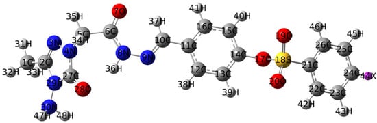
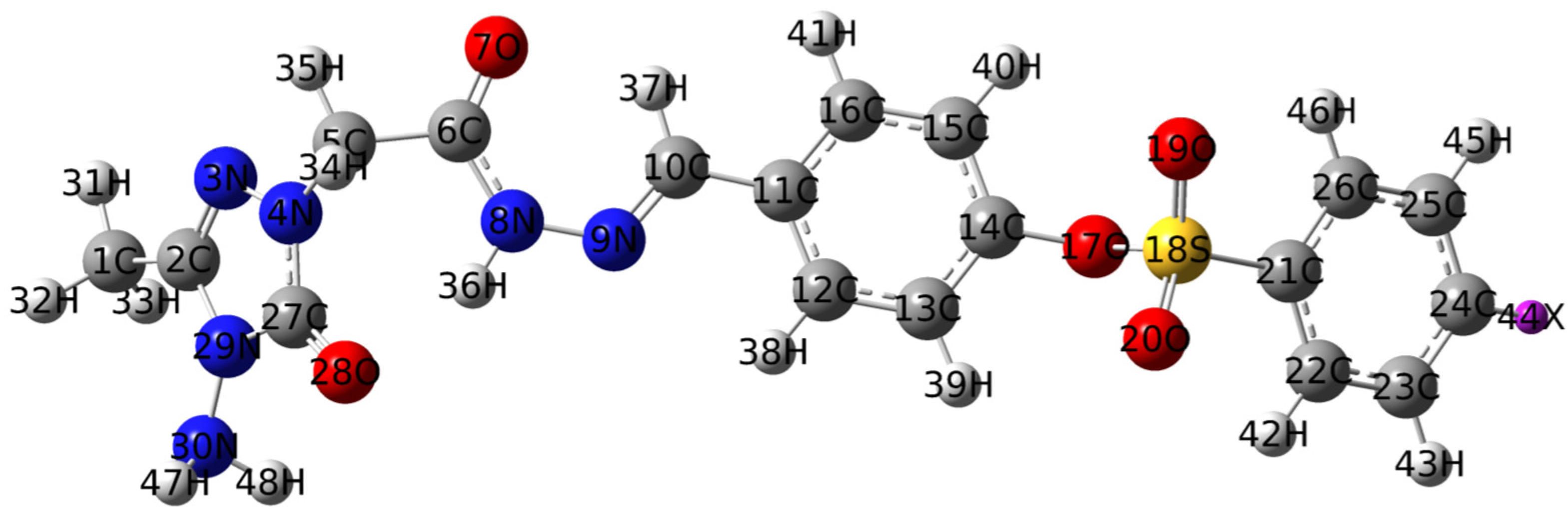
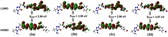
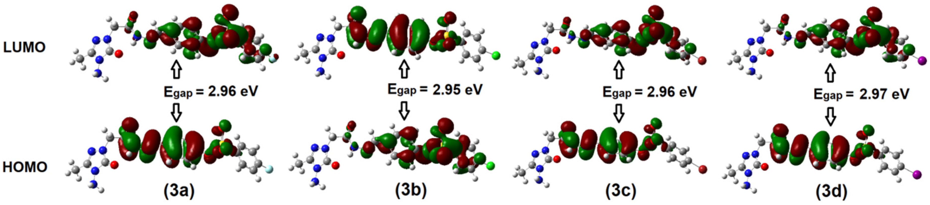
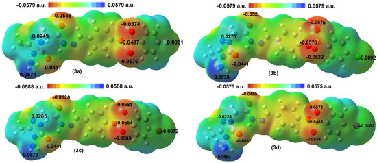
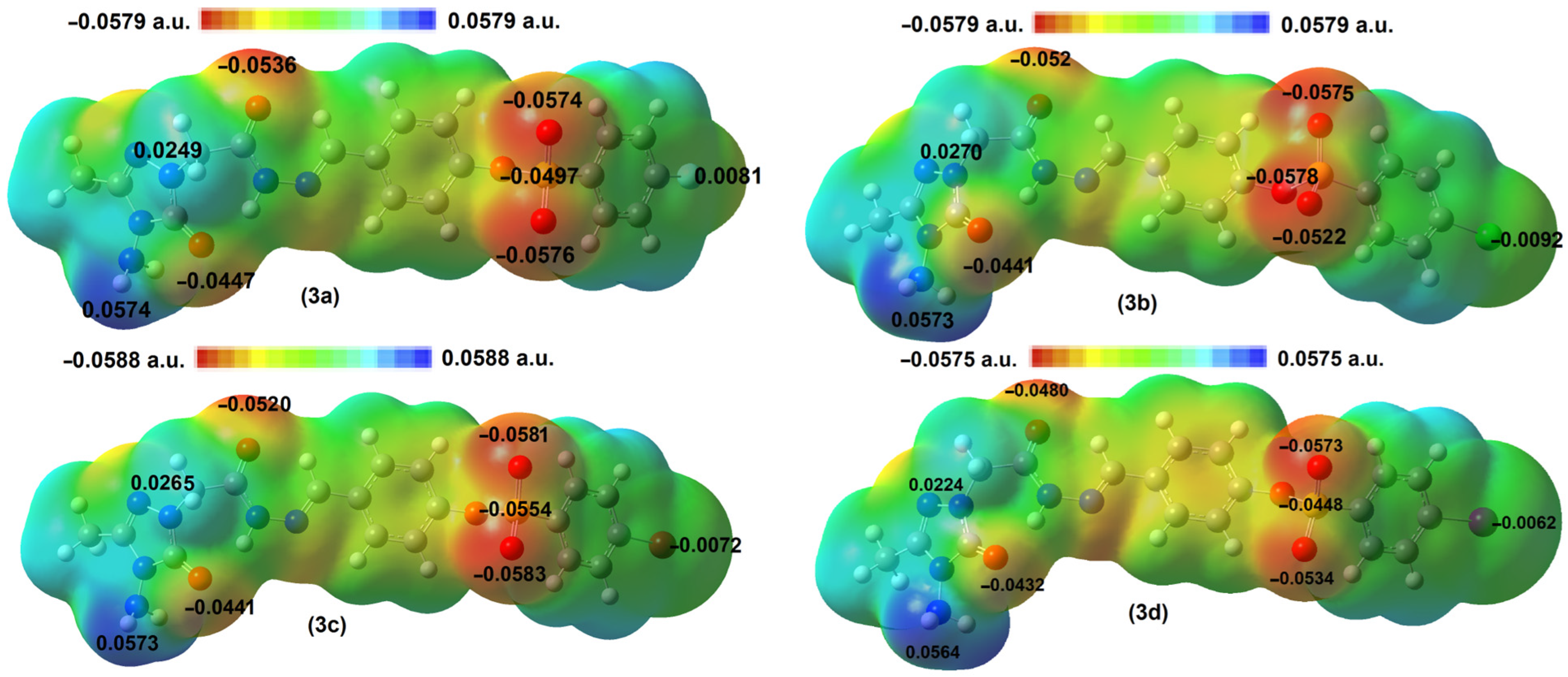
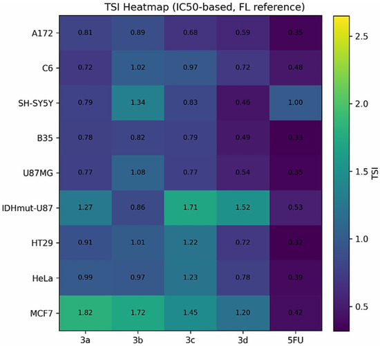
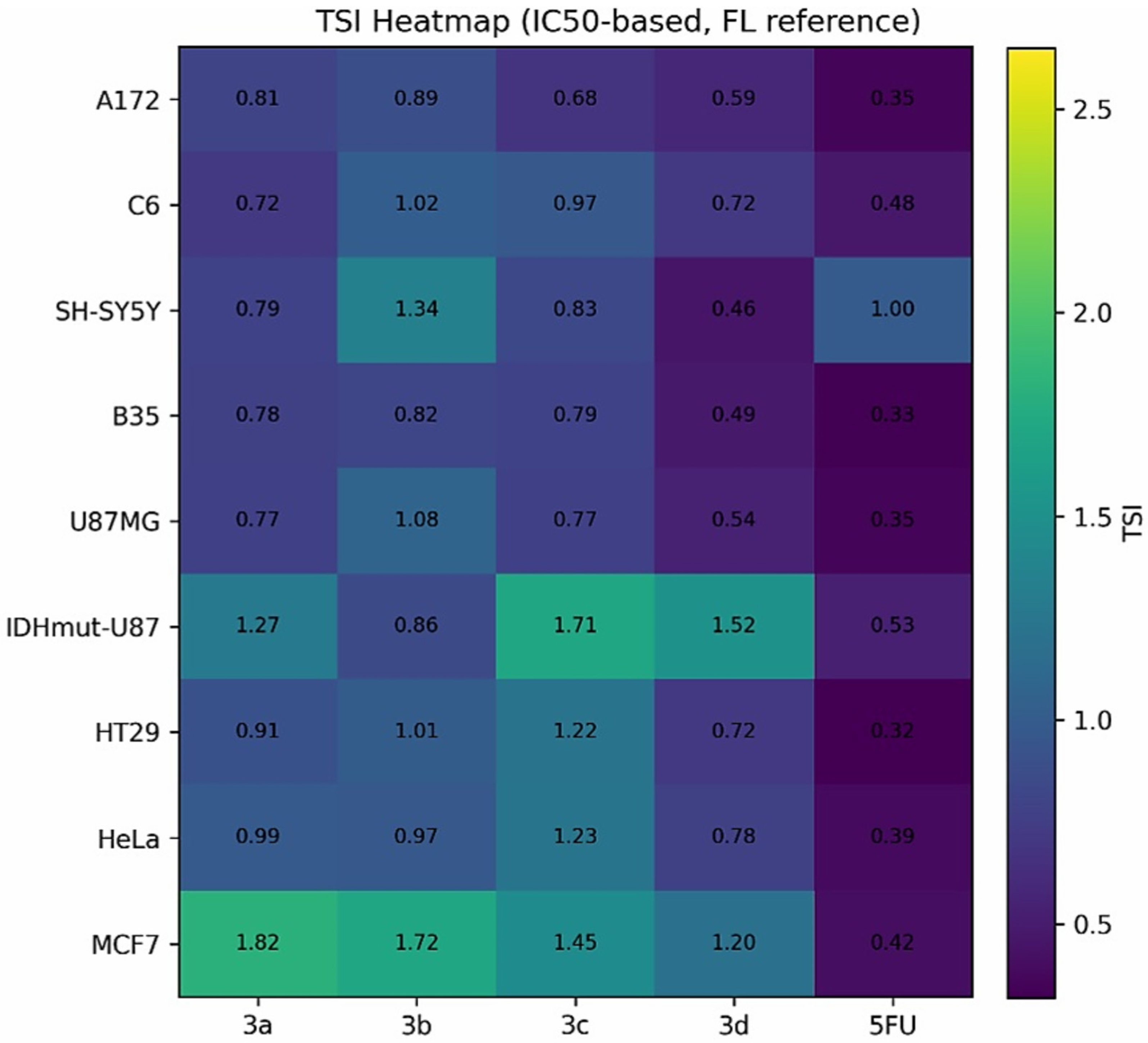
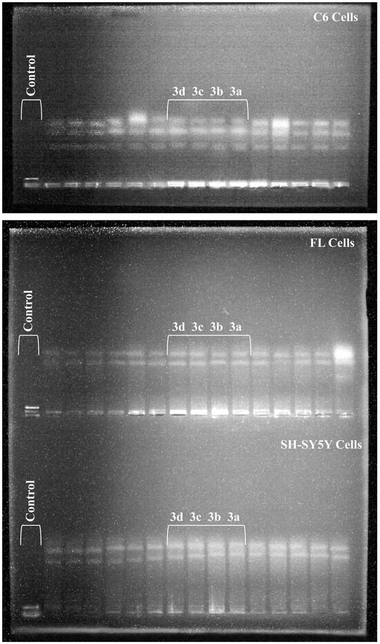
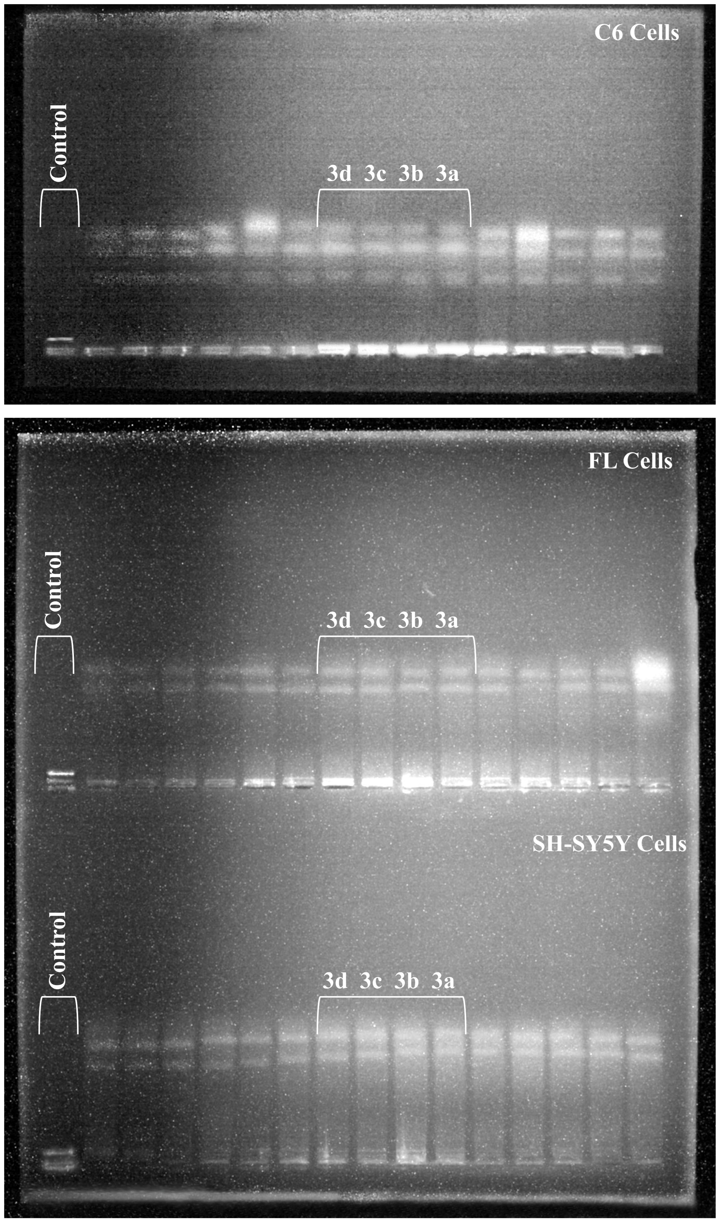
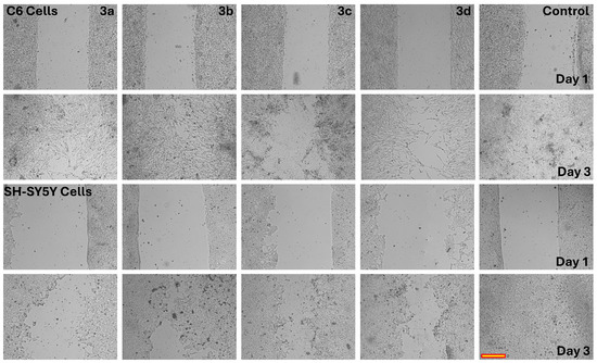
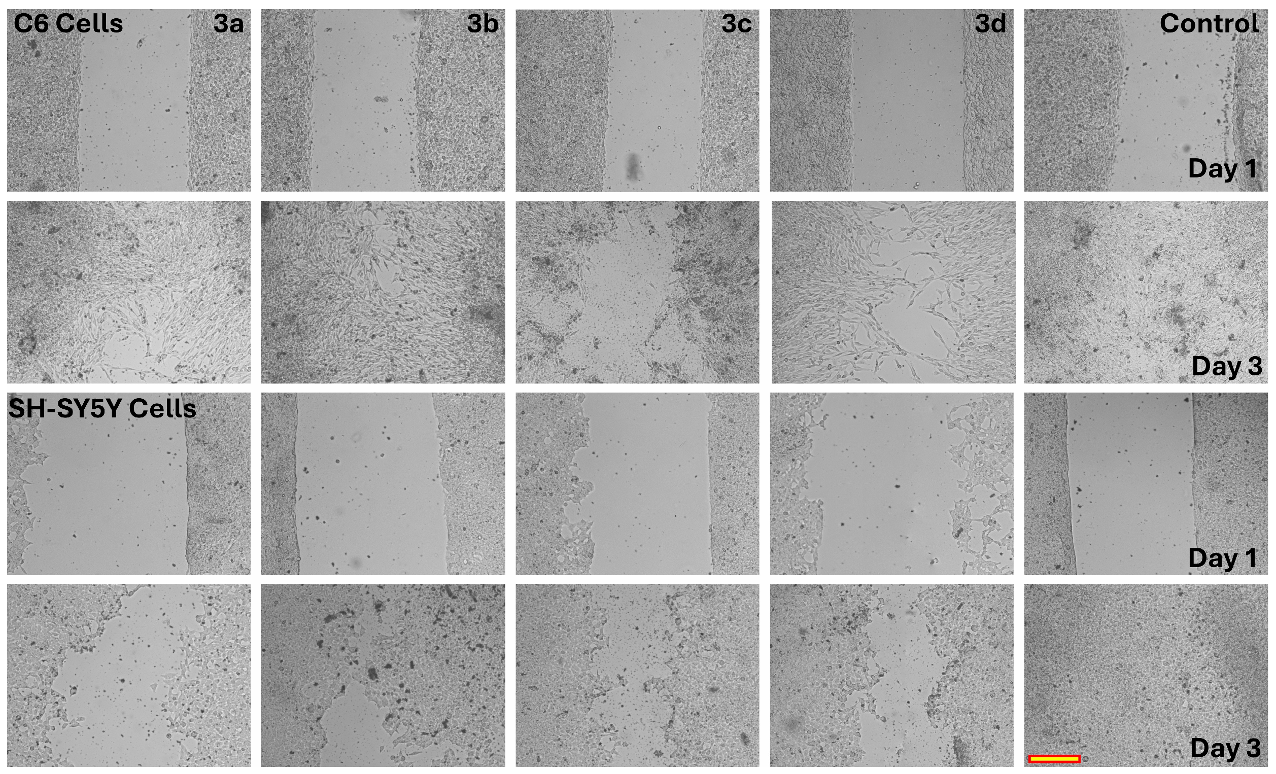
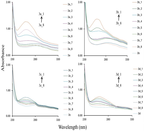
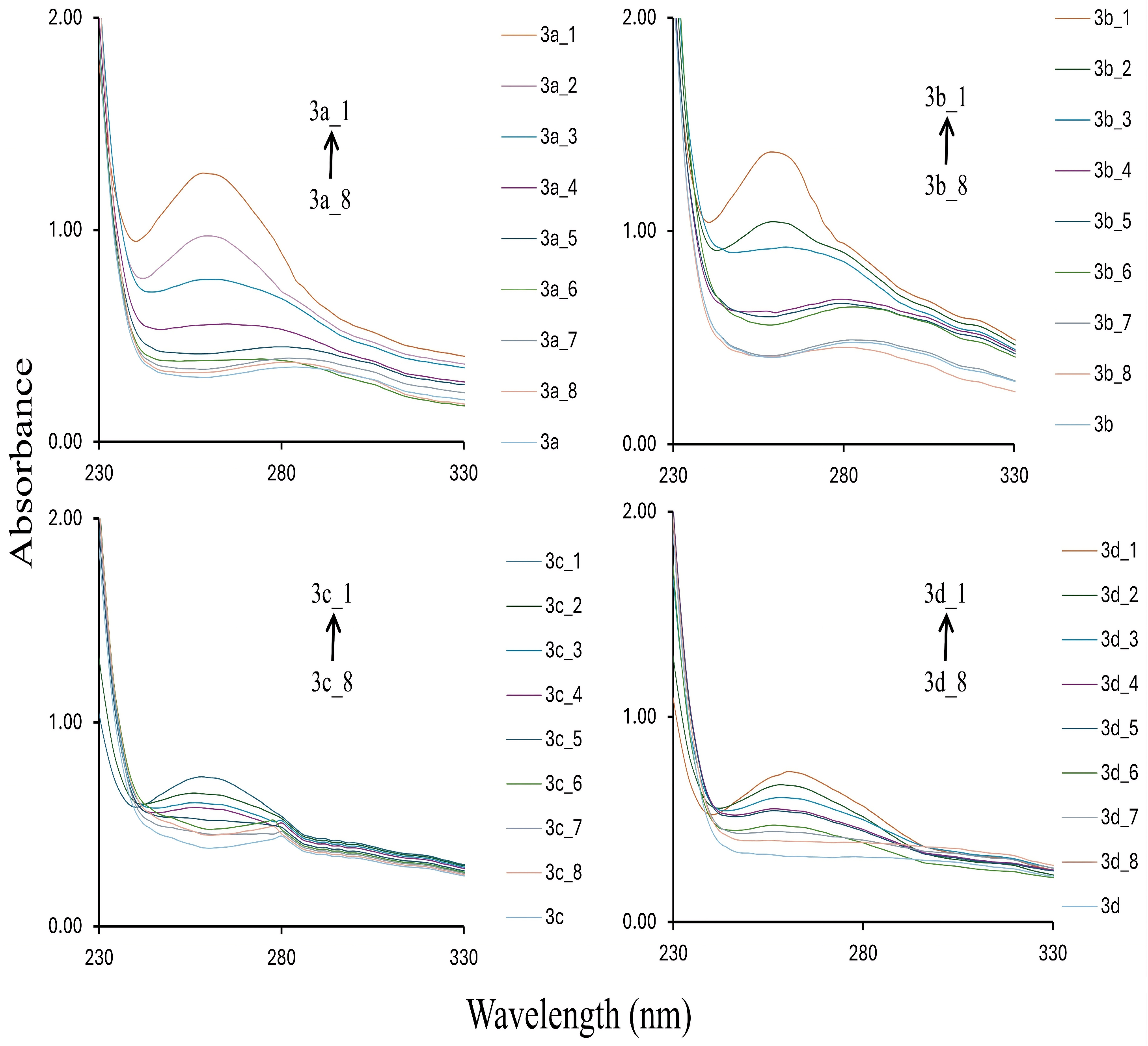
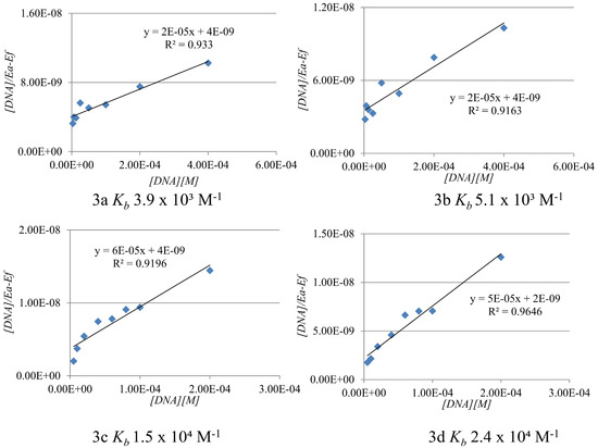
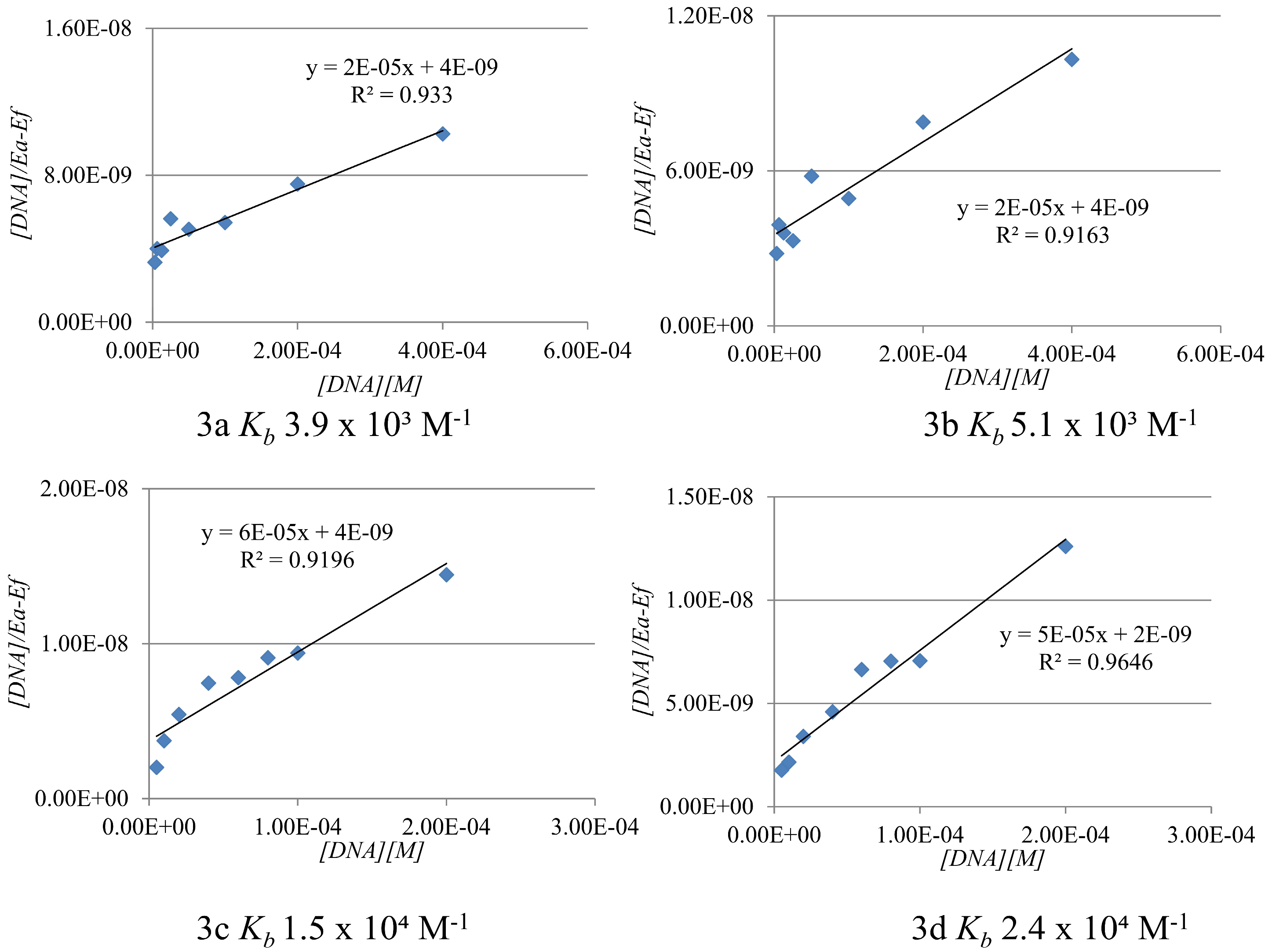


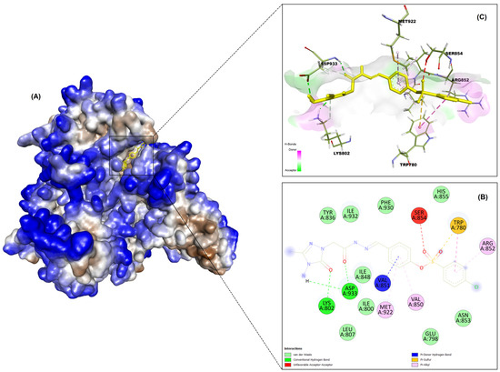
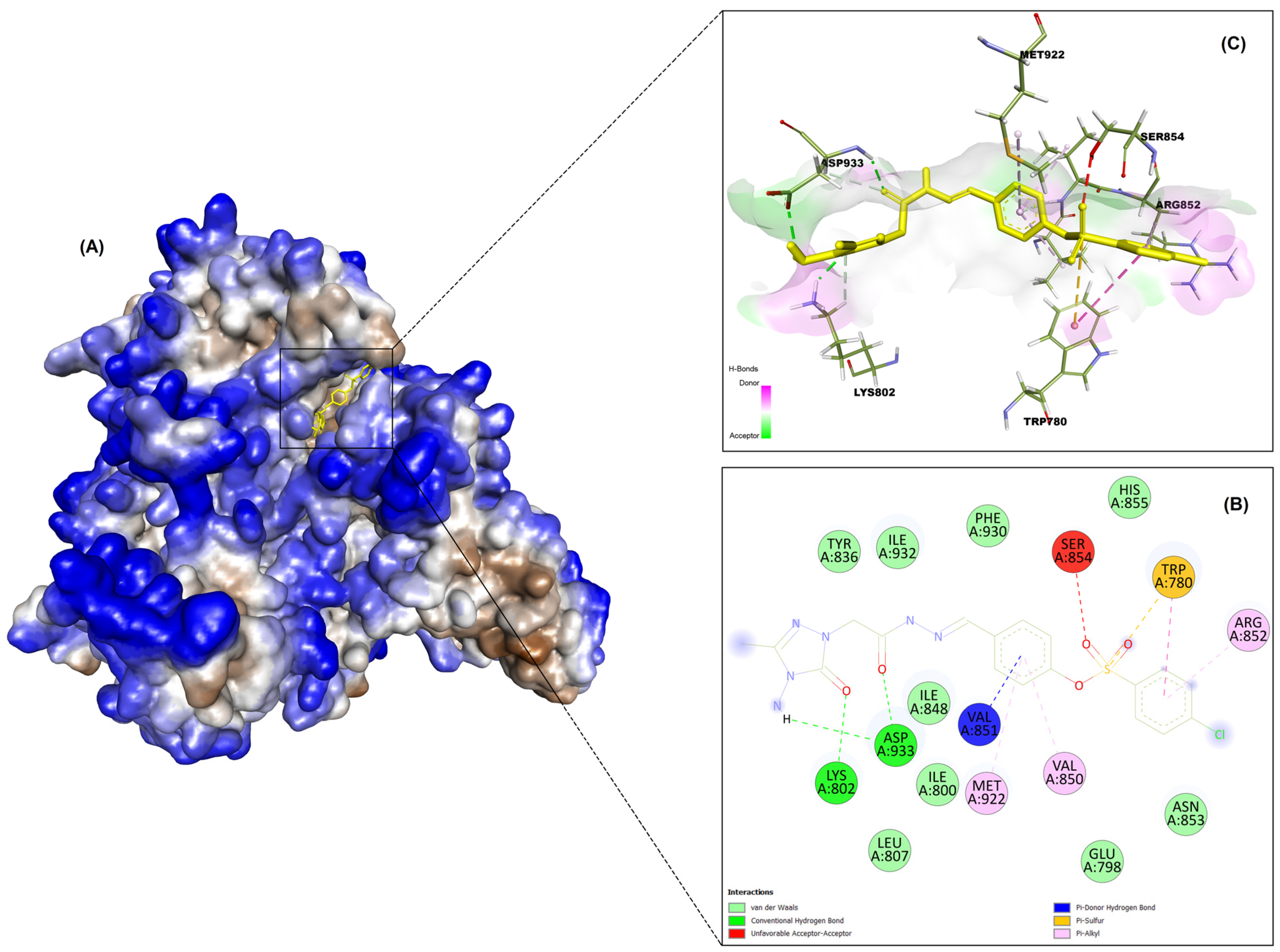
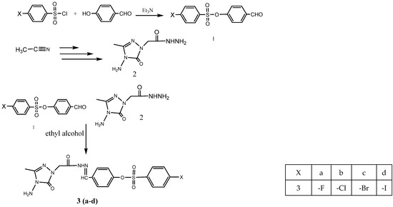
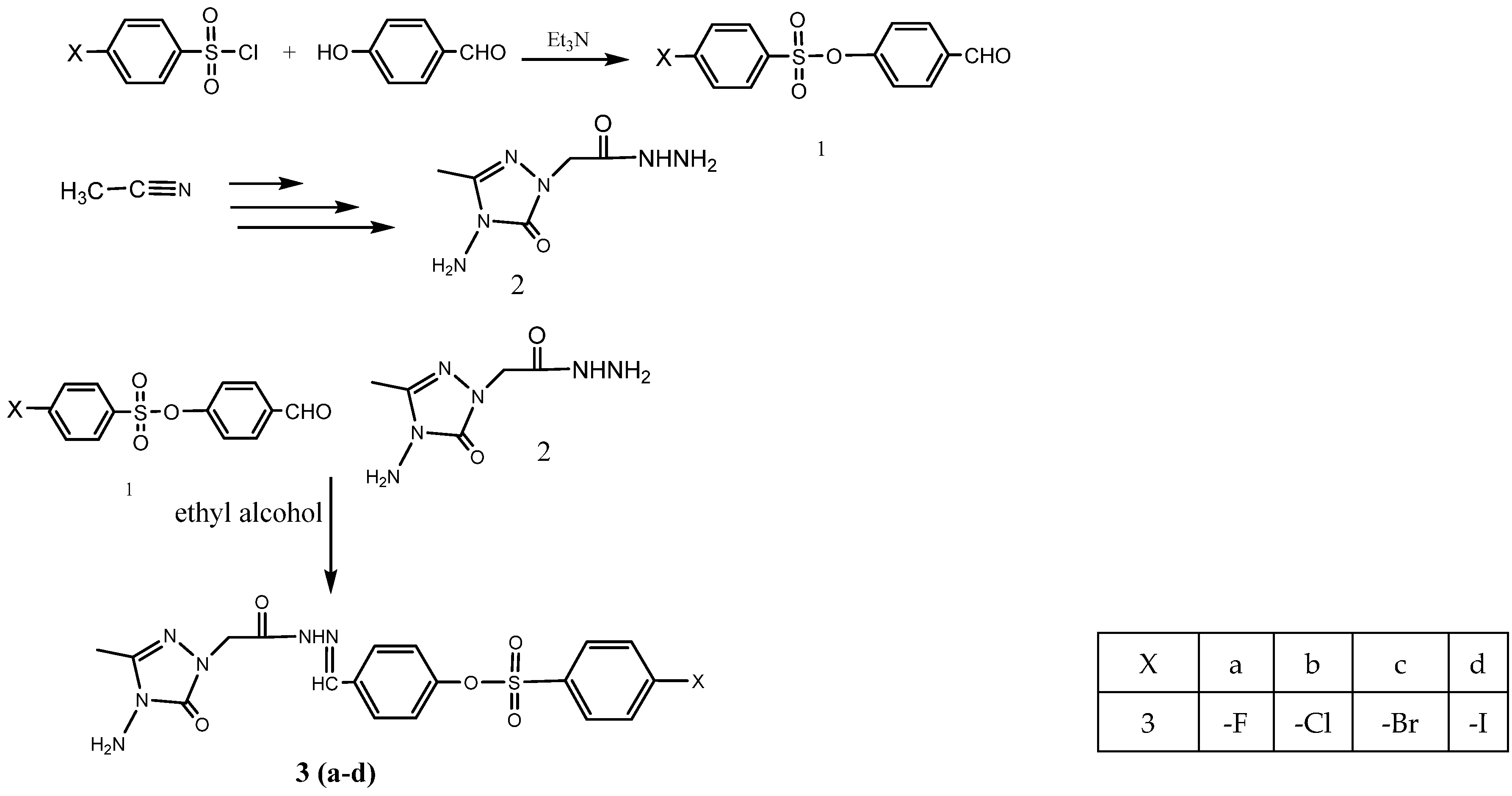

Disclaimer/Publisher’s Note: The statements, opinions and data contained in all publications are solely those of the individual author(s) and contributor(s) and not of MDPI and/or the editor(s). MDPI and/or the editor(s) disclaim responsibility for any injury to people or property resulting from any ideas, methods, instructions or products referred to in the content. |
© 2026 by the authors. Licensee MDPI, Basel, Switzerland. This article is an open access article distributed under the terms and conditions of the Creative Commons Attribution (CC BY) license.