


Abstract
Echinacoside (ECH), a natural phenylethanoid glycoside, was first isolated from Echinacea angustifolia DC. (Compositae) sixty years ago. It was found to possess numerous pharmacologically beneficial activities for human health, especially the neuroprotective and cardiovascular effects. Although ECH showed promising potential for treatment of Parkinson’s and Alzheimer’s diseases, some important issues arose. These included the identification of active metabolites as having poor bioavailability in prototype form, the definite molecular signal pathways or targets of ECH with the above effects, and limited reliable clinical trials. Thus, it remains unresolved as to whether scientific research can reasonably make use of this natural compound. A systematic summary and knowledge of future prospects are necessary to facilitate further studies for this natural product. The present review generalizes and analyzes the current knowledge on ECH, including its broad distribution, different preparation technologies, poor pharmacokinetics and kinds of therapeutic uses, and the future perspectives of its potential application.
1. Introduction—Treasure from the Garden: The Discovery and Distribution of ECH
Phenylethanoid glycosides (PhGs) are naturally occurring water-soluble compounds that are widely distributed in the plant kingdom, and most of which are isolated from garden plants and medicinal herbs. Structurally, these compounds are characterized by cinnamic acid and hydroxyl phenyl ethyl moieties that are attached to a β-glucopyranose (apiose, galactose, rhamnose, xylose, etc.) via a glycosidic bond. In recent years, interest has been growing in using PhGs [1] as their potential in the prevention and treatment of various human diseases and disorders.
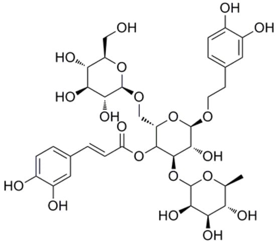
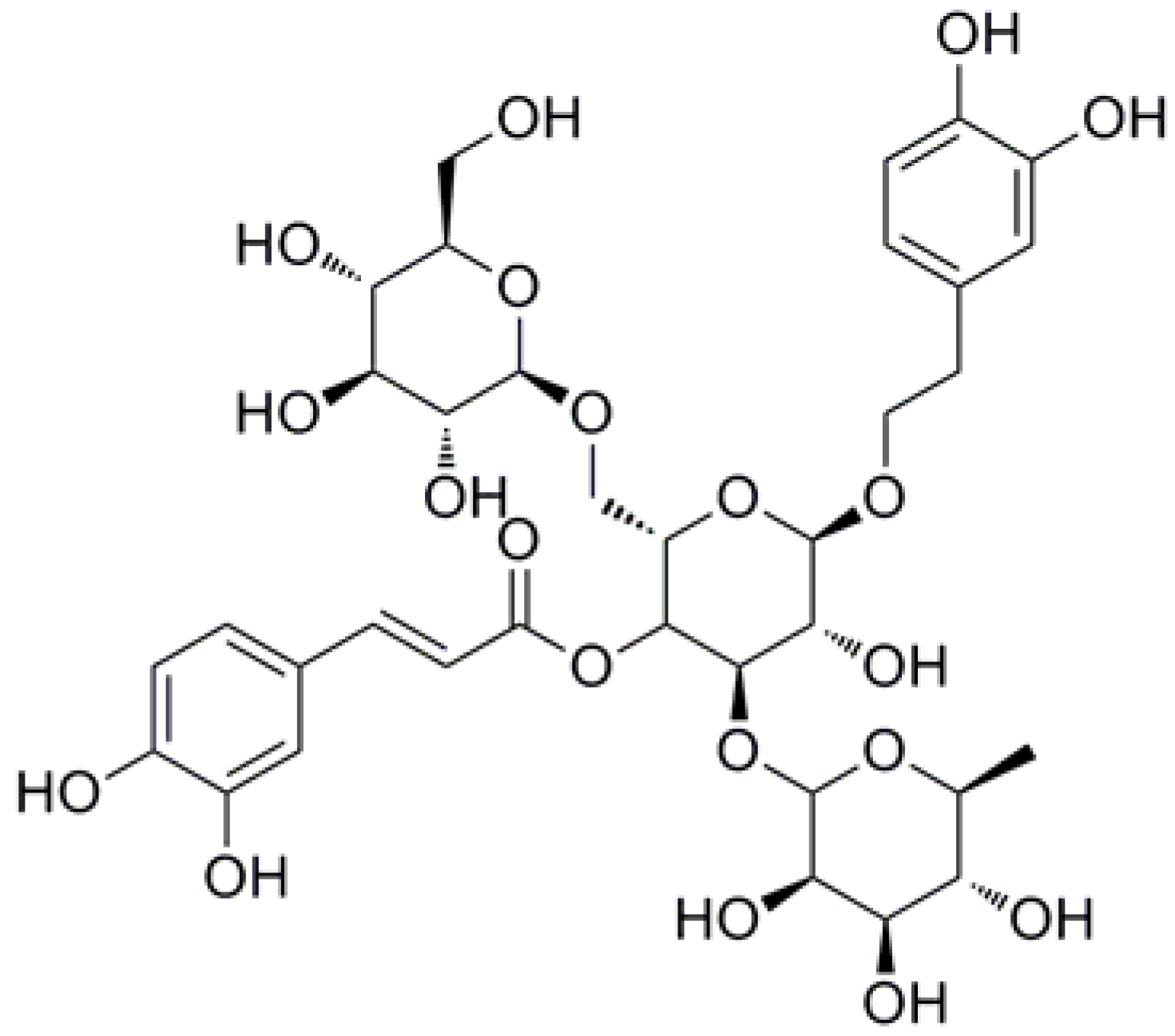
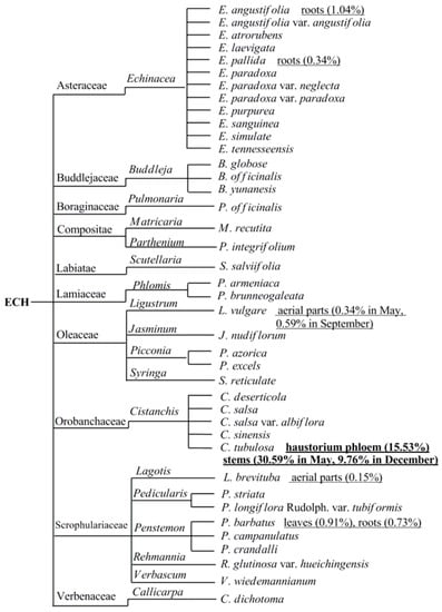
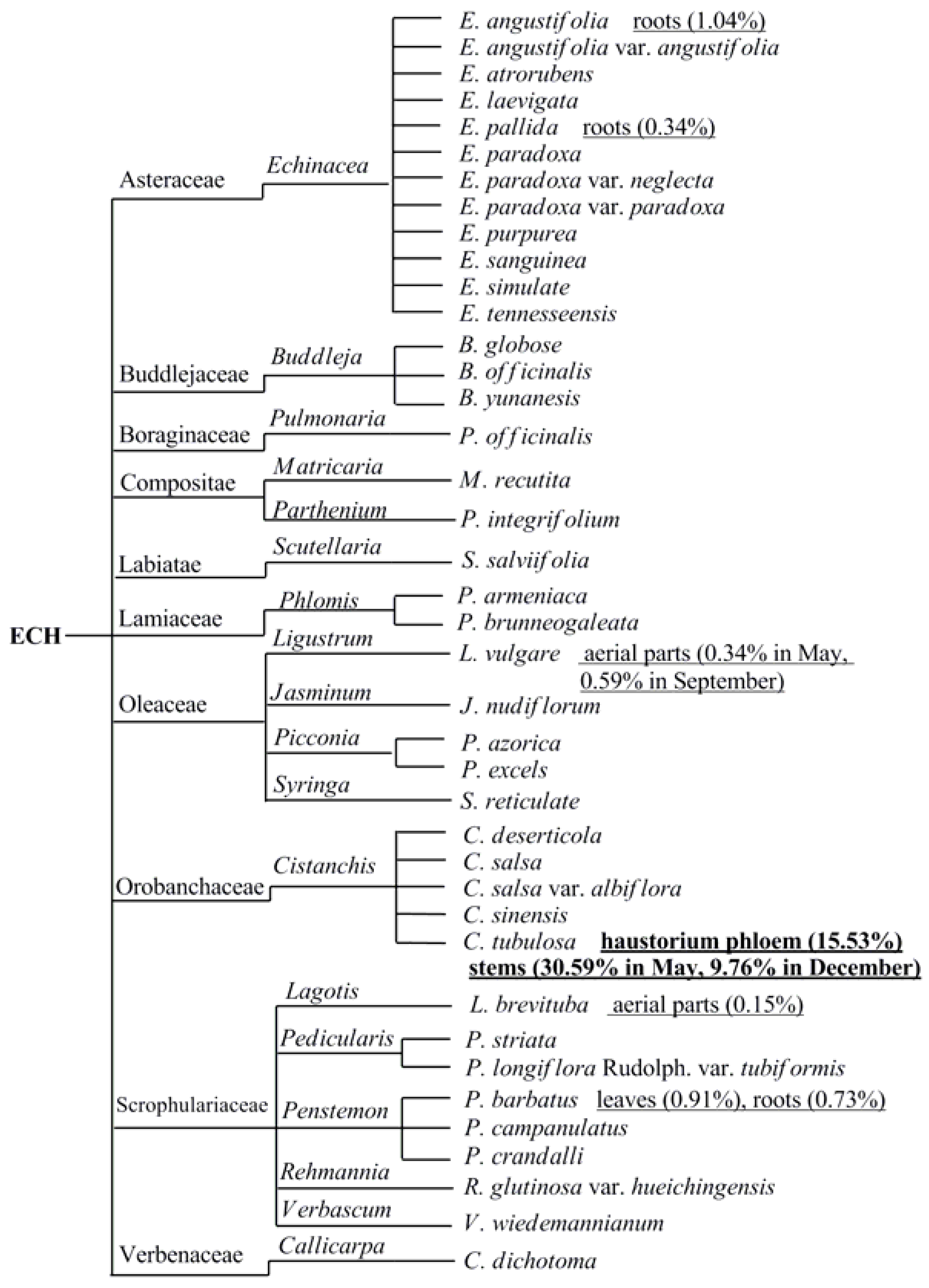
Echinacoside (ECH, Figure 1), a natural PhG, was first isolated from Echinacea angustifolia DC. (Compositae), a garden plant sixty years ago, [2] and subsequently prepared from the species of Cistanches [3] as well as the aerial part of landscape herb, Penstemon crandallii A. Nels. (Scrophulariaceae) [4], whole plants of Pedicularis striata Pall. [5] and now successively found in 40 plant species [6,7,8,9,10,11,12] belonging to 18 genus and 10 families (Figure 2). To date, the species of genus Cistanches (Orobanchaceae) and Echinacea (Asteraceae) were the main natural plant sources for the preparation of ECH.
Figure 1.
Chemical structure of Echinacoside (ECH) (Glu-Glu-Rha).
Figure 2.
The broad distribution and discovery of ECH in the plant kingdom.
ECH was found in both underground and aboveground parts of medicinal herbs but with widely varying levels of content (Figure 2), including different stages of plant growth [8,13], different parts of the same plant [14], and vice versa, the same parts of different plants [15,16]. Until now, the highest content of ECH was found in haustorium phloem of Cistanches tubulosa which was reached almost 15.5% [17], thus could be a good resource for the isolation of pure ECH.
2. Preparation of ECH
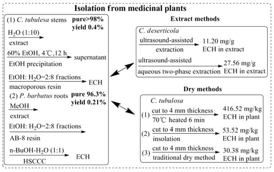
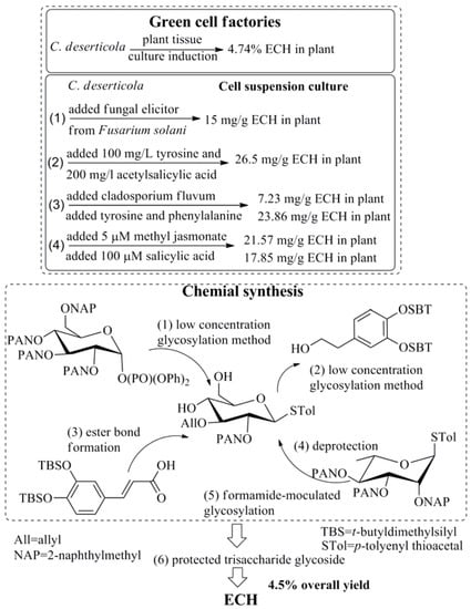
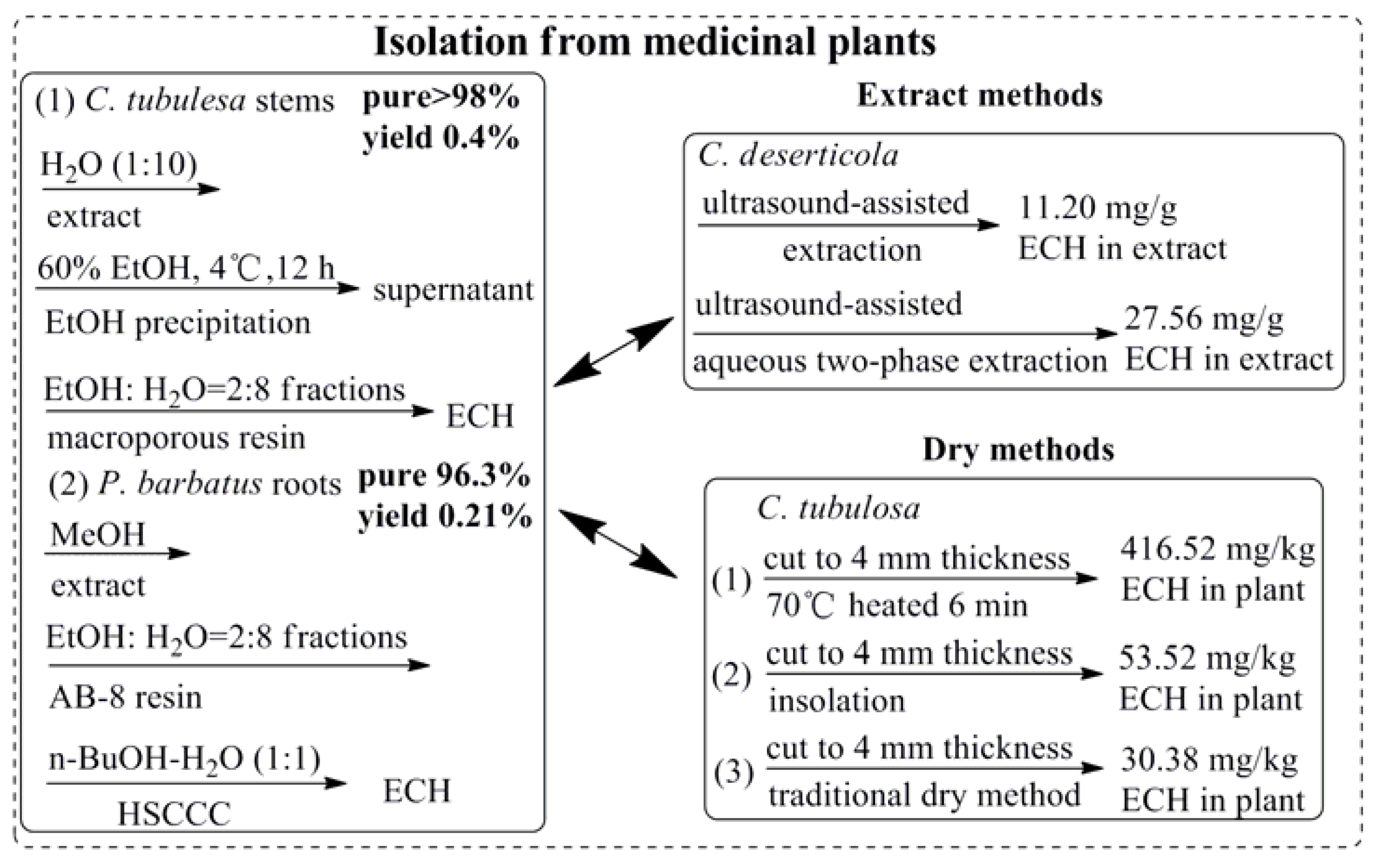
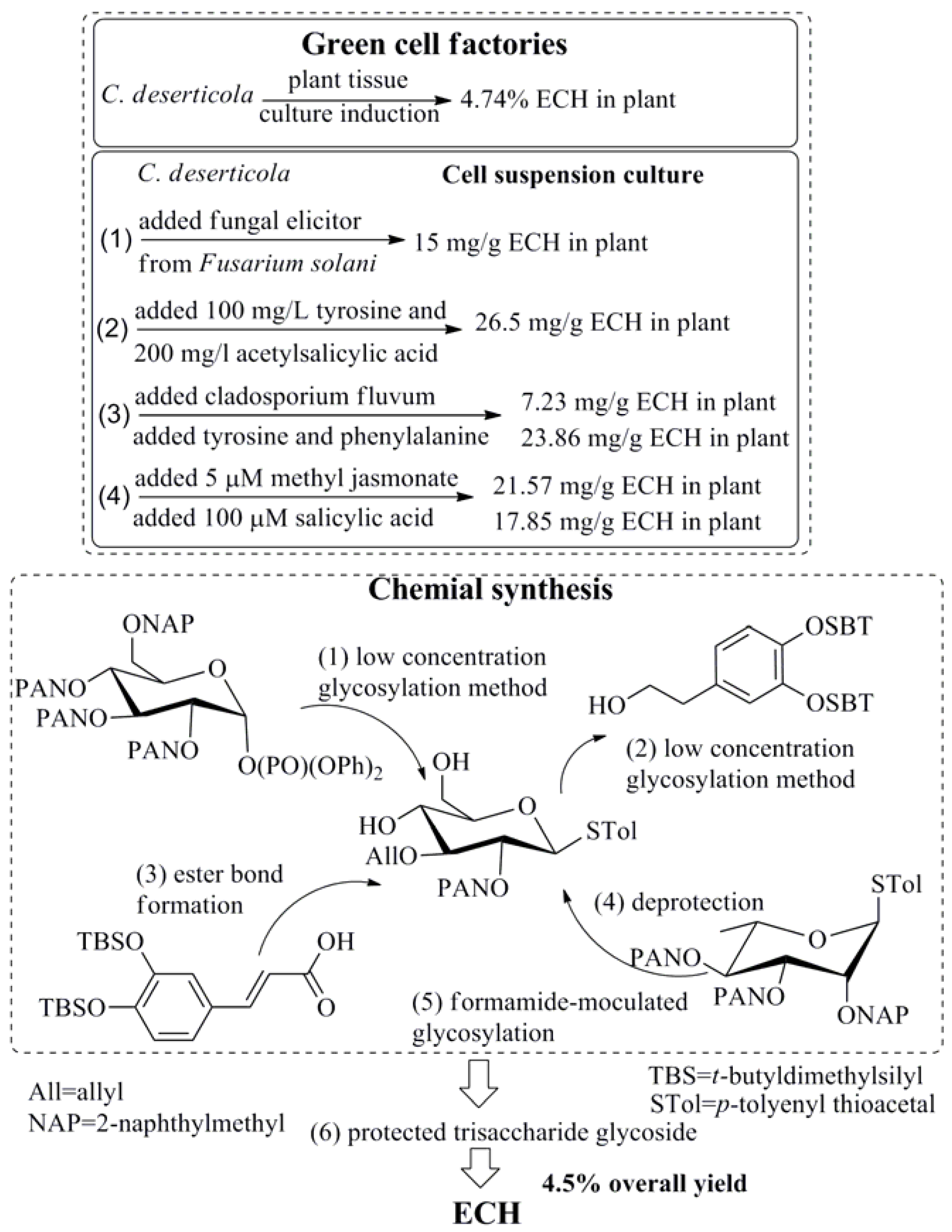
As PhG compounds exhibited significant activities in the prevention and treatment of various human diseases and disorders, it was important to develop sustainable methods to produce sufficient quantities of ECH for pharmaceutical applications. Firstly and also usually, like other natural bioactive compounds, high purity of ECH was obtained from medicinal plants by using classic isolation methods and semi-preparative liquid chromatography (LC) [18] or high-speed counter-current chromatography methods [14] as Figure 3 shown, and the yield of ECH was usually between 0.2%~0.4%. An efficient ultrasound-assisted aqueous two-phase extraction process for preparation of ECH from Cistanche deserticola enhanced the content of ECH in the extracts (27.56 mg/g) which was 2.46-fold higher than the amounts obtained in ultrasound-assisted extraction [19]. Interestingly, it was found the content of ECH in medicinal herbs was significantly influenced by the factors of preparation processing [20], including the slice thickness, drying temperature, and the time for inactivation of the enzyme [21]. Therefore, it was worth noting that ECH was demonstrated to be highly susceptible to “enzymic” degradation and oxidation in hydroalcoholic solutions during the extraction process, and ECH in biosamples was susceptible to degradation at a higher temperature during the whole process, thus the operation must be carried out carefully at a lower temperature. Secondly, besides the above classic isolation method, plant cell/tissue culture, called “green cell factories”, has become increasingly attractive as a cost-effective alternative to classical approaches for the sustainable mass production of plant-derived molecules [22]. Several published data demonstrated an increased accumulation of ECH in both plant tissue culture [23] and cell suspension culture [24] of Cistanche deserticola, and some revulsant including tyrosine, phenylalanine, cladosporium fulvum, methyl jasmonate and salicylic acid were found could promote the accumulation of ECH [25,26,27]. Thirdly, as an inestimable natural product which possesses a broad spectrum of beneficial activities, the chemical synthesis of ECH is needed to satisfy its comprehensive application. A group from National Taiwan University has completed the total synthesis of ECH from building blocks over 7 steps with yield was 4.5% [28] which was showed in Figure 3.
Figure 3.
Different methods of ECH obtaining.
3. Pharmacokinetics and Strategy
Generally, the systemic effects of natural PhGs mainly depend on their bioavailabilities through the gastrointestinal barrier. However, both in vitro and in vivo experimental data appeared to reflect their pitiful fates in the gut, including relatively poor bioavailabilities and rapid rates of excretion [29]. Dozens of studies have shown that the content of PhGs in plant within μg, following ingestion, they appeared in the circulation as phase II metabolites, and their plasma levels rarely exceed nM concentrations [30]. Animal study confirmed that the absolute bioavailability of ECH was only 0.83% [31], as Table 1 shown, the absorption and elimination of ECH was extremely fast in rats and the serum concentration was very low, and ECH could not be identified in any human plasma sample at any time after ECH tablet ingestion [32]. The serum concentration–time curves for intragastric and intravenous administration were fitted to one-compartment model and two-compartment model, respectively. The metabolites of ECH in rat feces were identified as acteoside, decaffeoylacteoside, lugrandosie and 3,4-dihydrophenyl ethanol after oral administration [33], and a portion of ECH was transformed into acteoside [34]. Now, to predict the absorption of orally administrated drugs, Caco-2 monolayer is widely used as a model of the human intestinal mucosa. It was shown that ECH permeated poorly through the Caco-2 monolayers although one potential metabolite, cinnamic acid, diffused readily with an apparent permeability of 1 × 10⁻⁴ cm/s. The data implied ECH was not likely to cross the normal intestinal barrier [35,36,37], but it can through the blood-brain barrier in permanent middle cerebral artery occlusion (MCAO) rats [38]. Furthermore, a recent study estimated the dynamic pharmacokinetic of ECH between Parkinson’s disease rat and normal rats showed that the plasma concentrations of ECH in Parkinson’s disease rats were higher than that in the normal rats after oral administration. The reasons why the elimination rate of ECH slowed down in Parkinson’s disease rats may be as follows: ECH is partially hydrolyzed to aglycone in the body and in the state of pathophysiology, a low activity of certain enzymes induced by 6-hydroxydopamine (6-OHDA) damage might lead to the decreased clearance rate and increased retention time of ECH. Even if ECH was mainly excreted in the urine, the decreased blood circulation of kidney induced by 6-OHDA damage might play an important role in the decreased elimination rate and increased retention time of ECH [39].
Table 1.
Pharmacokinetics of ECH with different models.
4. Pharmacological Properties and Underlying Mechanisms
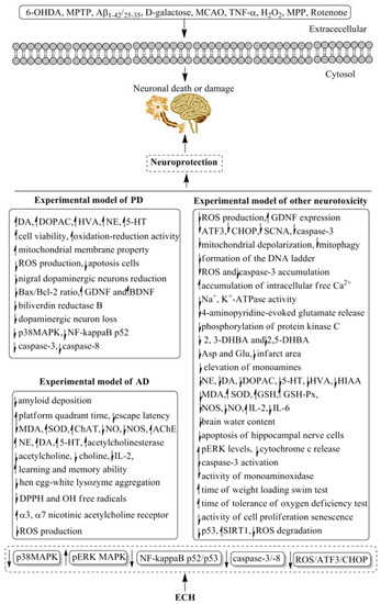
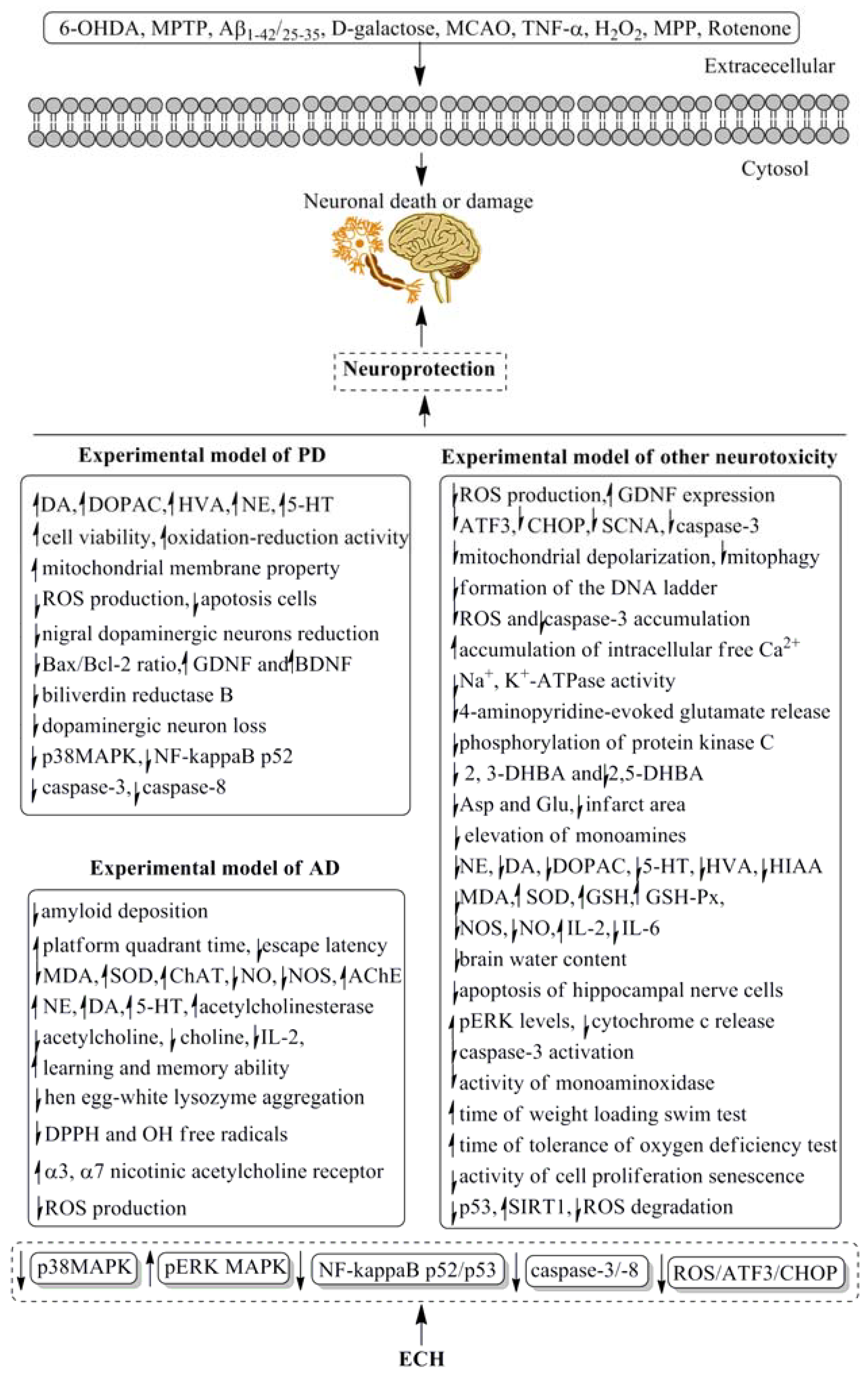
ECH was proved possessing kinds of pharmacological activities since it was found sixty years ago, the data of this review were mainly gathered by consulting the database of PubMed, Springer, Elsevier, and Scholar in the last 30 years. Among the broad range of therapeutic applications of ECH, its neuroprotective bioactive has attracted the more attention of pharmaceutical scientists than the others. Dozens of reports have discovered that ECH was effective in Parkinson’s and Alzheimer’s diseases by using both animal experiments and cell lines tests, which were shown in Table 2. Besides the neuroprotective action, the cardioactive property, anti-inflammatory activity, antioxidant and anti-osteoporotic activities as well as other pharmacological potentials of ECH were presented in Table 3, Table 4, Table 5, Table 6 and Table 7, respectively. The data of Table 2 and Figure 4 showed that ECH could prevent the progress of neurodegeneration in Parkinson’s and Alzheimer’s diseases. Several Parkinson’s or Alzheimer’s animal models induced by 6-OHDA, 1-methyl-4-phenyl-1,2,3,6-tetrahydropyridine (MPTP), d-galactose and β-amyloid Aβ-(25–35) as well as cerebral ischemia rats were used to estimate the neuroprotective effects of ECH. PC12 and neuroblastoma SH-SY5Y cell lines were employed to discover the related mechanisms, which were related to the mitogen-activated protein kinase (MAPK), NF-kappa B, caspase 3 and 8 as well as reactive oxygen species (ROS)/activating transcription factor 3 (ATF3)/C/EBP-homologous protein (CHOP) pathways, as Figure 4 showed. However, most of the above data were obtained by using cells or animals, the reliable clinical trials were limited, large-scale evidence-based human clinical trials with specific neuroprotective therapeutic settings are necessary. The same problems have been found by applying other evaluation methods of the different pharmacological properties.
Table 2.
Neuroprotective activity of ECH in selected models.
Table 3.
Cardioactive property of ECH in selected models.
Table 4.
Anti-inflammatory property of ECH in selected models.
Table 5.
Anti-oxidative property of ECH in selected models.
Table 6.
Anti-osteoporotic activity of ECH in selected models.
Table 7.
Other activities of ECH in selected models.
Figure 4.
The underlying mechanism of neuroprotective of ECH. AD, Alzheimer’s disease; PD, Parkinson’s disease.
One of the traditional uses of Cistanche deserticola was for treatment of irritable bowel syndrome disease, and ECH was the main bioactive ingredients in this herbal responsible for the activity. To date, dozens of in vivo studies demonstrated the anti-inflammatory property of ECH, the data of Table 4 showed that ECH could suppress the acute colitis in mice induced by dextran sulphate sodium [88], attenuate acute hepatotoxicity in rats induced by d-galactosamine/lipopolysaccharide [89] and carbon tetrachloride (CCl4) [90], increase hyaluronan levels and decrease wound contraction for wound healing, modulate inflammatory markers including transforming growth factor (TGF)-β1, NO, alanine aminotransferase (ALT), myeloperoxidase, inflammatory cytokines, and etc.; however, the molecular mechanisms of the anti-inflammatory of ECH were limited, which were only related to the expressions of TGF-β1, capase-3 and TNF-α.
It was worth mentioning that, to date, numerous in vitro and in vivo studies have demonstrated the strong antioxidant property of ECH (Table 5). In DPPH assay, the EC50 of ECH was 6.6 μM which was 9.5-fold than Trolox; on xanthine/xanthine oxidase generated superoxide anion radical test, the IC50 of ECH was 2.74 μM than tocopherol, and etc. [98,99,100,101]. In vivo experiments, ECH could prompt the ability of anti-oxication, anti-fatigue and anti-stress in vascular dementia rats or subacute aging mice model, and the indirect antioxidant activities of ECH due to the induction or/and activation of major endogenous antioxidant enzymes and inactivation of pro-oxidant enzymes. In addition, the molecular mechanisms of this activity showed that ECH reduced nuclear protein levels of transcription regulator protein BACH1, enhanced heme oxygenase 1 mRNA levels, down-regulated expression of p53, up-regulated the SIRT1 [102]. Furthermore, the structure-activity relationship of antioxidant property of ECH was also estimated. It was believed that the inhibitory oxidative hemolysis activity of ECH was related to the number of phenolic hydroxy groups. ECH, possessing four phenolic hydroxy groups, exhibited stronger antioxidant activities than cistanoside D possessing only two phenolic hydroxy groups, and compound permethylacteoside with no phenolic hydroxy group inhibited oxidative hemolysis weakly [103,104].
Cistanche deserticola is a traditional Chinese medicine (TCM) called “Desert ginseng” in China owing to its excellent medical functions and nourishing effect. According to the theory of TCM, Cistanche deserticola can supplement the kidney, and kidney stores essence and the essence can transform into bone marrow to nourish the bones, which means Cistanche deserticola could promote the formation of the bone [112]. As ECH is the main constituent of Cistanche deserticola, thus maybe possesses anti-osteoporotic property. And in the anti-osteoporotic tests as Table 6 shown, ECH exhibited anti-osteoporotic effect on the promotion of bone formation and suppression of bone resorption [113], and the molecular targets of ECH were also discovered that it could increase the osteoprotegerin (OPG) level and decrease the receptor activator for nuclear factor-κB Ligand (RANKL) level [114] as well as promoted the phosphorylation of ERK1/2 to activate MAPK/ERK pathway [115,116]; however, the dosage of ECH were so high that even reached at 270 mg/kg body weight/day, which made some difficult in the future clinical trials and enhanced the medicinal costs. Since the results of the report showed the dosage of 30 mg/kg body weight/day of ECH was also effective in ovariectomized (OVX) rats, a proper dosage of ECH in future treatment of osteoporosis disease should be selected with more tests.
Besides the above significant bioactive, ECH also proved to possess additional antidiabetic effect, antiviral activity, anti-hepatic fibrosis effect, anti-tumor property, testis and sperm injury protect activities as Table 7 shown.
5. Discussion
ECH, a natural PhGs compound has been isolated from dozens of medicinal or horticultural plants, exhibited highly positive activities in treatment of nervous, cardiovascular and bone disorders, especially for the prevention and treatment of a variety of nervous system disorders including Parkinson’s and Alzheimer’s diseases. Given the above potential in pharmaceutical applications, the preparations of ECH including the classic isolation from herbs, plant cell/tissue culture and even the chemical synthesis have attracted the interests of plenty pharmaceutical scientists. This review has presented the discovery of ECH including its distribution in the plant kingdom, and the preparation of ECH including the methods of classic isolation from medicinal plants, “green cell factories” of plant cell/tissue culture and chemical synthesis, and the pharmacokinetics data of ECH was also posted for further medicinal uses. Then, the most important section of this paper, the remarkable pharmacological properties of ECH were elucidated, including neuroprotective activity, cardioactive property, anti-osteoporotic effect, anti-inflammatory and antioxidant activities. However, there were four important questions should be pay attention before ECH was used for clinical applications: first, both in vivo and in vitro experiments of ECH reflected its dissatisfied pharmacokinetic property. Concerning the in vivo experiments, ECH exhibited a pitiful fates in the gut, including relatively poor bioavailability [31] (the absolute bioavailability was only 0.83%) and rapid rates of metabolism and excretion, following ingestion, ECH appeared in the circulation as phase II metabolites, and its plasma levels rarely exceed nM concentrations [30]; and in vitro tests showed that ECH permeated poorly through the Caco-2 monolayers, which implied that ECH was not likely to cross the intestinal and blood brain barriers, thus appeared failed to explain its neuroprotective activity and other bioactive [35]. However, it was reported that in neuronal cells and non-neuronal cells which were exposed to rotenone, ECH was able to cross the blood-brain barrier freely [74]. Second, it should be noted that several of the activities especially the anti-osteoporotic one occurred at relatively high concentrations of ECH (30–270 mg/kg/day, orally for 12 weeks) [113], and in the antidiabetic experiments [87], the dose of ECH even reached at 500 mg/kg p.o. And others occurred at normal concentrations about 5 to 50 mg/kg. Third, the underlying molecular mechanisms of ECH with neuroprotective and cardiovascular properties, anti-osteoporotic, and anti-inflammatory activities have not been elucidated in detail. Although some common molecular signal pathways and several distinct targets have been disclosed, the responses of molecular targets to ECH with the above effects remain unclear. Forth, sixty years after the discovery of ECH, to date, very little efforts are done for its clinical trials and the safety and toxicity tests, and reliable clinical data describing the health effects of ECH are limited. Therefore, the in vivo animal studies of ECH should be considered with caution and more clinical trial on its efficacy and safety should be performed.
6. Conclusions
In summary, ECH was an inestimable natural product that exhibited highly positive activities in nervous and cardiovascular system disorders as well as bone disease from the wealth of laboratory data, and thus was believed have a promising potential in the treatment of Parkinson’s and Alzheimer’s diseases, atherosclerosis, osteoporosis, acute colitis, wound injury, and hepatitis. The excellent antioxidant property, which was 9.5-fold greater than Trolox, also implied an ideal application of ECH in the future clinical trials. However, at present, despite the wealth of experimental data that was available describing the potent pharmacological effects of ECH, many issues remain unresolved with respect to effective clinical applications. First, the low bioavailability and extremely fast metabolism of ECH in animals reflected its dissatisfied pharmacokinetic property in the future clinical application. The rapid rates of metabolism and excretion of ECH was due to the multiple metabolic pathways that were involved to eliminate plant-derived toxins, thus further intensive studies are required to confirm the clinical potential of ECH, thereby enabling its acceptance as a therapeutic agent. Another important issue was that, although the present review findings provided a sound basis to confirm that ECH is a potential candidate for intervention in neurodegenerative diseases such as Alzheimer’s and Pakinson’s disorders, the molecular signal pathways, especially the molecular targets to ECH with the above effects, remain unclear. Thus, further studies are needed to interpret the directed molecular mechanisms. The last issue is also the most important problem, implying that large-scale evidence-based human studies with specific therapeutic settings are necessary. Although plenty of laboratory data shed light on the protection of ECH against dozens of diseases, the reliable clinical data are limited. More clinical trials on the safety and drugability of ECH are needed.
Funding
This work was supported by the grants from the National Natural Science Foundation of China (No.81560684) and Ningxia key research and invention program of science and technology cooperation of the East and the West (No. 2017BY084).
Conflicts of Interest
The authors declare no conflict of interest.
Abbreviations
The following abbreviations are used in this manuscript:
| ABTS | 2,2′-azino-bis 3-ethylbenzthiazoline-6-sulphonic acid |
| Aβ(25–35) | amyloid beta-protein fragment 25–35 |
| AchE | acetylcholinesterase |
| AD | Alzheimer’s disease |
| ALP | alkaline phosphatase |
| ALT | alanine aminotransferase |
| Asp | aspartic acid |
| AST | aspartate aminotransferase |
| ATF3 | activating transcription factors 3 |
| BDNF | brain-derived neurotrophic factor |
| BMD | bone mineral density |
| BMP | bone morphogenetic proteins |
| BPA | bisphenol A |
| CCl4 | carbon tetrachloride |
| ChAT | choline acetyltransferaxe |
| CHOP | C/EBP-homologous protein |
| CTAB | cetyl trimethylammonium bromide |
| CYP11A1 | cytochrome P450scc |
| CYP17A1 | cytochrome P450 17A1 |
| DA | dopamine |
| DHBA | dihydroxybenzoic acid |
| DOPAC | 3,4-dihydroxyphenyl acetic acid |
| DPPH | 2,2-diphenyl-1-picrylhydrazylhydrate |
| ECH | echinacoside |
| EDTA | ethylene diamine tetraacetic acid |
| ERK | extracellular signal regulated kinase |
| 5-FU | 5-fluorouracil |
| GalN/LPS | d-galactosamine/lipopolysaccharide |
| GDNF | glial cell line-derived neurotrophic factor |
| Glu | glutamic acid |
| GSH | glutathione |
| GSH-Px | glutathione peroxidase |
| 5-HT | 5-hydroxytryptamine |
| HPTLC | high-performance thin-layer chromatography |
| HIAA | hydroxyindoleacetic acid |
| 3β-HSD | 3β-hydroxysteroid dehydrogenase/Δ5-Δ4 isomerase |
| 17β-HSD | 17β-hydroxysteroid dehydrogenase |
| HVA | homovanillic acid |
| HSCCC | high speed countercurrent Chromatography |
| iNOS | inducible NO synthase |
| IL | interleukin |
| LC | liquid chromatography |
| LDH | lactate dehydrogenase |
| LDL | low-density lipoprotein |
| LOD | limit of detection |
| MAPK | mitogen-activated protein kinase |
| MCAO | middle cerebral artery occlusion |
| MDA | malondialdehyde |
| MPP | 1-methyl-4-phenylpyridinium |
| MPTP | 1-methyl-4-phenyl-1,2,3,6-tetrahydropyridine |
| NE | norepinephrine |
| NF-kappa B | nuclear factor-kappa B |
| NO | nitric oxide |
| NOS | nitric oxide synthase |
| 6-OHDA | 6-hydroxydopamine |
| OPG | osteoprotegerin |
| OPN | osteopontin |
| OVX | ovariectomized |
| PD | Parkinson’s disease |
| PhGs | phenylethanoid glycosides |
| PI3K | phosphatidylinositol 3-kinase |
| RANKL | receptor activator for nuclear factor-κB ligand |
| ROS | reactive oxygen species |
| SCNA | synuclein alpha |
| SIRT1 | silent mating type information regulation 2 homolog-1 |
| SOD | superoxide dismutase |
| StAR | steroidogenic acute regulatory protein |
| TCM | traditional Chinese medicine |
| TGF | transforming growth factor |
| TNF | tumor necrosis factor |
| Trk | tropomyosin-related tyrosine kinase |
| OPG | osteoprotegerin |
| OPN | osteopontin |
| OVX | ovariectomized |
| PD | Parkinson’s disease |
| PhGs | phenylethanoid glycosides |
| PI3K | phosphatidylinositol 3-kinase |
| RANKL | receptor activator for nuclear factor-κB ligand |
| ROS | reactive oxygen species |
| SCNA | synuclein alpha |
| SIRT1 | silent mating type information regulation 2 homolog-1 |
| SOD | superoxide dismutase |
| StAR | steroidogenic acute regulatory protein |
| TCM | traditional Chinese medicine |
| TGF | transforming growth factor |
| TNF | tumor necrosis factor |
| Trk | tropomyosin-related tyrosine kinase |
References
- Alipieva, K.; Korkina, L.; Orhan, I.E.; Georgiev, M.I. Verbascoside—A review of its occurrence, (bio)synthesis and pharmacological significance. Biotechnol. Adv. 2014, 32, 1065–1076. [Google Scholar] [CrossRef] [Scilit] [PubMed]
- Stoll, A.; Renz, J.; Brack, A. Isolation and constitution of echinacoside, a glycoside from the roots of Echinacea angustifolia DC. Helv. Chim. Acta 1950, 33, 1877–1893. [Google Scholar] [CrossRef] [Scilit]
- Kobayashi, H.; Komatsu, J. Studies on the constituents of Cistanchis herba. 1. Yakugaku Zasshi 1983, 103, 508–511. [Google Scholar] [CrossRef] [Scilit] [PubMed]
- Ismail, L.D.; el-Azizi, M.M.; Khalifa, T.I.; Stermitz, F.R. Verbascoside derivatives and iridoid glycosides from Penstemon crandallii. Phytochemistry 1995, 39, 1391–1393. [Google Scholar] [CrossRef] [Scilit]
- Zhang, G.G.; Yang, Z.B.; Wang, Y.; Yang, W.R. Effects of Astragalus membranaceus root processed to different particle sizes on growth performance, antioxidant status, and serum metabolites of broiler chickens. Poult. Sci. 2013, 92, 178–183. [Google Scholar] [CrossRef] [Scilit] [PubMed]
- Gousiadou, C.; Kokubun, T.; Martins, J.; Gotfredsen, C.H.; Jensen, S.R. Iridoid glucosides in the endemic Picconia azorica (Oleaceae). Phytochemistry 2015, 115, 171–174. [Google Scholar] [CrossRef] [Scilit] [PubMed]
- Chen, J.; Cheng, H.; Zhang, J.; Zhang, G.; Ding, W. Investigation on occurrence of lycium barbarum pests and its natural enemies at Ningxia. J. Chin. Med. Mater. 2003, 26, 391–394. [Google Scholar]
- Czerwinska, M.E.; Ziarek, M.; Bazylko, A.; Osinska, E.; Kiss, A.K. Quantitative Determination of Secoiridoids and Phenylpropanoids in Different Extracts of Ligustrum Vulgare L. Leaves by a Validated HPTLC-Photodensitometry Method. Phytochem. Anal. 2015, 24, 237–292. [Google Scholar]
- Spanakis, M.; Niopas, I. Determination of atenolol in human plasma by HPLC with fluorescence detection: Validation and application in a pharmacokinetic study. J. Chromatogr. Sci. 2013, 51, 128–132. [Google Scholar] [CrossRef] [Scilit] [PubMed]
- Dong, Y.; Guo, Q.; Liu, J.; Ma, X. Simultaneous determination of seven phenylethanoid glycosides in Cistanches Herba by a single marker using a new calculation of relative correction factor. J. Sep. Sci. 2018, 1–11. [Google Scholar] [CrossRef] [Scilit] [PubMed]
- Andary, C.; Tahrouch, S.; Marion, C.; Wylde, R.; Heitz, A. Caffeic glycoside esters from Jasminum nudiflorum and some related species. Phytochemistry 1992, 31, 885–886. [Google Scholar] [CrossRef] [Scilit]
- Xie, J.; Tan, F.; Zhu, J.; Yue, C.; Li, Q. Separation, purification and quantification of verbascoside from Penstemon barbatus (Cav.) Roth. Food Chem. 2012, 135, 2536–2541. [Google Scholar] [CrossRef] [Scilit] [PubMed]
- Yang, T.-X.; Lu, Y.-X.; Guo, Y.-H.; Zhai, Z.-X.; Wang, S.-A.; Lu, L.-Q.; Yu, G.-J. Stuided of dry matter accumulation and echinacoside ceontent of Cistanche tubulosa in Hubei plain. China J. Chin. Mater. Med. 2006, 31, 1317–1320. [Google Scholar]
- Xie, J.; Deng, J.; Tan, F.; Su, J. Separation and purification of echinacoside from Penstemon barbatus (Can.) Roth by recycling high-speed counter-current chromatography. Life Sci. 2010, 878, 2665–2668. [Google Scholar] [CrossRef] [Scilit] [PubMed]
- Perry, N.B.; Burgess, E.J.; Glennie, V.L. Echinacea standardization: Analytical methods for phenolic compounds and typical levels in medicinal species. J. Agric. Food Chem. 2001, 49, 1702–1706. [Google Scholar] [CrossRef] [Scilit] [PubMed]
- Xing, Y.-X.; Hu, F.-Z.; Dong, Q.; Peng, M. Determination of echinacoside and acteoside in Tibetan herb Lagotis brevituba Maxim. Chin. J. Pharm. Anal. 2012, 32, 1183–1185, 1195. [Google Scholar]
- Yang, T.X.; Zhang, X.H.; Cai, J.Z. Study on secondary metabolic organ of echinacoside in herbs of Cistanche tubulosa. China J. Chin. Mater. Med. 2007, 32, 2591–2594. [Google Scholar]
- Li, F.; Yang, Y.; Zhu, P.; Chen, W.; Qi, D.; Shi, X.; Zhang, C.; Yang, Z.; Li, P. Echinacoside promotes bone regeneration by increasing OPG/RANKL ratio in MC3T3-E1 cells. Fitoterapia 2012, 83, 1443–1450. [Google Scholar] [CrossRef] [Scilit] [PubMed]
- Dong, B.; Yuan, X.; Zhao, Q.; Feng, Q.; Liu, B.; Guo, Y.; Zhao, B. Ultrasound-assisted aqueous two-phase extraction of phenylethanoid glycosides from Cistanche deserticola Y. C. Ma stems. J. Sep. Sci. 2015, 38, 1194–1203. [Google Scholar] [CrossRef] [Scilit] [PubMed]
- Cai, H.; Bao, Z.; Jiang, Y.; Wang, X.Y.; Fan, X.T.; Aierken, M.; Tu, P.F. Study on processing method of Cistanche tubulosa. China J. Chin. Mater. Med. 2007, 32, 1289–1291. [Google Scholar]
- Li, F.; Yang, X.; Yang, Y.; Li, P.; Yang, Z.; Zhang, C. Phospholipid complex as an approach for bioavailability enhancement of echinacoside. Drug Dev. Ind. Pharm. 2015, 41, 1777–1784. [Google Scholar] [CrossRef] [Scilit] [PubMed]
- Lim, E.K.; Bowles, D. Plant production systems for bioactive small molecules. Curr. Opin. Biotechnol. 2012, 23, 271–277. [Google Scholar] [CrossRef] [Scilit] [PubMed]
- Zhu, Y.H.; Du, F.L.; Zhang, S. Echinacoside determined in Cistanche callus using chlorogenic acid as a internal standard by HPLC. J. Hunan Univ. Chin. Med. 2003, 23, 5–6. [Google Scholar]
- Lu, C.T.; Mei, X.G. Improvement of phenylethanoid glycosides production by a fungal elicitor in cell suspension culture of Cistanche deserticola. Biotechnol. Lett. 2003, 25, 1437–1439. [Google Scholar] [CrossRef] [Scilit] [PubMed]
- Zhong, L.; Wu, N.-Z. Effect of Echinacoside Content of Cistanche deserticola by Adding Precursors and Revulsant to Hosts. Biotechnology 2011, 21, 76–78. [Google Scholar]
- Lv, J.J.; Hu, G.S.; Li, J.K.; Jia, J.M. Effects of precursor feeding and fungal elicitors on secondary metabolits in cell suspension culture of Cistanche deserticola. J. Chin. Med. Mater. 2009, 32, 171–173. [Google Scholar]
- Xu, L.S.; Xue, X.F.; Fu, C.X.; Jin, Z.P.; Chen, Y.Q.; Zhao, D.X. Effects of methyl jasmonate and salicylic acid on phenylethanoid glycosides synthesis in suspension cultures of Cistanche deserticola. Chin. J. Biotechnol. 2005, 21, 402–406. [Google Scholar]
- Mulani, S.K.; Guh, J.H.; Mong, K.K. A general synthetic strategy and the anti-proliferation properties on prostate cancer cell lines for natural phenylethanoid glycosides. Org. Biomol. Chem. 2014, 12, 2926–2937. [Google Scholar] [CrossRef] [Scilit] [PubMed]
- Martin, K.; Appel, C. Polyphenols as dietary supplements: A double-edged sword. Nutr. Diet. Suppl. 2010, 2, 1–12. [Google Scholar] [CrossRef] [Scilit]
- Del Rio, D.; Rodriguez-Mateos, A.; Spencer, J.P.; Tognolini, M.; Borges, G.; Crozier, A. Dietary (poly) phenolics in human health: Structures, bioavailability, and evidence of protective effects against chronic diseases. Antioxid. Redox Signal. 2013, 18, 1818–1892. [Google Scholar] [CrossRef] [Scilit] [PubMed]
- Jia, C.; Shi, H.; Wu, X.; Li, Y.; Chen, J.; Tu, P. Determination of echinacoside in rat serum by reversed-phase high-performance liquid chromatography with ultraviolet detection and its application to pharmacokinetics and bioavailability. J. Chromatogr. B Anal. Technol. Biomed. Life Sci. 2006, 844, 308–313. [Google Scholar] [CrossRef] [Scilit] [PubMed]
- Matthias, A.; Addison, R.; Penman, K.; Dickinson, R.; Bone, K.; Lehmann, R. Echinacea alkylamide bioavailability and pharmacokinetics in humans after tablet ingestion. Life Sci. 2005, 77, 2018–2029. [Google Scholar] [CrossRef] [Scilit] [PubMed]
- Ma, Z.-G.; Yang, Z.-L.; Feng, Y.-P. Metabolites of Echinacoside in Rats Feces by HPLC-MSn. Chin. J. Nat. Med. 2008, 6, 387–390. [Google Scholar] [CrossRef] [Scilit]
- Lei, L.; Song, Z.H.; Tu, P.F.; Li, Y.Z.; Wu, L.J.; Chen, F.K. Metabolic regulation of phenylethanoid glycosides from Herba cistanches in dogs’ gastrointestine. Acta Pharm. Sin. 2001, 36, 432–435. [Google Scholar]
- Matthias, A.; Blanchfield, J.T.; Penman, K.G.; Toth, I.; Lang, C.S.; De Voss, J.J.; Lehmann, R.P. Permeability studies of alkylamides and caffeic acid conjugates from echinacea using a Caco-2 cell monolayer model. J. Clin. Pharm. Ther. 2004, 29, 7–13. [Google Scholar] [CrossRef] [Scilit] [PubMed]
- Matthias, A.; Penman, K.; Matovic, N.; Bone, K.; De Voss, J.; Lehmann, R. Bioavailability of Echinacea constituents: Caco-2 monolayers and pharmacokinetics of the alkylamides and caffeic acid conjugates. Molecules 2005, 10, 1242–1251. [Google Scholar] [CrossRef] [Scilit] [PubMed]
- Gao, Y.; Zong, C.; Liu, F.; Fang, L.; Cai, R.; Shi, Y.; Chen, X.; Qi, Y. Evaluation of the intestinal transport of a phenylethanoid glycoside-rich extract from Cistanche deserticola across the Caco-2 cell monolayer model. PLoS ONE 2015, 10, e0116490. [Google Scholar] [CrossRef] [Scilit] [PubMed]
- Wei, L.-L.; Chen, H.; Jiang, Y.; Tu, P.-F.; Zhong, M.; Liu, F.; Liu, C.-Y. Determination of ECH on cerebral ischemia injury rat plasma and brain tissue by HPLC method. Chin. Pharmacol. Bull. 2011, 27, 737–738. [Google Scholar]
- Zhou, J.; Zeng, P.; Sun, J.B.; Wang, F.Q.; Zhang, Q. Application of two-phase hollow fiber liquid phase microextraction coupled with high-performance liquid chromatography for the study of the echinacoside pharmacokinetics in Parkinson’s disease rat plasma. J. Pharm. Biomed. Anal. 2013, 81–82, 27–33. [Google Scholar] [CrossRef] [Scilit] [PubMed]
- Lu, H. Resolution of carotenoid isomers in Lycium barbarum L. by heuristic evolving latent projection. Chin. J. Chromatogr. 2005, 23, 415–417. [Google Scholar]
- Zhang, W.-X.; Ma, J.-Y.; Chen, H.; Jiang, Y.; Tu, P.-F.; Ding, H. Effect of echinacoside on striatal and hippocampus extracellular fluid of monoamine neurotransmitter in Parkinson’s disease rats. Chin. Pharmacol. Bull. 2014, 30, 1131–1136. [Google Scholar]
- Chen, H.; Jing, F.C.; Li, C.L.; Tu, P.F.; Zheng, Q.S.; Wang, Z.H. Echinacoside prevents the striatal extracellular levels of monoamine neurotransmitters from diminution in 6-hydroxydopamine lesion rats. J. Ethnopharmacol. 2007, 114, 285–289. [Google Scholar] [CrossRef] [Scilit] [PubMed]
- Jing, F.-C.; Chen, H.; Li, C.-L.; Yan, M.-Y. Effects of echinacoside on striatal extracellular levels of monomines neurotransmitters in 6-hydroxydopamine lesion rats. Chin. Pharmacol. Bull. 2007, 23, 1305–1308. [Google Scholar]
- Wang, Y.H.; Xuan, Z.H.; Tian, S.; Du, G.H. Echinacoside Protects against 6-Hydroxydopamine-Induced Mitochondrial Dysfunction and Inflammatory Responses in PC12 Cells via Reducing ROS Production. Evid. Based Complement. Alternat. Med. 2015, 189239. [Google Scholar] [CrossRef] [Scilit] [PubMed]
- Zhao, Q.; Gao, J.; Li, W.; Cai, D. Neurotrophic and neurorescue effects of Echinacoside in the subacute MPTP mouse model of Parkinson’s disease. Brain Res. 2010, 1346, 224–236. [Google Scholar] [CrossRef] [Scilit] [PubMed]
- Zhao, X.; Pu, X.-P.; Geng, X.-C. Effects of echinacoside on protein expression from substantia nigra and striatal tissue in mouse MPTP model of Parkinsons disease by using 2-dimensional electrophoresis analysis. Chin. Pharmacol. Bull. 2008, 24, 28–32. [Google Scholar]
- Zhang, J.; Zhang, Z.; Xiang, J.; Cai, M.; Yu, Z.; Li, X.; Wu, T.; Cai, D. Neuroprotective Effects of Echinacoside on Regulating the Stress-Active p38MAPK and NF-kappaB p52 Signals in the Mice Model of Parkinson’s Disease. Neurochem. Res. 2017, 42, 975–985. [Google Scholar] [CrossRef] [Scilit] [PubMed]
- Geng, X.; Tian, X.; Tu, P.; Pu, X. Neuroprotective effects of echinacoside in the mouse MPTP model of Parkinson’s disease. Eur. J. Pharmacol. 2007, 564, 66–74. [Google Scholar] [CrossRef] [Scilit] [PubMed]
- Zhao, Q.; Yang, X.; Cai, D.; Ye, L.; Hou, Y.; Zhang, L.; Cheng, J.; Shen, Y.; Wang, K.; Bai, Y. Echinacoside Protects Against MPP(+)-Induced Neuronal Apoptosis via ROS/ATF3/CHOP Pathway Regulation. Neurosci. Bull. 2016, 32, 349–362. [Google Scholar] [CrossRef] [Scilit] [PubMed]
- Wu, C.R.; Lin, H.C.; Su, M.H. Reversal by aqueous extracts of Cistanche tubulosa from behavioral deficits in Alzheimer’s disease-like rat model: Relevance for amyloid deposition and central neurotransmitter function. BMC Complement. Altern. Med. 2014, 14, 202. [Google Scholar] [CrossRef] [Scilit] [PubMed]
- Ding, H.; Chen, H.; Jiang, Y.; Tu, P.-F.; Ma, J.-Y.; Zhang, W.-X. Effects of echinacoside on monoamine neurotransmitters in hippocampus and cortex of rats with Alzheimer’s disease. Chin. Pharmacol. Bull. 2014, 30, 1564–1569. [Google Scholar]
- Liu, C.-L.; Chen, H.; Jiang, Y.; Tu, P.-F. Effects of echinacoside on behavior, oxygen free radical and cholinergic neurotransmitter metabolism rate of the rat model of vascular dementia. Chin. Pharmacol. Bull. 2013, 29, 1035–1036. [Google Scholar]
- Ding, H.; Chen, H.; Jiang, Y.; Tu, P.-F.; Ma, J.-Y.; Zhang, W.-X. Effect of echinacoside on learning-memory ability and oxygen free radicals on model rats with Alzheimer’s disease. Chin. Pharmacol. Bull. 2014, 30, 1302–1305. [Google Scholar]
- Tian, F.; Zhang, K.; Kang, A.-J.; Jiang, Y.; Zhou, S.-P.; Zheng, Z.-H. The Effect and Mechanism of Echinacoside on SAM-P/8’s Learning and Memory Ability. Lab. Anim. Sci. Manag. 2006, 23, 14–15, 21. [Google Scholar]
- Liu, C.-L.; Chen, H.; Jiang, Y.; Tu, P.-F.; Zhong, M.; Ma, J.-Y.; Ding, H.; Zhang, W.-X.; Jin, X.-M. Effects of echinacoside on extracellular acetylcholine and choline levels of hippocampus and striatum of cerebral ischemia rats. Acta Pharmacol. Sin. 2013, 48, 790–793. [Google Scholar]
- Zhang, D.; Li, H.; Wang, J.B. Echinacoside inhibits amyloid fibrillization of HEWL and protects against Abeta-induced neurotoxicity. Int. J. Biol. Macromol. 2015, 72, 243–253. [Google Scholar] [CrossRef] [Scilit] [PubMed]
- Qi, X.-L.; Xiao, H.-T.; Xiao, Y.; Hao, X.-Y.; Guang, Z.-Z. Effects of Echinacoside and Isoacteoside on the Expression of Nicotinic Receptors in Neuroblastoma Cells. Lishizhen Med. Mater. Med. Res. 2011, 22, 1561–1563. [Google Scholar]
- Zhu, M.; Zhou, M.; Shi, Y.; Li, W.W. Effects of echinacoside on MPP(+)-induced mitochondrial fragmentation, mitophagy and cell apoptosis in SH-SY5Y cells. Chin. J. Integr. Med. 2012, 10, 1427–1432. [Google Scholar] [CrossRef] [Scilit] [PubMed]
- Deng, M.; Zhao, J.Y.; Tu, P.F.; Jiang, Y.; Chen, J. Echinacoside rescues the SHSY5Y neruonal cells from TNFa-induced apoptosis. Chin. Pharmacol. Bull. 2005, 21, 169–174. [Google Scholar]
- Deng, M.; Zhao, J.Y.; Tu, P.F.; Jiang, Y.; Li, Z.B.; Wang, Y.H. Echinacoside rescues the SHSY5Y neuronal cells from TNFalpha-induced apoptosis. Eur. J. Pharmacol. 2004, 505, 11–18. [Google Scholar] [CrossRef] [Scilit] [PubMed]
- Shen, L.; Chen, H.; Zhu, Q.; Wang, Y.; Wang, S.; Qian, J.; Wang, Y.; Qu, H. Identification of bioactive ingredients with immuno-enhancement and anti-oxidative effects from Fufang-Ejiao-Syrup by LC-MS(n) combined with bioassays. J. Pharm. Biomed. Anal. 2016, 117, 363–371. [Google Scholar] [CrossRef] [Scilit] [PubMed]
- Kuang, R.; Sun, Y.; Yuan, W.; Lei, L.; Zheng, X. Protective effects of echinacoside, one of the phenylethanoid glycosides, on H2O2-induced cytotoxicity in PC12 cells. Planta Med. 2009, 75, 1499–1504. [Google Scholar] [CrossRef] [Scilit] [PubMed]
- Kuang, R.; Sun, Y.; Zheng, X. Suppression of nitric oxide implicated in the protective effect of echinacoside on H2O2-induced PC12 cell injury. Nat. Prod. Commun. 2010, 5, 571–574. [Google Scholar] [PubMed]
- Kuang, R.; Sun, Y.-G.; Deng, T.-L.; Zheng, X.-X. The protective effect and mechanisms of echinacoside on H2O2-injured PC12 cells. Chin. Pharmacol. Bull. 2009, 25, 515–518. [Google Scholar]
- Lu, C.W.; Lin, T.Y.; Huang, S.K.; Wang, S.J. Echinacoside Inhibits Glutamate Release by Suppressing Voltage-Dependent Ca(2+) Entry and Protein Kinase C in Rat Cerebrocortical Nerve Terminals. Int. J. Mol. Sci. 2016, 17. [Google Scholar] [CrossRef] [Scilit] [PubMed]
- Zhong, M.; Liu, C.-L.; Chen, H.; Jiang, Y.; Tu, P.F.; Wei, L.-L.; Liu, F. Effects of Echinacoside on Striatal Extracellular Levels of Hydroxyl Radical in Cerebral Ischemia Rats. Chin. Pharm. J. 2012, 47, 343–346. [Google Scholar]
- Zhong, M.; Chen, H.; Jiang, Y.; Tu, P.F.; Liu, C.-L.; Zhang, W.-X.; Ma, J.-Y.; Ding, H. Effects of echinacoside on striatal extracellular levels of amino acid neurotransmitter in cerebral ischemia rats. Chin. Pharmacol. Bull. 2012, 28, 361–365. [Google Scholar]
- Zhong, M.; Chen, H.; Jiang, Y.; Tu, P.F.; Liu, C.-L.; Wei, L.-L. Effects of echinacoside on monoamine neurotransmitters in bilateral brain tissue of rats with cerebral ischemia. Chin. J. New Drugs 2012, 21, 1283–1287. [Google Scholar]
- Wei, L.-L.; Chen, H.; Jiang, Y.; Tu, P.F.; Du, J.; Zhong, M.; Liu, F.; Liu, C.-L. Effects of echinacoside on Lipid Peroxidation in Cerebral Ischemia Rats. Chin. J. Inf. TCM 2011, 18, 36–37. [Google Scholar]
- Du, J.; Chen, H.; Jiang, Y.; Tu, P.F. Protective effect of echinacoside on cerebral ischemia rats. Lishizhen Med. Mater. Med. Res. 2010, 21, 1324–1325. [Google Scholar]
- Wei, L.L.; Chen, H.; Jiang, Y.; Tu, P.F.; Zhong, M.; Du, J.; Liu, F.; Wang, L.; Liu, C.Y. Effects of echinacoside on histio-central levels of active mass in middle cerebral artery occlusion rats. Biomed. Environ. Sci. 2012, 25, 238–244. [Google Scholar] [PubMed]
- Wei, L.L.; Chen, H.; Jiang, Y.; Tu, P.F.; Zhong, M.; Du, J.; Liu, F.; Liu, C.Y.; Wang, L. Effects of echinacoside on striatal extracellular levels of monoamine neurotransmitter in cerebral ischemia rats. Chin. Pharmacol. Bull. 2011, 27, 174–177. [Google Scholar]
- Feng, X.Y.; Zhu, M.; Zhang, Q.Q.; Chen, Y.P.; Li, W.W. Selective protection of nigral dopaminergic neurons by echinacoside in a rat model of Parkinson disease induced by rotenone. J. Chin. Integr. Med. 2012, 10, 777–783. [Google Scholar] [CrossRef] [Scilit]
- Zhu, M.; Lu, C.; Li, W. Transient exposure to echinacoside is sufficient to activate Trk signaling and protect neuronal cells from rotenone. J. Neurochem. 2013, 124, 571–580. [Google Scholar] [CrossRef] [Scilit] [PubMed]
- Ma, J.-Y.; Zhang, W.-X.; Chen, H.; Jiang, Y.; Tu, P.F.; Ding, H. The protective effects of echinacoside on oxidative stress injury in vascular dementia rats. Chin. Pharmacol. Bull. 2014, 30, 638–642. [Google Scholar]
- Gulinuer, M.; Lei, L.; Tu, P.F.; Guo, D.; Lu, J.-F. Study on Molecular Mechanism of Echinacoside for Against Aging. Acta Biochim. Biophys. Sin. 2004, 20, 183–187. [Google Scholar]
- Li, Y.; Song, Y.-Y.; Chu, C.-M.; Zhang, H.-Q. Study on the Anti-aging Effect of Echinacoside. Chin. Pharm. J. 2011, 46, 1077–1080. [Google Scholar]
- Li, Y.; Song, Y.-Y.; Zhang, H.-Q. Effect of Echinacoside on immune function and mitochondrial DNA relative content of aging mice. Chin. Pharmacol. Bull. 2010, 26, 810–813. [Google Scholar]
- Zhang, A.-X.; Lv, W.-H.; Xu, S.; Zhang, H.-Q. Study on the anti-oxidation effect of echinacoside. Pract. Geriatr. 2009, 23, 310–312. [Google Scholar]
- Zhu, H.; Cheng, C.; Zhang, C.; Wang, Z. Echinacoside suppresses cellular senscence of human fibroblastic cells by down-regulation of p53. J. Chin. Pharm. Sci. 2011, 20, 523–528. [Google Scholar] [CrossRef] [Scilit]
- Xie, H.; Zhu, H.; Cheng, C.; Liang, Y.; Wang, Z. Echinacoside retards cellular senescence of human fibroblastic cells MRC-5. Pharmazie 2009, 64, 752–754. [Google Scholar] [PubMed]
- Lindner, A.; Santilli, D.; Hodgett, J.; Nerlinger, C. Effects of 5-Fluorouracil on the Hematopoietic System of the Mouse. Cancer Res. 1960, 20, 497–502. [Google Scholar] [PubMed]
- Wang, S.; Zheng, G.; Tian, S.; Zhang, Y.; Shen, L.; Pak, Y.; Shen, Y.; Qian, J. Echinacoside improves hematopoietic function in 5-FU-induced myelosuppression mice. Life Sci. 2015, 123, 86–92. [Google Scholar] [CrossRef] [Scilit] [PubMed]
- Gai, X.Y.; Tang, F.; Ma, J.; Zeng, K.W.; Wang, S.L.; Wang, Y.P.; Wuren, T.N.; Lu, D.X.; Zhou, Y.; Ge, R.L. Antiproliferative effect of echinacoside on rat pulmonary artery smooth muscle cells under hypoxia. J. Pharmacol. Sci. 2014, 126, 155–163. [Google Scholar] [CrossRef] [Scilit] [PubMed]
- Li, H.; Song, A.-Q.; Xue, J.-H.; Zhou, Y.-H. Protective effect of echinacoside on vascular endothelial cells. J. Xi’an Jiaotong Univ. Med. Sci. 2013, 34, 387–392. [Google Scholar]
- He, W.J.; Fang, T.H.; Ma, X.; Zhang, K.; Ma, Z.Z.; Tu, P.F. Echinacoside elicits endothelium-dependent relaxation in rat aortic rings via an NO-cGMP pathway. Planta Med. 2009, 75, 1400–1404. [Google Scholar] [CrossRef] [Scilit] [PubMed]
- Morikawa, T.; Ninomiya, K.; Imamura, M.; Akaki, J.; Fujikura, S.; Pan, Y.; Yuan, D.; Yoshikawa, M.; Jia, X.; Li, Z.; et al. Acylated phenylethanoid glycosides, echinacoside and acteoside from Cistanche tubulosa, improve glucose tolerance in mice. J. Nat. Med. 2014, 68, 561–566. [Google Scholar] [CrossRef] [Scilit] [PubMed]
- Jia, Y.; Guan, Q.; Jiang, Y.; Salh, B.; Guo, Y.; Tu, P.; Du, C. Amelioration of Dextran Sulphate Sodium-Induced Colitis in Mice by Echinacoside-Enriched Extract of Cistanche tubulosa. Phytother. Res. 2014, 28, 110–119. [Google Scholar] [CrossRef] [Scilit] [PubMed]
- Morikawa, T.; Pan, Y.; Ninomiya, K.; Imura, K.; Matsuda, H.; Yoshikawa, M.; Yuan, D.; Muraoka, O. Acylated phenylethanoid oligoglycosides with hepatoprotective activity from the desert plant Cistanche tubulosa. Bioorg. Med. Chem. 2010, 18, 1882–1890. [Google Scholar] [CrossRef] [Scilit] [PubMed]
- Wu, Y.; Xu, G.-L.; Lou, M.; Zeng, Z. The protective effect of echinacoside on acute liver injury in rats. Chin. J. Gastroenterol. Hepatol. 2008, 17, 410–412. [Google Scholar]
- Jia, Y.; Guan, Q.; Guo, Y.; Du, C. Echinacoside stimulates cell proliferation and prevents cell apoptosis in intestinal epithelial MODE-K cells by up-regulation of transforming growth factor-beta1 expression. J. Pharmacol. Sci. 2012, 118, 99–108. [Google Scholar] [CrossRef] [Scilit] [PubMed]
- Xiong, Q.; Tezuka, Y.; Kaneko, T.; Li, H.; Tran, L.Q.; Hase, K.; Namba, T.; Kadota, S. Inhibition of nitric oxide by phenylethanoids in activated macrophages. Eur. J. Pharmacol. 2000, 400, 137–144. [Google Scholar] [CrossRef] [Scilit]
- Speroni, E.; Govoni, P.; Guizzardi, S.; Renzulli, C.; Guerra, M.C. Anti-inflammatory and cicatrizing activity of Echinacea pallida Nutt. root extract. J. Ethnopharmacol. 2002, 79, 265–272. [Google Scholar] [CrossRef] [Scilit]
- Rousseau, B.; Tateya, I.; Lim, X.; Munoz-del-Rio, A.; Bless, D.M. Investigation of anti-hyaluronidase treatment on vocal fold wound healing. J. Voice 2006, 20, 443–451. [Google Scholar] [CrossRef] [Scilit] [PubMed]
- Li, X.; Gou, C.; Yang, H.; Qiu, J.; Gu, T.; Wen, T. Echinacoside ameliorates d-galactosamine plus lipopolysaccharide-induced acute liver injury in mice via inhibition of apoptosis and inflammation. Scand. J. Gastroenterol. 2014, 49, 993–1000. [Google Scholar] [CrossRef] [Scilit] [PubMed]
- Wu, Y.; Li, L.; Wen, T.; Li, Y.Q. Protective effects of echinacoside on carbon tetrachloride-induced hepatotoxicity in rats. Toxicology 2007, 232, 50–56. [Google Scholar] [CrossRef] [Scilit] [PubMed]
- Senchina, D.S.; Strauch, J.H.; Hoffmann, G.B.; Shah, N.B.; Laflen, B.K.; Dumke, B.L.; Dao, C.T.; Dias, A.S.; Perera, M.A. Phytochemical and immunomodulatory properties of an Echinacea laevigata (Asteraceae) tincture. J. Altern. Complement. Med. 2011, 17, 375–377. [Google Scholar] [CrossRef] [Scilit] [PubMed]
- Pellati, F.; Benvenuti, S.; Magro, L.; Melegari, M.; Soragni, F. Analysis of phenolic compounds and radical scavenging activity of Echinacea spp. J. Pharm. Biomed. Anal. 2004, 35, 289–301. [Google Scholar] [CrossRef] [Scilit]
- Sloley, B.D.; Urichuk, L.J.; Tywin, C.; Coutts, R.T.; Pang, P.K.; Shan, J.J. Comparison of chemical components and antioxidants capacity of different Echinacea species. J. Pharm. Pharmacol. 2001, 53, 849–857. [Google Scholar] [CrossRef] [Scilit] [PubMed]
- Wang, P.; Zheng, R.; Gao, J.; Jia, Z.; Wang, W.; Yao, S.; Zhang, J.; Lin, N. Reaction of hydroxyl radical with phenylpropanoid glycosides from Pedicularis species: A pulse radiolysis study. Chin. Acad. Sci. 1996, 39, 154–158. [Google Scholar]
- Tai, B.H.; Jung, B.Y.; Cuong, N.M.; Linh, P.T.; Tung, N.H.; Nhiem, N.X.; Huong, T.T.; Anh, N.T.; Kim, J.A.; Kim, S.K.; et al. Total peroxynitrite scavenging capacity of phenylethanoid and flavonoid glycosides from the flowers of Buddleja officinalis. Biol. Pharm. Bull. 2009, 32, 1952–1956. [Google Scholar] [CrossRef] [Scilit] [PubMed]
- Hu, G.S.; Hur, Y.J.; Jia, J.M.; Lee, J.H.; Chung, Y.S.; Yi, Y.B.; Yun, D.J.; Park, S.K.; Kim, D.H. Effects of 2-aminoindan-2-phosphonic acid treatment on the accumulation of salidroside and four phenylethanoid glycosides in suspension cell culture of Cistanche deserticola. Plant Cell Rep. 2011, 30, 665–674. [Google Scholar] [CrossRef] [Scilit] [PubMed]
- Zheng, R.L.; Wang, P.F.; Li, J.; Liu, Z.M.; Jia, Z.J. Inhibition of the autoxidation of linoleic acid by phenylpropanoid glycosides from Pedicularis in micelles. Chem. Phys. Lipids 1993, 65, 151–154. [Google Scholar] [CrossRef] [Scilit]
- Li, J.; Wang, P.F.; Zheng, R.; Liu, Z.M.; Jia, Z. Protection of phenylpropanoid glycosides from Pedicularis against oxidative hemolysis in vitro. Planta Med. 1993, 59, 315–317. [Google Scholar] [PubMed]
- Mucaji, P.; Zahradnikova, A.; Bezakova, L.; Cupakova, M.; Rauova, D.; Nagy, M. HPLC determination of antilipoxygenase activity of a water infusion of Ligustrum vulgare L. leaves and some of its constituents. Molecules 2011, 16, 8198–8208. [Google Scholar] [CrossRef] [Scilit] [PubMed]
- Xiong, Q.; Kadota, S.; Tani, T.; Namba, T. Antioxidative effects of phenylethanoids from Cistanche deserticola. Biol. Pharm. Bull. 1996, 19, 1580–1585. [Google Scholar] [CrossRef] [Scilit] [PubMed]
- Sgarbossa, A.; Dal Bosco, M.; Pressi, G.; Cuzzocrea, S.; Dal Toso, R.; Menegazzi, M. Phenylpropanoid glycosides from plant cell cultures induce heme oxygenase 1 gene expression in a human keratinocyte cell line by affecting the balance of NRF2 and BACH1 transcription factors. Chem. Biol. Interact. 2012, 199, 87–95. [Google Scholar] [CrossRef] [Scilit] [PubMed]
- Facino, R.M.; Carini, M.; Aldini, G.; Saibene, L.; Pietta, P.; Mauri, P. Echinacoside and caffeoyl conjugates protect collagen from free radical-induced degradation: A potential use of Echinacea extracts in the prevention of skin photodamage. Planta Med. 1995, 61, 510–514. [Google Scholar] [CrossRef] [Scilit] [PubMed]
- Mensah, A.Y.; Sampson, J.; Houghton, P.J.; Hylands, P.J.; Westbrook, J.; Dunn, M.; Hughes, M.A.; Cherry, G.W. Effects of Buddleja globosa leaf and its constituents relevant to wound healing. J. Ethnopharmacol. 2001, 77, 219–226. [Google Scholar] [CrossRef] [Scilit]
- Dalby-Brown, L.; Barsett, H.; Landbo, A.K.; Meyer, A.S.; Molgaard, P. Synergistic antioxidative effects of alkamides, caffeic acid derivatives, and polysaccharide fractions from Echinacea purpurea on in vitro oxidation of human low-density lipoproteins. J. Agric. Food Chem. 2005, 53, 9413–9423. [Google Scholar] [CrossRef] [Scilit] [PubMed]
- Cervellati, R.; Renzulli, C.; Guerra, M.C.; Speroni, E. Evaluation of antioxidant activity of some natural polyphenolic compounds using the Briggs-Rauscher reaction method. J. Agric. Food Chem. 2002, 50, 7504–7509. [Google Scholar] [CrossRef] [Scilit] [PubMed]
- Wang, T.; Zhang, X.; Xie, W. Cistanche deserticola YC Ma, “Desert ginseng”: A review. Am. J. Chin. Med. 2012, 40, 1123–1141. [Google Scholar] [CrossRef] [Scilit] [PubMed]
- Li, F.; Yang, X.; Yang, Y.; Guo, C.; Zhang, C.; Yang, Z.; Li, P. Antiosteoporotic activity of echinacoside in ovariectomized rats. Phytomedicine 2013, 20, 549–557. [Google Scholar] [CrossRef] [Scilit] [PubMed]
- Yang, X.; Li, F.; Yang, Y.; Shen, J.; Zou, R.; Zhu, P.; Zhang, C.; Yang, Z.; Li, P. Efficacy and safety of echinacoside in a rat osteopenia model. Evid. Based Complement. Alternat. Med. 2013, 2013, 926928. [Google Scholar] [CrossRef] [Scilit] [PubMed]
- Fang, H.-L.; Li, J.-X.; Yao, L.-M.; Tao, Z. Echinacoside promotes cell proliferation of rat osteoblast through activating of ERK/BMP-2 signaling pathway. Med. Forum 2015, 19, 435–438. [Google Scholar]
- Xing, X.X.; Liu, Z.-J.; Han, B. Effects of Acteoside and Echinacoside on the Expression of the BMP2 in Rat Osteoblast. Prog. Vet. Med. 2011, 32, 45–48. [Google Scholar]
- Li, C.-H.; Liu, Z.-J.; Zheng, S.-J.; Han, B.; Wang, J.-F. Effect of Echinacoside on the Expression of the OPN mRNA and Protein in Rat Osteoblasts in vitro. Chin. Anim. Husb. Vet. Med. 2013, 40, 50–55. [Google Scholar]
- Vohra, S.; Adams, D.; Hudson, J.B.; Moore, J.A.; Vimalanathan, S.; Sharma, M.; Burt, A.J.; Lamont, E.; Lacaze, N.; Arnason, J.T.; et al. Selection of natural health products for clinical trials: A preclinical template. Can. J. Physiol. Pharmacol. 2009, 87, 371–378. [Google Scholar] [CrossRef] [Scilit] [PubMed]
- You, S.P.; Ma, L.; Zhao, J.; Zhang, S.L.; Liu, T. Phenylethanol Glycosides from Cistanche tubulosa Suppress Hepatic Stellate Cell Activation and Block the Conduction of Signaling Pathways in TGF-beta1/smad as Potential Anti-Hepatic Fibrosis Agents. Molecules 2016, 21, 102. [Google Scholar] [CrossRef] [Scilit] [PubMed]
- Wang, W.; Luo, J.; Liang, Y.; Li, X. Echinacoside suppresses pancreatic adenocarcinoma cell growth by inducing apoptosis via the mitogen-activated protein kinase pathway. Mol. Med. Rep. 2016, 13, 2613–2618. [Google Scholar] [CrossRef] [Scilit] [PubMed]
- Jiang, Z.; Wang, J.; Li, X.; Zhang, X. Echinacoside and Cistanche tubulosa (Schenk) R. wight ameliorate bisphenol A-induced testicular and sperm damage in rats through gonad axis regulated steroidogenic enzymes. J. Ethnopharmacol. 2016, 193, 321–328. [Google Scholar] [CrossRef] [Scilit] [PubMed]

© 2018 by the authors. Licensee MDPI, Basel, Switzerland. This article is an open access article distributed under the terms and conditions of the Creative Commons Attribution (CC BY) license (http://creativecommons.org/licenses/by/4.0/).