Skip to Content

Pathogens

Pathogens is an international, peer-reviewed, open access journal on pathogens and pathogen-host interactions published monthly online by MDPI.

Indexed in PubMed | Quartile Ranking JCR - Q2 (Microbiology)

Get Alerted

Add your email address to receive forthcoming issues of this journal.

All Articles (9,356)

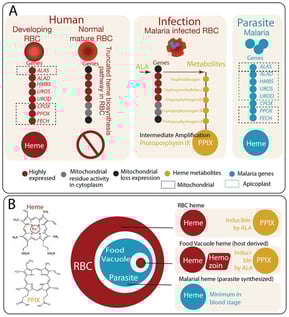
Heme metabolism is central to the biology of malaria parasites and to the mechanism of action of artemisinin-based therapies. Within malaria-infected red blood cells (RBCs), heme-related chemistry arises from multiple nested metabolic sources that function as “Russian dolls”: the truncated heme biosynthetic capacity of the host erythrocyte, the parasite’s own heme synthesis pathway, and host heme released through hemoglobin digestion in the parasite food vacuole. These overlapping metabolic layers create distinct pools of heme that can influence redox balance and drug activation. Recent studies highlight that exogenous 5-aminolevulinic acid (5-ALA) can perturb host heme biosynthesis in infected erythrocytes, potentially increasing intracellular levels of the heme intermediate protoporphyrin IX and sensitizing parasites to oxidative stress. However, the extent to which such metabolic perturbations affect artemisinin susceptibility depends strongly on parasite stage and exposure duration. Here we review the compartmentalized architecture of heme metabolism in malaria-infected RBCs and discuss how these nested vulnerabilities may be exploited for therapeutic intervention.

29 April 2026

Heme biosynthesis and compartmentalized metabolite dynamics in malaria-infected erythrocytes. (A) Differential organization of heme biosynthesis across erythroid maturation, host cell state, and parasite metabolism. (Left): Heme biosynthesis exhibits distinct configurations in developing erythroid cells, mature erythrocytes, and malaria-infected erythrocytes, reflecting differences in organelle content and metabolic capacity. In immature erythroid cells, a complete heme biosynthetic pathway operates across mitochondrial and cytosolic compartments, enabling efficient conversion of precursors to PPIX and subsequent ferrochelatase-mediated insertion of iron to form heme in a high-throughput, tightly regulated process. Upon maturation, erythrocytes expel mitochondria and other organelles, resulting in a truncated pathway with minimal capacity for de novo heme synthesis. (Middle): Following infection by Plasmodium spp., mature erythrocytes undergo extensive parasite-driven remodeling, including the formation of new permeability pathways that allow uptake of small molecules such as ALA. Under these conditions, exogenous ALA can drive accumulation of upstream intermediates, particularly PPIX, selectively in infected erythrocytes. (Right): In parallel, the parasite retains a complete, mitochondria and apicoplast associated heme biosynthetic pathway; however, its quantitative contribution to total heme production is relatively minor compared to host-derived heme from hemoglobin digestion. (B) Chemical distinction and “Russian doll” compartmentalization of heme and porphyrin intermediates. The defining biochemical difference between PPIX and heme is the insertion of a central ferrous iron (Fe2+) into the porphyrin ring. In malaria-infected erythrocytes, heme and its precursors are distributed across nested compartments that can be conceptualized as a “Russian doll” hierarchy. (Upper Right): At the outer layer, the host erythrocyte contains abundant hemoglobin-derived heme. Upon ALA exposure, the truncated host pathway in infected erythrocytes can be re-engaged to produce and accumulate PPIX. (Middle Right): Within the parasite, hemoglobin-derived heme is released in the food vacuole and detoxified through crystallization into hemozoin. This compartment may also interface with host-derived intermediates under conditions of increased precursor availability. (Lower Right): At the innermost layer, the parasite maintains its own endogenous heme biosynthetic pathway, contributing a smaller but functionally distinct pool of heme. Abbreviations: PPIX, protoporphyrin IX; ALA, 5-aminolevulinic acid; ALAS, 5-aminolevulinate synthase; ALAD, 5-aminolevulinate dehydratase; HMBS, hydroxymethylbilane synthase; UROS, uroporphyrinogen III synthase; UROD, uroporphyrinogen decarboxylase; CPOX, coproporphyrinogen oxidase; PPOX, protoporphyrinogen oxidase; FECH, ferrochelatase.

Influenza A(H3N2) Subclade K: Epidemiology, Molecular Evolution and Vaccine Effectiveness in Europe

  • Irene Scarvaglieri,
  • Maria Antonia De Francesco and
  • Eugenia Quiros-Roldan
  • + 5 authors

Background: Influenza A(H3N2) viruses remain a major public health concern due to their rapid antigenic evolution and association with severe disease, particularly among high-risk populations. During the 2025–2026 influenza season, a marked epidemiological shift was observed in Europe, with the emergence and predominance of the A(H3N2) subclade K (J.2.4.1). Objectives: This narrative review aims to provide an integrated overview of the epidemiology, evolutionary dynamics, and public health implications of subclade K, with a particular focus on its impact on vaccine effectiveness, in comparison with the 2024–2025 influenza season. Methods: A non-systematic literature review was conducted using major scientific databases and official public health sources, including WHO and ECDC reports. Recent surveillance data, genomic analyses, and epidemiological updates were included. Given the rapidly evolving evidence base, selected preprint studies were also considered and interpreted with caution. Results: The 2025–2026 influenza season in Europe was characterized by a relative genetic convergence, with subclade K accounting for the majority of A(H3N2) sequences. This variant demonstrated a clear selective advantage and was associated with an earlier and more intense epidemic peak. Molecular analyses indicate the accumulation of multiple mutations in the hemagglutinin protein, particularly within key antigenic sites, contributing to immune escape. These evolutionary changes have important implications for vaccine effectiveness, with current estimates suggesting moderate protection against infection but preserved effectiveness against severe outcomes. Antigenic mismatch, manufacturing constraints, and host-related factors further contribute to reduced vaccine performance. Conclusions: The emergence and rapid spread of subclade K highlight the dynamic nature of influenza virus evolution and its impact on public health. Continuous genomic surveillance and timely vaccine updates remain essential. Despite suboptimal effectiveness against infection, influenza vaccination continues to provide significant protection against severe disease and should remain a cornerstone of prevention strategies.

28 April 2026

Distribution of A/H3N2 subtypes and subclades in Europe during 2025–2026 influenza season according to Erviss database.

Background: The intracellular pathogen Brucella requires biotin for survival, yet the role of its de novo synthesis intermediate enzyme, BioA, in virulence remains undefined. This study investigates the contribution of BioA to the pathogenicity of Brucella abortus. Methods: We constructed a ΔBioA mutant in Brucella abortus 104M via homologous recombination and characterized its phenotype using growth assays, electron microscopy, macrophage infection models, and murine splenic colonization. Virulence gene expression was quantified by RT-qPCR. Results: The ΔBioA mutant exhibited severe growth auxotrophy in a biotin-deficient medium and displayed damaged outer membrane integrity. Furthermore, intracellular survival in macrophages was reduced by approximately 95% compared to the wild-type strain at 48 h post-infection. Notably, mice infected with the mutant showed a significant decrease in both splenic bacterial loads and spleen weight at 3 weeks, concomitant with a marked downregulation of VirB type IV secretion system (T4SS) genes. Conclusions: This study is the first to identify BioA as a critical nexus linking biotin metabolism to Brucella virulence. We demonstrate that BioA deficiency attenuates pathogenicity by impairing both structural integrity and the transcription of key virulence-related genes (VirB operon), thereby nominating BioA as a novel and promising target for anti-brucellosis interventions.

27 April 2026

A biotin-free environment can affect the expression of genes related to biotin synthesis in Brucella. (A) The operon model of the genes related to biotin synthesis of B. abortus 104M, B. suis S2 and B. melitensis 16M. (B) Detection model diagram of gene expression levels related to biotin synthesis in B. abortus 104M under biotin-deficient conditions. (C) Expression levels of biotin-synthesis-related genes in B. abortus 104M under biotin-deficient conditions. Data represent mean and standard deviation. *** p < 0.001, **** p < 0.0001 by one-way ANOVA followed by Dunnett’s multiple comparisons test.

A Neuronal Cell Line Model for Studying Camel Prions

  • Basant Abdulrahman,
  • Shabboo Rahimi Aqdam and
  • Hermann M. Schätzl
  • + 7 authors

Prion diseases are fatal neurodegenerative disorders that affect humans and animals, caused by the conformational conversion of the normal cellular prion protein (PrPC) into its misfolded, infectious isoform PrPSc. Recently, camel prion disease (CPrD) was identified in dromedary camels (Camelus dromedarius) in Algeria. Due to the potential implications for animal and human health, as well as the possible socio-economic impact in Mediterranean regions where camels play a pivotal role as a source of food, in-depth characterization of camel prions is important to increase our understanding of camel prion disease. We developed a neuronal cell line model for studying the molecular features of camel prion infection. We genetically edited mouse neuronal CAD5 cells to generate CAD5 PrP knockout (KO) cells. We then used lentiviral transduction to generate CAD5 cells expressing camel PrP (CAD5-camel-PrP). Following infection of these cells with a CPrD-positive camel brain homogenate, we observed PrPSc signals at various passages, as indicated by immunoblotting analysis. RT-QuIC (Real-Time Quaking-Induced Conversion) assays further supported these findings, demonstrating transient prion conversion activity in the CPrD-infected CAD5-camel-PrP cells. Taken together, our data describe the first neuronal cell line permissive to camel prion infection, a novel in vitro tool for mechanistic studies of camel prion disease.

27 April 2026

Fluorescence imaging and FACS analysis of CAD5-Camel-PrP transduced cells upon lentiviral transduction. (A) Fluorescence imaging of CAD5-Camel-PrP Cells. CAD5-PrP KO cells were transduced with 1 µL (top panel), 3 µL (middle panel), and 5 µL (bottom panel) of lentiviral particles encoding camel-PrP. The left panels show bright-field images, while the right panels display the corresponding fluorescence images. Fluorescence intensity correlates with the volume of lentiviral particles used, reflecting the level of PrP expression. The 1 µL condition shows minimal and sparse GFP fluorescence, indicating low transduction efficiency. The 3 µL condition yields increased GFP expression with improved distribution across the cell population. The 5 µL condition demonstrated stronger fluorescence expressed as a more uniform GFP signal. (B) CAD5 cells expressing camel-PrP upon lentiviral transduction. The top panels show scatter plots for all events, P1 gating, and SSC (side scatter) versus FSC (forward scatter), and histograms of GFP fluorescence intensity, with the P2 gate indicating cells with high GFP expression (middle panel). The tables summarize the number of events and GFP statistics for each population.

News & Conferences

Volumes

Latest Issues

Open for Submission

Editor's Choice

Reprints of Collections

Chagas Disease
Reprint

Chagas Disease

Celebrating the 115th Anniversary of the Discovery of <em>Trypanosoma cruzi</em>
Editors: Michel Tibayrenc
Parasitic Diseases of Fish
Reprint

Parasitic Diseases of Fish

Identification, Host-Parasite Interactions and Molecular Biology
Editors: Gokhlesh Kumar, Arun Sudhagar, Kandasamy Saravanan
XFacebookLinkedIn
Pathogens - ISSN 2076-0817