


Abstract
Viruses are an indispensable part of the environment we live in. The occurrence of seasonal and pandemic infections underscores the urgent need to develop new antiviral drugs or repurpose existing ones. Among the methods used in research on new antiviral molecules, surface plasmon resonance (SPR) has a well-established position due to its diverse applications in interaction analysis. It can be used to investigate various molecules (proteins, nucleic acids, small-molecular drugs) in different configurations and in real time. Although it is a gold-standard method for biomolecular interaction analysis, it is not free of constraints. Here, we review research on SPR in antiviral drug discovery. We focus on experimental design and discuss the application of SPR to investigate key stages of viral infection and to characterize antiviral interactions. In addition, we address the main limitations and challenges associated with SPR measurements and consider strategies for adapting the technique to meet the specific needs of antiviral research.
1. Introduction
Antiviral drug discovery is based on research, including both computational and empirical screening [1,2,3]. The first one is used to design molecules that can inhibit virus entry into cells, viral genetic material replication, or the processing of viral polyproteins. It is also used for the first selection of the most promising molecules. Although this strategy is quite fast and cost-effective, its results require experimental validation to ensure that effects predicted in silico are observed in vitro and in vivo. A wide variety of methods can be used to analyze the interaction between the putative drug and its molecular target, validate the putative drug’s safety in host cells, and, finally, assess efficacy in virus neutralization in vivo in animal models [1,2,3].
The designed small molecules or peptides predicted to prevent viral infection or inhibit viral propagation are first analyzed for their recognition and interactions with molecular targets. There are different strategies for identifying the most promising drugs: high-throughput screening (HTS), fragment-based drug discovery (FBDD), hit validation, or drug repurposing [4,5,6]. They rely on well-established methods for ligand–analyte interactions such as surface plasmon resonance (SPR) [7,8,9], Isothermal Titration Calorimetry (ITC) [10,11], MicroScale Thermophoresis (MST) [12,13], or Biolayer Interferometry (BLI) [11,14]. These methods are used for analysis of putative drugs against different viruses, e.g., severe acute respiratory syndrome coronavirus 2 (SARS-CoV-2) [9,12,13,15,16,17], human immunodeficiency virus 1 (HIV-1) [10,15], Hepatitis C virus (HCV) [12], herpes simplex virus type-1 (HSV-1) [18] and Influenza A virus [12,19,20].
Among the methods used to analyze biomolecular interactions, SPR is a gold-standard analytical tool. However, the specificity of interaction analysis using this technology makes SPR effective to varying degrees, depending on the approach chosen to identify the most active antiviral molecules. One of the biggest advantages of SPR over techniques such as MST or ITC is the determination of kinetic constants for putative drug interactions with viral or cellular targets [21,22]. SPR is an especially powerful technique when FBS is considered, because it allows detection of even very weak interactions, which constitutes the basis for this drug-discovery strategy. At the same time, it allows detection of binding of very small ligands (100–300 Da) [22]. These are undoubtedly advantages compared to a similar BLI technique, although SPR is not as high-throughput. The requirement of a small volume of label-free sample also often determines the choice of SPR over ITC (which usually requires higher concentrations of the molecules) [23] or MST (which requires fluorescent labeling of the ligand) [12,13]. On the other hand, both ITC and MST are solution-based methods that do not require ligand immobilization [11], which could be important when analyzing fragile or complexed viral proteins.
In this review, we will present the bases of SPR analysis with a focus on experimental design in antiviral drug discovery research and the utility of SPR in research targeting key steps of viral infection. We will highlight the limitations and challenges of SPR analysis and the potential for adapting the assay for antiviral research.
3. Targeting Steps of Viral Infection—Constraints and Adaptations in SPR Analysis
The process of viral infection can be divided into a few steps common to different viruses, despite their distinct structures, genetic material, modes of transmission, etc. [43]. First, the virus has to attach to and enter specific host cells; then, the viral genetic material should be released from the capsid, transported to the nucleus and replicated. To form new viral particles, viral genes must be expressed, leading to the synthesis and processing of viral proteins. When all viral components are present, they assemble into new virions and are released from the host cell, allowing the cycle to repeat. Antiviral strategies are based on inhibiting the most important steps of this cycle, e.g., adhesion and entry into cells, genome replication, and viral-polyproteins processing [44,45]. During the investigation of these steps, SPR can be successfully utilized if only the key constraints of this technique are considered.
3.1. Looking for Viral Entry Inhibitors
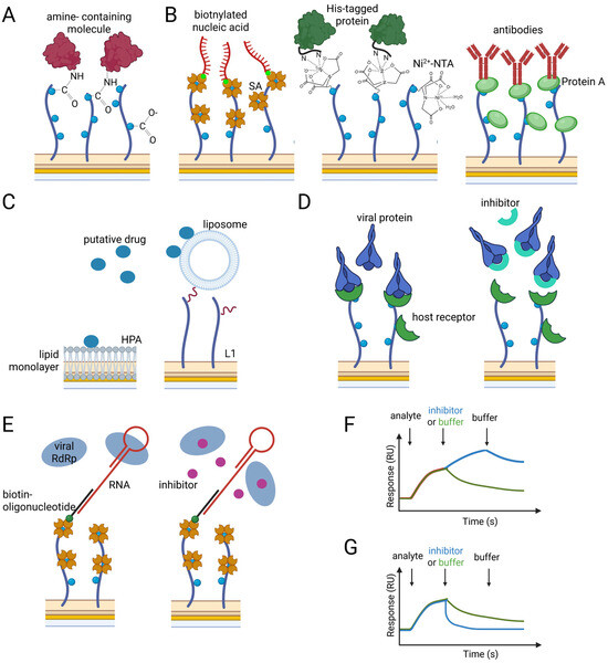
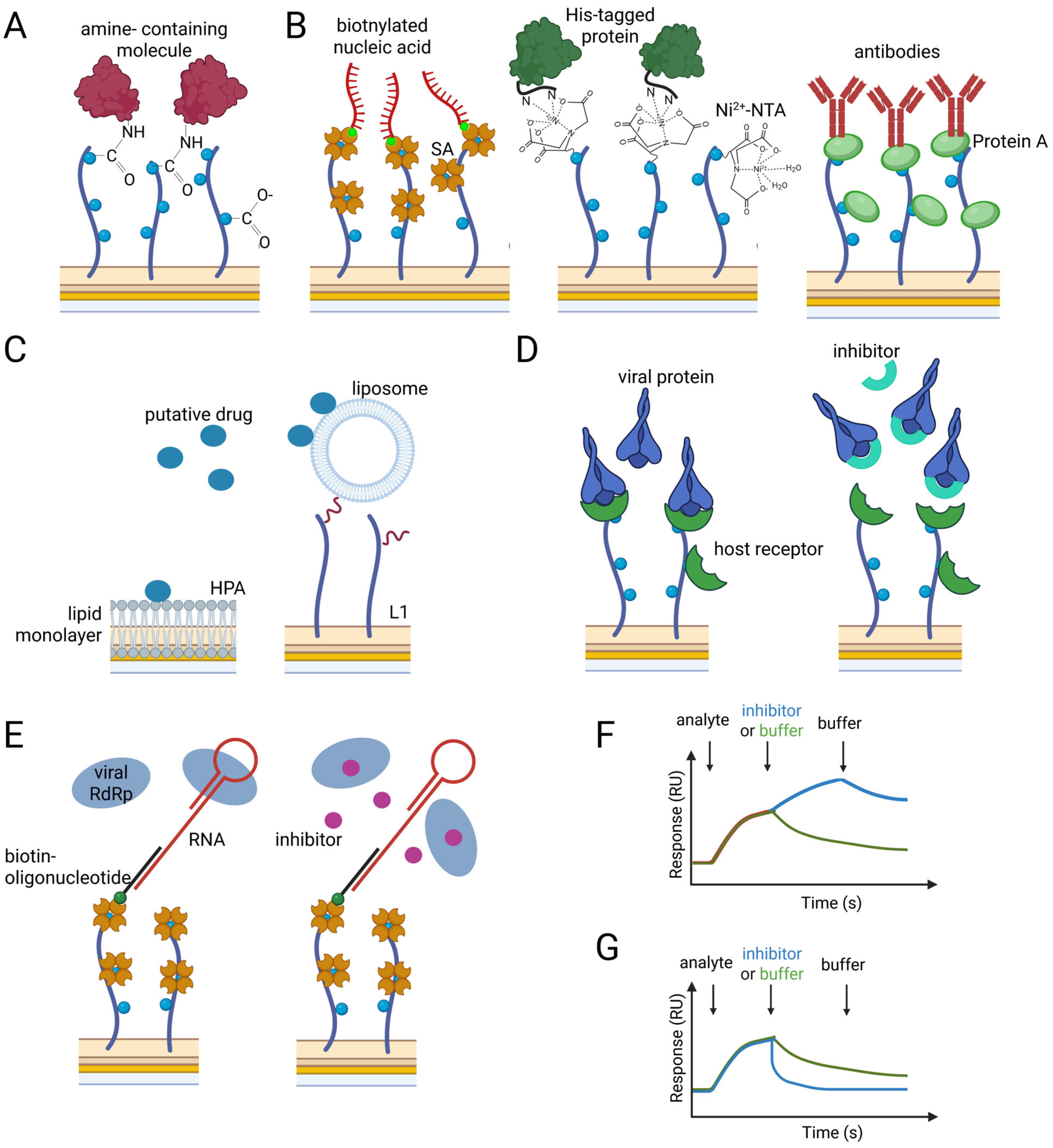
The best way to stop an infection is to act at the very first step, before the virus can spread through the body’s cells. To do this, molecules that inhibit the interaction between the viral envelope protein and its receptor on the host cell surface are designed. The molecule can bind either the viral protein or the host receptor in the interaction interface. Using SPR, these interactions are measured, as well as the decrease in complex formation when, prior to injection, the analyte is preincubated with the putative drug.
The typical targets when looking for viral entry inhibitors are quite large, complex (oligomeric) molecules, either when they are viral surface proteins (e.g., the SARS-CoV-2 virus’s spike protein (the S-glycoprotein) subunit S1; gp140 and gp120 of HIV-1) or host receptor (e.g., ACE2, CD4). The native proteins involved in viral entry are also membrane-associated; therefore, SPR analysis of full-length partners with proper quaternary structure is very difficult. Therefore, the research model is often simplified, and selected protein domains or monomeric proteins are analyzed. An example is the SARS-CoV-2 spike protein, where the S1 subunit is a trimer [46], but in SPR analysis, usually just its receptor-binding domain (RBD) is analyzed for interaction with ACE2 or neutralizing molecules [47,48,49,50]. Although this approach simplifies the analysis, it can yield apparent affinity and kinetic constants from the data because it does not account for multiple simultaneous interactions that occur in nature. Research comparing the interaction between the CD4 (presents on the surface of immune cells and constituates the primary receptor of HIV-1) and HIV-1 viral envelope proteins gp140 and gp120 purified as a stable trimer and a monomer, respectively, and investigating the neutralizing effect of selected antibodies on this interaction indicated significant differences resulting from the oligomeric state of the viral proteins [51]. The obtained results support the concept that gp140 trimers are better immunogens than gp120 monomers.
To support the determination of the most intrinsic interaction parameters, the availability of binding interfaces in both tested partners is crucial. Although in antiviral research the immobilization via amine group with the amine coupling method is also often applied (e.g., RBD-ACE2 interaction [47,49,52], CD4-gp120 [53]) to ensure the highest accessibility of binding site and to achieve a uniform orientation of the ligand, the immobilization via tag molecule is recommended (e.g., biotinylated ACE2, ACE2-Fc, RBD-Fc, His6-RBD [28,45,48,54]). Once the data are collected, the appropriate analysis is required. The default analysis usually applies the 1:1 Langmuir binding model. When considering the structural conditions mentioned above for the tested molecules, this model can provide an apparent description of the tested interaction. It fits well with simplified research systems, e.g., the use of RBD instead of the S1 protein [39]. However, as it was shown for S1-S2 stable spike protein interaction with ACE2, the application of a 1:1 model is burdened with error due to the failure to take into account minor conformational changes in the spike protein after initial receptor binding [39]. These limitations in applying the simplest model were overcome by using a two-state model, which resulted in a better fit, especially during the dissociation phase.
The application of a more complex model should also be considered in cases of investigation of antibody binding to neutralize the viral entry protein–host receptor interaction. Usually, the bivalent analyte model is applied when the antibodies are flowed over the immobilized target molecule. The 1:1 model is sufficient when antibodies are captured on the sensor chip surface, or monovalent nanobodies or Fab fragments are used as analytes. These kind of analyses were performed for investigation: nanobodies (e.g., H11 [48]) and antibodies (e.g., humanized mAbs MG1141A [30]) that should bind the S1 SARS-CoV-2 spike protein, preventing its ACE2 recognition; for investigation of binding antibodies, captured via immobilized human Fab binder or protein A, and HIV-1 viral proteins gp140 and gp120 as the analyte [52], or testing interaction between immobilized glycoprotein B (gB) of Epstein–Barr virus (EBV) and Fabs of neutralizing antibodies as analytes [55].
Unfortunately, correctly determining the kinetics of interaction at the stage of viral entry is not limited to selecting the appropriate immobilization method and binding model. Because the studied molecules are large, mass transfer and artifacts resulting from analyte rebinding can be significant limitations [34,56]. To increase the accessibility of the surface for the analyte, the low ligand immobilization level is applied (depending on the molecular weight of the ligand, usually 100–500 RU) and to remove the analyte from the surface immediately after dissociation, a high flow rate is set (up to 100 μL/min) [57,58,59].
These limitations do not occur when small antiviral molecules interact with their targets, whether viral envelope proteins or host receptors. The examples could be the interaction between immobilized HA and a small molecule; inhibiting membrane fusion of influenza virus and a host cell [60]; DCM205, compound-inhibiting interaction between the CD4 receptor and gp120 HIV-1 protein [54]; ficolin-1 binding GP glycoprotein of Ebola virus (EBOV) [61]; or mosquitoes salivary proteins AAEL000793, AAEL007420 and AAEL006347 binding the envelope protein of the Zika virus (ZIKV) [62] (Table 1). Small molecules binding to the ACE2 receptor were also analyzed. An example could be the analysis of interaction with ACE2, the derivatives of sartans (e.g., Losartan, Telmisartan, Olmesartan), which are known as Angiotensin II Receptor Blockers (ARBs) used for the treatment of hypertension and heart diseases [63]. These bis-alkylated imidazole derivatives bearing two anionic biphenyltetrazoles (bisartans) were designed to bind the ACE2 at its RBD interface [64,65]. In SPR analysis, it was shown that bisartan BV6 (bisalkylated) binds well to immobilized ACE2 protein and more strongly compared to losartan [52,65,66].
Table 1.
Examples of SPR analysis in research concerning antiviral molecules.
Although the disruption of direct contact between the viral protein and its host receptor is very important, and research is usually focused on this process, it is also worth noting that the inhibitory molecule can interact more or less specifically with the membrane in which the receptor is embedded. Studies using SPR by Cao and coworkers showed that peptide fusion inhibitors of HIV-1, sifuviride and enfuviride, might differ in efficacy due to higher local concentrations at the fusion site resulting from different interactions with rigid lipid areas [70].
During the viral entry into the host cell, the receptor is not always a protein. In many cases, the viral envelope protein (e.g., the RBD of the S1 spike protein [76], the envelope protein of Dengue virus [75], and the hemagglutinin (HA) of the influenza virus [77,78]) recognizes glycoconjugates present on the cell’s surface. Research conducted to identify this mechanism of viral entry by SPR usually utilizes the immobilization of tagged glycans (e.g., biotinylated glycans [78] or modified sialoglycopolymers [7] immobilized on a streptavidin-coated sensor chip immobilized surface). The interaction between glycoconjugates and viral protein can be eliminated by molecules structurally similar to the molecule of attachment. Usually it is heparine, but because it can cause some side effects such as bleeding [79,80], safer alternatives are being sought (e.g., the polyanionic compound Suramin behaving similarly to heparin, with a fast on-rate and an extremely slow off-rate during interaction with the envelope protein of the Dengue virus [75]; heparin mimetics interacting with RBD [67]). During analysis, a competition-binding assay is performed in which heparin can be immobilized on the sensor chip surface, and the viral protein, alone or in the presence of heparin mimetics, is flowed over it. Mimetics with affinity for viral proteins comparable to that of heparin are the most promising in preventing virus–host interaction [67]. Similarly, polyclonal antisera, monoclonal antibodies, or intravenous immunoglobulins can be analyzed [7], with respect to the constraints mentioned above.
3.2. SPR Analysis for Inhibition of Viral Enzymes
Although preventing virus entry into cells is the obvious target of antiviral drugs, research on new drug development also targets subsequent stages, such as expression of viral genes, proteolytic processing of viral polyprotein and replication of genetic material. The first two steps are required to obtain viral proteins, which can then act during viral genome replication, nuclear export, viral particle assembly, and their release. In SPR analysis, if the inhibition of polymerase activity or proteolytic processing of the polyprotein is investigated, the enzyme is usually immobilized on the sensor chip and viral enzyme inhibitors are flowed over the prepared surface (Table 1). To maintain enzyme activity, immobilization via a capture molecule (tag or antibody) is the most recommended method [57,58,59,60], although amine coupling is also often applied [69,72,81,82,83,84]. This kind of analysis was performed, for example, for the main protease of SARS-CoV-2 (3CLpro/Mpro), which is critical for viral genome replication and thus an excellent target for drugs, especially since no homolog of Mpro has been identified in humans [85]. Analysis indicated efficient binding to and inhibitory effects on Mpro by natural products, including chebulagic acid (CHLA) isolated from Terminalia chebula [17], hydrolyzable tannins (ellagitannins and gallotannins) [83], and cannabinoids [84]. SPR also identified efficient viral protease inhibitors when drug repurposing was considered. The already approved and investigational drugs were screened against ZIKV, and from them, temoporfin was shown to bind the ZIKV protease NS3 domain the strongest [74]. Also, tests of compounds with inhibitory activity against the NS3/NS4A protease of HCV identified those that bind the NS2B-NS3 protease of ZIKV as well [81]. This strategy with enzyme immobilization was also presented in the study of Lo and coworkers, which allowed for identification of compounds from a library of over two hundred, targeting the C-terminal domain of PA subunit (PAC) of RNA-dependent RNA polymerase (RdRp) of influenza virus [68], or in other studies for inhibitors of NS5B polymerase of HCV [71,86].
The strategy of immobilizing a viral protein and injecting a putative inhibitor was also applied to other viral proteins required for genome replication. An example is VP30, the protein critical for EBOV RNA synthesis [87,88]. Small molecules, embelin and Kobe2602, selected from a library of 8004 compounds, were tested for their inhibitory activity to VP30 with SPR in different configurations. They were either immobilized on the sensor chip surface and tested for VP30 binding at the interface, interacting with other proteins, or flowed over the surface with immobilized or captured VP30 [73]. Their binding and inhibitory effects were confirmed. Another example is the analysis of A9, a ligustrazine derivative, and its interaction with an immobilized His-tagged NP protein from the influenza virus [89]. This interaction was shown to inhibit the nuclear export of viral ribonucleoprotein (vRNP).
The level of enzyme immobilization has to be sufficient to detect the signal from the binding of inhibitors, which are usually very small molecules (200–800Da). Due to the size of the analyte and, consequently, low detected response, this research system is characterized by high sensitivity to bulk refractive index changes [90]. Therefore, the buffer has to be properly selected, and if an organic solvent such as DMSO is used, a solvent correction is mandatory [91]. Reversing the research system and immobilizing small molecules could reduce the limitations imposed by the small molecular weight of inhibitors, and keeping the enzyme in solution could increase its activity; however, the use of small analytes allows the application of the 1:1 binding model for analysis. Analytes with complex structures, such as oligomeric, multi-domain proteins (e.g., proteases and polymerases), may make it difficult to select the appropriate binding model. On the other hand, the binding of small molecules can occur too rapidly to allow reliable determination of kinetic constants. In this case, only the affinity at equilibrium can be determined [82].
Since viral polymerase activity depends on binding to nucleic acid, which can be easily immobilized if biotinylated, allowing the enzyme to remain in solution, this configuration can be successfully used for screening polymerase inhibitors. It was proposed by Mravinec and coauthors, who immobilized an RNA molecule composed of annealed oligonucleotides, one biotinylated and the other complementary, which interacted specifically with RdRp [31]. As an analyte, RdRp alone or after incubation with antiviral drug (ribavirin, favipiravir, sofosbuvir and suramin) was used. The comparison of RdRp binding to RNA before and after incubation with drugs indicates that suramin precluded interaction and even displaced RdRp from RNA when it was flowed over the surface with preformed RNA-RdRp complexes [31].
3.3. Assembly and Budding Inhibitors
The latest steps of viral infection, which can be targeted by antiviral drugs, are the assembly of viral particles and their release from cells. Since these processes involve multicomponent and multivalent interactions and are linked to lipid membrane interactions [84,85,86], SPR is not the most popular technique for assessing them. Usually, the binding of monomeric proteins instead of oligomers is analyzed. However, as shown for the HIV-1 capsid protein (CA), the oligomeric state of the target during SPR analysis may not only influence the determined kinetic parameters but also the occurrence of binding [92]. During the analysis, the CA protein was immobilized as a monomer, a hexamer, or just as its N-terminal domain (CA-NTA), and binding of the selected inhibitors (H22 and PF-74) was observed only with the hexamer. As for other small-molecule inhibitors, also in this case, the proper calculation of kinetic constants was difficult, and the obtained data were apparent compared to those obtained with other solution-based methods like ITC [92]. Therefore, only the equilibrium dissociation constant was calculated. Similarly, kinetic constants were not determined for CA inhibitors identified in other studies [93].
The use of antibodies as neutralizing molecules may overcome the constraints imposed by characteristic small-molecule binding, but in this case, mass transfer should be avoided. Neutralizing antibodies that inhibit this stage of viral infection were investigated, for example, for EBOV [94] and influenza virus [20,59,95]. In studies on EBOV, the viral protein VP40 was immobilized via amine coupling and tested for recognition by anti-VP40 antibodies. In the same assay, the selected antibodies were tested for recognition of other viral proteins to indicate possible off-targets and those that bound specifically to VP40 were identified [94]. In research on the influenza virus, the second major protein, neuraminidase (NA), which facilitates the release of viral particles from infected cells by cleaving sialic acid [96], was captured via a polyclonal anti-Avi antibody and the Avi-tag fused with NA [95] or via a modified known NA inhibitor (HAD-zanamivir) [20,59]. These strategies allow analyzing, on the surface of a single sensor chip, the interaction between different NA variants, e.g., those with diverse glycosylation [95] or wild-type and point substitution variants [20,59]. Research on capturing different NA variants on the sensor chip surface shows that this strategy could be used to rapidly screen newly emerging NA variants for their sensitivity to antiviral drugs. Even if the binding parameters are apparent, their comparison across different target variants allows the selection of the most prominent molecules for investigation with other solid- and cell-based techniques.
4. Limitations, Challenges and Future Perspectives
Although the SPR technique is well established in antiviral drug discovery, it is not without limitations (Table 2) and it should be used in combination with other methods, allowing for resolution of the complex structure and in vivo effects.
Table 2.
Constraints and SPR assay adaptations depending on the type of analyzed molecules.
SPR is considered a label-free method because detection does not require labeling, and immobilization on the sensor chip surface can be achieved via groups present in the molecules, e.g., the amine group of proteins. However, this type of immobilization can result in a different particle arrangement on the surface, which can influence interaction. Immobilization via a capture molecule bypasses these limitations but requires either tagging the ligand (e.g., biotin, His-tag) or the availability of capturing antibodies. Nevertheless, even if the molecules are deposited equally on the surface, it is necessary to ensure that the immobilization does not mask the interaction interface and that the interface remains accessible to the analyte. As shown, the oligomeric state may also influence binding, and purification of stable oligomers is required to achieve efficient complex formation [92,97]. It is also worth noting that many viral proteins involved in virus entry, as well as those connected with virus release, are embedded in membranes, and recreating native conditions (e.g., proper protein conformation) during SPR analysis can be complex and challenging, even when a lipid bilayer is used to capture the protein. When analyzing proteins with complex glycosylation, it is even more challenging to produce them homogeneously and thus achieve efficient, reproducible results. The system used for protein production can influence posttranslational modifications and, consequently, binding kinetics, as observed for the SARS-CoV-2 S1 protein produced in different cells: baculovirus-insect, Chinese hamster ovarian, and human embryonic kidney [98]. The glycosylation of viral proteins can also influence the background, since glycan groups can interact nonspecifically with the reference surface [99].
In SPR analysis, the standard 1:1 binding model is typically used to describe interaction kinetics, assuming a simple, single-step binding. Application of the simplest equations is usually sufficient for small monovalent analytes and single-domain proteins. This model also usually works when enzyme (e.g., polymerase or protease) active-site inhibitors are tested. However, viral–host interactions often involve multiple binding sites, are cooperative, or involve conformational changes, making simple models insufficient. Sevenich and coauthors showed, using the example of the SARS-CoV-2 spike protein and human ACE2 interaction, that choosing a binding model other than the 1:1 results in better fitting of experimental data [39]. Applying the model assuming a conformational change after the initial binding (the two-state model) better correlates with the data, especially when complex dissociation was measured over a longer time. The observed slower than expected dissociation usually indicates that multiple bonds have to break simultaneously, and the parameters calculated with a 1:1 binding model are apparent [34]. Thus, proper experimental design and data analysis to avoid artifacts are critical. If it is known that bivalent analytes or heterogeneous ligands are tested, then the dedicated model should be used. The solution could also involve inverting the orientation and use of a multivalent molecule as a ligand rather than an analyte. The ligand could also be immobilized at different densities, and the slower off-rate at the higher density might indicate multivalent rebinding [100]. Nevertheless, obtaining data from other methods (e.g., ITC, MST, cryo-electron microscopy, X-ray crystallography) should always be considered to get the most relevant information about the potential multivalency, activity and mechanism of action of the putative drug. On the other hand, even when complex formation is best characterized in vitro, it will not always reflect the actual cellular effect. Usually, data obtained from techniques such as SPR correlate well with antiviral efficacy for inhibitors that directly block the catalytic center of the well-characterized target. Also, the molecule’s long residence time is a good predictor of its inhibitory activity. However, very often in antiviral research, in solid-based techniques, isolated proteins’ domains or monomers are used, which do not reflect the conformational state observed in nature (the multiprotein complexes or oligomers, embedded in membranes). Above all, it shouldn’t be forgotten that SPR does not determine putative drug uptake, efflux, metabolic stability, cytotoxicity, or, finally, antiviral activity; thus, functional assays must be conducted because target engagement does not always reflect antiviral efficacy. In the work of Dierynck and coworkers, variants of HIV protease, derived from clinical isolates and harboring protease inhibitor resistance-associated mutations, were immobilized and analyzed for interaction with various protease inhibitors [69]. It was shown that many of them were bound to proteases containing substitutions in crucial residues, resulting in lower affinity and, consequently, lower antiviral activity. Darunavir was an exception, and a nearly 1000-fold decrease in binding affinity did not change its activity against HIV protease variants [69].
To improve the identification of putative antiviral molecules and their better characterization, machine learning (ML) can be applied [2,101,102,103]. It can be considered for anything from experimental design, through parameter setting analysis, to data processing [2,103,104,105,106]. The application of artificial intelligence, from the very first step of designing molecules to in silico validation, can significantly increase the success rate of experimental results. A study by Liu and coauthors used an integrated computational workflow (including large-scale virtual screening of over 10 million compounds, molecular mechanics/generalized born surface area (MM-GBSA) calculations, and molecular dynamics (MD) simulations) to identify novel influenza A nucleoprotein inhibitors, leading to the experimental validation of 16 candidates, of which 3 compounds showed the strongest target engagement in SPR assays [107]. Also, in research on other viruses, the initial in silico selection of antiviral molecules for further in vitro (with SPR [52,66,108,109,110]) and in vivo experiments was conducted [2,111,112,113,114], which limits the number of analyzed molecules. ML was also successfully applied to predict effective antiviral combinations of known antifvirals, confirmed by experiments (i.a., SPR) demonstrating increased binding affinity and viral suppression [115].
Regarding SPR analysis, ML is likewise particularly useful during the data processing step because it enables faster sensorgram classification based on binding models, aggregation, mass-transport limitations, or bulk artifacts [116]. It also provides better kinetic parameter calculations when the standard binding model is not accurate. Its potential can also be used to predict off-target interactions, which are missed entirely when binary interactions (viral protein–antiviral molecule) are measured by SPR [117]. The only limitation to applying ML to SPR data processing is the availability of a large training dataset. ML can also be utilized to design improved sensors that will provide higher sensitivity, selectivity, stability, and lower detection limits. The ML-designed improved sensors are now being tested, in which a classical gold or silver layer is replaced or modified with novel plasmonic materials (e.g., graphene) [24,118,119,120]. Therefore, leveraging the potential of artificial intelligence can accelerate research and help design drugs that target specific targets.
Author Contributions
K.E.W. wrote the manuscript with the help of J.M.M. All authors have read and agreed to the published version of the manuscript.
Funding
This research received no external funding.
Institutional Review Board Statement
Not applicable.
Informed Consent Statement
Not applicable.
Data Availability Statement
No new data were created or analyzed in this study.
Acknowledgments
J.M.M. acknowledges fruitful discussions with members of the “Drug Discovery and Development Consortium” established to promote research on drug discovery. The figure was created with BioRender.com. During the preparation of this manuscript, the authors used Grammarly for Microsoft Office version 6.8.263 for language proofreading. The authors have reviewed and edited the output and take full responsibility for the content of this publication.
Conflicts of Interest
The authors declare no conflicts of interest.
Abbreviations
The following abbreviations are used in this manuscript:
| SPR | Surface Plasmon Resonance |
| ITC | Isothermal Titration Calorimetry |
| MST | MicroScale Thermophoresis |
| BLI | Biolayer Interferometry |
| SARS-CoV-2 | The severe acute respiratory syndrome coronavirus 2 |
| HIV-1 | The human immunodeficiency virus 1 |
| HCV | Hepatitis C virus (HCV) |
| HSV-1 | herpes simplex virus type-1 |
| EBOV | Ebola virus |
| ZIKV | Zika virus |
| NHS | N-hydroxysuccinimide |
| EDC | 1-ethyl-3-(3-dimethylaminopropyl)carbodiimide |
| ACE2 | The receptor angiotensin-converting enzyme 2 |
| RDB | The receptor-binding domain (RBD) |
| HPA | hydrophobic association |
| DMSO | dimethyl sulfoxide |
| ML | machine learning |
| MD | molecular dynamics |
References
- Gan, Y.; Guo, F.; Roy, A.; Wang, X.; Shen, Y. Integrated In Silico, In Vitro, and In Vivo Studies Reveal Mangiferin as a Promising Antiviral Agent Against H1N1/pdm2009 Influenza Virus. Viruses 2025, 17, 873. [Google Scholar] [CrossRef]
- Izmailyan, R.; Matevosyan, M.; Khachatryan, H.; Shavina, A.; Gevorgyan, S.; Ghazaryan, A.; Tirosyan, I.; Gabrielyan, Y.; Ayvazyan, M.; Martirosyan, B.; et al. Discovery of new antiviral agents through artificial intelligence: In vitro and in vivo results. Antivir. Res. 2024, 222, 105818. [Google Scholar] [CrossRef] [PubMed]
- von Delft, A.; Hall, M.D.; Kwong, A.D.; Purcell, L.A.; Saikatendu, K.S.; Schmitz, U.; Tallarico, J.A.; Lee, A.A. Accelerating antiviral drug discovery: Lessons from COVID-19. Nat. Rev. Drug Discov. 2023, 22, 585–603. [Google Scholar] [CrossRef] [PubMed]
- Huber, W.; Mueller, F. Biomolecular interaction analysis in drug discovery using surface plasmon resonance technology. Curr. Pharm. Des. 2006, 12, 3999–4021. [Google Scholar] [CrossRef] [PubMed]
- Myszka, D.G.; Rich, R.L. Implementing surface plasmon resonance biosensors in drug discovery. Pharm. Sci. Technol. Today 2000, 3, 310–317. [Google Scholar] [CrossRef]
- Navratilova, I.; Hopkins, A.L. Emerging role of surface plasmon resonance in fragment-based drug discovery. Future Med. Chem. 2011, 3, 1809–1820. [Google Scholar] [CrossRef]
- Khalenkov, A.M.; Norton, M.G.; Scott, D.E. Method for screening influenza neutralizing antibodies in crude human plasma and its derivatives using SPR. Heliyon 2023, 9, e15651. [Google Scholar] [CrossRef]
- Wan, H.; Cui, H.; Liu, H.; Zhang, G.; He, L.; Liu, H.; Mahmud, S. Surface plasmon resonance biosensor chips: Fabrications and pharmaceutical applications. J. Pharm. Biomed. Anal. 2025, 265, 117018. [Google Scholar] [CrossRef]
- Wang, Q.; Liu, Z. Recent progress of surface plasmon resonance in the development of coronavirus disease-2019 drug candidates. Eur. J. Med. Chem. Rep. 2021, 1, 100003. [Google Scholar] [CrossRef]
- Ohtaka, H.; Velazquez-Campoy, A.; Xie, D.; Freire, E. Overcoming drug resistance in HIV-1 chemotherapy: The binding thermodynamics of Amprenavir and TMC-126 to wild-type and drug-resistant mutants of the HIV-1 protease. Protein Sci. 2002, 11, 1908–1916. [Google Scholar] [CrossRef]
- Zhou, Y.; Hu, H.; Huang, Q.; Tang, Y.; Liu, Y. Drug discovery through biophysical techniques: Methods and applications. Pharmacol. Ther. 2026, 277, 108947. [Google Scholar] [CrossRef] [PubMed]
- Magnez, R.; Bailly, C.; Thuru, X. Microscale Thermophoresis as a Tool to Study Protein Interactions and Their Implication in Human Diseases. Int. J. Mol. Sci. 2022, 23, 7672. [Google Scholar] [CrossRef] [PubMed]
- Nydegger, D.T.; Pujol-Gimenez, J.; Kandasamy, P.; Vogt, B.; Hediger, M.A. Applications of the Microscale Thermophoresis Binding Assay in COVID-19 Research. Viruses 2023, 15, 1432. [Google Scholar] [CrossRef] [PubMed]
- Haniff, H.S.; Knerr, L.; Chen, J.L.; Disney, M.D.; Lightfoot, H.L. Target-Directed Approaches for Screening Small Molecules against RNA Targets. SLAS Discov. 2020, 25, 869–894. [Google Scholar] [CrossRef]
- Lorson, Z.C.; McFadden, W.M.; Neilsen, G.; Castaner, A.E.; Slack, R.L.; Kirby, K.A.; Sarafianos, S.G. Characterization of antiviral compounds using Bio-Layer Interferometry. bioRxiv 2025. [Google Scholar] [CrossRef]
- Shahhamzehei, N.; Abdelfatah, S.; Efferth, T. In Silico and In Vitro Identification of Pan-Coronaviral Main Protease Inhibitors from a Large Natural Product Library. Pharmaceuticals 2022, 15, 308. [Google Scholar] [CrossRef]
- Zhang, M.; Wang, C.; Feng, L.; Yang, Q.; Cao, Y.; Zhao, Y.; Zhang, J.; Wang, Y.; Rao, Z.; Zhang, B. Targeting SARS-CoV-2 main protease for the discovery of a broad-spectrum COVID-19 inhibitor by intensive multi-tiered validation. Acta Pharm. Sin. B 2025, 15, 5789–5802. [Google Scholar] [CrossRef]
- Elste, J.; Rabbitt, M.; Wang, C.; Dordick, J.S.; Tiwari, V.; Zhang, F. Marine-derived sulfated glycans display a potent virostatic mechanism to block herpes simplex virus type-1 (HSV-1) entry and spread. Carbohydr. Polym. 2025, 368, 124141. [Google Scholar] [CrossRef]
- Jug, A.; Bratkovič, T.; Ilaš, J. Biolayer interferometry and its applications in drug discovery and development. TrAC Trends Anal. Chem. 2024, 176, 117741. [Google Scholar] [CrossRef]
- Somasundaram, B.; Fee, C.J.; Fredericks, R.; Watson, A.J.; Fairbanks, A.J.; Hall, R.J. A surface plasmon resonance assay for measurement of neuraminidase inhibition, sensitivity of wild-type influenza neuraminidase and its H274Y mutant to the antiviral drugs zanamivir and oseltamivir. J. Mol. Recognit. 2015, 28, 521–527. [Google Scholar] [CrossRef]
- Giannetti, A.M. Chapter Eight-From Experimental Design to Validated Hits: A Comprehensive Walk-Through of Fragment Lead Identification Using Surface Plasmon Resonance. In Methods in Enzymology; Kuo, L.C., Ed.; Academic Press: Cambridge, MA, USA, 2011; Volume 493, pp. 169–218. [Google Scholar]
- Neumann, T.; Junker, H.D.; Schmidt, K.; Sekul, R. SPR-based Fragment Screening: Advantages and Applications. Curr. Top. Med. Chem. 2007, 7, 1630–1642. [Google Scholar] [CrossRef]
- Freyer, M.W.; Lewis, E.A. Isothermal Titration Calorimetry: Experimental Design, Data Analysis, and Probing Macromolecule/Ligand Binding and Kinetic Interactions. In Methods in Cell Biology; Academic Press: Cambridge, MA, USA, 2008; Volume 84, pp. 79–113. [Google Scholar]
- Butt, M.A. Surface Plasmon Resonance-Based Biodetection Systems: Principles, Progress and Applications—A Comprehensive Review. Biosensors 2025, 15, 35. [Google Scholar] [CrossRef] [PubMed]
- Su, Y.W.; Wang, W. Surface plasmon resonance sensing: From purified biomolecules to intact cells. Anal. Bioanal. Chem. 2018, 410, 3943–3951. [Google Scholar] [CrossRef] [PubMed]
- Willander, M.; Al-Hilli, S. Analysis of biomolecules using surface plasmons. Methods Mol. Biol. 2009, 544, 201–229. [Google Scholar] [CrossRef] [PubMed]
- Piliarik, M.; Vaisocherova, H.; Homola, J. Surface plasmon resonance biosensing. Methods Mol. Biol. 2009, 503, 65–88. [Google Scholar] [CrossRef]
- Fischer, M.J. Amine coupling through EDC/NHS: A practical approach. Methods Mol. Biol. 2010, 627, 55–73. [Google Scholar] [CrossRef]
- Grandea, A.G., 3rd; Olsen, O.A.; Cox, T.C.; Renshaw, M.; Hammond, P.W.; Chan-Hui, P.Y.; Mitcham, J.L.; Cieplak, W.; Stewart, S.M.; Grantham, M.L.; et al. Human antibodies reveal a protective epitope that is highly conserved among human and nonhuman influenza A viruses. Proc. Natl. Acad. Sci. USA 2010, 107, 12658–12663. [Google Scholar] [CrossRef]
- Lee, S.; Jang, S.; Kang, J.; Park, S.B.; Han, Y.W.; Nam, H.; Kim, M.; Lee, J.; Cho, K.J.; Kim, J.; et al. MG1141A as a Highly Potent Monoclonal Neutralizing Antibody Against SARS-CoV-2 Variants. Front. Immunol. 2021, 12, 778829. [Google Scholar] [CrossRef]
- Mravinec, M.; Bajc, G.; Butala, M. Surface plasmon resonance approach to study drug interactions with SARS-CoV-2 RNA-dependent RNA polymerase highlights treatment potential of suramin. J. Virol. Methods 2021, 298, 114283. [Google Scholar] [CrossRef]
- Sakanovic, A.; Hodnik, V.; Anderluh, G. Methods in Molecular Biology. In Surface Plasmon Resonance for Measuring Interactions of Proteins with Lipids and Lipid Membranes; Humana: New York, NY, USA, 2019; Volume 2003, pp. 53–70. [Google Scholar] [CrossRef]
- Papo, N.; Shai, Y. Exploring peptide membrane interaction using surface plasmon resonance: Differentiation between pore formation versus membrane disruption by lytic peptides. Biochemistry 2003, 42, 458–466. [Google Scholar] [CrossRef]
- Schasfoort, R.B.M.; Tudos, A.J. Handbook of Surface Plasmon Resonance; RSC Publishing: Cambridge, UK, 2008; p. xxi. 403p. [Google Scholar]
- Topor, C.V.; Puiu, M.; Bala, C. Strategies for Surface Design in Surface Plasmon Resonance (SPR) Sensing. Biosensor 2023, 13, 465. [Google Scholar] [CrossRef] [PubMed]
- Howell, D. Surface Plasmon Resonance (SPR): Advances in Research and Applications; Nova Science Publishers: New York, NY, USA, 2017; p. ix. 130p. [Google Scholar]
- Eshaghi, G.; Kaiser, D.; Rasouli, H.R.; Ennaciri, R.; Frey, M.; Neumann, C.; Gary, D.; Fischer, T.; Frankenfeld, K.; Turchanin, A. Highly sensitive and label-free detection of SARS-CoV-2 proteins via surface plasmon resonance using biofunctionalization with 1 nm thick carbon nanomembranes. Sci. Rep. 2025, 15, 31248. [Google Scholar] [CrossRef] [PubMed]
- Zentner, I.; Sierra, L.J.; Maciunas, L.; Vinnik, A.; Fedichev, P.; Mankowski, M.K.; Ptak, R.G.; Martin-Garcia, J.; Cocklin, S. Discovery of a small-molecule antiviral targeting the HIV-1 matrix protein. Bioorg. Med. Chem. Lett. 2013, 23, 1132–1135. [Google Scholar] [CrossRef] [PubMed][Green Version]
- Sevenich, M.; van den Heuvel, J.; Gering, I.; Mohrluder, J.; Willbold, D. A So-Far Overlooked Secondary Conformation State in the Binding Mode of SARS-CoV-2 Spike Protein to Human ACE2 and Its Conversion Rate Are Crucial for Estimating Infectivity Efficacy of the Underlying Virus Variant. J. Virol. 2022, 96, e0068522. [Google Scholar] [CrossRef]
- Zannella, C.; Fernandez, F.R.; Santoro, F.; Bellavita, R.; Casciaro, B.; Rovero, P.; Nencioni, L.; De Angelis, M.; De Filippis, A.; Galdiero, M.; et al. Structure-Guided Design of Temporin-Derived Peptides Reveals Potent Dual-Mechanism Inhibitors of SARS-CoV-2. J. Med. Chem. 2026, 69, 3331–3342. [Google Scholar] [CrossRef]
- Callaway, H.M.; Hastie, K.M.; Schendel, S.L.; Li, H.; Yu, X.; Shek, J.; Buck, T.; Hui, S.; Bedinger, D.; Troup, C.; et al. Bivalent intra-spike binding provides durability against emergent Omicron lineages: Results from a global consortium. Cell Rep. 2023, 42, 112014. [Google Scholar] [CrossRef]
- Rosenberg, M.R.; Casarotto, M.G. Coexistence of two adamantane binding sites in the influenza A M2 ion channel. Proc. Natl. Acad. Sci. USA 2010, 107, 13866–13871. [Google Scholar] [CrossRef]
- Ryu, W.-S. Molecular Virology of Human Pathogenic Viruses; Academic Press: Cambridge, MA, USA, 2017. [Google Scholar] [CrossRef]
- Lee, M.F.; Tham, S.K.; Poh, C.L. Antiviral Strategies Targeting Enteroviruses: Current Advances and Future Directions. Viruses 2025, 17, 1178. [Google Scholar] [CrossRef]
- Muller, B.; Krausslich, H.G. Antiviral strategies. In Handbook of Experimental Pharmacology; Springer: Berlin/Heidelberg, Germany, 2009; Volume 189, pp. 1–24. [Google Scholar] [CrossRef]
- Zhang, J.; Xiao, T.; Cai, Y.; Chen, B. Structure of SARS-CoV-2 spike protein. Curr. Opin. Virol. 2021, 50, 173–182. [Google Scholar] [CrossRef]
- Jiang, W.; Jiang, Y.; Sun, H.; Deng, T.; Yu, K.; Fang, Q.; Ge, H.; Lan, M.; Lin, Y.; Fang, Z.; et al. Structural insight into broadening SARS-CoV-2 neutralization by an antibody cocktail harbouring both NTD and RBD potent antibodies. Emerg. Microbes Infect. 2024, 13, 2406300. [Google Scholar] [CrossRef]
- Huo, J.; Le Bas, A.; Ruza, R.R.; Duyvesteyn, H.M.E.; Mikolajek, H.; Malinauskas, T.; Tan, T.K.; Rijal, P.; Dumoux, M.; Ward, P.N.; et al. Neutralizing nanobodies bind SARS-CoV-2 spike RBD and block interaction with ACE2. Nat. Struct. Mol. Biol. 2020, 27, 846–854. [Google Scholar] [CrossRef]
- Shang, J.; Ye, G.; Shi, K.; Wan, Y.; Luo, C.; Aihara, H.; Geng, Q.; Auerbach, A.; Li, F. Structural basis of receptor recognition by SARS-CoV-2. Nature 2020, 581, 221–224. [Google Scholar] [CrossRef]
- Tang, X.; Chen, J.; Zhang, L.; Liu, T.; Ding, M.; Zheng, Y.W.; Zhang, Y. Interfacial subregions of SARS-CoV-2 spike RBD to hACE2 affect intermolecular affinity by their distinct roles played in association and dissociation kinetics. Commun. Biol. 2024, 7, 1621. [Google Scholar] [CrossRef] [PubMed]
- Kovacs, J.M.; Nkolola, J.P.; Peng, H.; Cheung, A.; Perry, J.; Miller, C.A.; Seaman, M.S.; Barouch, D.H.; Chen, B. HIV-1 envelope trimer elicits more potent neutralizing antibody responses than monomeric gp120. Proc. Natl. Acad. Sci. USA 2012, 109, 12111–12116. [Google Scholar] [CrossRef] [PubMed]
- Kelaidonis, K.; Ligielli, I.; Letsios, S.; Vidali, V.P.; Mavromoustakos, T.; Vassilaki, N.; Moore, G.J.; Hoffmann, W.; Wegrzyn, K.; Ridgway, H.; et al. Computational and Enzymatic Studies of Sartans in SARS-CoV-2 Spike RBD-ACE2 Binding: The Role of Tetrazole and Perspectives as Antihypertensive and COVID-19 Therapeutics. Int. J. Mol. Sci. 2023, 24, 8454. [Google Scholar] [CrossRef]
- Dey, B.; Svehla, K.; Xu, L.; Wycuff, D.; Zhou, T.; Voss, G.; Phogat, A.; Chakrabarti, B.K.; Li, Y.; Shaw, G.; et al. Structure-based stabilization of HIV-1 gp120 enhances humoral immune responses to the induced co-receptor binding site. PLoS Pathog. 2009, 5, e1000445. [Google Scholar] [CrossRef]
- Duong, Y.T.; Meadows, D.C.; Srivastava, I.K.; Gervay-Hague, J.; North, T.W. Direct inactivation of human immunodeficiency virus type 1 by a novel small-molecule entry inhibitor, DCM205. Antimicrob. Agents Chemother. 2007, 51, 1780–1786. [Google Scholar] [CrossRef][Green Version]
- Zhang, X.; Hong, J.; Zhong, L.; Wu, Q.; Zhang, S.; Zhu, Q.; Chen, H.; Wei, D.; Li, R.; Zhang, W.; et al. Protective anti-gB neutralizing antibodies targeting two vulnerable sites for EBV-cell membrane fusion. Proc. Natl. Acad. Sci. USA 2022, 119, e2202371119. [Google Scholar] [CrossRef]
- Sparks, R.P.; Jenkins, J.L.; Fratti, R. Use of Surface Plasmon Resonance (SPR) to Determine Binding Affinities and Kinetic Parameters Between Components Important in Fusion Machinery. Methods Mol. Biol. 2019, 1860, 199–210. [Google Scholar] [CrossRef]
- Forest-Nault, C.; Koyuturk, I.; Gaudreault, J.; Pelletier, A.; L’Abbe, D.; Cass, B.; Bisson, L.; Burlacu, A.; Delafosse, L.; Stuible, M.; et al. Impact of the temperature on the interactions between common variants of the SARS-CoV-2 receptor binding domain and the human ACE2. Sci. Rep. 2022, 12, 11520. [Google Scholar] [CrossRef]
- Jiang, M.; Dong, T.; Han, C.; Liu, L.; Zhang, T.; Kang, Q.; Wang, P.; Zhou, F. Regenerable and high-throughput surface plasmon resonance assay for rapid screening of anti-SARS-CoV-2 antibody in serum samples. Anal. Chim. Acta 2022, 1208, 339830. [Google Scholar] [CrossRef]
- Somasundaram, B.; Fee, C.J.; Fredericks, R.; Watson, A.J.; Fairbanks, A.J. Development of a surface plasmon resonance assay to measure the binding affinity of wild-type influenza neuraminidase and its H274Y mutant to the antiviral drug zanamivir. J. Mol. Recognit. 2015, 28, 87–95. [Google Scholar] [CrossRef] [PubMed]
- Yao, Y.; Kadam, R.U.; Lee, C.D.; Woehl, J.L.; Wu, N.C.; Zhu, X.; Kitamura, S.; Wilson, I.A.; Wolan, D.W. An influenza A hemagglutinin small-molecule fusion inhibitor identified by a new high-throughput fluorescence polarization screen. Proc. Natl. Acad. Sci. USA 2020, 117, 18431–18438. [Google Scholar] [CrossRef] [PubMed]
- Favier, A.L.; Gout, E.; Reynard, O.; Ferraris, O.; Kleman, J.P.; Volchkov, V.; Peyrefitte, C.; Thielens, N.M. Enhancement of Ebola Virus Infection via Ficolin-1 Interaction with the Mucin Domain of GP Glycoprotein. J. Virol. 2016, 90, 5256–5269. [Google Scholar] [CrossRef] [PubMed]
- Valenzuela-Leon, P.C.; Shrivastava, G.; Martin-Martin, I.; Cardenas, J.C.; Londono-Renteria, B.; Calvo, E. Multiple Salivary Proteins from Aedes aegypti Mosquito Bind to the Zika Virus Envelope Protein. Viruses 2022, 14, 221. [Google Scholar] [CrossRef]
- van Zwieten, P.A. Angiotensin II receptor antagonists (AT(1)-blockers, ARBs, sartans): Similarities and differences. Neth. Heart J. 2006, 14, 381–387. [Google Scholar]
- Agelis, G.; Resvani, A.; Durdagi, S.; Spyridaki, K.; Tumova, T.; Slaninova, J.; Giannopoulos, P.; Vlahakos, D.; Liapakis, G.; Mavromoustakos, T.; et al. The discovery of new potent non-peptide Angiotensin II AT1 receptor blockers: A concise synthesis, molecular docking studies and biological evaluation of N-substituted 5-butylimidazole derivatives. Eur. J. Med. Chem. 2012, 55, 358–374. [Google Scholar] [CrossRef]
- Ridgway, H.; Moore, G.J.; Mavromoustakos, T.; Tsiodras, S.; Ligielli, I.; Kelaidonis, K.; Chasapis, C.T.; Gadanec, L.K.; Zulli, A.; Apostolopoulos, V.; et al. Discovery of a new generation of angiotensin receptor blocking drugs: Receptor mechanisms and in silico binding to enzymes relevant to SARS-CoV-2. Comput. Struct. Biotechnol. J. 2022, 20, 2091–2111. [Google Scholar] [CrossRef]
- Ridgway, H.; Moore, G.J.; Gadanec, L.K.; Zulli, A.; Apostolopoulos, V.; Hoffmann, W.; Wegrzyn, K.; Vassilaki, N.; Mpekoulis, G.; Zouridakis, M.; et al. Novel benzimidazole angiotensin receptor blockers with anti-SARS-CoV-2 activity equipotent to that of nirmatrelvir: Computational and enzymatic studies. Expert. Opin. Ther. Targets 2024, 28, 437–459. [Google Scholar] [CrossRef]
- Sun, L.; Chopra, P.; Tomris, I.; van der Woude, R.; Liu, L.; de Vries, R.P.; Boons, G.J. Well-Defined Heparin Mimetics Can Inhibit Binding of the Trimeric Spike of SARS-CoV-2 in a Length-Dependent Manner. JACS Au 2023, 3, 1185–1195. [Google Scholar] [CrossRef]
- Lo, C.Y.; Li, O.T.; Tang, W.P.; Hu, C.; Wang, G.X.; Ngo, J.C.; Wan, D.C.; Poon, L.L.; Shaw, P.C. Identification of influenza polymerase inhibitors targeting C-terminal domain of PA through surface plasmon resonance screening. Sci. Rep. 2018, 8, 2280. [Google Scholar] [CrossRef]
- Dierynck, I.; De Wit, M.; Gustin, E.; Keuleers, I.; Vandersmissen, J.; Hallenberger, S.; Hertogs, K. Binding kinetics of darunavir to human immunodeficiency virus type 1 protease explain the potent antiviral activity and high genetic barrier. J. Virol. 2007, 81, 13845–13851. [Google Scholar] [CrossRef] [PubMed]
- Cao, P.; Dou, G.; Cheng, Y.; Che, J. The improved efficacy of Sifuvirtide compared with enfuvirtide might be related to its selectivity for the rigid biomembrane, as determined through surface plasmon resonance. PLoS ONE 2017, 12, e0171567. [Google Scholar] [CrossRef] [PubMed]
- Cleiren, E.; Devogelaere, B.; Fierens, K. Surface plasmon resonance as a tool to select potent drug candidates for hepatitis C virus NS5B polymerase. Methods Mol. Biol. 2013, 1030, 129–136. [Google Scholar] [CrossRef] [PubMed]
- Svahn Gustafsson, S.; Ehrenberg, A.; Schmuck, B.; Anwar, M.I.; Danielson, U.H. Identification of weak points of hepatitis C virus NS3 protease inhibitors using surface plasmon resonance biosensor-based interaction kinetic analysis and genetic variants. J. Med. Chem. 2014, 57, 1802–1811. [Google Scholar] [CrossRef] [PubMed]
- Ma, Y.H.; Hong, X.; Wu, F.; Xu, X.F.; Li, R.; Zhong, J.; Zhou, Y.Q.; Liu, S.W.; Zhan, J.; Xu, W. Inhibiting the transcription and replication of Ebola viruses by disrupting the nucleoprotein and VP30 protein interaction with small molecules. Acta Pharmacol. Sin. 2023, 44, 1487–1499. [Google Scholar] [CrossRef]
- Li, Z.; Brecher, M.; Deng, Y.Q.; Zhang, J.; Sakamuru, S.; Liu, B.; Huang, R.; Koetzner, C.A.; Allen, C.A.; Jones, S.A.; et al. Existing drugs as broad-spectrum and potent inhibitors for Zika virus by targeting NS2B-NS3 interaction. Cell Res. 2017, 27, 1046–1064. [Google Scholar] [CrossRef]
- Marks, R.M.; Lu, H.; Sundaresan, R.; Toida, T.; Suzuki, A.; Imanari, T.; Hernaiz, M.J.; Linhardt, R.J. Probing the interaction of dengue virus envelope protein with heparin: Assessment of glycosaminoglycan-derived inhibitors. J. Med. Chem. 2001, 44, 2178–2187. [Google Scholar] [CrossRef]
- Kearns, F.L.; Sandoval, D.R.; Casalino, L.; Clausen, T.M.; Rosenfeld, M.A.; Spliid, C.B.; Amaro, R.E.; Esko, J.D. Spike-heparan sulfate interactions in SARS-CoV-2 infection. Curr. Opin. Struct. Biol. 2022, 76, 102439. [Google Scholar] [CrossRef]
- Kumar, P.K.R. Systematic Screening of Viral Entry Inhibitors Using Surface Plasmon Resonance. Methods Mol. Biol. 2020, 2089, 131–145. [Google Scholar] [CrossRef]
- Suenaga, E.; Mizuno, H.; Penmetcha, K.K. Monitoring influenza hemagglutinin and glycan interactions using surface plasmon resonance. Biosens. Bioelectron. 2012, 32, 195–201. [Google Scholar] [CrossRef]
- Kim, S.Y.; Jin, W.; Sood, A.; Montgomery, D.W.; Grant, O.C.; Fuster, M.M.; Fu, L.; Dordick, J.S.; Woods, R.J.; Zhang, F.; et al. Characterization of heparin and severe acute respiratory syndrome-related coronavirus 2 (SARS-CoV-2) spike glycoprotein binding interactions. Antivir. Res. 2020, 181, 104873. [Google Scholar] [CrossRef] [PubMed]
- Mycroft-West, C.J.; Su, D.; Pagani, I.; Rudd, T.R.; Elli, S.; Gandhi, N.S.; Guimond, S.E.; Miller, G.J.; Meneghetti, M.C.Z.; Nader, H.B.; et al. Heparin Inhibits Cellular Invasion by SARS-CoV-2: Structural Dependence of the Interaction of the Spike S1 Receptor-Binding Domain with Heparin. Thromb. Haemost. 2020, 120, 1700–1715. [Google Scholar] [CrossRef] [PubMed]
- Lee, H.; Ren, J.; Nocadello, S.; Rice, A.J.; Ojeda, I.; Light, S.; Minasov, G.; Vargas, J.; Nagarathnam, D.; Anderson, W.F.; et al. Identification of novel small molecule inhibitors against NS2B/NS3 serine protease from Zika virus. Antivir. Res. 2017, 139, 49–58. [Google Scholar] [CrossRef]
- Lee, H.; Ren, J.; Pesavento, R.P.; Ojeda, I.; Rice, A.J.; Lv, H.; Kwon, Y.; Johnson, M.E. Identification and design of novel small molecule inhibitors against MERS-CoV papain-like protease via high-throughput screening and molecular modeling. Bioorg. Med. Chem. 2019, 27, 1981–1989. [Google Scholar] [CrossRef]
- Li, H.; Xu, F.; Liu, C.; Cai, A.; Dain, J.A.; Li, D.; Seeram, N.P.; Cho, B.P.; Ma, H. Inhibitory Effects and Surface Plasmon Resonance-Based Binding Affinities of Dietary Hydrolyzable Tannins and Their Gut Microbial Metabolites on SARS-CoV-2 Main Protease. J. Agric. Food Chem. 2021, 69, 12197–12208. [Google Scholar] [CrossRef]
- Liu, C.; Puopolo, T.; Li, H.; Cai, A.; Seeram, N.P.; Ma, H. Identification of SARS-CoV-2 Main Protease Inhibitors from a Library of Minor Cannabinoids by Biochemical Inhibition Assay and Surface Plasmon Resonance Characterized Binding Affinity. Molecules 2022, 27, 6127. [Google Scholar] [CrossRef]
- Hu, Q.; Xiong, Y.; Zhu, G.H.; Zhang, Y.N.; Zhang, Y.W.; Huang, P.; Ge, G.B. The SARS-CoV-2 main protease (M(pro)): Structure, function, and emerging therapies for COVID-19. Medcomm 2022, 3, e151. [Google Scholar] [CrossRef]
- Devogelaere, B.; Berke, J.M.; Vijgen, L.; Dehertogh, P.; Fransen, E.; Cleiren, E.; van der Helm, L.; Nyanguile, O.; Tahri, A.; Amssoms, K.; et al. TMC647055, a potent nonnucleoside hepatitis C virus NS5B polymerase inhibitor with cross-genotypic coverage. Antimicrob. Agents Chemother. 2012, 56, 4676–4684. [Google Scholar] [CrossRef]
- Martinez, M.J.; Biedenkopf, N.; Volchkova, V.; Hartlieb, B.; Alazard-Dany, N.; Reynard, O.; Becker, S.; Volchkov, V. Role of Ebola virus VP30 in transcription reinitiation. J. Virol. 2008, 82, 12569–12573. [Google Scholar] [CrossRef]
- Weik, M.; Modrof, J.; Klenk, H.D.; Becker, S.; Muhlberger, E. Ebola virus VP30-mediated transcription is regulated by RNA secondary structure formation. J. Virol. 2002, 76, 8532–8539. [Google Scholar] [CrossRef] [PubMed]
- Li, P.; Ju, H.; Zhang, Y.; Achi, J.G.; Kang, D.; Zou, J.; Du, R.; Cui, Q.; Liu, X.; Rong, L.; et al. Discovery of ligustrazine and chalcone derivatives as novel viral nucleoprotein nuclear export inhibitors against influenza viruses. J. Med. Virol. 2023, 95, e28968. [Google Scholar] [CrossRef] [PubMed]
- Chirco, A.; Meacci, E.; Margheri, G. A Tutorial Review on Surface Plasmon Resonance Biosensors: Applications in Biomedicine. ACS Bio Med. Chem. Au 2025, 5, 922–946. [Google Scholar] [CrossRef] [PubMed]
- Svirelis, J.; Andersson, J.; Stradner, A.; Dahlin, A. Accurate Correction of the “Bulk Response” in Surface Plasmon Resonance Sensing Provides New Insights on Interactions Involving Lysozyme and Poly(ethylene glycol). ACS Sens. 2022, 7, 1175–1182. [Google Scholar] [CrossRef]
- Xu, J.P.; Branson, J.D.; Lawrence, R.; Cocklin, S. Identification of a small molecule HIV-1 inhibitor that targets the capsid hexamer. Bioorg. Med. Chem. Lett. 2016, 26, 824–828. [Google Scholar] [CrossRef]
- Boulay, A.; Quevarec, E.; Malet, I.; Nicastro, G.; Chamontin, C.; Perrin, S.; Henriquet, C.; Pugniere, M.; Courgnaud, V.; Blaise, M.; et al. A new class of capsid-targeting inhibitors that specifically block HIV-1 nuclear import. EMBO Mol. Med. 2024, 16, 2918–2945. [Google Scholar] [CrossRef]
- Anderson, G.P.; Liu, J.L.; Zabetakis, D.; Legler, P.M.; Goldman, E.R. Label free checkerboard assay to determine overlapping epitopes of Ebola virus VP-40 antibodies using surface plasmon resonance. J. Immunol. Methods 2017, 442, 42–48. [Google Scholar] [CrossRef]
- Momont, C.; Dang, H.V.; Zatta, F.; Hauser, K.; Wang, C.; di Iulio, J.; Minola, A.; Czudnochowski, N.; De Marco, A.; Branch, K.; et al. A pan-influenza antibody inhibiting neuraminidase via receptor mimicry. Nature 2023, 618, 590–597. [Google Scholar] [CrossRef]
- McAuley, J.L.; Gilbertson, B.P.; Trifkovic, S.; Brown, L.E.; McKimm-Breschkin, J.L. Influenza Virus Neuraminidase Structure and Functions. Front. Microbiol. 2019, 10, 39. [Google Scholar] [CrossRef]
- San Felipe, C.J.; Batra, J.; Muralidharan, M.; Malpotra, S.; Anand, D.; Bauer, R.; Verba, K.A.; Swaney, D.L.; Krogan, N.J.; Grabe, M.; et al. Coupled equilibria of dimerization and lipid binding modulate SARS Cov 2 Orf9b interactions and interferon response. elife 2025, 14, RP106484. [Google Scholar] [CrossRef]
- Huang, C.; Tan, Z.; Zhao, K.; Zou, W.; Wang, H.; Gao, H.; Sun, S.; Bu, D.; Chai, W.; Li, Y. The effect of N-glycosylation of SARS-CoV-2 spike protein on the virus interaction with the host cell ACE2 receptor. iScience 2021, 24, 103272. [Google Scholar] [CrossRef]
- Le, H.T.; D’Ambrosio, E.A.; Mashayekh, S.; Grimes, C.L. Customized peptidoglycan surfaces to investigate innate immune recognition via surface plasmon resonance. Methods Enzymol. 2022, 665, 73–103. [Google Scholar] [CrossRef] [PubMed]
- Zheng, S.; Zou, M.; Shao, Y.; Wu, H.; Wu, H.; Wang, X. Two-dimensional measurements of receptor-ligand interactions. Front. Mol. Biosci. 2023, 10, 1154074. [Google Scholar] [CrossRef] [PubMed]
- Chen, Q.; Yamada, T.; Murata, A.; Sugai, A.; Matsushita, Y.; Nakatani, K. A machine learning approach toward generating the focused molecule library targeting CAG repeat DNA. Digit. Discov. 2024, 3, 243–248. [Google Scholar] [CrossRef]
- Liang, J.; Zhang, W.; Qin, Y.; Li, Y.; Liu, G.L.; Hu, W. Applying Machine Learning with Localized Surface Plasmon Resonance Sensors to Detect SARS-CoV-2 Particles. Biosensors 2022, 12, 173. [Google Scholar] [CrossRef]
- Zhao, Y.; Liu, Q.; Du, J.; Meng, Q.; Zhang, L. Machine learning methods for developments of binding kinetic models in predicting protein-ligand dissociation rate constants. Smart Mol. 2023, 1, e20230012. [Google Scholar] [CrossRef]
- Thadson, K.; Sasivimolkul, S.; Suvarnaphaet, P.; Visitsattapongse, S.; Pechprasarn, S. Measurement precision enhancement of surface plasmon resonance based angular scanning detection using deep learning. Sci. Rep. 2022, 12, 2052. [Google Scholar] [CrossRef]
- Volpe, J.; Costa, F.S.; Sachuk, B.; Camilo, I.; Faria, A.; de Andrade, H.M.; Silva, S.M.; Souto, D. Interpretating SPR-Derived Reaction Kinetics via Self-Organizing Maps for Diagnostic Applications. ACS Sens. 2025, 10, 8129–8139. [Google Scholar] [CrossRef]
- Xiang, Z.R.; Fan, S.R.; Ren, J.; Ruan, T.; Chen, Y.; Zhang, Y.W.; Wang, Y.T.; Yu, Z.Z.; Wang, C.F.; Sun, X.L.; et al. Utilizing artificial intelligence for precision exploration of N protein targeting phenanthridine sars-cov-2 inhibitors: A novel approach. Eur. J. Med. Chem. 2024, 279, 116885. [Google Scholar] [CrossRef]
- Liu, Z.; Guo, J.; Zhang, C.; Ding, Y.; Sun, S.; Yao, B.; Xing, C.; Liu, X.; Hu, C.; Xiao, J. Computational Discovery of Potent Nucleoprotein Inhibitors for Influenza A Virus: Validation Through QM/MM Analysis and Experimental Binding Assays. Molecules 2025, 30, 3960. [Google Scholar] [CrossRef]
- Chang, C.-k.; Jeyachandran, S.; Hu, N.-J.; Liu, C.-L.; Lin, S.-Y.; Wang, Y.-S.; Chang, Y.-M.; Hou, M.-H. Structure-based virtual screening and experimental validation of the discovery of inhibitors targeted towards the human coronavirus nucleocapsid protein. Mol. Biosyst. 2015, 12, 59–66. [Google Scholar] [CrossRef] [PubMed]
- He, P.; Shi, D.; Li, Y.; Xia, K.; Kim, S.B.; Dwivedi, R.; Farrag, M.; Pomin, V.H.; Linhardt, R.J.; Dordick, J.S.; et al. SPR Sensor-Based Analysis of the Inhibition of Marine Sulfated Glycans on Interactions between Monkeypox Virus Proteins and Glycosaminoglycans. Mar. Drugs 2023, 21, 264. [Google Scholar] [CrossRef] [PubMed]
- Yasaghi, M.; Dolatabadi, S.B.; Yamchi, A.; Tabarraei, A.; Moradi, A.; Hosseini, S.D. Insights into the antiviral mechanisms of beta-caryophyllene: Inhibiting viral spread and its synergy with acyclovir. BMC Complement. Med. Ther. 2025, 25, 245. [Google Scholar] [CrossRef] [PubMed]
- Cesaro, A.; Wan, F.; Shi, H.; Wang, K.; Maupin, C.M.; Barker, M.L.; Liu, J.; Fox, S.J.; Yeo, J.; de la Fuente-Nunez, C. Antiviral discovery using sparse datasets by integrating experiments, molecular simulations, and machine learning. Cell Rep. Phys. Sci. 2025, 6, 102554. [Google Scholar] [CrossRef]
- Dcunha, L.; Thomas, S.D.; John, L.; Thaikkad, A.; Francis, D.; Raju, R.; Hirani, N.; Jayanandan, A. AI-driven transfer learning and classical molecular dynamics for strategic therapeutic repurposing and rational design of antiviral peptides targeting monkeypox virus DNA polymerase. Comput. Biol. Med. 2026, 200, 111372. [Google Scholar] [CrossRef]
- Reilly, C.B.; Moore, J.; Lightbown, S.; Paul, A.; Bernier, S.G.; Carlson, K.E.; Ingber, D.E. Broad-spectrum coronavirus inhibitors discovered by modeling viral fusion dynamics. Front. Mol. Biosci. 2025, 12, 1575747. [Google Scholar] [CrossRef]
- Uslu, H.; Das, B.; Dagdogen, H.A.; Santur, Y.; Yılmaz, S.; Turkoglu, I.; Das, R. Discovery of new anti-HIV candidate molecules with an AI-based multi-stage system approach using molecular docking and ADME predictions. Chemom. Intell. Lab. Syst. 2025, 267, 105543. [Google Scholar] [CrossRef]
- Luo, P.; Li, K.; Xie, Y.; Huang, K.; Qin, Z.; Cai, J.-P.; Fu, Y.; Cao, J.; Cao, S.; Zhou, Z.; et al. Predictive modeling & mechanistic validation of synergistic pimodivir combinations for anti-influenza therapy via PB2cap affinity boost. npj Digit. Med. 2025, 8, 712. [Google Scholar] [CrossRef]
- de Moura, C.H.R.; da Silva, C.A.M.; Ferreira, J.L.; Macêdo, E.N.; Quaresma, J.N.N.; Cotta, R.M. Analysis of binding kinetics and mass transport in SPR-based biosensor using the Generalized Integral Transform Technique and the Markov Chain Monte Carlo Method. Comput. Biol. Med. 2025, 198, 111129. [Google Scholar] [CrossRef]
- Martelly, W.; Cook, R.L.; Agu, C.V.; Gushgari, L.R.; Moreno, S.; Kesiraju, S.; Mohan, M.; Takulapalli, B. Real-Time SPR Biosensing to Detect and Characterize Fast Dissociation Rate Binding Interactions Missed by Endpoint Detection and Implications for Off-Target Toxicity Screening. Biomolecules 2025, 15, 882. [Google Scholar] [CrossRef]
- Ehyaee, A.; Rahmati, A.; Bosaghzadeh, A.; Olyaee, S. Machine learning-enhanced surface plasmon resonance based photonic crystal fiber sensor. Opt. Express 2024, 32, 13369–13383. [Google Scholar] [CrossRef]
- Gomes, J.C.M.; Souza, L.C.; Oliveira, L.C. SmartSPR sensor: Machine learning approaches to create intelligent surface plasmon based sensors. Biosens. Bioelectron. 2021, 172, 112760. [Google Scholar] [CrossRef]
- Huang, J.; Qing, G.; Zhang, D.; Zhai, X.; Peng, J.; Xia, S.-X. Dynamically tunable and ultrastable plasmonic bound states in the continuum in bilayer graphene metagratings. Phys. Rev. B 2025, 112, 205421. [Google Scholar] [CrossRef]

Disclaimer/Publisher’s Note: The statements, opinions and data contained in all publications are solely those of the individual author(s) and contributor(s) and not of MDPI and/or the editor(s). MDPI and/or the editor(s) disclaim responsibility for any injury to people or property resulting from any ideas, methods, instructions or products referred to in the content. |
© 2026 by the authors. Licensee MDPI, Basel, Switzerland. This article is an open access article distributed under the terms and conditions of the Creative Commons Attribution (CC BY) license.