


Vision 2025, 9(3), 67; https://doi.org/10.3390/vision9030067 (registering DOI) - 1 Aug 2025
Abstract
►
Show Figures
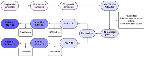
Our purpose is to evaluate the binocular contrast sensitivity function (CSF) in a presbyopic population and compare the results obtained with four different simultaneous-vision center-near multifocal contact lens (MCL) designs for distance vision under two illumination conditions. Additionally, chromatic CSF (red-green and blue-yellow)
[...] Read more.
Our purpose is to evaluate the binocular contrast sensitivity function (CSF) in a presbyopic population and compare the results obtained with four different simultaneous-vision center-near multifocal contact lens (MCL) designs for distance vision under two illumination conditions. Additionally, chromatic CSF (red-green and blue-yellow) was evaluated. A randomized crossover pilot study was conducted. Four daily disposable lens designs, based on simultaneous-vision and center-near correction, were compared. The achromatic contrast sensitivity function (CSF) was measured binocularly using the CSV1000e test under two lighting conditions: room light on and off. Chromatic CSF was measured using the OptoPad-CSF test. Comparison of achromatic results with room lighting showed a statistically significant difference only for 3 cpd (p = 0.03) between the baseline visit (with spectacles) and all MCLs. Comparison of achromatic results without room lighting showed no statistically significant differences between the baseline and all MCLs for any spatial frequency (p > 0.05 in all cases). Comparison of CSF-T results showed a statistically significant difference only for 4 cpd (p = 0.002). Comparison of CSF-D results showed no statistically significant difference for all frequencies (p > 0.05 in all cases). The MCL designs analyzed provided satisfactory achromatic contrast sensitivity results for distance vision, similar to those obtained with spectacles, with no remarkable differences between designs. Chromatic contrast sensitivity for the red-green and blue-yellow mechanisms revealed some differences from the baseline that should be further investigated in future studies.
Full article

Figure 1


