

Journal Description
Journal of Imaging
Journal of Imaging
is an international, multi/interdisciplinary, peer-reviewed, open access journal of imaging techniques, published online monthly by MDPI.
- Open Accessfree for readers, with article processing charges (APC) paid by authors or their institutions.
- High Visibility: indexed within Scopus, ESCI (Web of Science), PubMed, PMC, dblp, Inspec, Ei Compendex, and other databases.
- Journal Rank: JCR - Q2 (Imaging Science and Photographic Technology) / CiteScore - Q1 (Radiology, Nuclear Medicine and Imaging)
- Rapid Publication: manuscripts are peer-reviewed and a first decision is provided to authors approximately 21.3 days after submission; acceptance to publication is undertaken in 3.6 days (median values for papers published in this journal in the first half of 2026).
- Recognition of Reviewers: reviewers who provide timely, thorough peer-review reports receive vouchers entitling them to a discount on the APC of their next publication in any MDPI journal, in appreciation of the work done.
Impact Factor:
3.8 (2025);
5-Year Impact Factor:
3.6 (2025)
Latest Articles
Structuring Variability in Human Gait Datasets: A Covariate-Centered Taxonomy and Systematic Review of Image- and Depth-Based Collections
J. Imaging 2026, 12(7), 334; https://doi.org/10.3390/jimaging12070334 (registering DOI) - 22 Jul 2026
Abstract
Human gait datasets play a central role in the development and evaluation of computer vision models. However, the current dataset landscape remains highly heterogeneous, with inconsistent reporting of acquisition conditions, user variability, and sensing configurations, which limits reproducibility and hinders principled cross-dataset comparability.
[...] Read more.
Human gait datasets play a central role in the development and evaluation of computer vision models. However, the current dataset landscape remains highly heterogeneous, with inconsistent reporting of acquisition conditions, user variability, and sensing configurations, which limits reproducibility and hinders principled cross-dataset comparability. In this work, we propose a covariate-centered, modality-agnostic taxonomy for gait datasets, explicitly structuring variability across scene-level, user-level, and sensor-level factors. The proposed framework enables consistent characterization of datasets through a standardized set of covariates (A–R), bridging differences across application domains and sensing modalities. Following a systematic review protocol aligned with PRISMA 2020, we analyze 47 publicly available image- and depth-based human gait datasets spanning healthcare, biometric, and attribute-recognition application domains. Using the proposed taxonomy, we derive a quantitative analysis of covariate coverage, revealing systematic biases in current dataset design.
Full article
(This article belongs to the Section Computer Vision and Pattern Recognition)
Open AccessArticle
Decadal Changes in Institutional Diagnostic Reference Levels for X-Ray Angiography: A Retrospective Comparative Study
by
Ioannis Antonakos, Emmanouil Annousis, Tatiana Roko, Antonia Alexiadou, Maria Dmitropoulou, Dimitris Filippiadis, Stavros Spiliopoulos, Konstantinos Palialexis, Athanasios Giannakis, Niki Parmenidou and Efstathios Efstathopoulos
J. Imaging 2026, 12(7), 333; https://doi.org/10.3390/jimaging12070333 (registering DOI) - 22 Jul 2026
Abstract
Angiography is a key imaging modality for the diagnosis and treatment of vascular diseases, and the growing sophistication of interventional procedures has heightened the need for radiation dose optimization. Diagnostic Reference Levels (DRLs) are widely used to monitor patient exposure and to support
[...] Read more.
Angiography is a key imaging modality for the diagnosis and treatment of vascular diseases, and the growing sophistication of interventional procedures has heightened the need for radiation dose optimization. Diagnostic Reference Levels (DRLs) are widely used to monitor patient exposure and to support optimization in accordance with the ALARA principle. This study compared radiation dose metrics from a newly installed angiographic system at Attikon University Hospital with those obtained from the institution’s previous system and with values reported in the published literature. Radiation dose and procedural parameters were retrospectively collected for digital cerebral subtraction angiography (DSA), embolization, nephrostomy, vertebroplasty, transjugular intrahepatic portosystemic shunt (TIPS), chemoembolization, and injection procedures. Dose area product (DAP), fluoroscopy-related DAP, patient entrance dose indicators, and fluoroscopy time were analyzed. Median DAP values ranged from 5.72 to 349.60 Gy·cm2 depending on the procedure. Compared with data acquired approximately a decade earlier, DAP values increased by an average of 96.4%, whereas fluoroscopy times remained largely unchanged. Despite this increase, dose levels were generally lower than those reported in the international literature. Median DAP values ranged from 5.72 to 349.60 Gy·cm2 depending on the procedure. Compared with data acquired approximately a decade earlier, DAP values increased by an average of 96.4%, whereas fluoroscopy times remained largely unchanged. Although these differences may reflect the combined influence of technological developments, evolving procedural complexity, operator-related factors, and changes in clinical practice over time, these variables were not directly assessed in the present retrospective study. Nevertheless, the updated institutional Diagnostic Reference Levels provide a valuable benchmark for radiation dose optimization, quality assurance, and future multicenter studies aimed at supporting national DRL establishment.
Full article
(This article belongs to the Special Issue Diagnostic Imaging: From Basic Knowledge to Latest Advancements)
Open AccessArticle
LABFNet: A Restoration Network Guided by the LAB Colour Space and Frequency-Domain Constraints
by
Yaqian Zhang, Guanjun Wang, Quan Zhang and Bochao Zhou
J. Imaging 2026, 12(7), 332; https://doi.org/10.3390/jimaging12070332 - 22 Jul 2026
Abstract
In the restoration of mural images with rich colour information and complex texture structures, existing techniques typically extract the spatial-domain features in the Red–Green–Blue (RGB) colour space. However, the three RGB channels are physically decoupled without unified perceptual colour correlation constraints, which often
[...] Read more.
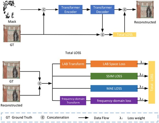
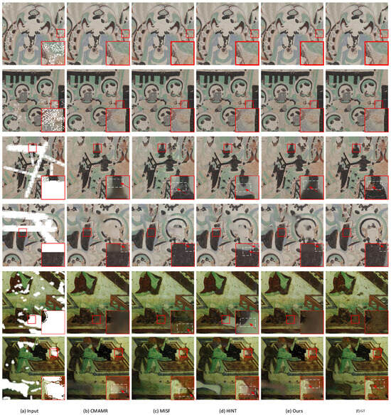
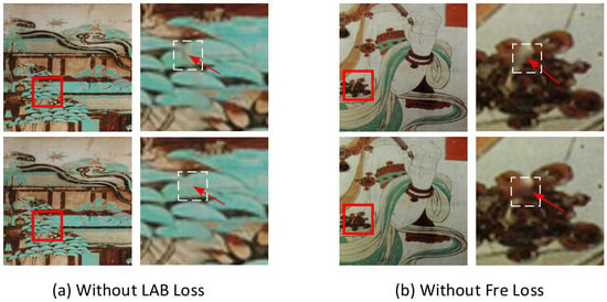
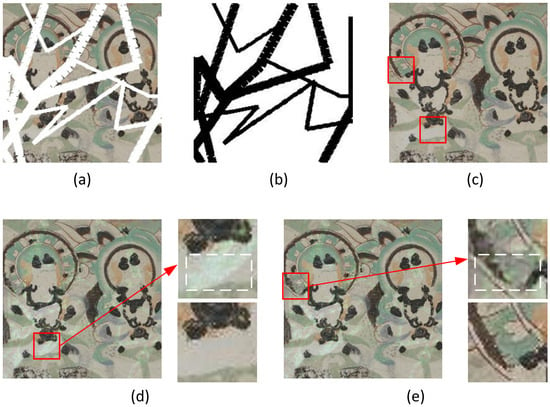
In the restoration of mural images with rich colour information and complex texture structures, existing techniques typically extract the spatial-domain features in the Red–Green–Blue (RGB) colour space. However, the three RGB channels are physically decoupled without unified perceptual colour correlation constraints, which often leads to noticeable colour deviation in damaged regions with large colour variations. In addition, restoring both high-frequency texture details and low-frequency global structures in a mixed-frequency spatial domain can create conflicts between frequencies, making it difficult to generate realistic high-frequency details. To address these issues, we propose the laboratory frequency network (LABFNet), a restoration network guided by the laboratory (LAB) colour space and frequency-domain constraints. Our model has two key improvements: (1) it incorporates colour parameters from the LAB space to model colour loss in murals, and (2) it decomposes the image into low- and high-frequency components and enforces frequency consistency during restoration. In the Dunhuang 20–40% mask-ratio setting, the Peak Signal-to-Noise Ratio (PSNR) and Structural Similarity Index (SSIM) improved by 1.58% and 0.27%, respectively, while the Mean Absolute Error (MAE), Learned Perceptual Image Patch Similarity (LPIPS) and CIEDE2000 decreased by 5.87%, 6.5%, and 21.65%, respectively. Experimental results on benchmark datasets show that LABFNet reduces colour deviation and structural defects.
Full article
(This article belongs to the Topic Computer Vision and Image Processing, 3rd Edition)
►▼
Show Figures

Figure 1
Open AccessArticle
Longitudinal CT Scanning for Explainable Early Detection of Postharvest Disorders: The ‘Braeburn’ Browning Case
by
Dirk Elias Schut, Rachael Maree Wood, Rob Schouten, Robert van Liere, Tristan van Leeuwen and Kees Joost Batenburg
J. Imaging 2026, 12(7), 331; https://doi.org/10.3390/jimaging12070331 - 21 Jul 2026
Abstract
This study presents two workflows for leveraging longitudinal computed tomography (CT) datasets when developing deep learning-based detection systems for gradually developing postharvest disorders. Workflow 1 (Longitudinal Benchmarking) benchmarks neural networks by training and testing them on images from different stages of disorder progression.
[...] Read more.
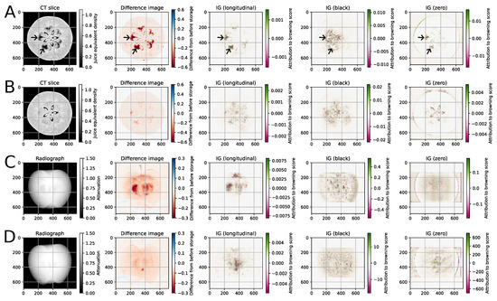
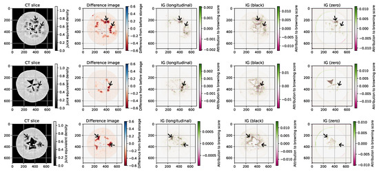
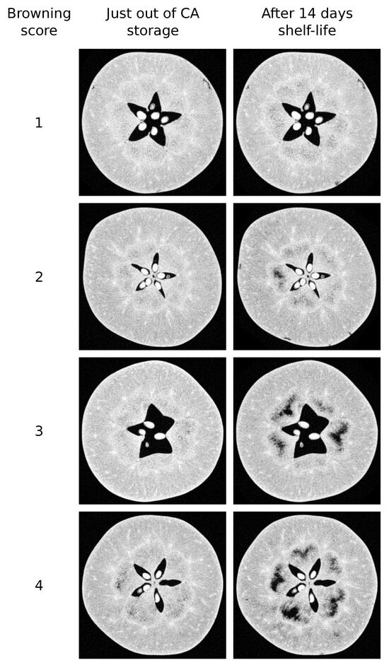
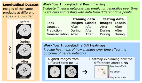
This study presents two workflows for leveraging longitudinal computed tomography (CT) datasets when developing deep learning-based detection systems for gradually developing postharvest disorders. Workflow 1 (Longitudinal Benchmarking) benchmarks neural networks by training and testing them on images from different stages of disorder progression. It examines the trade-off between detecting a disorder early or accurately and evaluates whether neural networks can generalize across time points. Workflow 2 (Longitudinal eXplainable Artificial Intelligence (XAI) Heatmaps) provides heatmaps that indicate how changes over time affect the outcomes of neural networks. It uses image registration to align an earlier-acquired image and then uses it as a baseline when calculating the heatmap. The workflows are demonstrated on a dataset of ‘Braeburn’ apples that were CT-scanned multiple times while developing internal browning during controlled-atmosphere (CA) storage and shelf life. The Longitudinal Benchmarking workflow was used to investigate whether images acquired immediately after CA storage can be used to predict the eventual browning after a shelf-life period, which is highly relevant in industrial practice. Moreover, the longitudinal XAI heatmaps avoided artifacts caused by out-of-distribution baselines or identical baseline regions, which occurred with conventional black or zero baselines.
Full article
(This article belongs to the Section AI in Imaging)
►▼
Show Figures

Figure 1
Open AccessArticle
Application of Machine Learning for Mean Glandular Dose Prediction Utilizing DICOM Mammography Images
by
Ali A. A. Alghamdi
J. Imaging 2026, 12(7), 330; https://doi.org/10.3390/jimaging12070330 - 21 Jul 2026
Abstract
The growing demand for raw and processed scientific data has encouraged many researchers and research institutions to adopt an open-source data policy. At present, data accessibility is of paramount importance due to the growing demand for artificial intelligence (AI) and machine learning (ML)
[...] Read more.
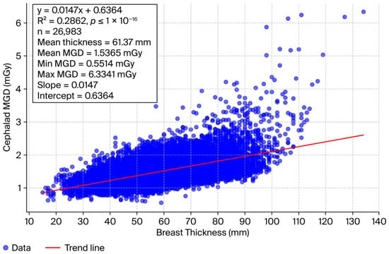
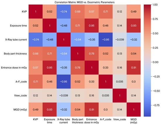
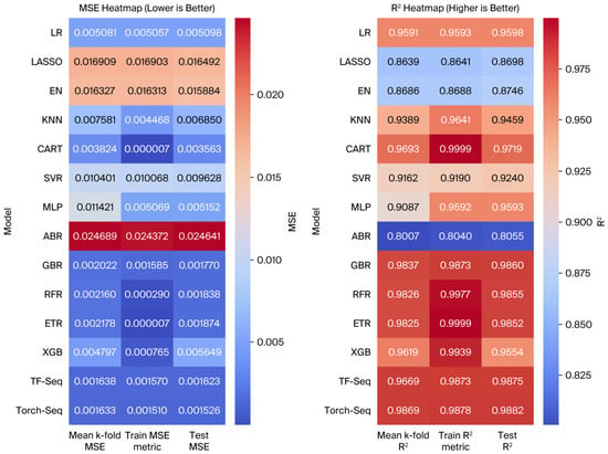
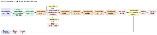
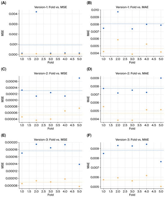
The growing demand for raw and processed scientific data has encouraged many researchers and research institutions to adopt an open-source data policy. At present, data accessibility is of paramount importance due to the growing demand for artificial intelligence (AI) and machine learning (ML) applications in various scientific fields, particularly medicine. Medium- to large-scale mammography datasets are widely used in breast cancer research to develop and evaluate computer-aided detection methods. However, there are only a few studies on using mammogram datasets for the prediction of the breast mean glandular dose (MGD) with AI or ML models. The aim of this study was to investigate the feasibility of using ML and deep ML for MGD prediction based on DICOM images and retrieved dosimetric data from DICOM mammogram images. A total of 26,988 mammography images in DICOM format were obtained from the Federated Research Data Repository (FRDR). Eleven regression algorithms and three neural network-based models were evaluated using five-fold cross-validation. In addition, a deep ML fusion model based on Vision Transformer (ViT) and tabular data was developed for the prediction of the MGD normalized conversion factor CF(DgN). A mean breast thickness of 61.37 mm and a mean MGD of 1.53 mGy (0.55–6.33 mGy) were calculated using this dataset. Regarding tabular data, the artificial neural network (ANN) sequential models outperformed other linear and tree-based models. The ViT deep ML fusion model was tested with three configuration versions differing on the number of features included. A comparison of the three versions revealed that the version with six features achieved the best overall predictor performance. This study demonstrates that ML and deep ML can effectively predict the MGD using dosimetric tabular data and mammography DICOM images. The use of ML with tabular data extracted from DICOM images can be further strengthened by incorporating larger and more diverse datasets.
Full article
(This article belongs to the Section Medical Imaging)
►▼
Show Figures

Figure 1
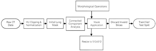
Open AccessArticle
Dynamic Convolution Enhanced Attention Network for Pulmonary Nodule Detection
by
Shengqun Zhang, Annie Anak Joseph and Kho Lee Chin
J. Imaging 2026, 12(7), 329; https://doi.org/10.3390/jimaging12070329 - 21 Jul 2026
Abstract
Pulmonary nodules are circular or irregular lesions visible on chest computed tomography (CT), and their early detection is critical for lung cancer screening. Deep learning detection algorithms have been widely adopted for pulmonary nodule diagnosis; existing lightweight models suffer from redundant network parameters
[...] Read more.
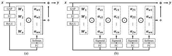
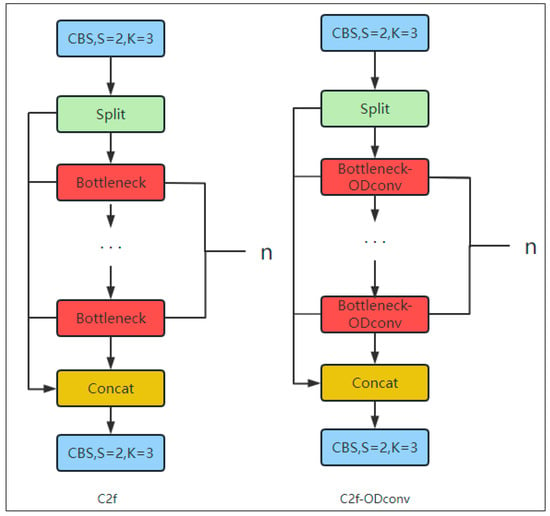
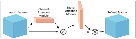
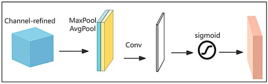
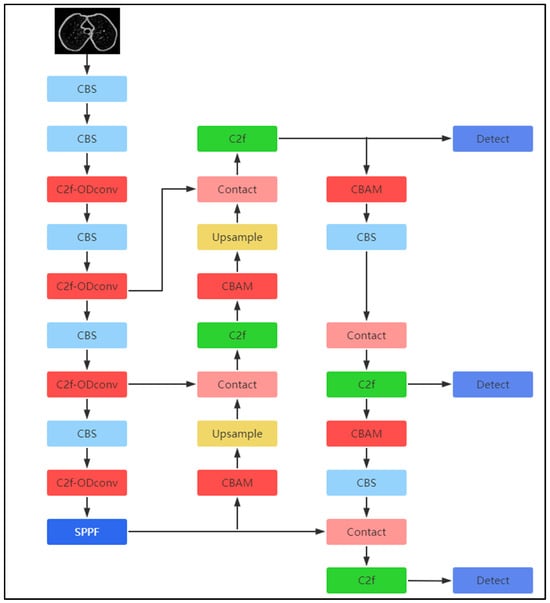
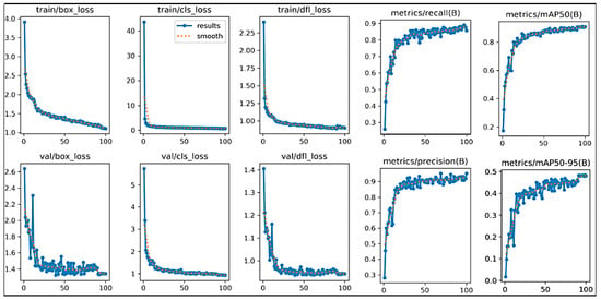
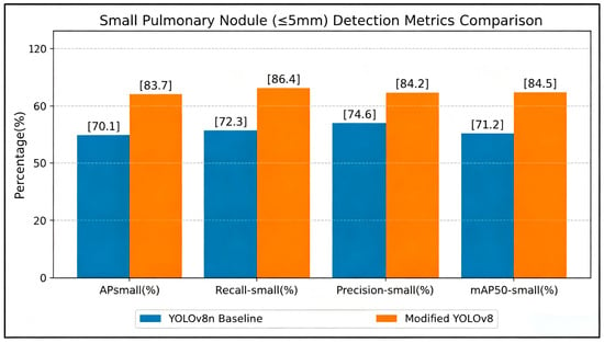
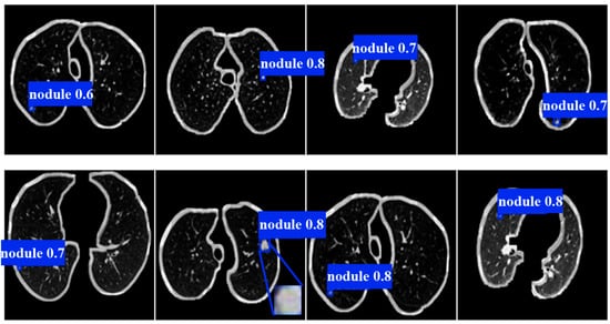
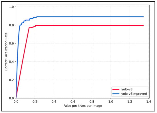
Pulmonary nodules are circular or irregular lesions visible on chest computed tomography (CT), and their early detection is critical for lung cancer screening. Deep learning detection algorithms have been widely adopted for pulmonary nodule diagnosis; existing lightweight models suffer from redundant network parameters and low detection accuracy for tiny lesions. To address these limitations, this study proposes an improved detection model based on YOLOv8n. First, Omni-Dimensional Dynamic Convolution (ODConv) replaces static convolution in the backbone to enhance multi-morphology nodule feature extraction. Second, the Convolutional Block Attention Module (CBAM) is embedded at multiple positions of the neck network to suppress background interference from blood vessels and normal lung parenchyma. Third, Complete Intersection over Union (CIoU) loss is substituted by Wise Intersection over Union (W-IoU) to optimize bounding box regression for hard samples with blurred boundaries. Experiments on the LUNA16 dataset show that compared with the original YOLOv8n, the proposed model improves Precision by 6.3%, Recall by 8.6%, mAP50 by 3.4%, and mAP50-95% by 2.7% while maintaining high inference speed. Additional generalization verification on the LIDC-IDRI multi-center dataset further proves the robustness of the proposed lightweight architecture, which achieves balanced accuracy and real-time performance compared with mainstream detection models.
Full article
(This article belongs to the Section Medical Imaging)
►▼
Show Figures

Figure 1
Open AccessArticle
Contrastive and Transfer Learning for Aligned Multimodal Neuroimaging Classification of Autism Spectrum Disorder
by
Raja Vavekanand, Ganesh Kumar, Muhammad Moazzam Jawaid, Shafiya Qadeer Memon and Teerath Kumar
J. Imaging 2026, 12(7), 328; https://doi.org/10.3390/jimaging12070328 - 20 Jul 2026
Abstract
Autism Spectrum Disorder (ASD) assessment remains challenging because behavioural instruments are partly observer-dependent and neuroimaging data are heterogeneous. This paper presents FAA (Fuse After Aligned), which is a multimodal classification framework that combines transfer learning for structural MRI (sMRI) representation learning with a
[...] Read more.
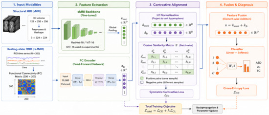
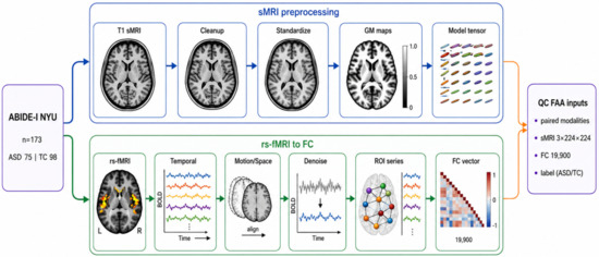
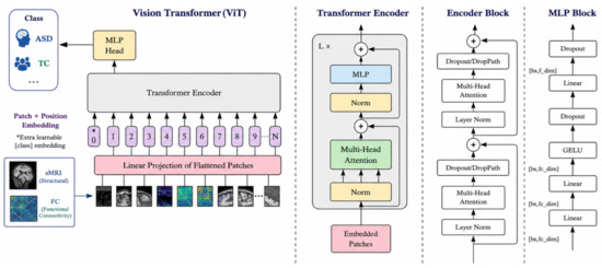
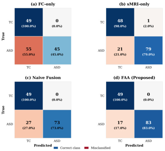
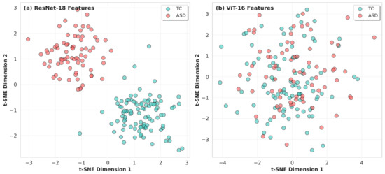
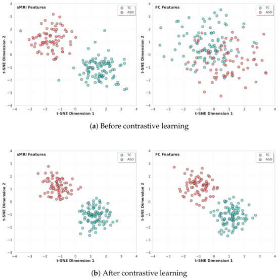
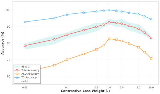
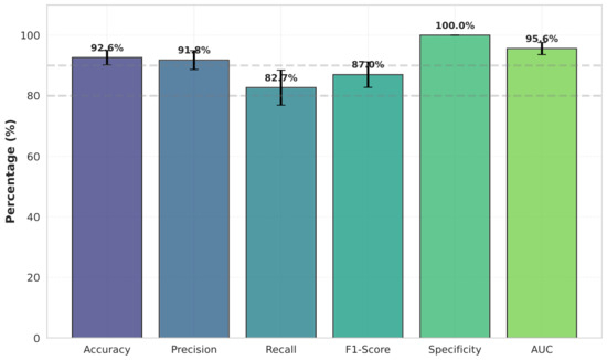
Autism Spectrum Disorder (ASD) assessment remains challenging because behavioural instruments are partly observer-dependent and neuroimaging data are heterogeneous. This paper presents FAA (Fuse After Aligned), which is a multimodal classification framework that combines transfer learning for structural MRI (sMRI) representation learning with a contrastive objective for the pre-fusion alignment of sMRI and resting-state functional MRI-derived functional connectivity (FC) features. Evaluation was restricted to the single-site ABIDE-I New York University subset comprising 75 participants with ASD and 98 typically developing controls. Under the reported five-fold internal cross-validation protocol, FAA achieved a mean accuracy of 92.6% compared with 87.4% for naive fusion and 90.9% for the sMRI-only baseline. Ablation analyses indicate that adding the contrastive objective is associated with improved classification performance and that ResNet-18 outperforms the evaluated ViT-16 configurations in this small-sample setting. These findings support the methodological value of pre-fusion feature alignment within the evaluated cohort. The framework offers a robust, computationally efficient, and clinically viable approach for objective ASD diagnosis with strong potential for generalisation to multi-site neuroimaging applications.
Full article
(This article belongs to the Section Medical Imaging)
►▼
Show Figures

Figure 1
Open AccessArticle
A Simplified CT Score for Thrombus Burden in Acute Pulmonary Embolism: Clinical Correlation and Reproducibility
by
Ignacio Díaz-Lorenzo, Rio Jorge Aguilar Torres, Paloma Caballero Sanchez-Robles, Raquel Caminero Garcia, Alfonso Canabal Berlanga, Alfonsa Friera Reyes and Alberto Alonso-Burgos
J. Imaging 2026, 12(7), 327; https://doi.org/10.3390/jimaging12070327 - 19 Jul 2026
Abstract
(1) Objectives: In acute pulmonary embolism (PE), detailed thrombus burden scores are often complex and time-consuming, limiting their integration into urgent radiology reports. We evaluated a simplified modified Ghanima score (GmScore and GmS) designed to provide a structured estimate of thrombus burden and
[...] Read more.
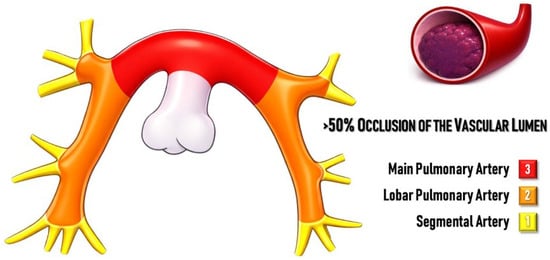
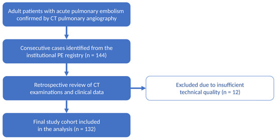
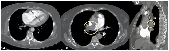
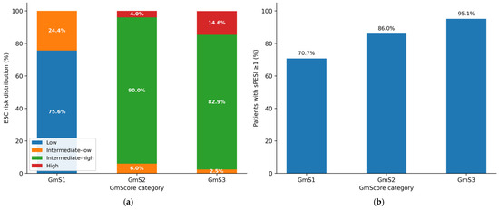
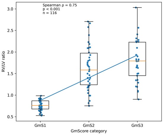
(1) Objectives: In acute pulmonary embolism (PE), detailed thrombus burden scores are often complex and time-consuming, limiting their integration into urgent radiology reports. We evaluated a simplified modified Ghanima score (GmScore and GmS) designed to provide a structured estimate of thrombus burden and assessed its clinical correlation and reproducibility. (2) Methods: In this retrospective single-center study, 132 consecutive patients with confirmed acute PE were classified according to the modified GmScore: GmS1 (segmental), GmS2 (lobar), and GmS3 (main pulmonary arteries), considering luminal obstruction ≥ 50%. European Society of Cardiology (ESC) risk category, simplified Pulmonary Embolism Severity Index (sPESI), CT right-to-left ventricular (RV/LV) ratio, echocardiographic right ventricular dysfunction, and 30-day mortality were recorded. Inter- and intraobserver agreement were assessed using weighted kappa. (3) Results: In 132 patients (mean age 64.8 ± 16.5 years; 77 men), a significant clinical gradient was observed across GmScore categories. ESC intermediate–high/high risk occurred in 0% of GmS1 and 95.6% of GmS2–3 patients (p < 0.001). The median RV/LV ratio increased progressively (0.76, 1.58, and 1.79 for GmS1–3; p < 0.001), with a strong correlation between the GmScore and RV/LV (Spearman ρ = 0.75). GmS2 and GmS3 showed no significant difference in ventricular repercussion (p = 0.938), whereas GmS1 differed markedly. Using GmS ≥ 2 to identify ESC intermediate–high/high risk yielded 100% sensitivity and negative predictive value. Interobserver agreement was excellent (κ = 0.92). Thirty-day mortality was 0% in GmS1, 2.0% in GmS2, and 14.6% in GmS3 (p = 0.005). (4) Conclusions: The modified GmScore is a simple, reproducible CT-based descriptor that aligns closely with right ventricular repercussion and ESC risk stratification.
Full article
(This article belongs to the Section Medical Imaging)
►▼
Show Figures

Figure 1
Open AccessArticle
Microscopic Pollen Image Classification via Contour-Signal Representation, Wavelet Analysis, and CNN
by
Abror Shavkatovich Buriboev, Akhram Nishanov, Shuxrat Isroilov, Inomjon Narzullaev, Umidjon Djumayozov, Shavkat Buriboyev, Temur Azamov, Parda Yuldashov, Davron Shodmonov, Djamshid Sultanov and Abbos Abduvaytov
J. Imaging 2026, 12(7), 326; https://doi.org/10.3390/jimaging12070326 - 18 Jul 2026
Abstract
Accurate classification of pollen grains in microscopic images remains challenging because of noise, structural variability, background complexity, weak texture, and intra-class similarity. To address these issues, this study proposes a hybrid framework that integrates contour-signal modeling, spectral–wavelet analysis, and deep learning for robust
[...] Read more.
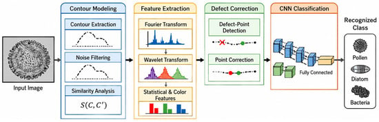
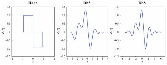
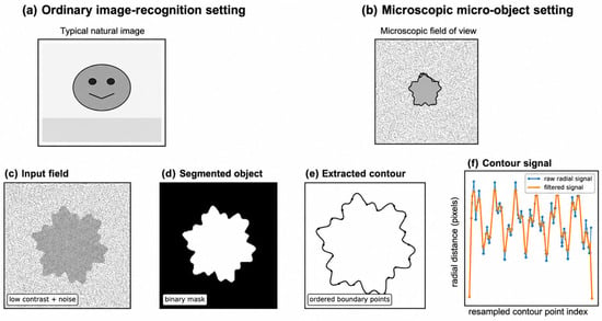
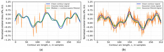
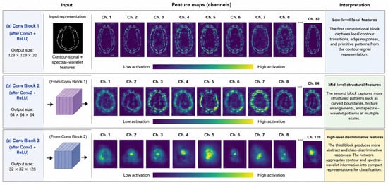
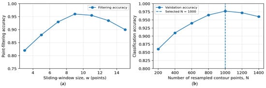
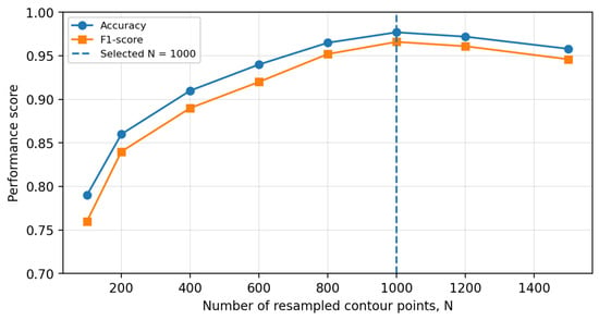
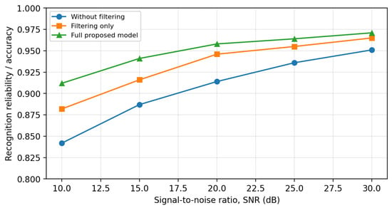
Accurate classification of pollen grains in microscopic images remains challenging because of noise, structural variability, background complexity, weak texture, and intra-class similarity. To address these issues, this study proposes a hybrid framework that integrates contour-signal modeling, spectral–wavelet analysis, and deep learning for robust microscopic pollen image recognition. In the proposed approach, microscopic pollen images are first converted into contour-based point-signal representations, allowing object boundaries to be analyzed as structured one-dimensional signals. To improve signal quality under real imaging conditions, the framework incorporates Gaussian, median, and contour-aware filtering together with defect-point detection and correction. The processed contour signals are then analyzed using Fourier transform, continuous wavelet transform, and discrete wavelet transform to extract complementary global and local descriptors. These enriched representations are provided to a convolutional neural network for final classification. Experiments conducted on a seven-class microscopic pollen-image dataset demonstrate that the proposed method outperforms conventional computer-vision and baseline deep-learning approaches. The best-performing hybrid configuration achieved an error rate of 6.4%, while the overall classification accuracy reached 0.977 with an F1-score of 0.966, compared with 0.837 for a traditional computer-vision pipeline. These results confirm that combining contour-based signal processing with hierarchical deep feature learning provides an effective and noise-robust strategy for microscopic pollen image recognition. However, the present validation is limited to pollen images, and further experiments on broader microscopic object datasets are required to assess generalization to other micro-object categories such as nanoparticles, fibers, rods, and synthetic microstructures.
Full article
(This article belongs to the Section Computer Vision and Pattern Recognition)
►▼
Show Figures

Figure 1
Open AccessArticle
Efficient Object Detection in Compressed Domain by Exploiting Knowledge Distillation from Pixel Domain
by
Serhat Dikyar and Behcet Ugur Toreyin
J. Imaging 2026, 12(7), 325; https://doi.org/10.3390/jimaging12070325 - 18 Jul 2026
Abstract
The proliferation of high-definition video data necessitates highly efficient processing pipelines for real-time edge analytics. However, traditional object detection architectures rely exclusively on pixel-domain inputs, which renders the computationally prohibitive decoding phase a latency bottleneck. In this paper, we propose a novel dual-phase
[...] Read more.
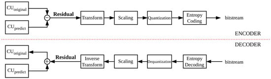
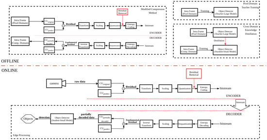
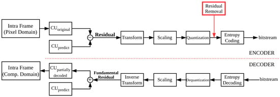
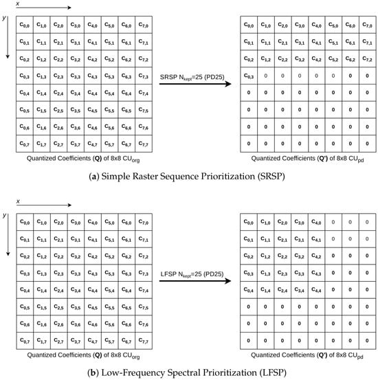
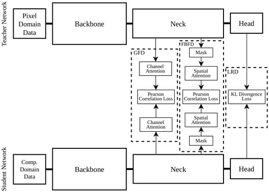
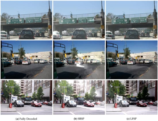
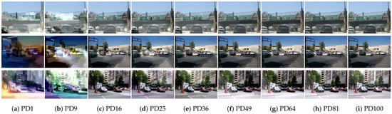
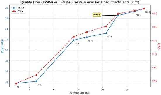
The proliferation of high-definition video data necessitates highly efficient processing pipelines for real-time edge analytics. However, traditional object detection architectures rely exclusively on pixel-domain inputs, which renders the computationally prohibitive decoding phase a latency bottleneck. In this paper, we propose a novel dual-phase framework designed to achieve fast and efficient object detection directly within the partially decoded compressed-domain data. First, we introduce a partial decoding paradigm featuring the Low-Frequency Spectral Prioritization method on the encoder side. By systematically discarding high-frequency residual coefficients and retaining only a sparse subset of fundamental spatial frequencies, this method dramatically reduces transmission payloads and accelerates the standard decoding process. Second, to recover the structural fidelity lost due to the intentional omission of residual data, we employ a multi-granularity cross-domain knowledge distillation architecture. This strategy aligns global contextual features, foreground boundary attention maps, and final response logits, transferring rich representational capacities from a high-performing pixel-domain teacher network to a lightweight compressed-domain student network. Comprehensive experiments utilizing RetinaNet, FCOS, and GFL object detection networks on the COCO-mini dataset demonstrate the superiority of the proposed framework. By retaining fundamental residual coefficients within the HEVC pipeline, the proposed method reduces average decoding latency while improving the mAP score by over the conventional fully decoded pixel-domain baseline on the COCO-mini dataset.
Full article
(This article belongs to the Section Computer Vision and Pattern Recognition)
►▼
Show Figures

Figure 1
Open AccessReview
Review: Techniques in Egocentric Multi-View Image Analysis: Advances, Challenges, and Future Directions
by
Duc Tri Phan and Hong Duc Nguyen
J. Imaging 2026, 12(7), 324; https://doi.org/10.3390/jimaging12070324 - 17 Jul 2026
Abstract
Egocentric multi-view image analysis refers to the processing of utilizing synchronized video streams captured from multiple wearable cameras worn on the head or body, providing complementary first-person perspectives of dynamic, real-world interactions. Unlike single-view egocentric vision, which may suffer from severe occlusions, motion
[...] Read more.
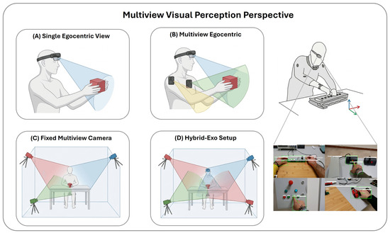
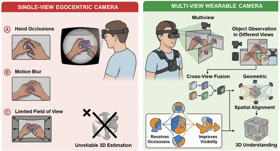
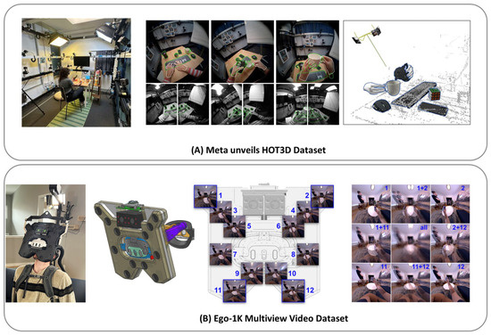
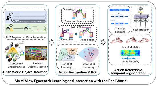
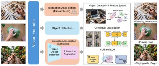
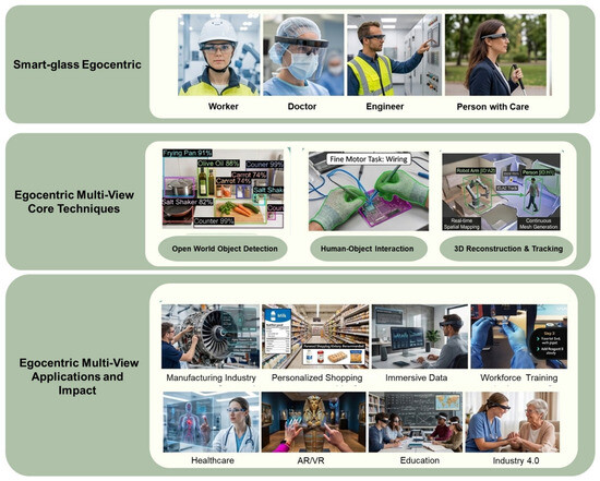
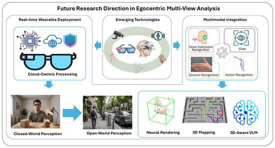
Egocentric multi-view image analysis refers to the processing of utilizing synchronized video streams captured from multiple wearable cameras worn on the head or body, providing complementary first-person perspectives of dynamic, real-world interactions. Unlike single-view egocentric vision, which may suffer from severe occlusions, motion blur, and limited field-of-view or traditional fixed-camera multi-view setups (assuming static geometry and controlled environments), egocentric multi-view systems leverage body-worn rigs to enable a more robust and flexible 3D understanding in open-world, mobile scenarios. In this work, we present a systematic survey of advancements in cross-view feature fusion, geometric consistency enforcement, open-world detection, human–object interaction (HOI) modeling, action segmentation, 3D reconstruction, and novel-view synthesis specifically tailored to wearable multi-camera platforms. Key datasets released between 2024 and 2026—including HOT3D (833 min of synchronized multi-view hand/object interactions from Project Aria and Quest 3), MultiEgo (first multi-egocentric dataset for 4D social scene reconstruction), and Ego-1K (large-scale 12-camera rig for dynamic 3D video synthesis) are thoroughly examined alongside an analysis of integrations with large language models (LLMs) and vision–language models that drive performance gains, typically in the 15–30% range over single-view baselines in hand tracking, HOI recognition, and reconstruction fidelity, although we show through a consolidated meta-analysis that this gain is task-dependent: larger for geometry-bottlenecked tasks such as in-hand object lifting, and smaller, method-dependent, or occasionally negative for semantic-recognition tasks such as keystep recognition under naive view fusion. These methods cover work in multi-view stereo, cross-view learning, and novel-view synthesis while addressing several real-time wearable constraints. Practical applications such as immersive Augmented Reality/Virtual Reality (AR/VR), assistive robotics, and healthcare monitoring are also discussed together with the challenges in motion calibration, benchmark diversity, and edge deployment ability. Thus, in this review, we attempt to fill a critical gap by focusing exclusively on wearable multi-view systems in an open-world setting, synthesizing the latest literature to chart future directions toward more embodied and continual learning agents.
Full article
(This article belongs to the Special Issue Techniques in Multi-View Image Analysis)
►▼
Show Figures

Figure 1
Open AccessArticle
Magnetic Resonance Imaging Preprocessing for Robust Spinal Cord Segmentation in Cervical Myelopathy
by
Hediyeh Toufani, Richard M. Dansereau, Philippe Phan, Jefferson R. Wilson and Eve C. Tsai
J. Imaging 2026, 12(7), 323; https://doi.org/10.3390/jimaging12070323 - 17 Jul 2026
Abstract
Accurate spinal cord segmentation is important for quantitative analysis of spinal cord magnetic resonance imaging, including measurement of cross-sectional area and diffusion-based microstructural characterization. In pathological conditions like cervical myelopathy, the shape deformation induced by cord compression is extreme, rendering automated segmentation particularly
[...] Read more.
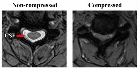
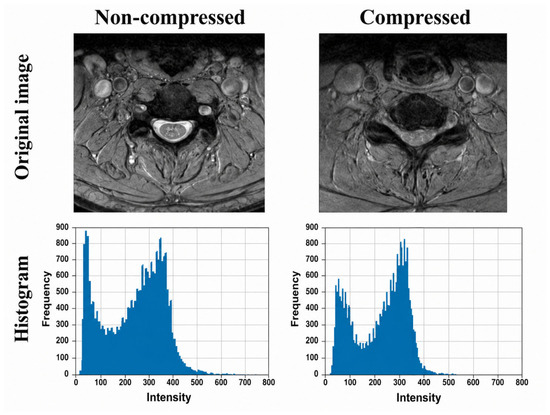
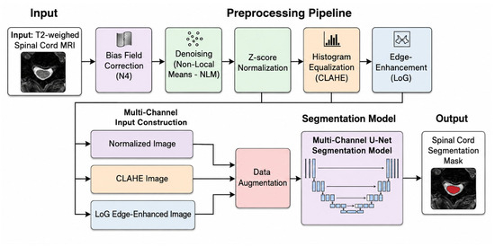
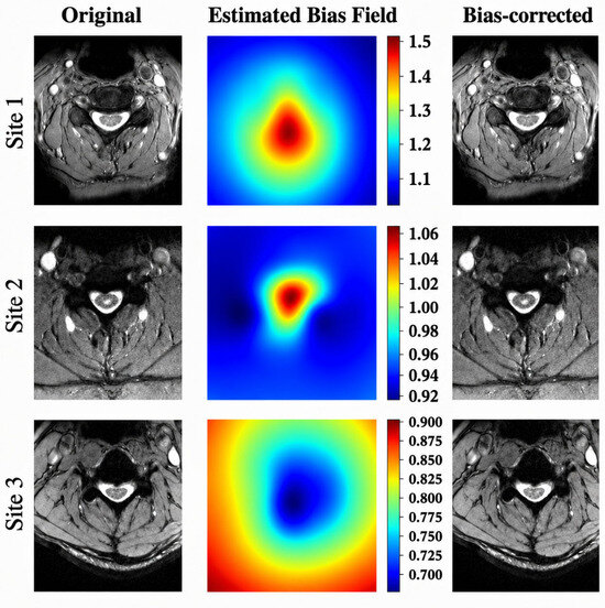
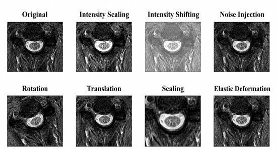
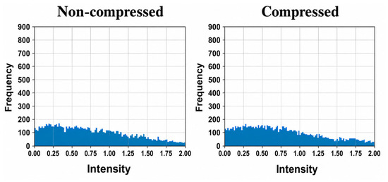
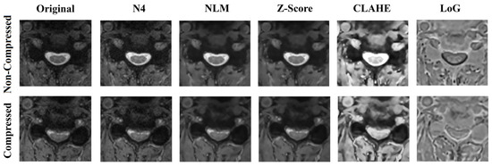
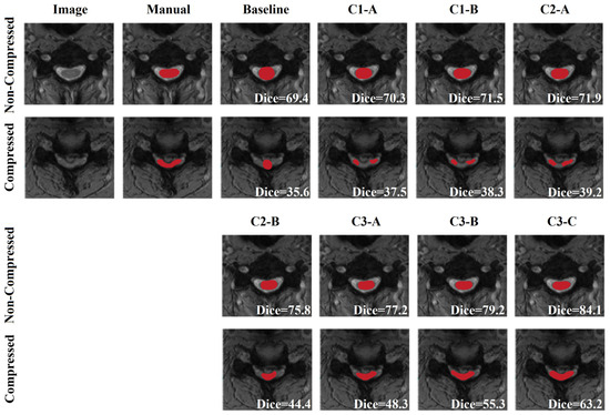
Accurate spinal cord segmentation is important for quantitative analysis of spinal cord magnetic resonance imaging, including measurement of cross-sectional area and diffusion-based microstructural characterization. In pathological conditions like cervical myelopathy, the shape deformation induced by cord compression is extreme, rendering automated segmentation particularly challenging. While deep learning-based methods yield good results in healthy or mildly pathological cases, their reliability suffers when anatomical assumptions fail under compression. In this work, we introduce a pathology-aware, boundary-focused preprocessing framework that directly aims to mitigate failure modes imposed by cord compression. Instead of generic preprocessing, each component aims to enhance intensity homogeneity, suppress noise and improve boundary visibility. At the core of this approach is a multi-representation input derived from a single T2*-weighted scan, whereby complementary intensity-, contrast- and edge-enhanced representations are fed to the U-Net model. The proposed framework is evaluated on spinal cord MRI data from three clinical centers (194 cervical myelopathy cases). The results demonstrate that the proposed preprocessing framework improves segmentation accuracy, robustness, and stability, particularly in anatomically challenging regions affected by compression. These findings highlight the importance of pathology-aware preprocessing for reliable spinal cord segmentation in cervical myelopathy.
Full article
(This article belongs to the Section Image and Video Processing)
►▼
Show Figures

Figure 1
Open AccessArticle
Sex Estimation Based on the Cranial Base of Three-Dimensional Skull Models from the Bosnia and Herzegovina Population Using Geometric Morphometrics
by
Zurifa Ajanović, Saleha Redžepi, Uzeir Ajanović, Naida Spahović, Amina Zorlak-Čavčić, Emina Dervišević, Admir Terzić and Mirza Pojskić
J. Imaging 2026, 12(7), 322; https://doi.org/10.3390/jimaging12070322 - 16 Jul 2026
Abstract
Sex estimation is a fundamental component of biological profiling in forensic anthropology, particularly when skeletal remains are incomplete or fragmented. This study aimed to evaluate sex estimation of the cranial base using geometric morphometrics and to assess the predictive value of cranial base
[...] Read more.
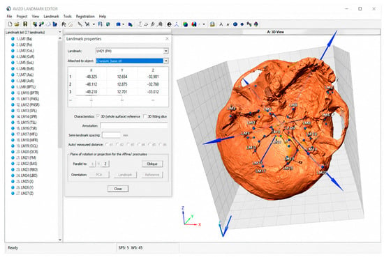
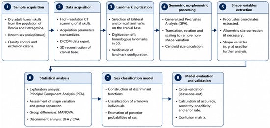
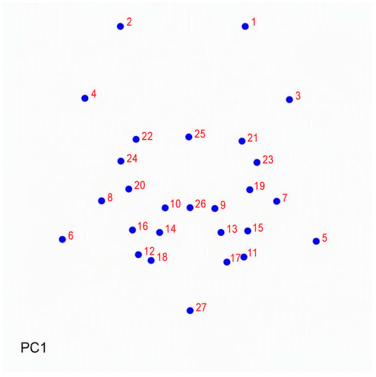
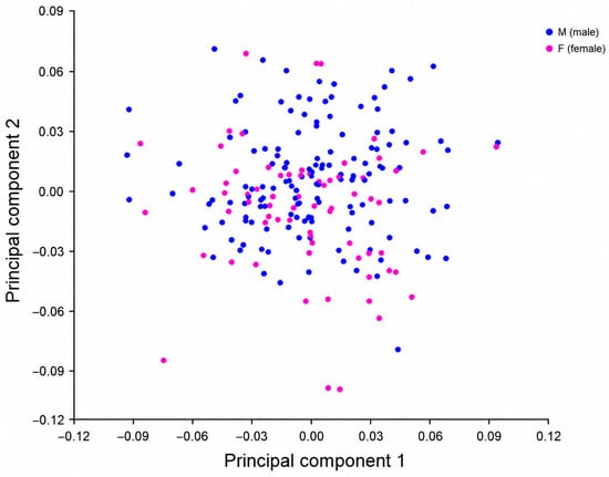
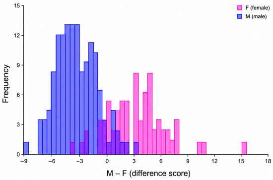
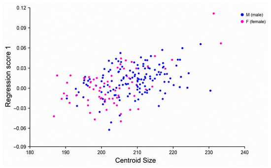
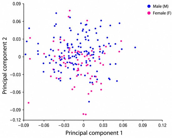
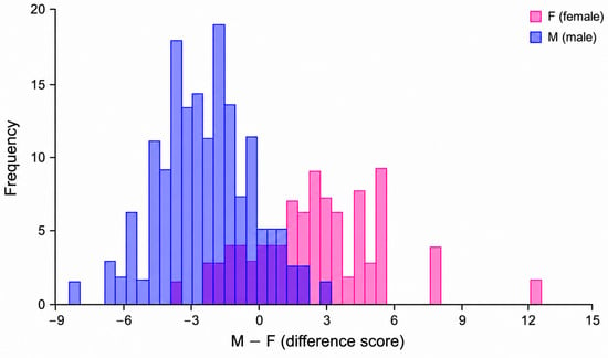
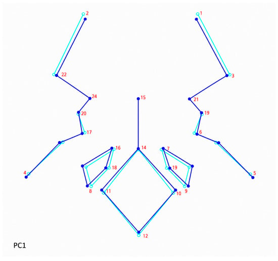
Sex estimation is a fundamental component of biological profiling in forensic anthropology, particularly when skeletal remains are incomplete or fragmented. This study aimed to evaluate sex estimation of the cranial base using geometric morphometrics and to assess the predictive value of cranial base morphology for sex estimation. The study included 211 adult skulls (139 male, 72 female) from the Bosnian population. Each skull was digitized to generate 3D models, and 27 anatomical landmarks were recorded. Landmark coordinates were standardized using Generalized Procrustes Analysis, Principal Component Analysis, Discriminant Function Analysis with permutation testing, and regression of shape on centroid size. Statistically significant sex estimation was observed at both the form (shape and size) and shape levels. Classification accuracy based on cranial base form reached 92.81% for males and 86.11% for females. Shape-based classification, after removal of size effects, also showed high accuracy (90.65% for males and 81.94% for females). Regression analysis indicated that size contributed significantly but modestly to shape variation. The cranial base exhibits stable sexually dimorphic patterns and may represent a reliable anatomical region for sex estimation. These findings contribute to population-specific standards for the Bosnia and Herzegovina population and support the forensic applicability of 3D geometric morphometric approaches.
Full article
(This article belongs to the Section Biometrics, Forensics, and Security)
►▼
Show Figures

Figure 1
Open AccessArticle
An Adaptive Attention-Driven Quadruplet Deep Hashing Method for Retrieving Histopathological Images
by
Seyed Mohammad Alizadeh, Henning Müller and Mohammad Sadegh Helfroush
J. Imaging 2026, 12(7), 321; https://doi.org/10.3390/jimaging12070321 - 15 Jul 2026
Abstract
Retrieving histopathological images can assist in the recognition and treatment planning of several diseases. Nevertheless, high-dimensional features can make this process complex and inefficient. These challenges can be addressed by encoding the feature domain into binary codes of different lengths utilizing deep hashing
[...] Read more.
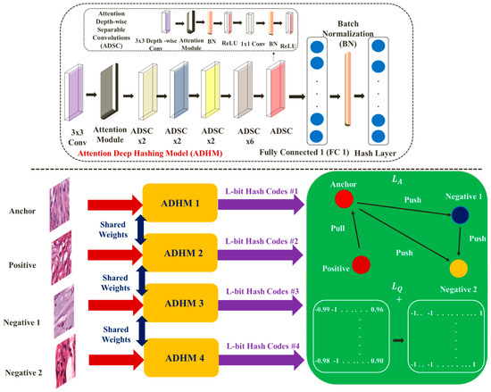
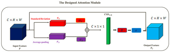
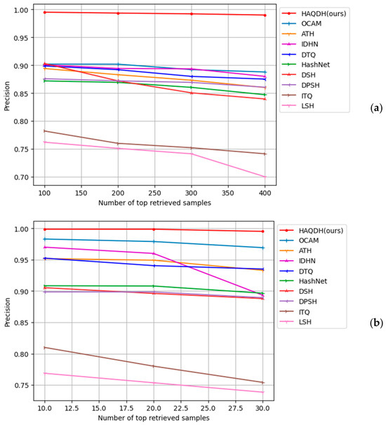
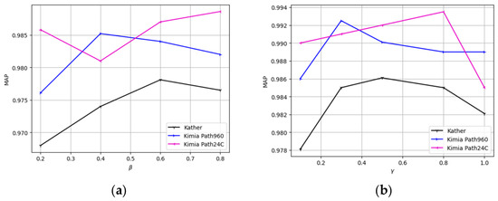
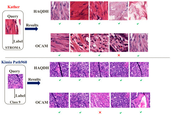
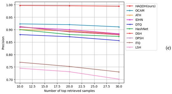
Retrieving histopathological images can assist in the recognition and treatment planning of several diseases. Nevertheless, high-dimensional features can make this process complex and inefficient. These challenges can be addressed by encoding the feature domain into binary codes of different lengths utilizing deep hashing approaches. Still, the vanishing gradient challenge remains a concern in these approaches. According to several studies, quadruplet deep hashing models have exhibited promising performance in retrieving images from multi-category datasets. Furthermore, adding an attention module to a convolutional neural network architecture can increase the efficiency of feature extraction. Thus, we introduce an adaptive quadruplet deep hashing model to retrieve histopathological images. Four designed deep hashing models with matching structures and parameters are utilized to produce hash codes. The resulting codes are trained according to a novel adaptive quadruplet loss function. The adaptive structure is capable of improving retrieval performance. The presented approach also suggests a novel hash layer for the vanishing gradient issue. In addition, a simple yet effective attention module is implemented to enhance feature extraction performance. Our model is evaluated on three publicly available histopathology datasets: Kather, Kimia Path960, and Kimia Path24C. The results indicate that the suggested approach achieves the highest mean average precision (MAP) of approximately 0.9940, 0.9983, and 0.9968 for the respective datasets. Based on experiments performed on the datasets, our model surpasses current hashing techniques.
Full article
(This article belongs to the Section AI in Imaging)
►▼
Show Figures

Figure 1
Open AccessArticle
Fast-CenLaneNet: A Lightweight Instance Segmentation-Based Network for Real-Time Lane Detection
by
Qidong Han, Shuo Feng, Yang Gao, Mengyao Li, Teng Meng, Ke Li and Yuhao Yang
J. Imaging 2026, 12(7), 320; https://doi.org/10.3390/jimaging12070320 - 13 Jul 2026
Abstract
Lane detection is a critical component of autonomous driving systems, requiring both high accuracy and real-time performance under complex driving scenarios. Unlike current methods that rely on predefined lane counts, instance segmentation methods can handle an arbitrary number of lanes, making them more
[...] Read more.
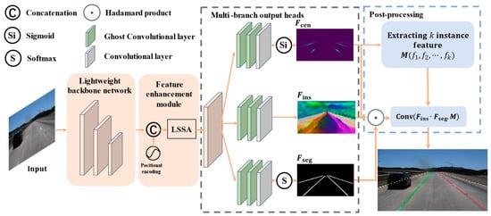
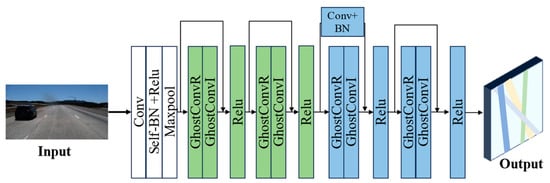
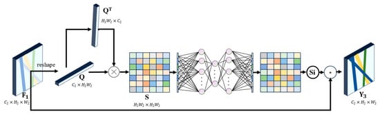
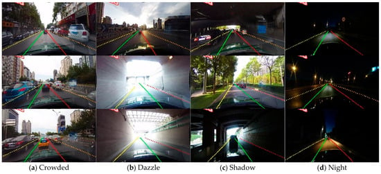
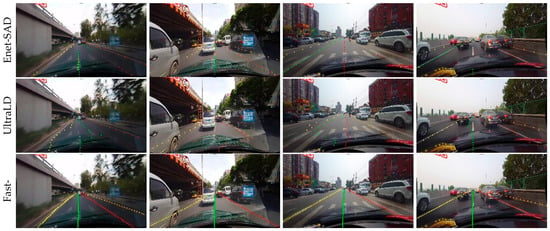
Lane detection is a critical component of autonomous driving systems, requiring both high accuracy and real-time performance under complex driving scenarios. Unlike current methods that rely on predefined lane counts, instance segmentation methods can handle an arbitrary number of lanes, making them more adaptable in real-world applications. However, this flexibility typically relies on dense pixel-level predictions, which necessitate large-scale networks and result in prohibitively high computational costs, hindering deployment on embedded platforms. To address these challenges, we present Fast-CenLaneNet, a lightweight architecture that improves inference efficiency while maintaining detection accuracy. Specifically, we design a lightweight backbone to reduce model parameters and computational cost, propose a learnable spatial similarity attention module to capture spatial dependencies within lane regions and enhance feature discriminability, and construct multi-branch output heads with Ghost convolutions to refine lane-related features with low computational overhead. Experiments on the TuSimple and CULane benchmarks demonstrate that Fast-CenLaneNet achieves a favorable accuracy–efficiency trade-off. On TuSimple, Fast-CenLaneNet obtains 96.40 ± 0.06% accuracy and 162.7 ± 6.8 FPS with 4.7 M parameters and 9.9 GFLOPs. Compared with CenLaneNet, it reduces the number of parameters by 89.1% and improves forward inference speed by 107.5%, with an accuracy decrease of only 0.08 percentage points.
Full article
(This article belongs to the Special Issue Computer Vision and Image Processing: Advances and Challenges)
►▼
Show Figures

Figure 1
Open AccessArticle
Exploring Adipose Tissue Behavior in CT: Impact of Age, Sex, and Contrast Media on Body Composition, Liver and Skeletal Muscle
by
Emil Matthisson, Hanns-Christian Breit, Markus Obmann, Jakob Wasserthal, Martin Segeroth and Daniel Boll
J. Imaging 2026, 12(7), 319; https://doi.org/10.3390/jimaging12070319 - 13 Jul 2026
Abstract
Objectives: To evaluate the impact of contrast phase, age, and sex on CT-derived body composition metrics—specifically attenuation and volume of subcutaneous adipose tissue (SAT), visceral adipose tissue (VAT), liver, and skeletal muscle. The potential of the proportion of muscle voxels below 0 Hounsfield
[...] Read more.
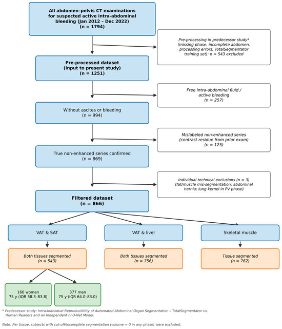
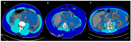
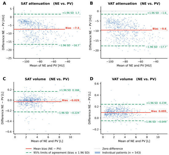
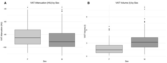
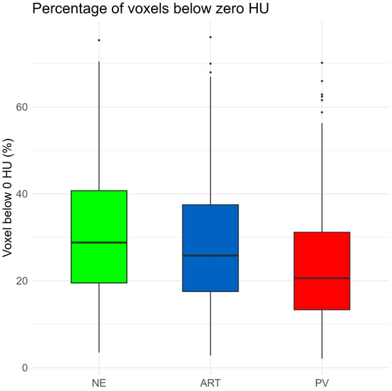
Objectives: To evaluate the impact of contrast phase, age, and sex on CT-derived body composition metrics—specifically attenuation and volume of subcutaneous adipose tissue (SAT), visceral adipose tissue (VAT), liver, and skeletal muscle. The potential of the proportion of muscle voxels below 0 Hounsfield units (HU) as a surrogate for fatty infiltration was also explored. Materials and Methods: A retrospective analysis of 866 multiphasic abdominal CT scans (non-enhanced [NE], arterial [ART], portal venous [PV]) from 2012 to 2022 was performed. Segmentation of SAT, VAT, liver, and skeletal muscle was conducted using the AI-based TotalSegmentator. Wilcoxon signed-rank tests and Bland–Altman analysis (mean bias and 95% limits of agreement) were applied to assess contrast-related effects; Spearman’s correlation coefficient was used to assess demographic associations. Results: Significant variation in attenuation and volume of SAT, VAT, and muscle was observed across contrast phases (p < 0.001). SAT attenuation was higher in NE and PV than in ART, while VAT attenuation was highest in PV. SAT volume increased and VAT volume decreased in contrast-enhanced phases. Attenuation and volume showed strong inter-phase correlation (ρ > 0.9). VAT attenuation was significantly higher in females, whereas VAT volume was significantly greater in males. VAT volume negatively correlated with liver attenuation (ρ = −0.33). Muscle voxels <0 HU were significantly reduced in contrast-enhanced scans. Conclusions: Contrast phase, age, and sex significantly influence CT-based body composition parameters. These confounding factors should be considered when using quantitative imaging biomarkers in clinical and research settings.
Full article
(This article belongs to the Section Medical Imaging)
►▼
Show Figures

Figure 1
Open AccessArticle
The Use of High-Frequency Skin Ultrasound in the Evaluation of Psoriatic Plaques—A Pilot Comparative Study Between Conventional and Biological Therapy
by
Adelina Filofteia Ghilencea, Daniel Octavian Costache, Constantin Căruntu, Maria Moga and Raluca Simona Costache
J. Imaging 2026, 12(7), 318; https://doi.org/10.3390/jimaging12070318 - 13 Jul 2026
Abstract
Introduction. Psoriasis is a chronic inflammatory disease histologically characterized by epidermal hyperproliferation, altered keratinocyte differentiation and dermal vascular remodeling. Although the diagnosis is mainly clinical, non-invasive imaging methods, such as high-frequency skin ultrasound, allow an objective assessment of skin changes and disease
[...] Read more.
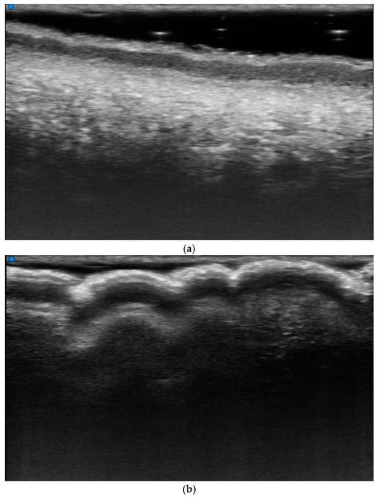
Introduction. Psoriasis is a chronic inflammatory disease histologically characterized by epidermal hyperproliferation, altered keratinocyte differentiation and dermal vascular remodeling. Although the diagnosis is mainly clinical, non-invasive imaging methods, such as high-frequency skin ultrasound, allow an objective assessment of skin changes and disease activity. Material and Methods. We conducted a pilot, observational, cross-sectional and comparative study, conducted within the Dermatovenerology Department of the Central Military Emergency Hospital “Dr. Carol Davila”, Bucharest, which included 40 patients diagnosed with psoriasis vulgaris, of whom 22 received conventional systemic treatment (methotrexate 15 mg/week), and 18 received biological therapy. For each patient, a representative, clinically active and recently appeared psoriatic plaque was evaluated with ultrasound, and the thickness of the epidermis, the thickness of the dermis, the thickness of the hypoechoic subepidermal band (SLEB) and the Doppler signal were analyzed. Statistical analysis was performed using SPSS v26. Results. Patients under biologic therapy had significantly lower ultrasound parameters compared to those under conventional systemic therapy, especially regarding epidermis thickness, hypoechoic subepidermal band thickness and Doppler signal. The PASI score was significantly higher in the conventionally treated group. Also, significant positive correlations were found between the PASI score and the hypoechoic subepidermal band thickness and the Doppler signal, Conclusions. Ultrasound parameters represent useful objective markers in the evaluation of psoriasis, reflecting disease activity. Patients under biologic therapy presented, at the time of evaluation, imaging parameters suggestive of reduced skin inflammation compared to those treated conventionally.
Full article
(This article belongs to the Section Medical Imaging)
►▼
Show Figures

Figure 1
Open AccessReview
Pedestrian Detection Techniques for Advanced Driver Assistance Systems: A Comprehensive Review
by
Dănuţ-Ovidiu Pop and Adrian-Silviu Roman
J. Imaging 2026, 12(7), 317; https://doi.org/10.3390/jimaging12070317 - 10 Jul 2026
Abstract
Pedestrian detection is a fundamental component of Advanced Driver Assistance Systems (ADAS) and plays a key role in collision avoidance and the safety of vulnerable road users. This paper presents a structured review of pedestrian detection methodologies developed between 2000 and 2025, spanning
[...] Read more.
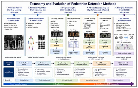
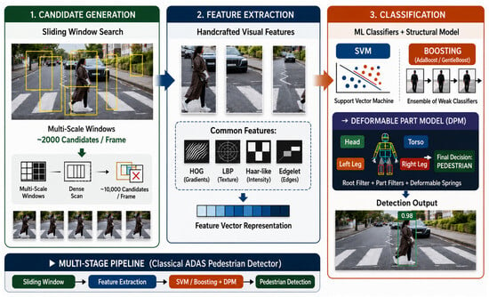
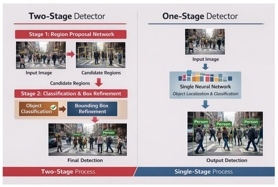
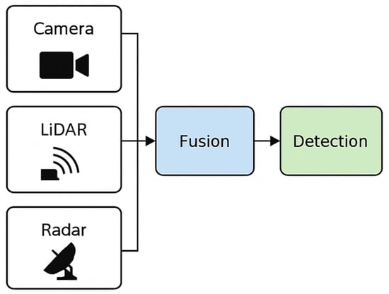
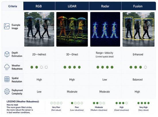
Pedestrian detection is a fundamental component of Advanced Driver Assistance Systems (ADAS) and plays a key role in collision avoidance and the safety of vulnerable road users. This paper presents a structured review of pedestrian detection methodologies developed between 2000 and 2025, spanning classical vision techniques and modern deep learning architectures. We organize the review into two phases. First, we examine classical methods, including Histogram of Oriented Gradients (HOG)+Support Vector Machine (SVM), Viola–Jones, Deformable Part Models, and Integral Channel Features, which established the conceptual foundations of the field. Then, we analyze state-of-the-art deep learning architectures, categorized by detector stage (one-stage vs. two-stage), localization strategy (anchor-based vs. anchor-free), feature extraction paradigm (Convolutional Neural Network (CNN)-based vs. transformer-based), output representation (bounding box vs. instance segmentation), and computational profile (lightweight vs. heavyweight). Several design principles introduced by classical methods remain visible in modern architectures, indicating that they were not fully superseded. The review also examines publicly available benchmark datasets and compares the strengths and limitations of camera-, Light Detection And Ranging (LiDAR)-, radar-, and multi-sensor-fusion-based systems for ADAS deployment. We close by identifying six open problems for the field: adversarial robustness, real-time inference under embedded constraints, detection under adverse weather, dataset bias and demographic fairness, the deployment of Bird’s-Eye View (BEV) and unified perception on automotive hardware, and explainability for safety-critical use.
Full article
(This article belongs to the Special Issue Computer Vision and Deep Learning: Trends and Applications (3rd Edition))
►▼
Show Figures

Figure 1
Open AccessArticle
NS-GUSL: Green U-Shaped Learning for Nuclei Segmentation from Histopathology Images
by
Catherine Aurelia Christie Alexander, Vasileios Magoulianitis, Jiaxin Yang and C.-C. Jay Kuo
J. Imaging 2026, 12(7), 316; https://doi.org/10.3390/jimaging12070316 - 10 Jul 2026
Abstract
Nuclei segmentation is a key task in digital histopathology, highlighting important aspects of nuclear morphology and topology in many cancer-related evaluations and studies. Variability in nuclear appearance both within and across different organs, stain heterogeneity, and inconsistencies in acquisition procedures contribute to the
[...] Read more.
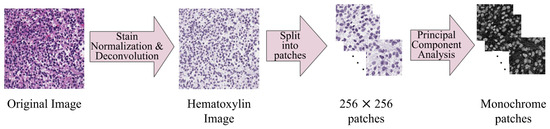
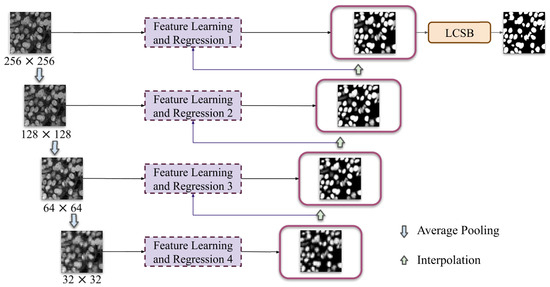
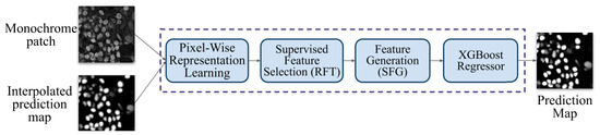
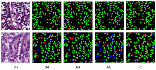
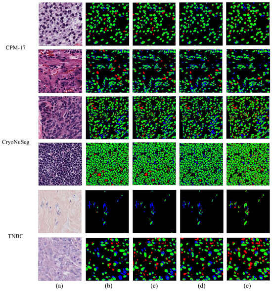
Nuclei segmentation is a key task in digital histopathology, highlighting important aspects of nuclear morphology and topology in many cancer-related evaluations and studies. Variability in nuclear appearance both within and across different organs, stain heterogeneity, and inconsistencies in acquisition procedures contribute to the complexity of the task. The existing nuclei segmentation methods apply deep learning to address these challenges, using models with millions of parameters, thereby significantly increasing computational complexity. They also face limitations in generalizing to unseen organs and slide preparations. In this paper, we propose a transparent and lightweight Green U-Shaped Learning model for nuclei segmentation (NS-GUSL). NS-GUSL features a multi-scale architecture for coarse-to-fine refinement of probability maps, which are subsequently binarized using a novel low-confidence sample binarization (LCSB) technique. The model features a modular, feed-forward feature learning scheme with unsupervised representation learning and supervised feature selection and generation. A final morphological post-processing step refines the segmentation maps to improve instance separation while preserving nuclei convexity. The model was trained and tested on the MoNuSeg dataset and compared against other deep learning baselines for segmentation performance. In addition, external validation experiments were conducted to evaluate the proposed model’s generalizability to unseen organs and staining procedures. NS-GUSL exhibits the best panoptic segmentation performance and competitive detection quality across all datasets. Moreover, our model is shown to be compact, low in computational complexity, and to have a minimal carbon footprint, compared to other deep learning models, making it a suitable choice for deployment on edge devices.
Full article
(This article belongs to the Special Issue AI-Driven Medical Image Processing and Analysis)
►▼
Show Figures

Figure 1
Open AccessArticle
B-Mode Ultrasound Radiomics for Differentiating Benign and Malignant Small Hyperechoic Renal Masses: An Exploratory Single-Center Experience
by
Fabrizio Urraro, Nicoletta Giordano, Vittorio Patanè, Marco Piscopo, Giovanni Ciani, Giovanni Balestrucci, Maria Chiara Brunese, Anna Russo, Mario Sansone and Alfonso Reginelli
J. Imaging 2026, 12(7), 315; https://doi.org/10.3390/jimaging12070315 - 10 Jul 2026
Abstract
Introduction: Small hyperechoic renal masses are frequently detected incidentally on conventional ultrasound and are often presumed to represent benign lesions, particularly angiomyolipomas. However, malignant renal tumors, including renal cell carcinoma, may also appear hyperechoic when small, creating a diagnostic challenge at first-line
[...] Read more.
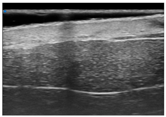
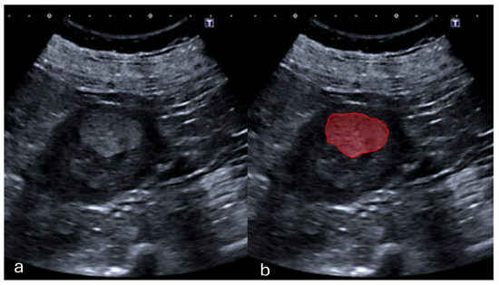
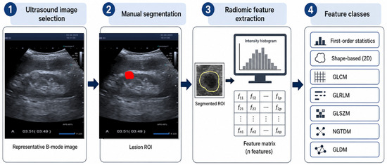
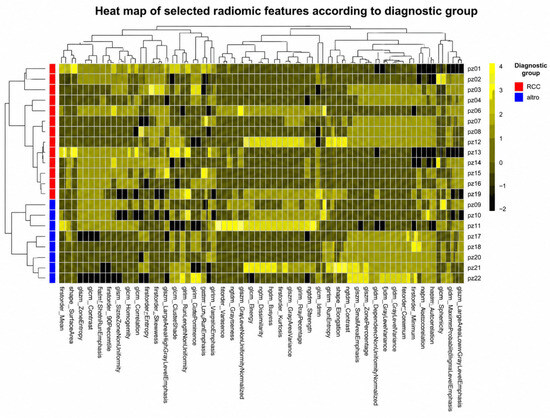
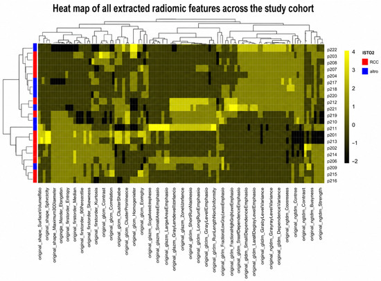
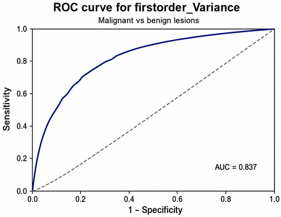
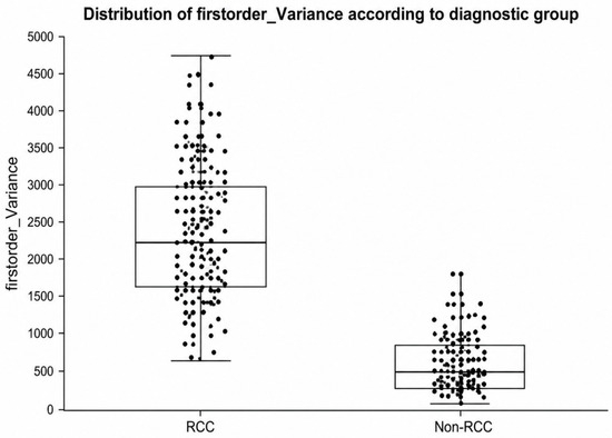
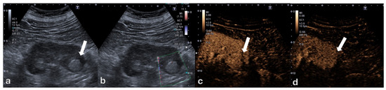
Introduction: Small hyperechoic renal masses are frequently detected incidentally on conventional ultrasound and are often presumed to represent benign lesions, particularly angiomyolipomas. However, malignant renal tumors, including renal cell carcinoma, may also appear hyperechoic when small, creating a diagnostic challenge at first-line imaging. This study aimed to evaluate the feasibility and exploratory diagnostic performance of B-mode ultrasound radiomics for differentiating benign and malignant small hyperechoic renal masses. Methods: This retrospective single-center study included adult patients with incidentally detected small hyperechoic renal masses measuring ≤3 cm and examined between July 2022 and April 2025. All lesions underwent standardized B-mode ultrasound assessment and multidisciplinary review. Final diagnosis was established by histopathology when available or by longitudinal ultrasound follow-up stability for lesions considered benign. Lesions were manually segmented on representative B-mode DICOM images, and original radiomic features were extracted using PyRadiomics version 3.0 according to standardized definitions compatible with the Image Biomarker Standardisation Initiative framework. A total of 114 original radiomic features were extracted from each lesion. The primary comparison was benign versus malignant lesions. Diagnostic performance was assessed using feature-level receiver operating characteristic analysis. Results: Forty-two lesions were included in the final radiomic cohort, including 26 malignant renal cell carcinomas and 16 benign angiomyolipomas. Malignant lesions included papillary renal cell carcinoma, chromophobe renal cell carcinoma, and clear-cell renal cell carcinoma. All malignant lesions were histologically confirmed. Among benign lesions, 14 angiomyolipomas were classified based on longitudinal ultrasound stability, whereas 2 were confirmed by ultrasound-guided percutaneous biopsy after mild dimensional increase during imaging surveillance. Among the extracted radiomic features, firstorder_Variance and firstorder_MeanAbsoluteDeviation showed the highest exploratory discriminatory performance, each achieving an area under the receiver operating characteristic curve of 0.837. Both features are first-order measures of gray-level dispersion within the segmented lesion. Higher values were observed in malignant lesions, suggesting greater intralesional grayscale heterogeneity compared with benign angiomyolipomas. Conclusions: B-mode ultrasound radiomics is feasible for the quantitative assessment of small hyperechoic renal masses and may provide complementary information for differentiating benign angiomyolipomas from malignant renal cell carcinomas. firstorder_Variance emerged as a representative candidate imaging biomarker of grayscale dispersion, with firstorder_MeanAbsoluteDeviation showing concordant performance as a related dispersion measure. These findings should be considered preliminary and hypothesis-generating and require validation in larger multicenter cohorts before clinical implementation.
Full article
(This article belongs to the Section Medical Imaging)
►▼
Show Figures

Figure 1
Highly Accessed Articles
Latest Books
E-Mail Alert
News
Topics
Topic in
Applied Sciences, Bioengineering, Diagnostics, J. Imaging, Signals
Signal Analysis and Biomedical Imaging for Precision Medicine
Topic Editors: Surbhi Bhatia Khan, Mo SaraeeDeadline: 31 August 2026
Topic in
BioMed, Cancers, Diagnostics, JCM, J. Imaging
Machine Learning and Deep Learning in Medical Imaging
Topic Editors: Rafał Obuchowicz, Michał Strzelecki, Adam Piórkowski, Karolina NurzynskaDeadline: 31 October 2026
Topic in
Applied Sciences, Electronics, J. Imaging, JMSE, Machines, Robotics, Sensors, Drones
Applications and Development of Underwater Robotics and Underwater Vision Technology, 2nd Edition
Topic Editors: Jingchun Zhou, Wenqi Ren, Qiuping Jiang, Yan-Tsung PengDeadline: 30 November 2026
Topic in
AI, Applied Sciences, Sensors, J. Imaging
Applied Computing and Machine Intelligence (ACMI): 2nd Edition
Topic Editors: Chuan-Ming Liu, Wei-Shinn KuDeadline: 31 December 2026
Conferences
Special Issues
Special Issue in
J. Imaging
From Code to Clinic: Trustworthy AI for Medical Imaging
Guest Editor: Soumick ChatterjeeDeadline: 31 July 2026
Special Issue in
J. Imaging
Self-Supervised Learning and Multimodal Foundation Models for AI-Driven Medical Imaging
Guest Editors: Andrea Loddo, Lorenzo Putzu, Cecilia Di Ruberto, Carsten MarrDeadline: 31 July 2026
Special Issue in
J. Imaging
Image and Video Forensics: Progress and Challenges
Guest Editors: Daniele Baracchi, Simone MagistriDeadline: 31 July 2026
Special Issue in
J. Imaging
AI-Driven Remote Sensing Image Processing and Pattern Recognition
Guest Editors: Zhenqi Liu, Jiaxin Li, Mengying Xie, Chuan Fu, Kaiwen WeiDeadline: 31 July 2026