


Abstract
Chronic limb-threatening ischemia (CLTI) in patients with no conventional targets for revascularization presents a formidable challenge in limb salvage. Deep venous arterialization (DVA) is an emerging endovascular approach that redirects arterial blood flow into the venous system to perfuse the ischemic foot. Despite early promising results, appropriate wound management of the ischemic foot following a DVA procedure has been described in the literature, albeit infrequently and with limited standardization. Here, we present a case of an 85-year-old male with multiple comorbidities, including peripheral artery disease and a prior right above-knee amputation (AKA), who underwent a successful left-sided DVA following an open transmetatarsal amputation (TMA) for infection. A staged wound care approach with guillotine amputation, delayed revision and skin grafting ultimately preserved his only remaining limb and allowed for ambulation. This case underscores the potential of DVA as a limb-saving option in complex “no-option” patients when paired with multidisciplinary care and tailored wound management.
1. Introduction
Chronic limb-threatening ischemia (CLTI) represents the end stage of peripheral artery disease, marked by rest pain, non-healing ulcers, and/or gangrene. CLTI carries a poor prognosis, with a one-year mortality of approximately 20–25% and five-year mortality approaching 60% [1]. In patients who undergo a major lower extremity amputation for CLTI, the outcomes are particularly grave, with one study noting post-amputation mortality of 44% at 1 year and 85% at 5 years [2]. Limb salvage is therefore paramount whenever possible.
A subset of “no-option” CLTI patients exhibit diffuse distal tibial and small pedal vessel disease, also known as “desert foot”. In these patients, small-vessel occlusion compromises distal runoff and precludes identification of a suitable outflow target for traditional surgical or endovascular revascularization [3]. For these patients, deep venous arterialization (DVA)—shunting arterial blood into the venous system of the foot—has emerged as a limb salvage option for the once “no-option” CLTI patient. The concept of venous arterialization is not new: early surgical attempts date back to the late 19th century and showed limited success. Modern iterations in the late 1970s demonstrated that anastomosing an artery to the foot’s venous arch could relieve ischemic pain and heal wounds in select patients [4,5]. Recent advances in endovascular technology have enabled percutaneous DVA using dedicated devices, making the technique viable for wider use. Early clinical reports and small case series have demonstrated successful limb salvage in no-option CLTI patients [6,7,8]. In particular, Migliara et al. and Cangiano et al. reported favorable outcomes using IVUS-guided percutaneous DVA techniques in diabetic and high-risk populations, while Alexandrescu et al. demonstrated improved perfusion and healing using deep calf vein arterialization guided by the angiosome model [6,7,8]. In the multi-center PROMISE II trial of percutaneous DVA using the LimFlow system, 76% of patients achieved limb salvage (avoidance of major amputation) at 6 months [9]. Powell et al. compared patients from the PROMISE II trial to a propensity-matched cohort of no-option CLTI patients from the CLariTI Registry who did not undergo DVA, demonstrating that DVA was associated with significantly improved outcomes, including a 29% increase in amputation-free survival [10]. Successful outcomes depend not only on the revascularization procedure but also on meticulous wound management and a multidisciplinary approach. In particular, integrating vascular surgery with podiatry and wound care (“toe and flow” team concept) is critical to maximize tissue preservation and functional outcome [11]. Here we present a case of a complex patient with no-option CLTI who underwent percutaneous deep vein arterialization after a transmetatarsal amputation (TMA), resulting in limb salvage, emphasizing the unique complexities of wound management after a partial foot amputation in the setting of a DVA procedure. We detail the clinical course, imaging findings, and wound care strategy, and discuss this case in the context of emerging literature on DVA and partial foot amputations.
2. Case Presentation
An 85-year-old male with a history of severe peripheral artery disease (PAD) and a history of a right AKA presented with chronic left forefoot gangrene that had acutely worsened with new drainage and signs of infection. His medical history included coronary artery disease, hypertension, polymyalgia rheumatica on chronic prednisone, and a prior gastrointestinal bleed. He had a former 65 pack-year smoking history and quit 9 years prior to presentation. Several months earlier, he had undergone endovascular intervention with left superficial femoral artery stenting and popliteal artery angioplasty for limb ischemia with superficial toe wounds. Despite prior revascularization attempts, his pedal perfusion remained poor, and the toes of the left foot progressively mummified (dry gangrene). He had been stable, treated with local wound care at home, until he developed nausea, vomiting, and increased malodorous discharge from the gangrenous toes, raising concern for transformation to wet gangrene. He presented to the emergency department with his wife, with elevated blood pressure (178/93 mmHg), elevated white blood cell count (14.4 × 10⁹/L), and mild lactic acidosis, indicating possible sepsis from foot infection.
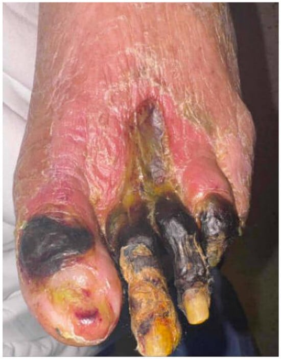
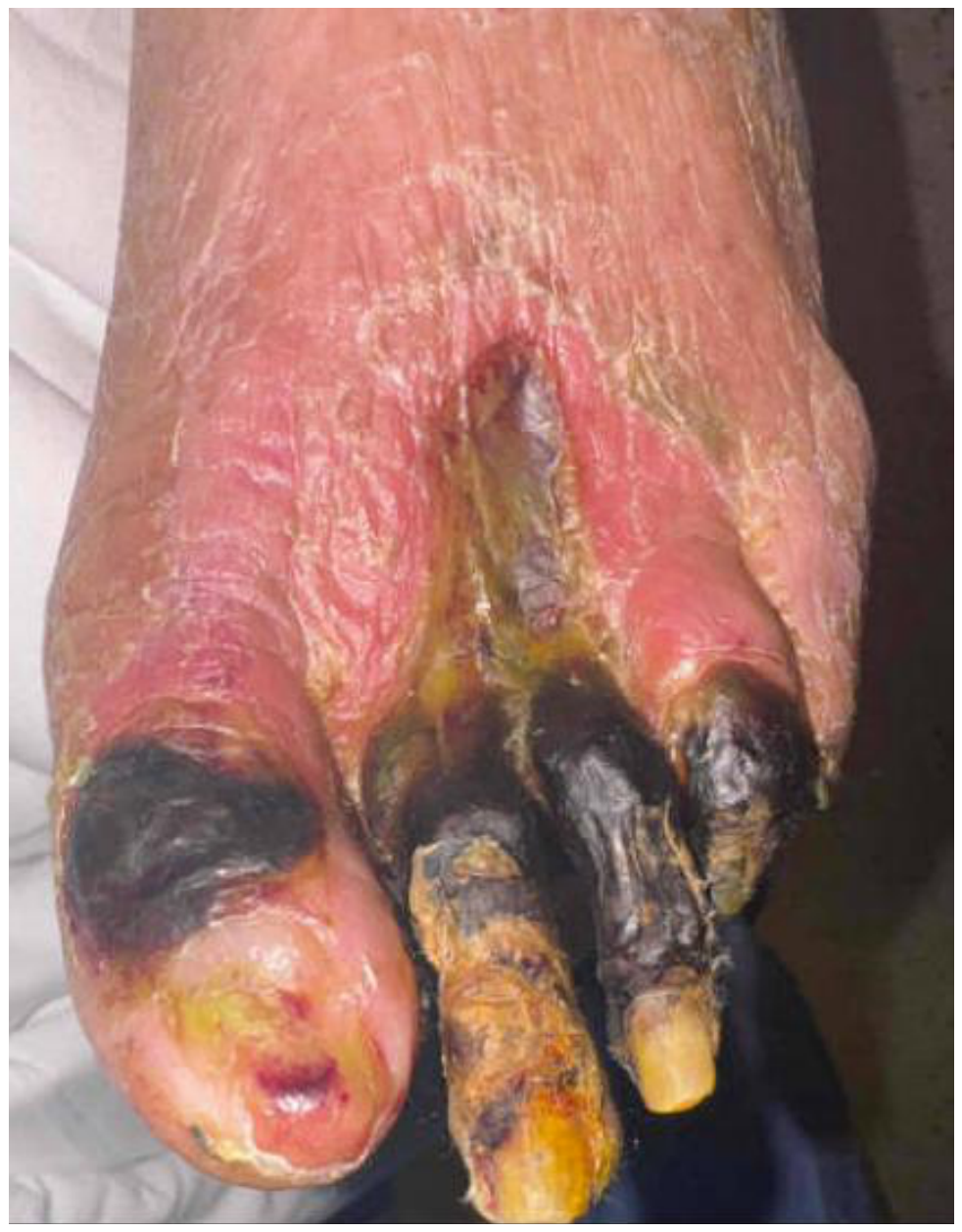
On examination, the left foot had multiple necrotic toes with foul discharge, consistent with wet gangrene (Figure 1). Given the source of sepsis in the foot and the absence of appreciable distal blood flow, an urgent limited amputation was deemed necessary for source control. The patient was transferred to the care of a specialized limb salvage team consisting of vascular surgery and podiatry. He was taken to the operating room for an urgent open guillotine transmetatarsal amputation of the left foot. The amputation was left open (no primary skin closure) with the placement of a wet-to-dry dressing. The lack of intraoperative bleeding raised concern that this midfoot stump would not heal without further intervention. The patient’s hemodynamics and inflammatory markers improved after the infected toes were removed, confirming sufficient source control.
Figure 1.
One week prior to TMA. Dry gangrene converting to wet gangrene.
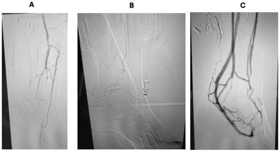
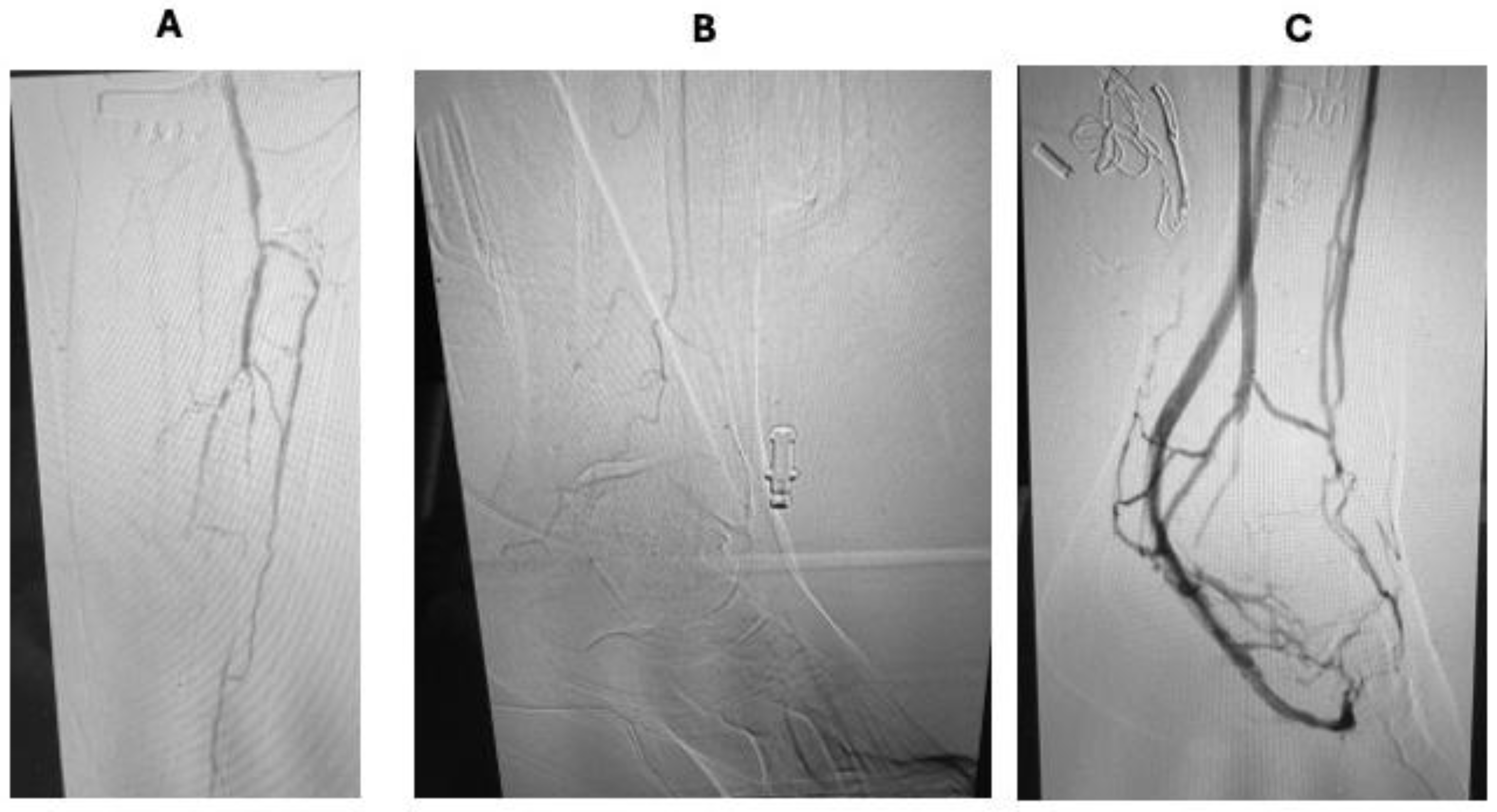
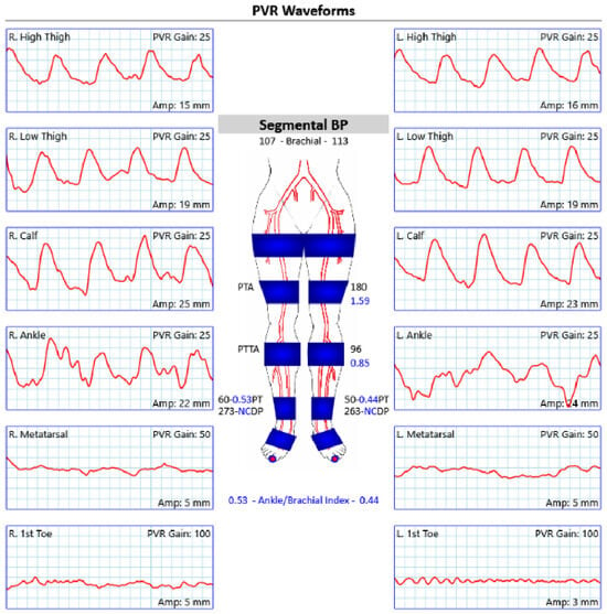
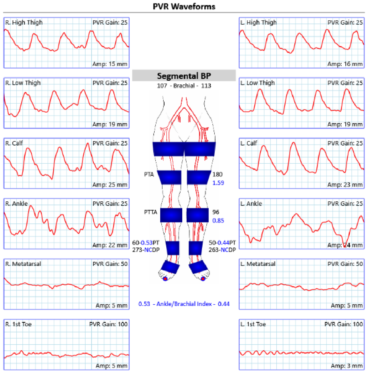
Diagnostic angiography demonstrated severe inframalleolar occlusive disease with markedly limited pedal runoff and absence of a suitable distal target vessel for bypass or conventional endovascular revascularization (Figure 2A,B). Noninvasive vascular testing demonstrated severe ischemia with an ankle-brachial index of 0.44, absent toe pressure, and severely dampened metatarsal and digital pulse volume recordings (Figure 3). These findings were consistent with a “desert foot” anatomy and supporting classification as a no-option CLTI. Given the patient’s “no-option” CLTI status (no bypass targets and failed angioplasty), the team considered deep vein arterialization for revascularization. Approximately two weeks after the TMA, following resolution of the acute infection, the patient underwent percutaneous DVA of the left lower extremity. Pre-procedure venous ultrasound mapping had shown patent deep veins in the calf and foot, including the lateral plantar vein (LPV) with a diameter ~4–5 mm (suitable for arterialization). The DVA procedure was performed in a standard fashion using the LimFlow kit (Limflow®, Inari, Irvine, CA, USA) [12]. The patient was placed in a supine position with his ipsilateral leg and foot prepped and draped. The lateral plantar vein was accessed using ultrasound guidance, and a wire was placed into the PT vein. Antegrade common femoral artery access was obtained, and an angiogram identified the PT artery remnant, which would serve as the donor site for the fistula. A crossing catheter was deployed to create a fistula between the PT artery remnant and the PT vein. A forward-facing valvulotome was passed, after which stents were placed (5 mm within the vein with a 3 mm crossing stent placed in the PT artery). The plantar venous loop was then accessed and balloon angioplastied to disrupt the valves and allow for forward arterial flow around the plantar loop. A completion angiogram showed brisk flow through the fistula into the foot (Figure 2C).
Figure 2.
Angiographic findings before and after DVA. (A) Preoperative angiography demonstrating severe tibial and pedal artery disease with markedly limited pedal runoff before DVA. (B) Preoperative angiography demonstrating poor perfusion to the foot. (C) Completion angiography demonstrating arterialized flow through the plantar venous system following DVA.
Figure 3.
Preoperative ankle-brachial index and pulse volume recording demonstrating severe bilateral peripheral arterial disease before right above-knee amputation, left transmetatarsal amputation, and deep venous arterialization. Findings demonstrate ABI values of 0.53 on the right and 0.44 on the left, absent toe pressures, and with markedly dampened pedal waveforms consistent with CLTI. Blue symbols indicate the sites of segmental pressure measurements and pulse volume recording acquisition, with the distal toe marker denoting toe pressure assessment.
The procedure was technically successful with no immediate complications. Postoperatively, the foot remained stable with no clinical signs of infection. The patient was started on anticoagulation and antiplatelet therapy for graft patency and monitored in the hospital.
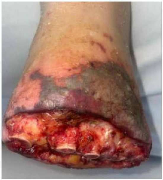
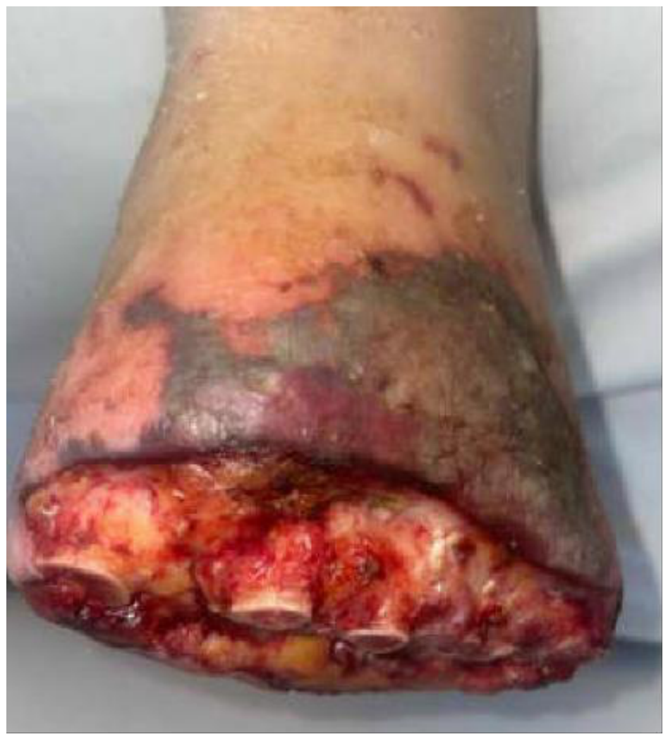
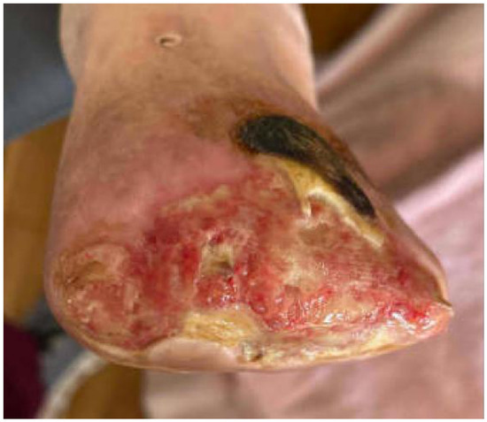
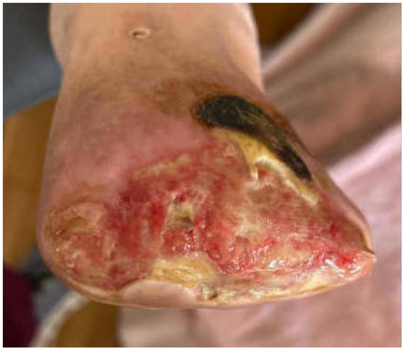
Over the next few days, the patient’s left foot pain improved and there were no signs of acute limb ischemia. There was, however, a mild progression of dry gangrene along the wound edges (Figure 4). He was fitted with a forefoot offloading postoperative shoe to protect the open TMA site and was allowed to bear weight on his heel. Once medically stable, he was transferred to an acute rehabilitation facility for continued wound care and to begin mobilization.
Figure 4.
Progression of ischemic changes to skin. Status at 16 days post-TMA and 2 days post-DVA.
3. Imaging and Procedural Follow-Up
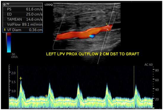
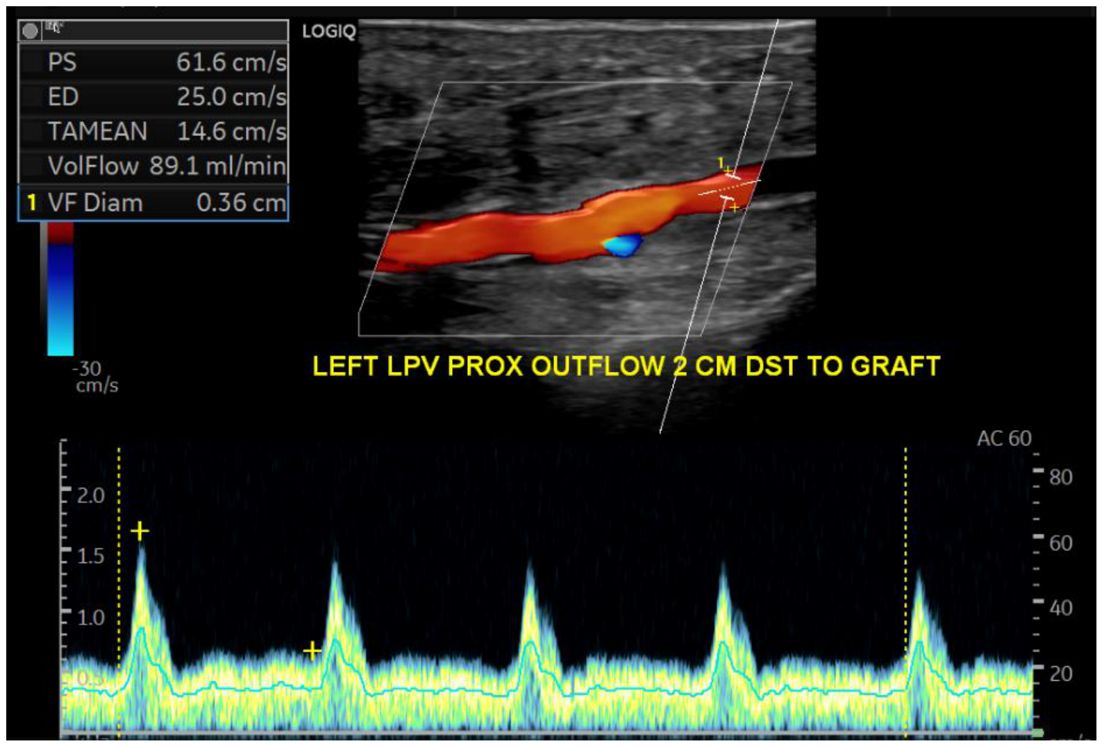
Follow-up vascular imaging demonstrated patency of the arterialized venous graft. A duplex ultrasound at approximately 17 days post-DVA showed normal monophasic flow throughout the stent graft, with no significant stenosis and forward flow into the lateral plantar vein. Peak systolic velocity measured 61.6 cm/s and volume flow within the outflow vein measured 89.1 mL/min (Figure 5). Peak systolic velocities within the graft were in the expected range (approximately 40–100 cm/s along the graft), and the outflow vein (arterialized LPV) was fully patent. These findings indicated a successful arterial-venous circuit with adequate perfusion to the foot. However, the duplex did detect an unrelated >50% stenosis in the mid-popliteal artery proximal to the graft. Given that this lesion was not limiting flow into the graft at that time, the team opted for conservative management and continued surveillance.
Figure 5.
Follow-up duplex ultrasound performed 17 days post-DVA demonstrating maturation of the DVA circuit with a patent arterialized lateral plantar vein and measured volume flow of 89.9 mL/min.
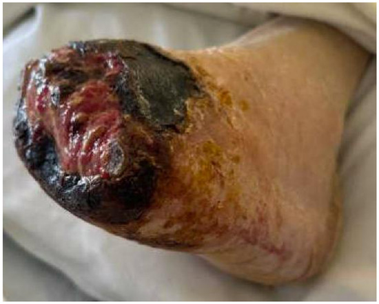
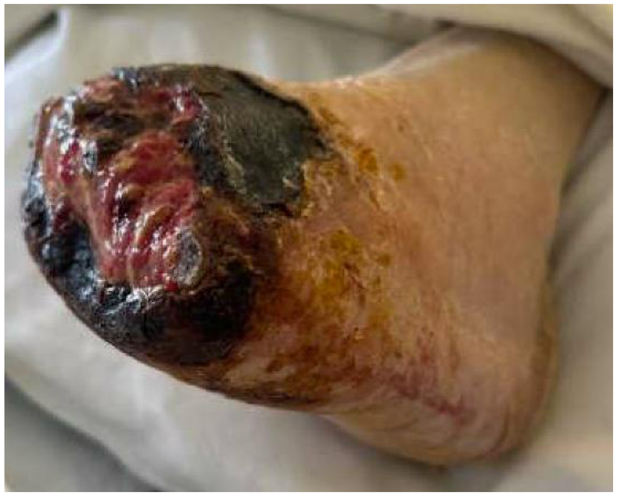
The open transmetatarsal amputation site was changed daily with Betadine paint to the skin edges and saline wet-to-dry dressings to the TMA site. As there was no evidence of acute infection, no additional debridement was performed for 3 months. The periwound necrosis initially worsened (Figure 6); however, six weeks after DVA, the midfoot wound began to show encouraging signs of healing: granulation tissue was forming within the wound base, and the necrosis was decreasing at the edges. A small heel ulcer (0.5 cm2) was also noted and was treated with offloading and local wound care.
Figure 6.
Status at 3 weeks post-DVA, initial progression of necrosis to the foot.
At approximately 11 weeks post-DVA, the foot wounds began to show more robust granulation tissue, partially covering the exposed metatarsal bones (Figure 7). During this interval, the patient’s pain had decreased and the previously gangrenous skin on the margins of the wound had begun to slough off.
Figure 7.
Healing of wound 3 months post-DVA and 3.5 months after TMA.
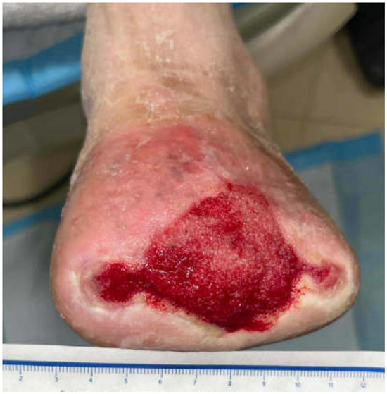
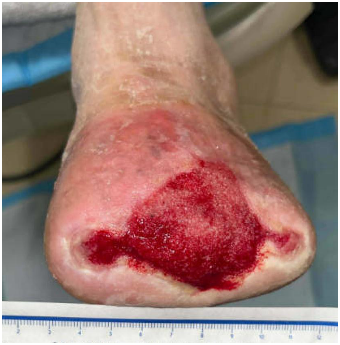
At 3 months post-DVA, the patient was brought back to the operating room for a planned TMA revision, inclusive of further resections of the metatarsals, and application of a skin substitute and wound vac. By this time, there was clear evidence of improved perfusion—the wound bed was granular and bleeding with healing wounds. Dorsalis pedis pulse was absent but a strong pulse could be palpated in the plantar venous arch of the foot. The plantar venous arch was marked using ultrasound in the preoperative area. The patient underwent revision of the TMA with resection of approximately 1 cm of the distal ends of the metatarsals to refresh the bone edges and remove any residual devitalized bone of distal metatarsals 1–5, excisional debridement of necrotic soft tissue, and application of a human umbilical tissue skin substitute (Stravix®, Smith + Nephew, Fort Worth, TX, USA) [13] on all of the wound sites which was secured with Vicryl sutures into the deep and subcutaneous tissues. No sutures were placed on the skin. A negative pressure wound therapy (Wound Vac, Solventum, Eagan, MN, USA) [14] was placed at low pressure, 75 mmHg continuous suction. Bone specimens were sent to pathology and microbiology to evaluate for chronic osteomyelitis, and empiric antibiotics were initiated. This staged approach, using a skin substitute and VAC, with delayed definitive skin grafting, was chosen to gradually achieve wound closure without compromising perfusion. The patient tolerated the procedure well and was treated with antibiotics for chronic osteomyelitis based on culture and pathology results. He was kept non-weight-bearing on the left foot for one week, then transitioned back to heel weight bearing. He was discharged back to his rehab facility and was evaluated biweekly at the wound care center.
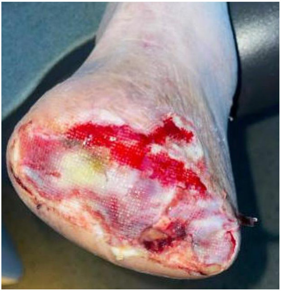
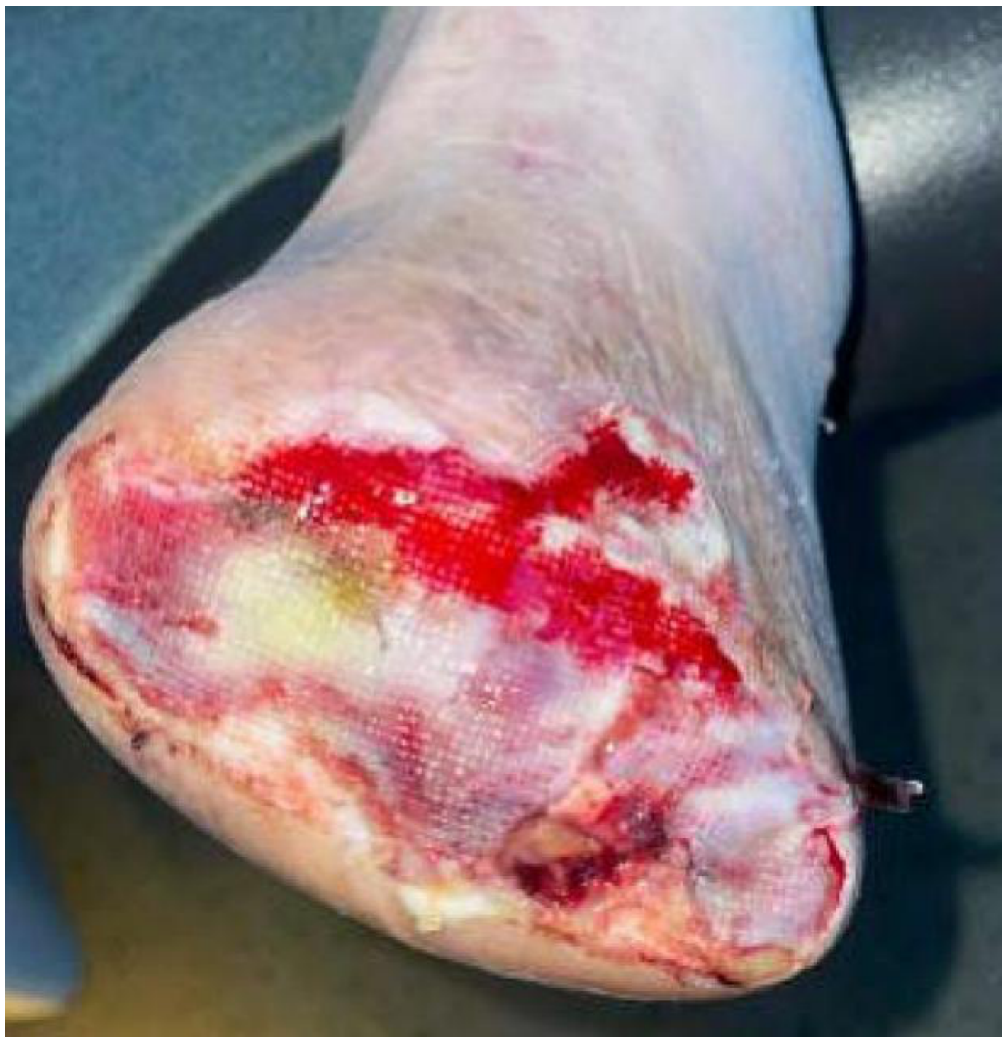
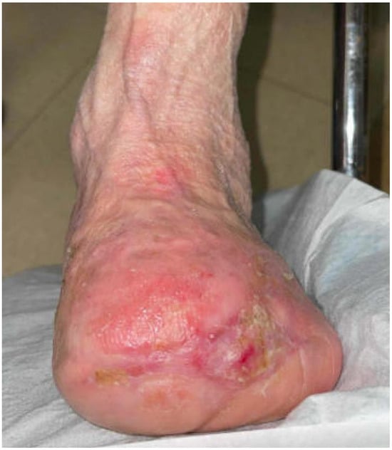
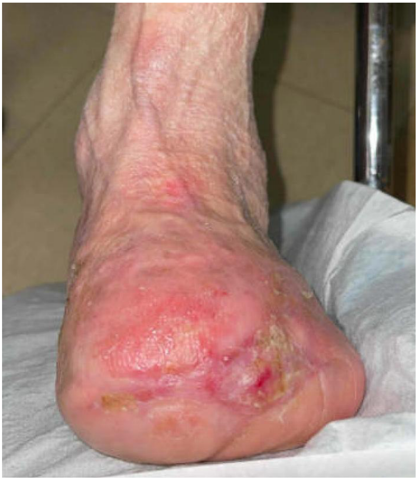
At two days post-debridement, early granulation and Stravix skin substitute adherence were evident (Figure 8). Four weeks after the TMA revision, robust granulation tissue was noted within the wound bed with no exposed bone (Figure 9). A plastic surgeon then successfully performed a split thickness skin graft over the wound site, with near-complete take of the graft (Figure 10). The wound has since fully healed, and the patient resumed full weight bearing in a diabetic shoe with a toe filler.
Figure 8.
Two days status post-debridement and application of skin substitute.
Figure 9.
Granulation tissue forming in wound bed. Status at 4 weeks post-debridement and skin substitute.
Figure 10.
Near-complete wound healing following split thickness skin grafting shown nearly 6 months after TMA.
4. Discussion
This case illustrates the successful use of percutaneous deep vein arterialization in conjunction with staged minor amputation to salvage a limb that otherwise would likely have required a below or above-knee amputation. The patient had advanced inframalleolar PAD with no distal arterial targets and gangrenous wounds, a scenario historically associated with inevitable limb loss. Conventional revascularization was not feasible, as confirmed by angiography showing no inline flow below the ankle despite previous revascularization attempts. DVA offered a last-resort option by rerouting blood flow into the venous plexus of the foot, thereby bypassing the occluded arterial tree. By converting the venous system into a low-pressure conduit for arterial blood, DVA can improve tissue perfusion in the ischemic foot. Experimental models and clinical observations have shown that within weeks of arterialization, the venous channels undergo morphological adaptation, and neo-angiogenesis occurs in the target tissues. In this patient, the return of bleeding and granulation tissue in the previously pulseless foot was a tangible sign of improved perfusion post-DVA [4].
Proper patient selection and timing are crucial to DVA success. In this case, the patient initially underwent an urgent guillotine transmetatarsal amputation to control infection, consistent with protocols that prioritize infection eradication even before full revascularization. Whenever feasible, definitive closure or reconstruction is deferred for at least 4–6 weeks post-DVA to allow for vascular maturation [15,16,17].
Postoperative foot care for patients who undergo DVA is not well-described. In our patient, we delayed wound closure by approximately 3 months. This staged approach, using a skin substitute and VAC, with delayed definitive skin grafting, was chosen to gradually achieve wound closure without compromising perfusion.
Immediately after DVA procedures, some degree of short-term pain and edema can be expected. It is theorized that this pain is due to some initial perfusion from a previous desert foot and/or demarcation of necrotic tissue. There should, however, always be a high index of suspicion for infection. In our experience, edema can occur in the treated limb and usually lasts for approximately one month. These limbs can be elevated, and light compress (≤20 mm Hg) compression can be used with caution. Moreover, it may be expected to see wound deterioration within the first 4–6 weeks after a DVA procedure. We recommend exercising caution in surgical debridement unless there is an active infection during this period. In our case, a TMA was utilized for initial source control, then the staged wound closure was delayed until 3 months after the DVA, when the perfusion was optimized and healing was most likely.
Perfusion to the skin after a DVA is found to be much more delicate than traditional revascularization. It is theorized that this is due to flow occurring through the venules rather than arterioles, which are much more sensitive to forces such as pressure and can close down more easily. The decision to leave the TMA open, rather than perform a primary closure, as well as using low-pressure settings on NPWT was therefore deliberate. Our group has found in previous cases that placing sutures to the skin after DVA nearly always leads to necrosis of the skin edges and the need for revisional surgery and a more proximal level of amputation. We have also found that low pressure (60–80 mmHg) is necessary to optimize healing, likely by decreasing the pressure on these newly forming capillary beds.
We do, however, face a challenge with closure in a wound with exposed bones after a TMA. Skin substitutes have been found to be useful to allow granulation tissue to form over these structures but cannot always achieve full closure in the larger wounds. As demonstrated in this case, a split thickness skin graft can achieve closure once the wound bed is optimized. These patients are generally poor candidates for rotational or full-thickness grafting given comorbidities and poor initial vasculature.
The importance of a multidisciplinary team cannot be overstated. Multidisciplinary coordination between vascular surgery, podiatry, plastic surgery, infectious disease, nursing and rehabilitation was essential in this case. Key decisions, including timing of amputation, wound closure, and imaging surveillance were made collaboratively, aligning with wound evolution and perfusion metrics. This approach mirrors the “toe and flow” model described by Rogers et al., which has been shown to significantly reduce major amputation rates in patients with CLTI [11].
Preserving a functional portion of the foot, as achieved with a TMA, offers significant biomechanical and rehabilitative advantages as compared to an above or below-knee amputation. TMA maintains ankle and knee joint function, reduces energy expenditure, and can facilitate ambulation with orthotic support rather than a full prosthesis in contrast to higher levels of amputation [18,19]. In contrast, higher-level amputations are associated with increased metabolic demand, reduced mobility, and greater reliance on assistive devices [20,21,22]. Our patient’s contralateral AKA dramatically raised the stakes of limb preservation. Bilateral lower-extremity amputation carries significantly higher morbidity and mortality, with 5-year mortality reported as between 61% and 69% in dysvascular populations [23,24].
Outcomes worsen with more proximal amputation, and bilateral above-knee amputation is associated with poor rehabilitation potential. In this case, deep venous arterialization enabled limb salvage and early return to heel-weight-bearing ambulation, highlighting the functional benefit of aggressive limb preservation in high-risk patients.
DVA, in this context, provided not just a chance to salvage a limb but to preserve the patient’s ability to ambulate, transfer, and engage in daily activities. The patient was heel-weight bearing with a walker within days of the guillotine TMA and is expected to progress to full weight bearing as rehabilitation and wound healing complete. This outcome underscores the functional value of successful DVA in high-risk patients, particularly those with limited remaining limb reserve.
Lepow et al. reported similar outcomes with staged open TMAs, skin substitutes, and delayed grafting, achieving limb salvage and a median time to ambulation of just two days after initial amputation [16]. Our strategy and results are consistent with this paradigm. By avoiding an immediate below-knee amputation, we not only potentially improved this patient’s survival chances but also preserved his mobility and independence to a much greater degree [2].
This case also underscores some challenges and considerations with DVA. Edema of the foot is commonly seen after DVA due to arterial pressure in the venous system; we mitigated this with limb elevation only, and it resolved in a few weeks as collateral pathways developed. Another consideration is the need for vigilant surveillance of the arterialized circuit as well as infection. Stenosis can develop in inflow or outflow segments. Indeed, our patient had a mid-popliteal arterial stenosis noted on surveillance ultrasound; although it was not flow-limiting early on, it could progress and may require angioplasty in the future. Lifelong follow-up with vascular imaging is indicated for DVA patients to monitor graft patency. Acute infection should always be treated urgently as this may quickly escalate and lead to limb loss even with a successful DVA. We were able to manage the acute infection initially with the TMA, and he remained stable with local wound care and close follow-up. Had this patient undergone DVA and there was no evidence of acute infection, a TMA would have been delayed until maturation of the arterialized circuit at 8–12 weeks post-procedure.
Finally, the favorable patency and hemodynamics of the DVA graft, confirmed on serial imaging, provide real-world validation of DVA’s potential. These findings align with recent long-term data from Migliara et al., who reported 24-month patency and sustained limb salvage after percutaneous DVA in no-option CLTI patients using an IVUS-guided approach [25]. Despite a mid-popliteal stenosis, the graft remained functional, and flow through the plantar venous arch was pulsatile and clinically effective. Vigilant follow-up remains essential, as prior studies have shown that post-procedural venous changes such as thrombosis can worsen limb outcomes in diabetic and ischemic patients [26]. This case supports the growing body of evidence that, in properly selected patients, DVA can offer durable perfusion and wound healing even in the absence of arterial runoff.
5. Conclusions
Percutaneous deep vein arterialization can be a limb-saving therapy in patients with chronic limb ischemia who have no conventional revascularization options. This case demonstrates that DVA, combined with prompt infection control and staged wound management, enabled healing of a transmetatarsal amputation in an octogenarian who likely would have otherwise lost his leg. Key factors in the successful outcome included careful patient selection, timing the reconstruction stages with the maturation of the arterialized circulation, and a multidisciplinary team approach to address vascular, infectious, and rehabilitative needs. As DVA is adopted more widely following the positive trial results, adherence to best practices, such as delaying definitive foot surgery until perfusion is established, avoiding primary closure with sutures, and using adjuncts like dermal substitutes and NPWT, will be essential to maximize limb salvage and functional recovery. This case adds to the growing body of evidence that even advanced peripheral artery disease with “desert foot” can be overcome with innovative revascularization techniques and collaborative care. Continued follow-up and reporting of outcomes are needed as we refine patient selection, procedural techniques, and postoperative protocols for DVA. In conclusion, deep vein arterialization offers new hope for no-option CLTI patients, and when paired with modern wound care, can achieve limb salvage and improve quality of life in this challenging population.
Author Contributions
Conceptualization, M.H., T.C., A.D. and S.R.-S.; Clinical care, S.R.-S.; Data curation, M.H.; Writing—original draft preparation, M.H.; Writing—review and editing, M.H., T.C., A.D. and S.R.-S. All authors have read and agreed to the published version of the manuscript.
Funding
This research received no external funding.
Institutional Review Board Statement
Ethical review and approval were waived for this study because it is a single-patient retrospective case report describing routine clinical care.
Informed Consent Statement
Verbal informed consent was obtained from the patient for publication of this case report and accompanying images. All identifying information has been removed to protect patient privacy.
Data Availability Statement
No data were used in this article.
Conflicts of Interest
The authors declare no conflicts of interest.
Abbreviations
The following abbreviations are used in this manuscript:
| TMA | Transmetatarsal amputation |
| DVA | Deep venous arterialization |
| CTLI | Chronic limb-threatening ischemia |
| AKA | Above knee amputation |
| LPV | Lateral plantar vein |
References
- Ventoruzzo, G.; Mazzitelli, G.; Ruzzi, U.; Liistro, F.; Scatena, A.; Martelli, E. Limb Salvage and Survival in Chronic Limb-Threatening Ischemia: The Need for a Fast-Track Team-Based Approach. J. Clin. Med. 2023, 12, 6081. [Google Scholar] [CrossRef] [Scilit] [PubMed]
- Klaphake, S.; de Leur, K.; Mulder, P.G.; Ho, G.H.; de Groot, H.G.; Veen, E.J.; Verhagen, H.J.; van der Laan, L. Mortality after major amputation in elderly patients with critical limb ischemia. Clin. Interv. Aging 2017, 12, 1985–1992. [Google Scholar] [CrossRef] [Scilit] [PubMed]
- Beckman, J.A.; Schneider, P.A.; Conte, M.S. Advances in Revascularization for Peripheral Artery Disease: Revascularization in PAD. Circ. Res. 2021, 128, 1885–1912. [Google Scholar] [CrossRef] [Scilit] [PubMed]
- Ho, V.T.; Gologorsky, R.; Kibrik, P.; Chandra, V.; Prent, A.; Lee, J.; Dua, A. Open, percutaneous, and hybrid deep venous arterialization technique for no-option foot salvage. J. Vasc. Surg. 2020, 71, 2152–2160. [Google Scholar] [CrossRef] [Scilit] [PubMed]
- Sheil, A.G.R. Treatment of critical ischaemia of the lower limb by venous arterialization: An interim report. Br. J. Surg. 1977, 64, 197–199. [Google Scholar] [CrossRef] [Scilit] [PubMed]
- Alexandrescu, V.; Ngongang, C.; Vincent, G.; Ledent, G.; Hubermont, G. Deep calf veins arterialization for inferior limb preservation in diabetic patients with extended ischaemic wounds, unfit for direct arterial reconstruction: Preliminary results according to an angiosome model of perfusion. Cardiovasc. Revasc. Med. 2011, 12, 10–19. [Google Scholar] [CrossRef] [Scilit] [PubMed]
- Migliara, B.; Mirandola, M.; Griso, A.; Feriani, G.; Nicoletti, C.; Lino, M. Totally Percutaneous Deep Foot Vein Arterialization in a Patient with No-Option Critical Limb Ischemia, Scheduled for Bilateral Major Amputation. J. Vasc. Interv. Radiol. 2020, 31, 1505–1507. [Google Scholar] [CrossRef] [Scilit] [PubMed]
- Cangiano, G.; Corvino, F.; Giurazza, F.; De Feo, E.M.; Fico, F.; Palumbo, V.; Amodio, F.; Silvestre, M.; Corvino, A.; Niola, R. Percutaneous Deep Foot Vein Arterialization IVUS-Guided in No-Option Critical Limb Ischemia Diabetic Patients. Vasc. Endovasc. Surg. 2021, 55, 58–63. [Google Scholar] [CrossRef] [Scilit] [PubMed]
- Shishehbor, M.H.; Powell, R.J.; Montero-Baker, M.F.; Dua, A.; Martínez-Trabal, J.L.; Bunte, M.C.; Lee, A.C.; Mugglin, A.S.; Mills, J.L.; Farber, A.; et al. Transcatheter Arterialization of Deep Veins in Chronic Limb-Threatening Ischemia. N. Engl. J. Med. 2023, 388, 1171–1180. [Google Scholar] [CrossRef] [Scilit] [PubMed]
- Powell, R.J.; Mullin, C.M.; Clair, D.G.; Shishehbor, M.H.; Dua, A. Comparison of Transcatheter Arterialization of Deep Veins to Standard of Care in Patients with No-Option Chronic Limb Threatening Ischemia. Ann. Vasc. Surg. 2024, 99, 50–57. [Google Scholar] [CrossRef] [Scilit] [PubMed]
- Rogers, L.C.; Andros, G.; Caporusso, J.; Harkless, L.B.; Mills, J.L.; Armstrong, D.G. Toe and flow: Essential components and structure of the amputation prevention team. J. Vasc. Surg. 2010, 52, 23S–27S. [Google Scholar] [CrossRef] [Scilit] [PubMed]
- LimFlow. LimFlow Avoids Vessel Trauma. Available online: https://limflow.com/us/technology/ (accessed on 6 August 2025).
- STRAVIX Umbilical Tissue Products|Smith+Nephew USA. Available online: https://www.smith-nephew.com/en-us/health-care-professionals/products/advanced-wound-management/stravix (accessed on 16 July 2025).
- V.A.C. Therapy. Available online: https://www.solventum.com/en-us/home/medical/advanced-wound-care/negative-pressure-wound-therapy/vac-therapy/ (accessed on 9 November 2025).
- Fitzpatrick, S.; Dunlap, E.; Nagarsheth, K. Utilization of Deep Vein Arterialization for Limb Salvage. J. Vasc. Ultrasound 2024, 48, 54–57. [Google Scholar] [CrossRef] [Scilit]
- Lepow, B.D. Latest Postprocedure Wound Care: Best Practices. Endovascular Today. Available online: https://evtoday.com/articles/2024-feb-supplement/latest-postprocedure-wound-care-best-practices (accessed on 3 April 2025).
- Dua, A.; Rose-Sauld, S.; Ferraro, L.; Sweeney, E. The Massachusetts General Hospital Approach to Transcatheter Arterialization of the Deep Veins for Advanced Limb Salvage: Protocols and Procedures; Springer Nature: Cham, Switzerland, 2023. [Google Scholar] [CrossRef] [Scilit]
- Anari, J.; Donegan, D. A Surgeon’s Perspective on Lower Extremity Amputation and Rehabilitation. Orthop. Res. Physiother. 2017, 3, 32. [Google Scholar] [CrossRef] [Scilit] [PubMed]
- Dudkiewicz, I.; Schwarz, O.; Heim, M.; Herman, A.; Siev-Ner, I. Trans-metatarsal amputation in patients with a diabetic foot: Reviewing 10 years experience. Foot 2009, 19, 201–204. [Google Scholar] [CrossRef] [Scilit] [PubMed]
- McCallum, R.; Tagoe, M. Transmetatarsal Amputation: A Case Series and Review of the Literature. J. Aging Res. 2012, 2012, 797218. [Google Scholar] [CrossRef] [Scilit] [PubMed]
- Waters, R.L.; Perry, J.; Antonelli, D.; Hislop, H. Energy cost of walking of amputees: The influence of level of amputation. J. Bone Jt. Surg. Am. 1976, 58, 42–46. [Google Scholar]
- Pinzur, M.S.; Gold, J.; Schwartz, D.; Gross, N. Energy demands for walking in dysvascular amputees as related to the level of amputation. Orthopedics 1992, 15, 1033–1036; discussion 1036-1037. [Google Scholar] [CrossRef] [Scilit] [PubMed]
- Inderbitzi, R.; Buettiker, M.; Enzler, M. The long-term mobility and mortality of patients with peripheral arterial disease following bilateral amputation. Eur. J. Vasc. Endovasc. Surg. 2003, 26, 59–64. [Google Scholar] [CrossRef] [Scilit] [PubMed]
- Inderbitzi, R.; Büttiker, M.; Pfluger, D.; Nachbur, B. The fate of bilateral lower limb amputees in end-stage vascular disease. Eur. J. Vasc. Surg. 1992, 6, 321–326. [Google Scholar] [CrossRef] [Scilit] [PubMed]
- Migliara, B.; Feriani, G.; Mirandola, M.; Griso, A.; Cappellari, T.F.; Nicoletti, C. Percutaneous Deep Venous Arterialization Using an IVUS-Guided Technique in no-Option Patients with Chronic Limb-Threatening Ischemia: 24-Month Results. Cardiovasc. Interv. Radiol. 2024, 47, 1727–1736. [Google Scholar] [CrossRef] [Scilit] [PubMed]
- Wang, P.C.; Chen, T.H.; Chung, C.M.; Chen, M.Y.; Chang, J.J.; Lin, Y.S.; Chu, P.H.; Peng, Y.S.; Lin, M.S. The effect of deep vein thrombosis on major adverse limb events in diabetic patients: A nationwide retrospective cohort study. Sci. Rep. 2021, 11, 8082. [Google Scholar] [CrossRef] [Scilit] [PubMed]
Disclaimer/Publisher’s Note: The statements, opinions and data contained in all publications are solely those of the individual author(s) and contributor(s) and not of MDPI and/or the editor(s). MDPI and/or the editor(s) disclaim responsibility for any injury to people or property resulting from any ideas, methods, instructions or products referred to in the content. |
© 2026 by the authors. Published by MDPI on behalf of the American Podiatric Medical Association (APMA). Licensee MDPI, Basel, Switzerland. This article is an open access article distributed under the terms and conditions of the Creative Commons Attribution (CC BY) license.