1. Introduction
Minimally invasive surgery (MIS) for hallux valgus correction has gained substantial traction due to its benefits over traditional open techniques, including reduced morbidity, faster recovery, and decreased soft tissue disruption [
1]. However, MIS techniques remain technically demanding, with no consensus on the ideal method for optimizing deformity correction [
2,
3]. One area of interest is the osteotomy location within the first metatarsal, particularly whether a more distal flat-cut osteotomy utilizing a dorsal distal to plantar proximal cut within the sagittal plane could utilize the medial eminence bone stock and thus enhance capital fragment translation and improve radiographic outcomes.
Recent studies have demonstrated reliable radiographic and clinical outcomes with MIS double bunionectomy techniques. Lewis et al. reported significant HVA improvement from 32.9° to 8.7° and IMA from 15.3° to 5.7° at two-year follow-up in 292 feet, with high patient satisfaction across all MOXFQ domains [
4]. Similarly, Nunes et al. showed correction even in severe deformities, with HVA improving from 41.2° to 11.6° and AOFAS scores rising from 41.2 to 90.9 [
5]. A meta-analysis from Alimy et al. comparing MIS and open techniques across 395 feet found no clinical differences in AOFAS, VAS, HVA, IMA, or DMAA correction, supporting the equivalence of MIS approaches [
6]. Despite these encouraging results, the optimal osteotomy positioning within the first metatarsal to maximize correction remains unclear.
The medial eminence, a characteristic bony prominence in hallux valgus deformity, represents a region of increased width at the distal metatarsal. Traditional MIS osteotomies are often performed proximally to the sesamoid apparatus, where the metatarsal narrows and may limit achievable lateral translation. In contrast, a flat cut placed obliquely through the medial eminence within the sagittal plane may take advantage of this wider region, theoretically allowing for greater lateral shift in the capital fragment and improved hallux valgus correction.
Previous studies, including those by Patel et al., have demonstrated average osteotomy positions approximately 22 mm proximal to the first metatarsophalangeal joint (MTPJ), with an average lateral shift of 6.78 mm. However, all osteotomies in those cases were performed proximally to the sesamoids, potentially limiting their corrective potential [
7]. The present study seeks to investigate whether utilizing the medial eminence as part of the osteotomy provides greater lateral translation and improved hallux valgus and forefoot width correction.
2. Methods
We conducted a retrospective radiographic analysis of 20 consecutive patients who underwent MIS bunion correction using a flat-cut osteotomy technique. All surgeries were performed by two board-certified foot and ankle surgeons. IRB approval was obtained by our institution. Inclusion criteria required the availability of preoperative and postoperative weight-bearing radiographs. Patients with prior foot surgery, inflammatory arthropathy, or incomplete radiographs were excluded.
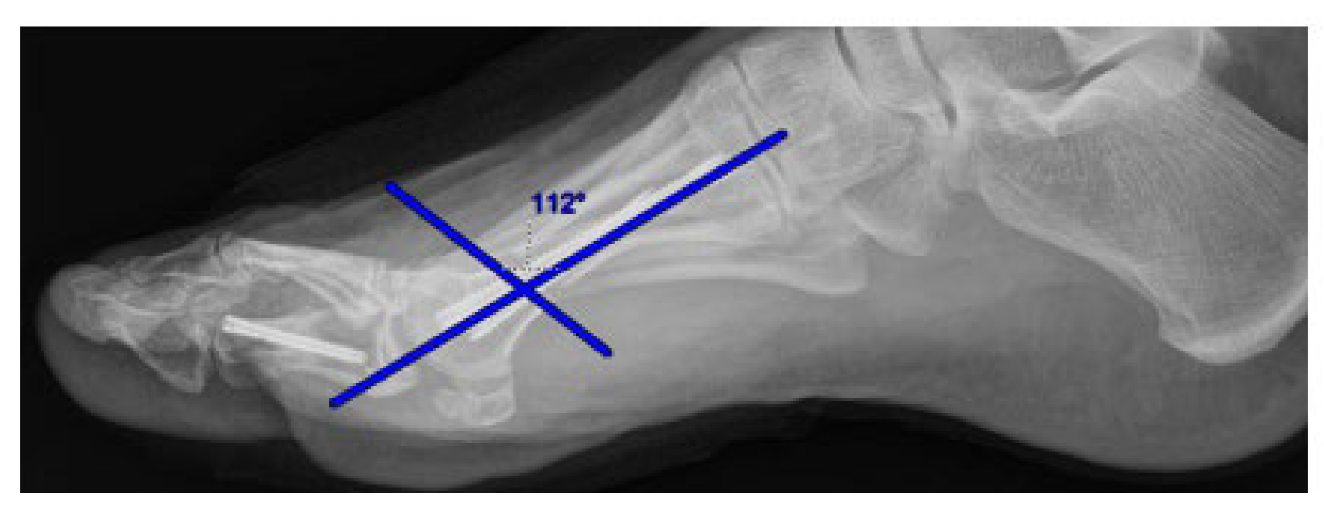
Radiographic measurements included hallux valgus angle (HVA), intermetatarsal angle (IMA), distal metatarsal articular angle (DMAA), sesamoid position, forefoot width (FW), the degree of lateral capital fragment shift expressed as a percentage (shift%), and the measurement of the angle of the 1st metatarsal osteotomy (
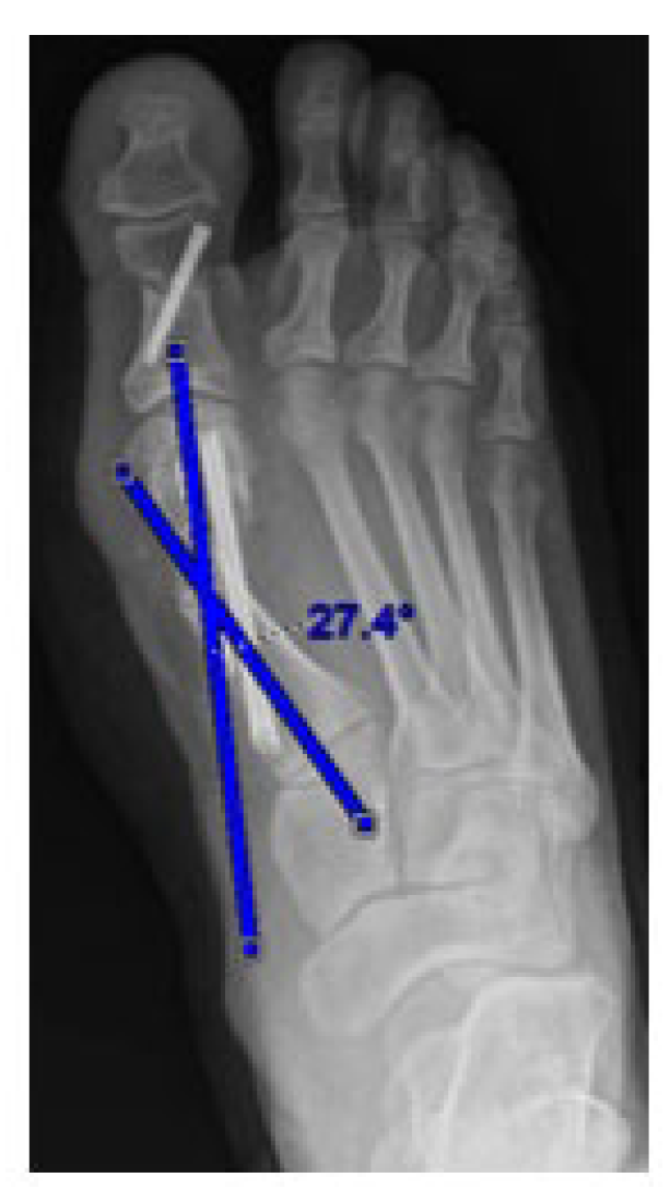
Figure 1). The osteotomy location was measured as the distance from the first MTPJ. Additionally, the width and angulation of the first metatarsal head (
Figure 2) were recorded.
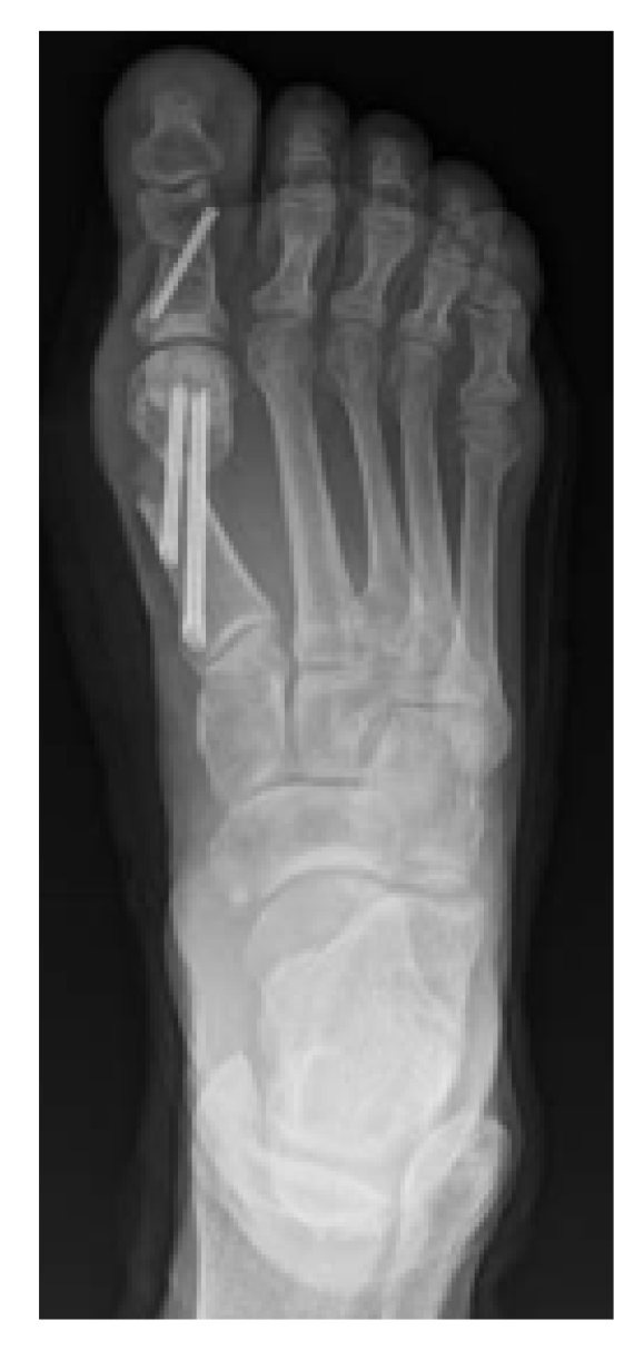
Surgical technique involved a flat-cut osteotomy within the distal first metatarsal with the plantar portion of the cut proximal to the sesamoid apparatus. The osteotomy was oriented from dorsal-distal to plantar-proximal within the sagittal plane. When the osteotomy was placed more distally dorsally, the cut traversed through the medial eminence with the prominent bony ridge at the medial aspect of the first metatarsal head. In these cases, the medial eminence served two potential biomechanical roles. First, the wider cross-sectional bone stock at the level of the medial eminence was hypothesized to provide a broader surface area for lateral translation of the capital fragment, potentially allowing greater shift before cortical overhang or instability occurred. The medial eminence was not resected separately; rather, it was incorporated into the osteotomy construct. In cases where the osteotomy was placed more proximally (away from the medial eminence), the cut traversed the narrower metatarsal neck region, where less bone stock was available for both translation and screw purchase. Fixation of the osteotomy involved a proximal 4.0 mm-cannulated fully threaded headless screw and distal 3.5 mm cannulated fully threaded headless screw. Akin osteotomies were included in all patients with fixation being a 2.5 mm cannulated fully threaded headless screw (
Figure 3).
Pearson correlation analysis was used to assess univariate associations between radiographic parameters. Multivariable linear regression was conducted to determine independent predictors of HVA and FW correction. Statistical significance was set at p < 0.05. Post hoc power analysis was performed using GPower 3.1 to evaluate the statistical power of the observed correlations and regression models given the sample size of 20 patients. Power was calculated for bivariate correlations using the exact test for the bivariate normal model, with α set at 0.05 and a two-tailed hypothesis. For multivariable regression models, power was assessed using the F-test for linear multiple regression (R2 deviation from zero). All patients were followed for a minimum of one year postoperatively. Complications measured recurrence of deformity (greater than 25% loss of correction of HAV angle), infection, non-union, or hardware failure.
3. Results
Twenty patients were included in the final analysis. The mean age was 50.5 years (range 39–64), with 19 females and one male. The average body mass index (BMI) was 30.7. No patients in the cohort had a history of smoking, chronic kidney disease, or diabetes.
The average osteotomy was performed 22 mm proximal to the first MTPJ, with a mean lateral shift of 6.78 mm. Significant correlations were observed between DMAA and HVA (r = 0.883,
p < 0.001), DMAA and IMA (r = 0.573,
p = 0.008), and HVA and shift percent (r = 0.541,
p = 0.014). Additionally, HVA showed a significant relationship with sesamoid position (r = 0.530,
p = 0.016), and forefoot width was significantly correlated with first metatarsal head angulation (r = −0.450,
p = 0.047) (
Table 1).
In multivariable linear regression modeling for HVA correction, the overall model was significant (R = 0.95, R
2 = 0.90, ANOVA
p < 0.001). Within this model, only DMAA remained independently associated with HVA correction (β = 0.679,
p < 0.001) (
Table 2). For FW correction, the regression model yielded R = 0.764 and R
2 = 0.434 (ANOVA
p = 0.020). Both first metatarsal head angulation (β = −0.514,
p = 0.017) and shift percent (β = 0.533,
p = 0.016) were significantly associated with forefoot width reduction (
Table 3).
Notably, neither the location of the osteotomy (distance from the MTPJ) nor the angle of the osteotomy (oblique > 100° vs. perpendicular 90–100°) demonstrated a statistically significant correlation with HAV correction or FW narrowing.
At one-year follow-up, there were no postoperative complications reported. Specifically, no patients developed recurrence of hallux valgus, infections, hardware-related issues, or signs of non-union.
Post hoc power analysis demonstrated that the study was well-powered to detect the observed large effect sizes. For the primary correlation between DMAA and HVA (r = 0.883), achieved power exceeded 0.99 at α = 0.05 with n = 20. For the correlation between HVA and shift percent (r = 0.541), achieved power was 0.89. However, for smaller observed effect sizes such as osteotomy location (r = −0.059) and angle of cut (r = −0.034), achieved power was approximately 0.07 and 0.06, respectively, indicating the study was underpowered to detect effects of this magnitude.
4. Discussion
This study attempted to determine whether utilizing the medial eminence in MIS bunion correction could increase capital fragment translation and improve radiographic correction of HAV and forefoot width. The medial eminence represents the widest region of the distal first metatarsal, and an osteotomy placed through this area would theoretically allow the capital fragment to be translated further laterally before reaching the limits of cortical contact, essentially using the wider bone as a “runway” for greater shift. Although theoretically appealing due to the increased bone stock present in the medial eminence, our findings suggest that osteotomy location and angle do not significantly influence the degree of radiographic correction.
The data indicates that angular deformity parameters, particularly the distal metatarsal articular angle, are more strongly associated with hallux valgus correction than the specific location or trajectory of the osteotomy cut. This suggests that the underlying deformity may play a more significant role than osteotomy location and angle. This finding may be important when considering open versus MIS technique. Kim et al., who compared MIS and open distal chevron osteotomies in 65 feet and found that MIS techniques with percutaneous K-wire fixation achieved superior DMAA correction (71.0% vs. 49.7%) compared to the open surgery group with less significant reduction in DMAA [
8]. Xu et al. similarly demonstrated that MIS approaches achieved greater DMAA correction than open techniques (14.8° to 6.3° vs. 15.1° to 8.7°), with equivalent functional outcomes [
9].
Furthermore, forefoot width reduction appeared to be more influenced by the amount of lateral shift achieved and by the coronal plane metatarsal head angulation, rather than by osteotomy cut characteristics. These findings may help guide surgical decision-making by emphasizing metatarsal head angulation and maximizing shift, rather than osteotomy positioning.
Our radiographic outcomes are comparable to those reported in larger MIS series. De Carvalho et al. reported HVA improvement from 30.4° to 11.1° and DMAA from 16.3° to 7.8° in 70 feet undergoing MIS bunion surgery for moderate-to-severe hallux valgus, with a 4.28% recurrence rate and 14.28% rate of painful hardware at minimum two-year follow-up [
10]. Lewis et al. demonstrated durable correction at five-year follow-up, maintaining radiographic correction with a 7.7% recurrence rate and 4.8% complication rate [
11]. In another study by Lewis et al. they reported on achieving HVA correction from 32.7° to 7.9° and DMAA from 18.5° to 5.6° at 12 months, with significant MOXFQ improvement [
12]. These studies collectively demonstrate that MIS techniques can achieve reliable and durable correction across a range of deformity severities.
While the hypothesis that utilizing the medial eminence would improve correction was not supported, this study contributes to the growing body of literature refining surgical techniques for MIS bunion correction. This study should be interpreted with several limitations. First, the study was limited by its retrospective design and relatively small sample size of 20 patients from a single institution, which may reduce the generalizability of the results and limit the ability to detect small associations between osteotomy characteristics and radiographic outcomes. Post hoc power analysis confirmed that the study was adequately powered to detect the large effect sizes observed for the primary correlations (DMAA and HVA power > 0.99; HVA and shift percent power = 0.89). However, the study was substantially underpowered to detect small-to-medium associations between osteotomy location, angle, and correction outcomes (achieved power 0.06–0.07). Approximately 85 patients would be required to detect a medium effect size (r = 0.3) with 80% power at α = 0.05. Therefore, the non-significant findings regarding osteotomy location and angle should be interpreted with caution, as clinically meaningful but smaller associations may exist that this study was unable to detect. A formal a priori power analysis was not performed prior to data collection, which is a limitation of the retrospective design. The present study was intended to evaluate whether utilization of the medial eminence during MIS bunion correction was associated with improved radiographic corrections rather than establishing definitive treatment recommendations. Despite the limited cohort size, significant associations were identified between several radiographic parameters and correction outcomes. Nevertheless, larger multicenter studies are warranted to confirm these findings, improve external validity, and further evaluate the influence of osteotomy location and morphology on hallux valgus correction. Additionally, this study did not include patient-reported outcome measures such as the AOFAS score, VAS, or MOXFQ. The AOFAS and VAS are the most commonly used PROMs in hallux valgus surgery research, though the MOXFQ has been recommended as a more valid alternative. Future prospective studies should incorporate these tools to correlate radiographic correction with patient satisfaction, pain, and functional improvement.
The absence of complications at one-year follow-up supports the use of MIS bunion correction using a flat-cut osteotomy. Despite concerns related to fixation and healing in MIS procedures, our findings suggest a low risk of recurrence, infection, non-union, or hardware failure in properly selected patients. This favorable safety profile is consistent with the broader MIS literature, though our small sample size limits generalizability. Miranda et al., in a systematic review of 1157 percutaneous hallux valgus procedures, reported complication rates including joint stiffness (18.47%), recurrence (15.2%), infection (7.6%), and transfer metatarsalgia (5.4%) [
13]. Lonati et al., in a systematic review of 22 MIS studies, found faster recovery, higher satisfaction, and fewer wound complications compared to open surgery, though open techniques were preferred for severe deformities due to the reliability of the procedure [
14]. Larger prospective studies are needed to confirm the low complication rates observed in the present cohort.
5. Conclusions
In this case series of patients undergoing MIS bunion correction with a flat-cut osteotomy, neither osteotomy location nor angle was significantly associated with improved hallux valgus or forefoot width correction. Greater correction was associated with angular deformity parameters, particularly distal metatarsal articular angle, and the degree of lateral capital fragment translation. While the medial eminence may theoretically provide increased surface area for lateral shift, its utilization during osteotomy did not result in significantly enhanced correction in this study.
Further research with multicenter studies for larger patient population is warranted to clarify the role of medial eminence utilization for MIS bunion correction.
Author Contributions
Conceptualization, D.L., J.H., L.R., V.A. and R.A.; methodology, D.L., J.H., L.R. and V.A.; software, C.-I.C.; validation, D.L. and C.-I.C.; formal analysis, D.L. and C.-I.C.; investigation, D.L., J.H., L.R. and C.-I.C.; resources, D.L., J.H. and L.R.; data curation, D.L. and C.-I.C.; writing—original draft preparation, D.L., J.H. and L.R.; writing—review and editing, D.L., J.H., L.R., V.A. and R.A.; visualization, D.L. and C.-I.C.; supervision, V.A. and R.A.; project administration, D.L. All authors have read and agreed to the published version of the manuscript.
Funding
This research received no external funding.
Institutional Review Board Statement
The study was conducted in accordance with the Declaration of Hlsinki and approved by the Institutional Review Board of Trinity Health Livonia protocol number E-24-1097 on 6 February 2024.
Informed Consent Statement
Patient consent was waived due to study being restrospective via chart review.
Data Availability Statement
Data unavailable due to privacy issues.
Acknowledgments
During the preparation of this manuscript/study, the authors used Chatgpt-5.5 for the purposes of proofreading. The authors have reviewed and edited the output and take full responsibility for the content of this publication.
Conflicts of Interest
The authors declare no conflicts of interest.
References
- Maffulli, N.; Longo, U.G.; Marinozzi, A.; Denaro, V. Hallux valgus: Effectiveness and safety of minimally invasive surgery. A systematic review. Br. Med. Bull. 2011, 97, 149–167. [Google Scholar] [PubMed]
- Brogan, K.; Voller, T.; Gee, C.; Borbely, T.; Palmer, S. Third-generation minimally invasive correction of hallux valgus: Technique and early outcomes. Int. Orthop. 2014, 38, 2115–2121. [Google Scholar] [CrossRef] [PubMed]
- Jowett, C.R.; Bedi, H.S. Preliminary results and learning curve of the minimally invasive chevron akin operation for hallux valgus. J. Foot Ankle Surg. 2017, 56, 445–452. [Google Scholar] [CrossRef] [PubMed]
- Lewis, T.L.; Ray, R.; Miller, G.; Gordon, D.J. Third-generation minimally invasive chevron and Akin osteotomies (MICA) in hallux valgus surgery: Two-year follow-up of 292 cases. J. Bone Jt. Surg. 2021, 103, 1203–1211. [Google Scholar]
- Nunes, G.A.; de Carvalho, K.A.M.; Ferreira, G.F.; Filho, M.V.P.; Baptista, A.D.; Zambelli, R.; Vega, J. Minimally invasive Chevron Akin (MICA) osteotomy for severe hallux valgus. Arch. Orthop. Trauma Surg. 2023, 143, 5507–5514. [Google Scholar] [CrossRef] [PubMed]
- Alimy, A.-R.; Polzer, H.; Ocokoljic, A.; Ray, R.F.; Lewis, T.L.M.; Rolvien, T.; Waizy, H. Does minimally invasive surgery provide better clinical or radiographic outcomes than open surgery in the treatment of hallux valgus deformity? A systematic review and meta-analysis. Clin. Orthop. Relat. Res. 2022, 481, 1143–1155. [Google Scholar] [CrossRef] [PubMed]
- Patel, R.; Siddiqui, N.; Dreyer, M.A.; Lam, K.; Ayyagari, V.; Onica, A. Radiographic and Cadaveric Analysis of Minimally Invasive Bunionectomy Osteotomy Position—“MIS Bunion Sweet Spot”. Foot Ankle Spec. 2024, 17, 358–364. [Google Scholar] [PubMed]
- Kim, J.; Oh, M.; Kyeong, T.H.; Na Choi, M.; Lee, S.Y. Radiographic comparison of open and minimally invasive distal chevron metatarsal osteotomy in patients with hallux valgus. J. Foot Ankle Surg. 2024, 63, 386–391. [Google Scholar] [CrossRef] [PubMed]
- Xu, Y.; Guo, C.-J.; Li, X.-C.; Xu, X.-Y. Radiographic and clinical outcomes of minimally invasive surgery versus open osteotomies for the correction of hallux valgus. Int. Orthop. 2022, 46, 1767–1774. [Google Scholar] [CrossRef] [PubMed]
- De Carvalho, K.A.M.; Baptista, A.D.; de Cesar Netto, C.; Johnson, A.H.; Dalmau-Pastor, M. Minimally invasive chevron-Akin for correction of moderate and severe hallux valgus deformities: Clinical and radiologic outcomes with a minimum 2-year follow-up. Foot Ankle Int. 2022, 43, 1317–1330. [Google Scholar] [CrossRef] [PubMed]
- Lewis, T.L.; Robinson, P.W.; Ray, R.; Dearden, P.M.C.; Goff, T.A.J.; Watt, C.; Lam, P. Five-year follow-up of third-generation percutaneous chevron and Akin osteotomies (PECA) for hallux valgus. Foot Ankle Int. 2023, 44, 104–117. [Google Scholar] [PubMed]
- Lewis, T.L.; Lau, B.; Alkhalfan, Y.; Trowbridge, S.; Gordon, D.; Vernois, J.; Lam, P.; Ray, R. Fourth-generation minimally invasive hallux valgus surgery with metaphyseal extra-articular transverse and akin osteotomy (META): 12 month clinical and radiologic results. Foot Ankle Int. 2023, 44, 178–191. [Google Scholar] [PubMed]
- Miranda, M.A.M.; Martins, C.; Cortegana, I.M.; Campos, G.; Pérez, M.F.M.; Oliva, X.M. Complications on percutaneous hallux valgus surgery: A systematic review. J. Foot Ankle Surg. 2021, 60, 548–554. [Google Scholar] [CrossRef] [PubMed]
- Lonati, D.; Kannegieter, E.; McHugh, D. A multi-dimensional systematic review of minimally invasive bunion surgery (MIBS). J. Clin. Med. 2025, 14, 2757. [Google Scholar] [CrossRef] [PubMed]
| Disclaimer/Publisher’s Note: The statements, opinions and data contained in all publications are solely those of the individual author(s) and contributor(s) and not of MDPI and/or the editor(s). MDPI and/or the editor(s) disclaim responsibility for any injury to people or property resulting from any ideas, methods, instructions or products referred to in the content. |
© 2026 by the authors. Published by MDPI on behalf of the American Podiatric Medical Association (APMA). Licensee MDPI, Basel, Switzerland. This article is an open access article distributed under the terms and conditions of the Creative Commons Attribution (CC BY) license.



 ,
,