1. Introduction
Drop foot, characterized by difficulty lifting the front of the foot, “toes to the nose” dorsiflexion, or complete lack of any dorsiflexion due to nerve damage, affects individuals of varying ages and backgrounds. While the exact incidence remains elusive, studies suggest that its prevalence ranges from 19 per 100,000 in the general population to 0.79% following specific procedures like total knee replacement [
1]. Common Peroneal Nerve (CPN) entrapment, where compression of the nerve disrupts its function, is a frequent etiology of drop foot, with a reported incidence similar to other well-known nerve entrapment syndromes such that it is the third most common type of nerve entrapment seen in human pathology [
2,
3]. Understanding the link between CPN entrapment and drop foot incidence paves the way for improved diagnosis, treatment (surgical decompression), and ultimately, improved patient outcomes. Zhong et al. demonstrated in diabetic laboratory rats that the compromise in peripheral nerve vascular perfusion due to the impairment of the microvasculature from entrapment can be reversed by surgical decompression [
4].
We have described the Phoenix Sign previously as a diagnostic peripheral nerve block that holds important and promising value not only in aiding an accurate diagnosis of peripheral nerve entrapment of the Common Peroneal (Fibular) nerve, but also with prognosis from surgical nerve decompression [
5]. The Phoenix Sign nerve block represents a diagnostic technique utilizing an ultrasound-guided subanesthetic dose of lidocaine administered to the common fibular nerve. This phenomenon was coined the “Phoenix Sign” because once non-functioning common fibular nerves with drop foot temporarily “rose from the ashes,” allowing patients to suddenly regain the ability to dorsiflex their foot. The technique involves ultrasound-guided infiltration of approximately 0.3–0.5 mL of 1% lidocaine adjacent to the common fibular nerve, with motor strength improvement typically occurring within four minutes post infiltration. The mechanism is believed to involve restoration of neural circulation through improved local blood flow to the vasa nervorum, producing temporary but significant strengthening of the affected muscles [
5,
6]. Nirenberg described a similar lidocaine injection test involving 4 cc of 1% lidocaine injected into the peroneus longus muscle at the site of common fibular nerve entrapment, which demonstrated positive results in 19 of 21 patients and successfully predicted surgical outcomes in 17 patients who underwent nerve decompression [
7]. The related ultrasound-guided lidocaine infiltration test (USLIT), described by Iborra and colleagues, applies similar principles to tarsal tunnel syndrome diagnosis, involving perineural injection of 0.5 mL of 1% lidocaine to the tibial nerve under real-time ultrasound guidance [
8]. The USLIT technique demonstrated improvements in both symptoms and nerve conduction velocity in a prospective study of 61 patients, serving as both a diagnostic tool and predictor of surgical decompression outcomes [
8].
In addition to an assessment of motor strength, which was evaluated in previous studies, this clinical study aims to evaluate microvascular and macrovasculature changes that have been previously observed clinically after an administration of a peripheral perineural infiltration of the Common Peroneal (Fibular) with 1% lidocaine without epinephrine, and to test the hypothesis that these objective findings may be due to a vasodilatory component of the lidocaine versus other possible neurological mechanisms. We performed pre- and post-infiltration evaluations of motor strength with dynamometry and manual motor testing, macrovasculature status measured with ultrasound Doppler waveform analysis and velocity of flow, and microvascular status via Kent Snapshot NIR imaging.
By comparing the block given with lidocaine versus papaverine (a known vasodilator), the vasodilation hypothesis was tested in this study.
2. Materials and Methods
A randomized control trial, NCT06919289, was conducted at Indiana Foot & Ankle in Jasper, Indiana, under the approval of the Memorial Hospital and Health Care Center Institutional Review Committee. This study was a proof-of-concept study that included four diabetic neuropathic patients with bilateral anterior compartment weakness of dorsiflexion. While small sample sizes are often a concern, we decided to test our hypothesis with a small pilot study (N = 4). We were encouraged that with only 4 subjects, there were statistically significant p-values, which can be exploratory, and note that this is an underpowered study. These subjects were included if they were diabetic and neuropathic. A documented history of diabetes mellitus had been medically established before their enrollment with demonstrative neurosensory symptoms consistent with diabetic peripheral neuropathy.
Exclusion criteria prevented enrollment of subjects who were neuropathic but in which the etiology was not confirmed to be due to diabetes. Trypanophobia was also included as an exclusion criterion, as well as any patient with a known allergy to local anesthetics or papaverine. Each of these study participants were also affected with severe sensory neuropathic impairment. A total of 8 extremities were evaluated in the 4 patients. A detailed informed consent process was completed for every patient per IRB approval.
Given this was an initial proof-of-concept pilot study aimed at exploring feasibility and preliminary efficacy, a formal sample size calculation was not performed. We intentionally selected a small sample size (n = 4) consisting of diabetic neuropathic patients with bilateral anterior compartment weakness of dorsiflexion to evaluate the potential impact of our intervention. Despite the limited number of participants, statistically significant outcomes with favorable p-values were obtained, suggesting meaningful preliminary evidence to warrant future studies with larger, adequately powered cohorts.
This study employed a randomized, double-blind design. Randomization of the first extremity was conducted using a simple coin toss method, determining allocation to receive either a perineural infiltration of 0.5 cc papaverine HCL (30 mg/mL, total dose 15 mg American Regent, Inc., Shirley, NY, USA) or 0.5 cc of 1% lidocaine without epinephrine (Hospira, Inc., Lake Forest, IL, USA). In order to maintain blinding in the study, the dosage of 1% lidocaine was increased to 0.5 cc to match the volume in the syringe to the 0.5 cc dose of papaverine. The lidocaine dosage used in previous studies was 0.3 cc, but the increase to 0.5 cc to assure blinding was within a range that would not affect outcomes of the infiltrations based on previous clinical usage experience.
The second extremity automatically received the alternate agent not assigned to the first side, thereby ensuring each participant served as their own control for intra-individual comparisons. Our use of contralateral limbs as the unit of analysis assumes independence between limbs, though shared patient-level factors introduce some statistical dependency. This may modestly affect our confidence intervals and should be considered when interpreting our findings.
Random allocation sequences were generated immediately prior to intervention through this coin toss procedure, and no blocking or other restrictions were applied due to the pilot nature and small sample size. Allocation concealment was achieved by having a separate clinical investigator who was not involved in performing infiltrations or outcome assessments, who drew up medications out of sight of both the patient and the clinician administering the infiltrations. Both participants and clinicians performing the infiltrations remained blinded to treatment allocation throughout the intervention and data collection phases, with unblinding occurring only after final data completion.
All infiltrations were performed under real-time ultrasound guidance using an Alpinion X-CUBE ultrasound system (Alpinion Medical Systems Co., Ltd., Seoul, Republic of Korea), with the Common Peroneal (Fibular) nerve identified at the fibular neck. Ultrasound imaging also confirmed accurate peri-neural administration, indicated by the development of a hypoechoic signal intensity adjacent to the nerve.
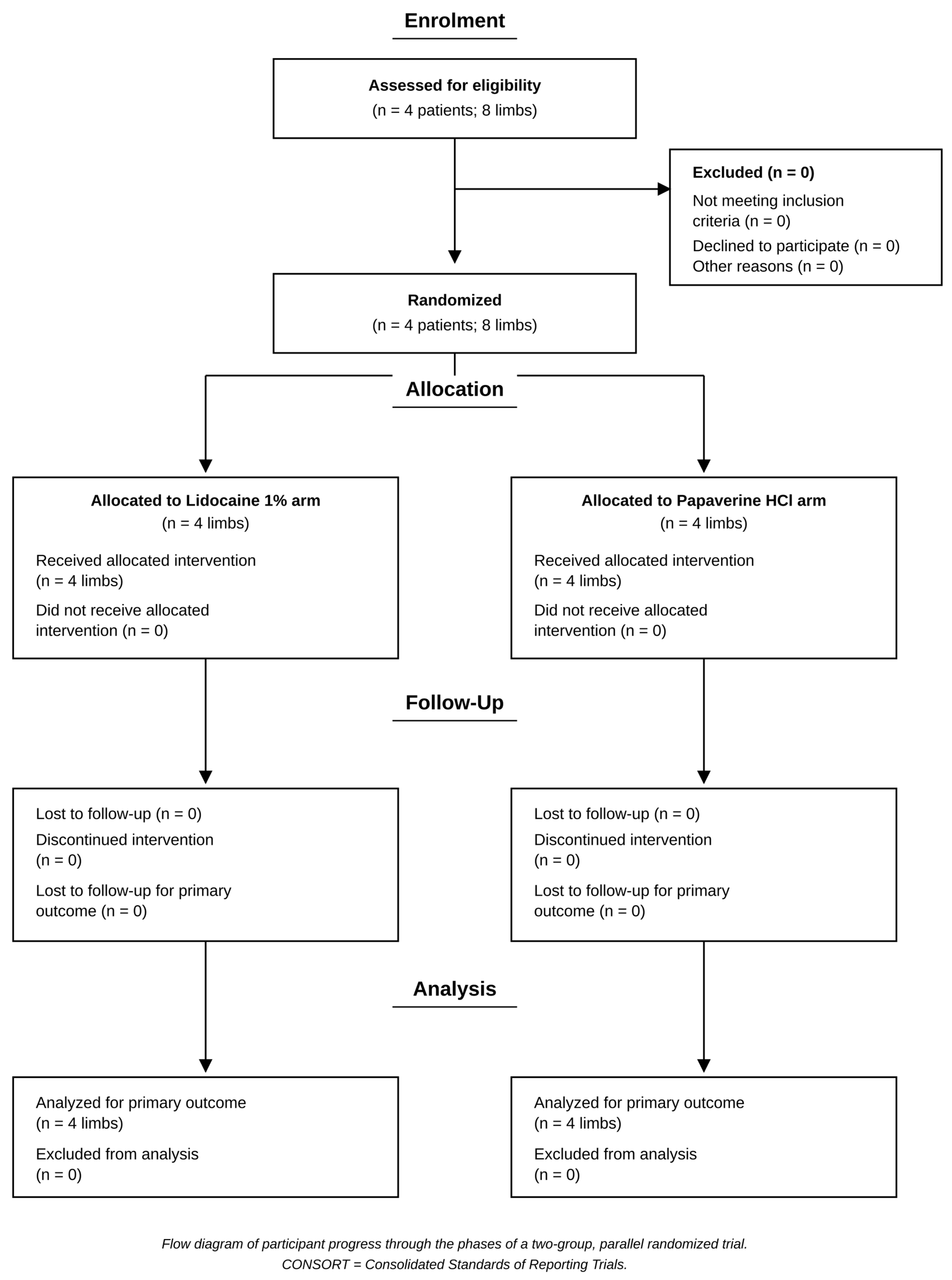
Participants were recruited between 6 and 8 March 2024. Follow-up evaluations were completed within 10 min post infiltration for each participant. No additional long-term follow-up was conducted due to the acute design of this proof-of-concept study. The trial concluded after successful enrollment of the planned pilot cohort (n = 4) and completion of all scheduled interventions and immediate follow-up assessments. The study ended as planned upon fulfilling its objective of assessing preliminary feasibility and effectiveness. There were no adverse events, safety issues, or logistical problems that prompted early termination (see
Table 1).
The macrovasculature was evaluated pre and post infiltration using ultrasound Doppler waveform analysis and velocity of flow at the level of the medial malleolus for the posterior tibial artery and on the dorsum of the foot at the level of the bases of the 1st and 2nd metatarsals for the dorsalis pedis artery, while the microvascular status was evaluated via measurements from the Kent Snapshot NIR camera (Kent Scientific Instruments Ltd. SnapShot NIR-2600, KentAnalyzer software Pro v3.2.1 Calgary, AB, Canada) measuring hemoglobin, deoxyhemoglobin, SPO2, and total hemoglobin. The Snapshot NIR camera allows for the simultaneous capture of data across multiple wavelengths, forming a “spectral image cube” that contains both spatial and spectral information [
9,
10].
The Snapshot NIR camera is being explored for its potential use in assessing dermal microvascular perfusion, which refers to the delivery of blood to tissues. Its application in this field leverages the unique properties of near-infrared (NIR) light. Specific NIR wavelengths are readily absorbed by hemoglobin, the oxygen-carrying protein in red blood cells. By measuring the absorption and scattering of NIR light within tissues, the Snapshot NIR camera can potentially estimate hemoglobin concentration and oxygenation levels, providing insights into blood flow and tissue health [
9,
10].
Movement Against Manual Resistance (MAMR) was evaluated using a scale of 1–5 out of 5. This was normalized so that 1/5 = 0.2, and 5/5 = 1.0. This was performed on three muscles of the anterior compartment, extensor hallucis longus (EHL), tibialis anterior (TA), and extensor digitorum longus (EDL) and the results for the three muscles were averaged. Each patient was placed in a seated supine position and tested by a single evaluator for measurement on each muscle tested, as MAMR is a semi-subjective testing modality. In order to account for possible errors in the intra-rater reliability, a minimum of 3 muscle strength assessments was performed to aid in consistency of assessment, in addition to use of the dynamometer for every muscle tested.
The EHL was evaluated with a dynamometer (Mxmoonfree digital force gauge; Mxmoonfree, Shenzhen, China), in Newtons (N), separately to augment intra-rater reliability.
Prior to infiltration the following data was collected:
Kent Snapshot NIR imaging of the dorsum of the foot (entire dorsum).
Kent Snapshot NIR imaging of the plantar aspect of the foot (entire plantar foot).
Motor strength of EHL.
Waveform of dorsalis pedis artery.
At 4 min post infiltration the following was measured:
Kent Snapshot NIR imaging of the dorsum of the foot (entire dorsum).
Kent Snapshot NIR imaging of the plantar aspect of the foot (entire plantar foot).
Motor strength of EHL.
Waveform of dorsalis pedis artery.
Additionally, at 10 min post infiltration the Kent Snapshot measurements were taken again of the dorsum and plantar aspects of the foot.
4. Discussion
In 2017, we first observed a phenomenon serendipitously in a patient who had a 5-year history of drop foot and was seen for a chief complaint of pain. She was given a diagnostic block with plain lidocaine at the level of the fibular neck to block the Common Peroneal (Fibular) nerve to determine whether this nerve distribution was primarily responsible for the majority of the patients presenting with pain. Her pain was relieved incidentally with the nerve block, but interestingly she recovered motor strength and was able to fully dorsiflex actively at the ankle. This motor strength was temporary and lasted only about 10 min. Because this was a highly unexpected clinical finding, she was brought back the following week to reconfirm the block again. The same results were observed. Finally, she was brought back for a third infiltration with lidocaine with epinephrine which did not show the return of any significant motor strength. This led us to the hypothesis that this phenomenon could be due to vasodilation. While primarily known for its local anesthetic properties, lidocaine also exhibits vasodilator effects, meaning it can cause blood vessels to widen and increase blood flow. This effect is believed to be attributed to two main mechanisms: Sodium channel blockade: Lidocaine primarily acts by blocking sodium channels in nerve cells, which prevents the generation of nerve impulses and numbs the area. However, these sodium channels are also present in the smooth muscle cells that line blood vessels. By blocking some sodium channels in these cells, lidocaine can interfere with their ability to constrict, leading to a relaxation and widening of the vessel [
11]. Indirect vasodilation: Lidocaine might also indirectly induce vasodilation by inhibiting the release of norepinephrine from sympathetic nerve endings. Norepinephrine is a neurotransmitter that causes blood vessels to constrict, so its reduced release due to lidocaine can lead to vasodilation [
11,
12,
13]. A dosage of 0.5 cc was based on achieving a sub-anesthetic dose of the agent and for blinding purposes as previously described.
It is important to note that lidocaine’s vasodilating effect is weaker compared to dedicated vasodilator medications like papaverine. Additionally, other factors like the concentration of lidocaine and the presence of other drugs like epinephrine (which causes vasoconstriction) can influence its overall vasodilatory effect.
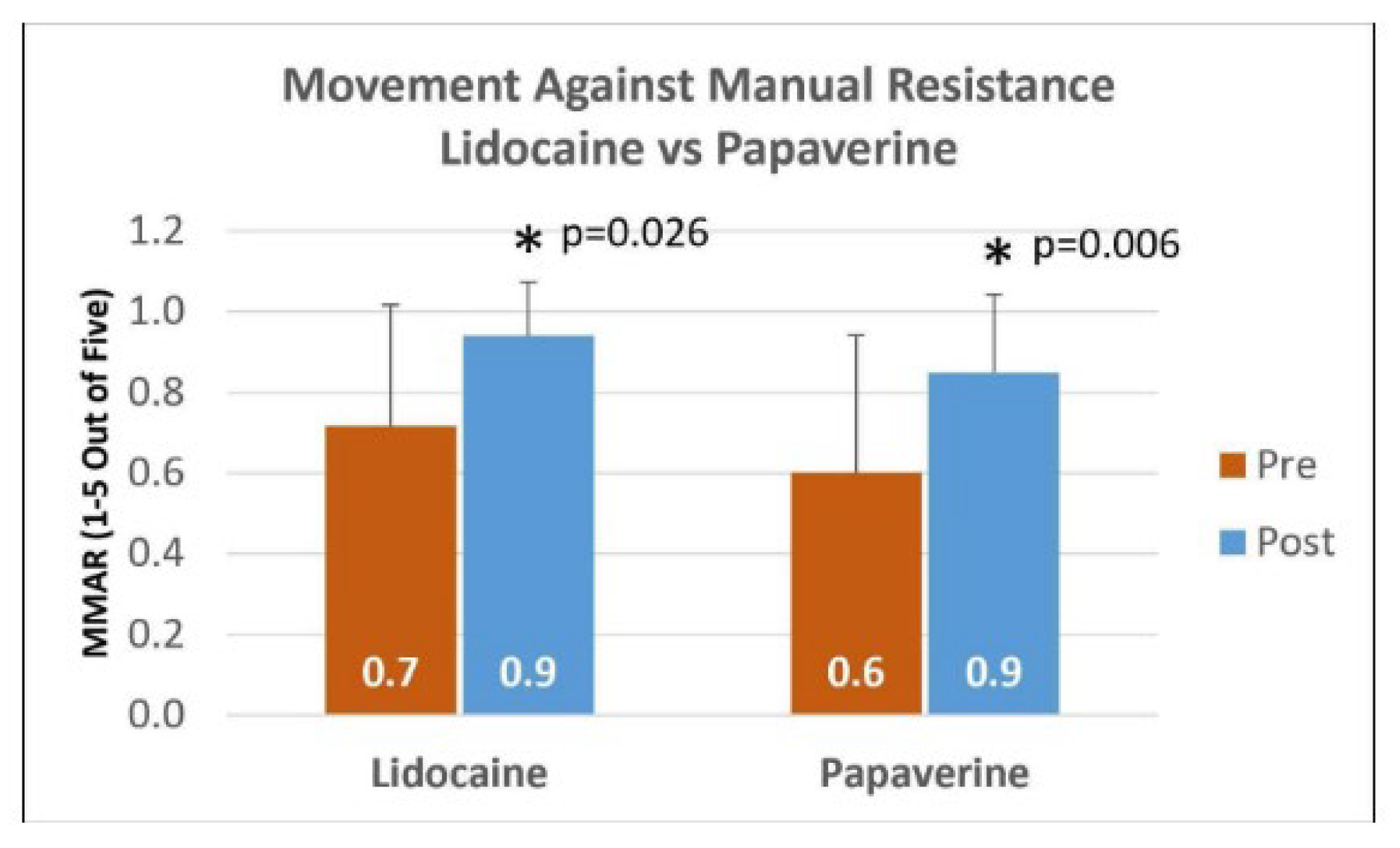
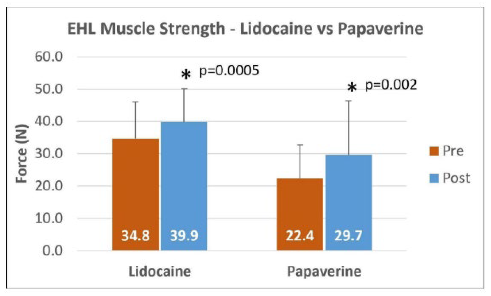
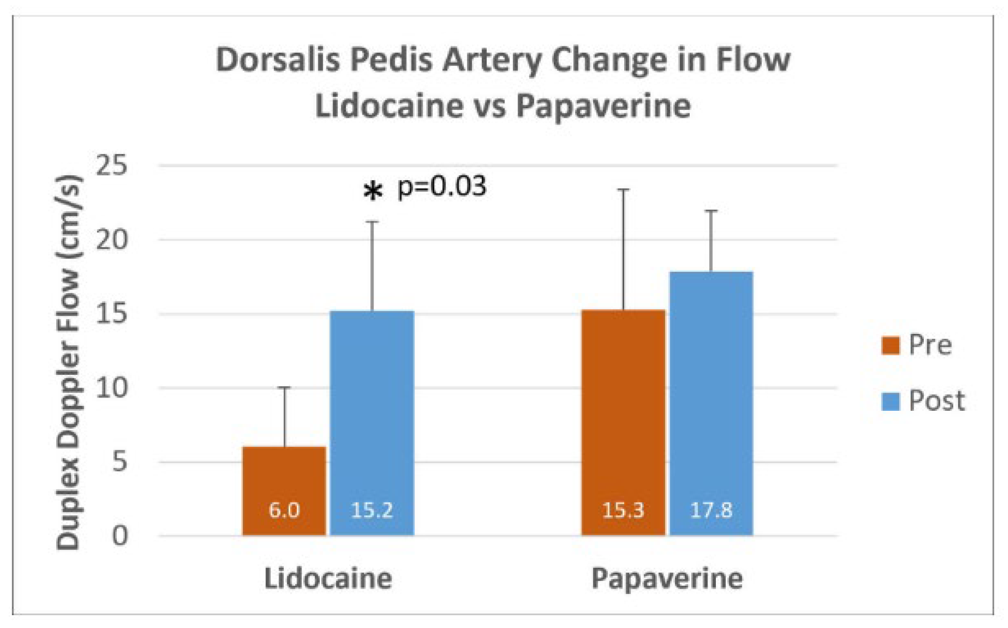
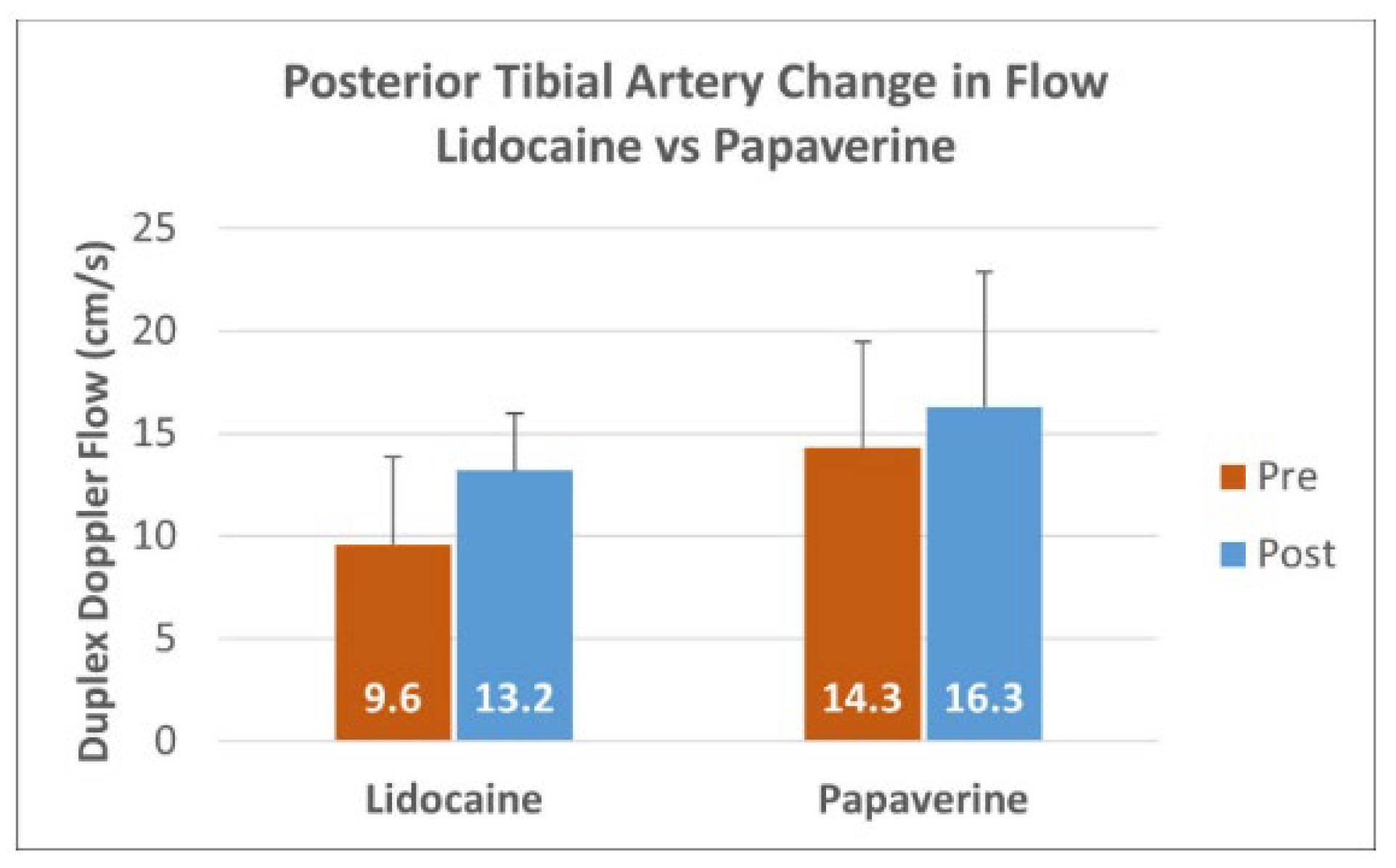
Our hypothesis is that the Phoenix Sign is due to lidocaine’s vasodilatory effect, which results in improved neural function. These results are consistent with this hypothesis in the following manner. (1) Both lidocaine and papaverine show significant improvement in motor function, and had similar success rates across the patient population, as determined by comparing the two proportions. (2) Lidocaine significantly improved blood flow in the dorsalis pedis, whereas papaverine did not. You would not expect a smooth muscle relaxant such as papaverine to affect this artery from the injection site at the knee. Also, neither drug showed significant improvement in flow in the posterior tibial artery, which is outside the expected innervation pattern of the Common Peroneal Nerve and its branches. (3) There were no significant differences in the microvasculature pre and post infiltration of either drug (
Figure 3 and
Figure 4). While this effect is more difficult to explain (i.e., there was an increase in dorsalis pedis flow, but not in the capillaries on the dorsal foot), it might be a temporal effect. This difference indicates that sympathetic axons to the large vessels behave differently than those to the arterioles. These points suggest that while papaverine and lidocaine work using different mechanisms (smooth muscle relaxant vs. blocking voltage-gated Na+ channels respectively), they both act by vasodilating the vasa nervorum to the treated nerves and the large arteries that they innervate. These in turn result in acute neuromuscular improvement to their target muscles.
Papaverine acts primarily as a smooth muscle relaxant by inhibiting phosphodiesterases (PDEs) and directly interacting with calcium channels in smooth muscle cells, which potentially hinders calcium influx and further contributes to relaxation [
11], which are crucial for smooth muscle relaxation [
11,
12]. Meanwhile, lidocaine blocks axonal voltage-gated Na+ channels. Hence, you would not expect a muscle relaxant to be injected at the Common Peroneal Nerve to have an effect on the posterior tibial artery, which was not observed. However, it is speculative conjecture by the authors that injecting lidocaine at that site could possibly inactivate autonomic innervation of the dorsalis pedis artery, as this was not directly measured. There could be additional physiological mechanisms at play, such as some form of direct or indirect neural effect, placebo, or measurement variability that warrant further investigation.
This neurological phenomenon has been observed clinically: after infiltration of a small amount of 1% lidocaine (usually less than or equal to 0.5 cc) adjacent to the Common Peroneal (Fibular) nerve under sonographic guidance, in patients with drop foot or significantly weakened motor potentials of the lower extremity dorsiflexors (extensor hallucis longus (EHL), tibialis anterior (TA), and extensor digitorum longus (EDL), the patient will regain partial or full dorsiflexion temporarily during the effect of this nerve block [
5]. This has been named the Phoenix Sign, in reference to ancient Greek folklore, as the non-functioning nerve (like the bird) becomes functional due to the effect of lidocaine, similar to the way that the mythical bird rose from the ashes. This sign has significant benefit to the diagnosis and assessment of Common Peroneal (Fibular) nerve palsy, as clinically, even when a nerve does not manifest a Tinel’s or Provocation sign, the nerve damage is believed to be so severe that peripheral nerve decompression will not likely benefit the patient.
Thus, many patients with this crippling condition are not receiving definitive treatment because of the inability of standard clinical work-up and diagnosis. A “positive Phoenix sign” also differentiates focal nerve compression peripherally versus a central CNS manifestation. It is hypothesized that this observation of improved nerve function is due to the vasodilatory nature of the local anesthetic. This hypothesis was supported in a previous double-blinded study that compared lidocaine to papaverine [
6].
Interestingly, no statistically significant changes were observed in the microvascular perfusion post infiltration while in every extremity there was an increase in the macrovasculature. One possibility for this discrepancy between micro- vs. macrovascular perfusion could be explained by the difference in anatomy and our sample population. Microvascular anatomy involves small blood vessels such as capillaries, arterioles, and venules, while macrovascular anatomy involves large blood vessels such as veins and arteries. Macrovascular perfusion can lead to changes in smooth muscle cell organization inducing vasoconstriction or vasodilation. Microvascular networks, on the other hand, function via oxygen exchange and vasomotion of arterioles. Chronic diabetes, through a series of damaging pathways, triggers endothelial cell injury and a decrease in nitric oxide production. This leads to local hypoxia and microvascular dysfunction [
14]. All of our patients were type II diabetic severely neuropathic patients. Diabetic neuropathy is inherently a complication of microvascular compromise; therefore, all of our subjects over time could have had structural and functional changes in the microvasculature around nerves [
15,
16]. These may not be easily altered with a vasodilatory nerve injection.
Another reason why the microvascular changes were not observed may be due to the proximal location and temporary duration of the diagnostic injection. Infiltration of lidocaine or papaverine around the Common Peroneal Nerve at the fibular neck is a targeted diagnostic injection that seems to have an indirect effect on temporarily increasing the macrovasculature but, as stated, we did not observe any changes in the microvasculature distally in the foot.
While small sample sizes are often a concern (N = 4), we decided to test our hypothesis with a small pilot study. Clearly the results of this study cannot be generalized to all cases of CPN entrapment. We were encouraged that with only four subjects, there were statistically significant results with favorable
p-values. Our motor results show clear and consistent patterns across each subject, suggesting there may be validity for our hypothesis. In addition, a story is emerging that the lidocaine injection inactivated the sympathetic fibers at the common fibular nerve, which hitchhike along the anterior tibial artery resulting in its denervation and subsequent dilation [
17,
18,
19,
20]. This could have occurred at higher levels of the artery, but only the distal portion at the dorsalis pedis was measured. Papaverine would not deactivate the sympathetics and did not result in vessel dilation, nor would deactivating the sympathetics at the common fibular nerve affect the posterior tibial artery, which is consistent with our results. It is possible that with a larger enrollment size, even the vascular results may show significance. Therefore, we feel that this pilot study is a valuable foundation for future research. Additionally, the consideration of statistical underpowering, the lack of complete independence between limbs, and reliance on short-term endpoints should be factored into further clinical investigation. There was no sham or placebo used that could possibly aid in the delineation of specific drug effects, consideration of this in future studies may greatly enhance our understanding of this phenomenon. Finally, while there was no reporting in this small cohort of confounding medications or co-morbidities, which theoretically could affect the outcome, the authors believe that regardless of the host status, if there is a positive increase in motor strength seen after infiltration, it is highly likely that a focal nerve entrapment exists.



 ,
,