


Cells 2021, 10(6), 1518; https://doi.org/10.3390/cells10061518 - 16 Jun 2021
Cited by 9 | Viewed by 3993
Abstract
►
Show Figures
Trace amine-associated receptor 1 (rodent Taar1/human TAAR1) is a G protein-coupled receptor that is mainly recognized for its functions in neuromodulation. Previous in vitro studies suggested that Taar1 may signal from intracellular compartments. However, we have shown Taar1 to localize apically and on
[...] Read more.
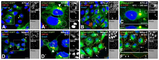
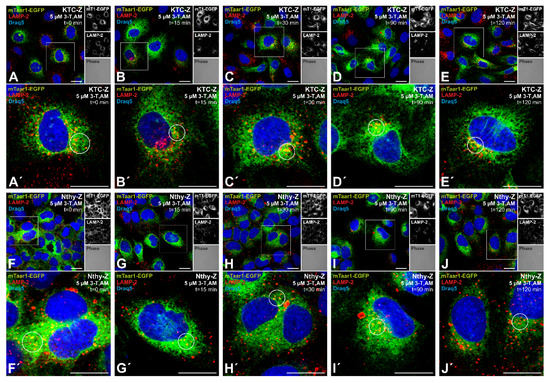
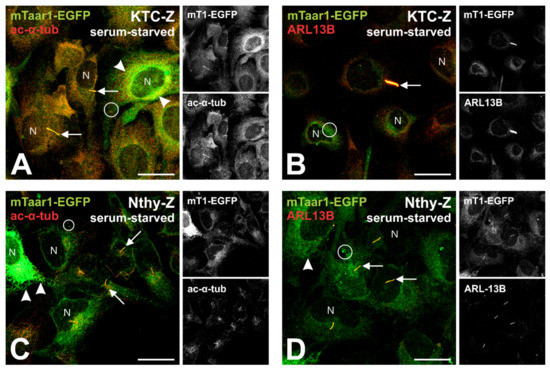
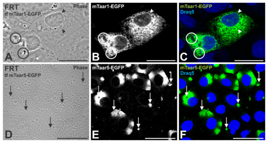
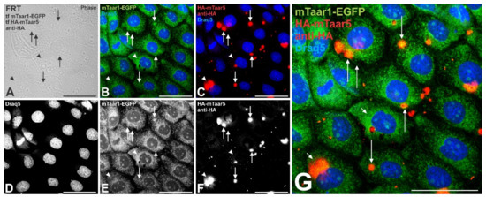
Trace amine-associated receptor 1 (rodent Taar1/human TAAR1) is a G protein-coupled receptor that is mainly recognized for its functions in neuromodulation. Previous in vitro studies suggested that Taar1 may signal from intracellular compartments. However, we have shown Taar1 to localize apically and on ciliary extensions in rodent thyrocytes, suggesting that at least in the thyroid, Taar1 may signal from the cilia at the apical plasma membrane domain of thyrocytes in situ, where it is exposed to the content of the follicle lumen containing putative Taar1 ligands. This study was designed to explore mouse Taar1 (mTaar1) trafficking, heterologously expressed in human and rat thyroid cell lines in order to establish an in vitro system in which Taar1 signaling from the cell surface can be studied in future. The results showed that chimeric mTaar1-EGFP traffics to the apical cell surface and localizes particularly to spherical structures of polarized thyroid cells, procilia, and primary cilia upon serum-starvation. Moreover, mTaar1-EGFP appears to form high molecular mass forms, possibly homodimers and tetramers, in stably expressing human thyroid cell lines. However, only monomeric mTaar1-EGFP was cell surface biotinylated in polarized human thyrocytes. In polarized rat thyrocytes, mTaar1-EGFP is retained in the endoplasmic reticulum, while cilia were reached by mTaar1-EGFP transiently co-expressed in combination with an HA-tagged construct of the related mTaar5. We conclude that Taar1 trafficking to cilia depends on their integrity. The results further suggest that an in vitro cell model was established that recapitulates Taar1 trafficking in thyrocytes in situ, in principle, and will enable studying Taar1 signaling in future, thus extending our general understanding of its potential significance for thyroid autoregulation.
Full article
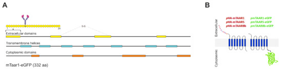
Figure 1