 Dr. Simona Bernardi
Dr. Simona Bernardi
E-Mail
Website
Collection Editor
Department Clinical and Experimental Sciences, University of Brescia, Viale Europa 11 (Brescia) Chair di Hematology, Unit of Bone Marrow Transplantation, ASST-Spedali Civili of Brescia, 25123 Brescia, Italy
Interests: acute myeloid leukemia (AML); chronic myeloid leukemia (CML); familiarity and genetic predisposition in hematologic malignancies; next generation sequencing (NGS); digital PCR (dPCR); exosomes and extracellular vesicles; liquid biopsy; zebrafish model; mesenchymal and hematopoietic stem cells (MSCs and HSCs); regenerative medicine; circulating endothelial cells (CEC)
 Dr. Carolina Balbi
Dr. Carolina Balbi
E-Mail
Website
Collection Editor
Cellular & molecular Cardiology Laboratory, Cardiocentro Ticino, Associated Institute of University of Zurich, 6900 Lugano, Switzerland
Interests: myocardial infarction (MI); drug cardiotoxicity; amniotic fluid stem cells (AFS); mesenchymal stem cells (MSC); cardiac progenitor cells (CPC); regenerative medicine; paracrine therapy; exosome and extracellular vesicles; biomarkers
Dear Colleagues,
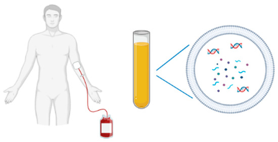
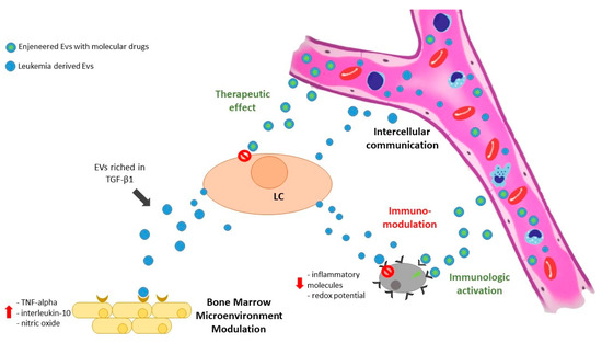
Intercellular communication is an essential hallmark of multicellular organisms and can be mediated through direct cell–cell contact or transfer of secreted molecules. In the last two decades, a third mechanism for intercellular communication has emerged that involves intercellular transfer of extracellular vesicles (EVs).
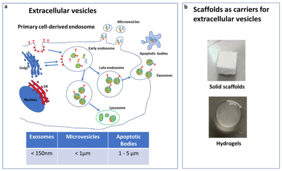
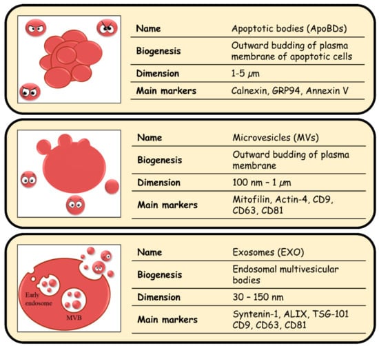
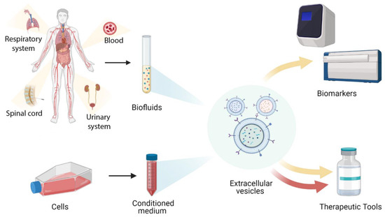
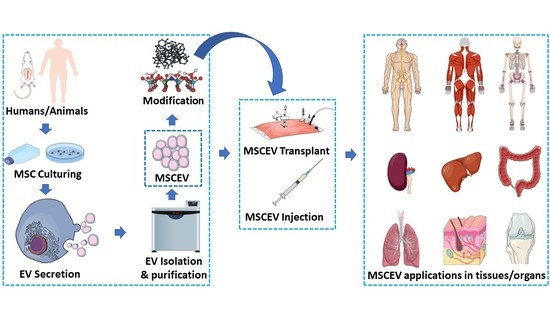
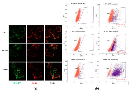
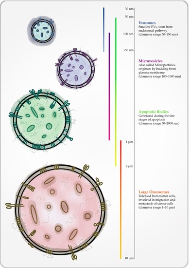
EVs are membranous vesicles of 30–5000 nm in size. Based on their dimension and biogenesis, EVs can be divided into different categories, such as microvesicles, apoptotic bodies, ectosomes, and exosomes. It has already been demonstrated that changing in protein, expressed on surfaces or content in these vesicles, may reflect the status of producing cells. For this reason, EVs, and exosomes in particular, are considered ideal biomarkers in several types of disease from cancer diagnosis to heart rejection. Furthermore, extracellular vesicles can be carriers of cytoprotective or cytotoxic factors and used as a therapeutic tool applied from regenerative medicine to target cancer therapy.
This Topical Collection focuses on recent findings pertaining to EV in different areas from biomarkers to therapeutic application. We warmly welcome experts to submit previously unpublished original scientific research with novel findings and reviews with a comprehensive overview of EVs.
Dr. Simona Bernardi
Dr. Carolina Balbi
Guest Editors
Manuscript Submission Information
Manuscripts should be submitted online at www.mdpi.com by registering and logging in to this website. Once you are registered, click here to go to the submission form. All submissions that pass pre-check are peer-reviewed. Accepted papers will be published continuously in the journal (as soon as accepted) and will be listed together on the collection website. Research articles, review articles as well as short communications are invited. For planned papers, a title and short abstract (about 250 words) can be sent to the Editorial Office for assessment.
Submitted manuscripts should not have been published previously, nor be under consideration for publication elsewhere (except conference proceedings papers). All manuscripts are thoroughly refereed through a single-anonymized peer-review process. A guide for authors and other relevant information for submission of manuscripts is available on the Instructions for Authors page. Biology is an international peer-reviewed open access semimonthly journal published by MDPI.
Please visit the Instructions for Authors page before submitting a manuscript.
The Article Processing Charge (APC) for publication in this open access journal is 2700 CHF (Swiss Francs).
Submitted papers should be well formatted and use good English. Authors may use MDPI's
English editing service prior to publication or during author revisions.


