Stem Cells in Domestic Animals: Applications in Health and Production
Printed Edition Available!
A printed edition of this Special Issue is available
here.
Share This Topical Collection
Editors
Prof. Dr. Eleonora Iacono
E-Mail
Website
Collection Editor
Department of Veterinary Medical Sciences, University of Bologna, Via Tolara di Sopra 50, 40064 Ozzano Emilia, Bologna, Italy
Interests: mesenchymal stromal cells; veterinary cell therapy; assisted reproduction; cell banking; animal reproduction
Special Issues, Collections and Topics in MDPI journals
Dr. Barbara Merlo
Dr. Barbara Merlo
E-Mail
Website
Collection Editor
Department of Veterinary Medical Sciences, University of Bologna, Via Tolara di Sopra 50, 40064 Ozzano Emilia, Bologna, Italy
Interests: regenerative medicine, mesenchymal stromal cells; cryopreservation; animal reproduction; fertility; assisted reproductive techniques
Special Issues, Collections and Topics in MDPI journals
Topical Collection Information
Dear Colleagues,
Over the past decade, stem cell research has emerged as an area of major interest for its potential applications, both in human and veterinary medicine. The effective management of companion and sport animals requires sophisticated new treatments and preventive strategies. Furthermore, also in livestock species, stem cell therapy could be used to treat several medical conditions that negatively affect meat and milk production or reproductive efficiency. In the era of antibiotic resistance, stem cells modified with therapeutic agents might be employed to combat mastitis in dairy cow, sheep, goat or buffalo, with enormous benefits also for human health. Reproductive performances could be improved by the integration of stem cells into the in vitro embryo production system, representing in another way the importance of stem cells in addressing commercial goals. Moreover, the importance of domestic animals as models for human diseases has been recognized. The quantity of tissue available for decellularization is greater than that in small animal models such as mice, and larger animal species more closely resembling some human behavior can be used for translational applications in cell therapy. Domestic animals are also models for stem cell secretome studies and clinical trials.
Prof. Dr. Eleonora Iacono
Dr. Barbara Merlo
Collection Editors
Manuscript Submission Information
Manuscripts should be submitted online at www.mdpi.com by registering and logging in to this website. Once you are registered, click here to go to the submission form. Manuscripts can be submitted until the deadline. All submissions that pass pre-check are peer-reviewed. Accepted papers will be published continuously in the journal (as soon as accepted) and will be listed together on the collection website. Research articles, review articles as well as short communications are invited. For planned papers, a title and short abstract (about 100 words) can be sent to the Editorial Office for announcement on this website.
Submitted manuscripts should not have been published previously, nor be under consideration for publication elsewhere (except conference proceedings papers). All manuscripts are thoroughly refereed through a single-blind peer-review process. A guide for authors and other relevant information for submission of manuscripts is available on the Instructions for Authors page. Animals is an international peer-reviewed open access semimonthly journal published by MDPI.
Please visit the Instructions for Authors page before submitting a manuscript.
The Article Processing Charge (APC) for publication in this open access journal is 2400 CHF (Swiss Francs).
Submitted papers should be well formatted and use good English. Authors may use MDPI's
English editing service prior to publication or during author revisions.
Keywords
- Stem cells
- Domestic animals
- Cellular therapy
- Translational medicine
- Cell culture
- Secretome
Published Papers (19 papers)
Open AccessArticle
In Situ Treatment of Refractory Perianal Fistulas in Dogs with Low-Dose Allogeneic Adipose-Derived Mesenchymal Stem Cells
by
Nathaly Enciso, Javier Enciso-Benavides, Juan Sandoval and Javier Enciso
Viewed by 804
Abstract
Stem cell therapy in dogs has increased considerably in recent years; however, there are few publications on the treatment of perianal fistulas (PF) in this species. The aim of this open-label clinical trial was to demonstrate the efficacy and safety of a new
[...] Read more.
Stem cell therapy in dogs has increased considerably in recent years; however, there are few publications on the treatment of perianal fistulas (PF) in this species. The aim of this open-label clinical trial was to demonstrate the efficacy and safety of a new protocol for the in situ administration of low-dose adipose-derived allogeneic stem cells (ASCs) for the treatment of refractory spontaneous perianal fistula. The methodology consisted of applying one to three doses of 5 × 10
6 cryopreserved allogeneic ASCs to each fistula. The study was performed in 14 dogs regardless of sex, breed, or age, with a clinical diagnosis of refractory PF. Cells diluted in phosphate-buffered saline were applied to five sites of the PF in an amount of 1 × 10
6 per application site. Efficacy was determined by the complete closure of the fistula, which was observed in 100% of the cases studied one month after therapy, with a subsequent follow-up of 12 to 48 months after therapy. Furthermore, safety was demonstrated by the absence of local or systemic adverse effects. In conclusion, the protocol used in this work demonstrates the efficacy without adverse effects of the in situ application of low doses of allogeneic ASCs, providing a simple, non-invasive, long-lasting and low-cost therapeutic option.
Full article
►▼
Show Figures
Open AccessArticle
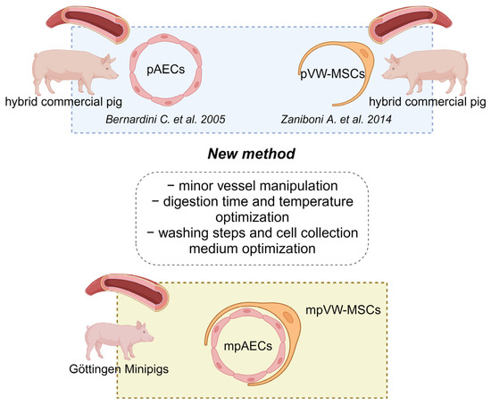
Isolation of Vascular Wall Mesenchymal Stem Cells from the Thoracic Aorta of Adult Göttingen Minipigs: A New Protocol for the Simultaneous Endothelial Cell Collection
by
Chiara Bernardini, Debora La Mantia, Roberta Salaroli, Domenico Ventrella, Alberto Elmi, Augusta Zannoni and Monica Forni
Cited by 2 | Viewed by 1503
Abstract
Two main classes of perivascular multipotent populations have been described: the microvascular pericytes and the vascular wall mesenchymal stem cells (VW-MSCs). VW-MSCs are isolated from large vessels in many species and they participate in vascular remodeling together with other cellular components such as
[...] Read more.
Two main classes of perivascular multipotent populations have been described: the microvascular pericytes and the vascular wall mesenchymal stem cells (VW-MSCs). VW-MSCs are isolated from large vessels in many species and they participate in vascular remodeling together with other cellular components such as endothelial cells. Considering that the Göttingen Minipigs are widely used in Europe as a translational model in the field of cardiovascular diseases, the aim of the present research was to isolate VW-MSCs from the adult aorta of Göttingen Minipigs while preserving and also collecting endothelial cells. The results obtained in the present research demonstrated that this new protocol allows us to obtain a pure population of VW-MSCs and endothelial cells. VW-MSCs from Göttingen Minipigs responded fully to the MSC minima international criteria, being positive to CD105, CD90, and CD44 and negative to CD45 and CD34. Moreover, VW-MSCs presented a differentiative potential towards osteogenic, chondrogenic, and adipogenic lineages. Overall, the present protocol, preserving the viability and phenotypic features of the two isolated populations, opens future possibilities of using minipig VW-MSCs and endothelial cells in in vitro vascular remodeling studies.
Full article
►▼
Show Figures
Open AccessEditorial
Stem Cells in Domestic Animals: Applications in Health and Production
by
Eleonora Iacono and Barbara Merlo
Viewed by 1992
Abstract
In the last decade, researchers described Mesenchymal Stem/stromal cells (MSCs) as a possible population of cells for cell-based therapies in regenerative medicine, both for humans and animals [...]
Full article
Open AccessArticle
Shipping Temperature, Time and Media Effects on Equine Wharton’s Jelly and Adipose Tissue Derived Mesenchymal Stromal Cells Characteristics
by
Eleonora Iacono, Aliai Lanci, Penelope Gugole and Barbara Merlo
Cited by 2 | Viewed by 1500
Abstract
To use Mesenchymal Stromal Cells (MSCs) in equine patients, isolation and expansion are performed in a laboratory. Cells are then sent back to the veterinary clinic. The main goal of storage conditions during cell transport is to preserve their biological properties and viability.
[...] Read more.
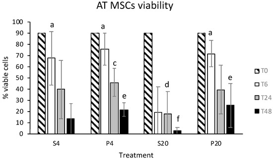
To use Mesenchymal Stromal Cells (MSCs) in equine patients, isolation and expansion are performed in a laboratory. Cells are then sent back to the veterinary clinic. The main goal of storage conditions during cell transport is to preserve their biological properties and viability. The aim of this study was to evaluate the effects of storage solutions, temperature and time on the characteristics of equine adipose tissue and Wharton’s jelly-derived MSCs. We compared two different storage solutions (plasma and 0.9% NaCl), two different temperatures (4 °C and room temperature) and three time frames (6, 24, 48 h). Cell viability, colony-forming units, trilineage differentiation, the expression of CD45 and CD90 antigens and adhesion potentials were evaluated. Despite the molecular characterization and differentiation potential were not influenced by storage conditions, viability, colony-forming units and adhesion potential are influenced in different way, depending on MSCs sources. Overall, this study found that, despite equine adipose tissue MSCs being usable after 24 h of storage, cells derived from Wharton’s jelly need to be used within 6 h. Moreover, while for adipose cells the best conservation solutions seems to be plasma, the cell viability of Wharton’s jelly MSCs declined in both saline and plasma solution, confirming their reduced resistance to conservation.
Full article
►▼
Show Figures
Open AccessReview
An Update on Applications of Cattle Mesenchymal Stromal Cells
by
Barbara Merlo, Penelope Maria Gugole and Eleonora Iacono
Cited by 3 | Viewed by 3399
Abstract
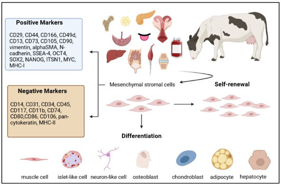
Attention on mesenchymal stromal cells (MSCs) research has increased in the last decade mainly due to the promising results about their plasticity, self-renewal, differentiation potential, immune modulatory and anti-inflammatory properties that have made stem cell therapy more clinically attractive. Furthermore, MSCs can be
[...] Read more.
Attention on mesenchymal stromal cells (MSCs) research has increased in the last decade mainly due to the promising results about their plasticity, self-renewal, differentiation potential, immune modulatory and anti-inflammatory properties that have made stem cell therapy more clinically attractive. Furthermore, MSCs can be easily isolated and expanded to be used for autologous or allogenic therapy following the administration of either freshly isolated or previously cryopreserved cells. The scientific literature on the use of stromal cells in the treatment of several animal health conditions is currently available. Although MSCs are not as widely used for clinical treatments in cows as for companion and sport animals, they have the potential to be employed to improve productivity in the cattle industry. This review provides an update on state-of-the-art applications of bovine MSCs to clinical treatments and reproductive biotechnologies.
Full article
►▼
Show Figures
Open AccessArticle
A Comparative Study of Canine Mesenchymal Stem Cells Isolated from Different Sources
by
Filip Humenik, Marcela Maloveska, Nikola Hudakova, Patricia Petrouskova, Lubica Hornakova, Michal Domaniza, Dagmar Mudronova, Simona Bodnarova and Dasa Cizkova
Cited by 15 | Viewed by 3273
Abstract
In this study, we provide comprehensive analyses of mesenchymal stem cells (MSCs) isolated from three types of canine tissues: bone marrow (BM-MSCs), adipose tissue (AT-MSCs) and amniotic tissue (AM-MSCs). We compare their morphology, phenotype, multilineage potential and proliferation activity. The BM-MSCs and AM-MSCs
[...] Read more.
In this study, we provide comprehensive analyses of mesenchymal stem cells (MSCs) isolated from three types of canine tissues: bone marrow (BM-MSCs), adipose tissue (AT-MSCs) and amniotic tissue (AM-MSCs). We compare their morphology, phenotype, multilineage potential and proliferation activity. The BM-MSCs and AM-MSCs showed fibroblast-like shapes against the spindle shape of the AT-MSCs. All populations showed strong osteogenic and chondrogenic potential. However, we observed phenotypic differences. The BM-MSCs and AT-MSCs revealed high expression of CD29, CD44, CD90 and CD105 positivity compared to the AM-MSCs, which showed reduced expression of all the analysed CD markers. Similarly, the isolation yield and proliferation varied depending on the source. The highest isolation yield and proliferation were detected in the population of AT-MSCs, while the AM-MSCs showed a high yield of cells, but the lowest proliferation activity, in contrast to the BM-MSCs which had the lowest isolation yield. Thus, the present data provide assumptions for obtaining a homogeneous MSC derived from all three canine tissues for possible applications in veterinary regenerative medicine, while the origin of isolated MSCs must always be taken into account.
Full article
►▼
Show Figures
Open AccessCommunication
Isolation and Characterization of Cat Olfactory Ecto-Mesenchymal Stem Cells
by
Marie-Laure Mollichella, Violaine Mechin, Dany Royer, Patrick Pageat and Pietro Asproni
Cited by 1 | Viewed by 1986
Abstract
The olfactory mucosa contains olfactory ecto-mesenchymal stem cells (OE-MSCs) which show stemness features, multipotency capabilities, and have a therapeutic potential. The OE-MSCs have already been collected and isolated from various mammals. The aim of this study was to evaluate the feasibility of collecting,
[...] Read more.
The olfactory mucosa contains olfactory ecto-mesenchymal stem cells (OE-MSCs) which show stemness features, multipotency capabilities, and have a therapeutic potential. The OE-MSCs have already been collected and isolated from various mammals. The aim of this study was to evaluate the feasibility of collecting, purifying and amplifying OE-MSCs from the cat nasal cavity. Four cats were included in the study. Biopsies of olfactory mucosa were performed on anesthetized animals. Then, the olfactory OE-MSCs were isolated, and their stemness features as well as their mesodermal differentiation capabilities were characterized. Olfactory mucosa biopsies were successfully performed in all subjects. From these biopsies, cellular populations were rapidly generated, presenting various stemness features, such as a fibroblast-like morphology, nestin and MAP2 expression, and sphere and colony formation. These cells could differentiate into neural and mesodermal lineages. This report shows for the first time that the isolation of OE-MSCs from cat olfactory mucosa is possible. These cells showed stemness features and multilineage differentiation capabilities, indicating they may be a promising tool for autologous grafts and feline regenerative medicine.
Full article
►▼
Show Figures
Open AccessReview
An Outstanding Role of Adipose Tissue in Canine Stem Cell Therapy
by
Marina Prišlin, Dunja Vlahović, Petar Kostešić, Ivana Ljolje, Dragan Brnić, Nenad Turk, Ivana Lojkić, Valentina Kunić, Tugomir Karadjole and Nina Krešić
Cited by 8 | Viewed by 3892
Abstract
Adipose tissue, previously known as connective tissue with a role in energy storage, is currently changing the course of treatments in veterinary medicine. Recent studies have revealed one particularly impressive function among all the newly discovered functions of adipose tissue. The interactive cells
[...] Read more.
Adipose tissue, previously known as connective tissue with a role in energy storage, is currently changing the course of treatments in veterinary medicine. Recent studies have revealed one particularly impressive function among all the newly discovered functions of adipose tissue. The interactive cells hosted by adipose tissue, the stromal vascular fraction (SVF), and their role in treating numerous diseases have provided a prospective course of research with positive outcomes in regenerative veterinary medicine (RVM). This review describes the main features of adipose tissue, emphasizing an eclectic combination of cells within the SVF and its thus far researched therapeutic possibilities in canine RVM. An afterwards focus is on a highly researched component of the SVF, adipose-derived mesenchymal stem cells (ASCs), which were shown to have an extraordinary impact relying on several proposed mechanisms of action on mitigating pathologies in canines. Furthermore, ASC therapy showed the most significant results in the orthopaedics field and in neurology, dermatology, ophthalmology, gastroenterology, and hepatology, which elevates the possibilities of ASC therapy to a whole new level. Therefore, this review article aims to raise awareness of the importance of research on cellular components, within abundant and easily accessible adipose tissue, in the direction of regenerative therapy in canines, considering the positive outcomes so far. Although the focus is on the positive aspects of cellular therapy in canines, the researchers should not forget the importance of identifying the potential negative aspects within published and upcoming research. Safe and standardized treatment represents a fundamental prerequisite for positively impacting the lives of canine patients.
Full article
►▼
Show Figures
Open AccessSystematic Review
Efficacy of Stem Cell Therapy in Large Animal Models of Ischemic Cardiomyopathies: A Systematic Review and Meta-Analysis
by
Debora La Mantia, Chiara Bernardini, Augusta Zannoni, Roberta Salaroli, Changzhen Wang, Silvia Bencivenni and Monica Forni
Cited by 11 | Viewed by 2893
Abstract
Stem-cell therapy provides a promising strategy for patients with ischemic heart disease. In recent years, numerous studies related to this therapeutic approach were performed; however, the results were often heterogeneous and contradictory. For this reason, we conducted a systematic review and meta-analysis of
[...] Read more.
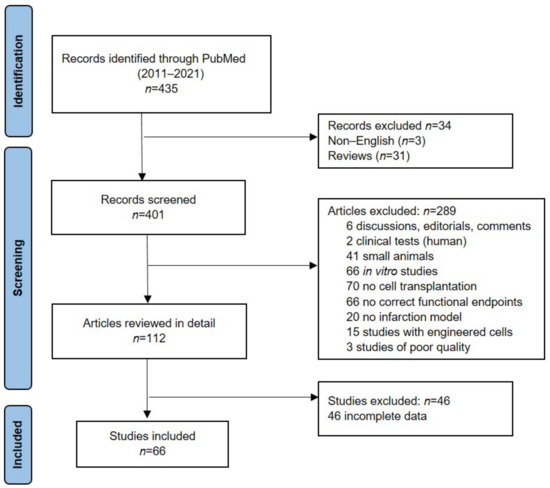
Stem-cell therapy provides a promising strategy for patients with ischemic heart disease. In recent years, numerous studies related to this therapeutic approach were performed; however, the results were often heterogeneous and contradictory. For this reason, we conducted a systematic review and meta-analysis of trials, reporting the use of stem-cell treatment against acute or chronic ischemic cardiomyopathies in large animal models with regard to Left Ventricular Ejection Fraction (LVEF). The defined research strategy was applied to the PubMed database to identify relevant studies published from January 2011 to July 2021. A random-effect meta-analysis was performed on LVEF mean data at follow-up between control and stem-cell-treated animals. In order to improve the definition of the effect measure and to analyze the factors that could influence the outcomes, a subgroup comparison was conducted. Sixty-six studies (
n = 1183 animals) satisfied our inclusion criteria. Ischemia/reperfusion infarction was performed in 37 studies, and chronic occlusion in 29 studies; moreover, 58 studies were on a pig animal model. The meta-analysis showed that cell therapy increased LVEF by 7.41% (95% Confidence Interval 6.23–8.59%;
p < 0.001) at follow-up, with significative heterogeneity and high inconsistency (I
2 = 82%,
p < 0.001). By subgroup comparison, the follow-up after 31–60 days (
p = 0.025), the late cell injection (>7 days,
p = 0.005) and the route of cellular delivery by surgical treatment (
p < 0.001) were significant predictors of LVEF improvement. This meta-analysis showed that stem-cell therapy may improve heart function in large animal models and that the swine specie is confirmed as a relevant animal model in the cardiovascular field. Due to the significative heterogeneity and high inconsistency, future translational studies should be designed to take into account the evidenced predictors to allow for the reduction of the number of animals used.
Full article
►▼
Show Figures
Open AccessArticle
Peptide Mediated Adhesion to Beta-Lactam Ring of Equine Mesenchymal Stem Cells: A Pilot Study
by
Barbara Merlo, Vito Antonio Baldassarro, Alessandra Flagelli, Romina Marcoccia, Valentina Giraldi, Maria Letizia Focarete, Daria Giacomini and Eleonora Iacono
Cited by 3 | Viewed by 2452
Abstract
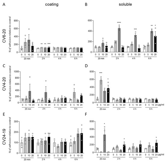
Regenerative medicine applied to skin lesions is a field in constant improvement. The use of biomaterials with integrin agonists could promote cell adhesion increasing tissue repair processes. The aim of this pilot study was to analyze the effect of an α4β1 integrin agonist
[...] Read more.
Regenerative medicine applied to skin lesions is a field in constant improvement. The use of biomaterials with integrin agonists could promote cell adhesion increasing tissue repair processes. The aim of this pilot study was to analyze the effect of an α4β1 integrin agonist on cell adhesion of equine adipose tissue (AT) and Wharton’s jelly (WJ) derived MSCs and to investigate their adhesion ability to GM18 incorporated poly L-lactic acid (PLLA) scaffolds. Adhesion assays were performed after culturing AT- and WJ-MSCs with GM18 coating or soluble GM18. Cell adhesion on GM18 containing PLLA scaffolds after 20 min co-incubation was assessed by HCS. Soluble GM18 affects the adhesion of equine AT- and WJ-MSCs, even if its effect is variable between donors. Adhesion to PLLA scaffolds containing GM18 is not significantly influenced by GM18 for AT-MSCs after 20 min or 24 h of culture and for WJ-MSCs after 20 min, but increased cell adhesion by 15% GM18 after 24 h. In conclusion, the α4β1 integrin agonist GM18 affects equine AT- and WJ-MSCs adhesion ability with a donor-related variability. These preliminary results represent a first step in the study of equine MSCs adhesion to PLLA scaffolds containing GM18, suggesting that WJ-MSCs might be more suitable than AT-MSCs. However, the results need to be confirmed by increasing the number of samples before drawing definite conclusions.
Full article
►▼
Show Figures
Open AccessArticle
Platelet Lysate for Mesenchymal Stromal Cell Culture in the Canine and Equine Species: Analogous but Not the Same
by
Alina Hagen, Heidrun Holland, Vivian-Pascal Brandt, Carla U. Doll, Thomas C. Häußler, Michaela Melzer, Julia Moellerberndt, Hendrik Lehmann and Janina Burk
Cited by 10 | Viewed by 2960
Abstract
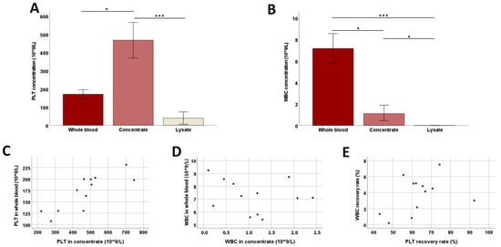
Platelet lysate (PL) is an attractive platelet-based therapeutic tool and has shown promise as xeno-free replacement for fetal bovine serum (FBS) in human and equine mesenchymal stromal cell (MSC) culture. Here, we established a scalable buffy-coat-based protocol for canine PL (cPL) production (n
[...] Read more.
Platelet lysate (PL) is an attractive platelet-based therapeutic tool and has shown promise as xeno-free replacement for fetal bovine serum (FBS) in human and equine mesenchymal stromal cell (MSC) culture. Here, we established a scalable buffy-coat-based protocol for canine PL (cPL) production (n = 12). The cPL was tested in canine adipose MSC (n = 5) culture compared to FBS. For further comparison, equine adipose MSC (n = 5) were cultured with analogous equine PL (ePL) or FBS. During canine blood processing, platelet and transforming growth factor-β1 concentrations increased (
p < 0.05 and
p < 0.001), while white blood cell concentrations decreased (
p < 0.05). However, while equine MSC showed good results when cultured with 10% ePL, canine MSC cultured with 2.5% or 10% cPL changed their morphology and showed decreased metabolic activity (
p < 0.05). Apoptosis and necrosis in canine MSC were increased with 2.5% cPL (
p < 0.05). Surprisingly, passage 5 canine MSC showed less genetic aberrations after culture with 10% cPL than with FBS. Our data reveal that using analogous canine and equine biologicals does not entail the same results. The buffy-coat-based cPL was not adequate for canine MSC culture, but may still be useful for therapeutic applications.
Full article
►▼
Show Figures
Open AccessEditor’s ChoiceReview
Current Status on Canine Foetal Fluid and Adnexa Derived Mesenchymal Stem Cells
by
Eleonora Iacono, Romina Marcoccia and Barbara Merlo
Cited by 2 | Viewed by 3086
Abstract
Effective standards of care treatment guidelines have been developed for many canine diseases. However, a subpopulation of patients is partially or completely refractory to these protocols, so their owners seek novel therapies such as treatments with MSCs. Although in dogs, as with human
[...] Read more.
Effective standards of care treatment guidelines have been developed for many canine diseases. However, a subpopulation of patients is partially or completely refractory to these protocols, so their owners seek novel therapies such as treatments with MSCs. Although in dogs, as with human medicine, the most studied MSCs sources have been bone marrow and adipose tissue, in recent years, many researchers have drawn attention towards alternative sources, such as foetal adnexa and fluid, since they possess many advantages over bone marrow and adipose tissue. Foetal adnexa and fluid could be considered as discarded material; therefore, sampling is non-invasive, inexpensive and free from ethical considerations. Furthermore, MSCs derived from foetal adnexa and fluid preserve some of the characteristics of the primitive embryonic layers from which they originate and seem to present immune-modulatory properties that make them a good candidate for allo- and xenotransplantation. The aim of the present review is to offer an update on the state of the art on canine MSCs derived from foetal adnexa and fluid focusing on the findings in their clinical setting.
Full article
Open AccessArticle
Cell Source-Dependent In Vitro Chondrogenic Differentiation Potential of Mesenchymal Stem Cell Established from Bone Marrow and Synovial Fluid of Camelus dromedarius
by
Young-Bum Son, Yeon Ik Jeong, Yeon Woo Jeong, Mohammad Shamim Hossein, Per Olof Olsson, Alex Tinson, Kuhad Kuldip Singh, Sang-Yun Lee and Woo Suk Hwang
Cited by 5 | Viewed by 2815
Abstract
Mesenchymal stem cells (MSCs) are promising multipotent cells with applications for cartilage tissue regeneration in stem cell-based therapies. In cartilage regeneration, both bone marrow (BM-MSCs) and synovial fluid (SF-MSCs) are valuable sources. However, the cellular characteristics and chondrocyte differentiation potential were not reported
[...] Read more.
Mesenchymal stem cells (MSCs) are promising multipotent cells with applications for cartilage tissue regeneration in stem cell-based therapies. In cartilage regeneration, both bone marrow (BM-MSCs) and synovial fluid (SF-MSCs) are valuable sources. However, the cellular characteristics and chondrocyte differentiation potential were not reported in either of the camel stem cells. The in vitro chondrocyte differentiation competence of MSCs, from (BM and SF) sources of the same
Camelus dromedaries (camel) donor, was determined. Both MSCs were evaluated on pluripotent markers and proliferation capacity. After passage three, both MSCs showed fibroblast-like morphology. The proliferation capacity was significantly increased in SF-MSCs compared to BM-MSCs. Furthermore, SF-MSCs showed an enhanced expression of transcription factors than BM-MSCs. SF-MSCs exhibited lower differentiation potential toward adipocytes than BM-MSCs. However, the osteoblast differentiation potential was similar in MSCs from both sources. Chondrogenic pellets obtained from SF-MSCs revealed higher levels of chondrocyte-specific markers than those from BM-MSCs. Additionally, glycosaminoglycan (GAG) content was elevated in SF-MSCs related to BM-MSCs. This is, to our knowledge, the first study to establish BM-MSCs and SF-MSCs from the same donor and to demonstrate in vitro differentiation potential into chondrocytes in camels.
Full article
►▼
Show Figures
Open AccessReview
Chicken Mesenchymal Stem Cells and Their Applications: A Mini Review
by
Andrea Svoradova, Vladimir Zmrhal, Eva Venusova and Petr Slama
Cited by 11 | Viewed by 4398
Abstract
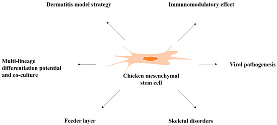
Mesenchymal stem cells (MSCs) are multipotent progenitor cells that adhere to plastic; express the specific markers CD29, CD44, CD73, CD90, and CD105; and produce cytokines and growth factors supporting and regulating hematopoiesis. MSCs have capacity for differentiating into osteocytes, chondrocytes, adipocytes, and myocytes.
[...] Read more.
Mesenchymal stem cells (MSCs) are multipotent progenitor cells that adhere to plastic; express the specific markers CD29, CD44, CD73, CD90, and CD105; and produce cytokines and growth factors supporting and regulating hematopoiesis. MSCs have capacity for differentiating into osteocytes, chondrocytes, adipocytes, and myocytes. They are useful for research toward better understanding the pathogenic potential of the infectious bursal disease virus, mineralization during osteogenesis, and interactions between MSCs as a feeder layer to other cells. MSCs are also important for immunomodulatory cell therapy, can provide a suitable strategy model for coculture with pathogens causing dermatitis disorders in chickens, can be cultured in vitro with probiotics and prebiotics with a view to eliminate the feeding of antibiotic growth promoters, and offer cell-based meat production. Moreover, bone marrow-derived MSCs (BM-MSCs) in coculture with hematopoietic progenitor/stem cells (HPCs/HSCs) can support expansion and regulation of the hematopoiesis process using the 3D-culture system in future research in chickens. MSCs’ several advantages, including ready availability, strong proliferation, and immune modulatory properties make them a suitable model in the field of stem cell research. This review summarizes current knowledge about the general characterization of MSCs and their application in chicken as a model organism.
Full article
►▼
Show Figures
Open AccessArticle
The Effect of a 7 Year-Long Cryopreservation on Stemness Features of Canine Adipose-Derived Mesenchymal Stem Cells (cAD-MSC)
by
Santina Di Bella, Vincenza Cannella, Francesco Mira, Patrizia Di Marco, Antonio Lastra, Francesca Gucciardi, Giuseppa Purpari and Annalisa Guercio
Cited by 6 | Viewed by 3515
Abstract
Mesenchymal stem cells (MSCs) are used in therapy in animal models and veterinary medicine, due to their capacity of inducing tissue regeneration and immunomodulation. Their clinical application requires a ready off-the-shelf amount of viable therapeutics doses. For this purpose, it is useful to
[...] Read more.
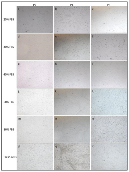
Mesenchymal stem cells (MSCs) are used in therapy in animal models and veterinary medicine, due to their capacity of inducing tissue regeneration and immunomodulation. Their clinical application requires a ready off-the-shelf amount of viable therapeutics doses. For this purpose, it is useful to cryopreserve MSCs to gain a ready and controlled source of abundant autologous stem cells. We evaluated the effect of 7 years cryopreservation using 10% dimethyl sulfoxide (DMSO) with different fetal bovine serum (FBS) concentrations (from 10 to 90%) on different passages of MSCs isolated from canine adipose tissue (cAD-MSCs). The study aimed to evaluate the most adequate cell passage and FBS percentage for the long-term cryopreservation of cells by maintaining the stemness features. Phenotype morphology, cell viability, osteogenic and adipogenic differentiation potentials, proliferative potential and expression of pluripotency markers were analyzed in thawed cells and compared with fresh ones. We demonstrated that cells cryopreserved with at least 80% FBS maintain unaltered the stemness characteristics of the freshly isolated cells. In particular, cells of P0–P1 passages have to be expanded in vitro and subsequently cryopreserved and cells of P2–P4 passages should be considered in the studies on therapeutic application and in vitro study of cAD-MSCs.
Full article
►▼
Show Figures
Open AccessArticle
Effect of Scrapie Prion Infection in Ovine Bone Marrow-Derived Mesenchymal Stem Cells and Ovine Mesenchymal Stem Cell-Derived Neurons
by
Laura García-Mendívil, Diego R. Mediano, Adelaida Hernaiz, David Sanz-Rubio, Francisco J. Vázquez, Belén Marín, Óscar López-Pérez, Alicia Otero, Juan J. Badiola, Pilar Zaragoza, Laura Ordovás, Rosa Bolea and Inmaculada Martín-Burriel
Cited by 6 | Viewed by 2567
Abstract
Scrapie is a prion disease affecting sheep and goats and it is considered a prototype of transmissible spongiform encephalopathies (TSEs). Mesenchymal stem cells (MSCs) have been proposed as candidates for developing in vitro models of prion diseases. Murine MSCs are able to propagate
[...] Read more.
Scrapie is a prion disease affecting sheep and goats and it is considered a prototype of transmissible spongiform encephalopathies (TSEs). Mesenchymal stem cells (MSCs) have been proposed as candidates for developing in vitro models of prion diseases. Murine MSCs are able to propagate prions after previous mouse-adaptation of prion strains and, although ovine MSCs express the cellular prion protein (PrP
C), their susceptibility to prion infection has never been investigated. Here, we analyze the potential of ovine bone marrow-derived MSCs (oBM-MSCs), in growth and neurogenic conditions, to be infected by natural scrapie and propagate prion particles (PrP
Sc) in vitro, as well as the effect of this infection on cell viability and proliferation. Cultures were kept for 48–72 h in contact with homogenates of central nervous system (CNS) samples from scrapie or control sheep. In growth conditions, oBM-MSCs initially maintained detectable levels of PrP
Sc post-inoculation, as determined by Western blotting and ELISA. However, the PrP
Sc signal weakened and was lost over time. oBM-MSCs infected with scrapie displayed lower cell doubling and higher doubling times than those infected with control inocula. On the other hand, in neurogenic conditions, oBM-MSCs not only maintained detectable levels of PrP
Sc post-inoculation, as determined by ELISA, but this PrP
Sc signal also increased progressively over time. Finally, inoculation with CNS extracts seems to induce the proliferation of oBM-MSCs in both growth and neurogenic conditions. Our results suggest that oBM-MSCs respond to prion infection by decreasing their proliferation capacity and thus might not be permissive to prion replication, whereas ovine MSC-derived neuron-like cells seem to maintain and replicate PrP
Sc.
Full article
►▼
Show Figures
Open AccessReview
The Usefulness of Mesenchymal Stem Cells beyond the Musculoskeletal System in Horses
by
Alina Cequier, Carmen Sanz, Clementina Rodellar and Laura Barrachina
Cited by 14 | Viewed by 4002
Abstract
The differentiation ability of mesenchymal stem cells (MSCs) initially raised interest for treating musculoskeletal injuries in horses, but MSC paracrine activity has widened their scope for inflammatory and immune-mediated pathologies in both equine and human medicine. Furthermore, the similar etiopathogenesis of some diseases
[...] Read more.
The differentiation ability of mesenchymal stem cells (MSCs) initially raised interest for treating musculoskeletal injuries in horses, but MSC paracrine activity has widened their scope for inflammatory and immune-mediated pathologies in both equine and human medicine. Furthermore, the similar etiopathogenesis of some diseases in both species has advanced the concept of “One Medicine, One Health”. This article reviews the current knowledge on the use of MSCs for equine pathologies beyond the locomotor system, highlighting the value of the horse as translational model. Ophthalmologic and reproductive disorders are among the most studied for MSC application. Equine asthma, equine metabolic syndrome, and endotoxemia have been less explored but offer an interesting scenario for human translation. The use of MSCs in wounds also provides a potential model for humans because of the healing particularities in both species. High-burden equine-specific pathologies such as laminitis have been suggested to benefit from MSC-therapy, and MSC application in challenging disorders such as neurologic conditions has been proposed. The available data are preliminary, however, and require further development to translate results into the clinic. Nevertheless, current evidence indicates a significant potential of equine MSCs to enlarge their range of application, with particular interest in pathologies analogous to human conditions.
Full article
Open AccessCommunication
Tissue Harvesting Site Effect on the Canine Adipose Stromal Vascular Fraction Quantity and Quality
by
Hanan Hendawy, Akiko Uemura, Danfu Ma, Ryosuke Namiki, Haney Samir, Mahmoud F. Ahmed, Ahmed Elfadadny, Hussein M. El-Husseiny, Cheng Chieh-Jen and Ryou Tanaka
Cited by 29 | Viewed by 3598
Abstract
Mesenchymal stem cells (MSCs) constitute a great promise for regenerative therapy, but these cells are difficultly recovered in large amounts. A potent alternative is the stromal vascular fraction (SVF), non-cultured MSCs, separated from adipose tissue (AT). We aim to evaluate AT harvesting site
[...] Read more.
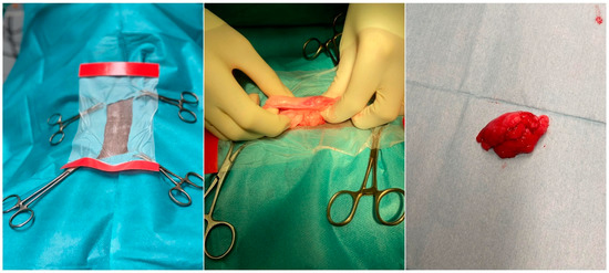
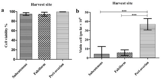
Mesenchymal stem cells (MSCs) constitute a great promise for regenerative therapy, but these cells are difficultly recovered in large amounts. A potent alternative is the stromal vascular fraction (SVF), non-cultured MSCs, separated from adipose tissue (AT). We aim to evaluate AT harvesting site effect on the SVF cells’ quantity and quality in dogs. Subcutaneous abdominal fat, falciform ligament and peri-ovarian fat were sampled. After SVF isolation, the trypan blue exclusion test and a hemocytometer were used to assess the cell viability and cellular yield. SVF cells were labeled for four surface antigenic markers, clusters of differentiation CD90, CD44, CD29, and CD45, and then examined by flow cytometry. Semi-quantitative RT-PCR was used to evaluate the gene expression of the former markers in addition to OCT-4 and CD34. SVF cells in the peri-ovarian AT recorded the highest viability% (99.63 ± 0.2%), as well as a significantly higher cellular yield (36.87 ± 19.6 × 10
6 viable cells/gm fat,
p < 0.001) and a higher expression of adipose-derived mesenchymal stem cells AD-MSCs surface markers than that of other sites. SVF cells from the peri-ovarian site revealed a higher expression of MSC markers (CD90, CD44, and CD29) and OCT-4 compared to the other sites, with weak CD45 and CD34 expressions. The positive OCT-4 expression demonstrated the pluripotency of SVF cells isolated from different sites. To conclude, the harvesting site is a strong determinant of SVF cells’ quantity and quality, and the peri-ovarian site could be the best AT sampling site in dogs.
Full article
►▼
Show Figures
Open AccessArticle
Optimized Approaches for the Induction of Putative Canine Induced Pluripotent Stem Cells from Old Fibroblasts Using Synthetic RNAs
by
Mirae Kim, Seon-Ung Hwang, Junchul David Yoon, Yeon Woo Jeong, Eunhye Kim and Sang-Hwan Hyun
Cited by 6 | Viewed by 3988
Abstract
Canine induced pluripotent stem cells (ciPSCs) can provide great potential for regenerative veterinary medicine. Several reports have described the generation of canine somatic cell-derived iPSCs; however, none have described the canine somatic cell reprogramming using a non-integrating and self-replicating RNA transfection method. The
[...] Read more.
Canine induced pluripotent stem cells (ciPSCs) can provide great potential for regenerative veterinary medicine. Several reports have described the generation of canine somatic cell-derived iPSCs; however, none have described the canine somatic cell reprogramming using a non-integrating and self-replicating RNA transfection method. The purpose of this study was to investigate the optimal strategy using this approach and characterize the transition stage of ciPSCs. In this study, fibroblasts obtained from a 13-year-old dog were reprogrammed using a non-integrating
Venezuelan equine encephalitis (VEE) RNA virus replicon, which has four reprogramming factors (collectively referred to as
T7-VEE-OKS-iG and comprised of
hOct4,
hKlf4,
hSox2, and
hGlis1) and co-transfected with the
T7-VEE-OKS-iG RNA and
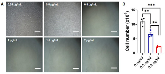
B18R mRNA for 4 h. One day after the final transfection, the cells were selected with puromycin (0.5 µg/mL) until day 10. After about 25 days, putative ciPSC colonies were identified showing TRA-1-60 expression and alkaline phosphatase activity. To determine the optimal culture conditions, the basic fibroblast growth factor in the culture medium was replaced with a modified medium supplemented with murine leukemia inhibitory factor (mLIF) and two kinase inhibitors (2i), PD0325901(MEK1/2 inhibitor) and CHIR99021 (GSK3β inhibitor). The derived colonies showed resemblance to naïve iPSCs in their morphology (dome-shaped) and are dependent on mLIF and 2i condition to maintain an undifferentiated phenotype. The expression of endogenous pluripotency markers such as
Oct4, Nanog, and
Rex1 transcripts were confirmed, suggesting that induced ciPSCs were in the late intermediate stage of reprogramming. In conclusion, the non-integrating and self-replicating VEE RNA replicon system can potentially make a great contribution to the generation of clinically applicable ciPSCs, and the findings of this study suggest a new method to utilize the VEE RNA approach for canine somatic cell reprogramming.
Full article
►▼
Show Figures
Planned Papers
The below list represents only planned manuscripts. Some of these
manuscripts have not been received by the Editorial Office yet. Papers
submitted to MDPI journals are subject to peer-review.
Title: Therapeutic application of Extracellular Vesicles derived from Mesenchymal Stem Cells in domestic animals
Authors: Aliai Lanci; Eleonora Iacono; Barbara Merlo
Affiliation: University of Bologna, Italy
Abstract: Recently, the therapeutic potential of extracellular vesicles (EVs) derived by mesenchymal stem cells (MSCs) has been extensively studied in both human and veterinary medicine. EVs are nano-sized particles containing biological components commonly found in other biological materials. For that reason, EVs isolation and characterization are critical to draw precise conclusions during their investigation. Research on EVs within veterinary medicine is still considered in its early phases, yet numerous papers were published in recent years. The conventional adult tissues for deriving MSCs include adipose tissue and bone marrow. Nonetheless, alternative sources such as synovial fluid, endometrium, gingiva, and milk have also been intermittently used. Fetal adnexa are amniotic membrane/fluid, umbilical cord and Wharton’s jelly. Cells derived from fetal adnexa exhibit an intermediate state between embryonic and adult cells, demonstrating higher proliferative and differentiative potential and longer telomeres compared to cells from adult tissues. There are summarized the principal and recent preclinical and clinical studies performed in domestic animals such as horse, cattle, dog and cat. To minimize the use of antibiotics and address the serious issue of antibiotic resistance as a public health concern, they will undoubtedly also be utilized in the future to treat infections in domestic animals. A number of concerns, including large-scale production with standardization of EV separation and characterization techniques, must be resolved for clinical application.