
Efficacy of Stem Cell Therapy in Large Animal Models of Ischemic Cardiomyopathies: A Systematic Review and Meta-Analysis

 , , , , and
Abstract
:Simple Summary
Abstract
1. Introduction
2. Materials and Methods
2.1. Search Strategy and Selection Criteria
2.2. Eligibility Criteria
2.3. Data Extraction
2.4. Statistical Analysis
3. Results
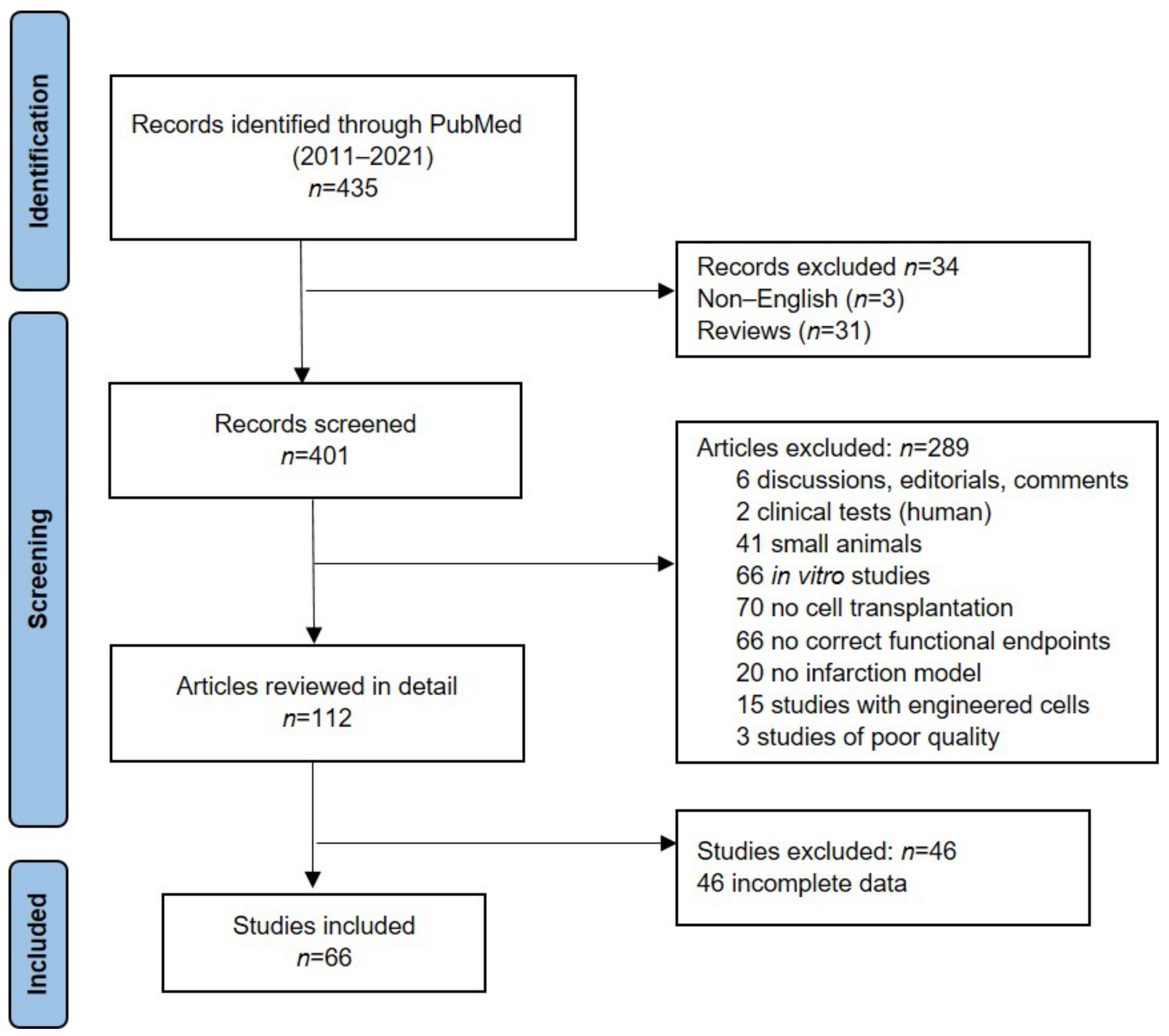
3.1. Study Selection
3.2. Included Studies Characteristics
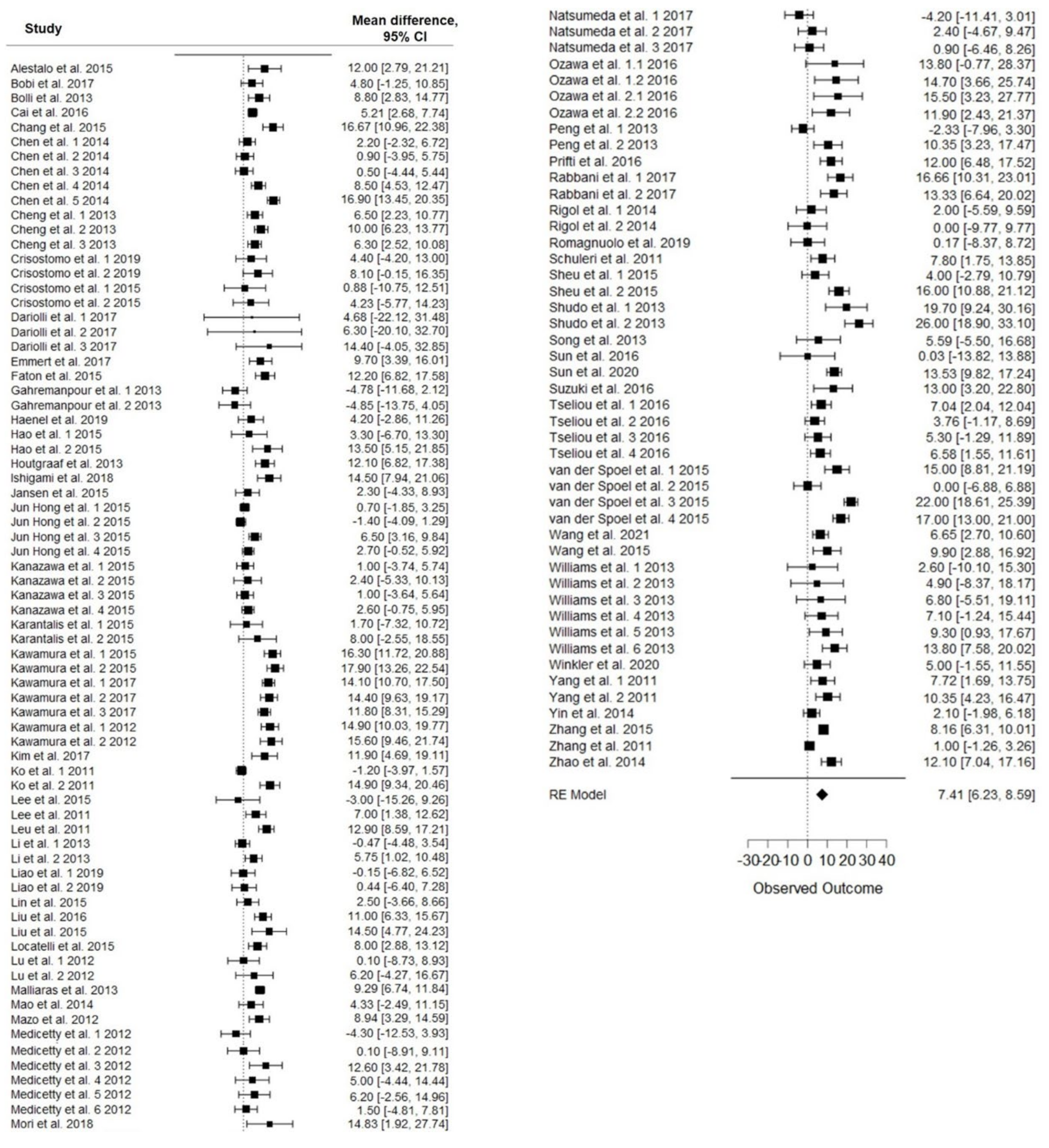
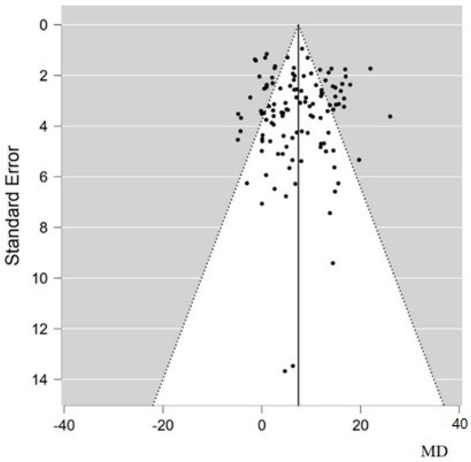
3.3. Meta-Analysis
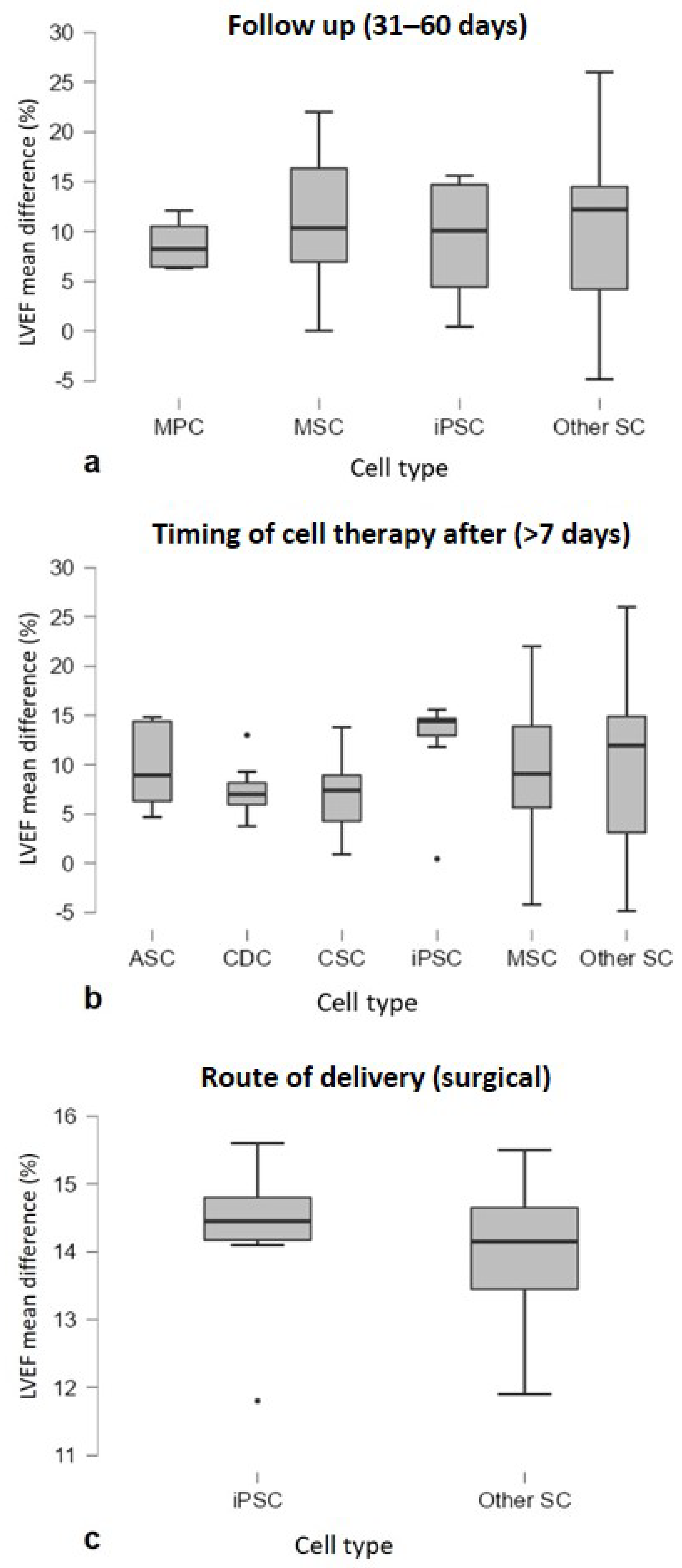
3.4. Subgroup Analysis
4. Discussion
Limitations
5. Conclusions
Author Contributions
Funding
Institutional Review Board Statement
Informed Consent Statement
Data Availability Statement
Conflicts of Interest
References
- Cardiovascular Diseases. Available online: https://www.who.int/westernpacific/health-topics/cardiovascular-diseases (accessed on 19 January 2022).
- Thygesen, K.; Alpert, J.S.; Jaffe, A.S.; Simoons, M.L.; Chaitman, B.R.; White, H.D. Writing Group on behalf of the Joint For the Universal Definition of Myocardial Infarction Third Universal Definition of Myocardial Infarction. Glob. Heart 2012, 7, 275. [Google Scholar] [CrossRef] [PubMed] [Green Version]
- Wang, Q.; He, X.; Wang, B.; Pan, J.; Shi, C.; Li, J.; Wang, L.; Zhao, Y.; Dai, J.; Wang, D. Injectable Collagen Scaffold Promotes Swine Myocardial Infarction Recovery by Long-Term Local Retention of Transplanted Human Umbilical Cord Mesenchymal Stem Cells. Sci. China Life Sci. 2021, 64, 269–281. [Google Scholar] [CrossRef] [PubMed]
- Perea-Gil, I.; Prat-Vidal, C.; Bayes-Genis, A. In Vivo Experience with Natural Scaffolds for Myocardial Infarction: The Times They Are a-Changin’. Stem Cell Res. Ther. 2015, 6, 248. [Google Scholar] [CrossRef] [PubMed] [Green Version]
- Dixit, P.; Katare, R. Challenges in Identifying the Best Source of Stem Cells for Cardiac Regeneration Therapy. Stem Cell Res. Ther. 2015, 6, 26. [Google Scholar] [CrossRef] [PubMed] [Green Version]
- Leri, A.; Kajstura, J.; Anversa, P. Cardiac Stem Cells and Mechanisms of Myocardial Regeneration. Physiol. Rev. 2005, 85, 1373–1416. [Google Scholar] [CrossRef] [Green Version]
- Gnecchi, M.; Zhang, Z.; Ni, A.; Dzau, V.J. Paracrine Mechanisms in Adult Stem Cell Signaling and Therapy. Circ. Res. 2008, 103, 1204–1219. [Google Scholar] [CrossRef]
- Tomita, S.; Li, R.-K.; Weisel, R.D.; Mickle, D.A.G.; Kim, E.-J.; Sakai, T.; Jia, Z.-Q. Autologous Transplantation of Bone Marrow Cells Improves Damaged Heart Function. Circulation 1999, 100, 247–256. [Google Scholar] [CrossRef] [Green Version]
- Pittenger, M.F.; Martin, B.J. Mesenchymal Stem Cells and Their Potential as Cardiac Therapeutics. Circ. Res. 2004, 95, 9–20. [Google Scholar] [CrossRef]
- Yang, D.; Wang, W.; Li, L.; Peng, Y.; Chen, P.; Huang, H.; Guo, Y.; Xia, X.; Wang, Y.; Wang, H.; et al. The Relative Contribution of Paracine Effect versus Direct Differentiation on Adipose-Derived Stem Cell Transplantation Mediated Cardiac Repair. PLoS ONE 2013, 8, e59020. [Google Scholar] [CrossRef]
- Sid-Otmane, C.; Perrault, L.P.; Ly, H.Q. Mesenchymal stem cell mediates cardiac repair through autocrine, paracrine and endocrine axes. J. Transl. Med. 2020, 18, 336. [Google Scholar] [CrossRef]
- Burchfield, J.S.; Dimmeler, S. Role of Paracrine Factors in Stem and Progenitor Cell Mediated Cardiac Repair and Tissue Fibrosis. Fibrogenesis Tissue Repair 2008, 1, 4. [Google Scholar] [CrossRef] [PubMed] [Green Version]
- Segers, V.F.M.; Tokunou, T.; Higgins, L.J.; MacGillivray, C.; Gannon, J.; Lee, R.T. Local Delivery of Protease-Resistant Stromal Cell Derived Factor-1 for Stem Cell Recruitment After Myocardial Infarction. Circulation 2007, 116, 1683–1692. [Google Scholar] [CrossRef] [PubMed] [Green Version]
- Chimenti, I.; Smith, R.R.; Li, T.-S.; Gerstenblith, G.; Messina, E.; Giacomello, A.; Marbán, E. Relative Roles of Direct Regeneration Versus Paracrine Effects of Human Cardiosphere-Derived Cells Transplanted Into Infarcted Mice. Circ. Res. 2010, 106, 971–980. [Google Scholar] [CrossRef] [PubMed]
- Sepantafar, M.; Maheronnaghsh, R.; Mohammadi, H.; Rajabi-Zeleti, S.; Annabi, N.; Aghdami, N.; Baharvand, H. Stem Cells and Injectable Hydrogels: Synergistic Therapeutics in Myocardial Repair. Biotechnol. Adv. 2016, 34, 362–379. [Google Scholar] [CrossRef] [PubMed] [Green Version]
- Chien, K.R.; Frisén, J.; Fritsche-Danielson, R.; Melton, D.A.; Murry, C.E.; Weissman, I.L. Regenerating the Field of Cardiovascular Cell Therapy. Nat. Biotechnol. 2019, 37, 232–237. [Google Scholar] [CrossRef]
- Van der Spoel, T.I.G.; Jansen of Lorkeers, S.J.; Agostoni, P.; van Belle, E.; Gyöngyösi, M.; Sluijter, J.P.G.; Cramer, M.J.; Doevendans, P.A.; Chamuleau, S.A.J. Human Relevance of Pre-Clinical Studies in Stem Cell Therapy: Systematic Review and Meta-Analysis of Large Animal Models of Ischaemic Heart Disease. Cardiovasc. Res. 2011, 91, 649–658. [Google Scholar] [CrossRef]
- Page, M.J.; Moher, D.; Bossuyt, P.M.; Boutron, I.; Hoffmann, T.C.; Mulrow, C.D.; Shamseer, L.; Tetzlaff, J.M.; Akl, E.A.; Brennan, S.E.; et al. PRISMA 2020 Explanation and Elaboration: Updated Guidance and Exemplars for Reporting Systematic Reviews. BMJ 2021, 372, n160. [Google Scholar] [CrossRef]
- PubMed. Available online: https://pubmed.ncbi.nlm.nih.gov/ (accessed on 24 January 2022).
- Higgins, J.P.T.; Thompson, S.G.; Deeks, J.J.; Altman, D.G. Measuring Inconsistency in Meta-Analyses. BMJ 2003, 327, 557–560. [Google Scholar] [CrossRef] [Green Version]
- Alestalo, K.; Korpi, R.; Mäkelä, J.; Lehtonen, S.; Mäkelä, T.; Yannopoulos, F.; Ylitalo, K.; Haapea, M.; Juvonen, T.; Anttila, V.; et al. High Number of Transplanted Stem Cells Improves Myocardial Recovery after AMI in a Porcine Model. Scand. Cardiovasc. J. 2015, 49, 82–94. [Google Scholar] [CrossRef]
- Bobi, J.; Solanes, N.; Fernández-Jiménez, R.; Galán-Arriola, C.; Dantas, A.P.; Fernández-Friera, L.; Gálvez-Montón, C.; Rigol-Monzó, E.; Agüero, J.; Ramírez, J.; et al. Intracoronary Administration of Allogeneic Adipose Tissue-Derived Mesenchymal Stem Cells Improves Myocardial Perfusion But Not Left Ventricle Function, in a Translational Model of Acute Myocardial Infarction. J. Am. Heart Assoc. 2017, 6, e005771. [Google Scholar] [CrossRef] [Green Version]
- Bolli, R.; Tang, X.-L.; Sanganalmath, S.K.; Rimoldi, O.; Mosna, F.; Abdel-Latif, A.; Jneid, H.; Rota, M.; Leri, A.; Kajstura, J. Intracoronary Delivery of Autologous Cardiac Stem Cells Improves Cardiac Function in a Porcine Model of Chronic Ischemic Cardiomyopathy. Circulation 2013, 128, 122–131. [Google Scholar] [CrossRef] [PubMed] [Green Version]
- Cai, M.; Shen, R.; Song, L.; Lu, M.; Wang, J.; Zhao, S.; Tang, Y.; Meng, X.; Li, Z.; He, Z.-X. Erratum: Bone Marrow Mesenchymal Stem Cells (BM-MSCs) Improve Heart Function in Swine Myocardial Infarction Model through Paracrine Effects. Sci. Rep. 2016, 6, 31528. [Google Scholar] [CrossRef] [PubMed] [Green Version]
- Chang, X.; Liu, J.; Liao, X.; Liu, G. Ultrasound-Mediated Microbubble Destruction Enhances the Therapeutic Effect of Intracoronary Transplantation of Bone Marrow Stem Cells on Myocardial Infarction. Int. J. Clin. Exp. Pathol. 2015, 8, 2221–2234. [Google Scholar] [PubMed]
- Chen, Y.; Teng, X.; Chen, W.; Yang, J.; Yang, Z.; Yu, Y.; Shen, Z. Timing of Transplantation of Autologous Bone Marrow Derived Mesenchymal Stem Cells for Treating Myocardial Infarction. Sci. China Life Sci. 2014, 57, 195–200. [Google Scholar] [CrossRef] [Green Version]
- Cheng, Y.; Yi, G.; Conditt, G.B.; Sheehy, A.; Kolodgie, F.D.; Tellez, A.; Polyakov, I.; Gu, A.; Aboodi, M.S.; Wallace-Bradley, D.; et al. Catheter-Based Endomyocardial Delivery of Mesenchymal Precursor Cells Using 3D Echo Guidance Improves Cardiac Function in a Chronic Myocardial Injury Ovine Model. Cell Transplant. 2013, 22, 2299–2309. [Google Scholar] [CrossRef] [Green Version]
- Crisostomo, V.; Baez, C.; Abad, J.L.; Sanchez, B.; Alvarez, V.; Rosado, R.; Gómez-Mauricio, G.; Gheysens, O.; Blanco-Blazquez, V.; Blazquez, R.; et al. Dose-Dependent Improvement of Cardiac Function in a Swine Model of Acute Myocardial Infarction after Intracoronary Administration of Allogeneic Heart-Derived Cells. Stem Cell Res. Ther. 2019, 10, 152. [Google Scholar] [CrossRef] [Green Version]
- Crisostomo, V.; Baez-Diaz, C.; Maestre, J.; Garcia-Lindo, M.; Sun, F.; Casado, J.G.; Blazquez, R.; Abad, J.L.; Palacios, I.; Rodriguez-Borlado, L.; et al. Delayed Administration of Allogeneic Cardiac Stem Cell Therapy for Acute Myocardial Infarction Could Ameliorate Adverse Remodeling: Experimental Study in Swine. J. Transl. Med. 2015, 13, 156. [Google Scholar] [CrossRef] [Green Version]
- Dariolli, R.; Naghetini, M.V.; Marques, E.F.; Takimura, C.K.; Jensen, L.S.; Kiers, B.; Tsutsui, J.M.; Mathias, W.; Lemos Neto, P.A.; Krieger, J.E. Allogeneic PASC Transplantation in Humanized Pigs Attenuates Cardiac Remodeling Post-Myocardial Infarction. PLoS ONE 2017, 12, e0176412. [Google Scholar] [CrossRef]
- Emmert, M.Y.; Wolint, P.; Jakab, A.; Sheehy, S.P.; Pasqualini, F.S.; Nguyen, T.D.L.; Hilbe, M.; Seifert, B.; Weber, B.; Brokopp, C.E.; et al. Safety and Efficacy of Cardiopoietic Stem Cells in the Treatment of Post-Infarction Left-Ventricular Dysfunction—From Cardioprotection to Functional Repair in a Translational Pig Infarction Model. Biomaterials 2017, 122, 48–62. [Google Scholar] [CrossRef] [Green Version]
- Fanton, Y.; Robic, B.; Rummens, J.-L.; Daniëls, A.; Windmolders, S.; Willems, L.; Jamaer, L.; Dubois, J.; Bijnens, E.; Heuts, N.; et al. Cardiac Atrial Appendage Stem Cells Engraft and Differentiate into Cardiomyocytes in Vivo: A New Tool for Cardiac Repair after MI. Int. J. Cardiol. 2015, 201, 10–19. [Google Scholar] [CrossRef]
- Gahremanpour, A.; Vela, D.; Zheng, Y.; Silva, G.V.; Fodor, W.; Cardoso, C.O.; Baimbridge, F.; Fernandes, M.R.; Buja, L.M.; Perin, E.C. Xenotransplantation of Human Unrestricted Somatic Stem Cells in a Pig Model of Acute Myocardial Infarction. Xenotransplantation 2013, 20, 110–122. [Google Scholar] [CrossRef] [PubMed]
- Haenel, A.; Ghosn, M.; Karimi, T.; Vykoukal, J.; Shah, D.; Valderrabano, M.; Schulz, D.G.; Raizner, A.; Schmitz, C.; Alt, E.U. Unmodified Autologous Stem Cells at Point of Care for Chronic Myocardial Infarction. World J. Stem Cells 2019, 11, 831–858. [Google Scholar] [CrossRef] [PubMed]
- Hao, L.; Hao, J.; Fang, W.; Han, C.; Zhang, K.; Wang, X. Dual Isotope Simultaneous Imaging to Evaluate the Effects of Intracoronary Bone Marrow-Derived Mesenchymal Stem Cells on Perfusion and Metabolism in Canines with Acute Myocardial Infarction. Biomed. Rep. 2015, 3, 447–452. [Google Scholar] [CrossRef] [PubMed] [Green Version]
- Houtgraaf, J.H.; de Jong, R.; Kazemi, K.; de Groot, D.; van der Spoel, T.I.G.; Arslan, F.; Hoefer, I.; Pasterkamp, G.; Itescu, S.; Zijlstra, F.; et al. Intracoronary Infusion of Allogeneic Mesenchymal Precursor Cells Directly after Experimental Acute Myocardial Infarction Reduces Infarct Size, Abrogates Adverse Remodeling, and Improves Cardiac Function. Circ. Res. 2013, 113, 153–166. [Google Scholar] [CrossRef] [Green Version]
- Ishigami, M.; Masumoto, H.; Ikuno, T.; Aoki, T.; Kawatou, M.; Minakata, K.; Ikeda, T.; Sakata, R.; Yamashita, J.K.; Minatoya, K. Human IPS Cell-Derived Cardiac Tissue Sheets for Functional Restoration of Infarcted Porcine Hearts. PLoS ONE 2018, 13, e0201650. [Google Scholar] [CrossRef] [Green Version]
- Jansen of Lorkeers, S.J.; Gho, J.M.I.H.; Koudstaal, S.; van Hout, G.P.J.; Zwetsloot, P.P.M.; van Oorschot, J.W.M.; van Eeuwijk, E.C.M.; Leiner, T.; Hoefer, I.E.; Goumans, M.-J.; et al. Xenotransplantation of Human Cardiomyocyte Progenitor Cells Does Not Improve Cardiac Function in a Porcine Model of Chronic Ischemic Heart Failure. Results from a Randomized, Blinded, Placebo Controlled Trial. PLoS ONE 2015, 10, e0143953. [Google Scholar] [CrossRef] [PubMed]
- Jun Hong, S.; Rogers, P.I.; Kihlken, J.; Warfel, J.; Bull, C.; Deuter-Reinhard, M.; Feng, D.; Xie, J.; Kyle, A.; Merfeld-Clauss, S.; et al. Intravenous Xenogeneic Transplantation of Human Adipose-Derived Stem Cells Improves Left Ventricular Function and Microvascular Integrity in Swine Myocardial Infarction Model. Catheter. Cardiovasc. Interv. Off. J. Soc. Card. Angiogr. Interv. 2015, 86, E38–E48. [Google Scholar] [CrossRef] [Green Version]
- Kanazawa, H.; Tseliou, E.; Malliaras, K.; Yee, K.; Dawkins, J.F.; De Couto, G.; Smith, R.R.; Kreke, M.; Seinfeld, J.; Middleton, R.C.; et al. Cellular Postconditioning: Allogeneic Cardiosphere-Derived Cells Reduce Infarct Size and Attenuate Microvascular Obstruction When Administered after Reperfusion in Pigs with Acute Myocardial Infarction. Circ. Heart Fail. 2015, 8, 322–332. [Google Scholar] [CrossRef] [PubMed] [Green Version]
- Karantalis, V.; Suncion-Loescher, V.Y.; Bagno, L.; Golpanian, S.; Wolf, A.; Sanina, C.; Premer, C.; Kanelidis, A.J.; McCall, F.; Wang, B.; et al. Synergistic Effects of Combined Cell Therapy for Chronic Ischemic Cardiomyopathy. J. Am. Coll. Cardiol. 2015, 66, 1990–1999. [Google Scholar] [CrossRef] [PubMed]
- Kawamura, M.; Miyagawa, S.; Fukushima, S.; Saito, A.; Toda, K.; Daimon, T.; Shimizu, T.; Okano, T.; Sawa, Y. Xenotransplantation of Bone Marrow-Derived Human Mesenchymal Stem Cell Sheets Attenuates Left Ventricular Remodeling in a Porcine Ischemic Cardiomyopathy Model. Tissue Eng. Part A 2015, 21, 2272–2280. [Google Scholar] [CrossRef] [Green Version]
- Kawamura, M.; Miyagawa, S.; Fukushima, S.; Saito, A.; Miki, K.; Funakoshi, S.; Yoshida, Y.; Yamanaka, S.; Shimizu, T.; Okano, T.; et al. Enhanced Therapeutic Effects of Human IPS Cell Derived-Cardiomyocyte by Combined Cell-Sheets with Omental Flap Technique in Porcine Ischemic Cardiomyopathy Model. Sci. Rep. 2017, 7, 8824. [Google Scholar] [CrossRef] [PubMed]
- Kawamura, M.; Miyagawa, S.; Miki, K.; Saito, A.; Fukushima, S.; Higuchi, T.; Kawamura, T.; Kuratani, T.; Daimon, T.; Shimizu, T.; et al. Feasibility, Safety, and Therapeutic Efficacy of Human Induced Pluripotent Stem Cell-Derived Cardiomyocyte Sheets in a Porcine Ischemic Cardiomyopathy Model. Circulation 2012, 126, S29–S37. [Google Scholar] [CrossRef] [PubMed] [Green Version]
- Kim, M.C.; Kim, Y.S.; Kang, W.S.; Lee, K.H.; Cho, M.; Hong, M.H.; Lim, K.S.; Jeong, M.H.; Ahn, Y. Intramyocardial Injection of Stem Cells in Pig Myocardial Infarction Model: The First Trial in Korea. J. Korean Med. Sci. 2017, 32, 1708–1712. [Google Scholar] [CrossRef] [PubMed]
- Ko, S.-F.; Yip, H.-K.; Lee, C.-C.; Sheu, J.-J.; Sun, C.-K.; Ng, S.-H.; Huang, C.-C.; Lin, Y.-C.; Chang, L.-T.; Chen, M.-C. Immediate Intramyocardial Bone Marrow-Derived Mononuclear Cells Implantation in Minipig Myocardium after Permanent Coronary Artery Ligation: Magnetic Resonance Imaging with Histopathologic and Immunochemical Correlation. Investig. Radiol. 2011, 46, 495–503. [Google Scholar] [CrossRef]
- Lee, H.W.; Lee, H.C.; Park, J.H.; Kim, B.W.; Ahn, J.; Kim, J.H.; Park, J.S.; Oh, J.-H.; Choi, J.H.; Cha, K.S.; et al. Effects of Intracoronary Administration of Autologous Adipose Tissue-Derived Stem Cells on Acute Myocardial Infarction in a Porcine Model. Yonsei Med. J. 2015, 56, 1522–1529. [Google Scholar] [CrossRef] [PubMed] [Green Version]
- Lee, S.-T.; White, A.J.; Matsushita, S.; Malliaras, K.; Steenbergen, C.; Zhang, Y.; Li, T.-S.; Terrovitis, J.; Yee, K.; Simsir, S.; et al. Intramyocardial Injection of Autologous Cardiospheres or Cardiosphere-Derived Cells Preserves Function and Minimizes Adverse Ventricular Remodeling in Pigs with Heart Failure Post-Myocardial Infarction. J. Am. Coll. Cardiol. 2011, 57, 455–465. [Google Scholar] [CrossRef] [PubMed] [Green Version]
- Leu, S.; Sun, C.-K.; Sheu, J.-J.; Chang, L.-T.; Yuen, C.-M.; Yen, C.-H.; Chiang, C.-H.; Ko, S.-F.; Pei, S.-N.; Chua, S.; et al. Autologous Bone Marrow Cell Implantation Attenuates Left Ventricular Remodeling and Improves Heart Function in Porcine Myocardial Infarction: An Echocardiographic, Six-Month Angiographic, and Molecular-Cellular Study. Int. J. Cardiol. 2011, 150, 156–168. [Google Scholar] [CrossRef] [PubMed]
- Li, X.; Zhang, F.; Song, G.; Gu, W.; Chen, M.; Yang, B.; Li, D.; Wang, D.; Cao, K. Intramyocardial Injection of Pig Pluripotent Stem Cells Improves Left Ventricular Function and Perfusion: A Study in a Porcine Model of Acute Myocardial Infarction. PLoS ONE 2013, 8, e66688. [Google Scholar] [CrossRef]
- Liao, S.; Zhang, Y.; Ting, S.; Zhen, Z.; Luo, F.; Zhu, Z.; Jiang, Y.; Sun, S.; Lai, W.-H.; Lian, Q.; et al. Potent Immunomodulation and Angiogenic Effects of Mesenchymal Stem Cells versus Cardiomyocytes Derived from Pluripotent Stem Cells for Treatment of Heart Failure. Stem Cell Res. Ther. 2019, 10, 78. [Google Scholar] [CrossRef] [Green Version]
- Lin, Y.-D.; Chang, M.-Y.; Cheng, B.; Liu, Y.-W.; Lin, L.-C.; Chen, J.-H.; Hsieh, P.C.H. Injection of Peptide Nanogels Preserves Postinfarct Diastolic Function and Prolongs Efficacy of Cell Therapy in Pigs. Tissue Eng. Part A 2015, 21, 1662–1671. [Google Scholar] [CrossRef]
- Liu, C.-B.; Huang, H.; Sun, P.; Ma, S.-Z.; Liu, A.-H.; Xue, J.; Fu, J.-H.; Liang, Y.-Q.; Liu, B.; Wu, D.-Y.; et al. Human Umbilical Cord-Derived Mesenchymal Stromal Cells Improve Left Ventricular Function, Perfusion, and Remodeling in a Porcine Model of Chronic Myocardial Ischemia. Stem Cells Transl. Med. 2016, 5, 1004–1013. [Google Scholar] [CrossRef] [PubMed]
- Liu, Y.-H.; Peng, K.-Y.; Chiu, Y.-W.; Ho, Y.-L.; Wang, Y.-H.; Shun, C.-T.; Huang, S.-Y.; Lin, Y.-S.; de Vries, A.A.F.; Pijnappels, D.A.; et al. Human Placenta-Derived Multipotent Cells (HPDMCs) Modulate Cardiac Injury: From Bench to Small and Large Animal Myocardial Ischemia Studies. Cell Transplant. 2015, 24, 2463–2478. [Google Scholar] [CrossRef] [PubMed] [Green Version]
- Locatelli, P.; Olea, F.D.; Hnatiuk, A.; De Lorenzi, A.; Cerdá, M.; Giménez, C.S.; Sepúlveda, D.; Laguens, R.; Crottogini, A. Mesenchymal Stromal Cells Overexpressing Vascular Endothelial Growth Factor in Ovine Myocardial Infarction. Gene Ther. 2015, 22, 449–457. [Google Scholar] [CrossRef] [PubMed]
- Lu, M.; Zhao, S.; Liu, Q.; Jiang, S.; Song, P.; Qian, H.; Zhang, Y.; Ling, J.; Yan, C.; Cheng, H.; et al. Transplantation with Autologous Mesenchymal Stem Cells after Acute Myocardial Infarction Evaluated by Magnetic Resonance Imaging: An Experimental Study. J. Thorac. Imaging 2012, 27, 125–135. [Google Scholar] [CrossRef] [PubMed]
- Malliaras, K.; Smith, R.R.; Kanazawa, H.; Yee, K.; Seinfeld, J.; Tseliou, E.; Dawkins, J.F.; Kreke, M.; Cheng, K.; Luthringer, D.; et al. Validation of Contrast-Enhanced Magnetic Resonance Imaging to Monitor Regenerative Efficacy after Cell Therapy in a Porcine Model of Convalescent Myocardial Infarction. Circulation 2013, 128, 2764–2775. [Google Scholar] [CrossRef] [PubMed] [Green Version]
- Mao, Q.; Lin, C.; Gao, J.; Liang, X.; Gao, W.; Shen, L.; Kang, L.; Xu, B. Mesenchymal Stem Cells Overexpressing Integrin-Linked Kinase Attenuate Left Ventricular Remodeling and Improve Cardiac Function after Myocardial Infarction. Mol. Cell. Biochem. 2014, 397, 203–214. [Google Scholar] [CrossRef] [PubMed]
- Mazo, M.; Hernández, S.; Gavira, J.J.; Abizanda, G.; Araña, M.; López-Martínez, T.; Moreno, C.; Merino, J.; Martino-Rodríguez, A.; Uixeira, A.; et al. Treatment of Reperfused Ischemia with Adipose-Derived Stem Cells in a Preclinical Swine Model of Myocardial Infarction. Cell Transplant. 2012, 21, 2723–2733. [Google Scholar] [CrossRef] [Green Version]
- Medicetty, S.; Wiktor, D.; Lehman, N.; Raber, A.; Popovic, Z.B.; Deans, R.; Ting, A.E.; Penn, M.S. Percutaneous Adventitial Delivery of Allogeneic Bone Marrow-Derived Stem Cells via Infarct-Related Artery Improves Long-Term Ventricular Function in Acute Myocardial Infarction. Cell Transplant. 2012, 21, 1109–1120. [Google Scholar] [CrossRef]
- Mori, D.; Miyagawa, S.; Yajima, S.; Saito, S.; Fukushima, S.; Ueno, T.; Toda, K.; Kawai, K.; Kurata, H.; Nishida, H.; et al. Cell Spray Transplantation of Adipose-Derived Mesenchymal Stem Cell Recovers Ischemic Cardiomyopathy in a Porcine Model. Transplantation 2018, 102, 2012–2024. [Google Scholar] [CrossRef]
- Natsumeda, M.; Florea, V.; Rieger, A.C.; Tompkins, B.A.; Banerjee, M.N.; Golpanian, S.; Fritsch, J.; Landin, A.M.; Kashikar, N.D.; Karantalis, V.; et al. A Combination of Allogeneic Stem Cells Promotes Cardiac Regeneration. J. Am. Coll. Cardiol. 2017, 70, 2504–2515. [Google Scholar] [CrossRef]
- Ozawa, H.; Miyagawa, S.; Fukushima, S.; Itoh, E.; Harada, A.; Saito, A.; Ueno, T.; Toda, K.; Kuratani, T.; Sawa, Y. Sirtuin1 Regulates the Stem Cell Therapeutic Effects on Regenerative Capability for Treating Severe Heart Failure in a Juvenile Animal Model. Ann. Thorac. Surg. 2016, 102, 803–812. [Google Scholar] [CrossRef] [PubMed] [Green Version]
- Peng, C.; Yang, K.; Xiang, P.; Zhang, C.; Zou, L.; Wu, X.; Gao, Y.; Kang, Z.; He, K.; Liu, J.; et al. Effect of Transplantation with Autologous Bone Marrow Stem Cells on Acute Myocardial Infarction. Int. J. Cardiol. 2013, 162, 158–165. [Google Scholar] [CrossRef] [PubMed]
- Prifti, E.; Di Lascio, G.; Harmelin, G.; Bani, D.; Briganti, V.; Veshti, A.; Bonacchi, M. Cellular Cardiomyoplasty into Infracted Swine’s Hearts by Retrograde Infusion through the Venous Coronary Sinus: An Experimental Study. Cardiovasc. Revasc. Med. Mol. Interv. 2016, 17, 262–271. [Google Scholar] [CrossRef] [PubMed]
- Rabbani, S.; Soleimani, M.; Sahebjam, M.; Imani, M.; Nassiri, S.M.; Atashi, A.; Daliri Joupari, M.; Ghiaseddin, A.; Latifpour, M.; Ahmadi Tafti, S.H. Effects of Endothelial and Mesenchymal Stem Cells on Improving Myocardial Function in a Sheep Animal Model. J. Tehran Heart Cent. 2017, 12, 65–71. [Google Scholar] [PubMed]
- Rigol, M.; Solanes, N.; Roura, S.; Roqué, M.; Novensà, L.; Dantas, A.P.; Martorell, J.; Sitges, M.; Ramírez, J.; Bayés-Genís, A.; et al. Allogeneic Adipose Stem Cell Therapy in Acute Myocardial Infarction. Eur. J. Clin. Investig. 2014, 44, 83–92. [Google Scholar] [CrossRef] [PubMed]
- Romagnuolo, R.; Masoudpour, H.; Porta-Sánchez, A.; Qiang, B.; Barry, J.; Laskary, A.; Qi, X.; Massé, S.; Magtibay, K.; Kawajiri, H.; et al. Human Embryonic Stem Cell-Derived Cardiomyocytes Regenerate the Infarcted Pig Heart but Induce Ventricular Tachyarrhythmias. Stem Cell Rep. 2019, 12, 967–981. [Google Scholar] [CrossRef] [Green Version]
- Schuleri, K.H.; Centola, M.; Choi, S.H.; Evers, K.S.; Dawoud, F.; George, R.T.; Lima, J.A.C.; Lardo, A.C. CT for Evaluation of Myocardial Cell Therapy in Heart Failure: A Comparison with CMR Imaging. JACC Cardiovasc. Imaging 2011, 4, 1284–1293. [Google Scholar] [CrossRef] [Green Version]
- Sheu, J.-J.; Lee, F.-Y.; Yuen, C.-M.; Chen, Y.-L.; Huang, T.-H.; Chua, S.; Chen, Y.-L.; Chen, C.-H.; Chai, H.-T.; Sung, P.-H.; et al. Combined Therapy with Shock Wave and Autologous Bone Marrow-Derived Mesenchymal Stem Cells Alleviates Left Ventricular Dysfunction and Remodeling through Inhibiting Inflammatory Stimuli, Oxidative Stress & Enhancing Angiogenesis in a Swine Myocardial Infarction Model. Int. J. Cardiol. 2015, 193, 69–83. [Google Scholar] [CrossRef]
- Shudo, Y.; Miyagawa, S.; Nakatani, S.; Fukushima, S.; Sakaguchi, T.; Saito, A.; Asanuma, T.; Kawaguchi, N.; Matsuura, N.; Shimizu, T.; et al. Myocardial Layer-Specific Effect of Myoblast Cell-Sheet Implantation Evaluated by Tissue Strain Imaging. Circ. J. Off. J. Jpn. Circ. Soc. 2013, 77, 1063–1072. [Google Scholar] [CrossRef] [Green Version]
- Song, L.; Yang, Y.-J.; Dong, Q.-T.; Qian, H.-Y.; Gao, R.-L.; Qiao, S.-B.; Shen, R.; He, Z.-X.; Lu, M.-J.; Zhao, S.-H.; et al. Atorvastatin Enhance Efficacy of Mesenchymal Stem Cells Treatment for Swine Myocardial Infarction via Activation of Nitric Oxide Synthase. PLoS ONE 2013, 8, e65702. [Google Scholar] [CrossRef] [Green Version]
- Sun, Q.-W.; Zhen, L.; Wang, Q.; Sun, Y.; Yang, J.; Li, Y.-J.; Li, R.-J.; Ma, N.; Li, Z.-A.; Wang, L.-Y.; et al. Assessment of Retrograde Coronary Venous Infusion of Mesenchymal Stem Cells Combined with Basic Fibroblast Growth Factor in Canine Myocardial Infarction Using Strain Values Derived from Speckle-Tracking Echocardiography. Ultrasound Med. Biol. 2016, 42, 272–281. [Google Scholar] [CrossRef] [PubMed]
- Sun, S.; Jiang, Y.; Zhen, Z.; Lai, W.-H.; Liao, S.; Tse, H.-F. Establishing a Swine Model of Post-Myocardial Infarction Heart Failure for Stem Cell Treatment. J. Vis. Exp. JoVE 2020, 159, e60392. [Google Scholar] [CrossRef] [PubMed]
- Suzuki, G.; Young, R.F.; Leiker, M.M.; Suzuki, T. Heart-Derived Stem Cells in Miniature Swine with Coronary Microembolization: Novel Ischemic Cardiomyopathy Model to Assess the Efficacy of Cell-Based Therapy. Stem Cells Int. 2016, 2016, 6940195. [Google Scholar] [CrossRef] [PubMed] [Green Version]
- Tseliou, E.; Kanazawa, H.; Dawkins, J.; Gallet, R.; Kreke, M.; Smith, R.; Middleton, R.; Valle, J.; Marbán, L.; Kar, S.; et al. Widespread Myocardial Delivery of Heart-Derived Stem Cells by Nonocclusive Triple-Vessel Intracoronary Infusion in Porcine Ischemic Cardiomyopathy: Superior Attenuation of Adverse Remodeling Documented by Magnetic Resonance Imaging and Histology. PLoS ONE 2016, 11, e0144523. [Google Scholar] [CrossRef]
- Van der Spoel, T.I.G.; Gathier, W.A.; Koudstaal, S.; van Slochteren, F.; Of Lorkeers, S.J.; Sluijter, J.P.G.; Hoefer, I.E.; Steendijk, P.; Cramer, M.J.M.; Doevendans, P.A.; et al. Autologous Mesenchymal Stem Cells Show More Benefit on Systolic Function Compared to Bone Marrow Mononuclear Cells in a Porcine Model of Chronic Myocardial Infarction. J Cardiovasc. Transl. Res. 2015, 8, 393–403. [Google Scholar] [CrossRef] [Green Version]
- Wang, X.; Zhen, L.; Miao, H.; Sun, Q.; Yang, Y.; Que, B.; Lopes Lao, E.P.; Wu, X.; Ren, H.; Shi, S.; et al. Concomitant Retrograde Coronary Venous Infusion of Basic Fibroblast Growth Factor Enhances Engraftment and Differentiation of Bone Marrow Mesenchymal Stem Cells for Cardiac Repair after Myocardial Infarction. Theranostics 2015, 5, 995–1006. [Google Scholar] [CrossRef] [Green Version]
- Williams, A.R.; Hatzistergos, K.E.; Addicott, B.; McCall, F.; Carvalho, D.; Suncion, V.; Morales, A.R.; Da Silva, J.; Sussman, M.A.; Heldman, A.W.; et al. Enhanced Effect of Combining Human Cardiac Stem Cells and Bone Marrow Mesenchymal Stem Cells to Reduce Infarct Size and to Restore Cardiac Function after Myocardial Infarction. Circulation 2013, 127, 213–223. [Google Scholar] [CrossRef] [Green Version]
- Winkler, J.; Lukovic, D.; Mester-Tonczar, J.; Zlabinger, K.; Gugerell, A.; Pavo, N.; Jakab, A.; Szankai, Z.; Traxler, D.; Müller, C.; et al. Quantitative Hybrid Cardiac [18F]FDG-PET-MRI Images for Assessment of Cardiac Repair by Preconditioned Cardiosphere-Derived Cells. Mol. Ther. Methods Clin. Dev. 2020, 18, 354–366. [Google Scholar] [CrossRef]
- Yang, K.; Xiang, P.; Zhang, C.; Zou, L.; Wu, X.; Gao, Y.; Kang, Z.; He, K.; Liu, J.; Peng, C. Magnetic Resonance Evaluation of Transplanted Mesenchymal Stem Cells after Myocardial Infarction in Swine. Can. J. Cardiol. 2011, 27, 818–825. [Google Scholar] [CrossRef]
- Yin, Q.; Pei, Z.; Wang, H.; Zhao, Y. Cyclosporine A-Nanoparticles Enhance the Therapeutic Benefit of Adipose Tissue-Derived Stem Cell Transplantation in a Swine Myocardial Infarction Model. Int. J. Nanomed. 2014, 9, 17–26. [Google Scholar] [CrossRef] [Green Version]
- Zhang, G.-W.; Gu, T.-X.; Guan, X.-Y.; Sun, X.-J.; Jiang, D.-Q.; Tang, R.; Qi, X.; Li, X.-Y. Delayed Enrichment for C-Kit and Inducing Cardiac Differentiation Attenuated Protective Effects of BMSCs’ Transplantation in Pig Model of Acute Myocardial Ischemia. Cardiovasc. Ther. 2015, 33, 184–192. [Google Scholar] [CrossRef] [PubMed]
- Zhang, G.-W.; Liu, X.-C.; Li-Ling, J.; Luan, Y.; Ying, Y.-N.; Wu, X.-S.; Zhao, C.-H.; Liu, T.-J.; Lü, F. Mechanisms of the Protective Effects of BMSCs Promoted by TMDR with Heparinized BFGF-Incorporated Stent in Pig Model of Acute Myocardial Ischemia. J. Cell. Mol. Med. 2011, 15, 1075–1086. [Google Scholar] [CrossRef] [PubMed]
- Zhao, J.-J.; Liu, X.-C.; Kong, F.; Qi, T.-G.; Cheng, G.-H.; Wang, J.; Sun, C.; Luan, Y. Bone Marrow Mesenchymal Stem Cells Improve Myocardial Function in a Swine Model of Acute Myocardial Infarction. Mol. Med. Rep. 2014, 10, 1448–1454. [Google Scholar] [CrossRef] [PubMed] [Green Version]
- Noiseux, N.; Borie, M.; Desnoyers, A.; Menaouar, A.; Stevens, L.M.; Mansour, S.; Danalache, B.A.; Roy, D.-C.; Jankowski, M.; Gutkowska, J. Preconditioning of Stem Cells by Oxytocin to Improve Their Therapeutic Potential. Endocrinology 2012, 153, 5361–5372. [Google Scholar] [CrossRef]
- Tan, S.C.; Gomes, R.S.; Yeoh, K.K.; Perbellini, F.; Malandraki-Miller, S.; Ambrose, L.; Heather, L.C.; Faggian, G.; Schofield, C.J.; Davies, K.E.; et al. Preconditioning of Cardiosphere-Derived Cells With Hypoxia or Prolyl-4-Hydroxylase Inhibitors Increases Stemness and Decreases Reliance on Oxidative Metabolism. Cell Transpl. 2016, 25, 35–53. [Google Scholar] [CrossRef]
- Araña, M.; Gavira, J.J.; Peña, E.; González, A.; Abizanda, G.; Cilla, M.; Pérez, M.M.; Albiasu, E.; Aguado, N.; Casado, M.; et al. Epicardial Delivery of Collagen Patches with Adipose-Derived Stem Cells in Rat and Minipig Models of Chronic Myocardial Infarction. Biomaterials 2014, 35, 143–151. [Google Scholar] [CrossRef]
- Rashedi, I.; Talele, N.; Wang, X.-H.; Hinz, B.; Radisic, M.; Keating, A. Collagen Scaffold Enhances the Regenerative Properties of Mesenchymal Stromal Cells. PLoS ONE 2017, 12, e0187348. [Google Scholar] [CrossRef] [Green Version]
- Müller, P.; Lemcke, H.; David, R. Stem Cell Therapy in Heart Diseases—Cell Types, Mechanisms and Improvement Strategies. Cell. Physiol. Biochem. 2018, 48, 2607–2655. [Google Scholar] [CrossRef]
- Xu, J.; Liu, D.; Zhong, Y.; Huang, R. Effects of Timing on Intracoronary Autologous Bone Marrow-Derived Cell Transplantation in Acute Myocardial Infarction: A Meta-Analysis of Randomized Controlled Trials. Stem Cell Res. Ther. 2017, 8, 231. [Google Scholar] [CrossRef] [Green Version]
- Siu, C.-W.; Liao, S.-Y.; Liu, Y.; Lian, Q.; Tse, H.-F. Stem Cells for Myocardial Repair. Thromb. Haemost. 2010, 104, 6–12. [Google Scholar] [CrossRef] [Green Version]
- Zwetsloot, P.P.; Végh, A.M.D.; Jansen of Lorkeers, S.J.; van Hout, G.P.J.; Currie, G.L.; Sena, E.S.; Gremmels, H.; Buikema, J.W.; Goumans, M.-J.; Macleod, M.R.; et al. Cardiac Stem Cell Treatment in Myocardial Infarction. Circ. Res. 2016, 118, 1223–1232. [Google Scholar] [CrossRef] [PubMed]
- Korf-Klingebiel, M.; Kempf, T.; Sauer, T.; Brinkmann, E.; Fischer, P.; Meyer, G.P.; Ganser, A.; Drexler, H.; Wollert, K.C. Bone Marrow Cells Are a Rich Source of Growth Factors and Cytokines: Implications for Cell Therapy Trials after Myocardial Infarction. Eur. Heart J. 2008, 29, 2851–2858. [Google Scholar] [CrossRef] [PubMed] [Green Version]
- Spannbauer, A.; Mester-Tonczar, J.; Traxler, D.; Kastner, N.; Zlabinger, K.; Hašimbegović, E.; Riesenhuber, M.; Pavo, N.; Goliasch, G.; Gyöngyösi, M. Large Animal Models of Cell-Free Cardiac Regeneration. Biomolecules 2020, 10, 1392. [Google Scholar] [CrossRef] [PubMed]
- Petitti, D.B. Meta-Analysis, Decision Analysis, and Cost-Effectiveness Analysis: Methods for Quantitative Synthesis in Medicine; Oxford University Press: New York, NY, USA, 2000; ISBN 978-0-19-513364-6. [Google Scholar]
- Gurevitch, J.; Koricheva, J.; Nakagawa, S.; Stewart, G. Meta-Analysis and the Science of Research Synthesis. Nature 2018, 555, 175–182. [Google Scholar] [CrossRef] [PubMed]
- Spector, T.D.; Thompson, S.G. The Potential and Limitations of Meta-Analysis. J. Epidemiol. Community Health 1991, 45, 89–92. [Google Scholar] [CrossRef] [PubMed] [Green Version]


| Author | n | Type of Animal | Type of Study | Type of Infarction | MI Model | Cell Type | Number of Cells | Autologous Cells (Yes or No) | Route of Delivery | Timing of Cell Therapy after MI a | Follow-Up (Days) |
|---|---|---|---|---|---|---|---|---|---|---|---|
| Alestalo et al. 2015 [21] | 24 | Pig | RCT | LCX | I/R | BMMNC | 6.2 × 10⁷–1.43 × 10⁸ | Yes | Surgical | 1.5 h | 21 |
| Bobi et al. 2017 [22] | 14 | Pig | RCT | LAD | I/R | ATMSC | 1 × 10⁷ | No | IC | 1 h | 60 |
| Bolli et al. 2013 [23] | 21 | Pig | No RCT | LAD | I/R | CSC | 5 × 10⁵ | Yes | IC | 90 d | 30 |
| Cai et al. 2016 [24] | 20 | Pig | RCT | LAD | No I/R | BMMSC | 3 × 10⁷ | Yes | IM | 0.5 h | 28 |
| Chang et al. 2015 [25] | 12 | Dog | RCT | LAD | No I/R | BMSC | 2 × 10⁷ | Yes | IC | 4 h | 28 |
| Chen et al. 2014 [26] | 50 | Pig | RCT | LAD | No I/R | BMSC | 1 × 10⁷ | Yes | IC | 3 h or 1 d or 3 d or 7 d or 14 d | 28 |
| Cheng et al. 2013 [27] | 39 | Sheep | RCT | LAD | I/R | MPC | 2.5 × 10⁷, 7.5 × 10⁷, 2.25 × 10⁸ | No | TE | 28 d | 56 |
| Crisostomo et al. 2019 [28] | 25 | Pig | RCT | LAD | I/R | CPC | 2.5 × 10⁷, 5.0 × 10⁷ | No | IC | 7 d | 70 |
| Crisostomo et al. 2015 [29] | 17 | Pig | No RCT | LAD | I/R | CSC | 2.5 × 10⁷ | No | IC | 2 h or 7 d | 70 |
| Dariolli et al. 2017 [30] | 25 | Pig | RCT | LCX | No I/R | pASC | 1 × 10⁶, 2 × 10⁶, 4 × 10⁶ | No | Surgical | 30 d | 30 |
| Emmert et al. 2017 [31] | 18 | Pig | RCT | LAD | I/R | cardiopoietic stem cells | 5 × 10⁷ | No | IM | 30 d | 30 |
| Fanton et al. 2015 [32] | 18 | Pig | RCT | LAD | I/R | CASC | 8.3 × 10⁷ ± 1.26 × 10⁸ | Yes | IM | 2 h | 60 |
| Gahremanpour et al. 2013 [33] | 30 | Pig | RCT | LAD | I/R | USSC | 3.02 × 10⁸ ± 2.3 × 10⁷ | No | TE | 10 d | 28–56 |
| Haenel et al. 2019 [34] | 17 | Pig | RCT | LAD | I/R | ADRC | 1.8 × 10⁷ | Yes | RCV | 28 d | 42 |
| Hao et al. 2015 [35] | 12 | Dog | RCT | LAD | I/R | MSC | 1 × 10⁷ | No | IC | 2–3 h | 70 |
| Houtgraaf et al. 2013 [36] | 34 | Sheep | RCT | LAD | I/R | MPC | 1.25 × 10⁷–3.75 × 10⁷ | No | IC | 1.5 h | 56 |
| Ishigami et al. 2018 [37] | 10 | Pig | RCT | LAD | No I/R | hiPSC | 1 × 10⁷ | No | Surgical | 14 d | 28 |
| Jansen of Lorkeers et al. 2015 [38] | 16 | Pig | RCT | LAD | I/R | hCMPC | 1 × 10⁷ | No | IC | 28 d | 28 |
| Jun Hong et al. 2015 [39] | 21 | Pig | RCT | LAD | No I/R | ASC | 1.5 × 10⁸, 5 × 107×3 | No | IV | 1 h | 2–28 |
| Kanazawa et al. 2015 [40] | 14 | Pig | RCT | LAD | I/R | CDC | 5 × 10⁶, 7.5 × 10⁶, 1 × 10⁷, 8.7 × 10⁶ | No | IC | 0.5 h | 2 |
| Karantalis et al. 2015 [41] | 20 | Pig | RCT | LAD | I/R | MSC/MSC+CSC | 2 × 10⁸/2 × 108+1 × 10⁶ | Yes | TE | 90 d | 90 |
| Kawamura et al. 2015 [42] | 12 | Pig | RCT | LAD | No I/R | BMMSC | 1 × 10⁸ | No | Surgical | 28 d | 28–56 |
| Kawamura et al. 2017 [43] | 11 | Pig | RCT | LAD | No I/R | hiPS-CM | 3.5 × 10⁷ | No | Surgical | 28 d | 30–60–90 |
| Kawamura et al. 2012 [44] | 12 | Pig | RCT | LAD | No I/R | hiPS-CM | 3.2 × 10⁷ | No | Surgical | 28 d | 28–56 |
| Kim et al. 2017 [45] | 18 | Pig | No RCT | LAD | No I/R | ATMSC | 1 × 10⁷ | No | percutaneous | 7 d | 21 |
| Ko et al. 2011 [46] | 12 | Pig | RCT | LAD | No I/R | BMDMNC | 3 × 10⁷ | Yes | Surgical | 0.25 h | 3–90 |
| Lee et al. 2015 [47] | 28 | Pig | No RCT | LAD | I/R | ADSC | 2 × 10⁶ | Yes | IC | 0.5 h | 28 |
| Lee et al. 2011 [48] | 21 | Pig | RCT | LAD | I/R | CDC | 1 × 10⁷ | Yes | Surgical | 28 d | 56 |
| Leu et al. 2011 [49] | 12 | Pig | No RCT | LAD | No I/R | BMDMNC | 3 × 10⁷ | Yes | Surgical | Immediately | 90 |
| Li et al. 2013 [50] | 24 | Pig | RCT | LAD | No I/R | iPS | 2 × 10⁷ | No | IM | 7 d | 7–42 |
| Liao et al. 2019 [51] | 24 | Pig | RCT | LCX | No I/R | CM/MSC | 2 × 10⁸/2 × 108 | No | IM | 56 d | 56 |
| Lin et al. 2015 [52] | 10 | Pig | No RCT | LAD | No I/R | MNC | 1 × 10⁸ | Yes | IM | Immediately | 90 |
| Liu et al. 2016 [53] | 12 | Pig | RCT | LCX | No I/R | UC-MSC | 3 × 10⁷ + 3 × 10⁷ | No | IC+IV | 28 d + 35–42 d | 28 |
| Liu et al. 2015 [54] | 12 | Pig | No RCT | LAD | No I/R | PDMC | 1 × 10⁷ | No | Surgical | Immediately | 56 |
| Locatelli et al. 2015 [55] | 16 | Sheep | RCT | LAD | No I/R | MSC | 2 × 10⁷ | No | Intramyocardial transepicardial | 7 d | 30 |
| Lu et al. 2012 [56] | 24 | Pig | RCT | LAD | I/R | MSC | 3 × 10⁷ | Yes | IC | 7 d | 3–42 |
| Malliaras et al. 2013 [57] | 10 | Pig | RCT | LAD | I/R | CDC | 1.25 × 10⁷ | No | IC | 14–21 d | 60 |
| Mao et al. 2014 [58] | 16 | Pig | RCT | LAD | I/R | MSC | 1.5 × 10⁷ | No | IM | 7 d | 28 |
| Mazo et al. 2012 [59] | 16 | Pig | RCT | LAD | I/R | ADSC | 2.1 × 10⁸ ± 4.2 × 10⁷ | Yes | Percutaneous myocardial | 9 d | 90 |
| Medicetty et al. 2012 [60] | 19 | Pig | No RCT | LAD | I/R | MAPC | 2 × 10⁷, 2 × 10⁸ | No | Percutaneous adventitial | 2 d | 2–30–90 |
| Mori et al. 2018 [61] | 12 | Pig | RCT | LAD | No I/R | ADSC | 1 × 10⁸ | No | Cell spray | 28 d | 28 |
| Natsumeda et al. 2017 [62] | 25 | Pig | RCT | LAD | I/R | MSC/CSC/MSC+CSC | 2 × 10⁸/1 × 106/2 × 108 + 1 × 10⁶ | No | TE | 90 d | 90 |
| Ozawa et al. 1 2016 [63] | 10 | Juvenile pig | No RCT | LAD | I/R | SSC | 4.5 × 10⁷−6 × 10⁷ | Yes | Surgical | 28 d | 28–56 |
| Ozawa et al. 2 2016 [63] | 10 | Adult Pig | No RCT | LAD | I/R | SSC | 1.5 × 10⁸ | Yes | Surgical | 28 d | 28–56 |
| Peng et al. 2013 [64] | 10 | Pig | RCT | LAD | I/R | MSC | 1 × 10⁸−2.3 × 10⁸ | Yes | IC | 7–14 d | 7–56 |
| Prifti et al. 2016 [65] | 25 | Pig | No RCT | LAD | I/R | Mouse skeletal C2C12 myoblasts | NA | No | Venous coronary sinus retrograde infusion | 30 d | 30 |
| Rabbani et al. 2017 [66] | 18 | Sheep | No RCT | LAD | No I/R | MSC/EC | 2.7 × 10⁷ | Yes | Surgical | Immediately | 60 |
| Rigol et al. 2014 [67] | 24 | Pig | No RCT | LAD | I/R | ATMSC | 1 × 10⁷ | No | IC | 0.25 h or 7 d | 21 |
| Romagnuolo et al. 2019 [68] | 10 | Pig | No RCT | LAD | I/R | hESC-CM | 1 × 10⁹ | No | Transepicardial | 21 d | 28 |
| Schuleri et al. 2011 [69] | 22 | Pig | RCT | LAD | I/R | MSC | 2 × 10⁸ | No | IM | 84 d | 84 |
| Sheu et al. 2015 [70] | 12 | Pig | No RCT | LAD | No I/R | BMMSC | 3 × 10⁷ | Yes | IM | 1 h | 4–60 |
| Shudo et al. 2013 [71] | 12 | Pig | RCT | LAD | I/R | SMB | 4.5 × 10⁸ | Yes | Cell sheets transepicardial | 28 d | 28–56 |
| Song et al. 2013 [72] | 14 | Pig | RCT | LAD | I/R | BMMSC | 3 × 10⁷ | Yes | IM | 2 h | 28 |
| Sun et al. 2016 [73] | 14 | Dog | RCT | LAD | No I/R | MSC | 1 × 10⁷ | Yes | RCV | 7 d | 40 |
| Sun et al. 2020 [74] | 16 | Pig | RCT | LCX | No I/R | hiPSC-MSC | 2 × 10⁸ | No | IM | 56 d | 56 |
| Suzuki et al. 2016 [75] | 11 | Pig | RCT | LAD | No I/R | CDC | 2 × 10⁷ | No | IC | 60 d | 28 |
| Tseliou et al. 1 2016 [76] | 15 | Pig | RCT | LAD | No I/R | CDC | 1.25 × 10⁷ | No | Single-vessel intracoronary (stop-flow or continuous-flow) | 21 d | 28 |
| Tseliou et al. 2 2016 [76] | 15 | Pig | RCT | LAD | No I/R | CDC | 1.25 × 10⁷ | No | Multi-vessel intracoronary (stop-flow or continuous-flow) | 21 d | 28 |
| van der Spoel et al. 2015 [77] | 17 | Pig | No RCT | LCX | I/R | MSC/BMMNC+MSC | 1 × 10⁷/1 × 107 + 1 × 10⁷ | Yes | TE | 28 d/28 d + 56 d | 28–56 |
| Wang et al. 2021 [3] | 30 | Pig | No RCT | LAD | I/R | MSC | 1 × 10⁸ | No | IM | 60 d | 90 |
| Wang et al. 2015 [78] | 8 | Dog | RCT | LAD | No I/R | MSC | 1 × 10⁸ | No | RCV | 7 d | 28 |
| Williams et al. 2013 [79] | 20 | Pig | No RCT | LAD | I/R | MSC/CSC/MSC+CSC | 2 × 10⁸/1 × 106/2 × 108 + 1 × 10⁶ | No | IM | 14 d | 14–28 |
| Winkler et al. 2020 [80] | 13 | Pig | RCT | LAD | I/R | CDC | 1 × 10⁷ | No | IC | 0.25 h | 30 |
| Yang et al. 2011 [81] | 25 | Pig | RCT | LAD | I/R | MSC | 9 × 10⁷−1.8 × 10⁸ | No | IC | 14 d | 42 |
| Yin et al. 2014 [82] | 10 | Pig | RCT | LAD | I/R | ASC | 4 × 10⁷ | Yes | IC | 7 d | 56 |
| Zhang et al. 2015 [83] | 12 | Pig | RCT | LAD | No I/R | BMSC | 2 × 10⁷ | Yes | IM | NA | 42 |
| Zhang et al. 2011 [84] | 12 | Pig | RCT | LAD | No I/R | BMSC | 2 × 10⁷ | Yes | IM | NA | 42 |
| Zhao et al. 2014 [85] | 20 | Pig | RCT | LAD | No I/R | BMSC | 1 × 10⁷ | NA | IM | Immediately | 180 |
Publisher’s Note: MDPI stays neutral with regard to jurisdictional claims in published maps and institutional affiliations. |
© 2022 by the authors. Licensee MDPI, Basel, Switzerland. This article is an open access article distributed under the terms and conditions of the Creative Commons Attribution (CC BY) license (https://creativecommons.org/licenses/by/4.0/).
Share and Cite
La Mantia, D.; Bernardini, C.; Zannoni, A.; Salaroli, R.; Wang, C.; Bencivenni, S.; Forni, M. Efficacy of Stem Cell Therapy in Large Animal Models of Ischemic Cardiomyopathies: A Systematic Review and Meta-Analysis. Animals 2022, 12, 749. https://doi.org/10.3390/ani12060749
La Mantia D, Bernardini C, Zannoni A, Salaroli R, Wang C, Bencivenni S, Forni M. Efficacy of Stem Cell Therapy in Large Animal Models of Ischemic Cardiomyopathies: A Systematic Review and Meta-Analysis. Animals. 2022; 12(6):749. https://doi.org/10.3390/ani12060749
Chicago/Turabian StyleLa Mantia, Debora, Chiara Bernardini, Augusta Zannoni, Roberta Salaroli, Changzhen Wang, Silvia Bencivenni, and Monica Forni. 2022. "Efficacy of Stem Cell Therapy in Large Animal Models of Ischemic Cardiomyopathies: A Systematic Review and Meta-Analysis" Animals 12, no. 6: 749. https://doi.org/10.3390/ani12060749
APA StyleLa Mantia, D., Bernardini, C., Zannoni, A., Salaroli, R., Wang, C., Bencivenni, S., & Forni, M. (2022). Efficacy of Stem Cell Therapy in Large Animal Models of Ischemic Cardiomyopathies: A Systematic Review and Meta-Analysis. Animals, 12(6), 749. https://doi.org/10.3390/ani12060749