
Journal Description
Journal of Molecular Pathology
is an international, peer-reviewed, open access journal on every topic related to modern histopathology and cytopathology, predictive pathology and molecular cytopathology, published quarterly online by MDPI.
- Open Access— free for readers, with article processing charges (APC) paid by authors or their institutions.
- High Visibility: indexed within Scopus, ESCI (Web of Science), CAPlus / SciFinder, and other databases.
- Journal Rank: JCR - Q2 (Pathology) / CiteScore - Q1 (Health Professions (miscellaneous))
- Rapid Publication: manuscripts are peer-reviewed and a first decision is provided to authors approximately 28.4 days after submission; acceptance to publication is undertaken in 5.7 days (median values for papers published in this journal in the first half of 2026).
- Recognition of Reviewers: APC discount vouchers, optional signed peer review, and reviewer names published annually in the journal.
Impact Factor:
2.5 (2025);
5-Year Impact Factor:
1.7 (2025)
Latest Articles
Metformin as an Upstream Substrate-Modifying Strategy for Atrial Fibrillation in Metabolic Dysfunction: Mechanistic Rationale and Clinical Evidence
J. Mol. Pathol. 2026, 7(3), 25; https://doi.org/10.3390/jmp7030025 - 1 Jul 2026
Abstract
►
Show Figures
Atrial fibrillation (AF) is the most prevalent sustained arrhythmia and is increasingly driven by cardiometabolic disease, including type 2 diabetes mellitus (T2DM), obesity, and insulin resistance. These conditions promote atrial electrical instability and a permissive substrate through mitochondrial dysfunction, oxidative stress, inflammation, calcium-handling
[...] Read more.
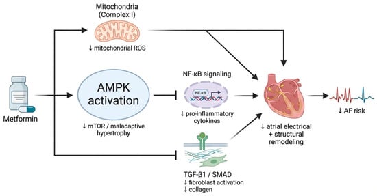
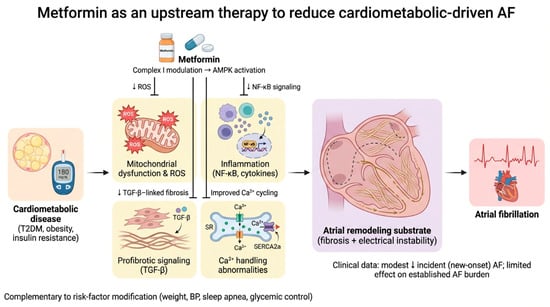
Atrial fibrillation (AF) is the most prevalent sustained arrhythmia and is increasingly driven by cardiometabolic disease, including type 2 diabetes mellitus (T2DM), obesity, and insulin resistance. These conditions promote atrial electrical instability and a permissive substrate through mitochondrial dysfunction, oxidative stress, inflammation, calcium-handling abnormalities, and profibrotic signaling, culminating in atrial fibrosis and conduction heterogeneity. Metformin, the foundational glucose-lowering therapy for T2DM, exerts pleiotropic actions that intersect with these upstream pathways. Beyond glycemic control, metformin induces mild mitochondrial complex I modulation with reduction of reverse electron transfer-derived reactive oxygen species, activates adenosine monophosphate (AMP) activated protein kinase, and attenuates nuclear factor kappa-light-chain-enhancer of activated B cells (NF-κB)-mediated cytokine signaling; experimental data further suggest favorable effects on adiponectin–sarcoendoplasmic reticulum calcium adenosine triphosphatase (SERCA) 2a-dependent calcium cycling, connexin expression, small-conductance Ca2+-activated K+ channel remodeling, lipid handling, and transforming growth factor-β (TGF)-β-associated fibrotic remodeling. Observational cohort studies have reported associations between metformin exposure and a modest reduction in incident AF, particularly with longer treatment duration and in higher-risk metabolic phenotypes; device-based surveillance cohorts support a preventive association for new-onset AF rather than reduction of established AF burden. Data after catheter ablation suggest improved freedom from recurrence in metformin-treated patients, whereas evidence in postoperative AF is largely neutral, likely reflecting distinct acute mechanisms. Collectively, metformin may be best conceptualized as a potential substrate-modifying, upstream therapy candidate; however, confounding, exposure misclassification, and heterogeneity in comparators limit causal inference, underscoring the need for prospective randomized trials with AF endpoints. In practice, integration with comprehensive risk-factor modification (blood pressure, weight, sleep apnea, and glycemic optimization) remains essential when considering AF prevention strategies.
Full article
Open AccessArticle
Comparative Cytotoxicity and Inflammatory Profiles of CeraSeal Versus AH Plus in Periodontal Tissue Repair: An In Vitro and In Vivo Study
by
Gulnihol Sharipova, Jasur Rizaev, Shuxrat Boymuradov, Mirzaakbar Kamolov, Adolat Mamadiyorova, Latipov Javdat, Umarov Doniyor and Nozimjon Ibrokhimov
J. Mol. Pathol. 2026, 7(2), 24; https://doi.org/10.3390/jmp7020024 - 15 Jun 2026
Abstract
Background/Objectives: Endodontic perforation repair requires biomaterials that balance sealing ability with minimal cellular injury. AH Plus (epoxy resin-based) remains widely used despite cytotoxicity concerns. CeraSeal (calcium silicate-based bioceramic) is a potentially more biocompatible alternative. However, comparative data on sealer-induced cytotoxicity and inflammatory
[...] Read more.
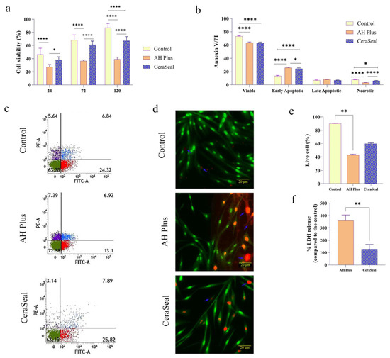
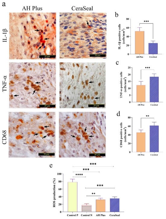
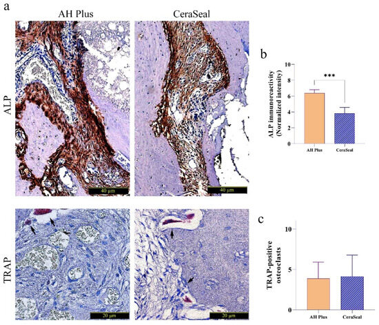
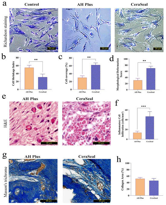
Background/Objectives: Endodontic perforation repair requires biomaterials that balance sealing ability with minimal cellular injury. AH Plus (epoxy resin-based) remains widely used despite cytotoxicity concerns. CeraSeal (calcium silicate-based bioceramic) is a potentially more biocompatible alternative. However, comparative data on sealer-induced cytotoxicity and inflammatory responses remain limited. This study compared the cytotoxicity and inflammatory profiles of CeraSeal and AH Plus using in vitro and in vivo approaches. Methods: Human periodontal ligament stem cells (hPDLSCs) were exposed to sealer extracts (1:4 AH Plus, 1:8 CeraSeal) for 120 h. Cell death was assessed by MTT, Live/Dead, LDH release, and Annexin V/PI flow cytometry. Oxidative stress was quantified via ROS generation (DCFH-DA). In a rat furcation perforation model (n = 8 teeth/group), inflammatory markers (TNF-α, IL-1β, CD68), osteogenic activity (ALP), and osteoclasts (TRAP) were evaluated. Results: AH Plus was associated with significantly greater necrotic cell death (357.6 ± 47.6% LDH release vs. CeraSeal 128.8 ± 37.5%; p = 0.0079) and reduced hPDLSC viability at all time points (p < 0.0001). ROS generation was comparable between sealers (~32–35%, p > 0.05). In vivo, IL-1β was higher in AH Plus-treated tissues (52.25 vs. 24.88 cells/mm2; p = 0.0002), while TNF-α and CD68 were greater in CeraSeal (p ≤ 0.0011). ALP was higher in AH Plus (median 6.15 vs. 3.68; p = 0.0002), with no difference in TRAP-positive osteoclasts. Morphometric analysis showed superior cellular preservation with CeraSeal (p = 0.0079), while inflammatory infiltration was higher in CeraSeal (p = 0.0002). Conclusions: AH Plus was associated with a necrotic-inflammatory profile with elevated IL-1β and higher ALP expression. CeraSeal demonstrated better cellular preservation, lower LDH release, and a distinct inflammatory signature (higher TNF-α and CD68). These findings establish comparative response profiles for the two sealers and support CeraSeal as a potentially biocompatible alternative, though further mechanistic studies are warranted.
Full article
(This article belongs to the Collection Feature Papers in Journal of Molecular Pathology)
►▼
Show Figures

Figure 1
Open AccessArticle
Pathomorphological Features of Diabetic Myopathy in Comorbid Conditions
by
Oksana Zhurakivska, Aleksandra Natalia Gabren-Syller, Oleh Koshkin, Viktor Mazurenko, Ivan Paliichuk and Nataliia Pyliachyk
J. Mol. Pathol. 2026, 7(2), 23; https://doi.org/10.3390/jmp7020023 - 5 Jun 2026
Abstract
►▼
Show Figures
Background/Objectives: Despite advances in diagnostic, preventive, and therapeutic strategies, diabetes mellitus remains a major global medico-social challenge with a high rate of complications, including diabetic myopathy. However, structural and functional changes in the masticatory muscles and disturbances of myodynamic balance in diabetes under
[...] Read more.
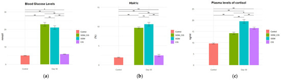
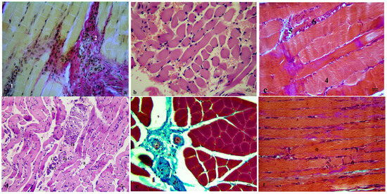
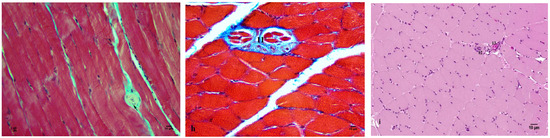
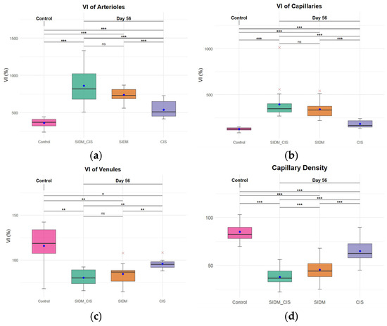
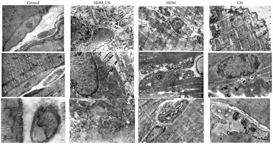
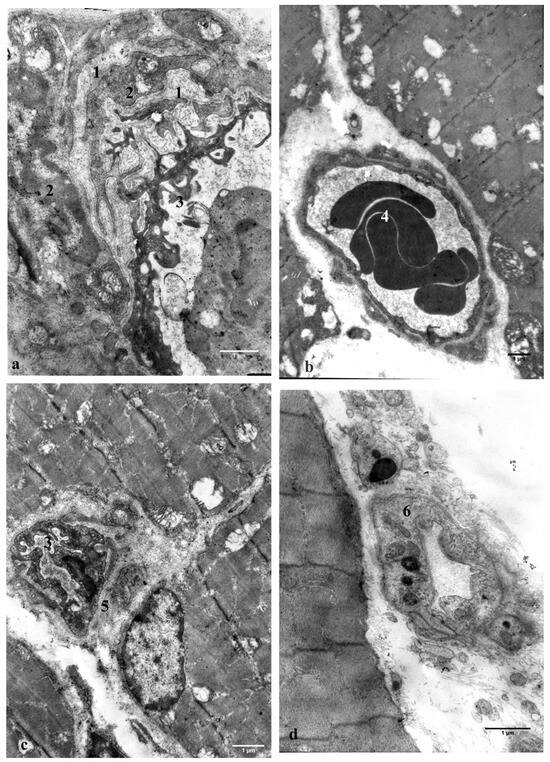
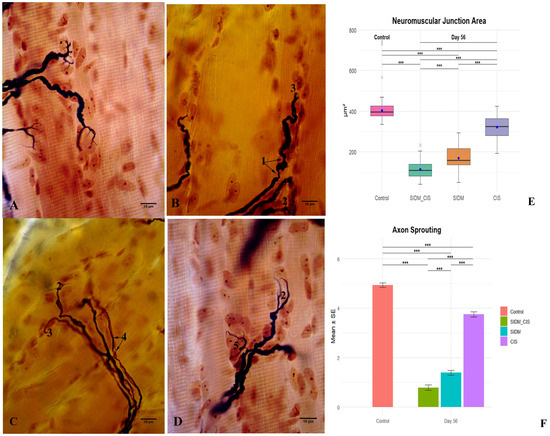
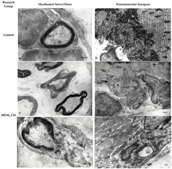
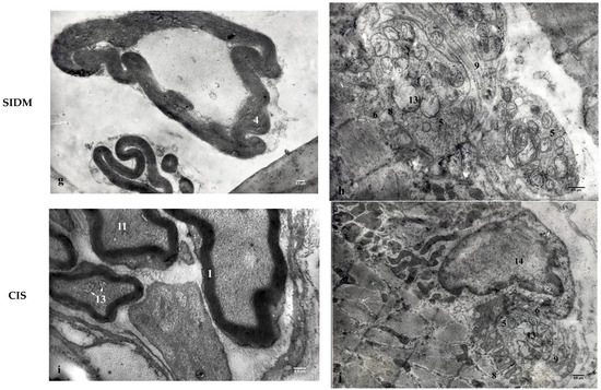
Background/Objectives: Despite advances in diagnostic, preventive, and therapeutic strategies, diabetes mellitus remains a major global medico-social challenge with a high rate of complications, including diabetic myopathy. However, structural and functional changes in the masticatory muscles and disturbances of myodynamic balance in diabetes under stress remain insufficiently studied. This research aimed to clarify the pathomorphological patterns of diabetic myopathy development under chronic immobilization stress. Methods: Twenty-eight six-month-old male albino rats were equally divided into four groups: Group 1—streptozotocin-induced diabetes mellitus (SIDM) combined with chronic immobilization stress (CIS); Group 2—SIDM; Group 3—CIS; Group 4—control. Samples were collected on day 56. Histological, electron microscopy, biochemical, and statistical methods were applied. Results: Myopathy in experimental conditions was characterized by aseptic myositis, focal fibrosis of the masticatory muscle, muscle fiber atrophy, and peripheral neuropathy. These changes developed on the background of pronounced diabetic microangiopathy, manifested by hemorheological disturbances, reduced vascular capacity of the haemomicrocirculatory bed (confirmed by an increased Vogenvort index), thickening and proliferation of the capillary basement membrane, and decreased capillary density. Degenerative muscle fiber alterations included vacuolar dystrophy, necrosis, ferroptosis, necroptosis, and autolysis. Neuropathic changes involved segmental demyelination, axonal atrophy, decreased sprouting, and extensive neuromuscular junction destruction, possibly leading to impaired neuromuscular transmission. Conclusions: Chronic immobilization stress acts as a trigger that aggravates diabetic myopathy progression by impairing microcirculation and inducing neuromuscular junction damage, resulting in subsequent muscle atrophy.
Full article

Figure 1
Open AccessArticle
ATM Immunohistochemistry as a Tool to Identify and Classify Somatic ATM Variants in Prostate Cancer
by
Roger Ferreira, Kassandra R. Bisson, Andrea Beharry, Champica Nicholas, Marco Iafolla, Samir Bidnur and Brandon S. Sheffield
J. Mol. Pathol. 2026, 7(2), 22; https://doi.org/10.3390/jmp7020022 - 4 Jun 2026
Abstract
►▼
Show Figures
Background/Objectives: Ataxia telangiectasia-mutated kinase is encoded by the ATM gene. This gene is altered in many cancer types; however, it is most relevant for eligibility of poly-ADP-ribose polymerase inhibitor (PARPi) therapy in metastatic castrate-resistant prostate cancer in Canada. Non-benign ATM alterations are relatively
[...] Read more.
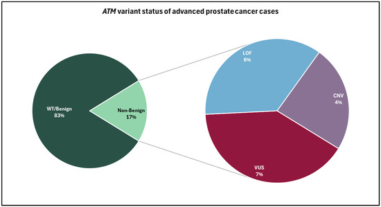
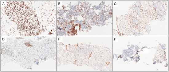
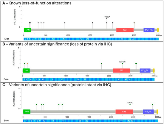
Background/Objectives: Ataxia telangiectasia-mutated kinase is encoded by the ATM gene. This gene is altered in many cancer types; however, it is most relevant for eligibility of poly-ADP-ribose polymerase inhibitor (PARPi) therapy in metastatic castrate-resistant prostate cancer in Canada. Non-benign ATM alterations are relatively uncommon, seen in less than 10% of prostate cancers; however, many of these are variants of uncertain significance with limited evidence of clinical significance or actionability. Methods: To aid in variant classification of next-generation sequencing results, ATM immunohistochemistry (IHC) is performed on patient tumour samples to serve as patient-specific functional evidence for ATM protein loss. Results: ATM IHC demonstrates strong concordance with known loss-of-function variants and copy number losses. Additionally, it can be used to clarify ATM protein loss in cases involving variants of uncertain significance (VUSs) or copy number deletions. Conclusions: Incorporation of ATM IHC into clinical testing for prostate cancer represents a relatively cost-effective orthogonal approach that can rescue poor-quality NGS results and clarify the functional impact of ATM VUSs on protein expression. Overall, this strategy may provide useful supportive evidence in prostate cancer biomarker testing and interpretation.
Full article

Figure 1
Open AccessCorrection
Correction: Vernygorodskyi et al. Cancer-Associated Fibroblasts and Epithelial–Mesenchymal Transition as Critical Contributors to Renal Cell Carcinoma Progression. J. Mol. Pathol. 2025, 6, 31
by
Sergii Vernygorodskyi, Anton B. Tonchev, Nikolai T. Evtimov and Kameliya Zhechkova Bratoeva
J. Mol. Pathol. 2026, 7(2), 21; https://doi.org/10.3390/jmp7020021 - 30 May 2026
Abstract
Following publication of the review article [...]
Full article
Open AccessArticle
Understanding of Microbial Causes, Clinicopathological Evaluation, Molecular Analysis, and Associated Risk Factors of Ear Infections in Dogs
by
Gopakrishna Mohanty, Prasana Kumar Rath, Bidyut Prava Mishra, Annushree Mishra, Shanta Swarupa Mishra, Aditya Prasad Acharya, Susen Kumar Panda, Rajeev Ranjan and Manoj Kumar Jena
J. Mol. Pathol. 2026, 7(2), 20; https://doi.org/10.3390/jmp7020020 - 19 May 2026
Abstract
►▼
Show Figures
Objective: Dog ear infections can have a variety of multifactorial causes, some of which are regarded as zoonotic. Thus, the present study was aimed at understanding the microbial causes, clinical evaluation, molecular analysis and associated risk factors of ear infections in dogs for
[...] Read more.
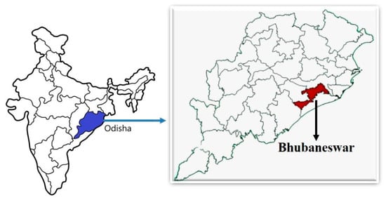
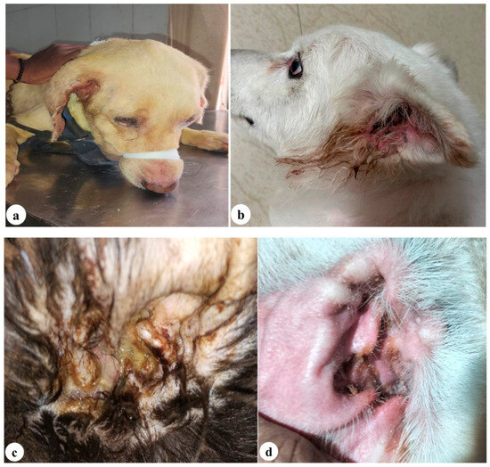
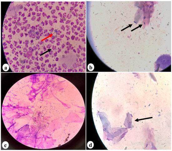
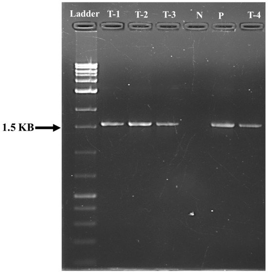
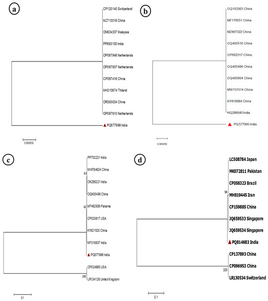
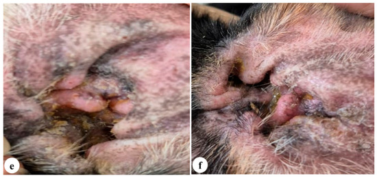
Objective: Dog ear infections can have a variety of multifactorial causes, some of which are regarded as zoonotic. Thus, the present study was aimed at understanding the microbial causes, clinical evaluation, molecular analysis and associated risk factors of ear infections in dogs for their management. Methods: A total of 167 dogs were screened for ear infections based on history and clinical signs. An auricular swab was collected and processed with standard methods. Head tilting to the afflicted side, ear pain when palpated, pawing at the ear and purulent discharges from the ear canal were typical clinical symptoms. Results: A total of 13.77% of dogs were positive for ear infection, and among these, 13.17% showed unilateral right-sided ear infections. Dogs with pendulous ears (56.52%), Labrador breeds (34.78%), males (56.52%), dogs older than 4 years (52.17%), and during the monsoon season (65.21%) had higher rates of ear infections among the total dogs screened (n = 167) for aural infections. Anaemia, leukocytosis, neutrophilia, and elevated levels of total protein, cholesterol, BUN, and AST were observed in dogs with ear infections. Cytological analyses showed the presence of yeast cells and bacteria, along with hyperkeratosis and degenerated neutrophils. Mammaliicoccus sciuri, Bacillus cereus, Klebsiella aerogenes, Pseudomonas aeruginosa, and Malassezia spp. were the organisms isolated. Bacterial isolates showed high sensitivity to gentamicin for the otitis treatment. Clinical Significance: This study highlights the need for preventive measures to curb the spread of potentially zoonotic pathogens such as Bacillus cereus and Klebsiella aerogenes, which can pose significant threats to both human and animal health.
Full article

Figure 1
Open AccessReview
The Tight Connection Between the Gut Microbiota and Childhood Acute Lymphoblastic Leukaemia: An Updated Review
by
Marta Arrabito, Giulio Pulvirenti, Maria Santagati, Stefania Stefani, Giovanna Russo and Luca Lo Nigro
J. Mol. Pathol. 2026, 7(2), 19; https://doi.org/10.3390/jmp7020019 - 12 May 2026
Abstract
►▼
Show Figures
The human microbiota, comprising bacteria, fungi, and viruses, primarily resides in the gut, skin, and oral and nasal cavities. The gut microbiota represents the largest and most functionally relevant microbial community, playing a crucial role in digestion, metabolism and immunity. Microbiota composition and
[...] Read more.
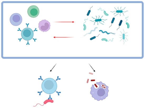
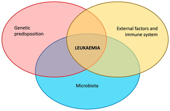
The human microbiota, comprising bacteria, fungi, and viruses, primarily resides in the gut, skin, and oral and nasal cavities. The gut microbiota represents the largest and most functionally relevant microbial community, playing a crucial role in digestion, metabolism and immunity. Microbiota composition and diversity have been associated with several diseases such as cancer, affecting drug efficacy, toxicity and clinical outcome. Interactions between the gut microbiota and the host immune system may contribute to the aetiology and progression of childhood acute lymphoblastic leukaemia (ALL). In this review, we summarized the general features of the gut microbiota in cancer patients and its specific role in aetiology, treatment and prognosis of paediatric ALL. Profiling the gut microbiota may help to shed the light on the mechanism of onset and to support the development of personalized treatment strategies and follow-up in children with ALL, contributing to precision medicine.
Full article

Figure 1
Open AccessArticle
Analytical Validation of Quantitative Polymerase Chain Reaction and AscentTM Low-Pass Whole Genome Sequencing to Report on Gene Copy Number Variants in Cerebrospinal Fluid Tumor-Derived DNA
by
Viriya Keo, Sakshi Khurana, Vindhya Udhane, Alexandra Larson, Jennifer N. Adams, Daniel Sanchez, Tarin Peltier, Anthony Acevedo, Kathleen Mitchell, Kala F. Schilter, Qian Nie and Honey V. Reddi
J. Mol. Pathol. 2026, 7(2), 18; https://doi.org/10.3390/jmp7020018 - 12 May 2026
Abstract
►▼
Show Figures
Background: Evaluation of gene-level copy number variants (CNVs) for diagnosis and therapeutic decision making has become standard of care with next-generation sequencing (NGS), immunohistochemistry (IHC), and/or fluorescence in situ hybridization (FISH) being used to detect gene amplifications/deletions in tumor tissue. In contrast to
[...] Read more.
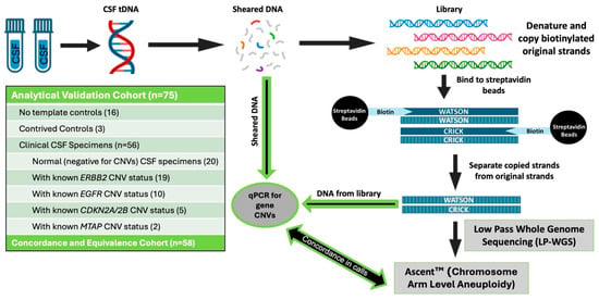
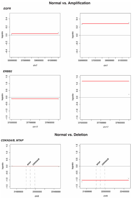
Background: Evaluation of gene-level copy number variants (CNVs) for diagnosis and therapeutic decision making has become standard of care with next-generation sequencing (NGS), immunohistochemistry (IHC), and/or fluorescence in situ hybridization (FISH) being used to detect gene amplifications/deletions in tumor tissue. In contrast to most solid tumors, CNS cancers are challenging to evaluate by resection and/or biopsy due to the associated risks with invasive brain surgery that can also result in death or associated morbidity and therefore alternate methods are required.Methods: This study presents the analytical validation of using quantitative PCR (qPCR) to detect gene CNVs directly from cerebrospinal fluid (CSF)-derived DNA and from the AscentTM low-pass whole genome sequencing (LP-WGS) libraries, demonstrating concordance with the gold standard of NGS/IHC/FISH used in tumor tissue. Results: The analytical sensitivity of qPCR to detect gene amplification calls for ERBB2 (erb-b2 receptor tyrosine kinase 2) was demonstrated to be 100% and that of EGFR (epidermal growth factor receptor) was 83%, with specificities of 96% and 100%, respectively. The analytical sensitivity of qPCR to detect gene deletions for CDKN2A/2B (cyclin-dependent kinase inhibitor 2A/2B) was 60% and that for MTAP (methylthioadenosine phosphorylase) was 100% with a specificity of 100% for all three genes. AscentTM was demonstrated to have a higher sensitivity (100%) when compared to qPCR for the same genes evaluated and demonstrated 100% positive agreement and 100% negative agreement with known CNV status. Conclusions: The results demonstrate that given the paucity of cells in CSF limiting the use of IHC and FISH, qPCR and AscentTM provide highly sensitive, novel, minimally invasive methods for the evaluation of gene copy number (CN) status to inform the diagnosis and management of CNS cancers.
Full article

Figure 1
Open AccessReview
A Narrative Approach to Mismatch Repair-Deficient Endometrial Cancer
by
Massimo Barberis and Yinxiu Zhan
J. Mol. Pathol. 2026, 7(2), 17; https://doi.org/10.3390/jmp7020017 - 15 Apr 2026
Abstract
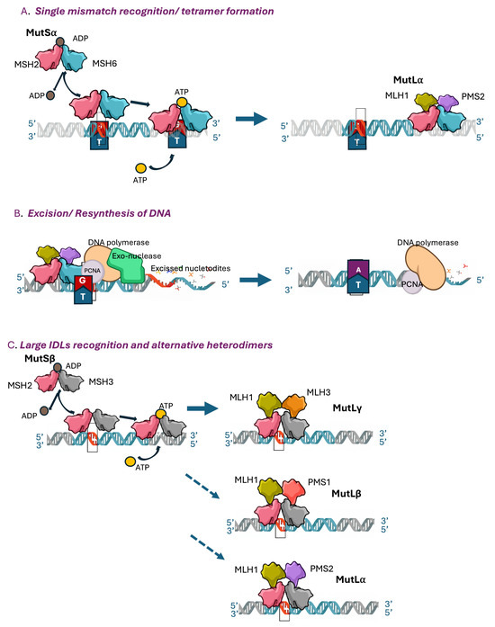
Endometrial cancer (EC) is the sixth most common cancer in women. Its overall incidence has increased by 132% over the past 30 years, reflecting an increase in the prevalence of risk factors. The mortality rate decreased by 15% in the last 30 years,
[...] Read more.
Endometrial cancer (EC) is the sixth most common cancer in women. Its overall incidence has increased by 132% over the past 30 years, reflecting an increase in the prevalence of risk factors. The mortality rate decreased by 15% in the last 30 years, despite the high number of endometrial cancer-related deaths occurring world-wide. An inverse relationship has been observed between the incidence of EC, mortality and socio-economic status: more patients living in low-income countries die from EC because they do not have access to timely and effective treatment. About 80% of EC cases are diagnosed in an early stage and have a good prognosis. However, about 20% of cases present in advanced stages and are characterized by a poor prognosis. The molecular classification proposed by The Cancer Genome Atlas (TGCA) and its surrogate for clinical use allowed the adoption of personalized treatments. The assessment of the status of the MMR has revolutionized the treatment of advanced ECs, leading to significant results both in terms of PFS and OS. In this review we will focus on MMR deficiency (dMMR)/microsatellite instability-hypermutated (MSI-H) tumors, which globally account for 20–30% of ECs. The dMMR group encompasses multiple etiologies, including sporadic defects in MMR genes, germline mutations, and hypermethylation of the MLH1 promoter. Currently, the combination of immunotherapy (I-O) with standard chemotherapy has become the new standard first-line treatment for dMMR advanced or recurrent ECs. Although the main clinical trials involving patients with MMRd/MSI-H ECs treated with I-O and chemotherapy have demonstrated efficacy and long-term control of the disease, a significant number of patients do not respond to treatment (intrinsic or primary resistance) and others develop progression during treatment (acquired or secondary resistance). In this narrative approach the biological and molecular bases of these tumors have been integrated with recent advances involving diagnostic techniques, therapeutic opportunities and the genomic and phenotypical alterations underpinning the mechanisms of resistance. Special attention was given to the need for robust, clinically affordable biomarkers to promptly identify responders and non-responders to the current treatment regimens.
Full article
Open AccessCase Report
Pediatric Ciliopathy Linked to TULP3 Variant—A Case Report
by
Mrunmayi Prashant Marathe, Snehavardhan Pandey, Anusha Kulkarni, Thenral S. Geetha and Ashish Bavdekar
J. Mol. Pathol. 2026, 7(2), 16; https://doi.org/10.3390/jmp7020016 - 14 Apr 2026
Abstract
►▼
Show Figures
Ciliopathies, initially known as fibrocystic liver diseases, encompass a group of inherited disorders characterized by cystic dilatation of intrahepatic bile ducts and portal fibrosis, frequently associated with renal anomalies. These disorders are now recognized as resulting from defects in primary cilia. The hepatic
[...] Read more.
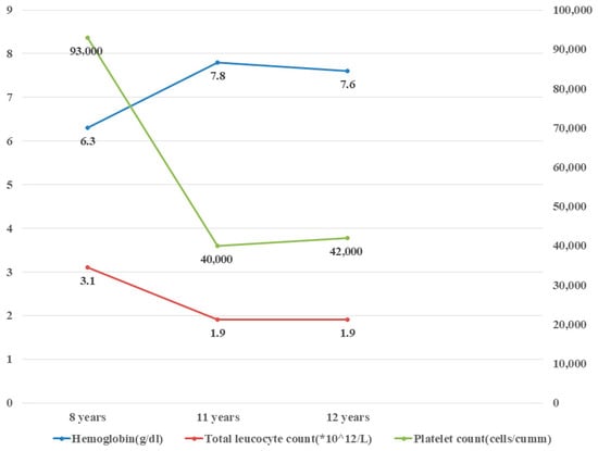
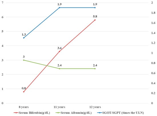
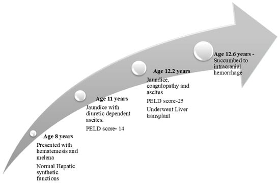
Ciliopathies, initially known as fibrocystic liver diseases, encompass a group of inherited disorders characterized by cystic dilatation of intrahepatic bile ducts and portal fibrosis, frequently associated with renal anomalies. These disorders are now recognized as resulting from defects in primary cilia. The hepatic manifestations, such as congenital hepatic fibrosis (CHF), Caroli syndrome, and polycystic liver disease, arise from ductal plate malformation. Recent studies have implicated variants in the TULP3 (Tubby related protein variant 3) gene in a novel monogenic ciliopathy affecting the liver, kidneys, and heart. We report an 8-year-old boy who presented with variceal bleeding and evolved to a progressive phenotype of CHF. Whole exome sequencing revealed a homozygous novel TULP3 mutation. The patient was managed by endotherapy and propranolol prophylaxis. Due to repeated episodes of variceal bleeding and progressive worsening of hepatic synthetic functions, he underwent a living donor liver transplantation at the age of 12 years.
Full article

Figure 1
Open AccessReview
Leveraging Epigenetic Biomarkers and CRISPR-Cas12a for Early Prostate Cancer Detection in Sub-Saharan Africa: Opportunities and Challenges
by
Niels K. Nguedia, Emmanuel C. Amadi, Irrinus F. Kintung, Olubanke O. Ogunlana and Shalom N. Chinedu
J. Mol. Pathol. 2026, 7(2), 15; https://doi.org/10.3390/jmp7020015 - 13 Apr 2026
Cited by 1
Abstract
►▼
Show Figures
Prostate cancer is a major cause of cancer-related deaths among men in Sub-Saharan Africa, where late-stage diagnoses are common due to limited access to affordable and sensitive diagnostic tools. Early detection is essential to improve survival and reduce the disease burden. This review
[...] Read more.
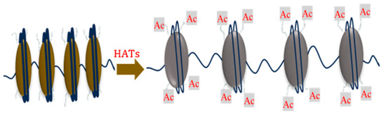
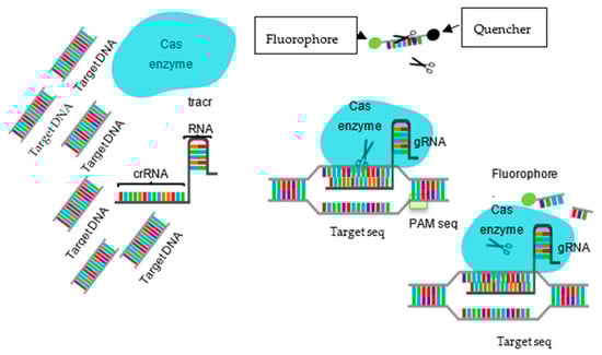
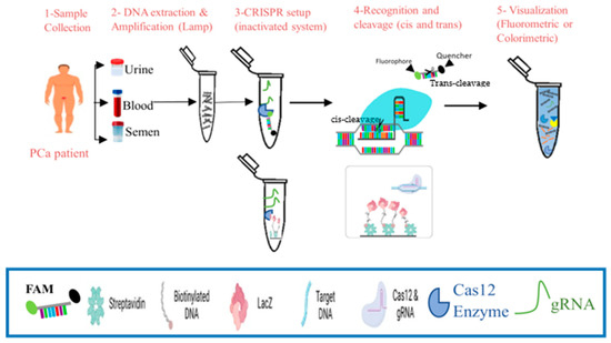
Prostate cancer is a major cause of cancer-related deaths among men in Sub-Saharan Africa, where late-stage diagnoses are common due to limited access to affordable and sensitive diagnostic tools. Early detection is essential to improve survival and reduce the disease burden. This review explores the integration of epigenetic biomarkers and CRISPR-Cas12a technology as a transformative approach for early, non-invasive prostate cancer detection in resource-limited settings. Among the many complexities of cancer development, molecular dysregulation plays a critical role. Epigenetic modifications including DNA methylation, histone changes, and non-coding RNA expression have emerged as stable and specific biomarkers with significant potential for the early detection and characterisation of prostate carcinogenesis. However, their low concentration in body fluids poses a significant challenge for detection. CRISPR-Cas12a, renowned for its high specificity and sensitivity, offers a promising solution. When integrated with isothermal amplification and liquid biopsy techniques, it enables rapid, point-of-care diagnostics. This review proposes a CRISPR-Cas12a-based diagnostic pipeline for the detection of specific epigenetic markers in liquid biopsies that could be associated with prostate cancer. The adoption of this technology in Sub-Saharan Africa has the potential to significantly improve early diagnosis, reduce mortality, and promote health equity.
Full article

Figure 1

Open AccessArticle
Differential Promoter Methylation of MMP-9 and E-Cadherin Genes in CLL: Evidence for a Pathogenic Role of MMP-9 Hypomethylation
by
Zeki Ali Mohamed
J. Mol. Pathol. 2026, 7(1), 14; https://doi.org/10.3390/jmp7010014 - 23 Mar 2026
Abstract
►▼
Show Figures
Background/Objectives: Chronic lymphocytic leukemia (CLL) is characterized by genetic and epigenetic alterations. This study aimed to assess the methylation status of E-Cadherin and MMP-9 gene promoters and to explore their relationships with disease pathogenesis and hematological parameters in CLL patients. Methods: A case–control
[...] Read more.
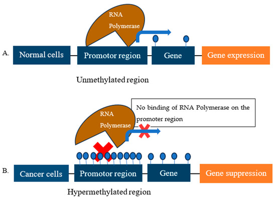
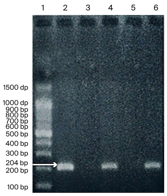
Background/Objectives: Chronic lymphocytic leukemia (CLL) is characterized by genetic and epigenetic alterations. This study aimed to assess the methylation status of E-Cadherin and MMP-9 gene promoters and to explore their relationships with disease pathogenesis and hematological parameters in CLL patients. Methods: A case–control study was conducted with 70 newly diagnosed CLL patients and 70 age- and sex-matched healthy controls. Promoter methylation of E-Cadherin and MMP-9 genes was evaluated using methylation-specific PCR (MSP) and methylation-sensitive restriction enzyme PCR (MSRE-PCR), respectively. Results: The median patient age was 62 years, and 68.5% were males. Binet stage A was the most common stage (57.3%). E-Cadherin promoter methylation was detected in 75.7% of CLL patients and 77.1% of controls (p = 0.91), showing no significant association with disease occurrence; however, it showed a significant correlation with higher lymphocyte counts (p = 0.01). In contrast, MMP-9 promoter methylation was significantly less frequent in CLL cases (70.0%) than in controls (100%, p = 0.001). Unmethylated MMP-9 correlated significantly with female gender (p = 0.02), lower hemoglobin (p = 0.031), reduced platelet counts (p = 0.001), and higher lymphocyte counts (p = 0.035). Conclusions: MMP-9 promoter hypomethylation may play a pathogenic role in CLL and is associated with female gender and cytopenia, whereas E-Cadherin methylation appears to be non-specific. MMP-9 methylation status could therefore serve as a potential biomarker for CLL biology and prognosis.
Full article

Figure 1
Open AccessCorrection
Correction: Markopoulos et al. Intraoperative Flow Cytometry Upon and Beyond the Cell Cycle: A Case Study of the Characterization of a Bone Metastasis. J. Mol. Pathol. 2023, 4, 225–233
by
Georgios S. Markopoulos, Emilios E. Pakos, Vasilios Gavrielatos, Dimitrios Kosmas, Ioannis Gkiatas, George A. Alexiou, Anna Batistatou, Evangeli Lampri and George Vartholomatos
J. Mol. Pathol. 2026, 7(1), 13; https://doi.org/10.3390/jmp7010013 - 20 Mar 2026
Abstract
Materials and Methods [...]
Full article
Open AccessArticle
Entosis in Colorectal, Lung, and Breast Cancer: Associations with Clinicopathological Features, Patient Outcomes, and Copy Number Alteration Landscape
by
Ksenia A. Gaptulbarova, Sergey V. Vtorushin, Marina K. Ibragimova, Irina A. Tsydenova, Natalia A. Tarabanovskaya, Vitaly P. Shubin, Aleksey S. Tsukanov, Evgeny O. Rodionov, Sergey I. Achkasov and Nikolai V. Litviakov
J. Mol. Pathol. 2026, 7(1), 12; https://doi.org/10.3390/jmp7010012 - 17 Mar 2026
Abstract
►▼
Show Figures
Objective: This study examined the frequency of entosis in solid tumors of various origins (colorectal cancer, breast cancer, and lung cancer) and its association with clinical and pathological characteristics. It also examined survival and copy number alterations (CNAs) in genes associated with
[...] Read more.
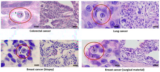
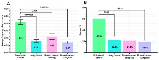
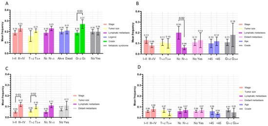
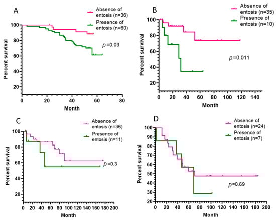
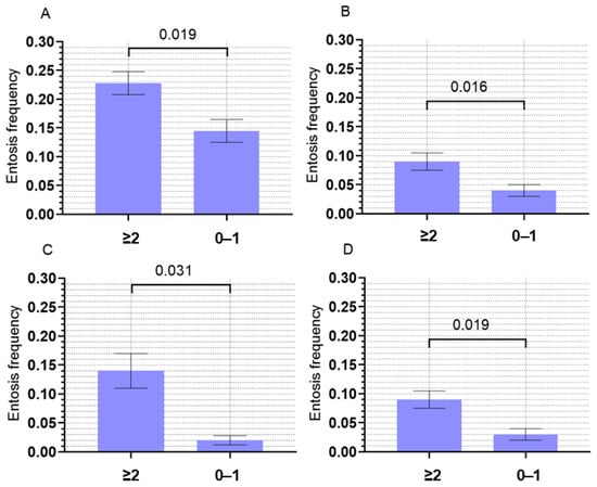
Objective: This study examined the frequency of entosis in solid tumors of various origins (colorectal cancer, breast cancer, and lung cancer) and its association with clinical and pathological characteristics. It also examined survival and copy number alterations (CNAs) in genes associated with stem cells. The aim was to assess the potential prognostic value of entotic events in tumors. Methods: A total of 238 patients were included: 96 with colorectal cancer (CRC), 45 with lung cancer (LC), and 97 with breast cancer (BC). Entotic cell-in-cell (CIC) structures were evaluated on hematoxylin–eosin–stained slides using Mackay’s criteria. A CIC frequency >0.1 per 20 high-power fields was considered positive. Clinicopathological parameters, overall survival (CRC), metastasis-free survival (LC and BC), and CNA profiles of stemness-related genes were analyzed. Amplifications of MAP1LC3A and other chromosomal loci were assessed. Results: CRC demonstrated the highest entosis rate, more than two-fold higher compared with BC and LC (p < 0.05). Entosis correlated with high tumor grade (G3) in CRC (p = 0.03). In LC, CIC-positive tumors were more frequent in patients with lymph-node metastases (p = 0.02), whereas in BC, the opposite trend was observed (p = 0.02). It was noted that in patients with stage III–IV LC, the frequency of entosis was significantly higher than in patients with stage I–II cancer (p = 0.03). CIC-positive status was associated with poorer overall survival in CRC (p = 0.03) and reduced metastasis-free survival in LC (p = 0.011). In breast cancer, no statistically significant survival differences were observed. Tumors harboring two or more stemness-gene amplifications showed significantly higher entosis frequency regardless of tumor site. A strong association was identified between entosis and MAP1LC3A amplification. Conclusions: Enosis is not a random morphological phenomenon but a process associated with unfavorable tumor characteristics, high malignancy, reduced survival, and amplification of stem cell-related genes. The results of this study confirm the working hypothesis that entosis may contribute to the emergence of aneuploid clones of tumor cells, including those containing amplifications of stem cell-associated genes. This positions entosis as a potential factor in tumor genetic heterogeneity, which is particularly important in the context of therapeutic selection pressure. The observed association between high entosis frequency and the presence of ≥2 stem cell gene amplifications, as well as its association with poor prognosis in colorectal and lung cancer, highlights its potential value as a prognostic indicator. Furthermore, MAP1LC3A amplification data may serve as a molecular marker of entotic activity and a potential therapeutic target.
Full article

Figure 1

Open AccessReview
From Dysbiosis to Tissue Destruction: Periodontal Pathogens as Inducers of Gingival Epithelial–Mesenchymal Transition (A Narrative Review)
by
Hadeel Mazin Akram and Saif Sehaam Saliem
J. Mol. Pathol. 2026, 7(1), 11; https://doi.org/10.3390/jmp7010011 - 4 Mar 2026
Cited by 4
Abstract
►▼
Show Figures
Periodontitis is a dysbiosis-driven inflammatory disease in which a pathogenic subgingival biofilm disrupts the host–microbe equilibrium and promotes progressive loss of tooth-supporting tissues. While periodontal destruction has traditionally been explained mainly through the host immune response, increasing experimental and clinical evidence suggests that
[...] Read more.
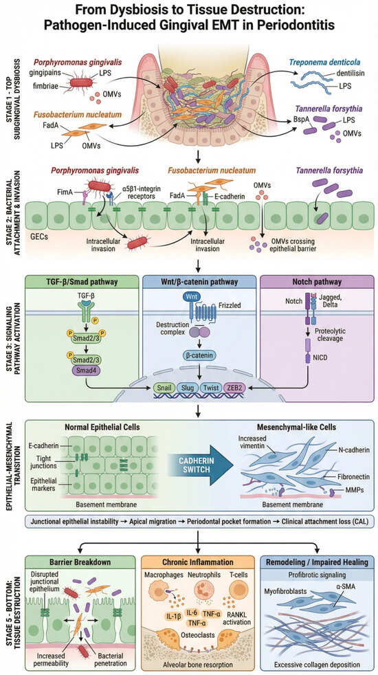
Periodontitis is a dysbiosis-driven inflammatory disease in which a pathogenic subgingival biofilm disrupts the host–microbe equilibrium and promotes progressive loss of tooth-supporting tissues. While periodontal destruction has traditionally been explained mainly through the host immune response, increasing experimental and clinical evidence suggests that epithelial–mesenchymal transition (EMT)-like changes in the gingival epithelium may contribute to barrier failure and tissue remodeling during disease progression. EMT is characterized by reduced epithelial adhesion and polarity, alongside a shift toward a mesenchymal-like phenotype with enhanced motility and impaired epithelial barrier function. This narrative review focuses on how periodontal pathogens, particularly red complex organisms and keystone species, may trigger gingival EMT through virulence factors such as gingipains, fimbriae, lipopolysaccharide, and outer membrane vesicles. These microbial signals can hijack host pathways including TGF-β/Smad, Wnt/β-catenin, and Notch to drive EMT-associated transcriptional changes and downstream functional consequences. Collectively, pathogen-induced gingival EMT may facilitate deeper microbial invasion, perpetuate chronic inflammation, impair wound healing, and contribute to fibrotic remodeling, ultimately linking microbial dysbiosis to connective tissue destruction. Understanding these mechanisms may support the development of EMT-related biomarkers and targeted interventions aimed at preserving epithelial barrier stability in periodontitis.
Full article

Figure 1

Open AccessArticle
Amyloid Deposits in Intramural Coronary Arteries of Feline Hearts: A Retrospective Histopathological Study
by
Izabela Janus-Ziółkowska, Joanna Bubak, Ewa Sawińska, Marcin Nowak and Agnieszka Noszczyk-Nowak
J. Mol. Pathol. 2026, 7(1), 10; https://doi.org/10.3390/jmp7010010 - 3 Mar 2026
Abstract
►▼
Show Figures
Background: Amyloidosis involving the heart is one of the types of the disease recognized in humans and has been previously described in dogs. To date, no data regarding the presence of amyloid in cardiac tissues of a large group of feline patients have
[...] Read more.
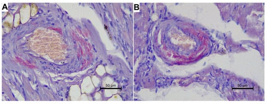
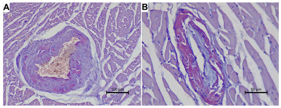
Background: Amyloidosis involving the heart is one of the types of the disease recognized in humans and has been previously described in dogs. To date, no data regarding the presence of amyloid in cardiac tissues of a large group of feline patients have been published. Our research aimed to analyze the presence and localization of amyloid in the atrial and ventricular cardiac tissue in retrospectively enrolled cats diagnosed with various types of primary cardiomyopathies, hyperthyroidism-induced cardiomyopathy, myocarditis, and generalized disorders. Methods: This study was conducted on atrial specimens obtained from 119 animals and on ventricular specimens obtained from 69 animals from that group. The atrial and ventricular specimens obtained from the enrolled animals were stained with Congo Red and evaluated in a light microscope and polarized light for the presence of amyloid deposits. Results: Five cases from the enrolled group turned out positive for amyloid deposits: three cats diagnosed with feline hyperthyroidism, one cat diagnosed with kidney glomerulonephritis, and one cat diagnosed with restrictive cardiomyopathy. In all positive cats, the amyloid deposits were present within the small intramural coronary arteries of the left ventricular free wall and interventricular septum and/or left and right atrium. No myocardial amyloid deposits were identified in the study group. Conclusions: In conclusion, cardiac coronary arterial amyloidosis, although infrequent, can be observed in cats.
Full article

Figure 1

Open AccessArticle
Evaluation of Predictive Markers for Immunotherapy in Colorectal Cancer: Concordance Between MMR Protein Expression and Microsatellite Instability in a Retrospective Series
by
Giulia Martinelli, Rossella Bruno, Marco Maria Germani, Anello Marcello Poma, Paola Vignali, Chiara Cremolini and Clara Ugolini
J. Mol. Pathol. 2026, 7(1), 9; https://doi.org/10.3390/jmp7010009 - 20 Feb 2026
Abstract
Background/Objectives: In metastatic colorectal cancer (mCRC) the evaluation of mismatch repair (MMR) and microsatellite instability (MSI) status is essential to identify patients eligible for treatment with immune-checkpoint inhibitors (ICI). This study aims to evaluate the potential utility of Comprehensive Genomic Profiling (CGP) in
[...] Read more.
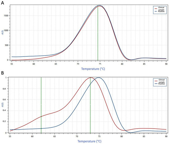
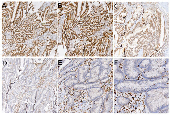
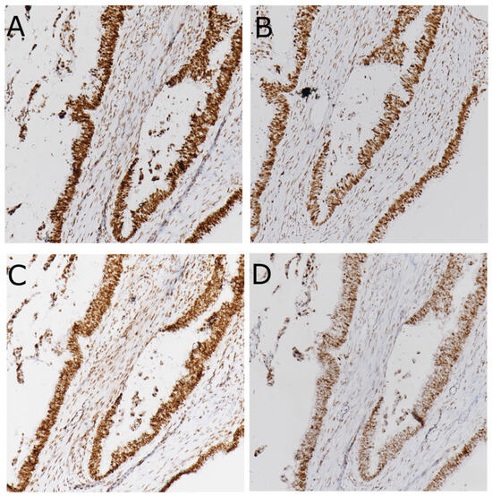
Background/Objectives: In metastatic colorectal cancer (mCRC) the evaluation of mismatch repair (MMR) and microsatellite instability (MSI) status is essential to identify patients eligible for treatment with immune-checkpoint inhibitors (ICI). This study aims to evaluate the potential utility of Comprehensive Genomic Profiling (CGP) in assessing MSI status, in addition to other immunotherapy-predictive biomarkers such as high tumor molecular burden (TMB) and the POLE and POLD1 mutations. Methods: A total of 138 mCRC tumor samples underwent a first-level molecular test (MMR status by immunohistochemistry, MSI by a melting-based PCR approach and RAS/BRAF mutational status by a small next-generation sequencing (NGS) panel) and second-level CGP analysis by the FoundationOne CDx assay. The prevalence of dMMR and MSI tumors was reported. Moreover, the concordance between the MMR and MSI status was determined, and discordant cases were discussed. Results: Twelve cases (8.7%) were MMR-deficient (dMMR); 10 showed high MSI and TMB (>10 mut/Mb). MSI status assessed by CGP and PCR was concordant in all cases except one MSH6-deficient tumor. Two dMMR cases were stable with low TMB. Moreover, in two MLH1/PMS2-deficient cases CGP revealed pathogenic alterations in the MSH2 and MSH6 genes; in both cases, the MLH1 promoter was hypermethylated. A high TMB was the only positive biomarker in 11 cases with a proficient MMR system and no MSI. Conclusions: MSI assessment by CGP analysis showed high concordance (98%) with MMR and was helpful in evaluating ICI eligibility in three out of twelve dMMR cases. Overall, compared to standard methods, analyzing a broader range of microsatellite loci and the simultaneous assessment of multiple predictive biomarkers by CGP may increase diagnostic accuracy and improve therapeutic assessment.
Full article
(This article belongs to the Special Issue Advancing Cancer Diagnosis: Integrating Molecular Pathology into Histopathology for Solid Tumors)
►▼
Show Figures

Figure 1
Open AccessArticle
Validated Approach for Flow Cytometric Quantification of Phospholipase C Zeta (PLCζ, PLCZ1) Protein Levels in Sperm
by
Marie-Helene Godin Pagé, Debbie Montjean, Cyntia Duval, Fabien Joao, Annabelle Calvé, Rosalie Cabry, Marie-Claire Bélanger, Moncef Benkhalifa and Pierre Miron
J. Mol. Pathol. 2026, 7(1), 8; https://doi.org/10.3390/jmp7010008 - 9 Feb 2026
Abstract
►▼
Show Figures
Background/Objectives: Phospholipase C zeta (PLCZ1; PLCζ) is a sperm-specific enzyme responsible for the Ca2+ oscillations required for oocyte activation, and altered PLCζ expression has been associated with fertilization failure in assisted reproductive technologies, particularly intracytoplasmic sperm injection (ICSI). This study aimed to
[...] Read more.
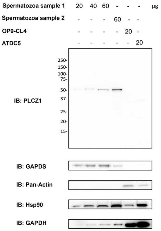
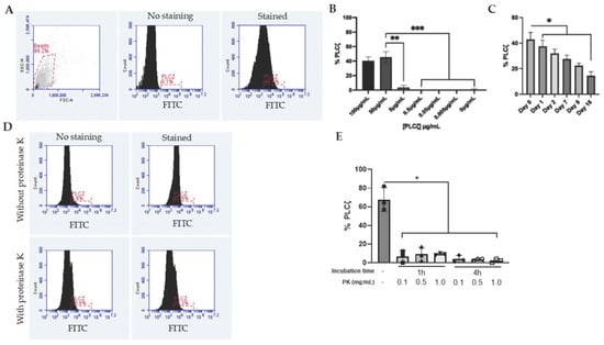
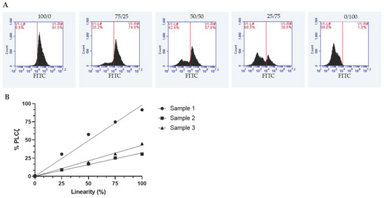
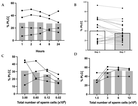
Background/Objectives: Phospholipase C zeta (PLCZ1; PLCζ) is a sperm-specific enzyme responsible for the Ca2+ oscillations required for oocyte activation, and altered PLCζ expression has been associated with fertilization failure in assisted reproductive technologies, particularly intracytoplasmic sperm injection (ICSI). This study aimed to develop and analytically validate a flow cytometry–based protocol for PLCζ quantification in human spermatozoa. Methods: The assay was established using normozoospermic samples and included validated positive and negative technical controls. Antibody specificity was confirmed by Western blot analysis. A defined gating strategy was used to assess linearity between fluorescence intensity and PLCζ expression. Analytical performance was evaluated for precision, reproducibility, stability, and sensitivity, including applicability to low sperm concentrations. Results: A linear relationship between fluorescence intensity and PLCζ expression was demonstrated. The assay showed high precision, reproducibility, and stability, with consistent results in samples stored up to 24 h at room temperature or up to one week post-fixation at 4 °C. Sensitivity testing confirmed suitability for low sperm concentrations. Conclusions: This work provides a standardized and analytically validated framework for PLCζ quantification using flow cytometry. Although the assay measures protein expression rather than functional competence or subcellular localization, it establishes a solid analytical basis for future studies to define clinically relevant PLCζ thresholds and assess its value as a biomarker of fertilization capacity.
Full article

Figure 1
Open AccessReview
Diabetic Retinopathy Therapeutics: Bridging Conventional Approaches and Gene Therapy with Focus on TXNIP-Targeted Interventions
by
Riddhi Tiwari, Archana Tiwari and Lalit P. Singh
J. Mol. Pathol. 2026, 7(1), 7; https://doi.org/10.3390/jmp7010007 - 6 Feb 2026
Abstract
►▼
Show Figures
Diabetic retinopathy (DR) is a progressive retinal disorder and a leading cause of vision impairment worldwide affecting the livelihood of millions. Its pathogenesis is driven by chronic hyperglycemia-induced neuronal and microvascular injury, leading to capillary occlusion, increased vascular permeability, and the eventual formation
[...] Read more.
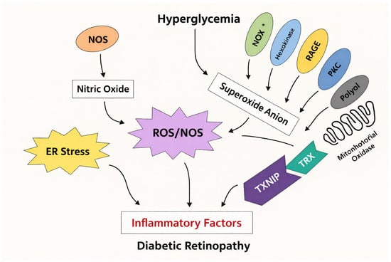
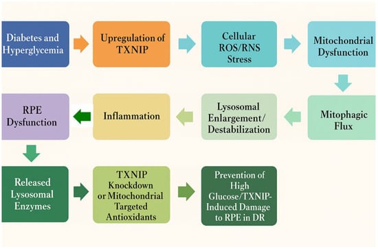
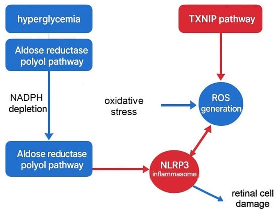
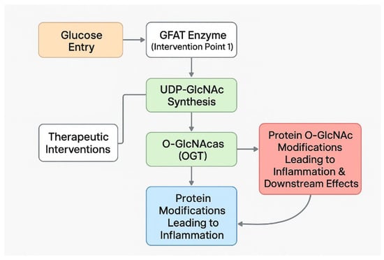
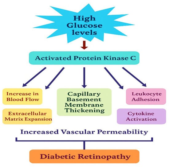
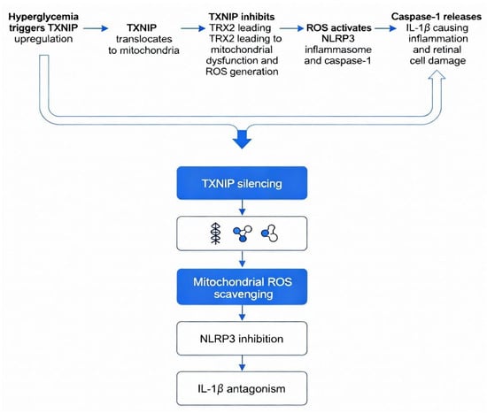
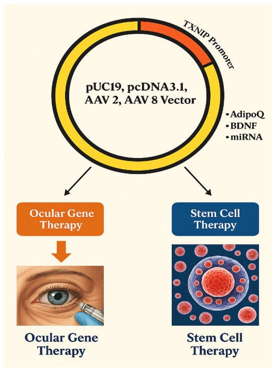
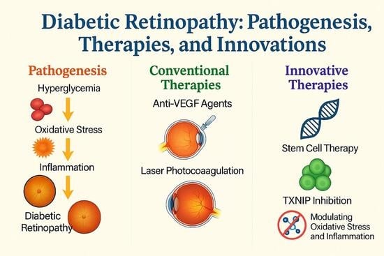
Diabetic retinopathy (DR) is a progressive retinal disorder and a leading cause of vision impairment worldwide affecting the livelihood of millions. Its pathogenesis is driven by chronic hyperglycemia-induced neuronal and microvascular injury, leading to capillary occlusion, increased vascular permeability, and the eventual formation of fragile neo vessels. These changes mark the progression from non-proliferative diabetic retinopathy (NPDR) to proliferative diabetic retinopathy (PDR). Diabetic macular edema (DME), characterized by blood–retinal barrier disruption and macular fluid accumulation, further contributes to vision loss. This review provides an integrative perspective on the cellular and molecular mechanisms of DR, highlighting both vascular and neuroglial contributions to retinal pathology. Current therapeutic approaches, including anti-VEGF agents and corticosteroids, offer symptomatic relief but are limited by the need for repeated administration and variability in patient response. Emerging evidence implicates the role of thioredoxin-interacting protein (TXNIP) as one of mediators of the disease progression. Strongly upregulated under hyperglycaemic stress, TXNIP induces oxidative damage, inflammation, and neuronal apoptosis, exacerbating neurovascular dysfunction. We explore potential therapeutic strategies such as gene therapy, TXNIP-targeted molecular interventions, and stem cell-based approaches aimed at achieving long-term modulation of disease mechanisms. This article thus attempts to address a comprehensive understanding of DR pathophysiology and innovative new strategies to improve long-term visual outcomes.
Full article

Graphical abstract
Open AccessArticle
Novel Combination of Icariin and Bone Xenograft Promotes the Expression of Collagen Fibers, BMP-2, and HIF-1α During Wound Healing in Wistar Rats
by
Christian Khoswanto and Ira Kusuma Dewi
J. Mol. Pathol. 2026, 7(1), 6; https://doi.org/10.3390/jmp7010006 - 4 Feb 2026
Abstract
►▼
Show Figures
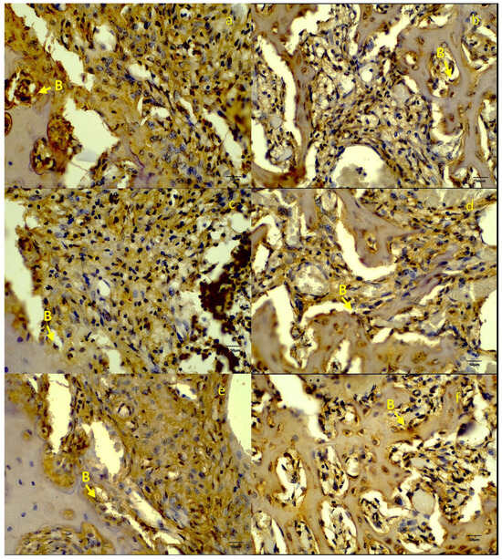
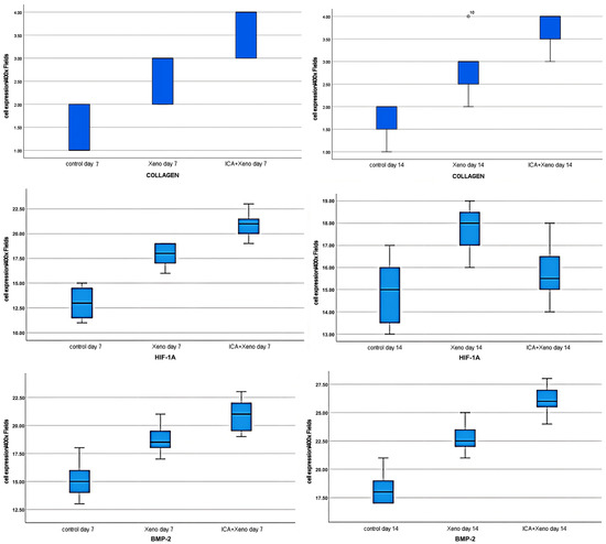
Objectives: This study evaluated the impact of combining icariin with a bovine xenograft on the enhancement of early socket healing in Wistar rats. Methods: Male Wistar rats underwent incisive extraction and were randomized into three groups (n = 8/group): (1) control, (2)
[...] Read more.
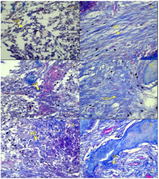
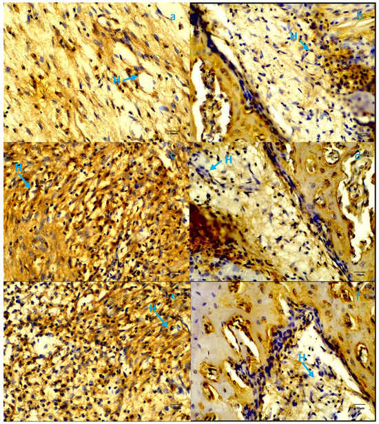
Objectives: This study evaluated the impact of combining icariin with a bovine xenograft on the enhancement of early socket healing in Wistar rats. Methods: Male Wistar rats underwent incisive extraction and were randomized into three groups (n = 8/group): (1) control, (2) xenograft, and (3) icariin–xenograft. On days 7 and 14, the animals were decapitated and their mandibles were examined. Histological analysis was conducted to assess the collagen matrix and the expression of BMP-2 and HIF-1α. Results: The icariin–xenograft group exhibited superior outcomes compared to the xenograft-alone and control groups. Histological analysis showed an earlier arrangement of connective tissue and an improved collagen matrix outcome in the icariin-treated sockets. Immunohistochemistry revealed elevated BMP-2 and HIF-1α expression in the icariin–xenograft group, indicating enhanced osteogenic and angiogenic signaling. Conclusions: Icariin-enhanced xenografts speed up the repair of early extraction sockets by enhancing the development of the collagen matrix and increasing the activity of pathways that promote osteogenesis and angiogenesis in low-oxygen conditions. This bioactive grafting technology appears to be a cost-effective method for preserving sockets and performing regenerative therapy in dentistry.
Full article

Figure 1

Highly Accessed Articles
Latest Books
E-Mail Alert
News
Topics
Topic in
Cells, JCM, Organoids, JMP
Novel Discoveries in Oncology 2nd Edition
Topic Editors: Michela Campolo, Giovanna Casili, Alessia Filippone, Marika LanzaDeadline: 31 January 2027
Topic in
Biomedicines, Biomolecules, Cardiogenetics, JCDD, JCRM, JMP, Metabolites, JPM
Advanced Imaging, Physiology, and Biomarkers in Ischemic Heart Disease
Topic Editors: Krishnaraj Sinhji Rathod, Alexandru CorlateanuDeadline: 10 March 2027
Topic in
Biomolecules, Cancers, Cells, JMP, Livers
Signaling Pathways in Liver Disease 2nd Edition
Topic Editors: Ralf Weiskirchen, Amedeo AmedeiDeadline: 31 March 2027
Topic in
Biomedicines, Cancers, IJMS, Vaccines, Viruses, JMP
Advances in HPV-Driven Head and Neck Cancer: Pathogenesis, Treatment, and Prevention
Topic Editors: Shilpi Gupta, Bhudev Chandra Das, Prabhat KumarDeadline: 20 April 2027
Special Issues
Special Issue in
JMP
New Insight of Genetic Alterations in Head and Neck and Salivary Gland Tumor
Guest Editor: Fuyuki SatoDeadline: 31 July 2026
Special Issue in
JMP
Selected Highlights of the 14th Molecular Cytopathology Meeting
Guest Editors: Giancarlo Troncone, Pasquale PisapiaDeadline: 30 September 2026
Special Issue in
JMP
Molecular Diagnostics in Cytopathology
Guest Editors: Eva M. Wojcik, Ryan HunterDeadline: 30 September 2026
Topical Collections
Topical Collection in
JMP
Feature Papers in Journal of Molecular Pathology
Collection Editors: Giancarlo Troncone, Pasquale Pisapia