Article

Comparative Cytotoxicity and Inflammatory Profiles of CeraSeal Versus AH Plus in Periodontal Tissue Repair: An In Vitro and In Vivo Study

1
Department of Hygiene No. 2, Bukhara State Medical Institute Named After Abu Ali Ibn Sino, Bukhara 200100, Uzbekistan
2
Department of Public Health and Healthcare Management, Samarkand State Medical University, Samarkand 140100, Uzbekistan
3
Department of Maxillofacial Surgery and Dentistry, Tashkent State Medical University, Tashkent 100000, Uzbekistan
4
Department of Maxillofacial Surgery, Andijan State Medical Institute, Andijan 170100, Uzbekistan
5
Department Prevention of Dental Diseases, Tashkent State Medical University, Tashkent 100000, Uzbekistan
6
Department of Medicine, Termez University of Economics and Service, Termez 190100, Uzbekistan
7
Department of Clinical Sciences, Ma’mun University, Urgench 220100, Uzbekistan
8
Department of Social Sciences, Bukhara State Pedagogical Institute, Bukhara 200100, Uzbekistan
*
Author to whom correspondence should be addressed.
J. Mol. Pathol. 2026, 7(2), 24; https://doi.org/10.3390/jmp7020024
Submission received: 28 April 2026 / Revised: 8 June 2026 / Accepted: 11 June 2026 / Published: 15 June 2026
(This article belongs to the Collection Feature Papers in Journal of Molecular Pathology)

Abstract

Background/Objectives: Endodontic perforation repair requires biomaterials that balance sealing ability with minimal cellular injury. AH Plus (epoxy resin-based) remains widely used despite cytotoxicity concerns. CeraSeal (calcium silicate-based bioceramic) is a potentially more biocompatible alternative. However, comparative data on sealer-induced cytotoxicity and inflammatory responses remain limited. This study compared the cytotoxicity and inflammatory profiles of CeraSeal and AH Plus using in vitro and in vivo approaches. Methods: Human periodontal ligament stem cells (hPDLSCs) were exposed to sealer extracts (1:4 AH Plus, 1:8 CeraSeal) for 120 h. Cell death was assessed by MTT, Live/Dead, LDH release, and Annexin V/PI flow cytometry. Oxidative stress was quantified via ROS generation (DCFH-DA). In a rat furcation perforation model (n = 8 teeth/group), inflammatory markers (TNF-α, IL-1β, CD68), osteogenic activity (ALP), and osteoclasts (TRAP) were evaluated. Results: AH Plus was associated with significantly greater necrotic cell death (357.6 ± 47.6% LDH release vs. CeraSeal 128.8 ± 37.5%; p = 0.0079) and reduced hPDLSC viability at all time points (p < 0.0001). ROS generation was comparable between sealers (~32–35%, p > 0.05). In vivo, IL-1β was higher in AH Plus-treated tissues (52.25 vs. 24.88 cells/mm2; p = 0.0002), while TNF-α and CD68 were greater in CeraSeal (p ≤ 0.0011). ALP was higher in AH Plus (median 6.15 vs. 3.68; p = 0.0002), with no difference in TRAP-positive osteoclasts. Morphometric analysis showed superior cellular preservation with CeraSeal (p = 0.0079), while inflammatory infiltration was higher in CeraSeal (p = 0.0002). Conclusions: AH Plus was associated with a necrotic-inflammatory profile with elevated IL-1β and higher ALP expression. CeraSeal demonstrated better cellular preservation, lower LDH release, and a distinct inflammatory signature (higher TNF-α and CD68). These findings establish comparative response profiles for the two sealers and support CeraSeal as a potentially biocompatible alternative, though further mechanistic studies are warranted.

1. Introduction

Iatrogenic furcation perforation is one of the most serious complications encountered during endodontic treatment, occurring in approximately 2–10% of root canal-treated teeth [1,2,3,4]. If left untreated or inadequately sealed, perforations create direct communication between the root canal system and the periodontal ligament space, leading to bacterial leakage, progressive inflammatory destruction of the periodontal attachment, and, ultimately, tooth loss [5]. The prognosis of a perforation depends critically on two interrelated factors: the ability to achieve an immediate and long-lasting seal, and the tissue response elicited by the repair material [6,7]. An ideal perforation repair material must therefore satisfy two seemingly opposing requirements: it should provide an impermeable seal against bacterial invasion while simultaneously exhibiting excellent biocompatibility to support periodontal and osseous regeneration [8]. However, the majority of currently available sealers were originally developed for conventional root canal obturation, not for direct contact with periodontal tissues [9]. Consequently, many exhibit favorable physical properties but unacceptable cytotoxicity, whereas others are biocompatible yet lack adequate sealing ability [10,11]. This therapeutic tension between sealing performance and biological safety represents the central clinical dilemma in perforation repair.
AH Plus, an epoxy resin-based sealer, is widely used as a reference material in endodontic research due to its well-established physicochemical properties and extensive documentation of its biological effects [12]. Its favorable performance stems from several key physicochemical properties: excellent flow and wettability, prolonged working time, minimal volumetric shrinkage upon setting, and high radiopacity [13]. While AH Plus is primarily indicated for root canal obturation, it has also been applied off-label for perforation repair procedures, and its cytotoxicity profile serves as a useful benchmark for comparing newer bioceramic materials [14]. These characteristics enable AH Plus to establish a durable, fluid-tight seal that effectively prevents bacterial microleakage [15]. Despite these advantages, a substantial body of evidence has consistently demonstrated that AH Plus exhibits significant cytotoxicity when placed in direct contact with viable tissues [16,17,18,19]. The epoxy resin matrix undergoes polymerization via an epoxy-amine crosslinking reaction, which is rarely complete and results in the long-term leaching of unreacted monomers as well as degradation byproducts [20]. Previous studies have shown that these released substances can compromise mitochondrial function and elevate intracellular reactive oxygen species (ROS) [17,21]. However, the precise pathways through which AH Plus eliminates cells remain incompletely defined, and whether cell death occurs predominantly via necrosis versus apoptosis has not been systematically compared against newer bioceramic alternatives.
In response to the limitations of epoxy resin-based materials, calcium silicate-based bioceramic sealers have been developed as a conceptually distinct class of repair materials [22]. Unlike epoxy resins, which rely on synthetic polymer matrices, bioceramic sealers undergo a hydration reaction upon exposure to tissue fluids, releasing calcium and hydroxyl ions and precipitating hydroxyapatite [23]. This physicochemical behavior has biological implications: the alkaline environment (pH ≈ 11–12) is intrinsically antibacterial, while released calcium ions may influence cellular responses [18,24]. CeraSeal (Meta Biomed Co., Ltd., Cheongju, Republic of Korea) represents a new-generation bioceramic sealer incorporating tricalcium silicate, zirconium oxide, and calcium aluminate. Recent studies have suggested that such compositions may alter host responses by shifting the balance from injury-driven inflammation toward tissue integration [25,26,27,28]. The immunomodulatory properties of dental biomaterials have increasingly become a focus of investigation, as the host inflammatory response is now recognized as a critical determinant of regenerative outcomes [29,30].
Nevertheless, several important questions remain unanswered. First, the specific patterns of cell death, inflammatory mediator expression, and tissue remodeling at the material interface have not been systematically characterized in direct comparative studies using both in vitro mechanistic assays and in vivo molecular profiling. Second, it remains unclear whether the superior cytocompatibility reported for CeraSeal in simple in vitro models translates into distinct inflammatory signatures (e.g., differential expression of TNF-α versus IL-1β) or osteogenic/osteoclastic activity in vivo [31,32,33,34,35,36]. Third, while oxidative stress has been implicated in sealer-induced toxicity, the relationship between ROS generation and downstream inflammatory outcomes has not been directly compared between epoxy resin and bioceramic materials [37,38]. The present study was designed to address these gaps by directly comparing CeraSeal and AH Plus within a single experimental framework spanning from cellular mechanisms to tissue-level outcomes. We hypothesized that the two sealers would induce qualitatively different patterns of cell death and inflammation, with CeraSeal demonstrating a more favorable cytocompatibility profile based on its distinct physicochemical properties. Specifically, we compared cytotoxicity, cell death pathways (apoptosis vs. necrosis), ROS generation, inflammatory marker expression (TNF-α, IL-1β, CD68), and osteogenic/osteoclastic activity using complementary in vitro (hPDLSC) and in vivo (rat furcation perforation) models.

2. Materials and Methods

2.1. Preparation of Sealer Extracts and Selection of Working Dilution

Endodontic sealers (AH Plus, Dentsply DeTrey, Konstanz, Germany; CeraSeal, Meta Biomed, Cheongju, Republic of Korea) were prepared following ISO 10993-5:2009 guidelines [39]. For each material, 0.1 g of freshly mixed sealer was immersed in 1 mL of serum-free Dulbecco’s Modified Eagle Medium (DMEM; Gibco, Grand Island, NY, USA) and incubated at 37 °C for 24 h. Eluates were filtered through 0.22 µm syringe filters (Sartorius AG, Göttingen, Germany) to ensure sterility. A pilot cytotoxicity screening using the MTT assay was conducted with serial dilutions (1:1, 1:2, 1:4, 1:6, 1:8, and 1:16) of each sealer extract (n = 5 per dilution) to determine the optimal working dilutions for subsequent mechanistic assays (Supplementary Figure S1 and Table S1). Based on these data, the 1:4 dilution for AH Plus (76.2 ± 11.3% viability at 24 h, p = 0.0556 vs. control) and the 1:8 dilution for CeraSeal (78.0 ± 16.3% viability at 24 h, p = 0.1710 vs. control) were selected. These dilutions produced comparable cell viability levels (approximately 76–78%) across both materials, enabling meaningful comparative analysis of cell death pathways, inflammatory responses, and oxidative stress profiles.
The use of different working dilutions follows ISO 10993-5:2009 guidelines, which recommend pilot-guided dilution selection for materials with distinct physicochemical properties [18,39,40,41]. Identical mass concentrations would be biologically inappropriate, as epoxy resin-based sealers release hydrophobic monomers that accumulate at the cell-material interface, while bioceramic sealers undergo gradual hydration and ion release [20,42,43]. All test preparations were freshly prepared under sterile conditions immediately before use and used within 30 min of preparation.

2.2. Cell Culture

Human periodontal ligament stem cells (hPDLSCs) were commercially obtained from the Department of Biology, Iskandarovna Laboratory (Bukhara, Uzbekistan; Catalog No.: hPDLSC-2023-01). According to the supplier’s certificate of analysis, cells were isolated from healthy premolars extracted from three systemically healthy donors (aged 18–25 years; two males, one female) and validated for stemness by positive expression of CD73, CD90, and CD105, negative expression of CD34 and CD45, and confirmed multilineage (osteogenic, adipogenic, chondrogenic) differentiation potential. Cells were received at passage 2 and used from passages 3–5 for all experiments. Cells from all three donors were pooled by the supplier to minimize donor-to-donor variability. hPDLSCs were cultured in complete DMEM supplemented with 10% fetal bovine serum (FBS; Gibco, Grand Island, NY, USA) and 1% penicillin–streptomycin (Gibco, Grand Island, NY, USA) at 37 °C in a humidified 5% CO2 atmosphere. Untreated cells served as the negative control.

2.3. MTT Assay

Cell metabolic activity was assessed using the MTT assay [44]. hPDLSCs were seeded in 96-well plates (1 × 10⁵ cells/well) and incubated for 24 h. Culture medium was replaced with sealer extracts or control medium, and viability was measured at 24, 72, and 120 h. At each time point, 50 µL of MTT solution (0.5 mg/mL; Sigma-Aldrich, St. Louis, MO, USA) was added for 2 h at 37 °C. Formazan crystals were dissolved in 100 µL isopropanol, and absorbance was read at 570 nm (Multiskan Sky, Thermo Fisher Scientific, Waltham, MA, USA). Cell viability was expressed as a percentage relative to control. All experiments were performed in five replicates per group and per time point. Cell viability was calculated as follows:
C e l l   v i a b i l i t y   ( % )   =   ( A b s o r b a n c e   o f   e x p e r i m e n t a l   g r o u p A b s o r b a n c e   o f   c o n t r o l   g r o u p     ) ×   100

2.4. Live/Dead Cell Staining Procedure

Long-term viability was evaluated using calcein-AM (2 µM) and ethidium homodimer-1 (4 µM) (Invitrogen, Carlsbad, CA, USA). hPDLSCs were seeded in 6-well plates (1 × 10⁵ cells/well), allowed to attach for 24 h, and exposed to sealer extracts for 120 h. After staining for 30 min at room temperature in the dark, cells were imaged under an inverted fluorescence microscope. Viable cells fluoresced green, whereas dead cells fluoresced red. Five random fields per well were analyzed using Fiji/ImageJ version 2.14.0 (NIH, Bethesda, MD, USA) to determine live cell percentage. All groups were analyzed in five replicates (n = 5). The percentage of live cells was quantified using ImageJ software (NIH, USA).

2.5. Lactate Dehydrogenase (LDH) Assay

Membrane damage was quantified using a commercial LDH assay kit (Promega, Madison, WI, USA). hPDLSCs were seeded in 96-well plates (1 × 10⁵ cells/well) and exposed to sealer extracts for 120 h. LDH activity in culture supernatants was measured at 450 nm and normalized to the negative control, according to the following formula:
L D H   R e l e a s e   ( % )   =   100   ×   [ ( E x p e r i m e n t a l   L D H   a b s o r b a n c e     C o n t r o l   a b s o r b a n c e )   /   C o n t r o l   a b s o r b a n c e ]

2.6. Richardson Staining for Morphological Evaluation

Richardson staining was performed after 120 h of incubation. Cells were seeded in 6-well tissue culture-treated plates at a density of 1 × 10⁵ cells/well and incubated for 24 h. Sealer extracts were added to the experimental groups, followed by incubation for 5 days under standard culture conditions. At the end of the incubation period, the culture medium was aspirated, and the wells were gently rinsed with PBS. Fixation was done using methyl ethanol (Merck KGaA, Darmstadt, Germany) for 10 min at room temperature. Plates were then air-dried under a laminar flow hood. Two staining solutions were freshly prepared: Solution I: 1% methylene blue in 1% sodium borate, and Solution II: 1% azure II in distilled water. Equal volumes of both solutions were mixed (1:1) and applied to the fixed monolayers for 3–5 min. Cells were gently rinsed with distilled water and left to air-dry. Images were captured using a light microscope at ×200 magnification. The following three morphological indices were evaluated based on five replicate images per group using ImageJ software. To quantify changes in cell shrinkage index (CSI), the following formula was applied:
C S I   ( % )   =   ( ( M e a n   C e l l   A r e a     _ C o n t r o l M e a n   C e l l   A r e a     _ T r e a t e d ) ) / M e a n   C e l l   A r e a     _ C o n t r o l   ×   100
Cell coverage (CC) measurements were performed manually for 20–30 cells per image using the freehand selection tool. Defined as the percentage of the microscopic field covered by adherent cells. Binary thresholding and particle analysis were used to determine the total occupied area, normalized to the field size:
C C   ( % ) = ( C e l l o c c u p i e d   a r e a T o t a l   i m a g e   a r e a ) × 100
The morphological deformation score (MDS) was based on qualitative assessment of cell rounding, granularity, and loss of cellular extensions, normalized on a scale from 0 (no deformation) to 100 (severe deformation) based on reference images. Assessment was performed by a blinded examiner. A composite index reflecting overall morphological integrity, calculated using the formula:
M D S = C S I   + ( 100 C C ) + N o r m a l i z e d   V D S 3

2.7. Annexin V/PI Assay

Apoptosis and necrosis were detected using the FITC Annexin V Apoptosis Detection Kit I (BD Biosciences, San Jose, CA, USA). hPDLSCs (1 × 10⁵ cells/mL) were exposed to sealer extracts for 24 h. Cells were stained with Annexin V-FITC and PI for 15 min in the dark and analyzed immediately by flow cytometry (COULTER® EPICS® XL™, Beckman Coulter, Inc., Brea, CA, USA).

2.8. Measurement of Reactive Oxygen Species (ROS)

Intracellular ROS generation was measured using 2′,7′-dichlorodihydrofluorescein diacetate (DCFH-DA, 20 µM; Sigma-Aldrich, St. Louis, MO, USA) [45]. hPDLSCs were exposed to sealer extracts for 120 h, stained with DCFH-DA for 2 h at 37 °C in the dark, and analyzed by flow cytometry. Mean fluorescence intensity (MFI) was quantified. Arachidonic acid (60 µM) served as the positive control.

2.9. Surgical Procedure

All surgical procedures were performed by a single experienced operator (G.S.) to ensure consistency. This in vivo experiment was performed on 16 maxillary first molars from ten adult male Sprague–Dawley rats (8–10 weeks old; weight: 250–300 g). Animals were randomly allocated to two groups (n = 8 teeth per group) according to the root canal sealer used for furcation perforation repair. Sample size was determined a priori using G*Power 3.1 software for a two-tailed independent t-test (α = 0.05, power = 0.80, Cohen’s d = 1.2), yielding a minimum of 7 teeth per group, consistent with previous rat furcation perforation studies [46]. The 40-day follow-up period was selected based on established rat perforation models demonstrating that 30 days is sufficient to capture early-to-intermediate reparative responses [47]. All procedures adhered to institutional and international ethical guidelines and were approved by the Bukhara State Medical Institute Animal Ethics Committee (approval code: BSMI/2023-042).
General anesthesia was induced via intraperitoneal ketamine (80 mg/kg) and xylazine (10 mg/kg) injections, with supplemental doses administered as required [48]. The operative site was disinfected with 2% chlorhexidine. Endodontic access cavities were prepared in the maxillary first molars using a high-speed handpiece under water cooling. A standardized perforation (0.5 mm diameter) was created at the pulp chamber floor in the center of the furcation area using a sterile round bur (#1/4, Dentsply Sirona, Charlotte, NC, USA) mounted on a high-speed handpiece under 20× magnification using a surgical operating microscope (Leica M320, Leica Microsystems, Wetzlar, Germany). The bur was oriented perpendicular to the chamber floor, and a through-and-through perforation was confirmed by direct visualization of the bur tip through the furcation area. Hemostasis was achieved using sterile cotton pellets moistened with sterile saline applied with gentle pressure for 60 s. The perforations were sealed immediately after hemostasis with the assigned test material (AH Plus or CeraSeal) mixed according to the manufacturer’s instructions and delivered using a sterile endodontic plugger (size 1, Hu-Friedy, Chicago, IL, USA). Excess material was removed with a sterile cotton pellet. The access cavity was restored with Filtek™ Z250 light-cured composite resin (3M ESPE, St. Paul, MN, USA) in two increments, each light-cured for 20 s using a LED curing light (Elipar DeepCure-S, 3M ESPE, St. Paul, MN, USA; intensity ≥ 1000 mW/cm2).
Postoperative care included subcutaneous buprenorphine (0.05 mg/kg) every 12 h for 2 days and daily monitoring of body weight, feeding, and behavior. At 40 days post-surgery, animals were euthanized by CO2 inhalation in accordance with AVMA guidelines. Maxillae were dissected, and treated molars with surrounding tissues were harvested and fixed in 10% neutral buffered formalin for histological and immunohistochemical evaluation.

2.10. Histological Processing

To ensure unbiased assessment, all tissue samples were randomly assigned numeric codes by a technician not involved in subsequent analyses. The code-key was stored separately and revealed only after all analyses were completed. Harvested specimens were decalcified in EDTA (Sigma-Aldrich, St. Louis, MO, USA) until adequately softened, washed, dehydrated in ascending ethanol series, cleared in xylene, and embedded in paraffin. All paraffin blocks were labeled only with numeric codes, with no treatment group information visible to personnel performing sectioning or staining. For histological evaluation, five non-serial sections (spaced at least 50 µm apart) were selected from each specimen. Longitudinal 5-µm sections through the perforation site were obtained using a rotary microtome (Leica RM2145, Leica Microsystems, Wetzlar, Germany) and mounted on glass slides. The perforation site was defined as the plane containing the root canals and the furcation area where the perforation was created. Only sections showing the entire perforation defect and surrounding periodontal ligament space were included for analysis.
Hematoxylin and Eosin (H&E) staining was used to assess inflammatory cell infiltration and overall tissue morphology. For inflammatory cell counts, five high-power fields (×400 magnification) were selected from each section using a systematic random sampling method. The observer first identified the perforation margin, then moved radially outward in 200-µm increments. Fields containing the perforation margin were excluded to avoid counting cells directly adjacent to the material. A random number generator was used to determine the direction of movement for field selection. The average number of cells per mm2 was calculated for each specimen [46].
Masson’s Trichrome staining was performed to assess collagen deposition. High-resolution images were analyzed with ImageJ software by applying a consistent threshold to the blue-stained collagen fibers. Five random fields per specimen were quantified using the same field selection criteria, and collagen content was expressed as collagen area fraction (%). All quantitative analyses were performed independently by two blinded examiners. Inter-observer agreement was excellent (intraclass correlation coefficient, ICC > 0.90 for all parameters). The code was broken only after all data were recorded, verified, and transferred to the statistical analysis database.

2.11. Immunohistochemistry

Paraffin sections (5 µm) were deparaffinized, rehydrated, and subjected to citrate buffer (pH 6.0) antigen retrieval in a pressurized decloaking chamber. Endogenous peroxidase activity was blocked with 3% hydrogen peroxide, and nonspecific binding sites were blocked with 1% bovine serum albumin (BSA). Sections were incubated overnight at 4 °C with the following primary antibodies: goat anti-TNF-α (catalog no. sc-1351, Santa Cruz Biotechnology, Dallas, TX, USA, dilution 1:100); rabbit anti-IL-1β (catalog no. sc-7884, Santa Cruz Biotechnology, Dallas, TX, USA, dilution 1:100); mouse anti-CD68 (catalog no. sc-20060, Santa Cruz Biotechnology, Dallas, TX, USA, dilution 1:100); and mouse anti-alkaline phosphatase (ALP) (catalog no. sc-166261, Santa Cruz Biotechnology, Dallas, TX, USA, dilution 1:50). After incubation with biotinylated secondary antibodies and streptavidin–HRP, immune complexes were visualized using DAB chromogen and counterstained with hematoxylin.
For quantitative assessment of TNF-α, IL-1β, and CD68, positively labeled cells were counted in standardized microscopic fields (1.2 mm × 0.6 mm) using the same systematic random sampling method described in Section 2.10, with the perforation margin as the reference point.
For ALP expression quantification, regions of interest (ROIs) were manually drawn along the alveolar bone surface within the periodontal ligament space adjacent to the perforation site (mid-root region of the mesial root, within 300 µm of the perforation margin). Each ROI included the bone surface and a 50-µm zone extending into the periodontal ligament space. Five ROIs per section were analyzed. The mean integrated density (corrected for background) was calculated using Fiji/ImageJ version 2.14.0 (NIH, Bethesda, MD, USA) according to the formula: Corrected Integrated Density = Integrated Density, (Area × Mean background intensity). The normalized staining intensity was expressed as the corrected integrated density per ROI area. All measurements were performed independently by two blinded examiners.

2.12. Tartrate-Resistant Acid Phosphatase (TRAP) Staining

To identify osteoclasts, sections were deparaffinized, hydrated, and stained with TRAP solution containing Fast Red and naphthol AS-BI phosphoric acid. Multinucleated TRAP-positive cells (containing three or more nuclei) were counted within resorption lacunae (Howship’s lacunae) on the alveolar bone surface. Counts were performed in the same anatomical region as for ALP (mid-root region of the mesial root, within 300 µm of the perforation margin), using the same systematic random sampling method described in Section 2.10 (200-µm increments outward from the perforation margin, fields containing the perforation margin excluded). Five fields per specimen were analyzed, and the number of TRAP-positive osteoclasts was expressed as the total count per specimen.

2.13. Standardization of Anatomical Regions

All histological and immunohistochemical images were captured from standardized anatomical regions to ensure comparability across specimens. The reference point for all analyses was the perforation margin. The primary anatomical region of interest was the periodontal ligament space in the mid-root region of the mesial root. For H&E analysis, fields were selected within 200 µm of the perforation margin. For immunohistochemistry (TNF-α, IL-1β, CD68), Masson’s trichrome, ALP, and TRAP analyses, fields were selected within 300 µm of the perforation margin. For ALP and TRAP analyses, the alveolar bone surface within the periodontal ligament space was specifically examined. Fields containing the perforation margin or the restorative material interface were excluded from all analyses to avoid counting cells directly adherent to the material surface or within the mechanical artifact zone adjacent to the perforation edge, where tissue disruption from the surgical procedure could confound accurate assessment of the biological response to the sealer material.

2.14. Statistical Analysis

Data normality was tested using the Shapiro–Wilk test. Parametric data were analyzed using unpaired t-tests (Welch’s correction if variances were unequal) or two-way ANOVA with Tukey’s post hoc test. Non-parametric data were analyzed using the Mann–Whitney U test or Kruskal–Wallis test with Dunn’s post hoc correction. Hodges–Lehmann estimators were calculated for median differences. Effect sizes (η2) were reported where applicable. Statistical significance was set at p < 0.05. Data are expressed as mean ± SD for normally distributed variables or median for non-normal variables.

3. Results

3.1. Comparative In Vitro Cytotoxicity and Cell Death Profiling of AH Plus and CeraSeal Sealers

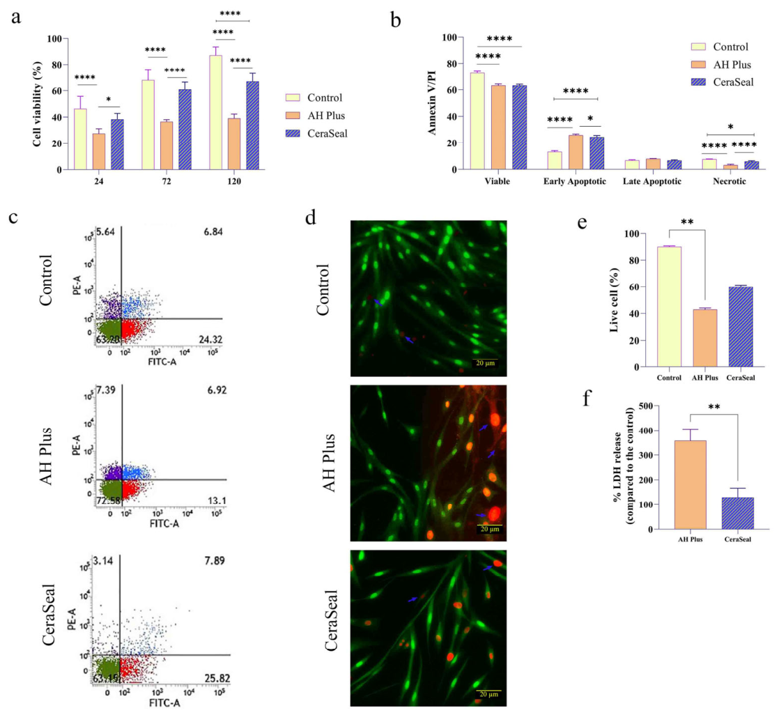
Cell viability and death profiles following exposure to AH Plus and CeraSeal were assessed using MTT, Live/Dead, LDH release, and Annexin V/PI assays (Figure 1). The MTT assay demonstrated a pronounced time- and material-dependent effect on metabolic activity (two-way ANOVA: time, group, and interaction, all p < 0.0001). AH Plus induced a significant reduction in viability at all time points compared with both control and CeraSeal (p < 0.0001; Figure 1a). In contrast, CeraSeal maintained viability comparable to the control at 24 h and 72 h (p > 0.05) and showed only a moderate decline at 120 h (p < 0.0001). To dissect the modes of cell death, quantitative analysis of viable, apoptotic, and necrotic cell subpopulations following 120 h exposure to AH Plus (1:4 dilution) and CeraSeal (1:8 dilution) is presented in Figure 1b, while representative two-dimensional dot plots for each group are shown in Figure 1c. Two-way ANOVA revealed significant effects of cell state (p < 0.0001) and a cell state–treatment interaction (p < 0.0001), without a main effect of treatment group (p = 0.9998). Viable cell proportions were significantly reduced in both AH Plus and CeraSeal compared to control (both p < 0.0001), with no difference between the sealers (p = 0.9993). Early apoptosis was elevated in both sealer groups versus control (both p < 0.0001), with AH Plus producing a modest but significant increase over CeraSeal (p = 0.0247). Late apoptosis did not differ significantly across groups. Necrosis was markedly higher in AH Plus compared with both control and CeraSeal (both p < 0.0001), while CeraSeal induced a smaller yet significant necrotic increase compared to control (p = 0.0143).
Consistent with these findings, the Live/Dead assay revealed significant group differences in live cell percentages (p < 0.0001, Kruskal–Wallis test). Representative fluorescence micrographs (Figure 1d) show abundant green-stained viable cells in the control and CeraSeal groups, whereas AH Plus samples displayed a marked predominance of red-stained dead cells. Quantitative analysis of live cell proportions (Figure 1e) confirmed that AH Plus significantly reduced viability compared to control (p = 0.0012), while CeraSeal exhibited intermediate values with no significant difference from either control or AH Plus (p = 0.23). The LDH release assay further confirmed greater cytotoxicity for AH Plus, with mean LDH release reaching 357.6 ± 47.6% versus 128.8 ± 37.5% for CeraSeal. This difference was significant (Mann–Whitney U = 0, p = 0.0079; Figure 1f).

3.2. Inflammatory and Oxidative Stress Responses Induced by AH Plus and CeraSeal

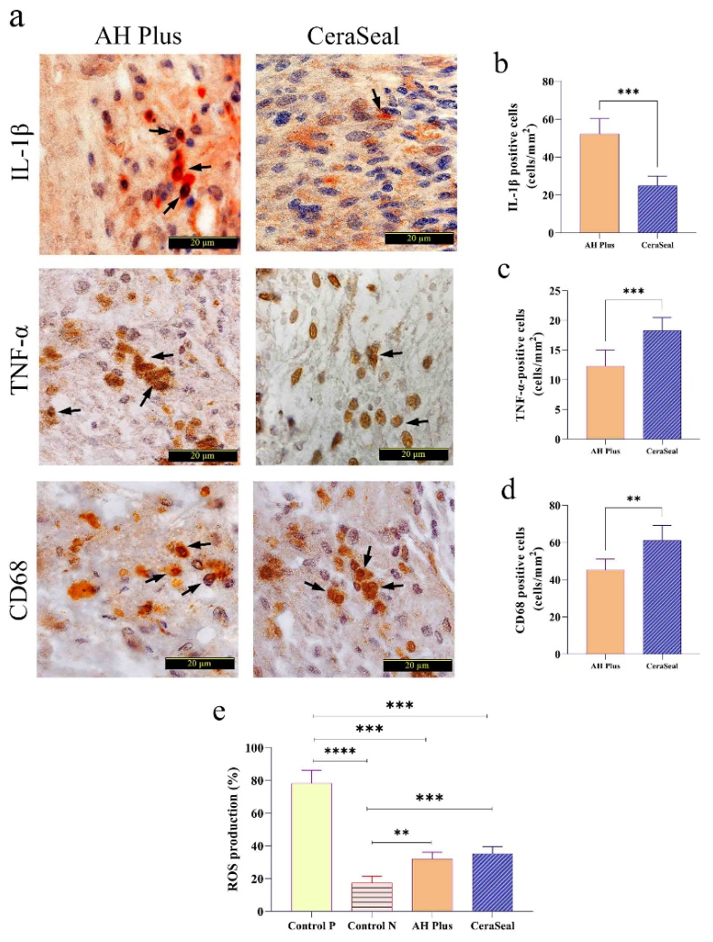
Immunohistochemical evaluation of inflammatory cell infiltration revealed distinct patterns between the two sealers (Figure 2a). The density of IL-1β immunostaining (Figure 2b) showed the opposite trend, with the AH Plus group exhibiting significantly higher expression levels (52.25 ± 7.73 cells/mm2) than CeraSeal (24.88 ± 4.82 cells/mm2; U = 0, p = 0.0002), suggesting an enhanced local pro-inflammatory response. In contrast, CeraSeal-treated tissues demonstrated a greater number of TNF-α-positive cells (18.25 ± 2.29 cells/mm2) than AH Plus-treated tissues (12.25 ± 2.43 cells/mm2; U = 0, p = 0.0002; Figure 2c). Similarly, CD68-positive macrophages (Figure 2d) were significantly higher in the CeraSeal group (61.13 ± 7.47 cells/mm2) compared with AH Plus (45.25 ± 5.57 cells/mm2; Mann–Whitney U = 3, p = 0.0011), indicating a sealer-specific cytokine activation profile.
The DCFH-DA assay further assessed oxidative stress by measuring intracellular ROS production (Figure 2e). Positive control samples confirmed assay validity by exhibiting markedly elevated ROS levels (78.2 ± 6.8%) compared to the negative control (17.4 ± 4.4%; p < 0.0001). Both AH Plus (32.0 ± 4.5%) and CeraSeal (35.2 ± 4.6%) induced significantly higher ROS production than the negative control (p = 0.0027 and p = 0.0008, respectively), but substantially lower levels than the positive control (p = 0.0001 and p = 0.0002, respectively). No significant difference in ROS generation was observed between AH Plus and CeraSeal (p = 0.7795), indicating comparable oxidative stress profiles despite differences in inflammatory marker expression.

3.3. Osteoclastic and Osteoblastic Activity in Response to AH Plus and CeraSeal

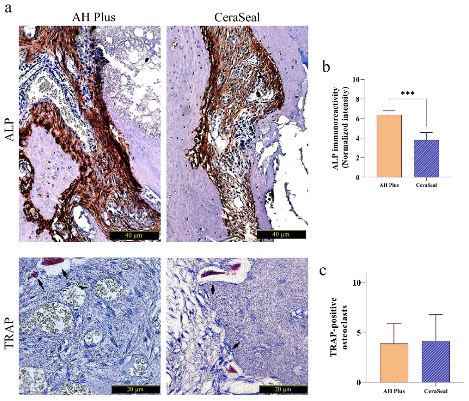
Representative histological micrographs showing ALP immunoreactivity and TRAP staining in AH Plus- and CeraSeal-treated tissues (Figure 3a). Assessment of bone-formative activity via quantitative ALP immunoreactivity revealed a significant intergroup difference (Figure 3b). AH Plus-treated tissues exhibited markedly higher normalized ALP intensity (median: 6.15) compared to CeraSeal (median: 3.68), with a Hodges–Lehmann estimated difference of −2.705 (p = 0.0002; Mann–Whitney U = 0). In contrast, histological quantification of bone-resorptive cells demonstrated that the mean number of TRAP-positive multinucleated osteoclasts was comparable between the AH Plus (3.88 ± 1.90) and CeraSeal (4.13 ± 2.10) groups (Figure 3c), with no statistically significant difference (unpaired t-test with Welch’s correction, p = 0.8352, η2 = 0.0034). Variance analysis further confirmed the absence of heterogeneity in cell count distribution between groups (F = 1.693, p = 0.5040), While this may suggest an enhanced osteogenic response in the AH Plus group, it is likely reflective of a reparative process secondary to greater tissue injury rather than an inherently superior bioinductive property of the material.

3.4. Cellular Morphology, Inflammatory Infiltration, and Extracellular Matrix Deposition

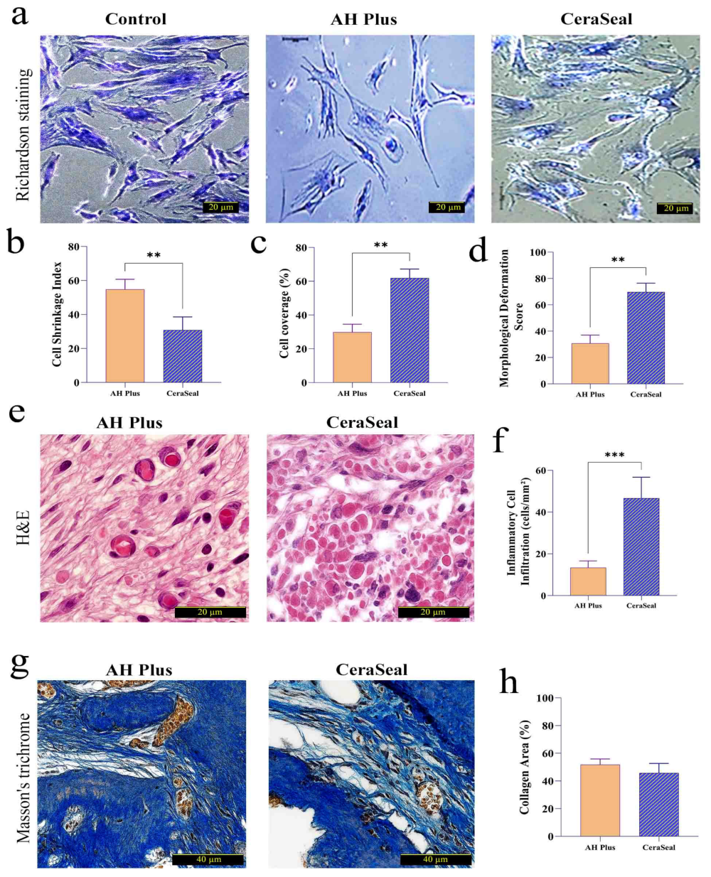
Quantitative Richardson staining analysis revealed pronounced differences in cell morphology between the two experimental sealers (Figure 4a). The cell shrinkage index (Figure 4b) was significantly higher in the AH Plus group (median: 55.2) compared to CeraSeal (median: 29.6), with a Hodges–Lehmann estimated difference of −24.8 (p = 0.0079; Mann–Whitney U = 0), indicating greater cytoplasmic condensation and morphological stress in AH Plus-treated cultures. Consistently, the percentage of cell-covered surface area (Figure 4c) was markedly greater in the CeraSeal group (median: 62.3%) than in the AH Plus group (median: 29.2%), yielding a Hodges–Lehmann difference of +33.0 (p = 0.0079; Mann–Whitney U = 0). These findings suggest superior monolayer preservation and confluence with CeraSeal, whereas AH Plus substantially reduced cell adhesion and surface expansion.
Similarly, the Morphological Deformation Score (Figure 4d), reflecting cellular preservation and structural integrity, was significantly higher in CeraSeal-treated cells (median: 68.2) compared to AH Plus (median: 32.4), with a Hodges–Lehmann difference of +38.0 (p = 0.0079; Mann–Whitney U = 0). Histological evaluation with H&E staining further demonstrated greater inflammatory cell infiltration in the CeraSeal group (median: 48.5 cells/mm2) compared to AH Plus (median: 13.0 cells/mm2; Hodges–Lehmann difference: +34.5; p = 0.0002; Mann–Whitney U = 0), indicating more pronounced immune cell recruitment with CeraSeal application (Figure 4e,f). Quantitative Masson’s trichrome analysis showed no statistically significant difference in collagen area fraction between AH Plus (median: 51.76%) and CeraSeal (median: 43.92%) (p = 0.0830; Mann–Whitney U = 15; Hodges–Lehmann difference: −6.86), despite a numerically higher collagen content in the AH Plus group (Figure 4g,h).

4. Discussion

The selection of an endodontic sealer for perforation repair involves a balance between material-induced cell death, inflammation, and tissue regeneration [49,50]. In this study, we compared AH Plus and CeraSeal using complementary in vitro and in vivo approaches. At the cellular level, AH Plus was associated with higher LDH release and a greater proportion of necrotic cells, whereas CeraSeal showed lower LDH release and a higher proportion of early apoptotic cells. In vivo, AH Plus-treated tissues exhibited elevated IL-1β, while CeraSeal-treated tissues showed higher TNF-α and CD68+ macrophage density. These findings indicate that the two materials elicit qualitatively different response patterns. However, the underlying molecular mechanisms were not directly investigated and remain to be elucidated.
AH Plus exposure resulted in significantly higher LDH release and a greater proportion of Annexin V/PI+ cells (operationally defined as necrosis), whereas CeraSeal was associated with a higher proportion of Annexin V+/PI cells (operationally defined as early apoptosis) [17,18,27]. Two caveats apply. First, Annexin V/PI staining alone cannot distinguish primary necrosis from secondary necrosis or late apoptotic disintegration [51,52,53]. Second, the use of different dilutions limits direct quantitative comparability, though this approach follows ISO 10993-5:2009 guidelines [39] and previous studies [18,41,52]. Our results should therefore be interpreted as material-specific response profiles. Based on prior literature, one might hypothesize that leaching of unreacted monomers from AH Plus contributes to mitochondrial dysfunction [43,54]. However, we did not measure mitochondrial membrane potential, ATP levels, or caspase activation. Conversely, the superior cytocompatibility of CeraSeal is consistent with previous reports [18,41,52,55,56], and it is plausible that gradual ion release supports cell survival [55,57,58,59]. However, we did not measure calcium signaling or downstream survival pathways. These interpretations remain hypothetical.
Both sealers induced comparable intracellular ROS levels, yet the in vivo inflammatory profiles diverged markedly. This dissociation is noteworthy: equal bulk ROS did not produce equal inflammation. This suggests that the quantity of ROS alone is not the critical determinant; rather, the source, compartmentalization, and kinetics of ROS production may shape downstream inflammatory outcomes [60].
AH Plus was associated with elevated IL-1β, a cytokine strictly dependent on NLRP3 inflammasome activation [61,62,63]. IL-1β maturation requires two signals: a priming signal (NF-κB-dependent pro-IL-1β synthesis) and an activation signal (often provided by mitochondrial ROS or DAMPs from necrotic cells) [64,65,66]. The concurrent observation of higher necrosis and higher IL-1β in AH Plus-treated tissues is therefore consistent with a sequence in which necrotic debris provides the activation signal for inflammasome assembly. In contrast, CeraSeal did not elevate IL-1β despite comparable ROS levels, suggesting that its ROS may not reach the mitochondrial compartment or may be rapidly buffered.
An apparent paradox was the higher inflammatory infiltration and CD68+ macrophage density in CeraSeal-treated tissues despite its lower in vitro cytotoxicity. This paradox may be resolved by considering that inflammation is not uniformly detrimental. The release of calcium and hydroxyl ions from CeraSeal can serve as chemotactic signals, recruiting macrophages without triggering the NLRP3-IL-1β axis [18,55,57]. This pattern of material-induced immune modulation is consistent with emerging concepts in regenerative dentistry, where bioactive materials are increasingly recognized for their ability to shape inflammatory responses rather than simply being inert [67,68,69,70]. TNF-α, which was elevated in CeraSeal-treated tissues, can be produced by both M1 and M2 macrophages. In the absence of sustained IL-1β, TNF-α may contribute to tissue remodeling, matrix metalloproteinase activation, and mesenchymal stromal cell recruitment rather than perpetuating injury [71,72]. However, without macrophage polarization markers (M1: iNOS, CD86; M2: CD163, CD206, Arg1), we cannot definitively classify this response as reparative versus pro-inflammatory [73,74,75]. Nevertheless, the combination of preserved tissue morphology, low IL-1β, and TNF-α elevation is more consistent with a regulated inflammatory response than with uncontrolled tissue injury.
AH Plus-treated tissues exhibited significantly higher ALP immunoreactivity than CeraSeal-treated tissues. Interpreted in isolation, this finding might suggest enhanced osteogenesis. However, three lines of evidence argue against this interpretation. First, elevated ALP occurred in the context of substantially greater cytotoxicity and tissue injury, raising the possibility of a stress-induced cellular response rather than true osteogenesis [76,77,78,79]. Second, there was no corresponding difference in osteoclast activity: TRAP-positive cell counts were comparable between groups, arguing against coordinated bone remodeling [80]. Third, CeraSeal, despite lower ALP, preserved cellular morphology significantly better, suggesting that a less injurious microenvironment does not require reactive ALP upregulation to maintain tissue integrity. Thus, the higher ALP in AH Plus-treated tissues is more plausibly explained as a compensatory or stress-induced response to greater tissue injury rather than a bona fide osteogenic advantage. Without complementary osteogenic markers or histomorphometric analysis of new bone formation, we caution against interpreting ALP elevation as evidence of enhanced regeneration.
Richardson staining showed that CeraSeal better preserved cellular morphology: lower cell shrinkage index, higher cell-covered surface area, and higher morphological deformation score. These findings are consistent with the lower cytotoxicity observed in CeraSeal-treated cultures. In contrast, the cytoskeletal disruption and cell rounding observed with AH Plus are consistent with known effects of epoxy-derived monomers on actin polymerization and focal adhesion turnover reported in other cell types [81,82,83].
H&E staining revealed greater inflammatory cell infiltration in CeraSeal-treated tissues compared to AH Plus-treated tissues. This pattern is consistent with previous studies showing that bioceramic sealers elicit greater macrophage infiltration than epoxy resin-based sealers [18,52,60,74,84,85]. Two opposing interpretations of this differential inflammatory response are possible. On one hand, lower inflammatory cell density in AH Plus-treated tissues could reflect immune cell toxicity and impaired recruitment, consistent with the lymphotoxic effects of epoxy monomers described in the literature [17,18,42,43,50]. On the other hand, higher inflammatory infiltration in CeraSeal-treated tissues could represent a host response to a bioactive material, but it could also indicate a foreign body reaction. Without additional markers of macrophage polarization (M1: iNOS, CD86; M2: CD163, CD206, Arg1), we cannot determine which interpretation is correct [59,73,74,84]. Masson’s trichrome staining showed no significant difference in collagen area fraction between groups, suggesting comparable extracellular matrix deposition at 40 days [4,5,47,86].
When the in vitro and in vivo data are considered together, two distinct response trajectories emerge. For AH Plus, the sequence appears to begin with cytotoxicity, which progresses to necrotic cell death, as evidenced by high LDH release and Annexin V/PI+ staining, and subsequently leads to elevated IL-1β expression in vivo. This pattern is consistent with injury-driven inflammation. For CeraSeal, a different trajectory is observed: preserved cell viability (lower LDH release and better morphological preservation) is followed by macrophage recruitment (higher CD68+ density) and TNF-α expression without commensurate IL-1β elevation. This pattern is consistent with a regulated inflammatory response. Whether this response ultimately supports tissue repair or represents a foreign body reaction remains to be determined, as definitive classification would require additional markers of macrophage polarization and functional outcomes. Contemporary reviews on bioactive materials in endodontics emphasize that the immunomodulatory properties of sealers may be as important as their physical properties in determining long-term regenerative success [87,88,89,90].
Several limitations should be acknowledged. First, different working dilutions limit direct quantitative comparability; results should be interpreted as material-specific response profiles. Second, DCFH-DA detects bulk ROS without discriminating species or subcellular sources. Third, lack of macrophage polarization markers prevents definitive classification of the inflammatory infiltrate. Fourth, the Annexin V/PI assay used in this study does not distinguish between primary necrosis, secondary necrosis, and necroptosis. The necrotic population reported may therefore include cells undergoing RIPK1/RIPK3/MLKL-mediated necroptosis. Future studies incorporating specific necroptosis markers would be necessary to determine the contribution of necroptosis to the observed cell death patterns.

5. Conclusions

This study demonstrates that AH Plus and CeraSeal elicit distinct biological response profiles in hPDLSCs and in a rat furcation perforation model. The main findings are as follows: (1) AH Plus was associated with significantly greater cytotoxicity, higher LDH release, and elevated IL-1β expression in vivo; (2) CeraSeal showed better preservation of cellular morphology, lower LDH release, and higher TNF-α and CD68+ macrophage density; (3) ALP immunoreactivity was higher in AH Plus-treated tissues, while TRAP-positive osteoclast counts did not differ between groups. These findings indicate that the two materials activate qualitatively different tissue response patterns. However, we emphasize that the underlying molecular mechanisms—including inflammasome activation, macrophage polarization, and specific death signaling pathways—were not directly investigated and remain to be elucidated. Our study is best understood as a comparative descriptive analysis that identifies patterns and generates hypotheses for future mechanistic investigation. From a clinical perspective, the higher cytotoxicity and IL-1β-associated inflammatory profile of AH Plus warrant caution for perforation repair applications where direct material-periodontal contact occurs. CeraSeal demonstrated more favorable cytocompatibility in this experimental system, supporting further investigation as a potentially biocompatible alternative. Extended in vivo studies and controlled clinical trials are necessary to validate long-term performance and to determine whether the observed differences translate into superior clinical outcomes.

Supplementary Materials

The following supporting information can be downloaded at: https://www.mdpi.com/article/10.3390/jmp7020024/s1, Figure S1: Pilot cytotoxicity screening of (a) AH Plus and (b) CeraSeal extracts in hPDLSCs after 24 h; Table S1: Selection of working dilutions for AH Plus and CeraSeal based on pilot cytotoxicity screening.

Author Contributions

Conceptualization, G.S. and J.R.; methodology, S.B. and M.K.; software, U.D. and N.I.; validation, G.S., J.R. and A.M.; formal analysis, S.B. and L.J.; investigation, M.K. and A.M.; resources, J.R. and G.S.; data curation, U.D. and N.I.; writing—original draft preparation, G.S., S.B. and A.M.; writing—review and editing, J.R., M.K., L.J. and U.D.; visualization, N.I. and L.J.; supervision, J.R. and G.S.; project administration, G.S.; funding acquisition, None. All authors have read and agreed to the published version of the manuscript.

Funding

This research received no external funding.

Institutional Review Board Statement

The animal study protocol was approved by the Institutional Review Board of Bukhara State Medical Institute Named After Abu Ali Ibn Sino (protocol code BSMI/2023-042, date of approval: 15 March 2023). All animal procedures were conducted in accordance with relevant institutional and national guidelines for the care and use of laboratory animals.

Informed Consent Statement

Not applicable. This study did not involve human subjects.

Data Availability Statement

The original data presented in this study are available on request from the corresponding author. Raw data (MTT, LDH, ROS, flow cytometry, and immunohistochemical quantifications) are not publicly available due to institutional data protection policies and ongoing follow-up studies. Requests for access should be directed to the corresponding author.

Conflicts of Interest

The authors declare no conflicts of interest.

Abbreviations

The following abbreviations are used in this manuscript:
ALPAlkaline phosphatase
BSABovine serum albumin
CCCell coverage
CD68Cluster of differentiation 68
CO2Carbon dioxide
CSICell shrinkage index
DAB3,3′-Diaminobenzidine
DCFH-DA2′,7′-Dichlorodihydrofluorescein diacetate
DMEMDulbecco’s Modified Eagle Medium
EDTAEthylenediaminetetraacetic acid
FBSFetal bovine serum
FITCFluorescein isothiocyanate
H&EHematoxylin and eosin
hPDLSCHuman periodontal ligament stem cell
HRPHorseradish peroxidase
IHCImmunohistochemistry
IL-1βInterleukin-1 beta
LDHLactate dehydrogenase
MDSMorphological deformation score
MTT3-(4,5-Dimethylthiazol-2-yl)-2,5-diphenyltetrazolium bromide
PBSPhosphate-buffered saline
PIPropidium iodide
ROIRegion of interest
ROSReactive oxygen species
TNF-αTumor necrosis factor-alpha
TRAPTartrate-resistant acid phosphatase

References

  1. Bhuva, B.; Ikram, O. Complications in Endodontics. Prim. Dent. J. 2020, 9, 52–58. [Google Scholar] [CrossRef] [PubMed]
  2. Zoya, A.; Ali, S.; Fatima, A. Management of Iatrogenic Root Perforation with Grade II Furcation Involvement through Guided Tissue Regeneration Technique: A Case with Comprehensive Review of Clinical Literature. Saudi Endod. J. 2022, 12, 129–137. [Google Scholar] [CrossRef]
  3. Tsesis, I.; Rosenberg, E.; Faivishevsky, V.; Kfir, A.; Katz, M.; Rosen, E. Prevalence and Associated Periodontal Status of Teeth with Root Perforation: A Retrospective Study of 2002 Patients’ Medical Records. J. Endod. 2010, 36, 797–800. [Google Scholar] [CrossRef]
  4. Mohsenpour, Z.; Jamshidi, A.; Gholami, A.A. Enhancing Diagnostic Accuracy of Periapical Lesions and Root Canal Morphology Using Artificial Neural Networks and Convolutional Neural Networks with CBCT Data. In Proceedings of the 3rd International Conference on Recent Advances in Engineering, Innovation and Technology, Bruaael, Belgium, 10 March 2025; pp. 102–113. [Google Scholar]
  5. Clauder, T. Repair of Pulp Chamber and Root Perforations. In Endodontic Advances and Evidence-Based Clinical Guidelines; John Wiley & Sons: Hoboken, NJ, USA, 2022; pp. 475–510. [Google Scholar]
  6. Wu, Z.; Vakalopoulos, K.A.; Boersema, G.S.A.; Kroese, L.F.; Lam, K.H.; van der Horst, P.H.; Mulder, I.M.; MBastiaansen-Jenniskens, Y.; Kleinrensink, G.-J.; Jeekel, J.; et al. The Prevention of Colorectal Anastomotic Leakage with Tissue Adhesives in a Contaminated Environment Is Associated with the Presence of Anti-Inflammatory Macrophages. Int. J. Colorectal Dis. 2014, 29, 1507–1516. [Google Scholar] [CrossRef] [PubMed]
  7. Cai, M.; Li, T.; Shi, W.; Guo, X.; Liu, J.; Jiang, X. Marine Organism-Inspired Tough and Adhesive Patch Based on Thermosensitive Quaternized Chitin for Tissue Sealing/Repair and Hemostasis. Carbohydr. Polym. 2025, 368, 124086. [Google Scholar] [CrossRef] [PubMed]
  8. Kakani, A.K.; Veeramachaneni, C.; Majeti, C.; Tummala, M.; Khiyani, L. A Review on Perforation Repair Materials. J. Clin. Diagn. Res. JCDR 2015, 9, ZE09. [Google Scholar] [CrossRef]
  9. Ma, X.; Xu, H.; Chen, X.; Zou, Q.; Wang, J.; Da, Y.; Yin, H. Modern Methods and Materials Used to Treat Root Perforation: Effectiveness Comparison. J. Mater. Sci. Mater. Med. 2024, 35, 1. [Google Scholar] [CrossRef]
  10. Özdemir, O.; Kopac, T. Cytotoxicity and Biocompatibility of Root Canal Sealers: A Review on Recent Studies. J. Appl. Biomater. Funct. Mater. 2022, 20, 22808000221076325. [Google Scholar] [CrossRef]
  11. Bapat, R.A.; Parolia, A.; Chaubal, T.; Dharamadhikari, S.; Abdulla, A.M.; Sakkir, N.; Arora, S.; Bapat, P.; Sindi, A.M.; Kesharwani, P. Recent Update on Potential Cytotoxicity, Biocompatibility and Preventive Measures of Biomaterials Used in Dentistry. Biomater. Sci. 2021, 9, 3244–3283. [Google Scholar] [CrossRef]
  12. Maneepraug, W.; Srisatjaluk, R.L.; Jantarat, J. The Comparison of Strip Perforation Repair Using Proroot MTA and Calcium Silicate-Based Sealer with Cold Hydraulic Technique; Bacterial Leakage and Micro-CT Studies. BMC Oral Health 2025, 25, 1457. [Google Scholar] [CrossRef]
  13. Shetty, A.; Sultana, H.A.; Srirekha, A.; Champa, C. Sealing Ability of Bioceramic Furcation Perforation Repair Materials—A Systematic Review. Endodontology 2025, 37, 10–15. [Google Scholar] [CrossRef]
  14. Uygun, A.D.; Şen, S.; Çelik, S. Comparison of Biocompatibility of Epoxy Resin and Bioceramic-Based Root Canal Sealers. Clin. Oral Investig. 2024, 28, 673. [Google Scholar] [CrossRef]
  15. Affaki, M.S. Comparative Analysis of the Degree of Microleakage of Two Endodontic Sealers. Master’s Thesis, University of Coimbra, Coimbra, Portugal, 2024. [Google Scholar]
  16. Miletić, I.; Devčić, N.; Anić, I.; Borčić, J.; Karlović, Z.; Osmak, M. The Cytotoxicity of RoekoSeal and AH plus Compared during Different Setting Periods. J. Endod. 2005, 31, 307–309. [Google Scholar] [CrossRef] [PubMed]
  17. Oncu, A.; Aras Tosun, D.; Celikten, B.; Buyuksungur, A. Cytotoxicity and Genotoxicity Evaluation of AH Plus, Well-Root ST, and AH Plus Bioceramic Sealer. BMC Oral Health 2025, 25, 867. [Google Scholar] [CrossRef] [PubMed]
  18. de Almeida-Junior, L.A.; de Campos Chaves Lamarque, G.; Herrera, H.; Arnez, M.F.M.; Lorencetti-Silva, F.; Silva, R.A.B.; Silva, L.A.B.; Paula-Silva, F.W.G. Analysis of the Cytotoxicity and Bioactivity of CeraSeal, BiorootTM and AH Plus® Sealers in Pre-Osteoblast Lineage Cells. BMC Oral Health 2024, 24, 262. [Google Scholar] [CrossRef]
  19. El-Tahan, A.I.; Tewfik, H.; Diab, A.H. Sealing Ability and Dislodgement Resistance of Three Materials for the Repair of Furcation Perforation (In-Vitro Study). J. Fundam. Clin. Res. 2021, 1, 121–137. [Google Scholar] [CrossRef]
  20. Ivanova, S.; Tomova, Z.; Vlahova, A.; Stoeva, I.L.; Vasileva, E.; Uzunova, Y.; Urumova, M.; Tomov, D.; Chonin, A. Contemporary Use of Polymers in Dentistry: A Narrative Review. Polymers 2026, 18, 138. [Google Scholar] [CrossRef] [PubMed]
  21. Abroumand Gholami, A.; Gheybi, F.; Molavi, A.M.; Tahmasebi, F.; Papi, A.; Babaloo, H. Effect of Polycaprolactone/Carbon Nanotube Scaffold Implantation along with Liposomal Ellagic Acid in Hippocampal Synaptogenesis after Spinal Cord Injury. Nanomed. J. 2023, 10, 197–209. [Google Scholar] [CrossRef]
  22. Shim, E.; Son, J.W.; Kwon, J.; Kim, H.-J.; Jang, J.-H.; Chang, S.W.; Oh, S. Retrieval of AH plus Bioceramic and Ceraseal versus AH plus in Endodontic Retreatment. J. Clin. Med. 2025, 14, 1826. [Google Scholar] [CrossRef]
  23. Kunam, D.; Uppalapati, Y.; Ponnapalli, A.T.; Krishna, C.N.V.M.; Bode, Y.; Yadatha, S. Comparative Evaluation of Wettability of AH plus, Ceraseal and Guttaflow Bioseal Root Canal Sealers on Root Canal Dentin: An in Vitro Study. J. Conserv. Dent. Endod. 2024, 27, 1021–1025. [Google Scholar] [CrossRef]
  24. Maharti, I.D.; Larasputri, I.; Herdianto, N.; Margono, A.; Tasomara, R.; Rosseti, R. A Comparative Analysis of Adhesion Abilities between AH Plus® Bioceramic, Ceraseal® and AH Plus® on Root Canal Dentine Surfaces. J. Conserv. Dent. Endod. 2025, 28, 881–885. [Google Scholar] [CrossRef]
  25. Eliwa, S.M.; Refa’ai, A.S.; Abou Shadi, M.M. Comparative Evaluation of Sealing Ability and Cytotoxicity of Ceraseal versus AH plus Sealers in Obturated Root Canals Using Different Obturation Techniques (an in Vitro Study). Braz. J. Pathol. Lab. Med. 2025, 61, 329–335. [Google Scholar]
  26. Dureja, A.; Purayil, T.P.; Ballal, N.V.; George, S.D. Effect of Different Chelating Agents on the Wettability of CeraSeal and AH Plus Sealers to Intraradicular Dentin: An in Vitro Study. Saudi Endod. J. 2023, 13, 248–253. [Google Scholar] [CrossRef]
  27. Kumar, A.; Kour, S.; Kaul, S.; Malik, A.; Dhani, R.; Kaul, R. Cytotoxicity Evaluation of Bio-C, CeraSeal, MTA-Fillapex, and AH Plus Root Canal Sealers by Microscopic and 3-(4, 5 Dimethythiazol-2yl)-2, 5-Diphenyltetrazolium Bromide (MTT) Assay. J. Conserv. Dent. 2023, 26, 73–78. [Google Scholar]
  28. Karami, A.; Babaloo, H.; Farhadian, N. Chapter 6—Encapsulation: Spray Chilling and Cooling. In Principles of Biomaterials Encapsulation: Volume One; Woodhead Publishing Series in Biomaterials; Sefat, F., Farzi, G., Mozafari, M., Eds.; Woodhead Publishing: Cambridge, UK, 2023; Volume 1, pp. 109–130. ISBN 978-0-323-85947-9. [Google Scholar]
  29. Hsu, C.-Y.; Pallathadka, H.; Jasim, S.A.; Rizaev, J.; Bokov, D.O.; Hjazi, A.; Mahajan, S.; Mustafa, Y.F.; Husseen, B.; Jawad, M.A. Innovations in Cancer Immunotherapy: A Comprehensive Overview of Recent Breakthroughs and Future Directions. Crit. Rev. Oncol./Hematol. 2025, 206, 104588. [Google Scholar] [CrossRef]
  30. Liu, Y.; Zhang, Y.; You, G.; Zheng, D.; He, Z.; Guo, W.; Antonina, K.; Shukhrat, Z.; Ding, B.; Zan, J.; et al. Tangeretin Attenuates Acute Lung Injury in Septic Mice by Inhibiting ROS-Mediated NLRP3 Inflammasome Activation via Regulating PLK1/AMPK/DRP1 Signaling Axis. Inflamm. Res. 2024, 73, 47–63. [Google Scholar] [CrossRef]
  31. Emam, S.A.; Mahran, A.H.; Elshafei, M.M. Evaluation of Cytotoxicity and Adaptability of a Novel Bioceramic Root Canal Sealer: An in Vitro and Scanning Electron Microscope Study. J. Conserv. Dent. Endod. 2024, 27, 326–330. [Google Scholar] [CrossRef] [PubMed]
  32. Piscopo, M.; Aliberti, A.; Gasparro, R.; Sammartino, G.; Coppola, N.; Ausiello, P. Cytotoxicity of Root Canal Sealers and Potential Clinical Implications: A Comprehensive Systematic Review of In Vitro Studies. J. Clin. Med. 2026, 15, 973. [Google Scholar] [CrossRef] [PubMed]
  33. Nayak, M.; Kumar, C.N.V.; Shetty, V.; Nair, A.V.; Prasada, L.K.; Nair, R.R. Comparative Evaluation of Cytotoxicity and Genotoxicity of Two Bioceramic Sealers on Fibroblast Cell Line: An in Vitro Study. J. Contemp. Dent. Pract. 2018, 19, 656–661. [Google Scholar] [CrossRef]
  34. Elgendy, A.Y.; Hassan, M.Y. A Comparative Analysis of Cytotoxicity of Three Different Root Canal Sealers. Int. J. Dent. Res. 2021, 6, 33–38. [Google Scholar] [CrossRef]
  35. Al-Haideri, M.; Tondok, S.B.; Safa, S.H.; Maleki, A.H.; Rostami, S.; Jalil, A.T.; Al-Gazally, M.E.; Alsaikhan, F.; Rizaev, J.A.; Mohammad, T.A.M.; et al. CAR-T Cell Combination Therapy: The next Revolution in Cancer Treatment. Cancer Cell Int. 2022, 22, 365. [Google Scholar] [CrossRef]
  36. Bayani, A.; Abosaoda, M.K.; Rizaev, J.; Nazari, M.; Jafari, Z.; Soleimani Samarkhazan, H. Aptamer-Based Approaches in Leukemia: A Paradigm Shift in Targeted Therapy. Clin. Exp. Med. 2025, 25, 186. [Google Scholar] [CrossRef]
  37. Mustafa, F.M.; Al-Hussainy, A.F.; Doshi, H.; Yadav, A.; Rekha, M.M.; Kundlas, M.; Sabarivani, A.; Kubaev, A.; Taher, S.G.; Alwan, M.; et al. TabNet and TabTransformer: Novel Deep Learning Models for Chemical Toxicity Prediction in Comparison with Machine Learning. J. Appl. Toxicol. 2025, 45, 1730–1749. [Google Scholar] [CrossRef] [PubMed]
  38. Sampathi, S.; Kulkarni, N.; Bhikshapathi, D.; Tawade, J.V.; Tarakaramu, N.; Rashid, R.F.; Kubaev, A. Optimizing Ibrutinib Bioavailability: Formulation and Assessment of Hydroxypropyl-β-Cyclodextrin-Based Nanosponge Delivery Systems. Curr. Res. Pharmacol. Drug Discov. 2025, 8, 100213. [Google Scholar] [CrossRef] [PubMed]
  39. ISO 10993-5:2009; Biological Evaluation of Medical Devices—Part 5: Tests for in Vitro Cytotoxicity. International Organization for Standardization: Geneva, Switzerland, 2009.
  40. Al-Gamal, A.G.; Gado, W.S.; Abo El-Khair, M.A.; Zakaria, K.; Ragab, A.A.; Kabel, K.I. ZnO Doped PAMAM for Asphalt Improvement as Anti-Corrosive Coatings. Sci. Rep. 2024, 14, 28352. [Google Scholar] [CrossRef] [PubMed]
  41. Sheela, S.; Nassar, M.; AlGhalban, F.M.; Gorduysus, M.O. In Vitro Cytotoxicity and Mineralization Potential of an Endodontic Bioceramic Material. Eur. J. Dent. 2023, 17, 548–555. [Google Scholar] [CrossRef]
  42. Rajkumar, D.S.; Padmanaban, R. Impact of Bisphenol A and Analogues Eluted from Resin-based Dental Materials on Cellular and Molecular Processes: An Insight on Underlying Toxicity Mechanisms. J. Appl. Toxicol. 2025, 45, 4–22. [Google Scholar] [CrossRef]
  43. Eldeniz, A.U.; Shehata, M.; Högg, C.; Reichl, F.X. DNA Double-strand Breaks Caused by New and Contemporary Endodontic Sealers. Int. Endod. J. 2016, 49, 1141–1151. [Google Scholar] [CrossRef]
  44. Moharreri, P.; Molavi, A.M.; Abroumand Gholami, A.; Rahmani, S.; Mokhtari, T.; Gheybi, F.; Haghir, H.; Kazemi Oskuee, R.; Babaloo, H. In Vitro Evaluation of Bioactive PCL/Alginate Fibers with Controlled Liposomal Silymarin Release for Mesenchymal Stem Cell Transplantation. Sci. Rep. 2025, 15, 35738. [Google Scholar] [CrossRef]
  45. Abroumand Gholami, A.; Rahmani, S.; Moharreri, P.; Amirazodi, E.; Molavi, A.M.; Mokhtari, T.; Tahmasebi, F.; Rabiei Rad, A.; Babaloo, H. Liposomal Ellagic Acid Enhances the Regenerative Potential of ADMSC-Laden Nanofibrous PCL Scaffolds in a Rat Model of Spinal Cord Injury. Sci. Rep. 2025, 15, 30202. [Google Scholar] [CrossRef]
  46. Silva, M.J.B.; Caliari, M.V.; Sobrinho, A.P.R.; Vieira, L.Q.; Arantes, R.M.E. An in Vivo Experimental Model to Assess Furcal Lesions as a Result of Perforation. Int. Endod. J. 2009, 42, 922–929. [Google Scholar] [CrossRef]
  47. Da Silva, G.F.; Guerreiro-Tanomaru, J.M.; Sasso-Cerri, E.; Tanomaru-Filho, M.; Cerri, P.S. Histological and Histomorphometrical Evaluation of Furcation Perforations Filled with MTA, CPM and ZOE. Int. Endod. J. 2011, 44, 100–110. [Google Scholar] [CrossRef]
  48. Shirzad, S.; Tayaranian Marvian, M.; Abroumand Gholami, A.; Gharehbaghi, M.; Marefati, N.; Salmani, H.; Mahdavizade, V.; Hosseini, M.; Vafaee, F. Unveiling the Effects of Left Hemispheric Intracerebral Hemorrhage on Long-Term Potentiation and Inflammation in the Bilateral Hippocampus: A Preclinical Study. J. Stroke Cerebrovasc. Dis. 2024, 33, 107523. [Google Scholar] [CrossRef]
  49. Koutroulis, A.; Kapralos, V.; Ørstavik, D.; Sunde, P.T. Root-Filling Materials for Endodontic Surgery: Biological and Clinical Aspects. Biomater. Investig. Dent. 2024, 11, 42172. [Google Scholar] [CrossRef]
  50. Gorduysus, M.; Avcu, N.; Gorduysus, O.; Pekel, A.; Baran, Y.; Avcu, F.; Ural, A.U. Cytotoxic Effects of Four Different Endodontic Materials in Human Periodontal Ligament Fibroblasts. J. Endod. 2007, 33, 1450–1454. [Google Scholar] [CrossRef] [PubMed]
  51. Souza, G.L.; Rosatto, C.M.P.; Silva, M.J.B.; Silva, M.V.; Rocha Rodrigues, D.B.; Moura, C.C.G. Evaluation of Apoptosis/Necrosis and Cytokine Release Provoked by Three Root Canal Sealers in Human Polymorphonuclears and Monocytes. Int. Endod. J. 2019, 52, 629–638. [Google Scholar] [CrossRef] [PubMed]
  52. Çelebi Keskin, İ.S.; Kabadayı, H.; Vatansever, H.S.; Erişen, F.R. Impact of Various Endodontic Sealers on HPDLF Cell Viability and Apoptosis. Sci. Rep. 2024, 14, 27270. [Google Scholar] [CrossRef] [PubMed]
  53. Rahmani, A.; Soleymani, A.; Almukhtar, M.; Behzad Moghadam, K.; Vaziri, Z.; Hosein Tabar Kashi, A.; Adabi Firoozjah, R.; Jafari Tadi, M.; Zolfaghari Dehkharghani, M.; Valadi, H. Exosomes, and the Potential for Exosome-based Interventions against COVID-19. Rev. Med. Virol. 2024, 34, e2562. [Google Scholar] [CrossRef] [PubMed]
  54. Malta, C.P.; Musachio, E.A.S.; Fernandes, E.J.; Escalante, E.S.S.; Benites, F.V.; Prigol, M.; Barcelos, R.C.S.; Morgental, R.D.; Segat, H.J. Oxidative Damage Analysis and Cell Viability of Drosophila Melanogaster Exposed to Three Different Endodontic Sealers: An in Vivo and Ex Vivo Study. Odontology 2024, 112, 1178–1185. [Google Scholar] [CrossRef]
  55. López-García, S.; Myong-Hyun, B.; Lozano, A.; García-Bernal, D.; Forner, L.; Llena, C.; Guerrero-Gironés, J.; Murcia, L.; Rodríguez-Lozano, F.J. Cytocompatibility, Bioactivity Potential, and Ion Release of Three Premixed Calcium Silicate-Based Sealers. Clin. Oral Investig. 2020, 24, 1749–1759. [Google Scholar] [CrossRef]
  56. Oh, H.; Kim, E.; Lee, S.; Park, S.; Chen, D.; Shin, S.-J.; Kim, E.; Kim, S. Comparison of Biocompatibility of Calcium Silicate-Based Sealers and Epoxy Resin-Based Sealer on Human Periodontal Ligament Stem Cells. Materials 2020, 13, 5242. [Google Scholar] [CrossRef] [PubMed]
  57. Maharti, I.D.; Rosseti, R.; Asrianti, D.; Herdianto, N.; Rianti, W. Calcium Silicate-Based Sealers: Apatite Deposition on Root Canal Dentin and PH Variation Analysis. Eur. J. Dent. 2025, 19, 374–381. [Google Scholar] [CrossRef] [PubMed]
  58. Rahmani, S.M.; Faridaalaee, G.; Dehkharghani, M.Z.; Pouryahya, P. Hyperkalaemia Induced Narrow QRS Complex Complete Heart Block. Emerg. Med. Trauma Care J. 2020, 2, 1–4. [Google Scholar]
  59. Abroumand Gholami, A.; Khachatryan, L.G.; Gulnoza, R.; Mortazavi, F.; Nilufar, N.; Tahmasebi, F.; Babaloo, H.; Davlatov, S. Spinal Cord Injury as a Window into Hippocampal Dysfunction: Linking Inflammation, Neurogenesis, and Network Oscillations to Cognitive Decline. Exp. Neurol. 2026, 403, 115837. [Google Scholar] [CrossRef]
  60. Krokosz, S.; Lis, V.E.; Zięba, S.; Maciejczyk, M.; Zalewska, E.; Obrycka, M.; Gołaś, E.; Żendzian-Piotrowska, M.; Ładny, J.; Skutnik-Radziszewska, A.; et al. Oxidative Stress, Pro-Inflammatory Response, Cytotoxicity and Apoptosis Induced by Contemporary Endodontic Sealers in Human Periodontal Ligament Fibroblasts. J. Funct. Biomater. 2026, 17, 105. [Google Scholar] [CrossRef]
  61. Xu, J.; Núñez, G. The NLRP3 Inflammasome: Activation and Regulation. Trends Biochem. Sci. 2023, 48, 331–344. [Google Scholar] [CrossRef] [PubMed]
  62. Huang, Y.; Xu, W.; Zhou, R. NLRP3 Inflammasome Activation and Cell Death. Cell. Mol. Immunol. 2021, 18, 2114–2127. [Google Scholar] [CrossRef]
  63. Abroumand Gholami, A.; Tahmasebi, F.; Haghir, H.; Babaloo, H. Targeting JAK/STAT Signaling Pathway by Curcumin: Implications for Spinal Cord Injury Neuroprotection. Inflammopharmacology 2025, 33, 4377–4395. [Google Scholar] [CrossRef]
  64. Murao, A.; Aziz, M.; Wang, H.; Brenner, M.; Wang, P. Release Mechanisms of Major DAMPs. Apoptosis 2021, 26, 152–162. [Google Scholar] [CrossRef]
  65. Zewinger, S.; Schumann, T.; Fliser, D.; Speer, T. Full Review Innate Immunity in CKD-Associated Vascular Diseases. Nephrol. Dial. Transplant. 2015, 31, 1813–1821. [Google Scholar] [CrossRef]
  66. Bahardoust, M.; Mousavi, S.; Dehkharghani, M.Z.; Arab, M.; Rashidi, H.; Gorgani, H.; Haghmoradi, M.; Askari, A. Association of Tramadol Versus Codeine Prescriptions with All-Cause Mortality and Cardiovascular Diseases among Patients with Osteoarthritis: A Systematic Review and Meta-Analysis of Propensity Score-Matched Population-Based Cohort Studies. Adv. Rheumatol. 2024, 64, 80. [Google Scholar] [CrossRef]
  67. Campana, M.D.; de Paolis, G.; Sammartino, G.; Bucci, P.; Aliberti, A.; Gasparro, R. Cannabinoids: Therapeutic Perspectives for Management of Orofacial Pain, Oral Inflammation and Bone Healing—A Systematic Review. Int. J. Mol. Sci. 2025, 26, 3766. [Google Scholar] [CrossRef]
  68. Wu, L.; Rizaev, J.; Fawy, K.F.; Kadham, M.J.; Dildora, N.; Kurbanova, S.; Atamuratova, Z.; Dauletbaev, A.; Lakshmaiya, N.; Al-Aouadi, R.F.A. The Evolution of Cellular-Based Immunotherapy in the Treatment of Gastric Cancer: An Overview of Clinical Trials. Clin. Transl. Oncol. 2026, 28, 1–19. [Google Scholar] [CrossRef]
  69. Zhao, H.; Gataa, I.S.; Alaridhee, Z.A.I.; Salahshour, S.; Sharma, P.; Kubaev, A.; Hashemian, M. Investigating the Effect of Functionalized Carbon Nanotube with COOH Group on the Drug Delivery Process of Doxorubicin in Capillary Networks around Cancer Tumors Using Molecular Dynamics Simulation. J. Mol. Struct. 2025, 1328, 141253. [Google Scholar] [CrossRef]
  70. Maadani, M.; Sarraf, N.S.; Alilou, S.; Aeinfar, K.; Sadeghipour, P.; Zahedmehr, A.; Fathollahi, M.S.; Ghadi, S.I.H.; Zavarehee, A.; Zolfaghari, M.; et al. Relationship between Preprocedural Lipid Levels and Periprocedural Myocardial Injury in Patients Undergoing Elective Percutaneous Coronary Intervention. Tex. Heart Inst. J. 2022, 49, e207384. [Google Scholar] [CrossRef] [PubMed]
  71. Bo, J.; Yang, J.; Zhu, D.; Li, J.; Yao, K. The Multiple Interactions between Growth Factors and Microenvironment in Vivo. Chin. Sci. Bull. 2006, 51, 761–769. [Google Scholar] [CrossRef]
  72. Kwon, Y.W.; Heo, S.C.; Jeong, G.O.; Yoon, J.W.; Mo, W.M.; Lee, M.J.; Jang, I.-H.; Kwon, S.M.; Lee, J.S.; Kim, J.H. Tumor Necrosis Factor-α-Activated Mesenchymal Stem Cells Promote Endothelial Progenitor Cell Homing and Angiogenesis. Biochim. Biophys. Acta (BBA) -Mol. Basis Dis. 2013, 1832, 2136–2144. [Google Scholar] [CrossRef] [PubMed]
  73. Wang, Z.; Sun, X.; Chen, Z.; Yu, J.; Chen, Z.; Chang, Z.; Zhu, L.; Wang, L. Evaluation of a Novel Bioceramic Sealer NeoSEALER Flo on the Activation and Polarization of Macrophage. Odontology. 2025. Online ahead of print.
  74. Guo, J.; Peters, O.A.; Hosseinpour, S. Immunomodulatory Effects of Endodontic Sealers: A Systematic Review. Dent. J. 2023, 11, 54. [Google Scholar] [CrossRef]
  75. Awasthi, S.; Dehkharghani, M.Z.; Fudolig, M. Emergence to Dominance: Estimating Time to Dominance of SARS-CoV-2 Variants Using Nonlinear Statistical Models. PLoS ONE 2025, 20, e0311459. [Google Scholar] [CrossRef]
  76. Min, Z.; Zou, Y.; Meng, Y.; Liu, X.; Li, H.; Liu, H.; Liu, J. Harnessing Redox: Biocomposites Modulate Macrophage-Stem Cell Dynamics in Osteo-Inflammation. Tissue Eng. Part B Rev. 2025, 31, 132–151. [Google Scholar] [CrossRef]
  77. Chawla, V.; Bundel, P.; Singh, Y. ALP-Mimetic Cyclic Peptide Nanotubes: A Multifunctional Strategy for Osteogenesis and Bone Regeneration. Biomacromolecules 2025, 26, 1686–1700. [Google Scholar] [CrossRef]
  78. Bahardoust, M.; Dehkharghani, M.Z.; Ebrahimi, P.; Najafirashed, M.; Mousavi, S.; Haghmoradi, M.; Khaleghian, M.; Tizmaghz, A. Effect of ABO Blood Group on Postoperative Overall Survival and Recurrence-Free Survival Rate in Patients with Hepatocellular Carcinoma after Hepatectomy: A Multi-Center Retrospective Cohort Study. BMC Surg. 2023, 23, 324. [Google Scholar] [CrossRef]
  79. Khatir, A.A.; Abbasi, A.; Sarandili, S.; Sepidarkish, M.; Fazlollahpour-Naghibi, A.; Arjmandi, D.; Rostami, A. The Association between Parkinson Disease and Toxocara Infection/Exposure: A Case-Control Study. J. Helminthol. 2025, 99, e40. [Google Scholar] [CrossRef] [PubMed]
  80. Ma, Q.; Liang, M.; Wu, Y.; Luo, F.; Ma, Z.; Dong, S.; Xu, J.; Dou, C. Osteoclast-Derived Apoptotic Bodies Couple Bone Resorption and Formation in Bone Remodeling. Bone Res. 2021, 9, 5. [Google Scholar] [CrossRef] [PubMed]
  81. Machesky, L.M.; Hall, A. Role of Actin Polymerization and Adhesion to Extracellular Matrix in Rac- and Rho-Induced Cytoskeletal Reorganization. J. Cell Biol. 1997, 138, 913–926. [Google Scholar] [CrossRef] [PubMed]
  82. Rad, A.R.; Mokhtari, T.; Amirazodi, E.; Molavi, A.M.; Moghadam, F.M.; Oskuee, R.K.; Gholami, A.A. Hippocampal Synaptic Markers and Cognitive Recovery in Spinal Cord Injury: The Therapeutic Potential of Neural Stem Cell-Laden Carbon Nanotube-Based Fiber Scaffolds with Liposomal Hesperidin. Res. Pharm. Sci. 2025, 20, 760–776. [Google Scholar] [CrossRef]
  83. Pirzaman, A.T.; Sepidarkish, M.; Alizadeh, F.; Al-Obidy, S.; Ebrahimi, P.; Kianifard, N.; Nooshabadi, M.S.; Tadi, M.J.; Dehkharghani, M.Z.; Mousavi, S.; et al. Prevalence of Human Schistosoma Mansoni Infection in Endemic Regions (2010–2024): A Systematic Review and Meta-Analysis. EClinicalMedicine 2024, 77, 102855. [Google Scholar] [CrossRef]
  84. Gaafar, S.S.; El Mekkawi, A.R.O.; Farag, R.A.; Gadelmawla, M.H.A.; Hussein, A.M.H.M.; Sayed, M.; Rayyan, M.; Basta, D.G.A. Comparative Analysis of the Inflammatory Response of Human Gingival Fibroblasts to NeoSEALER Flo and CeraSeal Bioceramic Sealers: An in Vitro Study. BMC Oral Health 2025, 25, 395. [Google Scholar] [CrossRef]
  85. Dehkharghani, M.Z.; Mousavi, S.; Kianifard, N.; Fazlzadeh, A.; Parsa, H.; Pirzaman, A.T.; Fazlollahpour-Naghibi, A. Importance of Long Non-Coding RNAs in the Pathogenesis, Diagnosis, and Treatment of Myocardial Infarction. IJC Heart Vasc. 2024, 55, 101529. [Google Scholar] [CrossRef]
  86. Moarrefzadeh, A.; Sarandili, S.; Motamed-Gorji, N.; Majdolashrafi, F.; Bahardoust, M.; Mousavi, S.; Hashemi, N.; Sarveazad, A.; Vazirizadeh-Mahabadi, M.; Habibi, S.A. Predictors of Quality of Life in Patients with Parkinson’s Disease: A Multicenter Case-Control Study. Basic Clin. Neurosci. 2025, 16, 667–676. [Google Scholar] [CrossRef]
  87. Abdellatif, D.; Iandolo, A.; De Benedetto, G.; Giordano, F.; Mancino, D.; Euvrard, E.; Pisano, M. Pulp Regeneration Treatment Using Different Bioactive Materials in Permanent Teeth of Pediatric Subjects. J. Conserv. Dent. Endod. 2024, 27, 458–484. [Google Scholar] [CrossRef]
  88. Ulmasov, F.; Juraev, M.; Suyarova, A.; Esankulova, B. Evaluation of the Complex Treatment Effectiveness of Malignant Non-Organ Retroperitoneal Tumorson a Relapse-Free Period. In Proceedings of the BIO Web of Conferences; EDP Sciences: Les Ulis, France, 2023; Volume 65, p. 5016. [Google Scholar]
  89. El-Sehrawy, A.A.M.A.; Khachatryan, L.G.; Kubaev, A.; Rekha, M.M.; Rab, S.O.; Kaur, M.; Singh, M.; Srivastava, M.; Kadhim, A.J.; Zwamel, A.H. Triglyceride–Glucose Index: A Potent Predictor of Metabolic Risk Factors and Eating Behavior Patterns among Obese Individuals. BMC Endocr. Disord. 2025, 25, 71. [Google Scholar] [CrossRef]
  90. Hajibeygi, R.; Mirghazanfari, S.M.; Pahlavani, N.; Jalil, A.T.; Alshahrani, S.H.; Rizaev, J.A.; Hadi, S.; Hadi, V.; Yekta, N.H. Effect of a Diet Based on Iranian Traditional Medicine on Inflammatory Markers and Clinical Outcomes in COVID-19 Patients: A Double-Blind, Randomized, Controlled Trial. Eur. J. Integr. Med. 2022, 55, 102179. [Google Scholar] [CrossRef]
Figure 1. Human periodontal ligament stem cells (hPDLSCs) were exposed to AH Plus (1:4 dilution) or CeraSeal (1:8 dilution) extracts for up to 120 h, and cell responses were evaluated using MTT, Annexin V/PI flow cytometry, Live/Dead fluorescence staining, and lactate dehydrogenase (LDH) release assays. (a) MTT assay showing a time- and material-dependent decline in metabolic activity, with AH Plus causing significantly greater reduction in viability at all time points compared to CeraSeal and control. (b) Quantitative distribution of viable, early apoptotic, late apoptotic, and necrotic cell subpopulations after 120 h exposure. (c) Representative two-dimensional Annexin V-FITC/PI dot plots: lower left quadrant (Annexin/PI), viable cells; lower right (Annexin+/PI), early apoptosis; upper right (Annexin+/PI+), late apoptosis; upper left (Annexin/PI+), necrosis. (d) Live/Dead assay micrographs showing viable cells (green, calcein-AM) and dead cells (red, ethidium homodimer-1); selected dead cells are highlighted with blue arrows for clarity. (e) Quantitative analysis of live cell percentages in groups. (f) Lactate dehydrogenase (LDH) release assay indicating markedly higher cytotoxicity in AH Plus versus CeraSeal. Data are mean ± SD; p < 0.05 considered significant. Significance levels are denoted by asterisks: * p < 0.05, ** p < 0.01, **** p < 0.0001.
Jmp 07 00024 g001
Figure 2. Representative immunohistochemical micrographs of (a) tissue sections showing tumor necrosis factor-alpha (TNF-α)-, interleukin-1 beta (IL-1β)-, and cluster of differentiation 68 (CD68)-positive cells in AH Plus- and CeraSeal-treated groups; nuclei counterstained with hematoxylin. Scale bars = 20 µm. Selected marker-positive cells are indicated with black arrows for clarity. Images were captured from the periodontal ligament space in the mid-root region of the mesial root, within 300 µm of the perforation margin. Fields containing the perforation margin or restorative material interface were excluded. Quantification of (b) TNF-α-positive cell, (c) IL-1β-positive cell, and (d) CD68-positive macrophage density (n = 8). (e) Intracellular reactive oxygen species (ROS) generation in hPDLSCs measured by the DCFH-DA (2′,7′-dichlorodihydrofluorescein diacetate) assay after 120 h exposure to sealer extracts (n = 5). Data are presented as mean ± SD; p < 0.05 considered significant. Significance levels are denoted by asterisks: ** p < 0.01, *** p < 0.001, **** p < 0.0001.
Jmp 07 00024 g002
Figure 3. (a) Representative histological micrographs showing alkaline phosphatase (ALP) immunoreactivity (osteoblastic marker, brown staining) and tartrate-resistant acid phosphatase (TRAP) staining (osteoclastic marker, red staining) in AH Plus- and CeraSeal-treated tissues. Nuclei counterstained with hematoxylin. TRAP-positive osteoclasts are indicated with black arrows. ALP images were taken from the alveolar bone surface within the periodontal ligament space, within 300 µm of the perforation margin. TRAP-positive osteoclasts were counted within resorption lacunae on the alveolar bone surface in the same anatomical region. (b) Quantitative analysis of ALP immunoreactivity demonstrated significantly higher normalized staining intensity in AH Plus-treated tissues compared to CeraSeal. (c) Quantification of TRAP-positive multinucleated osteoclasts revealed no significant difference between AH Plus and CeraSeal groups. Data are presented as mean ± SD (n = 8). Significance levels are denoted by asterisks: *** p < 0.001.
Jmp 07 00024 g003
Figure 4. Representative photomicrographs of (a) human periodontal ligament stem cells (hPDLSCs) cultures stained with Richardson–Anderson to assess cell morphology and monolayer coverage after 120 h exposure to AH Plus (1:4 dilution) and CeraSeal (1:8 dilution). (bd) Quantitative Richardson–Anderson analysis: (b) Cell shrinkage index, (c) percentage of cell-covered surface area, and (d) Morphological Deformation Score (MDS). AH Plus-treated cultures exhibited significantly higher shrinkage indices, reduced surface coverage, and lower MDS values, indicating greater cytoplasmic condensation and structural damage compared to CeraSeal (n = 5). (e) photoMicrographs of Hematoxylin and eosin (H&E) staining of tissue sections showing inflammatory cell infiltration, and (f) quantitative analysis of inflammatory cell density revealed markedly higher infiltration in CeraSeal-treated tissues compared to AH Plus. (g) Masson’s trichrome staining of extracellular matrix showing collagen deposition (blue), and (h) quantification of collagen area fraction demonstrated no significant difference between groups; images were captured from the periodontal ligament space adjacent to the perforation site. H&E fields were within 200 µm of the perforation margin. Masson’s trichrome fields were within 300 µm of the perforation margin (n = 8). Significance levels are denoted by asterisks: ** p < 0.01, *** p < 0.001.
Jmp 07 00024 g004
Disclaimer/Publisher’s Note: The statements, opinions and data contained in all publications are solely those of the individual author(s) and contributor(s) and not of MDPI and/or the editor(s). MDPI and/or the editor(s) disclaim responsibility for any injury to people or property resulting from any ideas, methods, instructions or products referred to in the content.