


J. Dev. Biol. 2026, 14(2), 24; https://doi.org/10.3390/jdb14020024 - 20 May 2026
Abstract
►
Show Figures
The proportion of spermatozoa with progressive motility is widely used to evaluate the quality of a single ejaculate. However, the cellular and physiological mechanisms underlying this trait remain unclear. The present study examined the association between the progressive motility of bovine spermatozoa, their
[...] Read more.
The proportion of spermatozoa with progressive motility is widely used to evaluate the quality of a single ejaculate. However, the cellular and physiological mechanisms underlying this trait remain unclear. The present study examined the association between the progressive motility of bovine spermatozoa, their quality and their fertilization competence in vitro, and subsequently the association with the developmental morphokinetics of the formed embryos. Fresh ejaculates were classified and divided into groups with high (HPM), medium (MPM), or low (LPM) progressive motility. Then, spermatozoa were evaluated for their morphology, plasma membrane integrity, mitochondrial membrane potential, oxidative status, and acrosome integrity. The findings revealed that spermatozoa from HPM ejaculates enhanced motility in association with higher mitochondrial membrane potential relative to the LPM group, suggesting higher metabolic potential. No differences were recorded in fertilization competence among groups; however, the developmental kinetics of the formed embryos, determined by a time-lapse system, differed; embryos derived from HPM spermatozoa cleaved earlier to the two-, three-, and four-cell stages than embryos derived from MPM spermatozoa, suggesting that HPM-derived embryos are of good quality. Our findings suggest that progressive motility is not only a motility characteristic; it also reflects cellular quality of spermatozoa and the formed embryo.
Full article
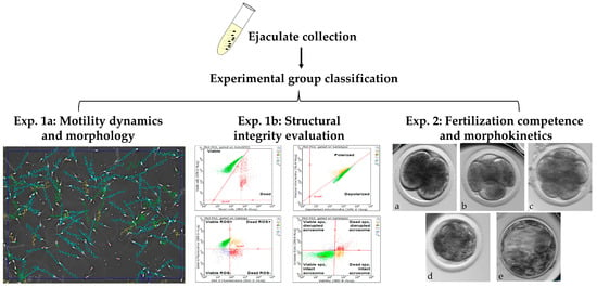
Figure 1


