


Abstract
Environmental exposures are increasingly recognized as important drivers of ocular surface inflammation, yet their combined contribution to the onset, exacerbation, and clinical burden of allergic conjunctivitis (AC) has not been comprehensively synthesized. This systematic review evaluated the evidence linking air pollutants, aeroallergens, and indoor or occupational exposures with allergic conjunctivitis. The review was conducted according to PRISMA 2020 and AMSTAR-2 guidelines and registered in PROSPERO (CRD420251162399). PubMed, Web of Science, and Scopus were searched from inception to 18 September 2025. Two independent reviewers screened studies, extracted data, and assessed methodological quality using the MINORS tool. Owing to substantial heterogeneity, findings were synthesized narratively. Twenty-nine studies were included, encompassing more than three million outpatient visits. Consistent associations were observed between particulate matter, nitrogen oxides, sulfur dioxide, carbon monoxide, and ozone with increased AC incidence and symptom severity, with variations by age, sex, and season. Pollen and air pollutants frequently acted synergistically. Indoor exposures were associated with increased risk in children, while occupational settings demonstrated exposure–response relationships. Experimental studies identified mechanisms involving epithelial barrier disruption, NF-κB activation, and thymic stromal lymphopoietin signaling. Overall, environmental exposures substantially contribute to allergic conjunctivitis and may inform improved prevention and personalized clinical management.
1. Introduction
Allergic conjunctivitis (AC) is a frequent ocular surface disease and one of the most common causes of ophthalmologic consultations worldwide. Its clinical manifestations include itching, redness, tearing, burning, photophobia, and mucous discharge, symptoms that considerably impair patients’ quality of life and productivity [1]. AC belongs to the broader group of allergic conjunctival diseases (ACDs), which also comprise perennial allergic conjunctivitis (PAC), seasonal allergic conjunctivitis (SAC), vernal keratoconjunctivitis (VKC), atopic keratoconjunctivitis (AKC), and giant papillary conjunctivitis (GPC) [2,3]. The disease spectrum ranges from mild, recurrent conditions such as SAC and PAC to severe, sight-threatening forms such as VKC and AKC, which can lead to keratoconus, cataracts, or retinal detachment [4,5].
Epidemiological evidence shows that AC prevalence is increasing worldwide. In the United States, up to 40% of the population report ocular allergic symptoms [6], with SAC and PAC affecting an estimated 15–25% of individuals [7,8,9]. In Europe, prevalence has reached nearly 50%, partly driven by the spread of highly allergenic species such as ragweed after 2009 [10]. In China, rapid industrialization and urbanization contributed to over 295 million cases of AC in 2020 [11]. Despite this substantial burden, AC remains underdiagnosed and undertreated, as many individuals perceive symptoms as normal until they receive specific treatment [12]. Comorbidity with asthma, rhinitis, and other atopic conditions is common, with ocular allergy affecting 40–80% of these subgroups [13,14].
Environmental exposures are central drivers of AC. The ocular surface is directly exposed to the atmosphere and therefore susceptible to allergens such as pollen, dust, and animal dander [15]. Ambient air pollution, recognized as a major contributor to systemic diseases including cardiovascular, respiratory, and neurological disorders, also threatens ocular health. The World Health Organization estimates that one in ten deaths in children under five years is attributable to air pollution [16]. Although the systemic consequences are well studied, ocular outcomes remain less investigated. Historically, photochemical smog in Los Angeles and Tokyo during the 1950s–1970s was already linked to “allergic eye syndromes” [17]. Since then, multiple pollutants, including particulate matter (such as PM10, PM2.5), diesel exhaust particles, nitrogen oxides (NOx), sulfur dioxide (SO2), carbon monoxide (CO), and ozone (O3), have been shown to trigger or exacerbate AC [18,19].
Pathophysiologically, AC is primarily mediated by IgE-driven hypersensitivity reactions, with mast cells and eosinophils releasing histamine, cytokines, and chemokines that sustain ocular inflammation [20]. Environmental pollutants can exacerbate these mechanisms by disrupting the epithelial barrier, prolonging allergen retention, and amplifying immune activation [6,21]. Moreover, the delayed or lagged effects of air pollutants complicates causal interpretation, as unmodeled inflammatory responses may bias results if not adequately modeled [21].
Overall, current evidence suggests that AC is not only a prevalent hypersensitivity disorder but also a sentinel condition for environmental health. However, the available literature remains fragmented across heterogeneous study designs, exposure indicators, and outcome definitions, limiting comparability and integrated interpretation. In particular, previous studies have rarely examined multiple environmental exposures jointly, their potential interactions (e.g., pollutants and pollen), or systematically linked epidemiological findings with mechanistic and experimental evidence.
To address these gaps, this systematic review was prospectively registered and conducted in accordance with PRISMA 2020 and AMSTAR-2 guidelines. We aimed to provide a comprehensive and methodologically rigorous synthesis of epidemiological, clinical, and experimental evidence on environmental determinants of allergic conjunctivitis, integrating population-level associations with mechanistic insights. By consolidating this evidence, the review seeks to inform future research priorities, support environmental risk-based prevention strategies, and contribute to more personalized clinical management and public health decision-making.
2. Materials and Methods
2.1. Research Question and PICOS Framework
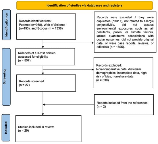
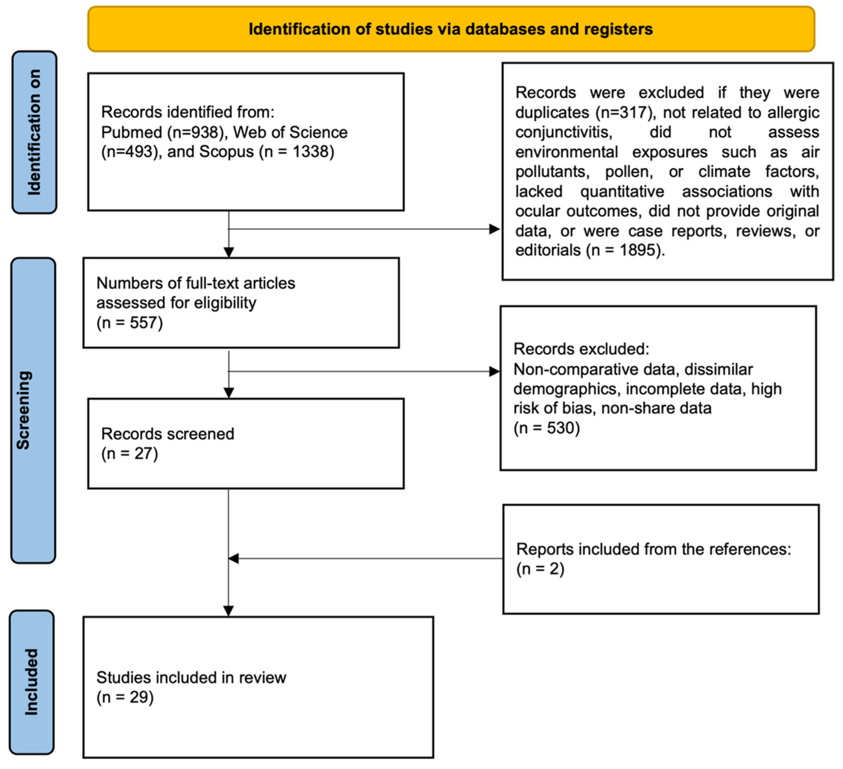
This systematic review was registered in PROSPERO (registration number: [CRD420251162399]) and conducted according to PRISMA 2020 guidelines [22] and AMSTAR-2 [23] methodological standards (Figure 1). A completed PRISMA checklist is provided as Supplementary Material (Supplementary File S1). The final literature search was completed on 18 September 2025. The research question was formulated using the PICOS framework to ensure methodological rigor and clinical relevance. Specifically, we examined whether environmental exposures, such as air pollutants, aeroallergens, meteorological variables, and occupational or indoor risk factors (Intervention/Exposure) are associated with the onset, exacerbation, or clinical burden of allergic conjunctivitis (Population), compared with unexposed groups or periods of lower exposure (Comparator). Eligible outcomes included incidence or prevalence of allergic conjunctivitis, frequency of outpatient visits, symptom severity, biomarker expression, and experimental evidence of immunological or pathophysiological mechanisms (Outcomes). All study designs providing quantitative or comparative data were considered (Study design), including ecological and case-crossover epidemiological studies, cross-sectional surveys, registry analyses, and experimental laboratory and animal models. Subgroup analyses considered exposure type (air pollution, pollen, indoor/occupational factors), geographic and demographic context, and study design as potential sources of heterogeneity. Through this comprehensive approach, the review aimed to synthesize evidence on environmental determinants of allergic conjunctivitis and to provide insights for clinical management, public health policies, and future research priorities.
Figure 1.
PRISMA flow diagram of study selection.
2.2. Eligibility Criteria
Studies were excluded if they met any of the following criteria: (i) case reports or single-patient descriptions lacking generalizable outcomes; (ii) review articles (systematic or narrative), editorials, letters, or conference abstracts without full data; (iii) duplicate publications derived from the same dataset or experimental series; or (iv) studies judged to present insufficient methodological detail or high risk of bias. Additional exclusions included reports that did not assess environmental exposures (air pollutants, aeroallergens, meteorological variables, or occupational/indoor factors), did not provide comparative or quantitative data linking exposures to allergic conjunctivitis outcomes, or lacked original data or presenting incomplete information that prevented meaningful synthesis.
2.3. Information Sources
A comprehensive and systematic literature search was conducted using three major electronic databases: PubMed, Web of Science, and Scopus, without restrictions on publication date or language. To maximize coverage across the thematic domains of allergic conjunctivitis, environmental exposures (including air pollutants, aeroallergens, meteorological variables, and occupational or indoor factors), and associated health outcomes, the reference lists of all eligible articles were also manually screened to identify additional relevant studies not captured in the initial database search.
2.4. Search Methods for Identification of Studies
The search strategy combined controlled vocabulary and free-text terms related to allergic conjunctivitis and environmental exposures. Keywords encompassed descriptors of ocular allergy (seasonal, perennial, vernal, atopic) and environmental domains (air pollution, particulate matter (PM), pollen, aeroallergens, ultraviolet (UV) radiation, climate-related factors, and general environmental exposures). Full search strategies tailored to each database are provided in Supplementary File S2. Two reviewers independently assessed study eligibility at both the title/abstract screening and full-text review stages. Discrepancies were resolved through discussion and consensus. No language restrictions were applied, and studies published in languages other than English or Portuguese were translated and included when relevant data were available.
2.5. Data Extraction and Data Items
Two authors (A.P.O. and C.M.P.) independently extracted data from all eligible studies. For each included article, key characteristics were collected, including the first author’s name, year of publication, country or institutional setting, study design (epidemiological, clinical, experimental, or animal model), type of environmental exposure assessed (air pollutants, aeroallergens, meteorological variables, occupational or indoor factors), comparator group or condition, and primary outcomes. Discrepancies in data extraction or study inclusion were resolved through discussion and consensus, without the need for a third reviewer. Record management, including duplicate removal and tracking of study eligibility, was conducted using Microsoft Excel.
Primary variables extracted included environmental exposures (e.g., PM2.5, PM10, nitrogen dioxide (NO2), SO2, O3, carbon dioxide (CO2), pollen, aeroallergens, UV radiation, indoor/occupational factors), study population characteristics (e.g., age group, sex distribution, clinical diagnosis of SAC, PAC, VKC, or AKC), and methodological approach (e.g., time-series analysis, case-crossover design, cross-sectional survey, registry analysis, biomarker assessment, experimental models). These variables were selected a priori to allow identification and qualitative exploration of potential sources of heterogeneity related to exposure indicators, outcome definitions, and study design. Additional variables, such as sample size, outcome domains (incidence or prevalence of allergic conjunctivitis, outpatient visits, symptom severity, biomarker expression, mechanistic pathways), and reported limitations or conflicts of interest (e.g., industrial or pharmaceutical funding), were also recorded to support synthesis and highlight methodological heterogeneity across studies.
2.6. Data Synthesis and Assessment of Results
Given the heterogeneity of study designs, exposures, populations, and reported outcomes, a quantitative meta-analysis was not feasible. Therefore, a narrative synthesis approach was adopted. Heterogeneity arose primarily from three sources. First, exposure indicators and metrics varied widely across studies, including differences in pollutant or allergen type, averaging periods, spatial resolution, and lag structures, which limits comparability of effect estimates. Second, outcome definitions and ascertainment were inconsistent, ranging from allergic conjunctivitis–specific clinical diagnoses to broader conjunctivitis categories, healthcare utilization outcomes, symptom-based assessments, biomarker measurements, and mechanistic endpoints. These differences reflect fundamentally distinct outcome constructs that are not directly poolable. Third, heterogeneity was inherent to study design, encompassing ecological time-series and case-crossover analyses, cross-sectional surveys, clinical investigations, and experimental animal models, each characterized by distinct confounding structures, sources of bias, and inferential scope. These sources of heterogeneity affect both effect magnitude and direction, limiting interpretability and precluding meaningful statistical pooling.
Accordingly, results from individual studies were tabulated and synthesized by study design, type of environmental exposure, and main outcome domain (incidence or prevalence of allergic conjunctivitis, outpatient visits, symptom severity, biomarker expression, or mechanistic findings). Evidence was organized thematically into epidemiological, clinical, and experimental domains to facilitate qualitative comparison across populations, exposures, and methodological approaches. No statistical pooling, effect size estimation, or formal assessment of publication bias was performed.
2.7. Risk of Bias Assessment
The methodological quality and risk of bias of the observational studies included were independently evaluated by two reviewers using the Methodological Index for Non-Randomized Studies (MINORS), originally proposed by Slim et al. [24] (Supplementary File S4). The MINORS instrument assesses essential elements of study design, including the clarity of study aims, consecutive enrolment of participants, appropriateness of eligibility criteria, objectivity of outcome measurements, and adequacy of follow-up procedures.
For comparative studies, the total MINORS score ranges from 0 to 24, with studies categorized as very low quality (0–6), low quality (7–10), moderate quality (11–15), or high quality (16–24). Due to the heterogeneity of study designs, a formal risk-of-bias assessment was performed only for observational comparative studies (cross-sectional, cohort, or case–control designs) using the MINORS tool. Ecological time-series studies, case-crossover analyses, registry-based studies, and experimental or animal studies were included in the qualitative synthesis but were not subjected to formal risk-of-bias scoring. Any discrepancies between reviewers were resolved through discussion and mutual agreement.
3. Results
3.1. Study Selection
A total of 2769 records were initially retrieved from PubMed (n = 938), Web of Science (n = 493), and Scopus (n = 1338) (Figure 1). After removal of duplicates (n = 317), 2452 records were screened based on titles and abstracts, of which 1895 were excluded due to irrelevance to allergic conjunctivitis, lack of assessment of environmental exposures (e.g., air pollutants, pollen, or climate factors), absence of quantitative associations with ocular outcomes, non-original data, or being case reports, reviews, or editorials. Subsequently, 557 full-text articles were assessed for eligibility, and 530 were excluded due to non-comparative data, differing population characteristics, incomplete information, high risk of bias, or unavailability of shared data. In addition, two relevant studies were identified through manual reference screening. Ultimately, 29 studies met the inclusion criteria and were included in the qualitative synthesis [6,25,26,27,28,29,30,31,32,33,34,35,36,37,38,39,40,41,42,43,44,45,46,47,48,49,50,51,52].
3.2. Study Characteristics
Supplementary File S4 summarizes the key characteristics of the 29 studies included in this synthesis, which investigated the relationship between environmental exposures and allergic conjunctivitis across diverse contexts. Investigated exposures included air pollutants (PM2.5, PM10, NO2, SO2, O3, CO), pollen and other aeroallergens, occupational exposures (e.g., hazelnut and pear orchards), and indoor or lifestyle-related factors. Study designs ranged from large-scale epidemiological time-series analyses and population-based surveys to clinical investigations, laboratory-based biomarker assessments, and experimental animal models.
Research objectives were highly variable, encompassing short-term effects of ambient air pollution and pollen interactions on outpatient visits and symptom exacerbations, to mechanistic studies of PM–induced ocular allergy in murine models, and the identification of molecular biomarkers such as eotaxin-1, NF-κB activation, or immune-related long noncoding RNAs. Several studies also investigated geographical and demographic determinants of allergic conjunctivitis, highlighting vulnerable subgroups such as children, elderly individuals, males or females depending on pollutant type, and occupationally exposed populations.
Methodological approaches included ecological and case-crossover studies linking air quality and pollen data to health records, cross-sectional epidemiological surveys using questionnaires and clinical diagnoses, retrospective registry analyses covering millions of outpatient visits, and in vivo or in vitro experiments evaluating immunological pathways, cytokine responses, and ocular surface integrity. Both clinical and preclinical studies assessed seasonal allergic conjunctivitis as well as more severe forms, such as VKC and AKC, employing conjunctival biopsies, provocation tests, and tear biomarker assays.
Regarding analytical strategies, ten of the 29 included studies applied multipollutant or mixed-pollutant models, typically adjusting for co-pollutants such as PM2.5, PM10, NO2, O3, SO2, or CO within the same regression framework. This represents approximately one third of the included evidence. In contrast, the remaining studies relied on single-pollutant models, descriptive exposure assessments, or experimental designs. This imbalance in analytical approaches contributed to the methodological heterogeneity observed across the evidence base and should be considered when comparing results across studies.
Environmental factors investigated spanned traffic-related pollutants, biomass and occupational exposures, seasonal and geographical variations in pollen load, and emerging toxicants such as chlorinated paraffins. Reported outcomes included increased incidence of allergic conjunctivitis incidence, symptom exacerbations during pollution peaks, synergistic effects between pollen and pollutants, mechanistic insights into macrophage polarization and Th2-driven inflammation, and evidence supporting the effectiveness of air quality interventions.
Collectively, these studies underscore the multifactorial nature of allergic conjunctivitis in relation to environmental exposures. Large-scale epidemiological evidence indicates that high-level or combined environmental exposures are associated with measurable increases in disease burden; for example, a multicity study reported an approximately 8% increase in allergic conjunctivitis outpatient visits per interquartile increase in ambient ozone concentrations [50]. Proposed implications emphasized the importance of mitigating environmental risks through pollution control, allergen-conscious urban planning, protective behavioral strategies (e.g., eyewear, limiting outdoor exposure), and identification of novel therapeutic targets, highlighting both population-level risks and mechanistic pathways relevant to prevention and management.
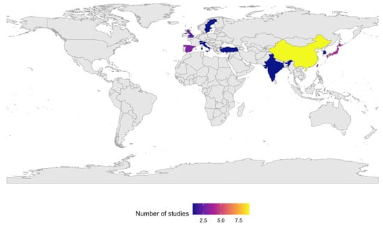
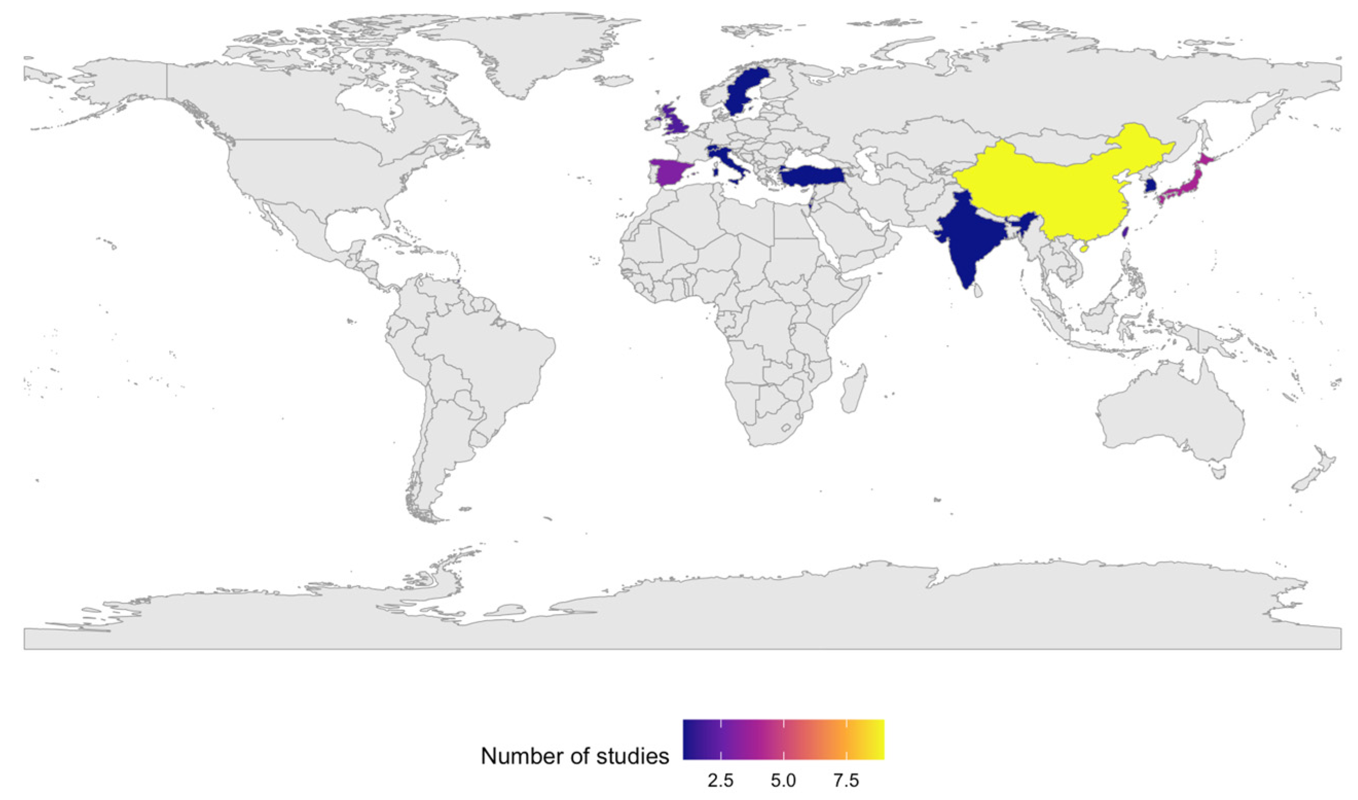
Overall, the 29 included studies spanned multiple cities and regions across several continents, including Asia, Europe, North America, South America, and the Caribbean. The evidence base comprised studies conducted in China, Japan, Taiwan, South Korea, India, Israel, Spain, the United Kingdom, Switzerland, Sweden, Italy, Turkey, the United States, Peru, and Trinidad and Tobago (Figure 2). Collectively, these studies encompassed more than three million cumulative individuals or outpatient visits across heterogeneous study designs, with publication dates ranging from 1997 to 2025.
Figure 2.
Geographic distribution of the included studies.
The main characteristics and methodological quality of the included studies are summarized in Table 1.
Table 1.
Characteristics of the included studies.
3.3. Outcomes
3.3.1. Environmental Factors and Exposures
Environmental exposures are central drivers of ocular allergy, especially AC. Epidemiological, clinical, and experimental evidence demonstrates that pollen, air pollution, indoor allergens, and occupational factors can act both independently and synergistically to increase disease burden. The 18 studies included in this review range from small occupational cohorts to national surveys and multicity time-series analyses, collectively demonstrating that AC as a sensitive indicator of environmental health challenges.
Occupational and aerobiological studies provide early and direct evidence of strong exposure–response relationships. Akcay Usta & Icoz [25] described ocular surface disease in hazelnut harvesters, linking pollen exposure to systemic inflammation and suggesting the value of blood biomarkers. Yanagisawa et al. [51] reported SAC in pear farmers exposed during artificial pollination, confirmed by eosinophil counts and IgE sensitization, with resolution after cromoglycate therapy. Calderón [28] identified Tipuana tipu pollen as a novel allergen, highlighting how local aerobiology can uncover new risks and inform urban greening policies. Chico-Fernández & Ayuga-Téllez [30,31] further demonstrated that common tree pollens and air pollutants interact synergistically to trigger conjunctivitis and rhinitis, emphasizing the need to integrate allergenicity into urban planning.
Large population-based time-series and registry studies provide quantitative evidence of age-related vulnerability to environmental exposures, although effect estimates are reported heterogeneously and cannot be directly pooled. In a nationwide registry study including over 3.2 million allergic conjunctivitis outpatient visits, Hong et al. [6] reported that individuals younger than 40 years were susceptible to multiple environmental factors (including NO2, O3, temperature, and humidity), whereas those aged 40 years and older showed more consistent associations primarily with NO2 exposure. Similarly, in a large time-series study from Urumqi (59,731 visits), Gui et al. [34] reported age-stratified relative risks, with significant associations for specific pollutants observed predominantly in young children and older adults, while intermediate adult age groups showed weaker or pollutant-specific effects. Although these age-specific estimates could not be pooled due to methodological and exposure heterogeneity, they consistently indicate greater relative vulnerability at the extremes of age. Liu et al. [39] showed lagged effects of particulate material and NO2, reinforcing temporal causality. Multicity studies from China Lu et al. [40] reported that even modest increases in PM2.5, PM10, NO2, SO2, and O3 were associated with elevated conjunctivitis risk, while Qiu et al. [50] focused on ozone, showing an 8% rise in AC visits per interquartile increase. These multicity studies confirm that associations between air pollutants and allergic conjunctivitis are robust and generalizable.
Japanese research refines the seasonal and mechanistic context. Mimura et al. [42] found PM2.5 associated with AC visits outside the pollen season, suggesting pollutant effects are more evident when allergen load is low. Mimura et al. [43] reported SPM and wind speed as the strongest predictors of AC across the year. Miyazaki et al. [44] provided prevalence estimates for SAC (45.4%), PAC (14.0%), AKC (5.3%), and VKC (1.2%), and linked traffic-related pollutants (NO2, NOx, PM10) with more severe keratoconjunctivitis forms, highlighting the environmental contribution to disease severity.
Regional and international studies show how local contexts shape ocular allergy burden. Ezinne et al. [33] found a prevalence above 40% in young adults, with associations to pollen, mites, cigarette smoke, and comorbid asthma and rhinitis, compounded by Saharan dust. Phiri et al. [46] (2025, Taiwan) identified indoor risk factors (furniture, carpets, mold, Der f 1) and outdoor PM10 as drivers of AC in preschool children, particularly boys. Gupta et al. [35] reported VKC prevalence above 1% in over 8000 children, with sunlight, dust, incense smoke, and seasonality as major triggers, and geographic variation with highest burden in the northern plains. These findings show how climate, culture, and housing shape exposure risks alongside pollutants.
Emerging contaminants expand the spectrum of environmental threats. Huang et al. [36] linked PM2.5-bound chlorinated paraffins (CPs) to allergic outcomes in over 130,000 children, with risks stronger in overweight individuals. Chlorinated paraffins are high-production-volume industrial chemicals increasingly detected in ambient air, household dust, and human biomonitoring samples, particularly in rapidly industrializing regions, raising concern about rising chronic low-level exposure in pediatric populations. These findings suggest new challenges for environmental ophthalmology beyond classical combustion-related pollutants.
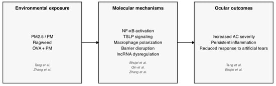
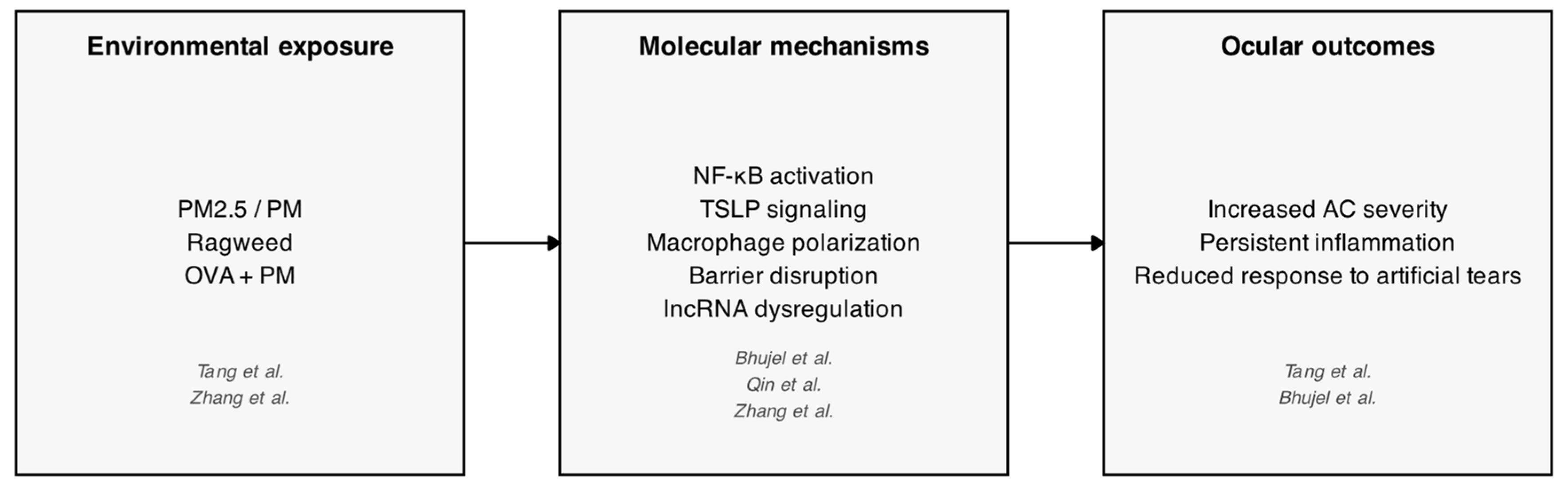
Experimental models provide mechanistic evidence. Bhujel et al. showed that PM exposure worsens ovalbumin (OVA)-induced allergic eye disease in mice through NF-κB activation and barrier disruption. Tang et al. reproduced PM2.5-induced AC, with symptoms resistant to artificial tears. Qin et al. identified thymic stromal lymphopoietin (TSLP) signaling and macrophage polarization as key mediators of PM2.5-induced inflammation, while Zhang et al. profiled lncRNAs in ragweed-induced AC, suggesting novel immune regulators. Figure 3 provides a schematic synthesis of the exposure–mechanism–outcome pathways identified across experimental models.
Figure 3.
Mechanistic pathways linking environmental exposures to allergic conjunctivitis in experimental models [27,48,49,52].
Several consistent themes emerge across studies. Pollen and pollutants act synergistically, with O3, NO2, and PM amplifying allergic responses. Meteorological conditions, such as temperature and wind, modulate these effects, often intensifying risk during warm seasons. Vulnerable populations groups (including children, the elderly, and individuals with asthma or eczema) are disproportionately affected, with some sex-specific sensitivities (e.g., women to CO, men to NO2). Geographic context also influences risk: rural Indian children experience high burden of VKC, Caribbean populations face dust exposure and limited awareness, and East Asian cities are characterized by traffic-related air pollution–driven risk. Emerging environmental exposures, such as chlorinated paraffins, further expand concern to industrial chemicals.
Collectively, these findings frame AC as more than a localized hypersensitivity. Its high prevalence, clear environmental triggers, and measurable burden on the healthcare system establish AC as a sentinel condition for environmental health. Public health strategies should include stricter air quality regulations, allergen-conscious urban planting, improved housing standards, and targeted protection for vulnerable populations. Clinicians are encouraged to integrate environmental histories into routine diagnosis and management, while researchers should prioritize multipollutant exposure models and explore molecular mediators as potential therapeutic targets.
3.3.2. Clinical Manifestations and Disease Burden
Population-based surveys and case-crossover analyses provide complementary insights into the epidemiology of ocular allergy and its environmental drivers. Singh et al. [47] analyzed data from NHANES III (1988–1994), showing that up to 40% of the U.S. population reported ocular allergy symptoms at least once over six years, the highest prevalence reported at that time. Ocular symptoms were more frequent than nasal ones in relation to pollen, animals, and dust, and older adults (>50 years) reported isolated ocular symptoms more often, whereas younger individuals experienced more combined ocular and nasal complaints. Skin prick testing confirmed strong associations with weed and pollen allergens, reinforcing the central role of aeroallergens in ocular allergy. This large dataset also underscored demographic and regional differences, with higher prevalence in the South and seasonal peaks in June–July.
Leonardi et al. [37] conducted one of the largest national surveys of ocular allergy in Europe, enrolling 3545 patients across 304 ophthalmology centers. They reported that SAC, PAC, VKC, and AKC together impose a substantial burden, yet allergy testing was performed in only 35% of cases, highlighting a diagnostic gap. The study revealed a mismatch between clinical practice and guidelines, with underuse of targeted therapies and overreliance on nonspecific medications. Importantly, it provided a detailed picture of triggers (pollen, dust, smoke, pollution) and comorbidities, offering a benchmark for clinical management across Europe.
Levanon et al. [38] advanced this evidence by linking VKC exacerbations to meteorological and air pollution exposures in a large population-based cohort of 6024 patients in southern Israel. Using a case-crossover design, they found that NO2, O3, and PM10 were strongly associated with exacerbations (ORs 1.9–2.3), alongside contributions from PM2.5, temperature, and solar radiation. Stratified analyses showed greater vulnerability in Bedouin children and socioeconomic subgroups, reflecting both biological and social determinants. These findings confirm that VKC flares are not solely seasonal phenomena but environmentally mediated events, sensitive to both pollutants and climatic extremes in semi-arid regions.
Together, these three studies illustrate the multilevel nature of ocular allergy research: national prevalence estimates defining scope (USA), large-scale clinical surveys exposing practice gaps (Italy), and environmental epidemiology pinpointing modifiable triggers (Israel). They converge on the conclusion that ocular allergy, including severe forms such as VKC, is highly prevalent, frequently underdiagnosed, and strongly influenced by both environmental exposures and healthcare practices.
3.3.3. Pathophysiological Mechanisms and Biomarkers
Experimental and clinical studies have provided key evidence on the immunological mechanisms and environmental factors underlying AC. In one of the earliest histological investigations, Anderson et al. [26] performed conjunctival biopsies in patients with seasonal allergic conjunctivitis (SAC), demonstrating that the disease is fundamentally mast cell–dependent and, unlike VKC and AKC, lacks extensive granulocyte infiltration. MacLeod et al. [41] expanded on this, showing that mast cells are not only effector cells but also as cytokine reservoirs, with IL-4 release playing a central role in amplifying ocular inflammation. Subsequently, Eperon et al. [32] reinforced these clinical findings by identifying eotaxin-1 as a biomarker of SAC, highlighting its role in eosinophil recruitment via the C-C chemokine type 3 (CCR3) receptor. Complementing these studies, Nivenius et al. [45] conducted conjunctival provocation tests (CPT) in patients with AKC and SAC, observing both early and late responses, which confirmed the chronic involvement of IgE alongside T cell– and cytokine-mediated pathways.
Animal models have been particularly useful in exploring the interaction between environmental pollutants and allergic responses. Bhujel et al. [27], using a murine model of allergic eye disease (AED), demonstrated that co-exposure to PM and OVA enhances allergic inflammation through NF-κB activation, disrupting epithelial integrity and exacerbating systemic responses. Qin et al. [49] expanded these findings by establishing a reproducible PM2.5-induced AC model in BALB/c mice, identifying macrophage polarization toward the M1 phenotype and emphasizing TSLP as a central mediator of the inflammatory cascade. Similarly, Tang et al. [48] developed an acute murine AC model by direct PM2.5 instillation over 18 days, reproducing clinical signs such as eyelid edema, hyperemia, and tearing, along with conjunctival eosinophilic infiltration and goblet cell hyperplasia. This provided solid experimental proof that fine particles alone can induce a clinical phenotype analogous to human AC. Complementarily, Zhang et al. [52] applied a transcriptomic approach to a murine model of AC induced by ragweed pollen, identifying significant differences in the expression of mRNAs and lncRNAs associated with mucosal immunity. Their findings revealed the involvement of molecules such as Bpifa1 and Reg3g, as well as activation of inflammatory pathways including mitogen-activated protein kinase (MAPK) and signal transducer and activator of transcription (STAT), pointing to novel potential therapeutic targets for ocular allergy management.
Taken together, these eight studies span from the histological and molecular characterization of AC in humans to animal models that clarify the effects of environmental particles and specific allergens. The evidence converges on the central role of mast cells and eosinophils in SAC, the relevance of biomarkers such as eotaxin-1 and TSLP, and the capacity of PM2.5 to act both as an adjuvant and as a direct trigger of conjunctival inflammation. Furthermore, the integration of transcriptomics and conjunctival provocation models provides a more refined understanding of mucosal immunity, underscoring the need for therapeutic approaches aimed not only at symptomatic control but also at modulation of specific immune pathways.
4. Discussion
This systematic review highlights that environmental exposures, including air pollutants, aeroallergens, and indoor or occupational factors, are key drivers of AC. Across 29 studies, consistent associations were observed between PM, NOx, O3, and pollen peaks with increased incidence and severity of ocular allergy. Mechanistic investigations further confirmed that these exposures amplify IgE-mediated inflammation by disrupting the epithelial barrier, prolonging allergen retention, and triggering cytokine cascades. Together, these findings reinforce the notion that AC is not only a prevalent hypersensitivity disorder but also a sentinel condition reflecting the broader health impacts of environmental change.
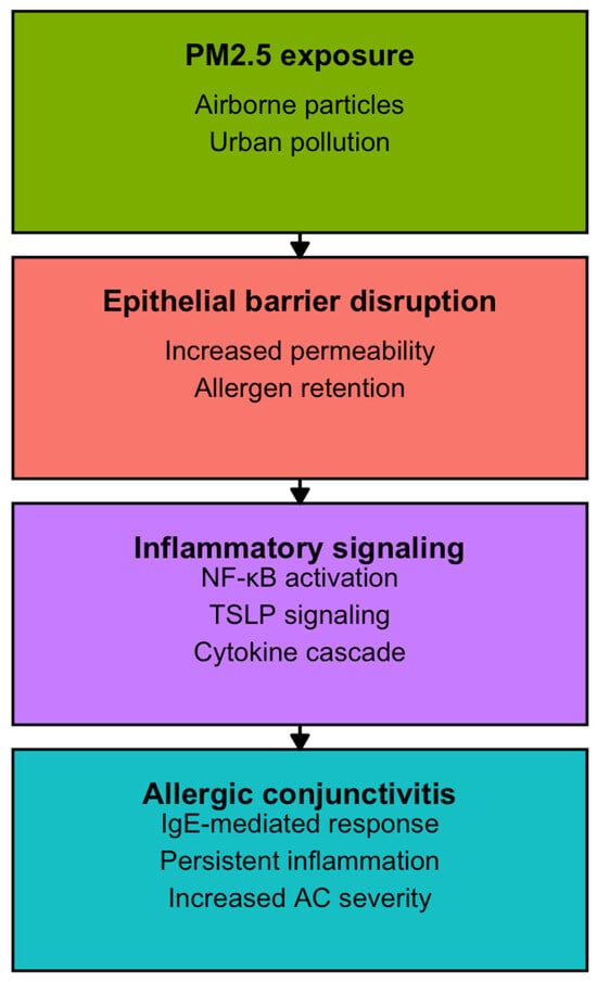
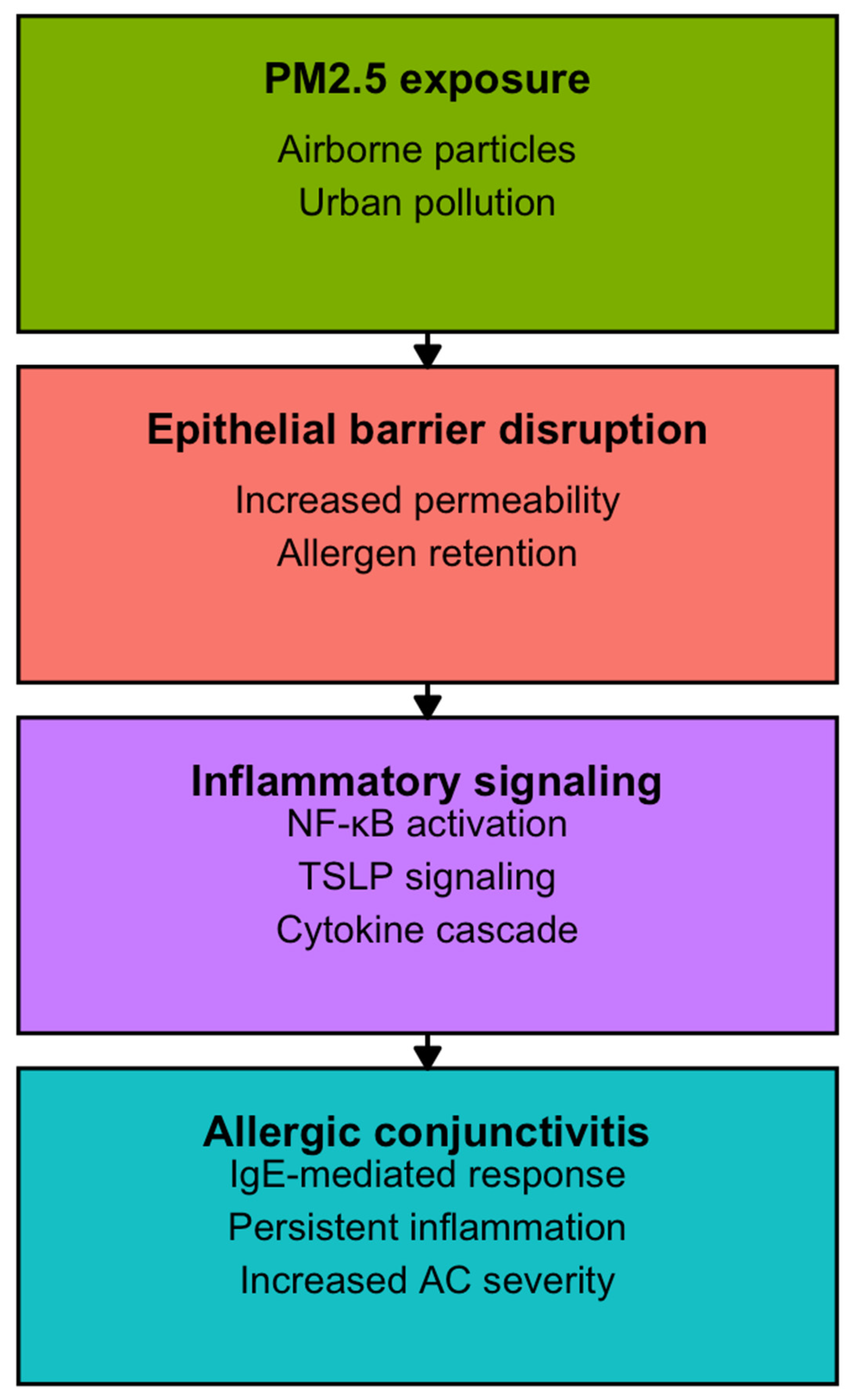
Our synthesis aligns with the report by Epstein et al. [53], which highlighted how climate change is reshaping aerobiology through longer pollen seasons, increasing allergenicity, and altered species distribution. While Epstein’s work was primarily predictive and focused on North America and Europe, our review consolidates empirical evidence from Asia, Africa, and Latin America, showing that these risks are already manifesting globally. Real-world support comes from registry analyses such as Hong et al. [6], which examined more than 3.2 million visits and identified NO2, O3, and temperature as consistent predictors, and by Gui et al. [34], which revealed pollutant-specific vulnerabilities by age and sex, corroborating the mechanisms anticipated by Epstein and colleagues. To integrate these epidemiological and experimental findings, Figure 4 provides a conceptual overview of the proposed pathways through which PM2.5 exposure disrupts epithelial barrier integrity, facilitates allergen penetration, and activates inflammatory signaling cascades, including NF-κB and TSLP. This framework links population-level associations with mechanistic evidence, illustrating how environmental exposures translate into amplified IgE-mediated ocular inflammation.
Figure 4.
Conceptual pathways linking PM2.5 exposure to allergic conjunctivitis.
Mechanistic evidence strengthens these associations. Sedghy et al. [54] reviewed how diesel exhaust and ozone can increase pollen allergenicity by enhancing protein release and modifying epitopes, a phenomenon supported by studies from Chico-Fernández and Ayuga-Téllez [30,31], which demonstrated synergistic effects of tree pollens and air pollutants in urban environments. Across these studies, pollutants are consistently recognized as adjuvants that potentiate allergic responses; the main distinction lies in the perspective: molecular studies describe how allergenic potency is modified, whereas epidemiological work documents how such changes translate into population-level disease burden. Our findings also align with the mechanistic review by Upaphong et al. [55], which showed that PM induces oxidative stress, DNA damage, and mitochondrial dysfunction in ocular surface cells. This toxicological evidence is consistent with the delayed increases in AC after pollution peaks reported by Liu et al. [39] and the seasonal patterns observed by Mimura et al. [42,43], indicating that PM can trigger conjunctivitis even outside pollen season. Across these studies, pollutants are identified as direct ocular hazards; differences arise in exposure scale, with in vitro experiments examining controlled doses and composition, while population studies reflect heterogeneous exposures shaped by geography and meteorology.
From a clinical perspective, our synthesis echoes the observations of Ackerman et al. [56], who emphasized that AC is frequently underdiagnosed and undertreated despite its high prevalence. This underrecognition was also evident in a European survey by Leonardi et al. [37], where only 35% of patients underwent allergy testing despite substantial clinical burden. Both studies highlight the gap between prevalence and diagnosis, although Ackerman’s conclusions were based on practice surveys and guideline analysis, whereas Leonardi provided multicenter quantitative data.
The role of biomarkers further illustrates the bridge between mechanistic and clinical research. Shoji [57] demonstrated the diagnostic utility of tear and ocular surface biomarkers, such as eotaxin-2 and periostin, while our review identified upregulation of eotaxin-1, NF-κB, and TSLP in response to environmental exposures. Both strands emphasize the relevance of molecular signatures, yet their application differs: Shoji proposed standardized use in clinical diagnosis, whereas most studies in our synthesis treated biomarker assessment as exploratory. In this context, emerging biosensing technologies may play a key role in translating molecular biomarkers into clinical practice by enabling sensitive, rapid, and clinically adaptable detection of immunological and inflammatory signals. Recent advances in optically active two-dimensional nanomaterials, such as MoS2-based biosensing platforms, have demonstrated high sensitivity and versatility for biomolecular detection, supporting their potential applicability in future ophthalmic and immunological biomarker assessment and strengthening the translational link between mechanistic research and clinical evaluation [58]. This gap underscores the need to translate experimental findings into routine practice.
Animal models also provide compelling evidence of causality. Groneberg et al. [59] showed that OVA and ragweed models clarified mast cell–driven hypersensitivity, while more recent work by Bhujel et al. [27] demonstrated that PM exacerbates allergic eye disease through mast cell degranulation, cytokine release, and conjunctival apoptosis. These findings are consistent with those of Qin et al. [49] and Tang et al. [48], who reproduced PM2.5-induced AC in mice and highlighted the role of TSLP signaling, macrophage polarization, and goblet cell hyperplasia. Over two decades of research, earlier models emphasized allergens as primary triggers, whereas recent experiments establish pollutants as independent pathogenic agents, bridging mechanistic insights with clinical relevance.
Despite broad agreement, notable discrepancies remain. Large registry studies consistently implicate NO2, O3, and PM as major contributors to AC, yet their effects vary across demographic groups. For example, Gui et al. [34] identified stronger associations in women, infants, and the elderly, while Levanon et al. [38] reported heightened vulnerability among Bedouin children and socioeconomically disadvantaged populations in Israel. Biomarker responses are similarly heterogeneous, with some studies demonstrating marked upregulation of eotaxin or TSLP, while others report modest changes. Such differences likely reflect variations in pollutant composition, geographical context, and methodological approaches, reinforcing the need for standardized exposure and outcome measures. Nevertheless, several themes emerge consistently. Pollen and pollutants act synergistically to amplify ocular allergy, meteorological conditions such as temperature and wind often exacerbate risk, and vulnerable groups, including children, the elderly, and patients with asthma or eczema, are disproportionately affected. Emerging contaminants, such as chlorinated paraffins [36], expand concern to industrial chemicals, highlighting environmental threats beyond classical combustion products.
Several important limitations of the available evidence must be acknowledged. The marked heterogeneity observed across studies (related to differences in exposure assessment, outcome definitions and ascertainment, and study design) limited direct comparability and precluded quantitative synthesis. This heterogeneity encompassed the use of diverse environmental indicators (e.g., PM2.5, PM10, NO2, O3, pollen), exposure metrics and lag structures, as well as variability in outcome assessment, ranging from allergic conjunctivitis–specific diagnoses to broader conjunctivitis categories and from healthcare utilization outcomes to symptom-based, clinical, or biomarker measures. In addition, heterogeneity inherent to study design, including ecological time-series and case-crossover analyses, cross-sectional surveys, clinical investigations, and experimental animal models, introduced distinct confounding structures and potential sources of bias that should be considered when interpreting the findings. Many investigations relied on ecological or city-level exposure estimates, which increase the risk of exposure misclassification and restrict the ability to account for individual-level confounders such as socioeconomic status, pre-existing atopy, or access to care. Second, most studies were conducted in East Asia and Europe. Although a small number of studies originated from Latin American or Caribbean settings and other tropical or subtropical regions, these were limited in number and scope and did not provide systematic or regionally representative evidence. No eligible studies were identified from Africa. Overall, tropical and low- and middle-income regions remain underrepresented. This geographic imbalance likely reflects gaps in research coverage and data availability rather than selective inclusion and limits the global generalizability of the findings. Third, although real-world exposures occur as complex mixtures, only a minority of studies adopted multipollutant models, and few applied advanced mixture methodologies despite strong collinearity among pollutants. In addition, although multiple clinical subtypes of allergic conjunctivitis (SAC, PAC, VKC, and AKC) were included, structured comparisons across subtypes were not feasible, as most studies focused on a single subtype or did not report outcomes stratified by clinical phenotype. Finally, longitudinal biomarker studies and subtype-specific analyses (e.g., SAC/PAC vs. VKC/AKC) remain scarce, limiting our understanding of temporal relationships between exposures, molecular pathways, and clinical manifestations.
On the other hand, most experimental animal models included in this review employed short-term and relatively high-dose PM2.5 exposure protocols to induce allergic conjunctivitis. While these models are valuable for elucidating mechanistic pathways and establishing causality, they do not fully recapitulate the chronic, low-dose, and cumulative exposure patterns characteristic of real-world human environmental exposure. Consequently, their translational relevance for long-term disease development, adaptation processes, and exposure–response relationships may be limited. Future experimental studies should prioritize chronic and repeated low-dose exposure paradigms to better reflect environmentally relevant conditions and improve alignment with epidemiological evidence. A key strength of this review lies in its integrative scope, combining epidemiological, clinical, and experimental evidence to provide a comprehensive overview of environmental determinants of allergic conjunctivitis. By synthesizing findings across multiple exposure domains, including air pollutants, aeroallergens, meteorological variables, and indoor or occupational factors, this work moves beyond isolated exposure–outcome associations and offers a unified framework linking environmental change to ocular allergic disease. The prospective registration and adherence to PRISMA and AMSTAR-2 standards further enhance methodological transparency and reproducibility.
Looking forward, several priorities emerge for advancing the field. Future studies should promote standardized outcome definitions and harmonized exposure assessment protocols to facilitate comparability across multi-center and multi-country cohorts. Longitudinal designs integrating detailed clinical phenotyping with molecular and immunological biomarkers are particularly needed to validate mechanistic pathways such as epithelial barrier dysfunction and TSLP signaling in human biospecimens, thereby strengthening translational relevance. From an analytical perspective, the adoption of multipollutant and mixture-based modeling approaches will be essential to quantify interaction effects among pollutants, aeroallergens, and meteorological factors under real-world conditions. Expanding research on emerging contaminants, including industrial chemicals, is also warranted given evolving exposure profiles. Finally, interventional and implementation studies evaluating the real-world effectiveness of preventive strategies, such as air filtration, exposure reduction policies, and protective eyewear, will be critical for translating mechanistic and epidemiological evidence into clinical practice and public health action.
5. Conclusions
Allergic conjunctivitis represents the intersection between environmental exposures and ocular immune responses, functioning not only as a prevalent clinical condition but also as a sensitive indicator of environmental health. By integrating epidemiological and mechanistic evidence, this review highlights the relevance of environmental drivers in shaping ocular allergic disease and underscores the need for coordinated clinical and public health strategies to mitigate environmentally driven risk.
Supplementary Materials
The following supporting information can be downloaded at: https://www.mdpi.com/article/10.3390/life16020271/s1, Supplementary File S1: PRISMA 2020 checklist; Supplementary File S2: Full electronic database search strategies (PubMed, Web of Science, Scopus); Supplementary File S3: Assessment of the quality of studies through Methodological Index for Non-Randomized Studies (MINORS); Supplementary File S4: Baseline characteristics of the 29 included studies.
Author Contributions
Conceptualization, C.M.-P. and A.P.O.; methodology, C.M.-P. and A.P.O.; software, C.M.-P. and A.P.O.; validation, C.M.-P. and A.P.O.; formal analysis, C.M.-P. and A.P.O.; investigation, C.M.-P. and A.P.O.; resources, C.M.-P. and A.P.O.; data curation, C.M.-P. and A.P.O.; writing—original draft preparation, C.M.-P. and A.P.O.; writing—review and editing, C.M.-P. and A.P.O.; visualization, C.M.-P. and A.P.O.; supervision, C.M.-P. and A.P.O.; project administration, C.M.-P. and A.P.O.; funding acquisition, C.M.-P. and A.P.O. All authors have read and agreed to the published version of the manuscript.
Funding
This research received no external funding.
Institutional Review Board Statement
Not applicable.
Informed Consent Statement
Not applicable.
Data Availability Statement
The original contributions presented in this study are included in the article/Supplementary Materials. Further inquiries can be directed to the corresponding author.
Conflicts of Interest
The authors declare no conflicts of interest.
References
- Cheng, P.; Liu, C.; Tu, B.; Zhang, X.; Chen, F.; Xu, J.; Qian, D.; Wang, X.; Zhou, W. Short-term effects of ambient ozone on the risk of conjunctivitis outpatient visits: A time-series analysis in Pudong New Area, Shanghai. Int. J. Environ. Health Res. 2023, 33, 348–357. [Google Scholar] [CrossRef] [Scilit]
- Takamura, E.; Uchio, E.; Ebihara, N.; Ohno, S.; Ohashi, Y.; Okamoto, S.; Kumagai, N.; Satake, Y.; Shoji, J.; Nakagawa, Y.; et al. Japanese guidelines for allergic conjunctival diseases 2017. Allergol. Int. 2017, 66, 220–229. [Google Scholar] [CrossRef] [Scilit]
- Kausar, A.; Akhtar, N.; Akbar, N. Epidemiological aspects of allergic conjunctivitis. Pak. J. Med. Health Sci. 2022, 34, 135–140. [Google Scholar] [CrossRef] [Scilit]
- Singhal, D.; Sahay, P.; Maharana, P.K.; Raj, N.; Sharma, N.; Titiyal, J.S. Vernal keratoconjunctivitis. Surv. Ophthalmol. 2019, 64, 289–311. [Google Scholar] [CrossRef] [Scilit]
- Tuft, S.J.; Kemeny, D.M.; Dart, J.K.G.; Buckley, R.J. Clinical features of atopic keratoconjunctivitis. Ophthalmology 1991, 98, 150–158. [Google Scholar] [CrossRef] [Scilit] [PubMed]
- Hong, J.; Zhong, T.; Li, H.; Xu, J.; Ye, X.; Mu, Z.; Lu, Y.; Mashaghi, A.; Zhou, Y.; Tan, M. Ambient air pollution, weather changes, and outpatient visits for allergic conjunctivitis: A retrospective registry study. Sci. Rep. 2016, 6, 23858. [Google Scholar] [CrossRef] [Scilit] [PubMed]
- Ono, S.J.; Abelson, M.B. Allergic conjunctivitis: Update on pathophysiology and prospects for future treatment. J. Allergy Clin. Immunol. 2005, 115, 118–122. [Google Scholar] [CrossRef] [Scilit] [PubMed]
- O’Brien, T.P. Allergic conjunctivitis: An update on diagnosis and management. Curr. Opin. Allergy Clin. Immunol. 2013, 13, 543–549. [Google Scholar] [CrossRef] [Scilit]
- Miraldi Utz, V.; Kaufman, A.R. Allergic eye disease. Pediatr. Clin. N. Am. 2014, 61, 607–620. [Google Scholar] [CrossRef] [Scilit]
- Burbach, G.J.; Heinzerling, L.M.; Röhnelt, C.; Bergmann, K.C.; Behrendt, H.; Zuberbier, T.; GA(2)LEN Study Group. Ragweed sensitization in Europe—GA2LEN study suggests increasing prevalence. Allergy 2009, 64, 664–665. [Google Scholar] [CrossRef] [Scilit]
- Azari, A.A.; Barney, N.P. Conjunctivitis: A systematic review of diagnosis and treatment. JAMA 2013, 310, 1721–1730. [Google Scholar] [CrossRef] [Scilit]
- Williams, D.C.; Edney, G.; Maiden, B.; Smith, P.K. Recognition of allergic conjunctivitis in patients with allergic rhinitis. World Allergy Organ. J. 2013, 6, 1–3. [Google Scholar] [CrossRef] [Scilit]
- Petricek, I.; Prost, M.; Popova, A. The differential diagnosis of red eye: A survey of medical practitioners from Eastern Europe and the Middle East. Ophthalmologica 2006, 220, 229–237. [Google Scholar] [CrossRef] [Scilit]
- Gomes, P.J. Trends in prevalence and treatment of ocular allergy. Curr. Opin. Allergy Clin. Immunol. 2014, 14, 451–456. [Google Scholar] [CrossRef] [Scilit]
- Helintö, M.; Renkonen, R.; Tervo, T.; Vesaluoma, M.; Saaren-Seppälä, H.; Haahtela, T.; Kirveskari, J. Direct in vivo monitoring of acute allergic reactions in human conjunctiva. J. Immunol. 2004, 172, 3235–3242. [Google Scholar] [CrossRef] [Scilit] [PubMed]
- World Health Organization. More than 90% of the World’s Children Breathe Toxic Air Every Day; WHO: Geneva, Switzerland, 2018.
- Gupta, X.P.D.; Muthukumar, A. Minor to chronic eye disorders due to environmental pollution: A review. J. Ocul. Infect. Inflamm. 2018, 2, 108. [Google Scholar]
- Kim, K. Influences of environmental chemicals on atopic dermatitis. Toxicol. Res. 2015, 31, 89–96. [Google Scholar] [CrossRef] [Scilit] [PubMed]
- Guarnieri, M.; Balmes, J.R. Outdoor air pollution and asthma. Lancet 2014, 383, 1581–1592. [Google Scholar] [CrossRef] [Scilit]
- Leonardi, A. Allergy and allergic mediators in tears. Exp. Eye Res. 2013, 117, 106–117. [Google Scholar] [CrossRef] [Scilit]
- Lu, C.-W.; Fu, J.; Liu, X.-F.; Chen, W.-W.; Hao, J.-L.; Li, X.-L.; Pant, O.P. Air pollution and meteorological conditions significantly contribute to the worsening of allergic conjunctivitis: A regional 20-city, 5-year study in Northeast China. Light Sci. Appl. 2021, 10, 190. [Google Scholar] [CrossRef] [Scilit]
- Page, M.J.; McKenzie, J.E.; Bossuyt, P.M.; Boutron, I.; Hoffmann, T.C.; Mulrow, C.D.; Shamseer, L.; Tetzlaff, J.M.; Akl, E.A.; Brennan, S.; et al. The PRISMA 2020 statement: An updated guideline for reporting systematic reviews. BMJ 2021, 372, n71. [Google Scholar] [CrossRef] [Scilit]
- Shea, B.J.; Reeves, B.C.; Wells, G.; Thuku, M.; Hamel, C.; Moran, J.; Moher, D.; Tugwell, P.; Welch, V.; Kristjansson, E.; et al. AMSTAR 2: A critical appraisal tool for systematic reviews. BMJ 2017, 358, j4008. [Google Scholar] [CrossRef] [Scilit]
- Slim, K.; Nini, E.; Forestier, D.; Kwiatkowski, F.; Panis, Y.; Chipponi, J. Methodological index for non-randomized studies (MINORS): Development and validation. ANZ J. Surg. 2003, 73, 712–716. [Google Scholar] [CrossRef] [Scilit] [PubMed]
- Akcay Usta, S.; Icoz, M. Evaluation of ocular surface parameters and systemic inflammatory biomarkers in hazelnut harvesters. Ocul. Immunol. Inflamm. 2024, 32, 2153–2158. [Google Scholar] [CrossRef] [Scilit]
- Anderson, D.F.; MacLeod, J.D.; Baddeley, S.M.; Bacon, A.S.; McGill, J.I.; Holgate, S.T.; Roche, W.R. Seasonal allergic conjunctivitis is accompanied by increased mast cell numbers in the absence of leucocyte infiltration. Clin. Exp. Allergy 1997, 27, 1060–1066. [Google Scholar] [CrossRef] [Scilit]
- Bhujel, B.; Oh, S.; Hur, W.; Lee, S.; Chung, H.S.; Lee, H.; Park, J.H.; Kim, J.Y. Effect of exposure to particulate matter on the ocular surface in an experimental allergic eye disease mouse model. Bioengineering 2024, 11, 498. [Google Scholar] [CrossRef] [Scilit] [PubMed]
- Calderón, O. Aerobiological study in Lima, Peru: Tipuana tipu, perhaps a new allergen? Rev. Alerg. Mex. 2024, 71, 58. [Google Scholar] [PubMed]
- Chen, R.; Yang, J.; Chen, D.; Liu, W.J.; Zhang, C.; Wang, H.; Li, B.; Xiong, P.; Wang, B.; Wang, Y.; et al. Air pollution and hospital outpatient visits for conjunctivitis: A time-series analysis in Tai’an, China. Environ. Sci. Pollut. Res. 2021, 28, 15453–15461. [Google Scholar] [CrossRef] [Scilit]
- Chico-Fernández, J.; Ayuga-Téllez, E. Tree pollen and pollutants and their interrelations with rhinitis and allergic conjunctivitis in Madrid. Appl. Sci. 2024, 14, 2965. [Google Scholar] [CrossRef] [Scilit]
- Chico-Fernández, J.; Ayuga-Téllez, E. Allergic asthma and tree pollen/pollutants in Madrid municipalities. Atmosphere 2025, 16, 425. [Google Scholar] [CrossRef] [Scilit]
- Eperon, S.; Sauty, A.; Lanz, R.; Leimgruber, A.; Lurati, F.; Guex-Crosier, Y. Eotaxin-1 up-regulation in tears during seasonal allergic conjunctivitis. Graefes Arch. Clin. Exp. Ophthalmol. 2004, 242, 966–970. [Google Scholar] [CrossRef] [Scilit]
- Ezinne, N.E.; Cheklie, T.; Coward, J.; Clarke, C.; Clopton, D.; Colthrust, A.; Constantine, D.; Cordner, A.; Dabiedeen, R.; Dalsou, P. Prevalence of ocular allergy among adults in Trinidad and Tobago. Int. Ophthalmol. 2025, 45, 235. [Google Scholar] [CrossRef] [Scilit]
- Gui, S.Y.; Wang, X.C.; Qiao, J.C.; Xiao, D.C.; Hu, C.Y.; Tao, F.B.; Liu, D.W.; Yi, X.L.; Jiang, Z.X. Short-term exposure to air pollution and outpatient visits for conjunctivitis: Urumqi, China. Environ. Sci. Pollut. Res. 2023, 30, 66400–66416. [Google Scholar] [CrossRef] [Scilit]
- Gupta, Y.; Tandon, R.; Vashisht, P.; Gupta, V.; Bhuyan, J.; Singh, S.; Murthy, G.V.S. Epidemiology of childhood vernal keratoconjunctivitis in India: EPIC VKC study. Ocul. Surf. 2025, 36, 41–55. [Google Scholar] [CrossRef] [Scilit]
- Huang, J.W.; Bai, Y.Y.; Wang, D.S.; He, W.T.; Zhang, J.L.; Tu, H.X.; Wang, J.Y.; Zhang, Y.T.; Wu, Q.Z.; Xu, S.L.; et al. Positive association between chlorinated paraffins and the risk of allergic diseases in children and adolescents. J. Hazard. Mater. 2024, 470, 134226. [Google Scholar] [CrossRef] [Scilit]
- Leonardi, A.; Piliego, F.; Castegnaro, A.; Lazzarini, D.; La Gloria Valerio, A.; Mattana, P.; Fregona, I. Allergic conjunctivitis: A cross-sectional study. Clin. Exp. Allergy 2015, 45, 1118–1125. [Google Scholar] [CrossRef] [Scilit] [PubMed]
- Levanon, E.; Peles, I.; Gordon, M.; Novack, L.; Tsumi, E. Air pollution and VKC exacerbations. Investig. Ophthalmol. Vis. Sci. 2023, 64, 37. [Google Scholar] [CrossRef] [Scilit]
- Liu, D.; Gui, S.; Wang, X.; Wang, Q.; Qiao, J.; Tao, F.; Tao, L.; Jiang, Z.; Yi, X. Long-term effects of air pollution on allergic conjunctivitis outpatient visits. Front. Public Health 2024, 12, 1325956. [Google Scholar] [CrossRef] [Scilit]
- Lu, P.; Zhang, Y.; Xia, G.; Zhang, W.; Li, S.; Guo, Y. Short-term exposure to air pollution and conjunctivitis outpatient visits: Multi-city study in China. Environ. Pollut. 2019, 254, 113030. [Google Scholar] [CrossRef] [Scilit]
- Macleod, J.D.; Anderson, D.F.; Baddeley, S.M.; Holgate, S.T.; McGill, J.I.; Roche, W.R. Immunolocalization of cytokines to mast cells in normal and allergic conjunctiva. Clin. Exp. Allergy 1997, 27, 1328–1334. [Google Scholar] [CrossRef] [Scilit] [PubMed]
- Mimura, T.; Ichinose, T.; Yamagami, S.; Fujishima, H.; Kamei, Y.; Goto, M.; Takada, S.; Matsubara, M. Airborne PM2.5 and allergic conjunctivitis prevalence in Japan. Sci. Total Environ. 2014, 487, 493–499. [Google Scholar] [CrossRef] [Scilit]
- Mimura, T.; Ichinose, T.; Inoue, K.I.; Yoshida, Y.; Fujishima, H. Airborne suspended particulate matter and allergic conjunctivitis prevalence in Japan. Cureus 2024, 16, e53292. [Google Scholar] [CrossRef] [Scilit]
- Miyazaki, D.; Fukagawa, K.; Fukushima, A.; Fujishima, H.; Uchio, E.; Ebihara, N.; Shoji, J.; Takamura, E.; Namba, K.; Ohashi, Y.; et al. Air pollution associated with severe ocular allergic inflammatory diseases. Sci. Rep. 2019, 9, 54841. [Google Scholar] [CrossRef] [Scilit]
- Nivenius, E.; Van der Ploeg, I.; Gafvelin, G.; Van Hage, M.; Montan, P.G. Conjunctival provocation with airborne allergen in atopic keratoconjunctivitis. Clin. Exp. Allergy 2012, 42, 58–65. [Google Scholar] [CrossRef] [Scilit]
- Phiri, Y.V.A.; Chen, Y.-H.; Zou, M.-L.; Adamkiewicz, G.; Jiang, C.-B.; Wu, C.-D.; Lung, S.-C.C.; Chien, L.-C.; Chao, H.J.; Spengler, J.D.; et al. Impact of urban environment and biological agents on childhood allergic diseases: Taipei. Int. J. Hyg. Environ. Health 2025, 269, 114626. [Google Scholar] [CrossRef] [Scilit]
- Singh, K.; Axelrod, S.; Bielory, L. Epidemiology of ocular and nasal allergy in the United States, 1988–1994. J. Allergy Clin. Immunol. 2010, 126, 778–783. [Google Scholar] [CrossRef] [Scilit]
- Tang, Y.J.; Chang, H.H.; Chiang, C.Y.; Lai, C.Y.; Hsu, M.Y.; Wang, K.R.; Han, H.H.; Chen, L.Y.; Lin, D.P. Murine model of acute allergic conjunctivitis induced by continuous PM2.5 exposure. Investig. Ophthalmol. Vis. Sci. 2019, 60, 2118–2126. [Google Scholar] [CrossRef] [Scilit] [PubMed]
- Qin, Y.; Bian, Y.; Hu, J.; Qu, Y.; Liu, R.; Ma, J.; Li, J.; Ma, B.; Pei, C.; Gao, N. PM2.5 stimulates macrophage-derived TSLP to induce allergic conjunctivitis in mice. Curr. Eye Res. 2025, 50, 684–694. [Google Scholar] [CrossRef] [Scilit] [PubMed]
- Qiu, Y.; Zhou, J.; Liao, Y.; Liu, W.; Song, Z.; Wang, Z.; Shan, W.; Guo, H.; Zheng, D.; Yang, L.; et al. Association between short-term ozone exposure and allergic conjunctivitis in China. Environ. Pollut. 2024, 358, 124506. [Google Scholar] [CrossRef] [Scilit] [PubMed]
- Yanagisawa, S.; Nagaki, Y.; Hiraki, S.; Kadoi, C.; Hayasaka, S.; Teranishi, H. Seasonal allergic conjunctivitis induced by Japanese pear pollen. Jpn. J. Ophthalmol. 1999, 43, 240–242. [Google Scholar] [CrossRef] [Scilit]
- Zhang, H.; Zhang, H.; Leng, Q.; Zheng, Y.J. Characterization of Mucosal Immune-Related lncRNAs and mRNAs in a Mouse Model of Allergic Conjunctivitis. J. Inflamm. Res. 2025, 18, 6061–6076. [Google Scholar] [CrossRef] [Scilit] [PubMed]
- Epstein, T.E.G.; Rorie, A.C.; Ramon, G.D.; Keswani, A.; Bernstein, J.; Codina, R.; Codispoti, C.; Craig, T.; Dykewicz, M.; Ferastraoaru, D.; et al. Impact of climate change on aerobiology, rhinitis, and allergen immunotherapy: Work Group Report from the Aerobiology, Rhinitis, Rhinosinusitis & Ocular Allergy, and Immunotherapy, Allergen Standardization & Allergy Diagnostics Committees of the American Academy of Allergy, Asthma & Immunology. J. Allergy Clin. Immunol. 2025, 155, 1767–1782.e2. [Google Scholar] [CrossRef] [Scilit] [PubMed]
- Sedghy, F.; Varasteh, A.R.; Sankian, M.; Moghadam, M. Interaction between air pollutants and pollen grains: The role on the rising trend in allergy. Curr. Allergy Asthma Rep. 2018, 18, 219–224. [Google Scholar]
- Upaphong, P.; Thonusin, C.; Wanichthanaolan, O.; Chattipakorn, N.; Chattipakorn, S.C. Consequences of exposure to particulate matter on the ocular surface: Mechanistic insights. Environ. Pollut. 2024, 345, 123488. [Google Scholar] [CrossRef] [Scilit]
- Ackerman, S.; Smith, L.M.; Gomes, P.J. Ocular itch associated with allergic conjunctivitis: Latest evidence and management. Ther. Adv. Chronic Dis. 2016, 7, 52–67. [Google Scholar] [CrossRef] [Scilit]
- Shoji, J. Ocular allergy tests and ocular surface biomarkers in allergic conjunctival diseases. Allergol. Int. 2020, 69, 496–504. [Google Scholar] [CrossRef] [Scilit]
- Ghosh, S.; Yang, C.J.; Lai, J.Y. Optically active two-dimensional MoS2-based nanohybrids for various biosensing applications: A comprehensive review. Biosens. Bioelectron. 2024, 246, 115861. [Google Scholar] [CrossRef] [Scilit]
- Groneberg, D.A.; Bielory, L.; Fischer, A.; Bonini, S.; Wahn, U. Animal models of allergic and inflammatory conjunctivitis. Allergy 2003, 58, 1101–1113. [Google Scholar] [CrossRef] [Scilit] [PubMed]

Disclaimer/Publisher’s Note: The statements, opinions and data contained in all publications are solely those of the individual author(s) and contributor(s) and not of MDPI and/or the editor(s). MDPI and/or the editor(s) disclaim responsibility for any injury to people or property resulting from any ideas, methods, instructions or products referred to in the content. |
© 2026 by the authors. Licensee MDPI, Basel, Switzerland. This article is an open access article distributed under the terms and conditions of the Creative Commons Attribution (CC BY) license.