


Abstract
Sexual dimorphism is a major source of intraspecific variation that might complicate the taxonomic interpretation of fossil penguins, potentially leading to an overestimation of species diversity when sex-related morphological variation is misinterpreted as taxonomic differences. Bone histology and microanatomy provide an independent line of evidence for sexual differentiation, as they preserve records of physiological processes associated with skeletal development and reproduction. In this study, we analysed the osteohistology and microanatomy of 37 thin sections from appendicular elements of Eocene Antarctic penguins from Marambio (Seymour) Island, including Delphinornis, Marambiornis, Anthropornis, Palaeeudyptes, Archaeospheniscus, Notodyptes, and undetermined specimens. The histological observations focused on bone matrix organisation across tissue regions, vascularisation, secondary remodelling, medullary microstructure, and the condition of the inner circumferential layer (ICL), particularly in the hindlimbs, to assess its possible significance for sex determination. No evidence of medullary bone was identified in any specimen, probably reflecting the transient nature and low preservation potential of this reproductive tissue. However, variation in ICL development and remodelling revealed three microstructural states associated with the reproductive cycle in extant penguins. Remodelled ICLs, interpreted as evidence of females that had completed at least one reproductive cycle, were identified in Delphinornis arctowskii, Anthropornis nordenskjoeldi and Palaeeudyptes gunnari. The presence of an unremodelled ICL, as observed in Anthropornis grandis, does not allow reliable sex determination because it may correspond either to males or to females outside the oviposition period. Likewise, the lack of ICL in Delphinornis larseni and Palaeeudyptes klekowskii could indicate that they are females. Comparisons among skeletal elements further indicate that hindlimbs preserve more informative histological signals than forelimbs, whose microanatomy is more strongly modified by biomechanical adaptations related to wing-propelled diving. These findings show that osteohistological and microanatomical evidence can substantially improve the recognition of sex-related variation in fossil penguins, providing a framework for interpreting morphological diversity. Incorporating these criteria into systematic studies may reduce taxonomic bias and refine the delineation of fossil species within Sphenisciformes.
1. Introduction
Penguins (Sphenisciformes) are highly specialized, flightless seabirds found almost exclusively in the Southern Hemisphere. Their fossil record dates back to the Paleocene, with the oldest known species recorded in New Zealand and Antarctica ([1,2,3] and references cited therein). By the Eocene, Antarctic penguins were highly diverse and abundant, particularly on Marambio (Seymour) Island, where numerous species have been described throughout the upper Paleocene to the upper Eocene/?lower Oligocene succession of the Cross Valley, La Meseta, and Submeseta formations [4].
Despite this exceptional fossil record, the systematics of Antarctic fossil penguins remains incompletely resolved. Articulated skeletons are exceedingly rare; most species have been described from isolated appendicular elements [5,6,7,8,9,10]. This reliance on isolated bones makes it difficult to assess the extent of normal intraspecific morphological variation and to distinguish taxonomic differences from variation related to ontogeny, individual variability, functional factors, or sexual dimorphism. As a result, some morphological differences currently regarded as taxonomically informative may instead reflect biological variation within species. Among these sources of variation, sexual dimorphism is particularly relevant because pronounced sexual size dimorphism is widespread among extant penguins. Accordingly, it has long been suggested that part of the morphological disparity observed in Eocene Antarctic penguins could reflect sex rather than taxonomy (e.g., [10,11]). The objective identification of males and females would therefore provide an important framework for reassessing species boundaries in taxa described from isolated elements, particularly in genera such as Anthropornis and Palaeeudyptes [5,9,11]. For example, some species-level differences, such as those between Anthropornis grandis and A. nordenskjoeldi, or between Palaeeudyptes gunnari and P. klekowskii, could represent sexual rather than taxonomic variation.
Because external morphology alone cannot distinguish between taxonomic and sexual variation, independent lines of evidence are required. In this context, bone histology provides complementary evidence to external morphology. It can yield information on skeletal maturity and growth, whereas the medullary bone constitutes a reliable indicator of reproductively active females in both extant birds and fossil avemetatarsalians [12,13,14,15,16,17]. Osteohistological studies of penguins remain comparatively scarce. Following the pioneering work of Meister [18], research on the bone microstructure of Sphenisciformes has expanded considerably during the last decade, encompassing both extant ([19,20,21] and references cited therein) and fossil species [22,23,24]. These studies have demonstrated that bone microstructure preserves information related to skeletal development, aquatic adaptations (e.g., [22]), biomechanical loading (e.g., [19,21]) and, more recently, sexual dimorphism ([19] and references cited therein). Nevertheless, histological data are available for only a limited number of fossil penguins; consequently, their potential contribution to understanding the intraspecific variation and improving taxonomic interpretations has received little attention [10,11].
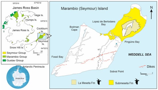
In the present contribution, we describe the microanatomy and histology of 37 thin sections of nine fossil penguin species, together with additional specimens referred to Palaeeudyptes sp., size groups sensu Jadwiszczak [25], or indeterminate specimens, all from the Eocene of Antarctica (Figure 1). We test the hypothesis that osteohistological characters associated with sex in extant penguins can also be identified in Eocene fossil penguins, enabling reassessment of morphological variation traditionally interpreted as taxonomic. Our main objective is to identify microstructural patterns associated with sexual dimorphism and evaluate their significance for interpreting morphological variation and refining the taxonomy of fossil penguins.
Figure 1.
Location map showing the outcrops of the La Meseta and Submeseta formations from which the materials examined here were collected. The inset on the left shows the James Ross Basin, including Marambio (Seymour) Island, located near the tip of the Antarctic Peninsula. Modified from Montes et al. [26].
2. Materials and Methods
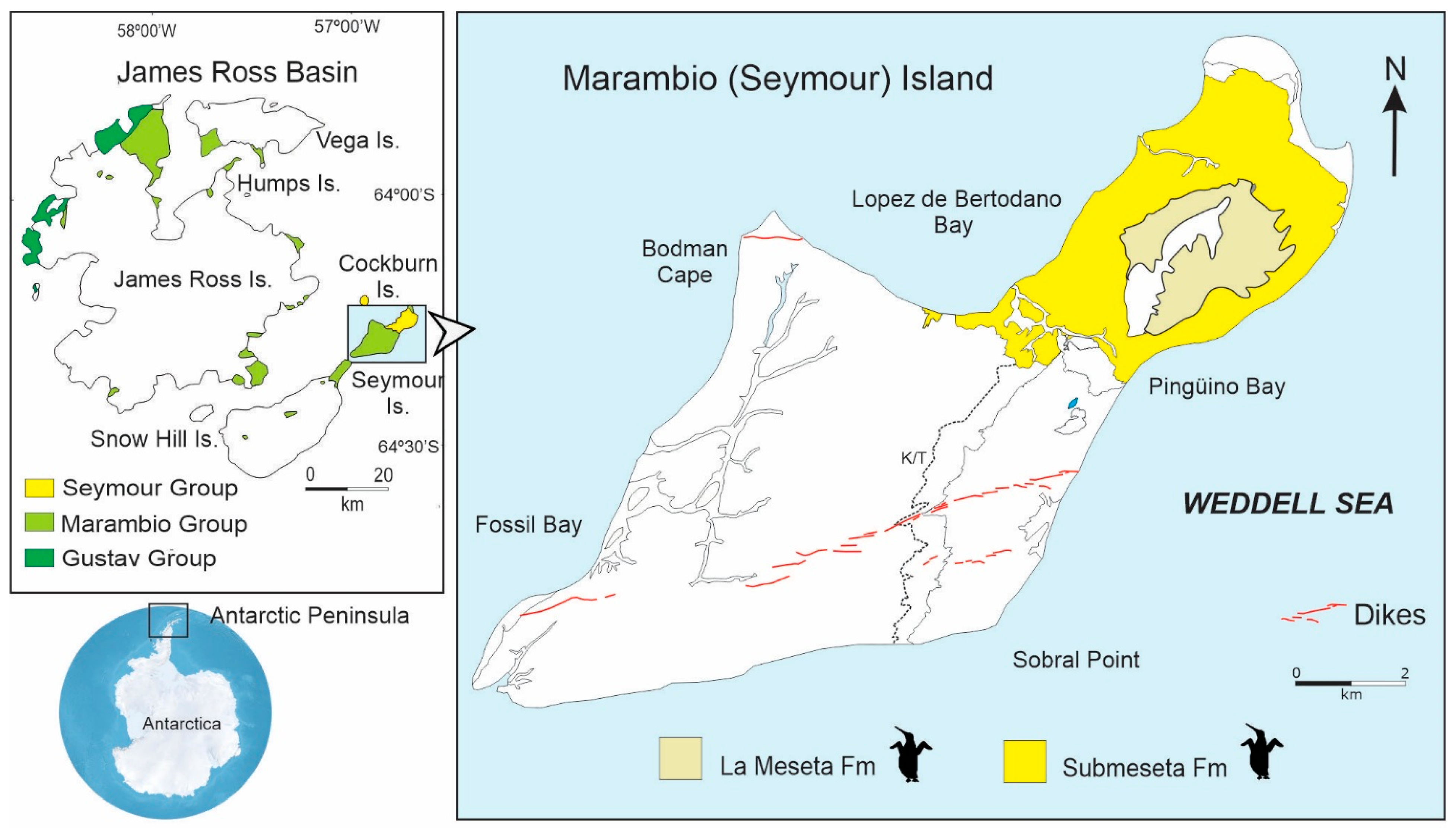
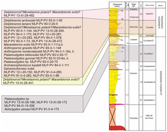
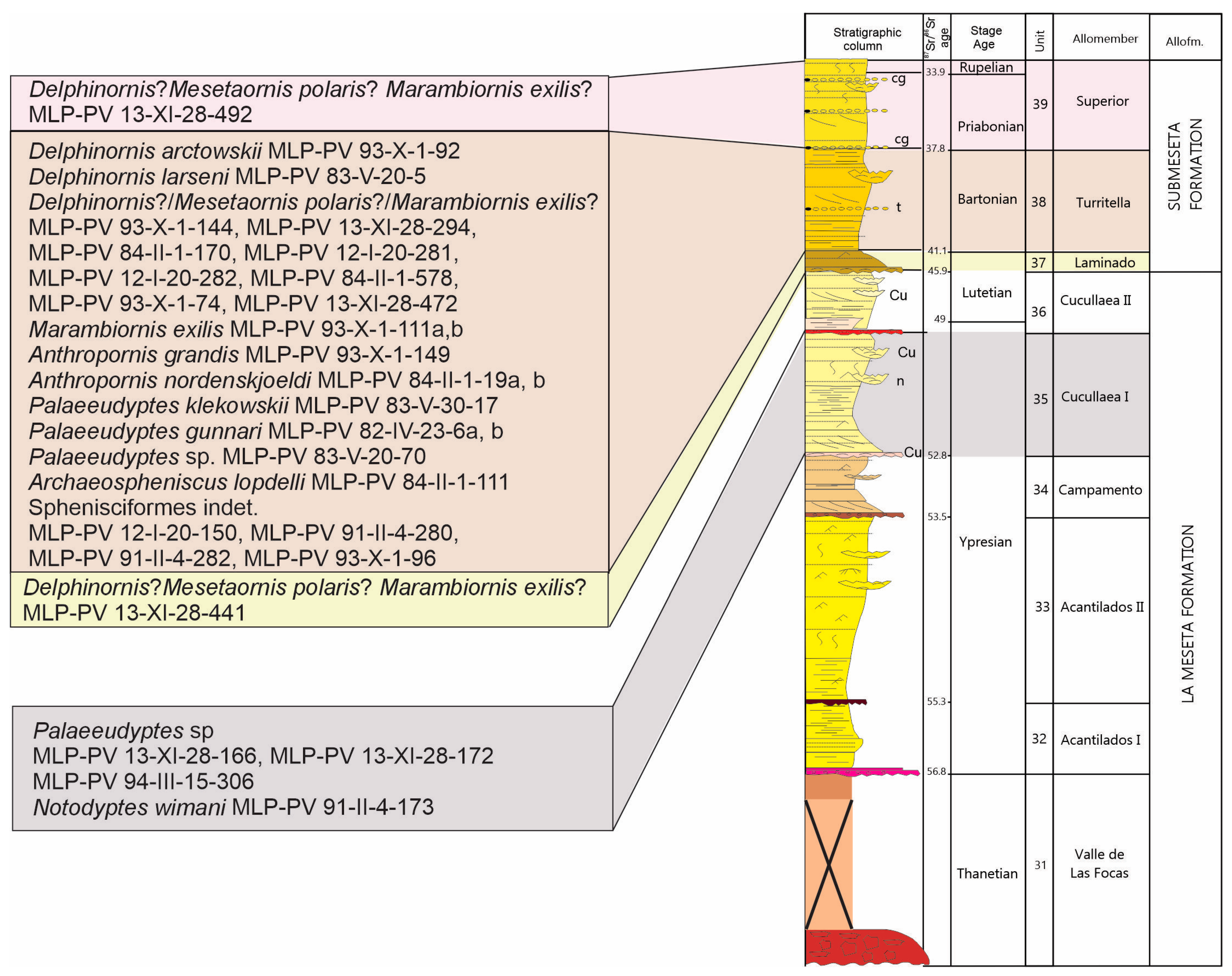
Thirty-seven thin sections corresponding to the following taxa were analysed: Delphinornis arctowskii, D. larseni, Marambiornis exilis, Anthropornis grandis, A. nordenskjoeldi, Palaeeudyptes klekowskii, P. gunnari, Archaeospheniscus lopdelli, Notodyptes wimani, Palaeeudyptes sp., an indeterminate tarsometatarsus tentatively assigned to Dephinornis?/Mesetaornis polaris?/Marambiornis exilis? (following the size criteria of Jadwiszczak [25]), and finally other indeterminate fossil Sphenisciformes specimens from the Eocene of Marambio Island (Figure 1 and Figure 2). Of this sample, 11 sections were prepared at the Department of Geology of the Universidad Nacional de San Luis (UNSL), in San Luis (Argentina), previously described by Cerda et al. [22], and 26 were prepared at the Thin Section Laboratory of the Instituto de Investigación en Paleobiología y Geología (CONICET-UNRN), in General Roca (Argentina), following the procedure described by Chinsamy and Raath [27] (Table 1).
Figure 2.
Stratigraphic column showing the allomembers of the La Meseta and Submeseta formations and indicating the levels from which the materials examined here were collected. Modified from Montes et al. [26].
Table 1.
Thin sections of the Antarctic fossil penguin specimens deposited in the Histological Collection (CH) of the Vertebrate Paleontology Division at the Museo de La Plata (MLP-PV).
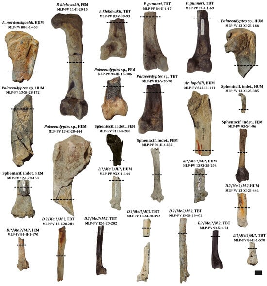
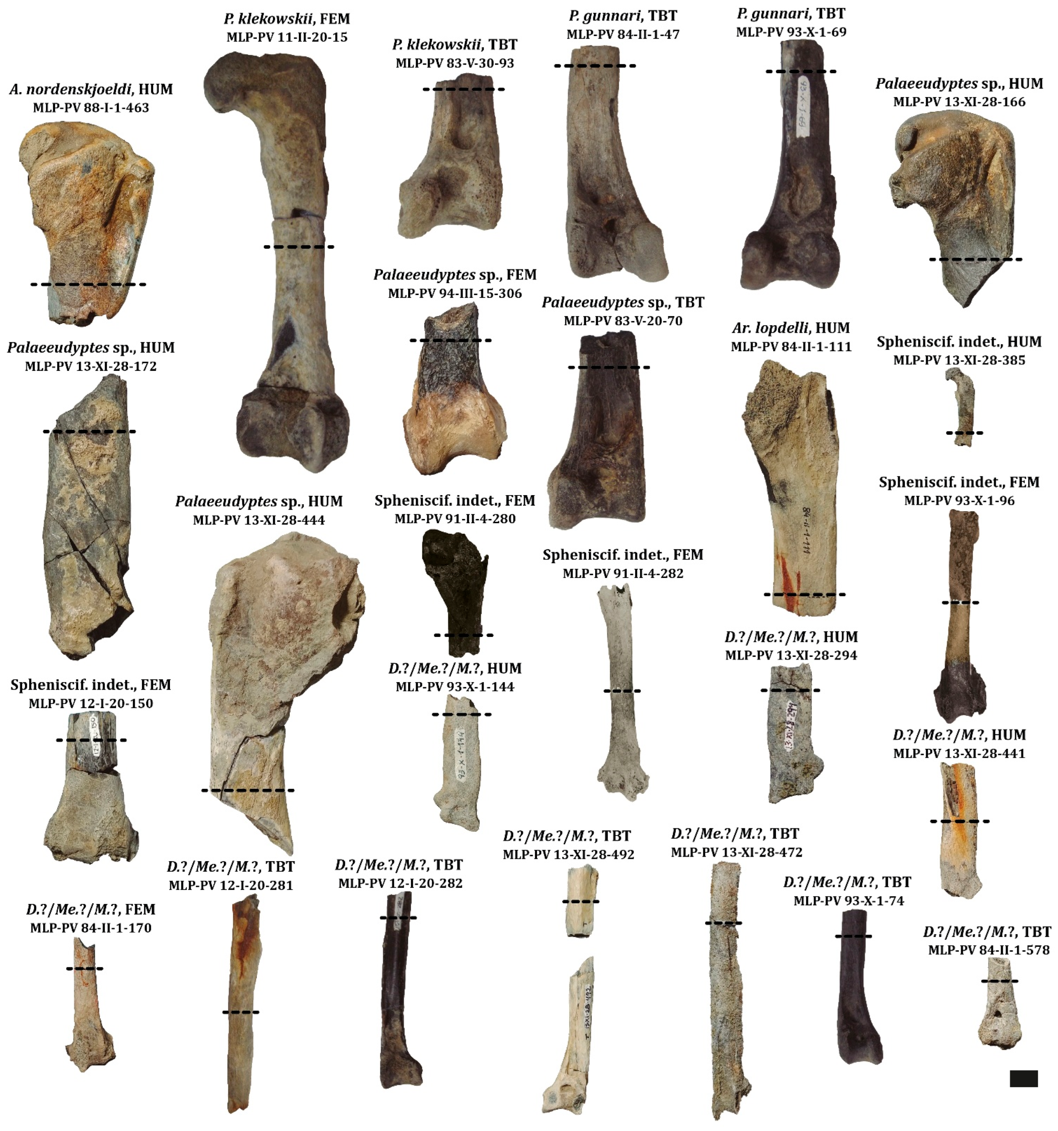
All materials belong to the collections of the Vertebrate Paleontology Division of the Museo de La Plata (MLP-PV), in the city of La Plata (Argentina) (Figure 3), and come from the La Meseta (late Thanetian–late Lutetian) and Submeseta (late Lutetian–early Rupelian) formations of Marambio (Seymour) Island, James Ross Basin, Antarctic Peninsula (Figure 2) (Montes et al. [26]).
Figure 3.
Fossil material sampled for thin-section preparation. Dashed lines indicate the levels from which samples were taken. Abbreviations: D.?/Me.?/M.?, Delphinornis?/Mesetaornis polaris?/Mariambiornis exilis?; Spheniscif. indet., indeterminate Sphenisciformes; HUM, humerus; FEM, femur; TBT, tibiotarsus; TMT, tarsometatarsus. Scale 1 cm.
The sections were analysed using a petrographic microscope (Zeiss Axio Imager, Carl Zeiss, Jena, Germany) under plane- and cross-polarised light. Images of the sections were captured with a digital camera (Zeiss Axiocam 105 color, Carl Zeiss, Jena, Germany) and processed using Adobe Photoshop 2020 v21.0 (image editing) and Adobe Illustrator 2022 v26.0 (figure preparation).
3. Results
First, the characteristics common to all the samples will be described, and subsequently, the specific features of each specimen and each bone element will be detailed.
3.1. Microanatomy
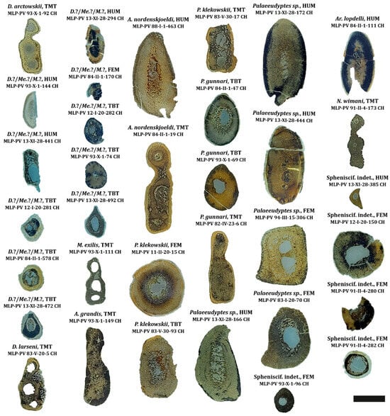
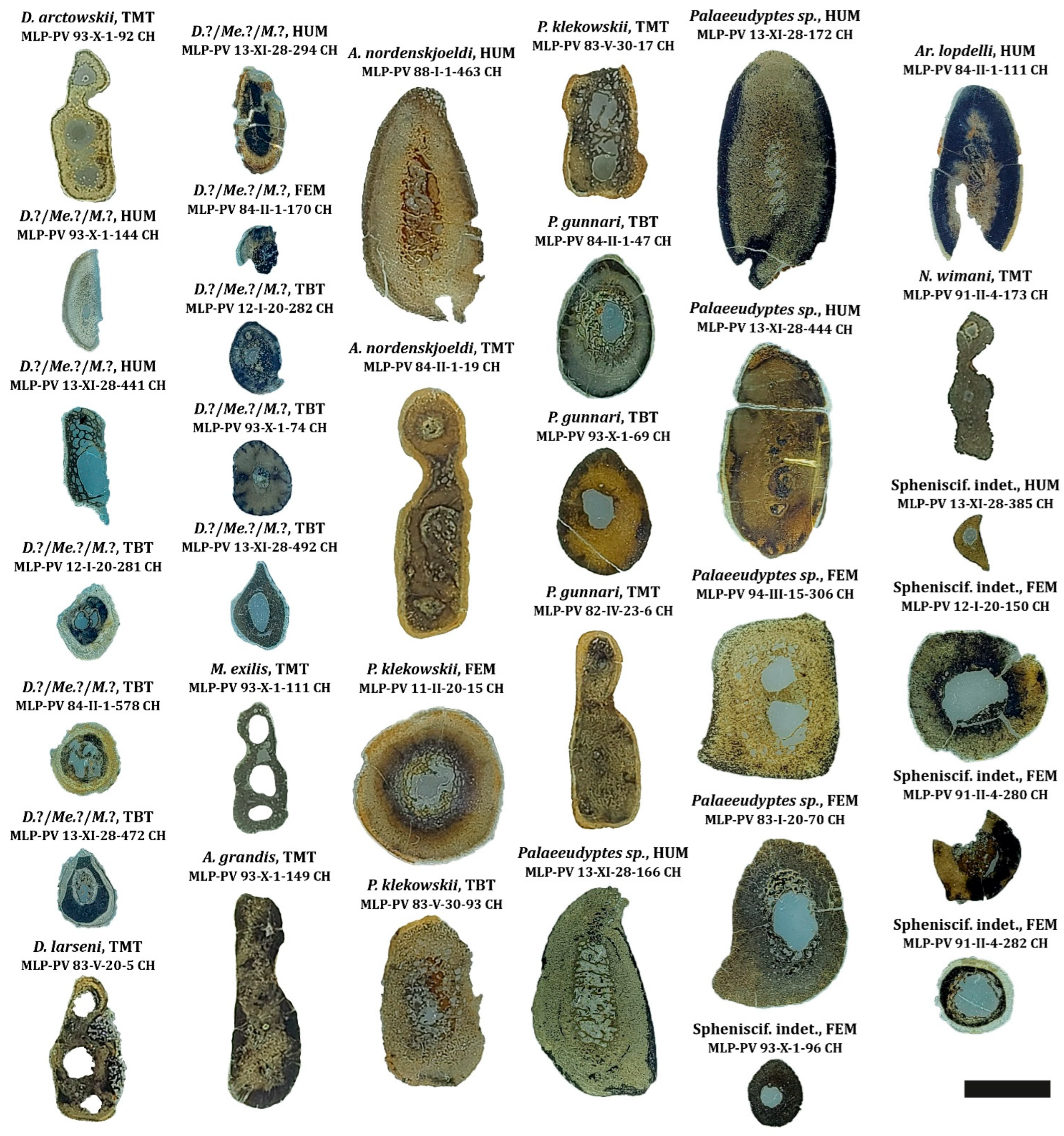
All the specimens analysed show clearly compact tissue and exhibit a high degree of bone remodelling. The most significant microanatomical variation is observed in the microstructure of the medullary spaces in the medullary region (Figure 4).
Figure 4.
Microanatomy of histological sections of the Antarctic fossil penguins examined here. Cross-sectional views of bone elements. Abbreviations: D.?/Me.?/M.?, Delphinornis?/Mesetaornis polaris?/Mariambiornis exilis?; Spheniscif. indet., indeterminate Sphenisciformes; HUM, humerus; FEM, femur; TBT, tibiotarsus; TMT, tarsometatarsus. Scale 1 cm.
In the forelimbs (humerus), compact tissue with varying degrees of compaction is observed (Figure 4). In the medullary region of some humeri (e.g., Delphinornis?/Mesetaornis polaris?/Marambiornis exilis? MLP-PV 13-XI-28-441 CH, Anthropornis nordenskjoeldi MLP-PV 88-I-1-463 CH, Palaeeudyptes sp. MLP-PV 13-XI-28-166 CH, MLP-PV 13-XI-28-172 CH, and MLP-PV 13 -XI-28-444 CH, and Archaeospheniscus lopdelli MLP-PV 84-II-1-111 CH) show several intertrabecular spaces, whereas in others (e.g., Delphinornis?/Mesetaornis polaris?/Marambiornis exilis? MLP-PV 93-X-1-144 CH, and MLP-PV 13-XI-28-294 CH) show scant trabecular development. Furthermore, in the proximal section of the humeral diaphysis of the undetermined specimen MLP-PV 13-XI-28-385 CH, a small medullary cavity with an irregular inner margin is observed.
By comparison, the bones of the hindlimbs exhibit greater microstructural variability (Figure 4), including: (1) a medullary cavity clearly delineated by surrounding tissue forming an inner circumferential layer (ICL) (e.g., Delphinornis arctowskii MLP-PV 93-X-1-92 CH; Delphinornis?/Mesetaornis polaris?/Marambiornis exilis? MLP-PV 93-X-1-74 CH, MLP-PV 13-XI-28-472 CH, MLP-PV 13-XI-28-492 CH; Marambiornis exilis MLP-PV 93-X-1-111 CH; Anthropornis grandis MLP-PV 93-X-1-149 CH; A. nordenskjoeldi MLP-PV 84-II-1-19 CH; Palaeeudyptes gunnari MLP-PV 93-X-1-69 CH, MLP-PV 82-IV-23-6 CH; Notodyptes wimani MLP-PV 91-II-4-173 CH; Sphenisciformes indet. MLP-PV 91-II-4-280 CH, MLP-PV 93-X-1-96 CH), and (2) a medullary cavity with an irregular margin, which may or may not converge with other intertrabecular spaces, without the presence of an ICL (e.g., Delphinornis larseni MLP-PV 83-V-20-5 CH; Delphinornis?/Mesetaornis polaris?/Marambiornis exilis? MLP-PV 12-I-20-281 CH; Palaeeudyptes klekowskii MLP-PV 11-II-20-15 CH, MLP-PV 83-V-30-93 CH, MLP-PV 83-V-30-17 CH; Palaeeudyptes gunnari MLP-PV 84-II-1-47 CH; Palaeeudyptes sp. MLP-PV 94-III-15-306 CH, MLP-PV 83-V-20-70 CH; Sphenisciformes indet. MLP-PV 91-II-4-282 CH). In the specific case of the sections Delphinornis?/Mesetaornis polaris?/Marambiornis exilis? MLP-PV 84-II-1-170 CH, MLP-PV 12-I-20-282 CH, MLP-PV 84-II-1-578 CH, and the undetermined specimen MLP-PV 12-I-20-150 CH, it is not possible to examine the medullary region in detail due to the high degree of weathering.
3.2. Histology
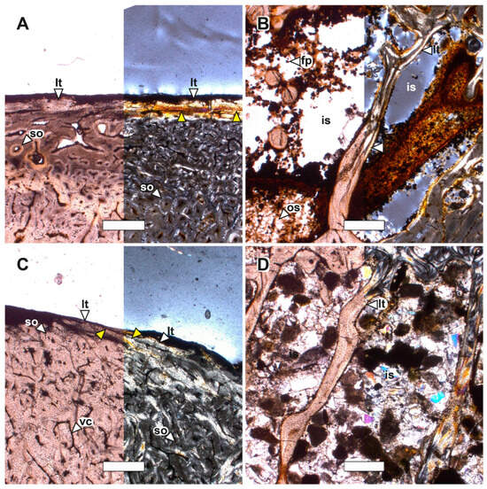
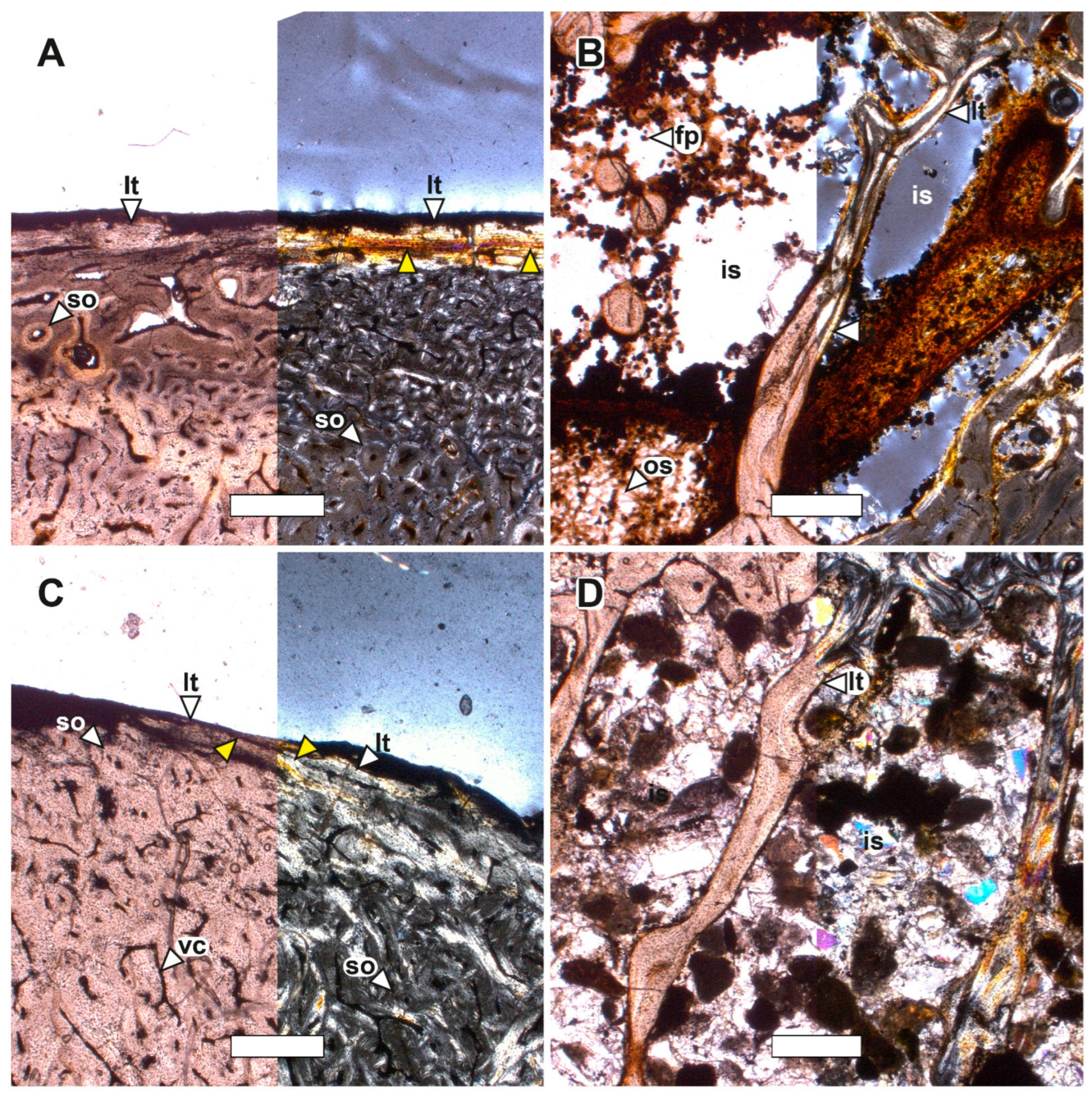
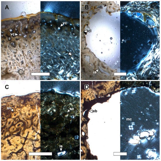
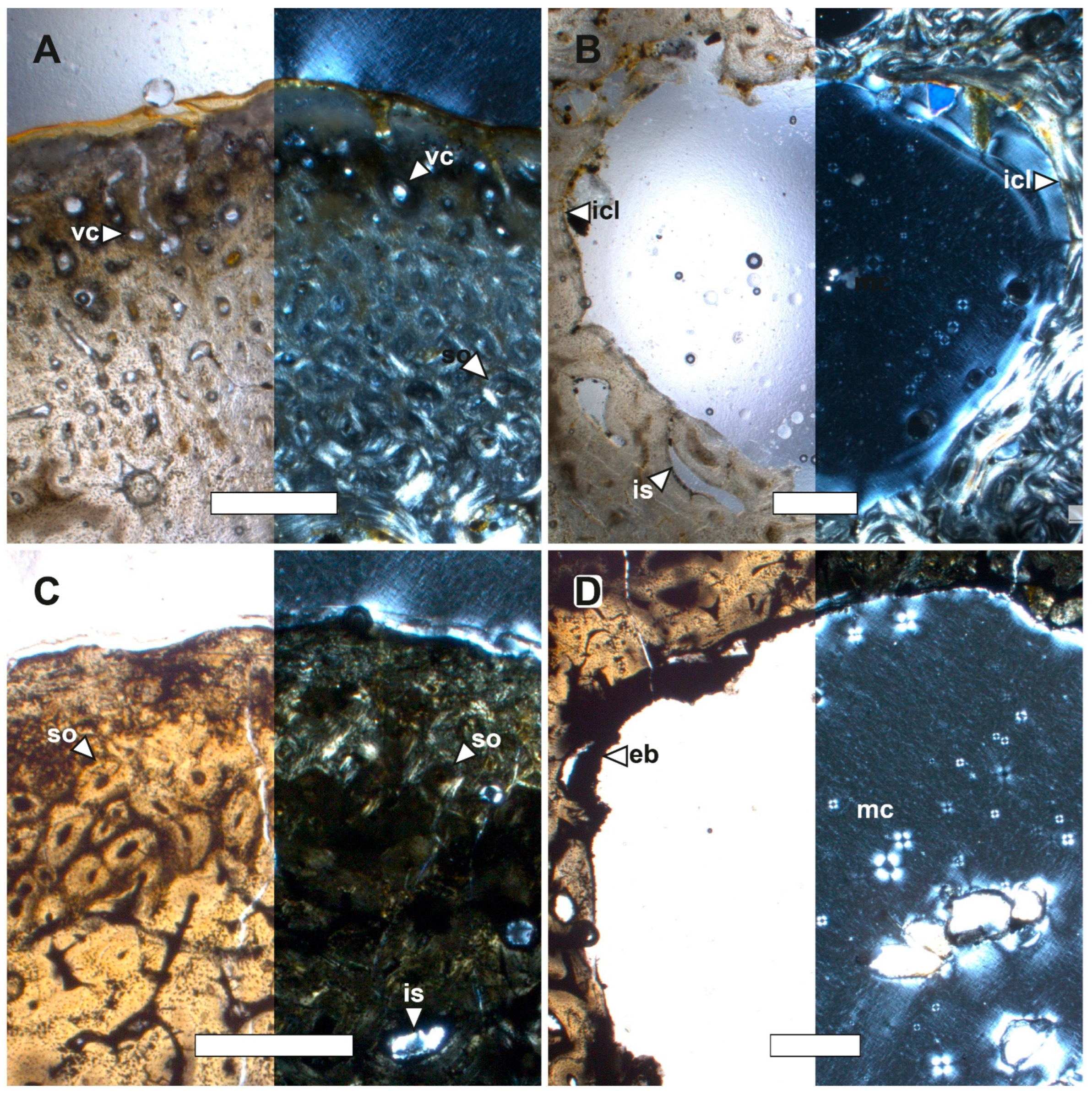
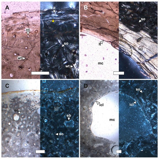
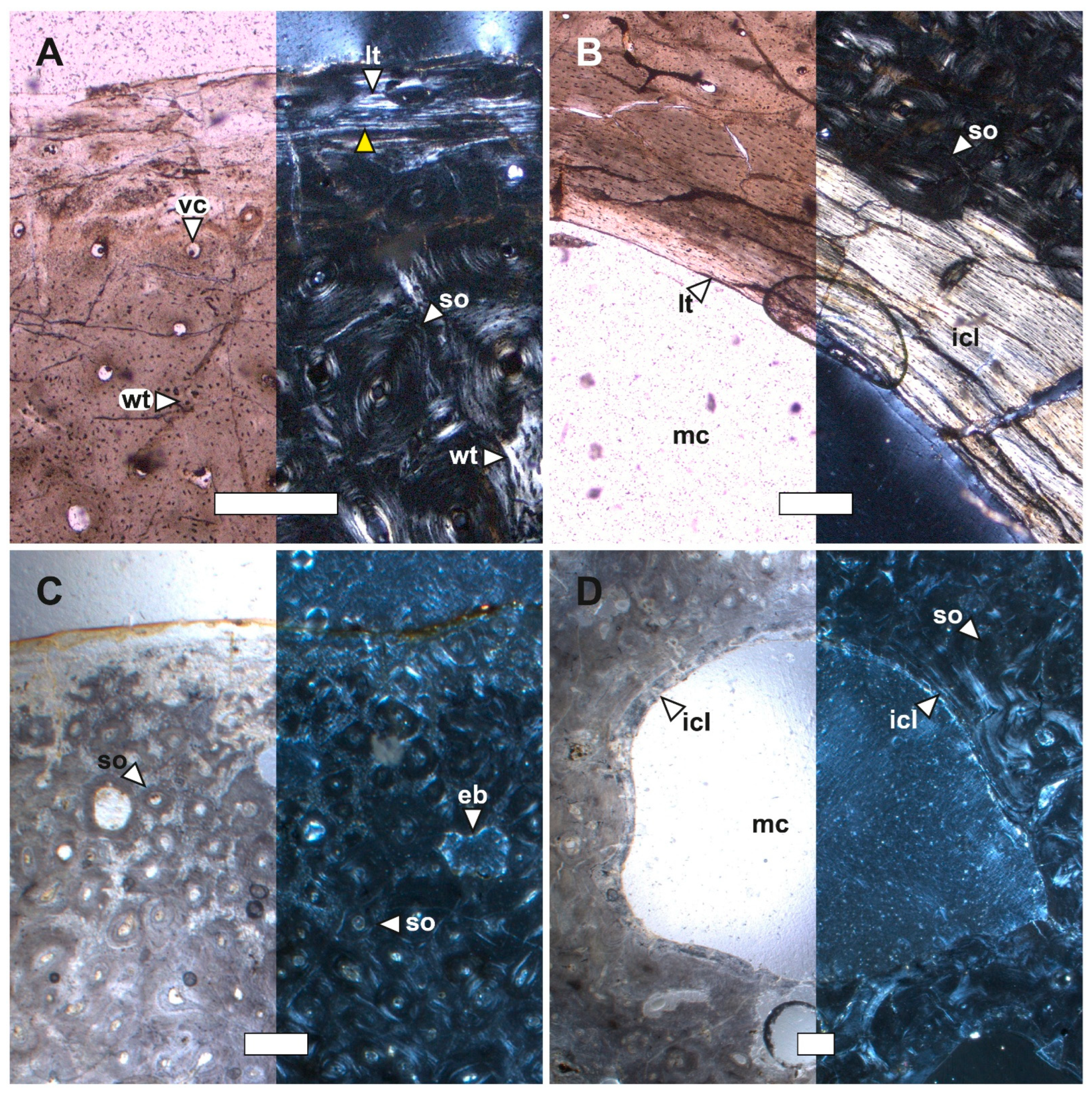
Most histological sections reveal various biological and physicochemical degradation processes affecting the bone tissue. These include filamentous structures, evidence of micro-bioerosion, and opaque mineral nodules (e.g., Figure 5B and Figure 6C). Only in a few sections was it possible to appreciate the bone matrix of the cortical region (e.g., MLP-PV 13-XI-28-441 CH, MLP-PV 13-XI-28-492 CH, MLP-PV 88 -I-1-463 CH, MLP-PV 11-II-20-15 CH, MLP-PV 13-XI-28-166 CH, MLP-PV 94-III-15-306 CH, MLP-PV 83-I-20-70 CH, and MLP-PV 91-II-4-280 CH). Overall, the vascular organisation is predominantly longitudinal; a high degree of bone compaction is observed in all sections; and the microanatomy and histology define three main regions: (1) a cortical region characterised by an avascular lamellar bone matrix and laminar vascular arrangements; (2) a perimedullary region characterised by a woven-fibered bone matrix, remodelled by secondary osteons in a longitudinal pattern; and (3) a medullary region where the organisation of the medullary spaces differs depending on the element analysed. In the medullary region of the forelimb, a secondarily compacted trabecular tissue is observed, in which the development of the intertrabecular spaces varies according to the degree of bone compaction. In the medullary region of the hindlimb, two microstructural patterns are recognised: a well-defined medullary cavity bounded by an inner circumferential layer (ICL), or a medullary cavity with an irregular margin, where adjacent intertrabecular spaces may converge with the medullary cavity and show no evidence of an ICL.
Figure 5.
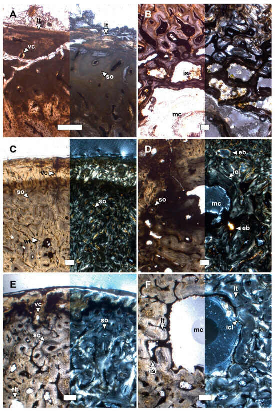
Histological features of the humerus of Anthropornis nordenskjoeldi (MLP-PV 88-I-1-463 CH), and Palaeeudyptes sp. (MLP-PV 13-XI-28-166 CH). (A,B) Anthropornis nordenskjoeldi and (C,D) Palaeeudyptes sp., under normal transmitted light (left) and polarized light (right). (A,C) Cortical and perimedullary region, (B,D) medullary region. (fp) framboidal pyrite, (is) intertrabecular space, (lt) lamellar tissue, (os) organic structure, (so) secondary osteon, (vc) vascular canal, and (yellow triangle) LAG. Scale 500 μm.
Figure 6.
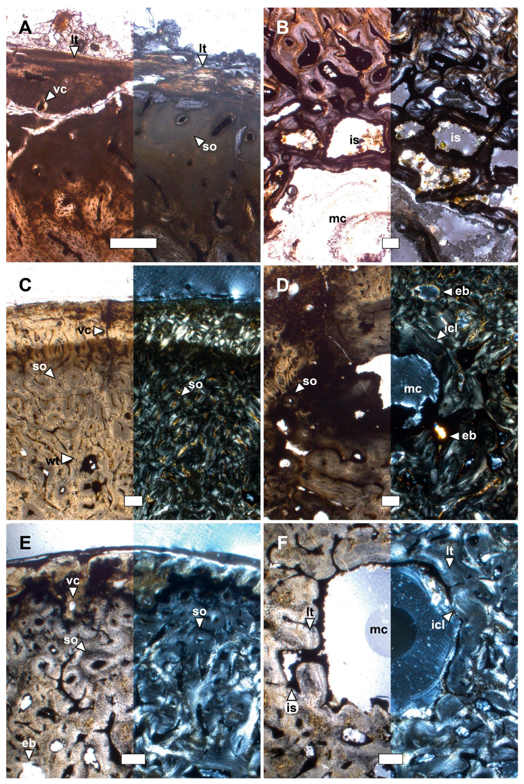
Histological features of the tarsometatarsus of Delphinornis arctowskii (MLP-PV 93-X-1-92 CH), and Delphinornis larseni (MLP-PV 83-V-20-5 CH). (A,B) Delphinornis arctowskii and (C,D) Delphinornis larseni, under normal transmitted light (left) and polarized light (right). (A,C) Cortical and perimedullary region, (B,D) medullary region. (eb) erosion bay, (icl) inner circumferential layer, (is) intertrabecular space, (mc) medullary cavity, (so) secondary osteon, and (vc) vascular canal. Scale 500 μm.
In the cortical region of the humerus of Anthropornis nordenskjoeldi (MLP-PV 88-I-1-463 CH) and Palaeeudyptes sp. (MLP-PV 13-XI-28-166 CH), avascular lamellar tissue and lines of arrested growth (LAGs) are observed (Figure 5A,C). In the perimedullary region, several secondary osteons remodelling a woven-fibered bone matrix (Figure 5A,C) are found, whereas in the medullary region, trabecular bone is observed, with extensive irregular intertrabecular spaces surrounded by lamellar tissue (Figure 5B,D). Furthermore, in the medullary region of Anthropornis nordenskjoeldi (MLP-PV 88-I-1-463 CH), several framboidal pyrite nuclei and a series of filamentous organic structures that may correspond to bacteria are present (Figure 5B).
In the tarsometatarsus of Delphinornis arctowskii (MLP-PV 93-X-1-92 CH) (Figure 6A,B), an internal circumferential layer (ICL) that has been partially remodelled in the medullary region can be observed (Figure 6B), whereas in the tarsometatarsus of Delphinornis larseni (MLP-PV 83-V-20-5 CH) (Figure 6C,D), an extensive medullary cavity is observed, with an irregular margin and no ICL present (Figure 6D). The small specimens, grouped here as Delphinornis?/Mesetaornis polaris?/Marambiornis exilis? (MLP-PV 93-X-1-144 CH, MLP-PV 13-XI-28-294 CH, MLP-PV 13-XI-28-441 CH, MLP-PV 84-II-1-170 CH, MLP-PV 12-I-20-281 CH, MLP-PV 12-I-20-282 CH, MLP-PV 84-II-1-578 CH, MLP-PV 93-X-1-74 CH, MLP-PV 13-XI-28-472 CH, and MLP-PV 13-XI-28-492 CH) (Figure 7A,B) show considerable variation in the organization of the medullary region of the tibiotarsus. In some of these, cavities clearly demarcated by the surrounding tissue are observed, without forming a very extensive ICL (e.g., MLP-PV 12-I-20-281 CH, MLP-PV 93-X-1-74 CH, and MLP-PV 13-XI-28-472 CH), a medullary cavity showing clear development of an ICL (e.g., MLP-PV 13-XI-28-492 CH), and a medullary cavity with an irregular margin that converges with other intertrabecular spaces (e.g., MLP-PV 84-II-1-578 CH) (Figure 7B).
Figure 7.
Histological features of the tibiotarsus assigned to the size group Dephinornis?/Mesetaornis polaris?/Marambiornis exilis? (MLP-PV 13-XI-28-492 CH), and the tarsometatarsus of Marambiornis exilis (MLP-PV 93-X-1-111 CH). (A,B) size group Dephinornis?/Mesetaornis polaris?/Marambiornis exilis? and (C,D) Marambiornis exilis, under normal transmitted light (left) and polarized light (right). (A,C) cortical and perimedullary region, (B,D) medullary region. (eb) erosion bay, (icl) inner circumferential layer, (lt) lamellar tissue, (mc) medullary cavity, (so) secondary osteon, (vc) vascular canal, (wt) woven-fibered tissue, and (yellow triangle) LAG. Scale 200 μm.
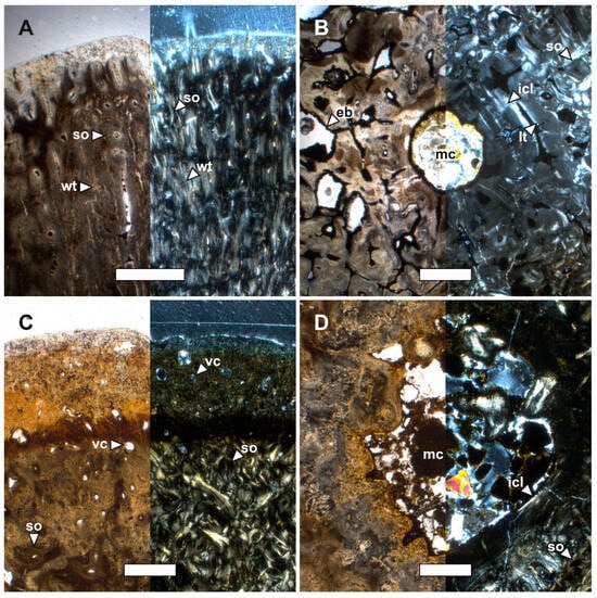
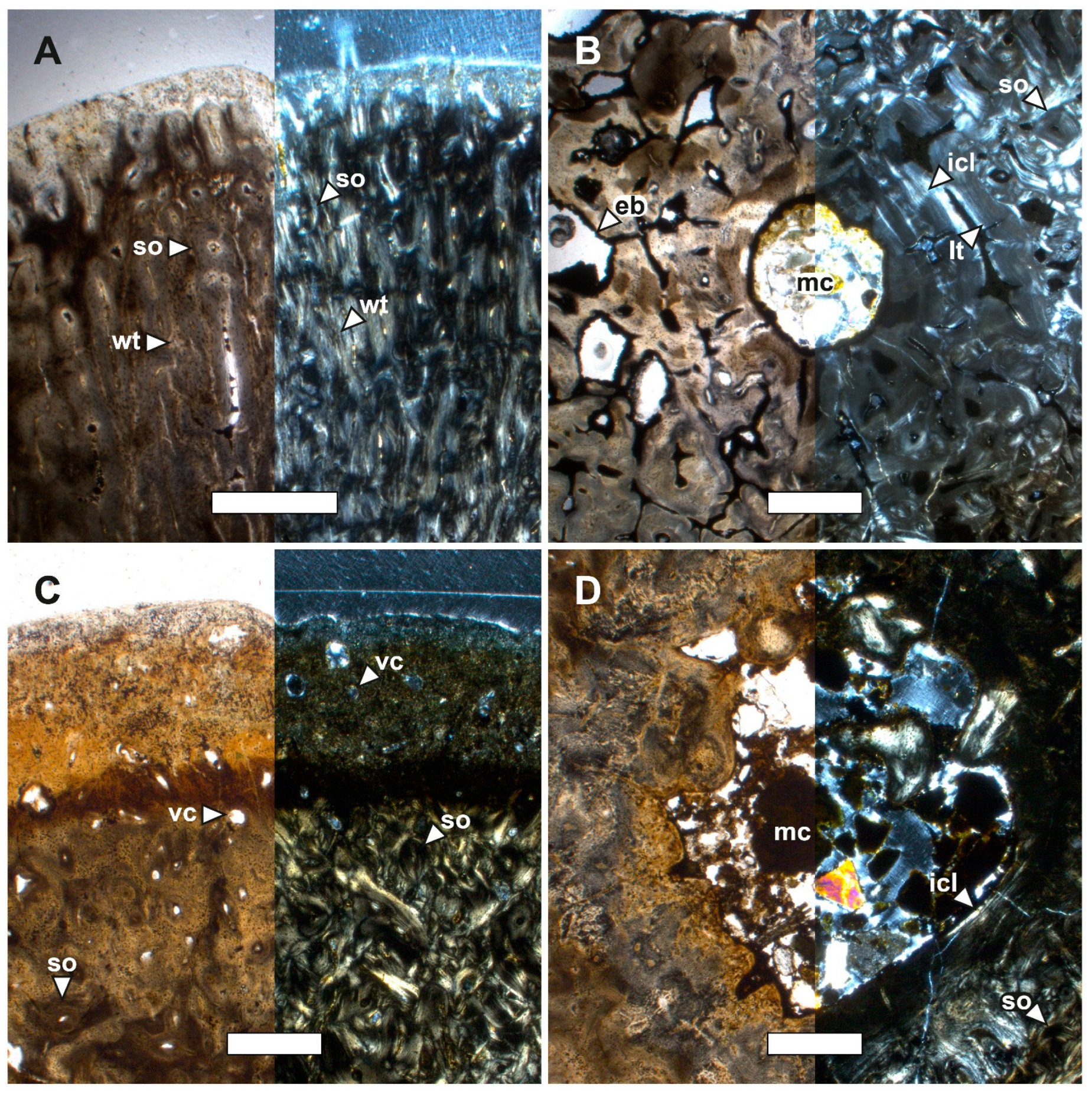
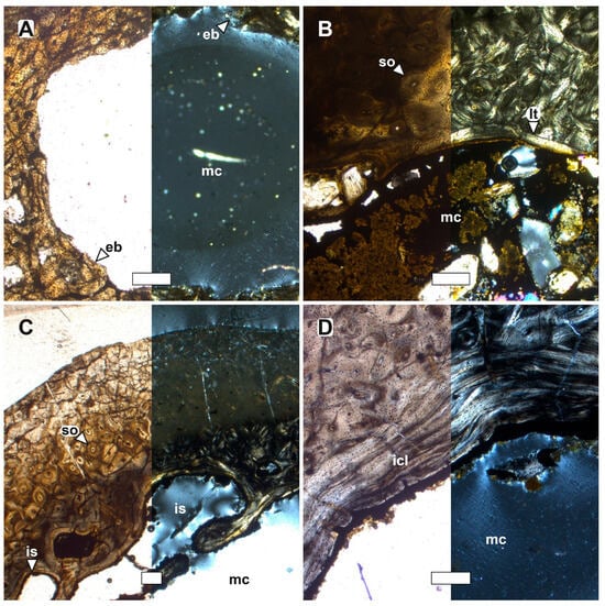
A well-developed ICL is also observed in the tarsometatarsus of Marambiornis exilis (MLP-PV 93-X-1-111 CH) (Figure 7C,D), and Anthropornis grandis (MLP-PV 93-X-1-149 CH) (Figure 8A,B). Furthermore, an ICL is visible in the tarsometatarsus of Anthropornis nordenskjoeldi (MLP-PV 84-II-1-19 CH) (Figure 8C,D), although it shows signs of remodelling.
Figure 8.
Histological features of the tarsometatarsus of Anthropornis grandis (MLP-PV 93-X-1-149 CH), and Anthropornis nordenskjoeldi (MLP-PV 84-II-1-19 CH). (A,B) Anthropornis grandis and (C,D) Anthropornis nordenskjoeldi, under normal transmitted light (left) and polarized light (right). (A,C) cortical and perimedullary region, (B,D) medullary region. (eb) erosion bay, (icl) inner circumferential layer, (lt) lamellar tissue, (mc) medullary cavity, (so) secondary osteon, (vc) vascular canal, and (wt) woven-fibered tissue. Scale 500 μm.
Palaeeudyptes klekowskii exhibits, in the femur, tibiotarsus, and tarsometatarsus, a medullary cavity with an irregular margin that converges with other intertrabecular spaces (e.g., MLP-PV 11-II-20-15 CH, MLP-PV 83-V-30-93 CH, and MLP-PV 83 -V-30-17 CH) (Figure 9A,B), whilst Palaeeudyptes gunnari exhibits the same microstructural patterns in the tibiotarsus (MLP-PV 84-II-1-47 CH). However, in other sections of the tibiotarsus (MLP-PV 93-X-1-69 CH), P. gunnari exhibits a well-developed ICL and, in the tarsometatarsus (MLP-PV 82-IV-23-6 CH), a partially remodelled ICL is observed (Figure 9C,D). Similarly, the tarsometatarsus of Notodyptes wimani exhibits a well-developed ICL (Figure 9E,F).
Figure 9.
Histological features of the femur of Palaeeudyptes klekowskii (MLP-PV 11-II-20-15 CH), the tarsometatarsus of Palaeeudyptes gunnari (MLP-PV 82-IV-23-6 CH), and the tarsometatarsus of Notodyptes wimani (MLP-PV 91-II-4-173 CH). (A,B) Palaeeudyptes klekowskii, (C,D) Palaeeudyptes gunnari, and (E,F) Archaeospheniscus wimani, under normal transmitted light (left) and polarized light (right). (A,C,E) cortical and perimedullary region, (B,D,F) medullary region. (eb) erosion bay, (icl) inner circumferential layer, (is) intertrabecular space, (lt) lamellar tissue, (mc) medullary cavity, (so) secondary osteon, (vc) vascular canal, and (wt) woven-fibered tissue. Scale 200 μm.
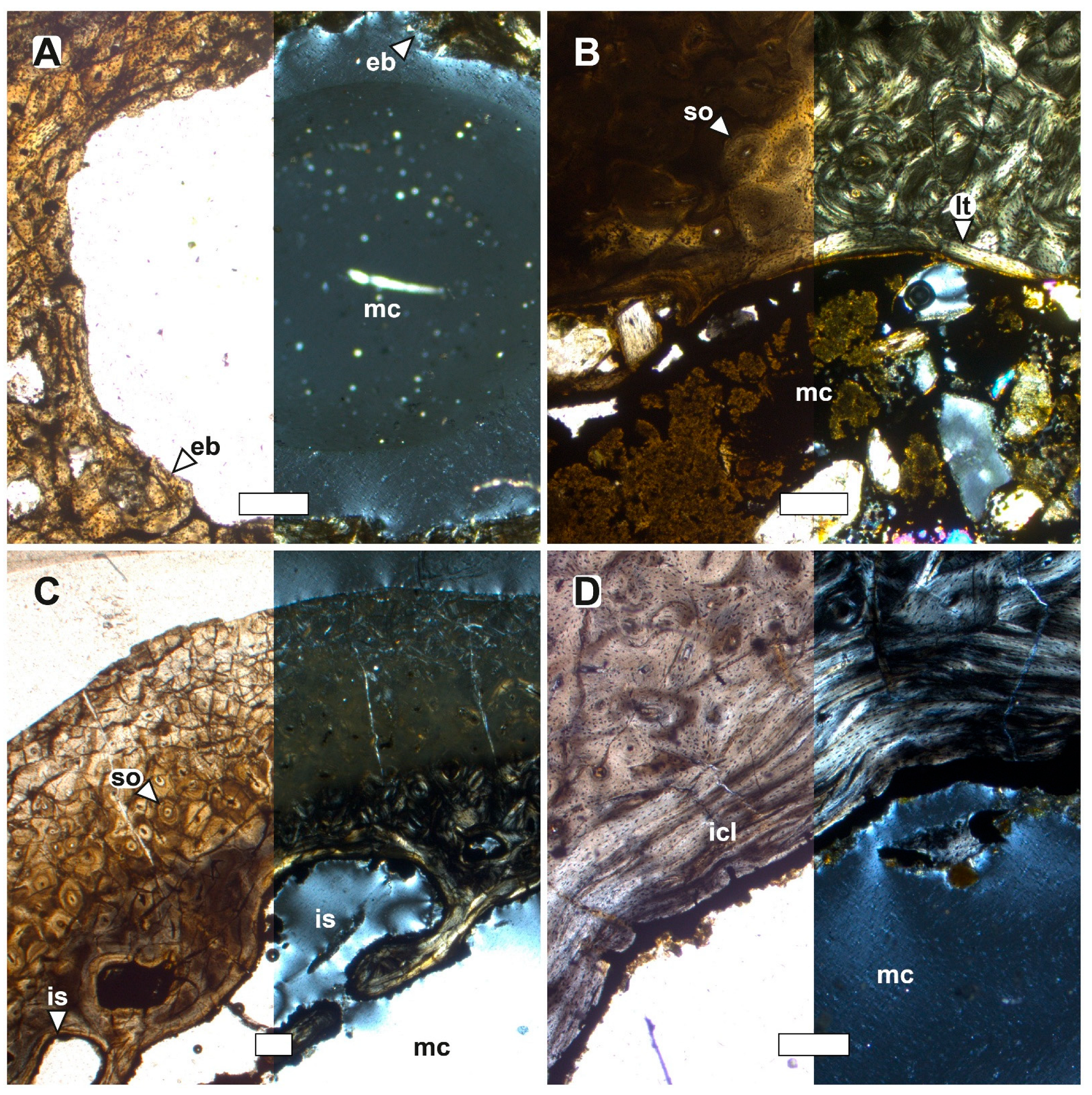
Finally, considerable microstructural variability is observed in the femur sections of the indeterminate specimens. The femur MLP-PV 12-I-20-150 CH (Figure 10A) exhibits a well-defined medullary cavity, but no surrounding tissue could be observed. The femur MLP-PV 91-II-4-280 CH (Figure 10B) has a well-defined medullary cavity surrounded by lamellar tissue, forming a short ICL. The femur MLP-PV 91-II-4-282 CH (Figure 10C) exhibits a medullary cavity with an irregular margin that may converge with other intertrabecular spaces, whilst the femur MLP-PV 93-X-1-96 CH (Figure 10D) shows a medullary cavity clearly surrounded by an ICL.
Figure 10.
Histological features of the femur of taxonomically indeterminate Sphenisciformes (MLP-PV 12-I-20-150 CH, MLP-PV 91-II-4-280 CH, MLP-PV 91-II-4-282 CH, and MLP-PV 93-X-1-96 CH). (A) MLP-PV 12-I-20-150 CH, (B) MLP-PV 91-II-4-280 CH, (C) MLP-PV 91-II-4-282 CH, and (D) MLP-PV 93-X-1-96 CH, under normal transmitted light (left) and under polarized light (right). (A–D) Perimedullary region and medullary region. (eb) erosion bay, (icl) inner circumferential layer, (is) intertrabecular space, (lt) lamellar tissue, (mc) medullary cavity, and (so) secondary osteon. Scale 200 μm.
4. Discussion
Our results demonstrate that osteohistological features previously associated with sex in extant penguins are also recognizable in Eocene Antarctic penguins. Although these characters require further validation before they can be regarded as definitive indicators of sex in fossil material, they provide an independent line of evidence for assessing biological variation within fossil populations. Consequently, osteohistology offers a promising framework for distinguishing sexual dimorphism from taxonomic differences and therefore contributing to a more robust interpretation of morphological diversity in fossil penguins.
Among birds, medullary bone remains the only unequivocal osteohistological indicator of female sex because it forms exclusively in reproductively active females during oviposition and functions as a temporary calcium reservoir for eggshell formation [12,13,14,15,16,17,28]. However, as in previous studies of fossil penguins [22,23,24], medullary bone was not identified in the material analysed here. This absence is not unexpected because medullary bone is a transient tissue that forms shortly before oviposition and is rapidly resorbed after reproduction. In addition, its high vascularization and relatively low degree of mineralization make it particularly susceptible to taphonomic degradation, substantially reducing its preservation potential in the fossil record [16].
Because medullary bone is rarely preserved, additional osteohistological markers of reproductive physiology are particularly valuable in fossil taxa. Garat et al. ([20,21] and references cited therein) proposed that the condition of the inner circumferential layer (ICL) provides such information in penguins. Based on extant taxa, three microstructural patterns were recognized: (1) a well-developed ICL, (2) a remodelled ICL, and (3) the absence of an ICL in adult individuals. Within this framework, remodelling of the ICL is interpreted as the result of endosteal resorption associated with calcium mobilization during reproduction, and, therefore, represents the strongest histological evidence currently available for identifying probable females when medullary bone is absent. Conversely, an unremodelled ICL may correspond either to males or females outside the reproductive period, whereas the absence of an ICL may also indicate females if somatic maturity can be independently established. Accordingly, the ICL should not be regarded as an absolute indicator of sex but as a probabilistic osteohistological marker that must be interpreted altogether with ontogenetic and anatomical evidence.
The microstructural patterns observed in the present study are consistent with this interpretative framework. Specimens of Delphinornis arctowskii (MLP-PV 93-X -1-92 CH), Anthropornis nordenskjoeldi (MLP-PV 84-II-1-19 CH), and Palaeeudyptes gunnari (MLP-PV 82-IV-23-6 CH) exhibit remodelled ICL and are therefore interpreted as probable females. In contrast, Anthropornis grandis (MLP-PV 93-X-1-149 CH) retains an unremodelled ICL, a condition that may represent either a male individual or a female outside the oviposition period and therefore cannot be considered diagnostic. Similarly, adult specimens of Delphinornis larseni (MLP-PV 83-V-20-5 CH) and Palaeeudyptes klekowskii (MLP-PV 11-II-20-15 CH, MLP-PV 83-V-30-93 CH, and MLP-PV 83-V-30-17 CH) lacking an ICL may also represent probable females, particularly when accompanied by evidence of endosteal remodelling. This interpretation is further supported by specimens of Palaeeudyptes sp. (MLP-PV 94-III-15-306 CH and MLP-PV 83-V-20-70 CH) and the indeterminate specimen MLP-PV 91-II-4-282 CH, which preserve extensive endosteal remodelling despite the complete absence of ICL. Although these interpretations remain provisional, they demonstrate that osteohistological data can provide biologically meaningful information that is inaccessible through external morphology alone.
Interpretation of these microstructural patterns should also consider functional differences among skeletal elements. Comparative studies of extant penguins indicate that forelimb bones are more strongly modified by the biomechanical demands of wing-propelled diving, whereas hindlimb elements retain a better-defined medullary region and preserve histological features associated with reproductive physiology [19,21]. Consequently, hindlimb elements provide more reliable information for evaluating putative sex-related microstructural variation. This anatomical constraint also limits comparisons among taxa represented by different skeletal elements, explaining why direct comparisons between Archaeospheniscus lopdelli and Notodyptes wimani were not possible in the present study.
One of the principal implications of these findings concerns the taxonomy of fossil penguins. Antarctic fossil penguins are commonly represented by isolated skeletal elements, making it difficult to distinguish taxonomic differences from normal intraspecific variation. As a result, sexual dimorphism has long been proposed as a potential explanation for part of the morphological disparity observed in giant Eocene penguins [5,9,11]. The osteohistological evidence presented here provides independent support for this hypothesis. Within Anthropornis, the occurrence of a remodelled ICL in A. nordenskjoeldi and an unremodelled ICL in A. grandis suggests that at least some morphological differences traditionally used to distinguish these species could instead reflect sexual variation. Likewise, specimens assigned to Palaeeudyptes klekowskii consistently display histological features compatible with probable females, whereas P. gunnari includes individuals with remodelled and unremodelled ICLs, suggesting that part of the variation currently interpreted as interspecific may instead represent sexual or reproductive variation. Although these hypotheses require testing with larger sample sizes and additional histological data, they illustrate the potential of the osteohistology to reassess species boundaries in taxa described primarily from isolated bones.
Several limitations should nevertheless be acknowledged. The sample size remains relatively small, medullary bone is absent in all analysed specimens, and the proposed relationship between ICL remodelling and sex is currently based on comparisons with extant penguins rather than on fossil individuals of known sex. Consequently, the histological patterns identified here should be regarded as hypotheses of sex attribution rather than definitive diagnoses. Future studies incorporating larger fossil samples, additional skeletal elements, and broader comparative datasets from extant penguins will be essential for testing the robustness and taxonomic applicability of these osteohistological criteria.
Despite these limitations, this study provides the first evidence that osteohistological signatures associated with reproductive biology in extant penguins are also preserved in Eocene Antarctic penguins. These findings demonstrate that osteohistology provides an independent source of evidence for recognizing biological variation within fossil populations and represents a valuable complement to traditional morphological analyses. Incorporating histological data into systematic studies therefore offers a promising avenue for distinguishing sexual from taxonomic variation, ultimately improving our understanding of the diversity and evolution of fossil penguins.
5. Conclusions
Sex determination in fossil penguins based on osteohistological features remains challenging. Medullary bone may be absent from the preserved skeleton either because the individual was not reproductively active at the time of death or because this tissue has limited potential for preservation under taphonomic conditions. However, traces of the osteoclastic activity associated with medullary bone formation may persist after the tissue itself has been resorbed. These microstructural features can therefore provide indirect evidence that medullary bone was present during the individual’s lifetime.
Within this framework, the microstructure of hindlimb bones may provide three different patterns relevant to sex determination. The first is an unremodelled inner circumferential layer (ICL), which by itself does not allow sex to be inferred, as the ICL develops in both males and females during ontogeny. A remodelled ICL, in contrast, may evidence the female condition, as remodelling can be associated with medullary bone development. Finally, the absence of an ICL may also be compatible with a female individual. This interpretation, however, requires independent evidence of somatic maturity, since the ICL may be absent in immature individuals. These patterns are particularly relevant to hindlimb elements. Conversely, forelimb bones are more strongly affected by biomechanical demands associated with aquatic locomotion and may therefore retain less informative osteohistological signals for sex determination.
These microstructural patterns may therefore provide an independent source of evidence for recognising potential females and males in fossil penguins, although none of these conditions should be regarded as an absolute diagnostic criterion. Consequently, increasing the available sample size could help to evaluate more accurately whether the presence, absence, or degree of ICL remodelling is consistently associated with sex across different penguin taxa.
Under this interpretative approach, the remodelled ICL observed in Delphinornis arctowskii, Anthropornis nordenskjoeldi and Palaeeudyptes gunnari may indicate female individuals. Similarly, the absence of an ICL in Delphinornis larseni and Palaeeudyptes klekowskii could also be interpreted as evidence of females. In contrast, taxa preserving an unremodelled ICL, such as Anthropornis grandis, cannot be assigned confidently to either sex because this condition may represent either males or females outside the oviposition period. These interpretations may help reassess the taxonomic significance of morphological variation in Antarctic fossil penguins, particularly in Anthropornis and Palaeeudyptes, where some differences traditionally attributed to distinct species could instead reflect sexual variation.
Author Contributions
Conceptualization, L.M.G., C.A.H. and M.T.; Formal Analysis, L.M.G.; Investigation, L.M.G.; Writing—Original Draft Preparation, L.M.G.; Writing—Review and Editing, C.A.H. and M.T.; Project Administration, C.A.H. and M.T.; Funding Acquisition, C.A.H. and M.T. All authors have read and agreed to the published version of the manuscript.
Funding
This work was partially supported by Universidad Nacional de Río Negro PIUNRN 40-A-1230, Universidad Nacional de La Plata 11/N1044 and 11/N1063, and Consejo Nacional de Investigaciones Científicas y Técnicas (PIP 0096).
Data Availability Statement
The original contributions presented in this study are included in the article. Further inquiries can be directed to the corresponding authors.
Acknowledgments
We thank Martín de los Reyes (MLP) and Laura Yanina Herrera (MLP) for providing access to skeletal material, and Juan Ignacio Ison (IIPG) for technical support during thin-section preparation. We also thank to two anonymous reviewers for their constructive and helpful comments, which contributed to improving the quality of this manuscript.
Conflicts of Interest
The authors declare no conflicts of interest.
References
- Tambussi, C.P.; Reguero, M.A.; Marenssi, S.A.; Santillana, S.N. Crossvallia unienwillia, a new Spheniscidae (Sphenisciformes, Aves) from the Late Paleocene of Antarctica. Geobios 2005, 38, 667–675. [Google Scholar] [CrossRef] [Scilit]
- Slack, K.E.; Jones, C.M.; Ando, T.; Harrison, G.L.; Ewan Fordyce, R.; Arnason, U.; Penny, D. Early Penguin Fossils, Plus Mitochondrial Genomes, Calibrate Avian Evolution. Mol. Biol. Evol. 2006, 23, 1144–1155. [Google Scholar] [CrossRef] [Scilit] [PubMed]
- Mayr, G.; De Pietri, V.L.; Proffitt, L.; Blokland, J.C.; Clarke, J.A.; Love, L.; Mannering, A.A.; Crouch, E.M.; Reid, C.; Scofield, R.P. Multiple exceptionally preserved fossils from the Paleocene Waipara Greensand inform the diversity of the oldest stem group Sphenisciformes and the formation of their diving adaptations. Zool. J. Linn. Soc. 2025, 204, zlaf080. [Google Scholar] [CrossRef] [Scilit]
- Acosta Hospitaleche, C.; Jadwiszczak, P.; Clarke, J.A.; Cenizo, M. The fossil record of birds from the James Ross Basin, West Antarctica. Adv. Polar Sci. 2019, 30, 251–273. [Google Scholar]
- Jadwiszczak, P.; Mörs, T. First partial skeleton of Delphinornis larseni Wiman, 1905, a slender-footed penguin from the Eocene of Antarctic Peninsula. Palaeontol. Electron. 2019, 22.2.32A, 1–31. [Google Scholar] [CrossRef] [Scilit] [PubMed]
- Wiman, C. Vorläufige Mitteilung über die alttertiären Vertebraten der Seymourinsel. Bull. Geol. Inst. Univ. Upsala 1905, 6, 247–253. [Google Scholar]
- Wiman, C. Über die alttertiären Vertebraten der Seymourinsel. In Wissenschaftliche Ergebnisse der Schwedischen Südpolar-Expedition, 1901–1903; Stockholm Lithographisches Institut des Generalstabs: Sergelgatan, Stockholm, 1905; Volume 3, pp. 1–37. [Google Scholar]
- Myrcha, A.; Jadwiszczak, P.; Tambussi, C.; Noriega, G.F.; Gazdzicki, A.; Tatur, A.; Del Valle, R. Taxonomic revision of Eocene Antarctic penguins based on tarsometatarsal morphology. Pol. Polar Res. 2002, 23, 5–46. [Google Scholar]
- Acosta Hospitaleche, C.; Reguero, M. Palaeeudyptes klekowskii, the best-preserved penguin skeleton from the eocene–oligocene of Antarctica: Taxonomic and evolutionary remarks. Geobios 2014, 47, 77–85. [Google Scholar] [CrossRef] [Scilit]
- Jadwiszczak, P.; Mörs, T. Aspects of diversity in early Antarctic penguins. Acta Palaeontol. Pol. 2011, 56, 269–277. [Google Scholar] [CrossRef] [Scilit]
- Jadwiszczak, P.; Acosta Hospitaleche, C. Distinguishing between two Antarctic species of Eocene Palaeeudyptes penguins: A statistical approach using tarsometatarsi. Pol. Polar Res. 2013, 34, 237–252. [Google Scholar] [CrossRef] [Scilit]
- Bloom, W.; Acosta Bloom, M.; McLean, F.C. Calcification and ossification. Medullary bone changes in the reproductive cycle of female pigeons. Anat. Rec. 1941, 81, 443–475. [Google Scholar] [CrossRef] [Scilit]
- Dacke, C.G.; Arkle, S.; Cook, D.J.; Wormstone, I.M.; Jones, S.; Zaidi, M.; Bascal, Z.A. Medullary bone and avian calcium regulation. J. Exp. Biol. 1993, 184, 63–88. [Google Scholar] [CrossRef] [Scilit]
- Wilson, S.; Thorp, B.H. Estrogen and cancellous bone loss in the fowl. Calcif. Tissue Int. 1998, 62, 506–511. [Google Scholar] [CrossRef] [Scilit] [PubMed]
- Bailleul, A.M.; O’Connor, J.; Schweitzer, M.H. Dinosaur paleohistology: Review, trends and new avenues of investigation. PeerJ 2019, 7, e7764. [Google Scholar] [CrossRef] [Scilit] [PubMed]
- Canoville, A.; Schweitzer, M.H.; Zanno, L.E. Systemic distribution of medullary bone in the avian skeleton: Ground truthing criteria for the identification of reproductive tissues in extinct Avemetatarsalia. BMC Evol. Biol. 2019, 19, 71. [Google Scholar] [CrossRef] [Scilit] [PubMed]
- Canoville, A.; Schwitzer, M.H.; Zanno, L. Identifying medullary bone in extinct avemetatarsalians: Challenges, implications and perspectives. Philos. Trans. R. Soc. B Biol. Sci. 2020, 375, 20190133. [Google Scholar] [CrossRef] [Scilit] [PubMed]
- Meister, W. Histological Structure of the Long Bones of Penguins. Anat. Rec. 1962, 143, 377–387. [Google Scholar] [CrossRef] [Scilit] [PubMed]
- Canoville, A.; Robin, J.P.; de Buffrénil, V. Ontogenetic development of limb bone microstructure in the king penguin, Aptenodytes patagonicus (Miller, 1778), with considerations for palaeocological inferences in Sphenisciformes. Zool. J. Linn. Soc. 2024, 203, zlae002. [Google Scholar] [CrossRef] [Scilit]
- Garat, L.M.; Talevi, M.; Acosta Hospitaleche, C. Penguin osteohistology: The key role of the inner circumferential layer in sex determination. Polar Biol. 2025, 48, 38. [Google Scholar] [CrossRef] [Scilit]
- Garat, L.M.; Talevi, M.; Acosta Hospitaleche, C. Microstructural Variations in the Bone of Pygoscelis antarctica (Aves, Sphenisciformes) During the Postnatal Ontogeny. Biology 2026, 15, 703. [Google Scholar] [CrossRef] [Scilit] [PubMed]
- Cerda, I.A.; Tambussi, C.P.; Degrange, F.J. Unexpected microanatomical variation among Eocene antarctic stem penguins (Aves: Sphenisciformes). Hist. Biol. 2015, 27, 549–557. [Google Scholar] [CrossRef] [Scilit]
- Ksepka, D.T.; Werning, S.; Sclafani, M.; Boles, Z.M. Bone histology in extant and fossil penguins (Aves: Sphenisciformes). J. Anat. 2015, 227, 611–630. [Google Scholar] [CrossRef] [Scilit] [PubMed]
- Garcia Marsà, J.A.; Tambussi, C.P.; Cerda, I.A. First evidence of globuli ossei in bird (Aves, Spheniciformes). Implications on paleohistology and bird behaviour. Hist. Biol. 2020, 32, 570–573. [Google Scholar] [CrossRef] [Scilit]
- Jadwiszczak, P. Eocene penguins of Seymour Island, Antarctica: Taxonomy. Pol. Polar Res. 2006, 27, 3–62. [Google Scholar]
- Montes, M.; Nozal, F.; Santillana, S. Geología y Geomorfología de isla Marambio (Seymour); Instituto Geológico y Minero de España: Madrid, Spain, 2019; 300p. [Google Scholar]
- Chinsamy, A.; Raath, M.A. Preparation of fossil bone for histological examination. Palaeontol. Afr. 1992, 29, 39–44. [Google Scholar]
- de Buffrénil, V.; Francillon-Vieillot, H. Ontogenetic changes in bone compactness in male and female Nile monitors (Varanus niloticus). J. Zool. 2001, 254, 539–546. [Google Scholar] [CrossRef] [Scilit]

Disclaimer/Publisher’s Note: The statements, opinions and data contained in all publications are solely those of the individual author(s) and contributor(s) and not of MDPI and/or the editor(s). MDPI and/or the editor(s) disclaim responsibility for any injury to people or property resulting from any ideas, methods, instructions or products referred to in the content. |
© 2026 by the authors. Licensee MDPI, Basel, Switzerland. This article is an open access article distributed under the terms and conditions of the Creative Commons Attribution (CC BY) license.