


Abstract
The three-dimensional (3D) documentation of the Similaun mummy (Ötzi the Iceman) and the associated Copper Age equipment and clothing presents unique challenges due to diverse material properties and strict conservation constraints. This study presents a comprehensive digital preservation workflow, primarily utilizing Structure from Motion (SfM) close-range photogrammetry (a method that reconstructs precise 3D geometry from overlapping 2D digital photographs), integrated with Image-Based Modeling (IBM) and Neural Radiance Field (NeRF) algorithms (a machine learning approach that models a complex scene as a continuous volumetric function method). To overcome the non-Lambertian properties of the mummy’s protective ice layer and wet skin (surfaces that reflect light specularly rather than diffusely, creating glares that can disorient standard reconstruction algorithms), a specialized Polarized Light Photography (PLP) strategy was implemented using custom-built hardware. This integration required advanced anatomical segmentation to resolve postural discrepancies caused by taphonomic processes. The resulting web-based application provides the scientific community with a metrically accurate digital twin, featuring interactive tools for cross-sectioning and X-ray visualization. By adopting a Free/Libre and Open-Source Software (FLOSS) ecosystem, this project establishes a sustainable, modular framework for future forensic investigations and diachronic monitoring, ensuring the long-term digital life of one of the world’s most significant archaeological finds.
1. Introduction
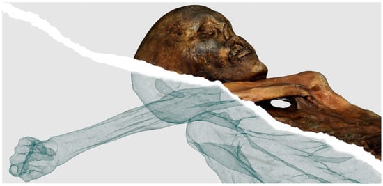
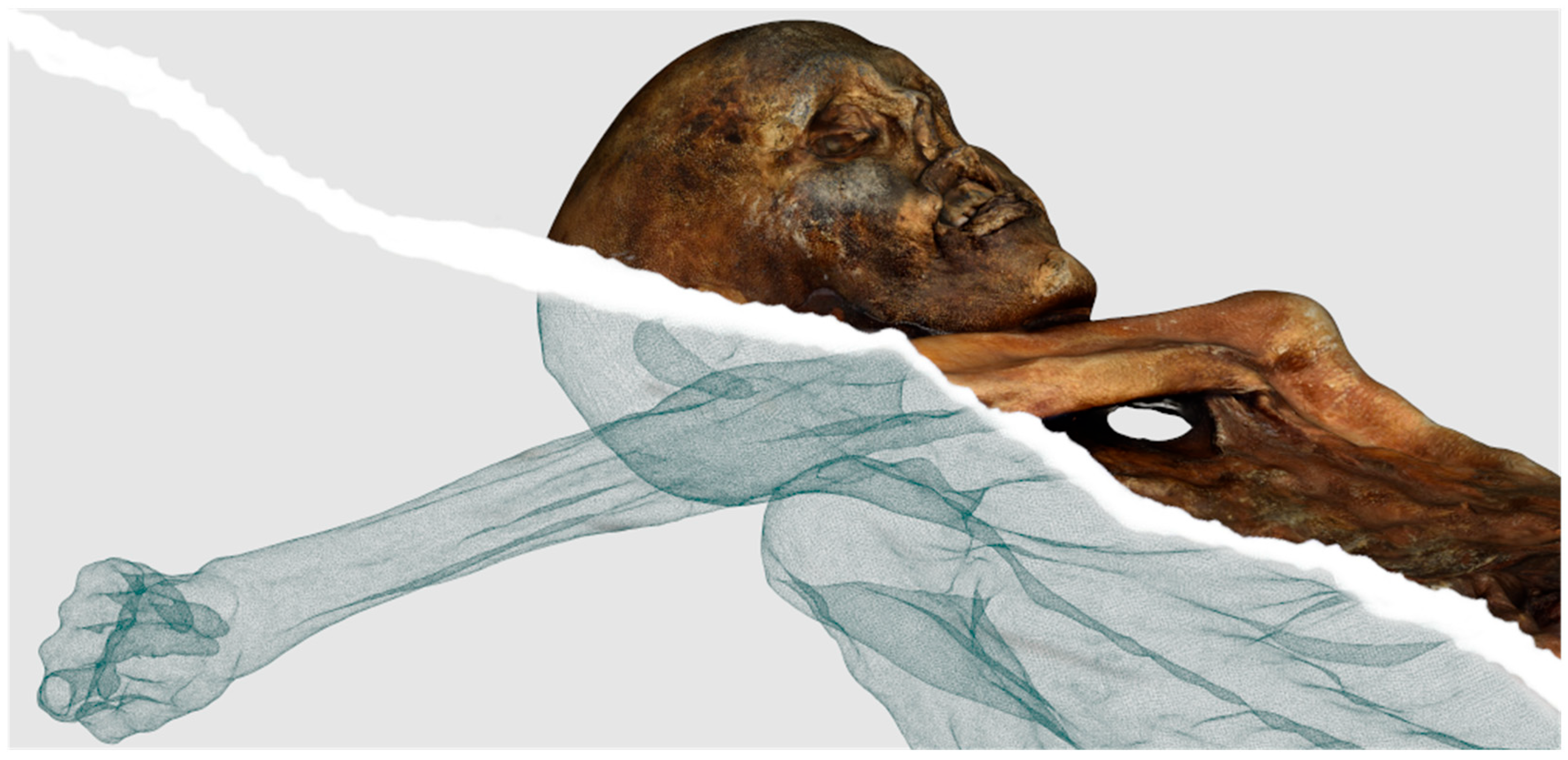
The primary aim of this paper is to present a complete, multi-modal digital twin of one of the most famous mummies in the world, Ötzi the Iceman, designing a sustainable framework to support long-term conservation, micro-spatial monitoring, multidisciplinary research, and high-fidelity public outreach, ensuring both the ethical stewardship and accessibility of the remains (Figure 1).
Figure 1.
The digital twin of Ötzi the Iceman. The image shows both the high-resolution textured 3D mesh and its underlying polygonal wireframe representation.
The Copper Age mummy [1] was discovered in 1991 by hikers on an alpine glacier [2] close to the Austrian–Italian border (46.7789° N, 10.8392° E). Commonly known as ‘Ötzi’ (derived from the Ötztal Alps where the discovery took place) or the Similaun mummy (in reference to the nearby Similaun peak), this individual was murdered by an arrowshot [3].
The exceptional nature of this discovery lies not only in the high level of natural preservation of the human remains over five millennia but also in the recovery of the complete personal equipment [4]. Consequently, the associated clothing, yew bow, copper axe, and other tools found in context offer an unprecedented opportunity for technological study and museum exhibition.
While 3D documentation methodologies are well-established in cultural heritage, their application within museum institutions has become increasingly significant. The creation of digital twins offers key advantages for preservation, research, and public dissemination [5]. From a research perspective, high-fidelity digital replicas enable scholars worldwide to conduct detailed morphological analyses and virtual simulations without physically handling fragile artifacts, thereby minimizing physical conservation risks. Additionally, comparative 3D recording allows conservators to monitor the structural integrity and preservation state of physical objects over time. Finally, these digital assets enhance museum exhibitions and public outreach, providing interactive and scientifically grounded educational experiences.
Recognizing that the direct conservation of physical artifacts remains the primary mandate of museum institutions, digital preservation offers a systematic method for long-term data permanence. Organic materials are inherently susceptible to natural degradation over time. High-resolution digital twins provide a permanent, non-destructive record of an object’s precise physical state at a specific point in time, mitigating the risk of irreversible data loss and ensuring the perpetual availability of structural and contextual information for future research.
Although computerized tomography scans of the mummy have been utilized in past research [6], the resulting DICOM datasets lack radiometric information, meaning they do not capture surface color or texture. For the systematic monitoring of sensitive organic remains, color documentation can be highly significant. The photogrammetric approach presented here, based on high-resolution photographic coverage, captures minute variations on the body’s surface, providing a critical baseline to detect physical deterioration. Furthermore, this documentation allows for the recording of superficial features (such as the mummy’s historic tattoos [7]) which are not visible in sub-surface CT data due to their lack of density contrast.
This project presents the first multimodal 3D reconstruction of the mummy that integrates clinical CT data with high-resolution photogrammetric surveys. This approach enables the co-registration of both sub-surface anatomical structures and outer surface textures into a single, comprehensive digital twin. By establishing this integrated methodology, future recordings (possibly conducted at regular intervals) will facilitate quantitative comparative analyses, allowing for the early detection and mitigation of potential subtle conservation anomalies or structural changes.
Hypothetical taphonomic variations over time can include micro-dehydration and localized volumetric mass loss, as minimal water sublimation still occurs even within the strictly controlled microclimate of the preservation chamber. Furthermore, this monitoring can identify potential surface degradation resulting from necessary maintenance handling and mechanical stress, as well as any early stages of unexpected biological or chemical alterations. Quantifying these possible geometric variations is crucial to scientifically validate the long-term efficacy of the current preservation protocols and to proactively adjust the environmental parameters before any macroscopic damage occurs.
2. Materials and Methods
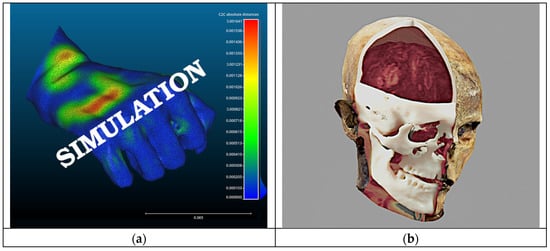
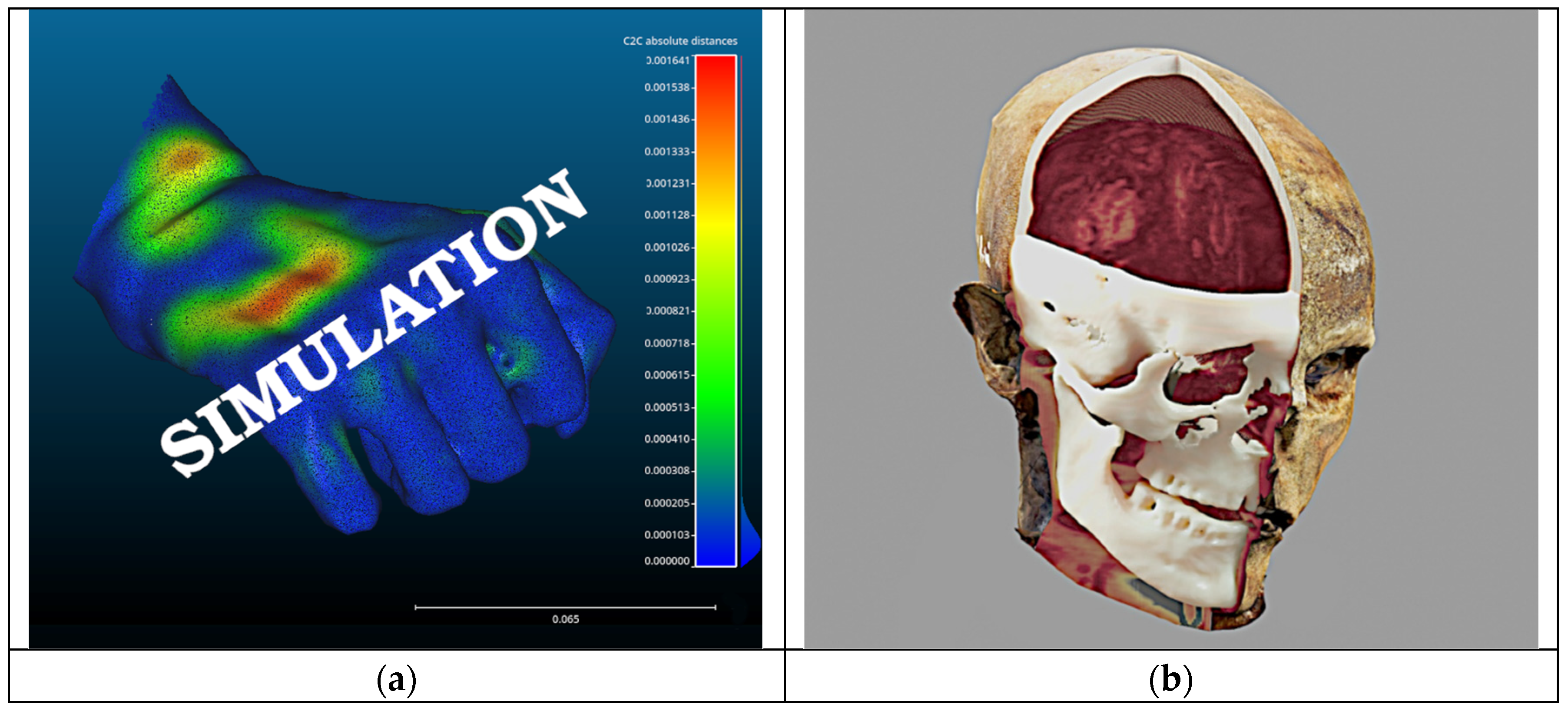
A project based on the 3D documentation of a museum heritage such as that of the Similaun mummy aims to address diverse museological, conservative [8], and scientific requirements. Among these, a priority is the necessity to enhance microbiological monitoring [9] of both the artifacts and the mummy itself. This is achieved by integrating 3D technologies with regular photogrammetric surveys: ideally, every 3–5 years for stable archaeological objects (kept in standard climatized museum showcases at room temperature) and every 7 years for the wet mummified human remains (preserved in a high-vulnerability state of cryo-conservation). Such a strategy is capable of detecting potential chromatic or morphological changes (Figure 2a).
Figure 2.
(a) Surface deviation analysis simulating morphological micro-changes within CloudCompare (v.2.12) software; (b) multimodal visualization rendered in Blender (v.4.0.2) via the OrtogOnBlender (v.2.91) add-on for exhibition purposes, integrating the SfM-derived skin 3D mesh with the skeletal 3D mesh and brain volumetric data (voxels) derived from DICOM scans.
This approach was adopted and further developed as it enables continuous, long-term monitoring of artifacts and the identification of alterations, while fully avoiding invasive procedures [10,11]. Equally significant are the outreach objectives, related to the use of the generated outputs for exhibitions and displays (Figure 2b). Furthermore, the project serves pure research purposes by enabling the remote sharing of a simplified ‘digital twins’ from such an extraordinary context within the scientific community. The following sections will present the methodology employed throughout the project, specifically illustrating the challenges encountered and the solutions proposed.
2.1. Critical Issues and Project Constraints
Given the project’s premises, the entire 3D documentation process must be oriented toward a low archaeological tolerance, which translates into the attempt to achieve the highest level of detail, characterized by high precision and high accuracy. Nevertheless, the find context (an Alpine glacier that has preserved a vast amount of organic material) and the heterogeneity of the artifacts make the entire operation highly challenging. Indeed, in addition to the mummy itself, the 3D survey involved much of the clothing and equipment found in situ (the original archaeological context), namely: the quiver, the bearskin cap, the axe, the belt with pouch, the loincloth, the shoes, the leggings, the coat, and, recently, the longbow [4]. Such a variety of artifacts necessarily corresponds to an equally wide range of materials, including leather [12], fur [13,14], wood [15], copper [16,17], plant fibers [18], flint, skin, and ice. Each of these is characterized by different physical properties that entail distinct challenges for 3D documentation. Among these differences, a primary factor is undoubtedly how the surface of the object interacts with light, ranging from materials that partially absorb light to those that primarily exhibit diffuse reflection, up to the most problematic ones characterized by specular reflection.
Beyond these technical difficulties, logistical constraints must also be considered, as the Similaun mummy is currently preserved in a specialized cold cell at the South Tyrol Museum of Archaeology, maintained at −6.5 °C and 99% relative humidity. These extreme parameters are calibrated to replicate the glacial environment, thereby preventing tissue desiccation, mass loss, and opportunistic microbial proliferation.
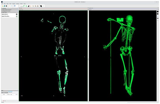
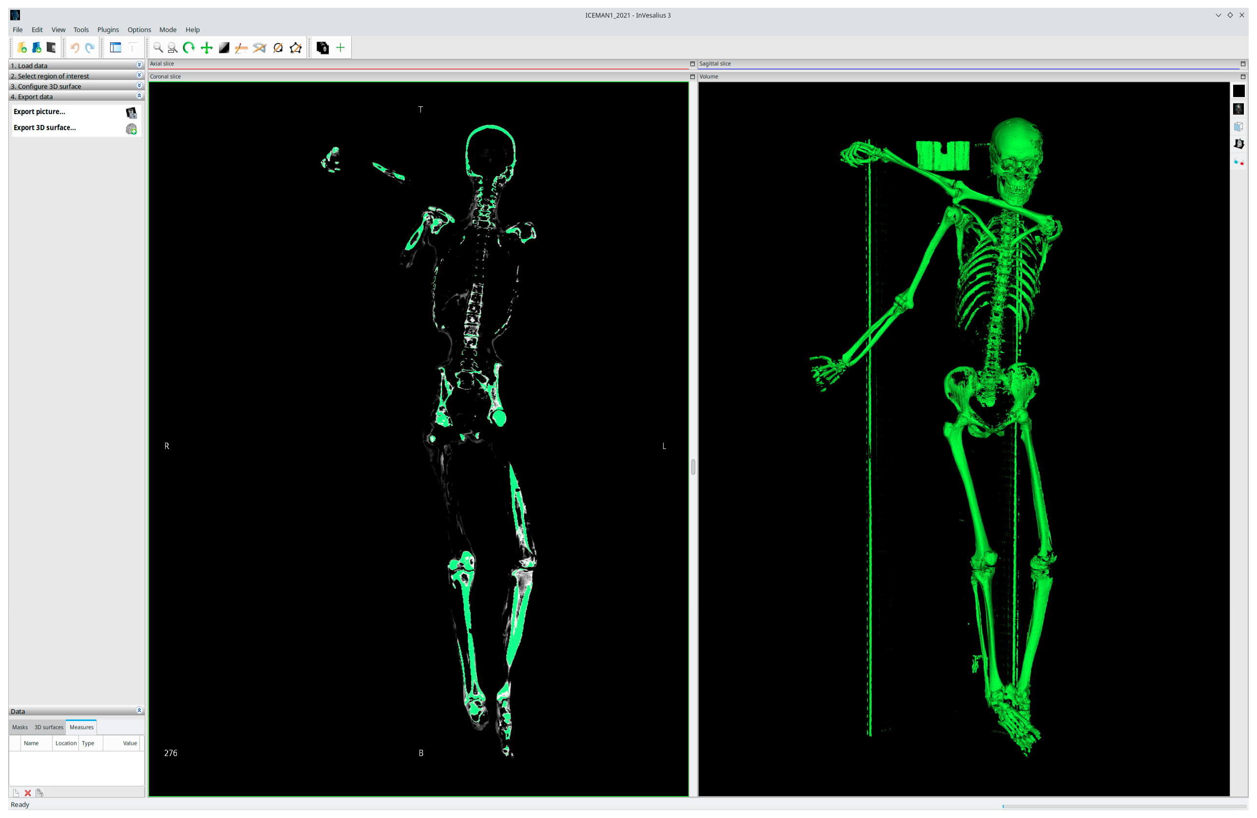
Furthermore, the project required the integration of heterogeneous data, such as the 3D survey outputs and CT scan data (Figure 3), based on DICOM standards and volumetric (voxel-based) graphics. Given this complex scenario, the fundamental prerequisite for the project’s success was the adoption of a survey technique capable of addressing and resolving all the aforementioned difficulties. This technique, as explained in the following section, was identified as photogrammetry based on Structure from Motion (SfM).
Figure 3.
Representation, within InVesalius (v.3.1.9), of the whole-body CT scan of the Similaun mummy performed at Bolzano Hospital (2021).
2.2. Technical Workflow and Applied Solutions
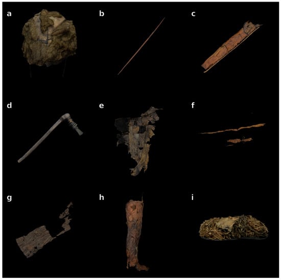
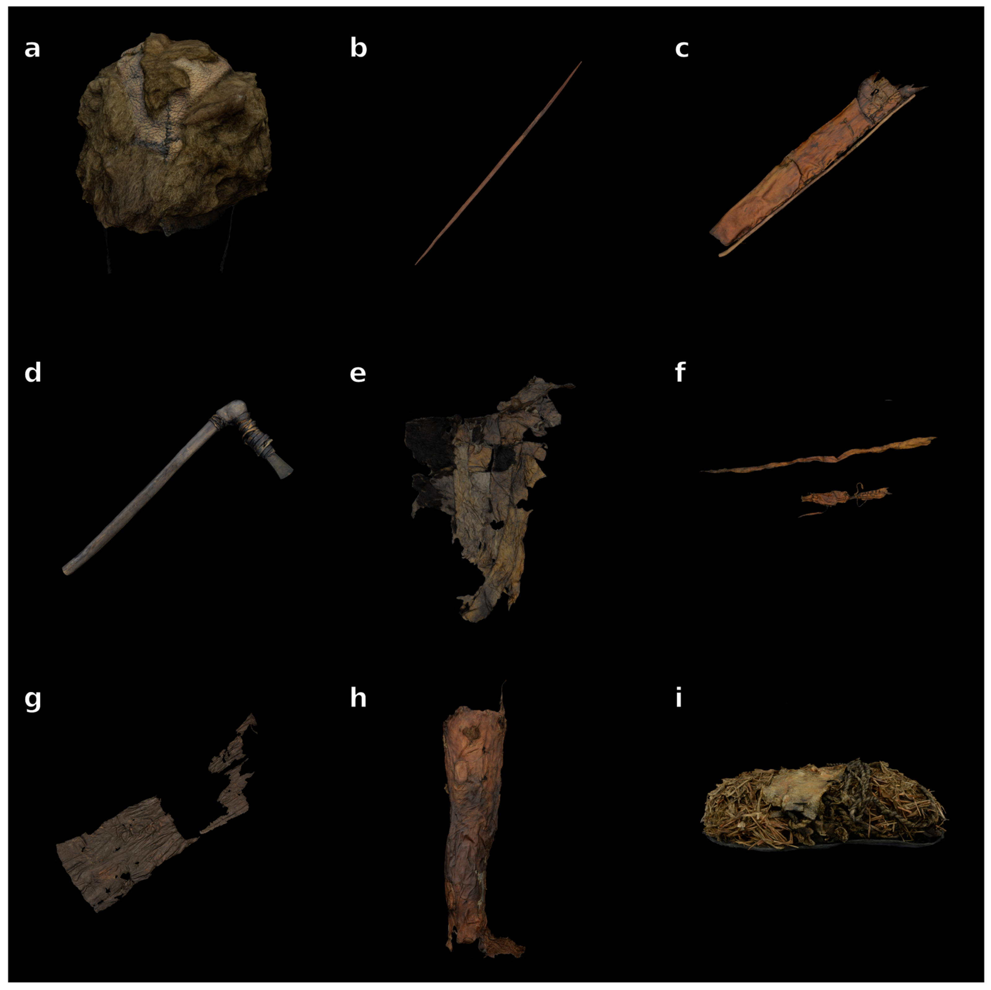
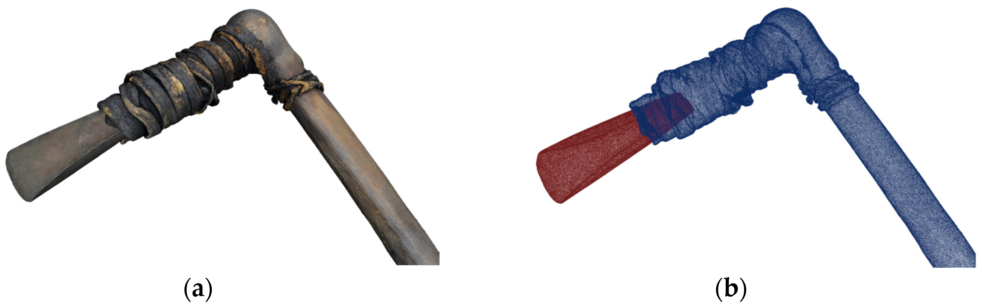
As previously mentioned, to address all the difficulties encountered during the project, focus was given to choosing a 3D survey methodology versatile enough to adapt to the peculiar needs of archaeological documentation. This choice landed on Structure from Motion (SfM, Figure 4 and Figure 5a,b), a technique already widely utilized in archaeology since 2008, thanks to the Open Software Bundler [19], and, as a branch of photogrammetry, increasingly popular in recent years [20]. When coupled with appropriate robotic devices [21,22], SfM has been proven to deliver acceptable results even under challenging technical conditions and in extreme environments [23,24]. As a photogrammetric methodology, SfM stands out for its versatility during both the delicate data acquisition and the subsequent data processing stages: in both steps, the procedure can be optimized at various levels. For instance, while the acquisition can be tailored to different materials or environmental contexts, the processing stage allows for an increased level of detail by integrating the workflow with specific Image-Based Modeling (IBM) techniques.
Figure 4.
3D digital documentation of some of the Iceman’s archaeological garments and equipment obtained via Structure from Motion (SfM): (a) bearskin cap; (b) bow; (c) quiver; (d) yew axe; (e) hide coat; (f) belt and pouch; (g) loincloth; (h) legging; (i) shoe. Note: The objects are not represented to the same scale; each item has been scaled independently to optimize individual visibility and detail preservation within the layout.
Figure 5.
(a) Digital twin of the copper axe (high-resolution textured mesh); (b) wireframe representation of the model, illustrating the assembly details and the structural connection between the copper blade (in red) and the wooden handle through the leather lashing (in blue).
Nevertheless, for the conservation requirements of the project, the Structure from Motion approach proved insufficient for the specific case of the bearskin cap. Consequently, it was integrated with the experimental use of Neural Radiance Field (NeRF) algorithms [25]. This integration was relatively straightforward, as both techniques rely on processing the same photographic dataset, making them highly compatible. Regardless, the application of SfM throughout the entire 3D documentation process required several technical adjustments, which will be described in detail in the following sections in relation to the specific challenges addressed.
2.3. Archaeological Tolerance and Non-Lambertian Surfaces
The challenges encountered in adopting SfM techniques for the survey of the Similaun mummy and its archaeological context primarily stemmed from three types of materials: leather, fur, and ice. Briefly, these difficulties can be summarized as follows: first, the level of detail achieved in the scanning of certain leather elements and the bearskin cap was insufficient to meet the pre-established archaeological tolerance; second, significant issues arose due to the specular reflection of light on non-Lambertian surfaces, such as the protective ice layer covering the mummy.
As is evident, these are common problems in all 3D archaeological survey methods, whether based on photographic acquisition, Time of Flight (ToF) instruments (devices that measure distance by calculating the time a laser pulse takes to return to the sensor), or structured light emission. Only the recent integration of blue laser technology into semi-professional handheld scanners, which occurred between 2024 and 2025 (years subsequent to the project presented here, dating back to 2022 for the finds and 2023–2024 for the mummy), has partially resolved the difficulties related to specular reflection on small objects. However, these advancements still do not guarantee the possibility of a totally passive (non-contact and markerless) survey for sensitive, medium-to-large-scale objects such as the Similaun mummy. For this type of intervention, SfM remains the only viable solution today.
2.3.1. Survey of the Archaeological Artifacts
Regarding the need to align the 3D survey’s level of detail with the required archaeological tolerance (specifically for the project’s conservation goals) action was taken in two directions: IBM-based post-processing techniques were adopted for the leather artifacts, while NeRF technology was experimentally employed as a complementary tool for the fur.
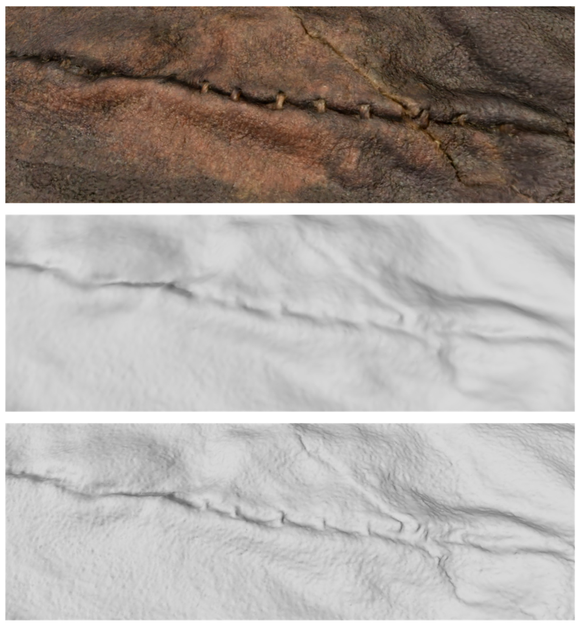
In the first case, after generating the 3D models (digital twins) of the leather finds through the combined use of openMVG (open Multiple View Geometry, v.2.1) and openMVS (open Multi-View Stereo, v.2.3.0) [26], the level of detail was enhanced by recovering information from the 2D photographic dataset. This operation, which leverages IBM techniques, follows a methodology successfully tested by Arc-Team in 2018 during the 3D documentation of the ‘incorrupt’ body of Saint Catherine Fieschi Adorno (Genoa, Italy). That project was aimed at Forensic Facial Approximation (FFA) [27] and performed using the OrtogOnBlender (v.2.91) application [28] (an open-source digital surgical planning and forensic facial approximation toolkit integrated with Blender). On that occasion, due to the impossibility of opening the glass reliquary, the 3D survey was conducted via SfM using photographs taken from a distance through the glass panels. Since the resulting model resolution was insufficient for the planned FFA operations, the detail was enhanced by experimenting for the first time with Displacement Mapping. This process involves extracting high-frequency geometric details from 2D photographic data and translating them into physical deformations of the 3D surface. Unlike Bump or Normal Mapping techniques, which only simulate the appearance of detail through shading and light manipulation without altering the actual geometry, Displacement Mapping effectively modifies the mesh topology by shifting the vertices based on a ‘height map’ derived from the source images. This allowed us to recover the subtle rugosity and micro-textures of ancient leather, ensuring that the digital twin accurately reflects the morphological complexity of the original find (Figure 6).
Figure 6.
Detail of a leather artifact from the Iceman’s equipment, illustrating the geometric enhancement pipeline: (top) the final textured 3D mesh; (center) the medium-to-low-resolution mesh as initially generated by the SfM process; (bottom) the high-definition mesh after Displacement Mapping post-processing, highlighting the recovery of micro-textures and surface rugosity.
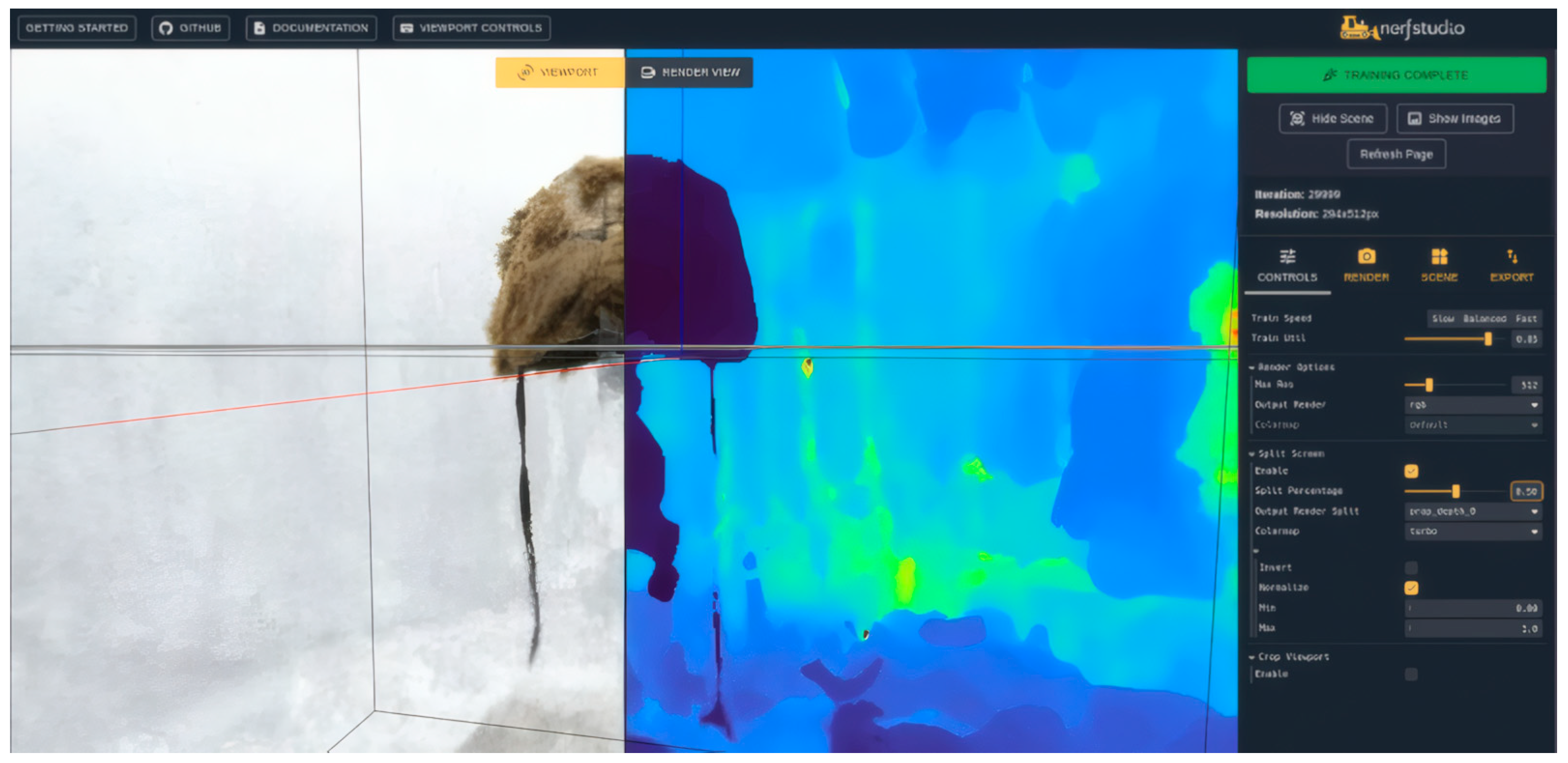
In the case of the fur (represented by a unique artifact, the bearskin cap) an integrative technique was required to render the details of the thick hair preserved in large, virtually intact areas. Within these areas, extraneous hairs were also detected; these do not belong to the original bear hide but were deposited onto the artifact through various post-depositional processes [13]. While other garments, such as the striped goat and sheepskin coat, featured sparse fur that could be successfully reconstructed using standard SfM-IBM workflows, the bear pelt’s dense and overlapping fur presented significant challenges. In such cases, traditional photogrammetry often fails to resolve the fine, non-Lambertian structures of individual hairs, resulting in ‘noisy’ or over-smoothed geometries. For this reason, the integration of Neural Radiance Fields (NeRFs) proved essential. We adopted a dual approach: first, a 3D model was generated via SfM to capture the overall volumes and the primary clusters of fur as accurately as possible. Simultaneously, using the same photographic dataset within the NerfStudio (v.1.0.2) framework [29], we obtained an interactive system that recorded the exact state of the artifact at the time of documentation (Figure 7).
Figure 7.
The bearskin cap visualized within the NerfStudio framework using a split-screen mode: the left side displays the RGB output render (standard color representation), while the right side shows the Depth output render (depth map estimation in false colors), illustrating the neural network’s ability to interpret the complex volumetric geometry of the fur.
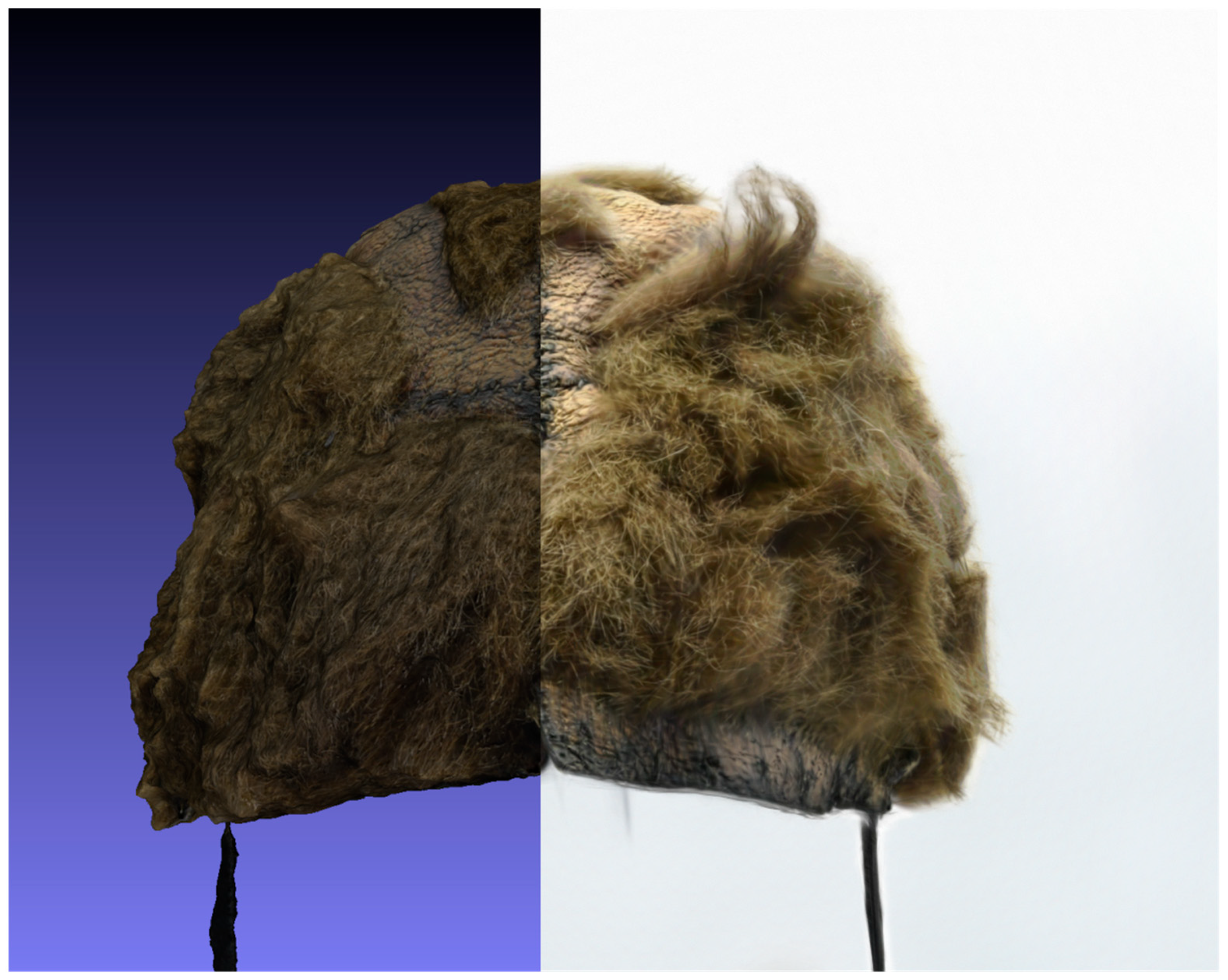
This was made possible by the NeRF algorithms, which represent the scene as a continuous volumetric function. Instead of defining a surface with discrete vertices, NeRFs optimize a multilayer perception (MLP) to map spatial coordinates and viewing angles (x, y, z, θ, ϕ) to specific color and density values. This allows the system to generate high-resolution 2D renderings in real-time from any perspective, effectively capturing complex light interactions and the intricate volumetric nature of the dense fur that traditional mesh-based models cannot fully represent. In other words, while SfM provided a metric–morphological documentation of the bearskin cap (resulting in a discrete and measurable 3D model that is necessarily simplified) the NeRF approach provided an optical–volumetric documentation. This yielded a continuous and implicit representation of the find, based on its radiance field, capable of fully covering its complex nature (Figure 8). This volumetric documentation approach also has significant implications, as it enables detailed visualization of possible ongoing taphonomic processes and surface alterations that may support future investigations.
Figure 8.
Comparative visualization of the bearskin cap: (left) the discrete 3D mesh processed via SfM and visualized in MeshLab (v.2023.12), providing a metrically accurate but simplified geometric surface; (right) the implicit NeRF representation within the NerfStudio (v.1.0.2) framework, capturing the high-fidelity volumetric detail of the dense fur. The side-by-side comparison highlights the complementary nature of these methodologies for the 3D documentation of complex, non-Lambertian archaeological organic materials.
2.3.2. Survey of the Mummy
Unlike the survey of the archaeological finds, the 3D documentation of the mummy presented challenges primarily related to the non-Lambertian surfaces created by the protective ice layer and wet skin.
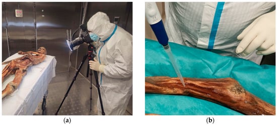
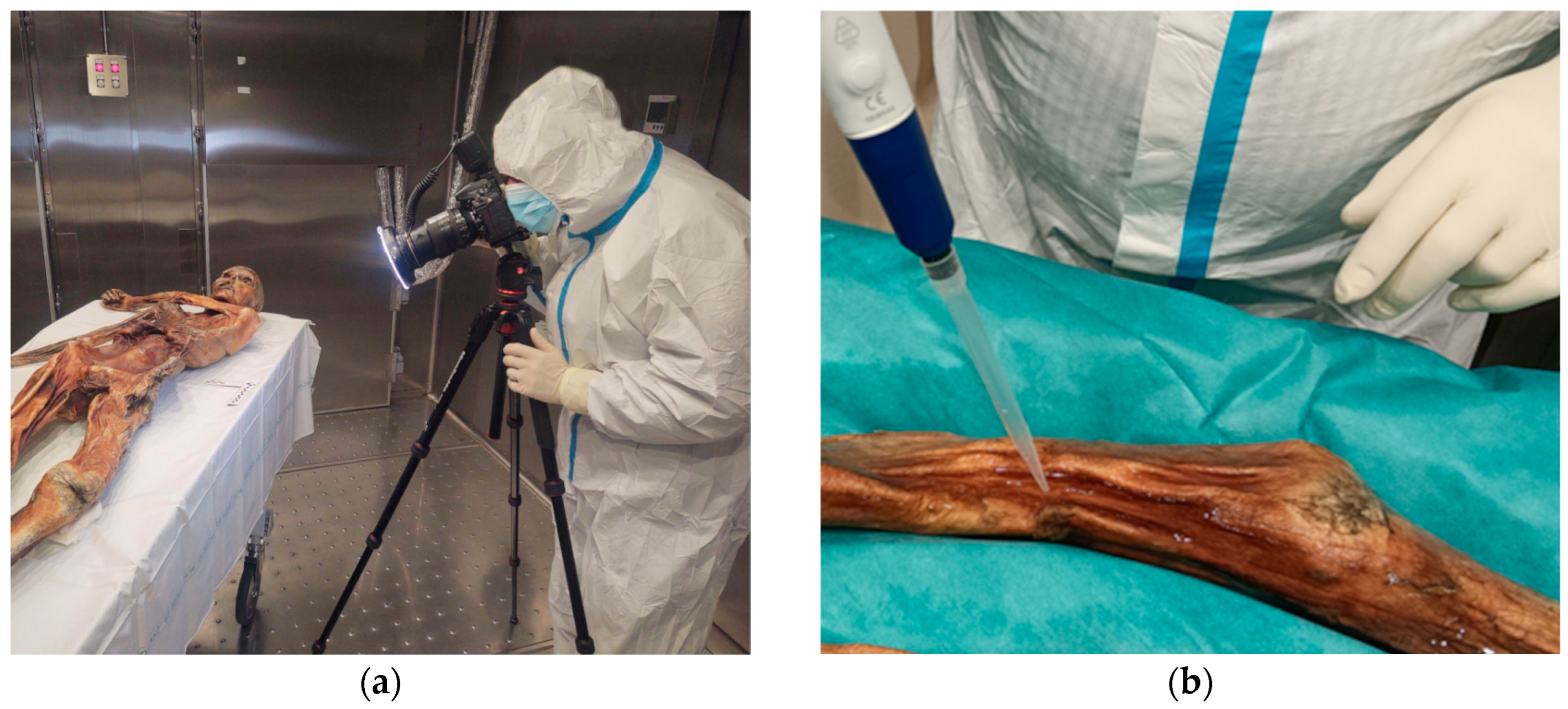
To obtain the most accurate digital twin possible, the documentation (Figure 9a) followed an exceptional analysis of the mummy’s state of preservation, aimed at excluding modern bacterial contamination (Figure 9b). During this analysis, the mummy was temporarily and partially defrosted to allow the collection of meltwater samples, thereby reducing the ice layer on the skin surface (an optimal condition for 3D scanning). The survey was conducted in the interval between this exceptional procedure and the subsequent routine nebulization with sterilized water, intended to counteract the natural weight loss of the mummified tissues.
Figure 9.
(a) Photographic acquisition phase inside the cleanroom laboratory chamber, conducted under strictly controlled temperature (−6.5 °C) and aseptic conditions; (b) meltwater sampling from the mummy’s protective ice layer for microbiological monitoring, performed during the extraordinary maintenance procedure prior to the 3D survey.
The workflow began with the sterilization of the photographic equipment and the personnel (three specialized archaeological operators who rotated during the nearly six-hour acquisition phase). Access was gained through a decontamination dressing room, a Class 10,000 cleanroom (at rest) according to Federal Standard 209e (corresponding to ISO Class 7, 14644-1 [30]), which utilizes U.V. germicidal irradiation and maintains a temperature of 20 °C. Following the sampling for microbiological monitoring, the archaeologists were granted access to the laboratory chamber. This environment was kept aseptic by an advanced air conditioning and filtration system, where the mummy, transported from the conservation or backup chambers (both at −6.5 °C and 99% relative humidity), was protected by a flow of sterile, decontaminated air [31]. Inside the laboratory, the temperature was gradually brought back to −6.5 °C while two operators at a time performed the photographic acquisition for SfM.
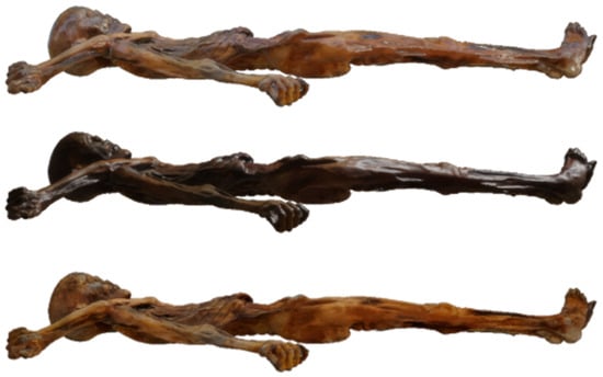
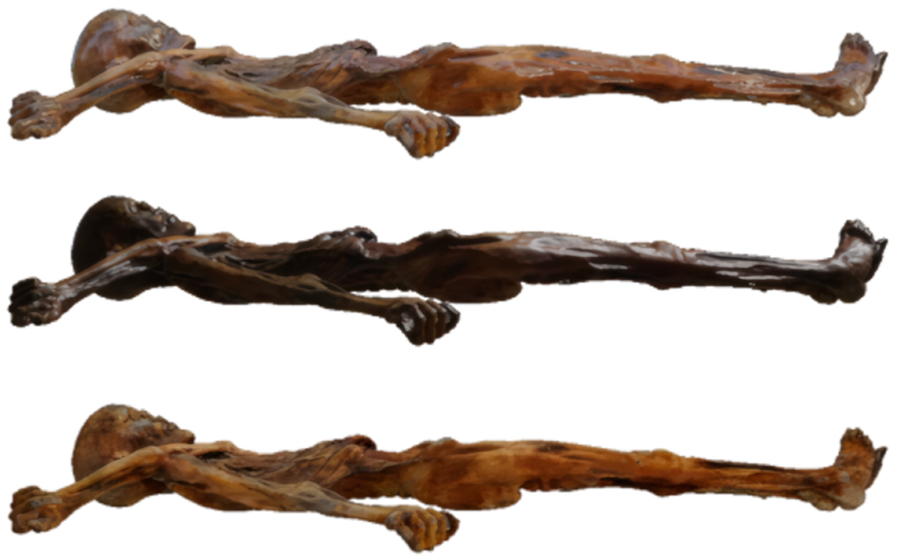
Given the challenges of non-Lambertian surfaces, the Arc-Team crew implemented a strategy similar to the one used for the 3D documentation of the ‘Ori di Taranto’ (2020–2022), a project documenting the Hellenistic and Roman gold artifacts from the National Archaeological Museum of Taranto [32]. That strategy relied on Polarized Light Photography (PLP) within a controlled darkroom environment at the National Archaeological Museum of Taranto (MaRTA), using a single zenithal polarized light source. While this allowed for reflection-free acquisition, the strategy could not be replicated identically due to the impossibility of darkening the laboratory chamber or controlling its multiple light sources with filters. Consequently, the team adapted the approach using a more versatile setup: a D-ring LED equipped with a custom-built polarizing filter. The polarized ring-light effectively countered the ambient laboratory lighting (and shadows), allowing a second polarizing filter on the camera lens to eliminate specular reflections (Figure 10).
Figure 10.
Comparison of light reflection components on the mummy’s surface: (top) the original unpolarized image featuring both diffuse and specular reflections; (center) the isolated specular reflection component, highlighting the glare from the ice layer and wet tissues; (bottom) the isolated diffuse reflection component (Lambertian surface) obtained through cross-polarization. This final, glare-free dataset was used for the SfM processing to ensure high-fidelity geometric reconstruction.
Once the issues of light reflection were resolved, the acquisition strategy had to ensure a complete survey while minimizing direct contact. It was decided to perform the survey in two main stages: one for the anterior aspect and one for the posterior aspect. The archaeologists avoided direct interaction with the body, delegating all necessary handling to specialized medical personnel (Dr. Oliver Peschel, forensic pathologist at Munich University, and Dr. Martina Tauber, pathologist at Bolzano/Bozen Regional Hospital).
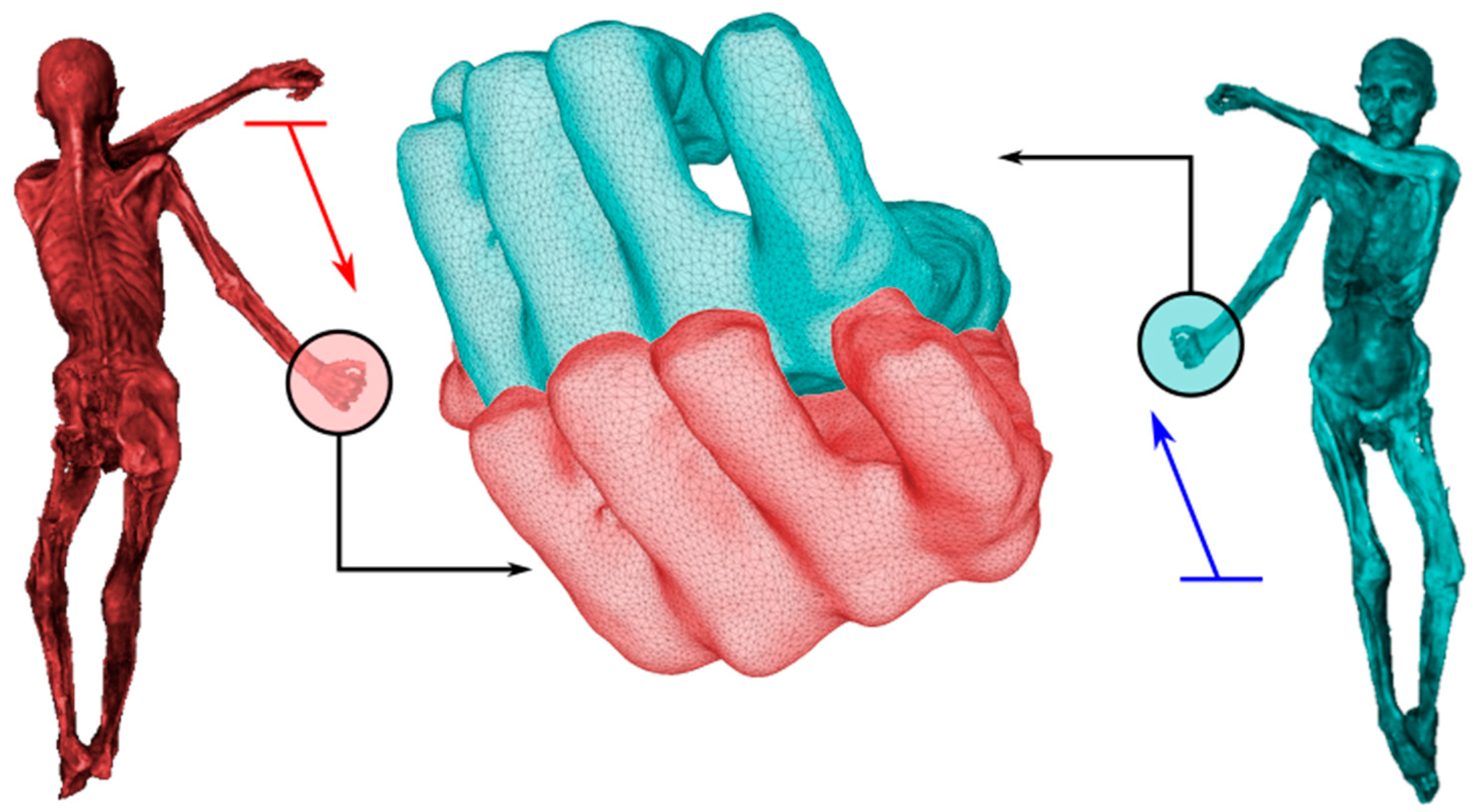
During acquisition, it was assumed that such conservation precaution would not affect data processing, provided the survey rotation was centered on the mummy’s head, theoretically allowing sequential photo processing. However, as was observed a posteriori (afterward), even minimal handling (especially during partial defrosting) causes slight, often imperceptible shifts in the body’s posture (in this case, a displacement of approximately 5 cm for the right hand and 1 cm for the left hand; Figure 11). This condition created unforeseen difficulties in the SfM pipeline, which were resolved through a segmented data processing approach. The model was divided into four sectors and subsequently reassembled digitally in Meshlab (v.2023.12):
Figure 11.
Visualization of the postural shift observed during the integration of different datasets: the overlay highlights a misalignment of approximately 5 cm in the right hand between the 3D survey of the dorsal side (left, shown in red) and the ventral side (right, shown in blue).
- Head, torso, lower limbs, and part of the left arm;
- Left forearm and hand;
- Upper right arm;
- Lower right arm.
This process ultimately yielded a high-fidelity digital twin of the Similaun mummy, derived from diffuse-light imagery and free from specular noise, successfully meeting the pre-established parameters of archaeological tolerance.
3. Results
Given the successful results obtained from the mummy’s scan, the project was expanded by integrating simplified 3D digital twin with DICOM data from the most recent CT whole-body scan available, performed at the Bolzano Hospital in 2021. This scan was conducted using a Siemens Somatom Definition Flash system, according to the parameters detailed in Table 1.
Table 1.
CT parameters of the image whole-body image session of 2021.
To this end, data processing was performed using InVesalius (v.3.1.9) software [33], through the automated extraction of the bone tissue 3D model from the whole-body CT DICOM dataset. This FLOSS solution was selected because it is specifically optimized for the precise thresholding and segmentation of medical DICOM datasets.
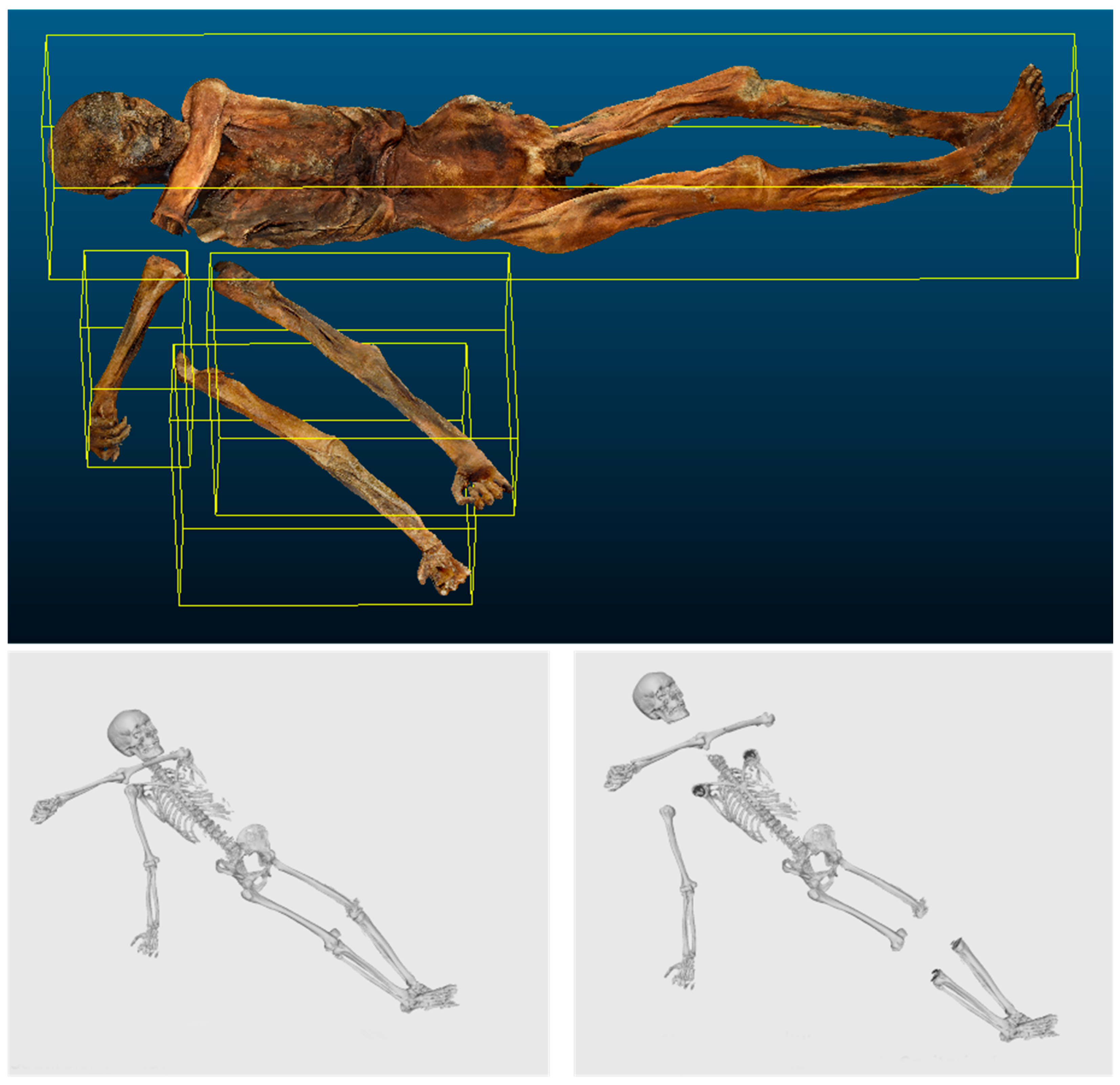
As observed during the SfM survey, the skeletal 3D model derived from the 2021 CT scan exhibited a slightly different posture compared to the 2023 digital twin, making them incompatible, particularly regarding the upper and lower limbs. To standardize the datasets and obtain a unified model, a new segmentation was required, this time performed in post-processing on the skeletal 3D model within Blender. The intervention focused primarily on the joints: for the lower limbs, by disarticulating the femorotibial joint and separating the femurs and patellae from the tibiae and fibulae; for the upper limbs, by disarticulating the humeral heads from the glenoid cavity of the shoulder; and for the cervical region, by separating the atlanto-axial joint. These operations resulted in five distinct anatomical blocks (cranium, right upper limb, left upper limb, torso with lower limbs down to the patella, and the distal part of the lower limbs), which were then realigned in Blender with the 2023 digital twin (Figure 12).
Figure 12.
Multi-segment anatomical reconstruction strategy to compensate for postural shifts: (top) the SfM-derived skin mesh partitioned into four segments (torso including head, left forearm, upper right arm, and lower right arm); (bottom) comparison with the CT-derived skeletal model, showing the full skeleton (right) and its five-segment partition ((left): cranium, right upper limb, left upper limb, torso with lower limbs down to the patellae, and distal lower limbs).
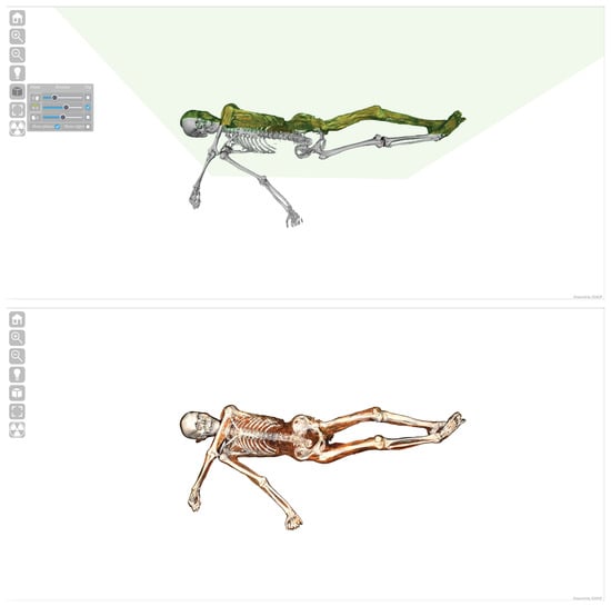
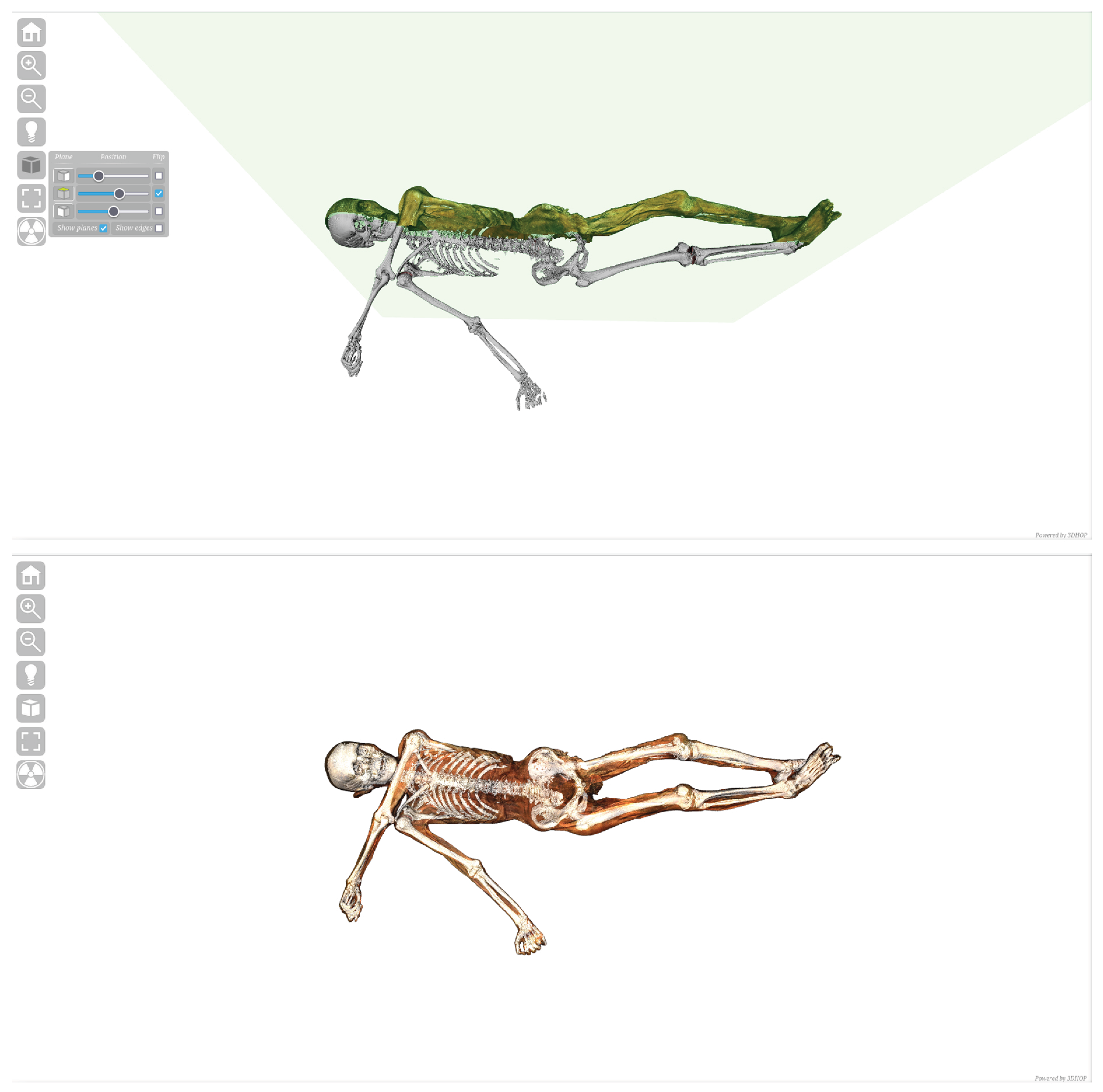
Once two morphologically and metrically correct models (consistent in posture) were obtained, a web-based platform for interactive visualization of the 3D data was used to create a virtual environment for their integration: the open-source 3DHOP (v.4.3) (3D Heritage Online Presenter) framework [34]. This led to the development of a web-based application that allows researchers to remotely visualize both the mummy’s digital twin and the internal skeletal 3D model simultaneously. This application represents the first instance of merging DICOM data (from CT) with a digital twin (from SfM). Furthermore, the application leverages 3DHOP’s core features to provide additional research tools: from basic functions, like zooming and measurement, to more refined ones such as ambient lighting control. To allow for the visualization of the skeletal system, the mummy’s model can be sectioned along the three axes using the ‘Plane Sections’ tool, revealing the underlying bone structure. Additionally, by customizing the 3DHOP source code, a new ‘X-ray’ tool was implemented, allowing users to adjust the opacity of the mummy’s 3D model and view the skeletal model through a transparency effect (Figure 13).
Figure 13.
The interactive 3D web-visualizer developed using the 3DHOP framework: (top) the Plane Sections tool, enabling the real-time exploration of internal sections; (bottom) the X-ray visualization tool, custom-developed for this project to allow the simultaneous inspection of the integrated skin and skeletal datasets.
Technical Notes
The entire project was conducted exclusively using FLOSS (Free/Libre and Open-Source Software) within a GNU/Linux Debian 12 ‘bookworm’ distribution derived from the ArcheOS project. This approach, broadly related to Open Science [35,36,37] and recently applied to Archaeology and Cultural Heritage [38], specifically follows the old Open Archaeology model, the advantages of which (in terms of transparency and scientific sustainability) have been extensively discussed in the earlier literature [39,40,41].
Specifically, all SfM (Structure from Motion) operations were carried out using openMVG and openMVS [26]. Quality control and mesh analysis were performed using MeshLab [42] and CloudCompare [43]. Further IBM (Image-Based Modeling) procedures, such as Displacement Mapping, were executed within the Blender suite, partially utilizing the OrtogOnBlender add-on [28] (which also supports the full SfM pipeline and 3D surface extraction from DICOM data). The 3D documentation of the bearskin cap using NeRF (Neural Radiance Field) algorithms was processed within the NerfStudio framework [29], while the extraction of the skeletal 3D model from CT DICOM data was performed in InVesalius [33]. Finally, the web-based application for remote analysis and data fusion was developed using the 3DHOP framework [34].
The photographic survey complied with the technical guidelines established by the Ufficio Beni Archeologici/Amt fuer Bodendenkmaeler (Soprintendenza ai Beni Culturali/Abteilung Denkmalpflege della Provincia Autonoma di Bolzano/Alto Adige—Autonome Provinz Bozen/Suedtirol), which require capturing images in RAW + Fine format alongside physical scale bars (to ensure spatial and metrological accuracy) and colorimetric targets (to ensure colorimetric calibration protocol), even for standard 2D archaeological photos. Data acquisition was conducted using a Nikon D810 camera (Nikon Corporation, Tokyo, Japan) with a 24–120 mm zoom lens, mounted on a Manfrotto Befree Advanced AS tripod (Manfrotto, Cassola, Italy). In the specific case of the mummy, a Godox Ring72 illuminator (Godox Photo Equipment Co., Ltd., Shenzhen, China) was employed, equipped with a custom-built polarizing filter constructed from high-efficiency linear polarizing sheets optimized for the visible spectrum (400–700 nm, extinction ratio equal to or greater than 99.9%) to manage specular reflections. The RAW files were then processed using the open-source software Darktable. Finally, data processing was performed on a Dell G5 15 workstation (Dell Inc., Round Rock, TX, USA), equipped with an Intel Core i7 10th Generation processor (Intel Corporation, Santa Clara, CA, USA), 16 GB of RAM, and an NVIDIA GeForce RTX GPU (NVIDIA Corporation, Santa Clara, CA, USA). The dataset regarding the Iceman mummy was based on 1294 effective high-resolution images (640 for the front and 654 for the back). The close-range acquisition guaranteed a sub-millimetric Ground Sample Distance (GSD < 0.5 mm/pixel). To ensure long-term usability on the museum’s local hardware infrastructure, the final optimized 3D mesh delivered consists of 1,552,744 faces and 776,355 vertices, preserving high geometric fidelity while maintaining data accessibility. Based on the sub-millimetric Ground Sample Distance (GSD < 0.5 mm/pixel), the removal of specular noise via cross-polarized lighting, and the integration of physical scale bars, the acquisition pipeline provides a sub-millimetric spatial resolution with a theoretical geometric accuracy and local precision within the range of ±0.5 mm (an error acceptable in other highly demanding scientific fields, such as Virtual Surgical Planning [44].
4. Discussion
The 3D documentation project of the Similaun mummy and the associated archaeological artifacts was primarily carried out using SfM (Structure from Motion) technologies. This choice dates to 2022; however, it remains the only viable approach in such a constrained context, despite the recent emergence of portable blue-light laser scanners capable of documenting non-Lambertian surfaces. In fact, these instruments are not yet capable of operating on medium-to-large sensitive surfaces with a markerless approach (avoiding the application of tracking markers directly onto the find). Furthermore, many semi-professional scanners currently on the market feature low-resolution camera sensors that are inadequate for the high-quality texturing required by archaeological standards. While this limitation is likely to be overcome by future market developments, the proposed workflow represents an effective and rigorous methodological framework for non-invasive documentation. The methodological transparency and open-source approach adopted in this project reflect a commitment to open science, reproducibility, and ethical stewardship, in the attempt to disclose digital heritage to the scientific community and grant long-term sustainable access to both data and methods.
At present, the integration of SfM techniques with NeRF-based algorithms provides results suitable for complex multidisciplinary studies, meeting the strict parameters of archaeological tolerance. It is important to emphasize that these two methodologies are inherently complementary, as they represent two distinct processing pipelines for the same photographic dataset. From a single acquisition, it is possible to generate both a discrete, measurable SfM model (ideal for metric analysis and diachronic morphological monitoring) and an implicit NeRF representation, which excels at capturing the complex appearance of materials like the dense bear-fur cap.
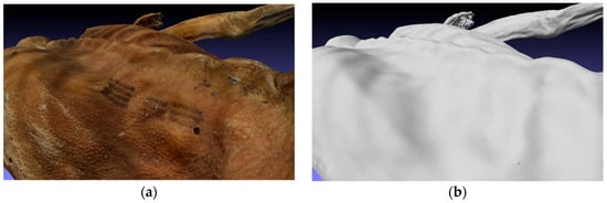
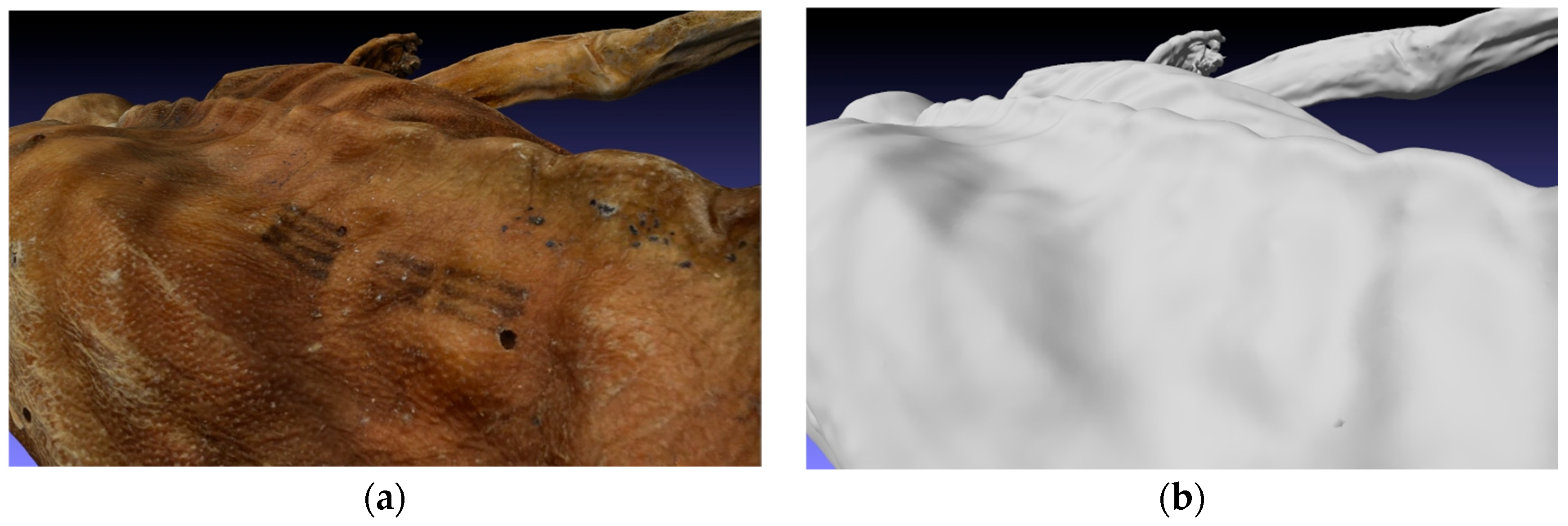
However, beyond the survey techniques used for the project, one of its most relevant achievements is the multimodal integration within the 3DHOP framework: the fusion of SfM-derived surfaces (the skin) with 3D skeletal models extracted from DICOM datasets. This result opens significant perspectives for further integration with other ice-preserved internal organs, granting both a long-term monitoring method and a new instrument for future forensic investigations. The integrated 3DHOP model is not a final output but rather a modular framework. Looking ahead, it will be possible to incorporate new reconstructive models of systems (such as the vascular network) that are difficult to segment directly from the mummy’s CT scans due to taphonomic processes and tissue shrinkage. These reconstructions could be developed using proxy anatomical data from anonymous patients (modern CT datasets). By applying deformable registration and warping techniques, these modern anatomical templates can be adapted to the specific morphology of the Similaun mummy, providing a high-fidelity virtual representation of his original internal anatomy. This methodology has been previously employed by the Arc-Team research group in the forensic study of a Ptolemaic mummy, yielding successful reconstructive results [45]. Furthermore, the 3DHOP application remains fully expandable. It can be implemented, for instance, to track ante-mortem and peri-mortem traumas, visually differentiating them (via dedicated hotspots and infoboxes) from post-mortem damage occurred during the mummy’s recovery or from early forensic investigations, such as the 1991 exploratory drillings in Innsbruck (the so called ‘Austrian windows’ [46]). Similarly, the framework could integrate previous research on the mummy’s 61 tattoos, building upon the comprehensive mapping conducted in 2015 [7] (Figure 14a,b).
Figure 14.
Comparison between surface and internal imaging for the documentation of epidermal features: (a) detail of the high-resolution textured 3D mesh, where a specific tattoo on the mummy’s back is clearly visible; (b) the same area visualized on the non-textured CT-derived model, highlighting the limitations of tomographic data in capturing superficial pigmentations and skin markings.
Regardless of future research directions, the choice of a FLOSS ecosystem like 3DHOP not only grants the global scientific community remote access to the data but also ensures the continuous integration of both legacy and new datasets. This infrastructure could be further enhanced by coupling it with an open-source Database Management System (DBMS) such as PostgreSQL (v.18.6), ensuring long-term scientific sustainability and transparency.
Ultimately, from an ethical and museological standpoint, the resulting dematerialization of the Iceman mummy through this multimodal digital twin does not merely represent a technological achievement but serves as an ethical tool for preventive conservation. The ethics of digital human remains is a complex, globally debated topic. However, in the context of such an exceptionally fragile specimen, high-fidelity digitization offers a non-invasive solution to a fundamental museological dilemma: balancing the need for scientific access with the preservation of biological integrity. By shifting future diagnostic, forensic, and taphonomic investigations to a virtual environment (such as the 3DHOP framework, v.4.3), we dramatically reduce the necessity for physical manipulation and exposure of the actual remains. Thus, the virtual twin becomes an ethical proxy, ensuring global scientific transparency and long-term monitoring while safeguarding the physical mummy in its controlled environment.
Author Contributions
Conceptualization, A.P., A.B., L.B. and R.G.; methodology, L.B., A.B. and R.G.; software, L.B., C.M.; validation, A.B., L.B., R.G. and A.P.; formal analysis, A.B., L.B., R.G. and C.M.; investigation, A.B., L.B. and R.G.; resources, E.V.; data curation, A.B., R.G., L.B., C.M., P.P.; writing—original draft preparation, L.B.; writing—review and editing, E.E.G., M.T., A.B., L.B., O.P. and A.P.; visualization, A.B. and L.B.; supervision, A.P.; project administration, A.P. and E.V.; funding acquisition, E.V. and A.P. All authors have read and agreed to the published version of the manuscript.
Funding
This research was funded by the South Tyrol Museum of Archaeology as part of an official public assignment under the project code CUP H53C23000260005.
Institutional Review Board Statement
This research was conducted in agreement with the ICOM Code of Ethics for Museums (2004 and following revisions).
Data Availability Statement
Data will soon be available on the official South Tyrol Museum of Archaeology website.
Acknowledgments
The authors would like to thank the South Tyrol Health Authority (Azienda Sanitaria dell’Alto Adige/Südtiroler Sanitätsbetrieb) for their assistance. We also extend our gratitude to the staff of the South Tyrol Museum of Archaeology, in particular to Alexander Sanin and Andrea Battagin, for their valuable support. During the preparation of this manuscript the authors used Gemini Google (April 2026) for the purposes of grammatical review and improving clarity and accuracy in English. The authors have reviewed and edited the output and take full responsibility for the content of this publication.
Conflicts of Interest
Authors Luca Bezzi, Alessandro Bezzi, and Rupert Gietl are founding partners of the company Arc-Team (Italy), which received financial compensation from the South Tyrol Museum of Archaeology to perform the technical 3D documentation described in this study. The funder (the Museum) was actively involved in the study design, logistical coordination, data collection, and the decision to submit the paper for publication. The remaining authors declare no commercial or financial relationships that could be construed as a potential conflict of interest.
Abbreviations
The following abbreviations are used in this manuscript:
| 3D | Three-Dimensional |
| CT | Computed Tomography |
| DBMS | Database Management System |
| DICOM | Digital Imaging and COmmunications in Medicine |
| FFA | Forensic Facial Approximation |
| FLOSS | Free/Libre and Open-Source Software |
| FOV | Field Of View |
| GB | Gigabyte |
| GNU | Gnu is Not Unix |
| GPU | Graphics Processing Unit |
| IBM | Image-Based Modeling |
| ISO | International Organization for Standardization |
| kV | Kilovolt |
| LED | Light-Emitting Diode |
| MLP | Multi-Layer Perceptron |
| NeRF | Neural Radiance Field |
| PLP | Polarized Light Photography |
| UV | Ultraviolet |
| RAM | Random Access Memory |
| RGB | Red, Green, Blue |
| SfM | Structure from Motion |
| ToF | Time of Flight |
References
- Bonani, I.; Niklaus, S.; Housley, B.; van Klinken, H. Altersbestimmung von Milligrammproben der Ötztaler Gletscherleiche mit der Beschlunigermassenspektrometrie-Methode (AMS) [Age Determination of Milligram Samples of the Ötztal Glacier Mummy using Accelerator Mass Spectrometry (AMS)]. In Der Mann im Eis [The Man in the Ice], Band 1, 1st ed.; Höpfel, F., Platzer, W., Spindler, K., Eds.; Eigenverlag der Universität Innsbruck: Innsbruk, Austria, 1992; Volume 1, pp. 108–117. [Google Scholar]
- Rastbichler-Zissnerig, E. Der Mann im Eis—Die Fundgeschichte: Die Interpretation der Quellen als Grundlage für die Rekonstruktion des archäologischen Befunds [The Man in the Ice—The Discovery History: The Interpretation of Sources as a Basis for Reconstructing the Archaeological Context], 1st ed.; Eigenverlag der Universität Innsbruck: Innsbruck, Austria, 2006. [Google Scholar]
- Pernter, P.; Gostner, P.; Egarter Vigl, E.; Jakobus Rühli, F. Radiologic proof for the Iceman’s cause of death (ca. 5’300BP). J. Archaeol. Sci. 2007, 34, 1784–1786. [Google Scholar] [CrossRef] [Scilit]
- Egg, M.; Spindler, K. Kleidung und Ausrüstung Der Gletschermumie aus den Ötztaler Alpen [Clothing and Equipment of the Glacier Mummy from the Ötztal Alps], 1st ed.; Römisch-Germanisches Zentralmuseum: Mainz, Germany, 2009. [Google Scholar]
- Dagnaw, G.A.; Capuano, R.; Muccini, H. Digital Twins for Cultural Heritage: A Systematic Analysis of the State of the Art. ACM Comput. Surv. 2026, 58, 237. [Google Scholar] [CrossRef] [Scilit]
- Murphy, W.; Nedden, D.; Gostner, P.; Knapp, R.; Recheis, W.; Seidler, H. The Iceman: Discovery and Imaging. Radiology 2003, 226, 614–629. [Google Scholar] [CrossRef] [Scilit]
- Samadelli, M.; Melis, M.; Miccoli, M.; Vigl, E.; Zink, A. Complete mapping of the tattoos of the 5300-year-old Tyrolean Iceman. J. Cult. Herit. 2015, 16, 753–758. [Google Scholar] [CrossRef] [Scilit]
- Egarter Vigl, E. Die Konservierung der Mumie des Mannes aus dem Eis im Südtiroler Archäologiemuseum [The Conservation of the Mummy of the Man from the Ice in the South Tyrol Museum of Archaeology]. In Die Gletschermumie aus der Kupferzeit 2 [The Copper Age Glacier Mummy 2]; Fleckinger, A., Ed.; Folio Verlag: Bozen, Italy; Vienna, Austria, 2003; pp. 35–41. [Google Scholar]
- Sarhan, M.S.; Samadelli, M.; Zink, A. The Iceman’s microbiome: Unveiling millennia of microbial diversity and continuity. Microbiome 2026, 14, 135. [Google Scholar] [CrossRef] [Scilit]
- Zandasrar, F. Non-Invasive Documentation of Archaeological Textiles: Scythian Fabrics from Southern Ukraine. Master’s Thesis, Università degli Studi di Padova, Padova, Italy, 2024. [Google Scholar]
- Brandolini, F.; Patrucco, G. Structure-from-Motion (SFM) Photogrammetry as a Non-Invasive Methodology to Digitalize Historical Documents: A Highly Flexible and Low-Cost Approach? Heritage 2019, 2, 2124–2136. [Google Scholar] [CrossRef] [Scilit]
- O’Sullivan, N.; Teasdale, M.; Mattiangeli, V.; Maixner, F.; Pinhasi, R.; Bradley, D.; Zink, A. A whole mitochondria analysis of the Tyrolean Iceman’s leather provides insights into the animal sources of Copper Age clothing. Sci. Rep. 2016, 6, 31279. [Google Scholar] [CrossRef] [Scilit]
- Goedecker-Ciolek, R. Konservierung der Beifunde einer Gletschermumie vom Ende der Steinzeit I—Bekleidung [Conservation of the Associated Finds of a Glacier Mummy from the End of the Stone Age I—Clothing]. Arbeitsblätter Für Restaur. 1994, 27, 105–127. [Google Scholar]
- Lange, J. Weitere Untersuchungen zu den Felllederproben des Mannes aus dem Eis [Further Investigations on the Fur and Leather Samples of the Man from the Ice]. In Kleidung und Ausrüstung des Mannes im Eis [Clothing and Equipment of the Man in the Ice]; Egg, M., Spindler, K., Eds.; Verlag des Römisch-Germanischen Zentralmuseums: Mainz, Germany, 2009; pp. 211–247. [Google Scholar]
- Schoch, W.; Oeggl, K. Dendrochronological analyses of artefacts and other remains. In The Iceman and His Natural Environment; Bortenschlager, S., Oeggl, K., Eds.; The Man in the Ice; Springer: Vienna, Austria; New York, NY, USA, 2000; Volume 4, pp. 29–63. [Google Scholar]
- Artioli, G.; Angelini, I.; Canovaro, C.; Kaufmann, G.; Villa, I.M. Lead isotopes of prehistoric copper tools define metallurgical phases in Late Neolithic and Eneolithic Italy. Sci. Rep. 2024, 14, 5382. [Google Scholar] [CrossRef] [Scilit]
- Artioli, G.; Angelini, I.; Kaufmann, G.; Canovaro, C.; Dal Sasso, G.; Villa, I.M. Long-distance connections in the Copper Age: New evidence from the Alpine Iceman’s copper axe. PLoS ONE 2017, 12, e0179263. [Google Scholar] [CrossRef] [Scilit]
- Pfeifer, K.; Oeggl, K. Analysis of the bast used by the Iceman as binding material. In The Iceman and His Natural Environment; Bortenschlager, S., Oeggl, K., Eds.; The Man in the Ice; Springer: Vienna, Austria; New York, NY, USA, 2000; Volume 4, pp. 69–77. [Google Scholar]
- Bezzi, A.; Bezzi, L.; Ducke, B. Computer Vision e Structure from Motion, nuove metodologie per la documentazione archeologica tridimensionale: Un approccio aperto [Computer Vision and Structure from Motion, New Methodologies for Three-Dimensional Archaeological Documentation: An Open Approach]. Insul. Diomedeae 2011, 19, 103–111. [Google Scholar]
- Sapirstein, P.; Murray, S.A. Establishing Best Practices for Photogrammetric Recording during Archaeological Excavation. J. Field Archaeol. 2017, 42, 337–350. [Google Scholar] [CrossRef] [Scilit]
- Bezzi, L.; Bezzi, A.; Gietl, R.; Naponiello, G.; Feistmantl, K. Archeorobotics. Applicazioni robotiche aperte e archeologia estrema [Archeorobotics. Open Robotic Applications and Extreme Archaeology]. Archeol. Calc. 2019, 30, 467–470. [Google Scholar] [CrossRef] [Scilit]
- Themistocleous, K. The Use of UAVs for Cultural Heritage and Archaeology. In Remote Sensing for Archaeology and Cultural Landscapes: Best Practices and Perspectives Across Europe and the Middle East; Hadjimitsis, D.G., Agapiou, A., Lasaponara, R., Masini, N., Eds.; Springer: Cham, Switzerland, 2019; pp. 241–269. [Google Scholar] [CrossRef] [Scilit]
- Bezzi, L.; Bezzi, A.; Boscaro, C.; Feistmantl, K.; Gietl, R.; Naponiello, G.; Ottati, F.; de Guzman, M. Commercial Archaeology and 3D Web Technologies. J. Field Archaeol. 2018, 43, S45–S59. [Google Scholar] [CrossRef] [Scilit]
- Westoby, M.J.; Brasington, J.; Glasser, N.F.; Hambrey, M.J.; Reynolds, J.M. ‘Structure-from-Motion’ photogrammetry: A low-cost, effective tool for geoscience applications. Geomorphology 2012, 179, 300–314. [Google Scholar] [CrossRef] [Scilit]
- Mildenhall, B.; Srinivasan, P.P.; Tancik, M.; Barron, J.T.; Ramamoorthi, R.; Ng, R. NeRF: Representing Scenes as Neural Radiance Fields for View Synthesis. In Lecture Notes in Computer Science; Springer: Cham, Switzerland, 2020; pp. 405–421. [Google Scholar] [CrossRef] [Scilit]
- Moulon, P.; Monasse, P.; Perrot, R.; Marlet, R. OpenMVG: Open Multiple View Geometry. In Lecture Notes in Computer Science; Springer: Cham, Switzerland, 2017; pp. 60–74. [Google Scholar] [CrossRef] [Scilit]
- Bezzi, L.; Bezzi, A.; Moraes, C. Ricostruzione Facciale Forense di S.Caterina Fieschi Adorno [Forensic Facial Reconstruction of Saint Catherine Fieschi Adorno]; Technical Report; Arc-Team: Cles, Italy, 2018; pp. 1–15. [Google Scholar] [CrossRef]
- Moraes, C.; Moulon, P.; Bezzi, A.; Moura, L.; Rosa, E.; Dornelles, R.; Bezzi, L. Photogrammetry in OrtogOnBlender XP: The Story of an (R)Evolution. OrtogOnLineMag 2025, 7, 9–16. [Google Scholar] [CrossRef]
- Tancik, M.; Weber, E.; Ng, E.; Li, R.; Yi, B.; Wang, T.; Kristoffersen, A.; Austin, J.; Salahi, K.; Ahuja, A.; et al. Nerfstudio: A Modular Framework for Neural Radiance Field Development. In Proceeding of the Special Interest Group on Computer Graphics and Interactive Techniques Conference, Los Angeles CA, USA, 6–10 August 2023; pp. 1–12. [Google Scholar] [CrossRef] [Scilit]
- ISO 14644-1:2015; Cleanrooms and associated controlled environments—Part 1: Classification of air cleanliness by particle concentration. International Organization for Standardization: Geneva, Switzerland, 2015.
- Angelantoni, C. Il sistema di conservazione “dell’uomo venuto dal ghiaccio” [The Preservation System of the “Man Came from the Ice”]. Available online: https://www.centrogalileo.it/nuovaPA/Articoli%20tecnici/otzi.htm (accessed on 3 May 2026).
- Bezzi, L.; Bezzi, A.; Gietl, R.; Moraes, C.; Naponiello, G.; Airò, S.; Putzer, A.; Silvestri, E. Digital twins of archaeological finds: Open source technologies applied to 3D scanning. Archeol. Calc. 2024, 35, 203–214. [Google Scholar] [CrossRef]
- Moraes, T.; Amorim, P.; Azevedo, F.; Silva, J. In Vesalius: An Open-Source Software for Reconstruction of Medical Images. World J. Urol. 2011, 30, 687–691. [Google Scholar] [CrossRef] [Scilit]
- Potenziani, M.; Callieri, M.; Dellepiane, M.; Corsini, M.; Ponchio, F.; Scopigno, R. 3DHOP: 3D Heritage Online Presenter. Comput. Graph. 2015, 52, 129–141. [Google Scholar] [CrossRef] [Scilit]
- Wilkinson, M.D.; Dumontier, M.; Aalbersberg, I.J.; Appleton, G.; Axton, M.; Baak, A.; Blomberg, N.; Boiten, J.W.; da Silva Santos, L.B.; Bourne, P.E.; et al. The FAIR Guiding Principles for scientific data management and stewardship. Sci. Data 2016, 3, 160018. [Google Scholar] [CrossRef] [Scilit]
- Barker, M.; Chue Hong, N.P.; Katz, D.S.; Lamprecht, A.L.; Martinez, C.; Psomopoulos, F.; Harrow, J.; Castro, L.J.; Gruenpeter, M.; Martinez, P.A.; et al. Introducing the FAIR Principles for research software. Sci. Data 2022, 9, 622. [Google Scholar] [CrossRef] [Scilit]
- Prlić, A.; Procter, J.B. Ten Simple Rules for the Open Development of Scientific Software. PLoS Comput. Biol. 2012, 8, e1002802. [Google Scholar] [CrossRef] [Scilit]
- Marwick, B. Computational Reproducibility in Archaeological Research: Basic Principles and a Case Study of Their Implementation. J. Archaeol. Method Theory 2017, 24, 424–450. [Google Scholar] [CrossRef] [Scilit]
- Bagnara, R.; Macchi Jánica, G. Introduzione. In Open Source, Free Software e Open Format nei Processi di Ricerca Archeologici. Atti del I Workshop (Grosseto, 8 Maggio 2006); Bagnara, R., Macchi Jánica, G., Eds.; Centro Editoriale Toscano: Florence, Italy, 2007; pp. 1–6. [Google Scholar]
- Costa, S.; Pesce, G.L. Introduzione. In Open Source, Free Software e Open Format nei Processi di Ricerca Archeologica: Atti del II Workshop (Genova, 11 Maggio 2007); Costa, S., Pesce, G.L., Eds.; Ubiquity Press: London, UK, 2013; pp. 1–8. [Google Scholar] [CrossRef] [Scilit]
- Bezzi, L.; Francisci, D.; Grossi, P.; Lotto, D. Introduzione [Introduction]. In Proceedings of the Open Source, Free Software e Open Format nei Processi di Ricerca Archeologica. Atti del III Workshop, Padova, Italia, 8–9 Maggio 2008; Bezzi, L., Francisci, D., Grossi, P., Lotto, D., Eds.; Quazar: Rome, Italy, 2009; pp. 9–11. [Google Scholar]
- Cignoni, P.; Callieri, M.; Corsini, M.; Dellepiane, M.; Ganovelli, F.; Ranzuglia, G. MeshLab: An Open-Source Mesh Processing Tool. In Proceedings of the Eurographics Italian Chapter Conference, Salerno, Italy, 2–4 July 2008. [Google Scholar] [CrossRef]
- Girardeau-Montaut, D. Détection de Changement sur des Données Géométriques Tridimensionnelles. Ph.D. Thesis, ENST (École Nationale Supérieure des Télécommunications), Paris, France, 2006. [Google Scholar]
- Vitorino, N.d.S.; Dias, F.J.d.N.; Daruge, R.J. Photorealistic 3D Facial Meshes Obtained by Low-Cost Photogrammetry: Are They Precise Enough to Replace Colorless CT-scan Meshes in Virtual Surgical Planning? J. Funct. Biomater. 2023, 14, 468. [Google Scholar] [CrossRef] [Scilit]
- Carrara, N.; Scattolin, G.; Moraes, C.; Bezzi, L. CT scans of a Ptolemaic mummy from Heliopolis: Evidences of probable lethal injuries. Digit. Appl. Archaeol. Cult. Herit. 2026, 40, e00498. [Google Scholar] [CrossRef] [Scilit]
- Spindler, K. Der Mann im Eis: Die Ötztaler Mumie Verrät die Geheimnisse der Steinzeit, 1st ed.; Bertelsmann: Munich, Germany, 1993. [Google Scholar]

Disclaimer/Publisher’s Note: The statements, opinions and data contained in all publications are solely those of the individual author(s) and contributor(s) and not of MDPI and/or the editor(s). MDPI and/or the editor(s) disclaim responsibility for any injury to people or property resulting from any ideas, methods, instructions or products referred to in the content. |
© 2026 by the authors. Licensee MDPI, Basel, Switzerland. This article is an open access article distributed under the terms and conditions of the Creative Commons Attribution (CC BY) license.