


Abstract
This study examines the in vivo tongue resting configuration in relation to lingual mobility. While biomechanical models suggest the tongue may exhibit properties consistent with a muscular hydrostat, direct large-scale clinical observation of resting posture remains limited. The objective was to characterize tongue resting configuration before and after removal of lingual mechanical restriction (ankyloglossia), and to evaluate whether observed behavior aligns with hydrostatic principles. A retrospective cohort study was conducted at a single center pediatric specialty practice from September 2017 to November 2025. Patients were categorized by procedural setting, including an awake infant cohort (n = 3182) and a general anesthesia cohort (n = 355). Resting tongue position was assessed before and after the intervention using standardized observational techniques, with paired analyses performed. Full-length palatal contact increased from 0.0% pre-procedure to 99.5% postoperatively in the awake cohort (p < 0.001). In the general anesthesia cohort, 100% of patients demonstrated full-length palatal contact intraoperatively following restoration of unrestricted mobility (p < 0.001), regardless of age or palatal morphology. These findings demonstrate that, when unrestricted, the tongue consistently assumes a palatal resting configuration in vivo. This behavior is consistent with muscular hydrostat principles and supports consideration of the living tongue as a muscular hydrostat from a functional perspective.
1. Introduction
The human tongue is composed of intrinsic and extrinsic muscle groups oriented in different directions that permit movement and structural support without skeletal elements [1]. As such, the tongue exhibits suction-like behavior, similar to the tentacles of an octopus, permitting it to passively suction against the hard palate at rest [2].
During rest (defined here as periods outside of eating and speaking), the tongue has been theorized to distribute broadly within the oral cavity, potentially establishing passive contact with the hard palate in the presence of a moist, enclosed environment. The intraoral vacuum effect helps maintain palatal contact without active muscle force [3].
Despite this biomechanical framework, many anatomical depictions portray the resting tongue positioned inferiorly toward the floor of the mouth. This representation is largely derived from cadaveric observation, where suction-like behavior cannot be fully appreciated. In vivo examination is therefore essential for understanding tongue function under physiologic conditions [3,4].
The resting position of the tongue establishes its baseline spatial relationship within the oral cavity. Accurate characterization of this reference state is fundamental to describing both typical anatomy and structural deviation.
Beyond classical descriptions of the tongue, advances in imaging and biomechanical modeling over the past decade have further clarified its three-dimensional structural organization and deformation capabilities. Diffusion tensor imaging, magnetic resonance imaging (MRI), and computational modeling studies have demonstrated the complex interplay of intrinsic and extrinsic muscle fibers that allow volumetric redistribution without skeletal support [3,4,5]. These studies confirm that tongue shape and position reflect mechanical equilibrium within the spatial constraints of the oral cavity rather than simple gravitational positioning.
Muscular hydrostats such as the octopus tentacle and elephant trunk exhibit similar deformation principles, wherein internal pressure is redistributed within a closed muscular compartment bounded by surrounding structures [2]. In the human oral cavity, the hard palate represents the superior structural boundary of this compartment. Under physiologic conditions, the oral cavity is moist and enclosed, creating an environment capable of sustaining passive surface contact. This environment differs fundamentally from preserved cadaveric specimens, in which tissue hydration, neuromuscular tone, and pressure dynamics are altered [1,3].
Recent anatomical investigations have emphasized the variability and adaptability of tongue musculature across individuals and developmental stages [3]. However, most of the available literature evaluates tongue motion during speech, swallowing, or respiration, rather than passive resting posture. Imaging studies typically capture dynamic tasks or supine positioning under experimental conditions, which may not fully represent natural resting equilibrium [3,6,7].
Resting tongue posture has been evaluated using imaging modalities, clinical observation, and, less commonly, instrumental approaches; however, each method has limitations, including non-physiologic testing conditions, task dependence, or lack of standardization [3,6,7,8,9]. Consequently, no single approach has emerged as a definitive method for assessing passive resting configuration.
Observation of true resting tongue posture presents inherent methodological challenges. In awake individuals, clinical assessment of tongue position may be influenced by voluntary activation, task dependence, guarding, or awareness of the examination itself, making passive resting configuration difficult to assess directly. Natural resting posture may therefore be more readily observed in infancy, when passive resting states are frequently encountered during clinical examination. Outside of infancy, it may be more reliably evaluated under conditions in which conscious motor control is minimized, such as sleep, sedation, or general anesthesia. These conditions provide complementary opportunities to evaluate passive tongue equilibrium within the living oral cavity and are consistent with prior imaging-based approaches used to characterize tongue position and upper-airway anatomy in vivo [7,8,9,10].
Within the field of orofacial myology, tongue resting posture has been discussed as a component of functional balance and dentofacial development [11,12,13]. The orthodontic literature has proposed that habitual tongue position may influence palatal morphology and transverse maxillary growth patterns [14,15,16]. Despite these conceptual models, objective large-scale in vivo documentation of resting configuration remains limited.
Resting tongue posture has also been considered clinically relevant in the evaluation and management of ankyloglossia. Functional assessments often incorporate observation of tongue position at rest, as inferior or anterior resting posture has been associated with altered oral function, mouth breathing, and dentofacial development [11,12,13,14,15,16]. In the clinical and sleep medicine literature, ankyloglossia has been associated with functional phenotypes including altered airway dynamics and compensatory tongue positioning [11], while surgical and therapeutic interventions have been explored in relation to functional outcomes and tongue posture [12]. Despite its frequent inclusion in clinical decision-making frameworks, objective characterization of resting tongue posture remains limited, and criteria for its assessment are not standardized across studies.
This gap is particularly relevant in the context of lingual restriction, where it remains unclear whether altered resting posture reflects intrinsic neuromuscular patterning or a mechanical limitation of mobility. If the tongue functions as a muscular hydrostat, unrestricted mobility should permit it to assume a mechanically stable resting configuration characterized by broad palatal contact within the enclosed oral environment. Conversely, persistent inferior or partial positioning would suggest either structural constraints or deviations from hydrostatic behavior.
By examining resting posture before and after removal of lingual mechanical restriction in a large pediatric cohort, the present study provides an opportunity to evaluate this relationship in vivo. Rather than inferring resting configuration from dynamic tasks or imaging simulations alone, direct clinical visualization allows assessment of whether restoration of mobility is associated with a predictable shift toward full-length palatal distribution.
The primary objective of this study was to examine tongue resting position in infants and children before and after removal of lingual mechanical restriction (ankyloglossia), with the aim of evaluating whether in vivo resting configuration is consistent with the expected behavior of a muscular hydrostat when mechanical constraint is removed.
2. Materials and Methods
2.1. Study Design and Ethical Approval
A retrospective cohort study of pediatric patients at a single center was conducted based on records from September 2017 to November 2025. Clinical observations were performed by a single practitioner (the author), who was not blinded to the patient’s status. This observational study was reported in accordance with the Strengthening the Reporting of Observational Studies in Epidemiology (STROBE) guidelines to ensure transparency and completeness of reporting.
Patients were included if they underwent removal of lingual mechanical restriction (ankyloglossia) and had documented assessment of tongue resting posture both before and after the procedure.
The study was approved by an independent ethics board, Advarra (Protocol #00091702; approved 12 December 2025). The institutional review board granted a waiver of informed consent due to the retrospective design and use of de-identified clinical data. The study was conducted in accordance with the Declaration of Helsinki (1975, revised 2013).
2.2. Study Groups and Assessment Timing
Patients were categorized according to procedural setting at the time of removal of lingual mechanical restriction:
- Awake cohort—treated without general anesthesia
- Age range: 1–442 days
- Median age: 52 days (IQR 25–108)
- General Anesthesia cohort—treated under general anesthesia
- Age range: 1–18 years
- Median age: 5 years (IQR 3–8)
Although age ranges overlapped, assignment to cohort was determined by clinical and procedural considerations rather than chronological age alone. Indications for general anesthesia included the presence of erupted primary molars, which may limit postoperative stretching, and parental or guardian preference.
General anesthesia was also selected in patients with limited cooperation, allowing for controlled surgical conditions, precise release of lingual restriction, and standardized assessment of resting tongue posture. In this cohort, surgical closure with sutures was performed following release, facilitating optimal healing and prevention of adherence between the ventral tongue and the floor of the mouth. This approach minimized the influence of voluntary movement, guarding, gag reflex, and muscular fatigue while avoiding the use of local anesthesia.
For both cohorts, palatal morphology was assessed and categorized as wide, rounded, or narrow. In the awake cohort, classification was determined through operator palpation of the palate. In the general anesthesia cohort, assessment was based on palpation in conjunction with observation of dental spacing and/or crowding, as well as inter-cuspid and inter-molar measurements (at the level of the second primary molars or first permanent molars).
For both cohorts, tongue resting posture was assessed before removal of lingual mechanical restriction and reassessed following release.
In the awake cohort, postoperative reassessment occurred at a routine two-week follow-up visit. In the general anesthesia cohort, reassessment was performed intraoperatively immediately following release while patients remained under the same anesthetic conditions. Patients in the general anesthesia cohort were nasally intubated, and neuromuscular blocking agents were not administered at any time during the procedure.
2.3. Variables and Categorization
2.3.1. Awake Cohort (Infants)
Tongue resting posture was assessed immediately prior to the removal of lingual mechanical restriction. When the mouth was closed at rest, the mandible was gently lowered using light pressure applied to the chin to permit visualization of tongue position. When the mouth was open, light pressure was applied along the inferior border of the mandible to achieve passive mouth closure, followed by gentle mandibular depression to observe the resting position.
Care was taken to avoid direct contact with the tongue or stimulation of active movement during assessment, and the maneuver was performed without eliciting cry or voluntary tongue activation. The assessment was repeated twice to confirm the consistency of observations.
Visualization of palatal contact during mandibular depression was performed with consideration of palatal morphology, as the degree of mouth opening required to maintain intraoral suction may vary across individuals. Mandibular opening was adjusted as needed to preserve passive palatal contact during observation. Resting posture was determined based on observation of tongue position immediately upon gentle mandibular opening, often accompanied by an audible release of intraoral suction, consistent with prior palatal contact.
In infants, natural resting states are frequently observed during clinical examination, permitting visualization of passive tongue position following assisted mandibular positioning.
Postoperative reassessment at two weeks was performed using the same technique. This visit represents a routine evaluation, at which time mucosal healing is typically complete and oral tissues have stabilized.
Resting posture was categorized as either:
- Full-length palatal contact—broad distribution of the tongue along the entire hard palate from anterior to posterior
- Non-palatal resting configuration—inferior positioning toward the floor of the mouth, anterior interposition between the gum pads or teeth, or partial anterior (blade-only) palatal contact
For analytic purposes, full-length palatal contact was defined as the physiologic resting configuration, whereas non-palatal configurations were considered restricted or altered resting positions.
2.3.2. General Anesthesia Cohort (Children)
Resting posture was assessed intraoperatively immediately prior to and immediately following removal of lingual mechanical restriction. Mandibular guidance and visualization were performed using the same technique described for the awake cohort. Observations were documented with photographic and video recording, and the maneuver was repeated three times to confirm consistency.
Visualization of palatal contact during mandibular depression was performed with consideration of palatal morphology, as the degree of mouth opening required to maintain intraoral suction may vary across individuals. Mandibular opening was adjusted as needed to preserve passive palatal contact during observation.
In this cohort, achievement of unrestricted tongue mobility with full-length palatal contact was used as a procedural completion criterion. Post-release assessment, therefore, reflects the observed resting configuration at the time the surgical endpoint was achieved.
Resting posture was categorized into four mutually exclusive configurations:
- Tongue positioned inferiorly toward the floor of the mouth
- Tongue positioned anteriorly between the teeth
- Partial anterior (blade-only) palatal contact
- Full-length palatal contact extending along the entire hard palate
For comparative analysis with the awake cohort, these categories were collapsed into binary variables: full-length palatal contact versus non-palatal resting configuration.
2.4. Statistical Analysis
Statistical analyses were performed by an independent statistician who was not involved in the clinical procedures. Demographic and clinical variables were summarized descriptively. Categorical variables were reported as counts and percentages, and continuous variables as medians with ranges or interquartile ranges (IQR), as appropriate.
Because resting posture was assessed in the same participants before and after removal of lingual mechanical restriction, analyses accounted for paired data.
In the awake cohort, resting posture was classified as a binary variable (full-length palatal contact vs. non-palatal resting configuration). Pre–post changes were evaluated using McNemar’s test for paired dichotomous outcomes. Preoperative and postoperative proportions, absolute differences in proportions, and exact p-values were calculated.
In the general anesthesia cohort, resting posture was initially categorized into four mutually exclusive configurations. Pre–post changes in distribution were evaluated using the Stuart–Maxwell test, a generalization of McNemar’s test for paired nominal variables with more than two categories. For comparative purposes with the awake cohort, a secondary binary analysis (full-length palatal contact vs. non-palatal resting configuration) was performed using McNemar’s test.
No imputation was required, as no missing data were present.
All analyses were conducted in R (version 4.4.0). Statistical significance was defined as a two-sided p < 0.05.
3. Results
3.1. Awake Cohort (Infants)
A total of 3182 infants were included (age range: 1–442 days; median age: 51 days [IQR: 25–108]). Of these, 1701 (53.5%) were male, and 1480 (46.5%) were female (Table 1). The median postoperative follow-up interval was 16 days (range: 7–33 days).
Table 1.
Demographic Characteristics by Procedural Cohort.
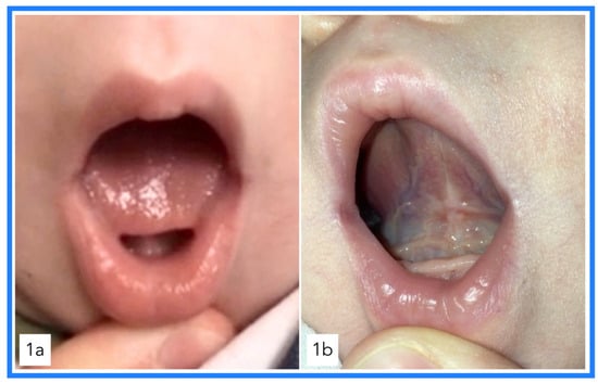
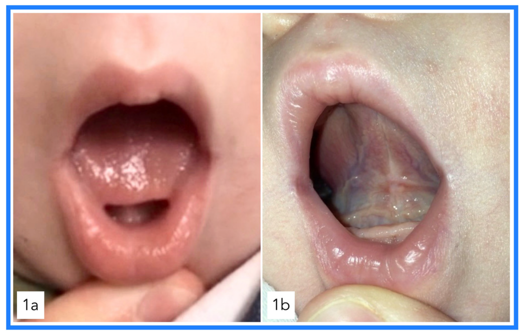
Full-length palatal contact increased from 0/3182 infants (0.0%) prior to removal of lingual mechanical restriction to 3166/3182 (99.5%) at postoperative reassessment (absolute increase: 99.5 percentage points; McNemar p < 0.001), irrespective of palatal morphology (Table 2). A representative example from the awake cohort is shown in Figure 1.
Table 2.
Tongue Resting Configuration in the Awake Cohort Before and After Removal of Lingual Mechanical Restriction.
Figure 1.
Representative example from the Awake Cohort. Pre-procedure image (a) demonstrates a non-palatal resting configuration with the tongue positioned inferiorly toward the floor of the mouth. Postoperative image at two-week follow-up (b) demonstrates full-length palatal contact in the same patient.
An observational difference in visualization during mandibular depression was noted across palatal morphologies, although this was minimal in the infant cohort. In some infants with narrower palatal morphology, continuous palatal contact during mouth opening was maintained with a slightly reduced degree of mandibular depression compared to those with wider palates. This variation in visualization did not affect the observed resting configuration.
Sixteen infants (0.5%) demonstrated persistent non-palatal resting configuration at follow-up.
3.2. General Anesthesia Cohort
A total of 355 patients were included in the general anesthesia cohort (age range: 13 months–18 years; median age: 5 years [IQR 3–8]). Of these, 233 (65.6%) were male, and 122 (34.4%) were female (Table 1).
Resting configuration was assessed intraoperatively immediately prior to and immediately following removal of lingual mechanical restriction while patients remained under the same anesthetic conditions.
Preoperatively, all 355 patients (100%) demonstrated a non-palatal resting configuration, distributed among three categories: inferior positioning toward the floor of the mouth, anterior positioning between the teeth, or partial anterior (blade-only) palatal contact.
Following removal of mechanical restriction, full-length palatal contact was observed in 355/355 patients (100%) intraoperatively, irrespective of palatal morphology. No variability in outcome was observed across age, palatal morphology, or preoperative configuration.
An observational difference in visualization during mandibular depression, distinct from the resting outcome, was infrequently noted across palatal morphologies. In a small subset of patients with markedly narrow, high-vaulted palates, maintenance of continuous palatal contact was preserved over a relatively reduced range of mandibular opening compared to patients with wider or more rounded palates. In contrast, patients with wider palatal morphology were able to maintain continuous palatal contact over a greater degree of mouth opening. This variation in visualization did not alter the observed resting configuration across patients.
As described in the Methods, achievement of unrestricted tongue mobility with full-length palatal contact was used as a procedural completion criterion in this cohort.
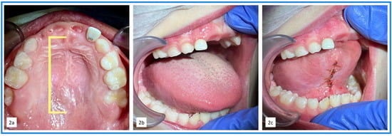
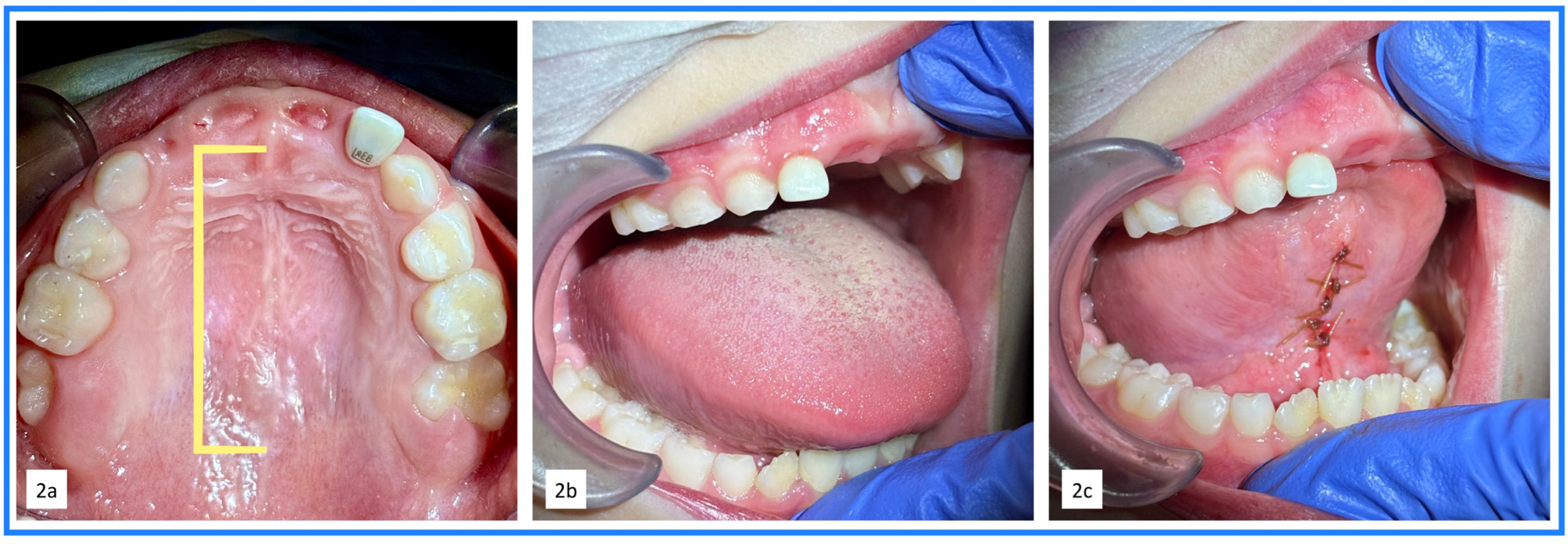
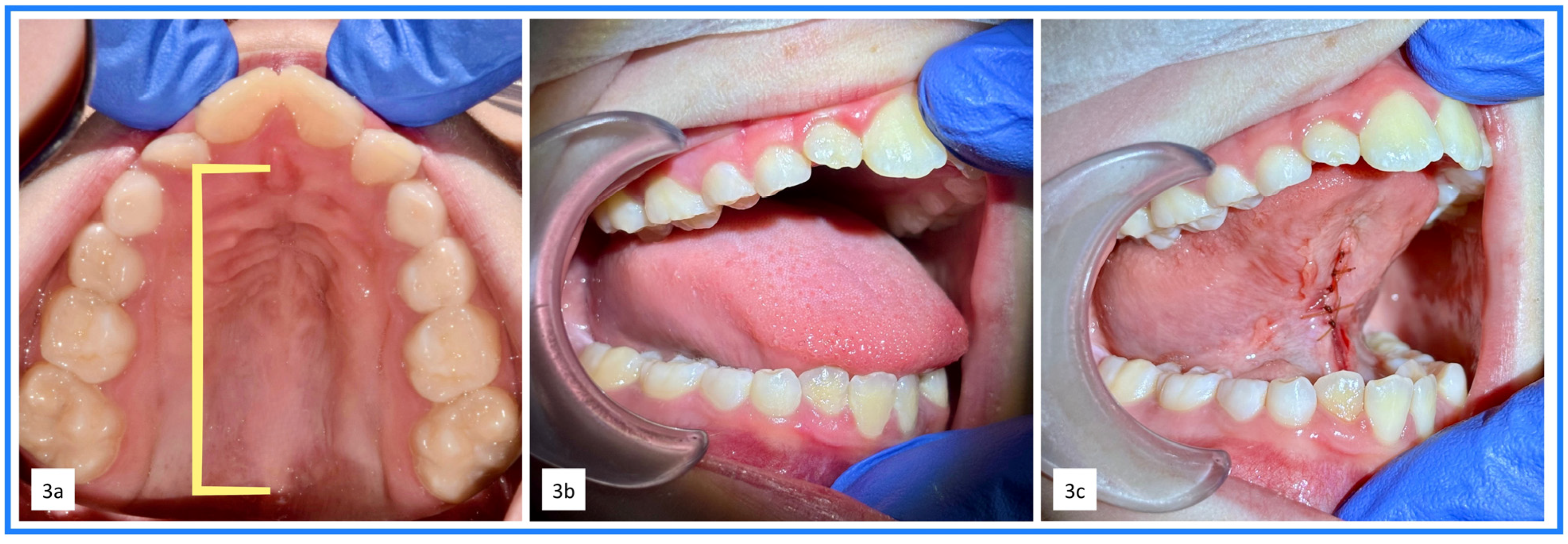
The overall distribution of resting configuration changed significantly following release (Stuart–Maxwell test, p < 0.001). When categories were collapsed into binary variables (full-length palatal contact vs. non-palatal resting configuration), McNemar’s test likewise demonstrated a statistically significant shift (p < 0.001; Table 3). A representative example from the general anesthesia cohort with a round palate is shown in Figure 2. A representative example from the general anesthesia cohort with a narrow palate is shown in Figure 3.
Table 3.
Resting Configuration in the General Anesthesia Cohort Before and After Removal of Lingual Mechanical Restriction.
Figure 2.
Representative example from the General Anesthesia Cohort. (a). Visualization of the round hard palate and palatal rugae extending from the incisive papilla to the posterior junction of the hard and soft palate. Depicted by the yellow bracket, this region corresponds to the area of full-length palatal contact observed following removal of lingual mechanical restriction. (b). Pre-procedure image demonstrates a non-palatal resting configuration with the tongue positioned inferiorly toward the floor of the mouth. (c). Intraoperative post-release image demonstrates full-length palatal contact in the same patient.
Figure 3.
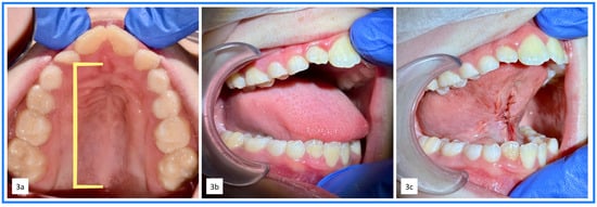
Representative example from the General Anesthesia Cohort. (a). Visualization of the narrow hard palate and palatal rugae extending from the incisive papilla to the posterior junction of the hard and soft palate. Depicted by the yellow bracket, this region corresponds to the area of full-length palatal contact observed following removal of lingual mechanical restriction. (b). Pre-procedure image demonstrates a non-palatal resting configuration with the tongue positioned inferiorly toward the floor of the mouth. (c). Intraoperative post-release image demonstrates full-length palatal contact in the same patient.
Based on patient records and documented follow-up, there were no clinically significant postoperative complications observed in either cohort, including but not limited to excessive bleeding, infection, postoperative swelling, wound dehiscence, suture failure, or behavioral trauma (e.g., tongue biting).
4. Discussion
Accurate interpretation of anatomical structure depends on understanding baseline spatial relationships in living tissue. In this study, removal of lingual mechanical restriction was consistently associated with full-length palatal contact across two pediatric cohorts assessed under distinct clinical conditions.
In the awake cohort, full-length palatal contact was observed in 99.5% of infants at routine postoperative reassessment. In the general anesthesia cohort, full-length palatal contact was observed intraoperatively in all patients at the time unrestricted tongue mobility was achieved.
Importantly, in the general anesthesia cohort, achievement of full-length palatal contact was used as a procedural completion criterion. Thus, postoperative findings in that cohort reflect the resting configuration observed at the time unrestricted mobility was confirmed. In contrast, postoperative assessment in the awake cohort occurred following routine healing. The consistency of palatal distribution across both contexts strengthens the mechanical plausibility of the observed configuration.
Together, these findings demonstrate that when mechanical constraint is absent, the tongue consistently distributes broadly across the hard palate, consistent with muscular hydrostat behavior.
The resting configuration must also be considered in the context of craniofacial growth and postnatal anatomical maturation. During infancy, the tongue occupies a proportionally larger volume within the oral cavity. Structural changes, including descent of the epiglottis and elongation of the pharyngeal airway, alter the spatial relationships between the tongue and surrounding structures during early development [17,18,19]. Despite these maturational changes, the present findings demonstrate consistent full-length palatal distribution across a broad pediatric age range when lingual mobility is unrestricted.
The interaction between tongue posture and palatal morphology has been explored within orthodontic and craniofacial research. Clinical and experimental observations suggest that sustained lingual pressure may influence transverse palatal development and dentofacial form [14,15,16,20]. Although the present study does not directly evaluate growth outcomes, documentation of consistent palatal contact provides anatomical context for future investigations into structure–function relationships during development.
In the orofacial myofunctional literature, resting tongue posture has been described as a determinant of equilibrium within the stomatognathic system [8,11,12]. However, much of this discussion has relied on clinical observation or therapeutic frameworks rather than large-scale anatomical documentation. The current dataset provides objective evidence that full-length palatal distribution is consistently observed when mechanical constraints are removed, supporting the mechanical plausibility of this configuration.
While previous studies have reported functional improvements following treatment of ankyloglossia, including enhanced tongue mobility and orofacial function [11,12], direct in vivo evidence characterizing resting tongue configuration after release is limited. The present findings extend this literature by demonstrating consistent full-length palatal contact as a reproducible resting outcome following removal of mechanical restriction.
Factors such as low muscle tone, restricted oral cavity space, and palatal morphology may influence baseline tongue posture and function. However, these findings suggest that, following adequate release of lingual restriction, regardless of palatal morphology, the tongue is capable of achieving full palatal suction, which would be consistent with its behavior as a muscular hydrostat.
The safety profile of the present cohort also provides important clinical context for interpreting these findings. The observed change toward full-length palatal contact occurred without clinically significant postoperative complications in either cohort. This safety profile provides important context for interpreting the primary finding of this study: restoration of lingual mobility was associated with the tongue assuming an elevated palatal resting configuration consistent with muscular hydrostat behavior, without documented excessive bleeding, infection, clinically significant postoperative swelling, wound dehiscence, suture failure, salivary gland or floor-of-mouth injury, oral aversion or feeding refusal, scar-related tethering requiring revision, or behavioral trauma such as tongue biting.
These findings compare favorably with prior reports. Zaghi et al. reported no major complications following lingual frenuloplasty with myofunctional therapy in 348 patients older than two years, including older children, adolescents, and adults; however, minor complications occurred in fewer than 5% of cases and included prolonged bleeding, temporary tongue-tip numbness, salivary gland issues, minor wound infection or inflammation, and revision to excise restrictive scar tissue [21]. In contrast, these minor and major postoperative issues were not identified in the present awake infant or pediatric general anesthesia cohorts. The absence of clinically significant complications in the awake infant cohort is particularly notable given the smaller operative field, developing anatomy, and greater technical demands of treatment in this age group.
Several factors may help explain differences between studies. First, the patient populations differed: the present study included awake infants and pediatric patients aged 13 months to 18 years treated under general anesthesia, whereas Zaghi et al. included patients older than 2 years [21]. Second, although the present manuscript is not intended as a surgical-technique paper, differences in release method, wound management, anesthetic approach, postoperative protocols, follow-up duration, and method of complication capture may influence reported adverse-event rates.
In the present cohort, a functional scissor release was used rather than laser or thermal ablation. This approach involved clamp stabilization, controlled incision of the restrictive tissue, and functional fascial mobilization/stretching with intraoperative functional checkpoints to confirm adequate release. Scissors permit direct mechanical engagement of restrictive tissue and allow real-time adaptation to visible and palpable anatomy, including variable or distorted vascular anatomy, floor-of-mouth structures, and salivary tissues. This may help limit unnecessary involvement of adjacent soft tissues and blood vessels and may be relevant to the absence of excessive bleeding, salivary gland injury, or floor-of-mouth injury in the present cohort. No topical or infiltrative local anesthetic was used in either cohort, which may also preserve immediate postoperative sensation, movement, and functional feedback. This is relevant to the present study’s emphasis on postoperative tongue resting posture and muscular hydrostat behavior and may, in theory, reduce the risk of altered early tongue use or inadvertent biting while sensation remains diminished.
These comparisons should be interpreted cautiously, as the present study was not designed as a direct comparative trial. Nevertheless, the absence of major complications and minor adverse events described in prior reports supports the safety of complete functional release in the studied awake infant and pediatric general anesthesia cohorts. Importantly, these safety findings remain secondary to, and supportive of, the primary focus of this manuscript: following adequate release of restrictive tethering, the tongue can assume an elevated palatal resting posture consistent with its role as a muscular hydrostat.
It is important to distinguish between the visualization of palatal contact during mandibular opening and the true resting configuration of the tongue. While intraoperative and clinical visualization often involve gentle mandibular depression to demonstrate palatal suction, this maneuver does not represent a functional or physiologic endpoint. Rather, it serves as a method of observation and documentation.
The clinically relevant resting state is the closed-mouth condition, in which the tongue is passively suctioned against the palate within an enclosed oral cavity, consistent with muscular hydrostat behavior and lingual-palatal suction described in prior functional studies [1,2,12]. Under these conditions, the tongue occupies the available intraoral space defined by the freeway space, or interocclusal rest space, typically characterized by a small vertical separation between the maxillary and mandibular dentition of approximately 2–3 mm at rest [22]. Differences observed during mouth opening, therefore, reflect the limits of maintaining intraoral suction under changing spatial conditions, rather than variation in true resting posture.
This distinction is clinically important because visualization of palatal suction requires temporary mandibular opening, whereas the relevant physiologic state is the closed-mouth resting configuration. In awake older children, conscious awareness, cooperation, guarding, or task-related tongue movement may alter the resting state being observed. The general anesthesia cohort, therefore, provided a complementary observation condition in which voluntary motor control was minimized, allowing passive tongue configuration to be assessed immediately following restoration of unrestricted mobility. Importantly, the observed resting configuration involved a broad distribution of the tongue against the hard palate rather than isolated anterior or tongue-tip contact alone. The reproducibility of full-length palatal contact under these conditions supports interpretation of this configuration as a passive mechanical equilibrium, consistent with muscular hydrostat principles of deformation, volume conservation, and structural support without skeletal elements [1,2].
Adjunctive therapies, such as myofunctional therapy, bodywork, and neuromuscular rehabilitation, are often considered in clinical practice to support functional outcomes before and/or after treatment of lingual restriction. In the present study, adjunctive therapy exposure differed by cohort. Infants in the awake cohort did not receive preoperative adjunctive neuromuscular or myofunctional therapy. Postoperatively, caregivers were instructed in a twice-daily stretching protocol to support wound mobility and healing, and age-appropriate tongue movement exercises, including lateralization, protrusion, and sucking, were introduced when appropriate. Therefore, no structured pre-release therapy was required for the observed change in resting posture in this cohort. In the general anesthesia cohort, myofunctional exercise protocols were individualized according to patient age, cooperation, maturity, and clinical needs, and preoperative compliance or preparation varied among patients. Despite this variability, complete passive palatal suction was consistently observed once unrestricted tongue mobility was achieved.
These findings suggest that achieving full palatal suction at rest is not dependent on adjunctive therapy but rather reflects the innate biomechanical behavior of the unrestricted tongue as a muscular hydrostat. At rest, when the tongue is not engaged in speech, mastication, or swallowing, restoration of sufficient mobility appears to permit the tongue to assume a passive palatal suction posture within the enclosed oral cavity. This interpretation is further supported by the awake infant cohort, in which the same resting configuration was observed despite the absence of preoperative adjunctive therapy.
This observation should not be interpreted to mean that adjunctive therapy is unnecessary in all cases. Rather, the present findings are specific to passive resting posture and do not address broader functional outcomes. Previous studies and reviews have suggested that outcomes may be improved when orofacial myofunctional therapy is incorporated before and after lingual frenectomy, particularly when treatment goals include speech, swallowing, nasal breathing, oral habits, compensatory mandibular or floor-of-mouth recruitment, tongue strength and endurance, wound mobility, and long-term neuromuscular adaptation [23,24]. The apparent difference between these reports and the present study likely reflects differences in outcome measures: immediate or early passive palatal resting posture may emerge following complete release of mechanical restriction, whereas adjunctive therapy may be more relevant for optimizing active function, reducing compensatory patterns, supporting postoperative wound mobility, and maintaining long-term functional stability.
Importantly, these findings do not imply conscious or volitional positioning of the tongue. Rather, they support the interpretation that full-length palatal contact represents a mechanically stable equilibrium state under physiologic conditions [1,2]. Mechanical restriction appears to alter this equilibrium, resulting in inferior or partial distribution patterns that may reflect structural constraint rather than intrinsic resting preference. These findings suggest that the inferior resting tongue posture observed clinically may reflect mechanical constraints rather than intrinsic neuromuscular preference.
Taken together, these findings provide large-scale in vivo clinical evidence supporting the interpretation of the human tongue as a muscular hydrostat [1,2]. Classical biomechanical descriptions of muscular hydrostats emphasize deformation, volume conservation, and structural support in the absence of skeletal elements [1,2]. At the same time, more recent imaging and anatomical studies have demonstrated the complex three-dimensional muscular architecture that enables these properties in the human tongue [3,4,5]. The present study extends this literature by showing that, when mechanical restriction is removed, the tongue consistently assumes a broad palatal distribution at rest under living physiologic conditions. In this context, the observed resting configuration is not merely compatible with muscular hydrostat theory, but provides in vivo clinical evidence demonstrating how these principles are expressed under physiologic conditions.
These observations do not intend to imply that surgical intervention alone addresses all aspects of orofacial function or development. Rather, these findings indicate that restoration of adequate tongue mobility permits the tongue to assume an optimal palatal position, consistent with its function as a muscular hydrostat.
From a clinical perspective, these findings may have implications for the evaluation and management of lingual mechanical restriction. The consistent observation of full-length palatal contact following adequate release suggests that restoration of tongue mobility permits the tongue to assume a palatal resting configuration innately, without the need for active or volitional positioning. This may provide clinicians with a functional indicator of sufficient release at the time of procedure and during postoperative assessment.
Recognition of this palatal resting configuration may also be relevant to airway function, nasal breathing, and craniofacial development, as the prior literature has linked tongue posture with upper airway behavior and dentofacial form [11,14,15,16,20]. While these downstream outcomes were not directly measured in the present study, the observed configuration provides an anatomical and functional framework for interpreting these relationships. It supports the use of a palatal-contact model of resting tongue posture in clinical teaching and future research.
Future investigations incorporating advanced imaging modalities such as MRI or ultrasound may further quantify contact surface patterns and refine characterization of hydrostatic resting behavior [6,7,9]. Longitudinal assessment may also clarify the stability of the resting configuration over time following the removal of mechanical restriction.
The complex three-dimensional arrangement of intrinsic and extrinsic tongue musculature permits deformation and redistribution without skeletal support [3,4]. In the present study, the clinically observed area of full-length palatal contact corresponded to the hard palate, including the palatal rugae extending from the incisive papilla anteriorly to the posterior junction of the hard and soft palate (Figure 2 and Figure 3). This structural correspondence supports the interpretation of palatal distribution as a mechanically and anatomically coherent resting configuration within the living oral cavity.
Because the lateral borders of the tongue contact the sides of the palate, direct clinical observation cannot fully characterize the midsagittal relationship between the dorsal posterior tongue and the soft palate/velum. Therefore, the present study does not claim to objectively quantify velar contact. However, MRI-based studies have demonstrated the ability to visualize the tongue, soft palate, and surrounding upper-airway structures in vivo, providing complementary support for representing their spatial relationship within the living oral cavity [10].
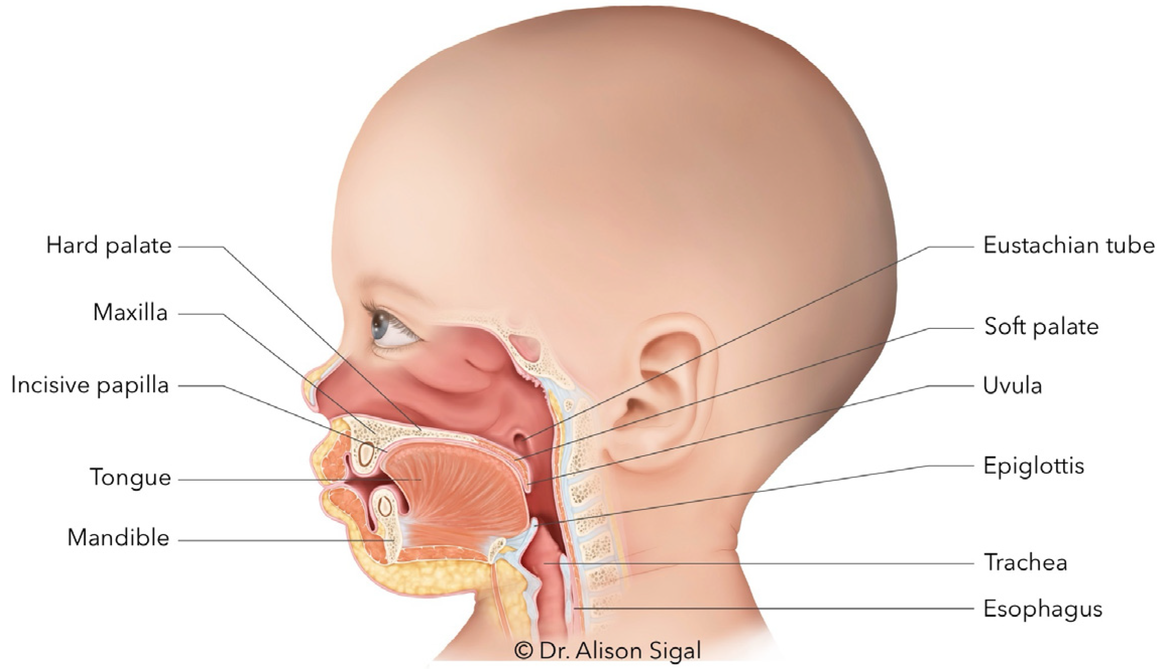
Traditional anatomical illustrations commonly depict the tongue positioned inferiorly within the oral cavity in midsagittal views [25]. Because hydrostatic behavior cannot be fully appreciated in cadaveric tissue, in vivo observation offers complementary insight into functional resting configuration [1,2]. The present findings support full-length hard-palate contact as the mechanically stable resting configuration when lingual mobility is unrestricted, consistent with muscular hydrostat principles [1,2]. The schematic in Figure 4 is not intended to replace objective imaging of the velum, but rather to integrate the present clinical observations with existing MRI-based understanding of in vivo tongue–palate–soft palate spatial relationships.
Figure 4.
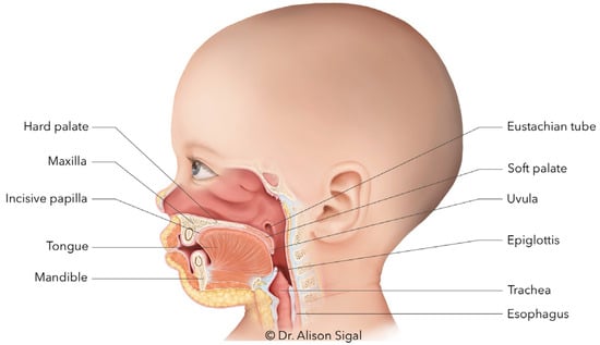
Schematic sagittal illustration depicting full-length palatal contact of the tongue within the oral cavity, consistent with the resting configuration observed following removal of lingual mechanical restriction.
Clarifying resting configuration is not a purely descriptive exercise. Rest posture defines baseline spatial relationships between the tongue and palate from which dynamic functions such as swallowing and speech begin. Accurate representation of this baseline is essential for interpreting both typical anatomy and structural deviation. This interpretation supports not only a conceptual understanding of the tongue as a muscular hydrostat but also the need for anatomical representations that reflect its functional resting configuration in vivo. Accordingly, the schematic in Figure 4 is intended to provide a more functionally and anatomically representative model of resting tongue posture for future educational, anatomical, and clinical reference.
4.1. Implications for Future Research
The present observations underscore the importance of clearly defining resting configuration when investigating orofacial function. Many imaging and modeling studies assess tongue deformation during active tasks such as speech and swallowing; however, the baseline resting state from which these dynamic movements originate is less frequently documented. Standardized criteria for evaluating passive resting configuration may enhance consistency across future investigations.
Beyond descriptive characterization, future research may examine whether full-length palatal contact is associated with measurable functional or structural outcomes. Comparative studies evaluating individuals with persistent non-palatal resting configurations may help clarify potential relationships between resting posture and clinical signs, symptom patterns, or craniofacial development. Prospective designs with blinded examiners, incorporating objective functional assessments, could further determine whether restoration of unrestricted mobility is associated with reproducible changes in observable parameters.
Additionally, the consistent intraoperative observation of full-length palatal contact as a marker of unrestricted mobility suggests that resting configuration may serve as a candidate anatomical endpoint for future objective measurement development. Quantifiable criteria—potentially incorporating imaging, pressure mapping, or biomechanical modeling—may assist in standardizing assessment of lingual mechanical restriction and refining surgical decision-making frameworks.
Further research using objective imaging modalities, such as MRI or ultrasound, in conjunction with clinical observation, may refine the understanding of hydrostatic resting behavior and quantify surface contact patterns more precisely. Longitudinal studies may also clarify the stability of resting configuration over time following the removal of mechanical restriction.
4.2. Strengths
This study includes a large pediatric cohort spanning infancy through adolescence, enabling examination of resting configuration across developmental stages. All assessments were performed by a single practitioner using standardized observational techniques, providing a high degree of procedural consistency across the dataset.
Observations were reproducible across two distinct clinical contexts: postoperative evaluation in awake infants and intraoperative assessment under general anesthesia. The consistency of findings across age groups and assessment settings strengthens the mechanical plausibility of the observed configuration. The large sample size and uniformity of data acquisition over time further support the robustness and internal consistency of the findings.
Paired pre–post statistical analysis further enhances internal validity.
4.3. Limitations
This study is retrospective and based on clinical observation without blinded assessment. All evaluations and procedures were performed by a single practitioner at a single center; therefore, formal inter-rater reliability was not assessed, intra-rater reliability was not formally quantified, and some degree of observer bias is expected.
Resting configuration was categorized descriptively rather than through imaging-based or instrument-derived measurement. In the awake cohort, reassessment occurred at short-term follow-up, and long-term stability was not systematically evaluated. In the general anesthesia cohort, full-length palatal contact was used as a procedural completion criterion, which may limit the interpretation of postoperative distribution as an independent outcome measure. Prospective studies incorporating objective imaging modalities, blinded assessment, and independent multi-operator replication would further characterize hydrostatic resting behavior.
5. Conclusions
Interpretation of the tongue as a muscular hydrostat is best informed by observation in living subjects, in whom its dynamic and suction-based properties can be appreciated. When lingual mobility is unrestricted, the tongue consistently distributes along the full length of the hard palate in vivo. Recognition of this mechanically stable resting configuration provides an essential anatomical reference point for understanding spatial relationships within the oral cavity and may inform future investigation into structural and functional interactions of the tongue across development.
Funding
No sponsorship or external funding was used to support this study.
Institutional Review Board Statement
The study was approved by an independent ethics board, Advarra (Protocol #00091702; approved 12 December 2025). The study was conducted in accordance with the Declaration of Helsinki (1975, revised 2013).
Informed Consent Statement
The institutional review board granted a waiver of informed consent due to the retrospective design and use of de-identified clinical data.
Data Availability Statement
The data that support the findings of this study are available from the corresponding author upon reasonable request.
Acknowledgments
The author thanks Myanca Rodrigues for statistical consultation and Natalie Cormier for the creation of the medical illustration used in this manuscript.
Conflicts of Interest
The author declares no financial or non-financial conflicts of interest related to this work.
References
- Gilbert, R.J.; Napadow, V.J.; Gaige, T.A.; Wedeen, V.J. Anatomical basis of lingual hydrostatic deformation. J. Exp. Biol. 2007, 210, 4069–4082. [Google Scholar] [CrossRef] [Scilit] [PubMed]
- Kier, W.M.; Smith, K.K. Tongues, tentacles and trunks: The biomechanics of movement in muscular-hydrostats. Zool. J. Linn. Soc. 1985, 83, 307–324. [Google Scholar] [CrossRef] [Scilit]
- Stone, M.; Woo, J.; Lee, J.; Poole, T.; Seagraves, A.; Chung, M.; Kim, E.; Murano, E.Z.; Prince, J.L.; Blemker, S.S. Structure and variability in human tongue muscle anatomy. Comput. Methods Biomech. BioMed Eng. Imaging Vis. 2018, 6, 499–507. [Google Scholar] [CrossRef] [Scilit] [PubMed]
- Sanders, I.; Mu, L. A three-dimensional atlas of human tongue muscles. Anat. Rec. 2013, 296, 1102–1114. [Google Scholar] [CrossRef] [Scilit] [PubMed]
- Mijailovich, S.M.; Stojanovic, B.; Kojic, M.; Liang, A.; Wedeen, V.J.; Gilbert, R.J. Derivation of a finite-element model of lingual deformation during swallowing from the mechanics of mesoscale myofiber tracts obtained by MRI. J. Appl. Physiol. 2010, 109, 1500–1514. [Google Scholar] [CrossRef] [Scilit] [PubMed]
- Brown, E.C.; Cheng, S.; McKenzie, D.K.; Butler, J.E.; Gandevia, S.C.; Bilston, L.E. Tongue and lateral upper airway movement with mandibular advancement. Sleep 2013, 36, 397–404. [Google Scholar] [CrossRef] [Scilit] [PubMed]
- Woo, J.; Xing, F.; Lee, J.; Stone, M.; Prince, J.L. A spatio-temporal atlas and statistical model of the tongue during speech from cine-MRI. Comput. Methods Biomech. Biomed. Eng. Imaging Vis. 2018, 6, 520–531. [Google Scholar] [CrossRef] [Scilit] [PubMed]
- Fukuda, T.; Kohzuka, Y.; Almeida, F.R.; Iijima, T.; Masuda, R.; Tsuiki, S. Control of Tongue Position in Patients with Obstructive Sleep Apnea: Concept and Protocol for a Randomized Controlled Crossover Trial. Int. J. Environ. Res. Public Health 2023, 20, 6026. [Google Scholar] [CrossRef] [Scilit] [PubMed]
- Ovsenik, R.; Marolt Mušič, M.; Primožič, J. Changes in the swallowing pattern and tongue posture during the transition from deciduous to mixed dentition—A longitudinal ultrasonography study. Eur. J. Orthod. 2024, 46, cjad066. [Google Scholar] [CrossRef] [Scilit]
- Welch, K.C.; Foster, G.D.; Ritter, C.T.; Wadden, T.A.; Arens, R.; Maislin, G.; Schwab, R.J. A novel volumetric magnetic resonance imaging paradigm to study upper airway anatomy. Sleep 2002, 25, 530–541. [Google Scholar] [CrossRef] [Scilit]
- Guilleminault, C.; Huseni, S.; Lo, L. A frequent phenotype for paediatric sleep apnoea: Short lingual frenulum. ERJ Open Res. 2016, 2, 00043-2016. [Google Scholar] [CrossRef] [Scilit] [PubMed]
- Govardhan, C.; Murdock, J.; Norouz-Knutsen, L.; Valcu-Pinkerton, S.; Zaghi, S. Lingual and Maxillary Labial Frenuloplasty with Myofunctional Therapy as a Treatment for Mouth Breathing and Snoring. Case Rep. Otolaryngol. 2019, 2019, 3408053. [Google Scholar] [CrossRef] [Scilit] [PubMed]
- Wang, W.; Huang, J.; Lin, Q.; Liu, X.; Cao, J.; Dai, J. Effect of maxillary expansion combined with orofacial myofunctional therapy on tongue position in children with mouth breathing. Lin Chuang Er Bi Yan Hou Tou Jing Wai Ke Za Zhi 2023, 37, 648–651. [Google Scholar] [CrossRef] [Scilit] [PubMed]
- Feștilă, D.; Ciobotaru, C.D.; Suciu, T.; Olteanu, C.D.; Ghergie, M. Oral Breathing Effects on Malocclusions and Mandibular Posture: Complex Consequences on Dentofacial Development in Pediatric Orthodontics. Children 2025, 12, 72. [Google Scholar] [CrossRef] [Scilit] [PubMed]
- Principato, J.J. Upper airway obstruction and craniofacial morphology. Otolaryngol. Head Neck Surg. 1991, 104, 881–890. [Google Scholar] [CrossRef] [Scilit] [PubMed]
- Trabalon, M.; Schaal, B. It Takes a Mouth to Eat and a Nose to Breathe: Abnormal Oral Respiration Affects Neonates’ Oral Competence and Systemic Adaptation. Int. J. Pediatr. 2012, 2012, 207605. [Google Scholar] [CrossRef] [Scilit] [PubMed]
- Sasaki, C.T.; Levine, M.S.; Laitman, J.T.; Crelin, E.S. Postnatal Descent of the Epiglottis in Man: A Preliminary Report. Arch. Otolaryngol. 1977, 103, 169–171. [Google Scholar] [CrossRef] [Scilit] [PubMed]
- Sato, K.; Chitose, S.I.; Sato, K.; Sato, F.; Ono, T.; Umeno, H. Growth and Development of Epiglottis and Preepiglottic Space of Larynx as It Acquires Vocal Tract. Laryngoscope Investig. Otolaryngol. 2024, 9, e1288. [Google Scholar] [CrossRef] [Scilit] [PubMed]
- Maynard, T.M.; Zohn, I.E.; Moody, S.A.; LaMantia, A.S. Suckling, Feeding, and Swallowing: Behaviors, Circuits, and Targets for Neurodevelopmental Pathology. Annu. Rev. Neurosci. 2020, 43, 315–336. [Google Scholar] [CrossRef] [Scilit] [PubMed]
- Moayedi, Y.; Michlig, S.; Park, M.; Koch, A.; Lumpkin, E.A. Somatosensory innervation of healthy human oral tissues. J. Comp. Neurol. 2021, 529, 3046–3061. [Google Scholar] [CrossRef] [Scilit]
- Zaghi, S.; Valcu-Pinkerton, S.; Jabara, M.; Norouz-Knutsen, L.; Govardhan, C.; Moeller, J.; Sinkus, V.; Thorsen, R.S.; Downing, V.; Camacho, M.; et al. Lingual frenuloplasty with myofunctional therapy: Exploring safety and efficacy in 348 cases. Laryngoscope Investig. Otolaryngol. 2019, 4, 489–496. [Google Scholar] [CrossRef] [Scilit]
- Dawson, P.E. Functional Occlusion: From TMJ to Smile Design; Mosby: St. Louis, MO, USA, 2007. [Google Scholar]
- González Garrido, M.D.P.; Garcia-Munoz, C.; Rodríguez-Huguet, M.; Martin-Vega, F.J.; Gonzalez-Medina, G.; Vinolo-Gil, M.J. Effectiveness of myofunctional therapy in ankyloglossia: A systematic review. Int. J. Env. Res. Public Health 2022, 19, 12347. [Google Scholar] [CrossRef] [Scilit]
- Zaghi, S.; Ramirez, A.; Espadas, S.; Nguyen, G.; Kupiec, L.M.; Ghodousi-Zaghi, N.; Nouri-Norouz, M.; Gonzalez, S.; Valcu-Pinkerton, S.; Rodriguez, J.; et al. Lingual frenuloplasty with myofunctional therapy: Improving outcomes for the treatment of ankyloglossia (tongue-tie) with refined techniques and endpoints. Int. J. Orofac. Myol. Myofunct. Ther. 2025, 51, 11. [Google Scholar] [CrossRef] [Scilit]
- Netter, F.H. Netter Atlas of Human Anatomy, 8th ed.; Elsevier: Philadelphia, PA, USA, 2022. [Google Scholar]

Disclaimer/Publisher’s Note: The statements, opinions and data contained in all publications are solely those of the individual author(s) and contributor(s) and not of MDPI and/or the editor(s). MDPI and/or the editor(s) disclaim responsibility for any injury to people or property resulting from any ideas, methods, instructions or products referred to in the content. |
© 2026 by the author. Published by MDPI on behalf of the International Association of Orofacial Myology (IAOM). Licensee MDPI, Basel, Switzerland. This article is an open access article distributed under the terms and conditions of the Creative Commons Attribution (CC BY) license.