


Abstract
Pancreatic ductal adenocarcinoma (PDAC) is one of the most lethal solid malignancies, characterised by rising incidence, poor long-term survival, and profound resistance to systemic therapy. Its poor prognosis reflects late diagnosis, early metastatic spread, and a uniquely desmoplastic, immunosuppressive tumour microenvironment that limits both drug delivery and effective antitumour immunity. Although cytotoxic chemotherapy remains the therapeutic backbone, its clinical benefit is modest. In parallel, molecular profiling has revealed a limited but expanding set of actionable vulnerabilities; however, immuno-oncology has not yet replicated in PDAC the transformative impact observed in other tumour types. This review examines the evolving immuno-oncology landscape in PDAC, focusing on four principal domains: immune checkpoint blockade in MSI-H/dMMR disease, tumour microenvironment-directed therapies, adoptive cellular therapies, and therapeutic cancer vaccines. Taken together, available evidence suggests that progress in PDAC immuno-oncology will depend on biomarker-guided patient selection, earlier disease settings, minimal residual disease-directed approaches, and rational combinations that address immune exclusion, stromal and myeloid barriers, impaired T-cell priming, and adaptive resistance. While durable clinical benefit remains limited and most strategies remain in early-phase evaluation, emerging data from molecularly selected cohorts, adoptive cellular therapies, and personalised or shared-antigen vaccines provide a compelling rationale for continued refinement of immune-based approaches in PDAC.
Keywords:
pancreatic ductal adenocarcinoma (PDAC); immuno-oncology; tumour microenvironment (TME); immune checkpoint blockade; MSI-H/dMMR; adoptive cellular therapy; tumour-infiltrating lymphocyte (TIL) therapy; chimeric antigen receptor (CAR)-T cell therapy; T-cell receptor (TCR)-engineered T-cell therapy; cancer vaccines 1. Introduction
Pancreatic ductal adenocarcinoma (PDAC) is one of the most lethal solid malignancies worldwide. Globally, PDAC accounts for approximately 2.6% of all cancer diagnoses but a disproportionate share of all cancer-related mortality (4.8% of cancer-related deaths), with over 510,000 new cases and 467,000 deaths reported annually [1]. Incidence rates continue to rise and long-term survival remains poor: the 5-year overall survival (OS) rate is approximately 13%, varying substantially by stage from 44% for localised disease to 17% for regional disease and 3% for distant disease [2,3]. The majority of patients present with advanced or metastatic disease, and recurrence rates remain high even after curative intent surgery, reflecting persistent challenges in early detection and effective treatment [4].
Chemotherapy remains the backbone of PDAC treatment, with regimens such as FOLFIRINOX (5-fluorouracil, leucovorin, irinotecan and oxaliplatin) or gemcitabine-based combinations providing modest survival benefits at the cost of significant toxicity and inevitable therapeutic resistance [5,6]. In metastatic PDAC trial populations, outcomes remain poor with a median OS of 11.1 months and progression-free survival (PFS) of 6.4 months in the first-line setting (PRODIGE 4/ACCORD 11, NCT00112658), and a median OS of 6.1 months at best in the second-line setting (NAPOLI-1, NCT01494506) [7].
Although immuno-oncology in PDAC has been reviewed extensively, the field has changed materially in recent years. Emerging data from personalised mRNA neoantigen vaccines, minimal residual disease (MRD)-directed KRAS vaccines, KRAS-specific TCR-engineered T cells and CLDN18.2-directed cellular therapies, together with increasingly biomarker-integrated trial designs, have shifted the central question from whether PDAC can respond to immunotherapy to which biological subsets and disease settings are most permissive. Accordingly, this review does not simply catalogue immune-based modalities. It uses both positive and negative clinical studies to identify recurrent mechanisms of resistance, distinguish pharmacodynamic or immunological activity from evidence of clinical efficacy, and consider where emerging strategies may fit across the PDAC disease continuum from perioperative and MRD-positive disease to treatment-refractory metastatic cancer. The principal biological barriers and therapeutic targets within the PDAC tumour microenvironment are illustrated in Figure 1, while the major therapeutic strategies and their current evidence base are summarised in Table 1.
Figure 1.
Immuno-oncology targets within the PDAC tumour microenvironment. Schematic overview of key cellular and molecular interactions contributing to immune exclusion and immune suppression in PDAC, including tolerogenic dendritic cells, regulatory T cells, suppressive myeloid populations, tumour-associated macrophages, cancer-associated fibroblasts, endothelial cells, NK cells and CD8+ T cells. Annotated therapeutic strategies correspond to approaches discussed in this review. Therapeutic vaccines aim to enhance antigen presentation and T-cell priming; CD40, STING and TLR agonists target antigen-presenting cells and innate immune activation; immune checkpoint inhibitors target inhibitory PD-1/PD-L1 and CTLA-4 signalling; TIL/TCR-T-cell therapies and CAR-T/CAR-NK therapies supply or redirect tumour-reactive effector cells; PEGPH20 and certepetide/CEND-1 target stromal barriers and tumour penetration; CCR2, CCR2/CCR5 and CXCR4 blockade target suppressive myeloid recruitment and chemokine-mediated immune exclusion; and FAK and TGF-β pathway inhibition aim to remodel stromal and immune-suppressive signalling. Abbreviations: Arg-1: arginase-1; CAF: cancer-associated fibroblast; CAR: chimeric antigen receptor; CEA: carcinoembryonic antigen; CSF2: colony-stimulating factor 2; CSF3: colony-stimulating factor 3; CTLA-4: cytotoxic T lymphocyte-associated protein-4; CXCL12: C-X-C motif chemokine 12; DC: dendritic cell; FAK: focal adhesion kinase; FGF2: fibroblast growth factor; IL-1β: interleukin-1β; IL-4: interleukin-4; IL-6: interleukin-6; IL-8: interleukin-8; IL-10: interleukin-10; IL-27: interleukin-27; IL-35: interleukin-35; IDO: indoleamine 2,3-dioxygenase; iNOS: inducible nitric oxide synthase; LAG-3: lymphocyte-activation gene 3; MDSC: myeloid-derived suppressor cell; MMP: matrix metalloprotease; NK: natural killer; NO: nitric oxide; PD-1: programmed cell death protein-1; ROS: reactive oxygen species; TAA: tumour-associated antigen; TAM: tumour-associated macrophage; TCR-T: T-cell receptor-engineered T cell; TIL: tumour-infiltrating lymphocyte; TGFβ: transforming growth factor-β; TLR: Toll-like receptor; TNFα: tumour necrosis factor-α; TRAIL: TNF-related apoptosis-inducing ligand; Treg: regulatory T cell; VEGF: vascular endothelial growth factor. The graphic was created with BioRender.com.
Table 1.
Clinical development and evidence level of immuno-oncology strategies in PDAC.
The relevant literature was identified through PubMed/MEDLINE and ClinicalTrials.gov searches, supplemented by reference-list review and targeted searches for major guidelines, conference proceedings and recent studies. Searches included the literature published up to March 2026 and combined terms relating to pancreatic ductal adenocarcinoma with immune checkpoint blockade, MSI/dMMR, tumour microenvironment, stromal and myeloid targeting, CD40, STING/TLR, CXCR4, adoptive cellular therapies, cancer vaccines, KRAS, microbiome, bispecific T-cell engagers, oncolytic viruses, radiotherapy, ctDNA and spatial immune profiling. Priority was given to peer-reviewed PDAC-specific clinical and translational studies, with mixed-tumour studies, conference abstracts and trial-registry data included where relevant evidence was limited or rapidly evolving. This was a narrative rather than systematic review; no formal protocol, quality assessment or meta-analysis was undertaken.
2. MSI-H/dMMR PDAC
Microsatellite instability-high (MSI-H) and mismatch repair-deficiency (dMMR) are rare in PDAC, generally reported in approximately 1–3% of cases, with prevalence varying by cohort, assay, and histological composition [93]. Immune checkpoint inhibitors have demonstrated tumour-agnostic activity across MSI-H/dMMR malignancies, leading to tumour-agnostic FDA approval of the anti-PD-1 antibody pembrolizumab; however, PDAC-specific outcomes appear more heterogeneous.
Within the phase II KEYNOTE-158 trial, the PDAC cohort of 22 previously treated patients who received pembrolizumab achieved an overall response rate (ORR) of 18.2%, with a median PFS of 2.1 months and a median OS of 3.7 months [8]. Pembrolizumab therefore represents an established tumour-agnostic treatment option for patients with MSI-H/dMMR disease, although the limited PDAC cohort in KEYNOTE-158 underscores that MSI-H/dMMR status does not confer uniform sensitivity in this tumour type [9].
By contrast, in the real-world AGEO European cohort of advanced dMMR/MSI-H PDAC, immune checkpoint blockade demonstrated substantial clinical activity with a favourable safety profile, largely in the second-line or later-line setting [10]. Among 31 evaluable patients treated predominantly with single-agent anti-PD-1 therapy, the objective response rate was 48.4%. After a median follow-up of 18 months, median PFS was 26.7 months and median OS was not reached, indicating durable benefit in a subset of patients. Disease control was particularly high in less heavily pre-treated patients, with a disease control rate (DCR) of 76.5% among those who had received only one prior line of therapy. Notably, no grade 3–4 treatment-related adverse events were reported, underscoring the tolerability of immune checkpoint inhibitors in this molecularly selected PDAC population.
The variability between KEYNOTE-158 and real-world/retrospective cohorts likely reflects both biological and clinical heterogeneity within MSI-H/dMMR PDAC, including differences in tumour mutational burden, antigen presentation, TME context, line of therapy, performance status and prior treatment exposure. Although MSI-H/dMMR remains the most clinically actionable immunotherapy biomarker in PDAC, its low prevalence and variable PDAC-specific response to checkpoint blockade should be recognised. MSI/MMR assessment may be incorporated within broader molecular profiling, with positive findings potentially informing treatment selection and prompting consideration of germline evaluation for Lynch syndrome where appropriate. Testing may be performed using mismatch repair protein immunohistochemistry, PCR-based MSI testing or validated next-generation sequencing assays. Importantly, MSI-H/dMMR status alone does not guarantee response to PD-1 blockade, highlighting the need for complementary biomarkers of sensitivity and resistance.
Outside molecularly selected MSI-H/dMMR disease, immune checkpoint blockade has shown limited clinical activity in PDAC. Single-agent CTLA-4 blockade with Ipilimumab produced no RECIST objective responses in a phase II trial of locally advanced or metastatic pancreatic adenocarcinoma, while durvalumab monotherapy or durvalumab plus tremelimumab produced ORRs of 0% and 3.1%, respectively, in previously treated metastatic PDAC [11,12]. This limited efficacy likely reflects the low neoantigen burden of most microsatellite-stable PDACs, poor effector T-cell infiltration, myeloid-dominant immunosuppression, and stromal immune exclusion.
For patients who have progressed on immune checkpoint inhibitors, independent CRISPR–Cas9 loss-of-function screens have identified WRN (Werner syndrome RecQ helicase) as an essential gene in MSI-H/dMMR cancer cells [94,95]. Early-phase clinical trials are underway in MSI-H/dMMR advanced solid tumours, including gastrointestinal cancers, using first-in-class WRN inhibitors HRO761 (NCT05838768) and RO7589831/VVD-133214 (study code BP44474; NCT06004245) as monotherapy or in combination regimens. For HRO761, eligible patients must have progressed after or be intolerant to prior standard therapy; and Arms A and C specifically require progression after a prior line of immune checkpoint inhibitor therapy. For RO7589831, eligibility is generally second-line or later, although the study also includes a first-line metastatic colorectal pembrolizumab combination cohort.
3. Tumour Microenvironment (TME) Modifiers
3.1. Targeting Stromal Depletion: PEGPH20
Targeting the dense hyaluronan-rich stroma in PDAC has been pursued as a strategy to reduce interstitial pressure, decompress tumour vasculature, and enhance chemotherapy delivery.
In the phase II HALO-202 trial, conducted in previously untreated unselected metastatic PDAC, PEGPH20 (pegylated recombinant human hyaluronidase PH20) plus gemcitabine/nab-paclitaxel provided only a modest PFS benefit in the overall, unselected population compared with chemotherapy alone (6.0 vs. 5.3 months) [13]. In the prespecified hyaluronan-high subgroup, median PFS was 9.2 versus 5.2 months, ORR was approximately 45%, and median OS was 11.5 versus 8.5 months. However, this signal did not translate into clinical benefit in the subsequent phase III biomarker-enriched HALO-301 trial, in which median OS was 11.2 versus 11.5 months [14].
3.2. Vascular and Penetration Enhancement: CEND-1
CEND-1 (iRGD/LSTA1, now also referred to as certepetide) is a tumour-penetrating peptide designed to enhance intratumoural delivery of co-administered therapies through sequential binding to αV integrins and neuropilin-1 [15]. In a first-in-human phase I trial (NCT03517176), CEND-1 was combined with gemcitabine/nab-paclitaxel as first-line treatment for metastatic PDAC [16]. Among 29 evaluable patients the combination achieved an ORR of 59% (including one complete response) and a median OS of 13.2 months. Given the small, single-arm design and selected trial population, these outcomes cannot establish comparative efficacy relative to historical gemcitabine/nab-paclitaxel cohorts and should be regarded as hypothesis-generating pending randomised validation.
Certepetide is being evaluated in randomised studies in combination with first-line gemcitabine plus nab-paclitaxel. The ASCEND trial (NCT05042128) is a randomised, double-blind, placebo-controlled phase II/IIb study evaluating certepetide versus placebo added to gemcitabine plus nab-paclitaxel in patients with previously untreated metastatic PDAC [17]. Preliminary results from cohort B showed a median PFS of 7.5 versus 4.7 months (HR 0.60; 95% CI 0.34–1.07); however, this comparison did not reach statistical significance (p = 0.08) [18]. The ORR (45.2% vs. 19.0%) and DCR (85.7% vs. 61.9%) were higher with certepetide, but these preliminary findings remain insufficient to establish clinical benefit and OS data have not yet been reported. A separate randomised phase II trial (NCT06261359) is also underway evaluating CEND-1 in combination with first-line gemcitabine plus nab-paclitaxel in patients with locally advanced unresectable or metastatic PDAC. Efficacy results from this study are awaited.
In the phase I/IIa CENDIFOX trial (NCT05121038), neoadjuvant certepetide combined with modified FOLFIRINOX was feasible and well tolerated in patients with resectable or borderline resectable PDAC [19]. In preliminary data from the PDAC cohort (n = 35), most grade ≥ 3 adverse events were consistent with expected mFOLFIRINOX-related toxicities. Ten patients (29%) proceeded to resection of the pancreatic primary, and among these resected/evaluable cases, the reported R0 resection rate was only 50% and the pathologic partial response rate (defined as Tumour Regression Grade 2) was 70%. These findings should be interpreted with caution given the small number of resected patients and the absence of mature survival outcomes.
These data raise the possibility that certepetide may be better suited to selected chemotherapy backbones, although current mechanistic evidence supports enhanced intratumoural delivery of co-administered therapies broadly rather than preferential synergy with FOLFIRINOX or gemcitabine/nab-paclitaxel specifically. Clinically, the gemcitabine/nab-paclitaxel backbone has the stronger evidence base to date, while FOLFIRINOX data remain immature.
3.3. Innate Immune Activation: STING and Toll-like Receptor (TLR) Agonists
Activation of the Stimulator of Interferon Genes (STING) pathway has been explored as a means of converting the immunologically “cold” PDAC TME into an inflamed, T-cell-permissive state. In a first-in-human phase I trial (NCT02675439) of intratumoural MIW815/ADU-S100, a first-generation STING agonist, in patients with pre-treated advanced solid tumours or lymphomas, treatment was generally tolerable and induced pharmacodynamic evidence of immune activation, but antitumour activity was limited, with only one confirmed partial response reported [20].
When MIW815 was evaluated in combination with the anti-PD-1 antibody Spartalizumab/PDR001 in a phase Ib trial (NCT03172936), clinical activity was again modest with an ORR of 10.4% among evaluable solid-tumour patients and no clear evidence of meaningful activity in PDAC [21].
Toll-like receptor (TLR) agonists represent another approach to innate immune priming in PDAC, aiming to enhance antigen presentation and remodel suppressive myeloid populations. The TLR9 agonist SD-101 has been evaluated in a phase I trial (NCT04050085) in combination with the anti-PD-1 antibody Nivolumab and radiotherapy in patients with chemotherapy-refractory metastatic PDAC. However, efficacy outcomes have not yet been reported.
The dual TLR7/8 agonist BDB001 has also been investigated in the phase II AGADIR basket trial (NCT03915678) in combination with the PD-L1 inhibitor atezolizumab and immunogenic SBRT in heavily pre-treated patients lacking suitable standard-of-care therapeutic options. Immunogenic SBRT was given as an immunomodulatory strategy intended to enhance systemic antitumour immunity—including potential abscopal effects. In the PDAC cohort SBRT was directed to at least one metastatic lesion rather than specifically to the pancreatic primary. Among 32 enrolled patients in the PDAC cohort, 21 had undergone central review at the time of reporting; the DCR was 38%, with two confirmed partial responses (9.5%), six cases of stable disease (28.5%), and 13 cases of progressive disease (62.0%) [22].
3.4. Focal Adhesion Kinase (FAK) Inhibition and Immune-Stromal Remodelling
Focal adhesion kinase (FAK) inhibition represents another strategy to remodel the immunosuppressive TME in PDAC. Preclinical studies suggest that FAK inhibition can reduce stromal fibrosis and suppressive immune-cell recruitment, including cancer-associated fibroblast (CAF)-associated signalling, myeloid-derived suppressor cells, tumour-associated macrophages, and regulatory T cells (Tregs), thereby increasing CD8+ T-cell infiltration and sensitising tumours to immune checkpoint blockade [23].
In a phase I dose-escalation and expansion study of defactinib, an oral small-molecule inhibitor of focal adhesion kinase (FAK/PTK2) and proline-rich tyrosine kinase 2 (PYK2), plus pembrolizumab and gemcitabine in treatment-refractory advanced/metastatic PDAC, the combination was tolerable and showed preliminary activity [24]. Among 20 treated patients, the DCR was 80%, with one partial response and 15 patients achieving stable disease; median PFS and OS were 3.6 and 7.8 months, respectively. In the maintenance cohort, among 10 evaluable patients, the DCR was 70% with one partial response and six patients achieving stable disease; median PFS and OS on study treatment were 5.0 and 8.3 months, respectively. Paired biopsy analyses demonstrated increased infiltrating CD8+ T cells and decreased Tregs after treatment, supporting pharmacodynamic immune modulation.
Defactinib is also being evaluated in a phase II randomised trial (NCT03727880), which investigates pembrolizumab with or without defactinib, following chemotherapy as a neoadjuvant and adjuvant treatment for patients with resectable PDAC. Preliminary translational reporting suggests reduced fibroblast infiltration and increased antitumour immune activation with defactinib, including higher pro-inflammatory (M1) macrophage and CD8+ T-cell infiltration [25]. However, mature peer-reviewed clinical efficacy outcomes have not yet been reported.
3.5. CD40 Agonism, Myeloid Reprogramming and Chemokine-Axis Blockade
Beyond stromal depletion, vascular penetration enhancement, innate immune agonism and FAK inhibition, additional TME-directed strategies have focused on modulating the myeloid compartment and chemokine networks that contribute to immune exclusion in PDAC. These approaches include CD40 agonism to activate antigen-presenting cells and remodel macrophage function, CCR2/CCR5-directed strategies to limit suppressive monocyte and macrophage recruitment, and CXCL12-CXCR4 blockade to improve T-cell trafficking into tumour tissue.
CD40 agonists activate antigen-presenting cells, particularly dendritic cells and macrophages, thereby enhancing T-cell priming and promoting myeloid remodelling. Early translational studies showed that CD40 activation could remodel the PDAC stroma and induce macrophage-mediated antitumour activity in preclinical models and patients with PDAC [26]. In a phase I trial (NCT00711191) of 22 chemotherapy-naïve patients with advanced/metastatic PDAC, CP-870,893 (an agonist CD40 monoclonal antibody) combined with gemcitabine was tolerable and produced partial responses in 4 of 21 evaluable patients, with an ORR of 19%, stable disease in 11 patients, median PFS of 5.6 months and median OS of 7.4 months [27]. In the phase Ib PRINCE trial (NCT03214250), sotigalimab/APX005M plus gemcitabine/nab-paclitaxel, with or without Nivolumab, was tolerable and produced responses in 14 of 24 evaluable patients [28]. However, in the randomised phase II PRINCE trial, only Nivolumab plus chemotherapy met the primary 1-year OS endpoint (57.7%; median OS 16.7 months), whereas sotigalimab plus chemotherapy (48.1%; median OS 11.4 months) and the triplet regimen (41.3%; median OS 10.1 months) did not [29]. These data suggest context-dependent activity for CD40 agonism, while highlighting unresolved questions around biomarker selection and optimal combination strategies.
Myeloid-cell targeting represents another rational strategy, given the abundance of tumour-associated macrophages, inflammatory monocytes, neutrophils and myeloid-derived suppressor cells within PDAC. CCR2 inhibition has been explored as a means of limiting recruitment of suppressive inflammatory monocytes. PF-04136309 is a small-molecule CCR2 antagonist that inhibits CCL2–CCR2-mediated recruitment of inflammatory monocytes into the PDAC TME. In a phase Ib trial (NCT01413022) of patients with borderline resectable or locally advanced PDAC, 16 of 33 radiographically evaluable patients achieved an objective response with PF-04136309 plus FOLFIRINOX, corresponding to an ORR of 49%; local tumour control was observed in 32 of 33 patients (97%) [30]. However, in a separate phase Ib trial (NCT02732938) of 21 patients with previously untreated metastatic PDAC, PF-04136309 combined with gemcitabine plus nab-paclitaxel produced an ORR of 23.8%, but was associated with pulmonary toxicity in 24% of patients, including grade 4 hypoxia. Although CD14+CCR2+ inflammatory monocytes decreased in peripheral blood, the regimen did not show an efficacy signal above gemcitabine/nab-paclitaxel alone and raised concern for synergistic pulmonary toxicity [31]. Dual CCR2/CCR5 blockade has also been investigated using BMS-813160. In a phase Ib/II trial (NCT03184870) of first-line metastatic PDAC, BMS-813160 combined with gemcitabine plus nab-paclitaxel produced an ORR of 26% compared with 28% for gemcitabine plus nab-paclitaxel alone, while the addition of Nivolumab to BMS-813160 plus gemcitabine/nab-paclitaxel produced an ORR of 37% [32]. Median duration of response was longer with BMS-813160-containing regimens, but the ORR was not clearly improved over chemotherapy alone, and the regimen remains investigational.
Chemokine-axis blockade has also been investigated as a method to reverse immune exclusion. CXCL12–CXCR4 signalling has been implicated in restricting T-cell infiltration into PDAC, providing a rationale for CXCR4 inhibition in combination with immune checkpoint blockade. In the phase IIa COMBAT/KEYNOTE-202 trial (NCT02826486), the CXCR4 antagonist BL-8040/motixafortide was evaluated with pembrolizumab, with or without chemotherapy, in metastatic PDAC. In the chemotherapy-resistant cohort treated with BL-8040 plus pembrolizumab, the DCR was 34.5%, including one partial response and nine cases of stable disease, with a median OS of 3.3 months. In the chemotherapy-combination cohort, BL-8040 plus pembrolizumab and chemotherapy produced an ORR of 32%, DCR of 77% and median duration of response of 7.8 months. Treatment was associated with increased CD8+ effector T-cell tumour infiltration and reductions in myeloid-derived suppressor cells and circulating regulatory T cells, supporting pharmacodynamic immune modulation [33].
TGF-β pathway inhibition has also been explored as a strategy to reduce stromal and immune-mediated suppression in PDAC. Galunisertib/LY2157299, an oral TGF-β receptor I/ALK5 kinase inhibitor, was evaluated with gemcitabine in the phase Ib/randomised phase II H9H-MC-JBAJ study (NCT01373164) in patients with unresectable locally advanced or metastatic pancreatic cancer. In the randomised phase II portion, galunisertib plus gemcitabine modestly improved median OS compared with gemcitabine alone (8.9 vs. 7.1 months; HR 0.79), with minimal added toxicity [34]. A subsequent exploratory biomarker analysis of the same trial suggested that high baseline plasma CCL3 may identify a subgroup more likely to benefit from galunisertib plus gemcitabine: high CCL3 was associated with a poor outcome in the placebo/gemcitabine arm, but with an improved outcome in the galunisertib/gemcitabine arm [35]. However, subsequent evaluation of galunisertib with durvalumab in recurrent/refractory metastatic PDAC (NCT02734160) demonstrated limited activity, with one partial response, seven cases of stable disease, a DCR of 25.0%, median PFS of 1.87 months and median OS of 5.72 months among 32 patients treated at the recommended phase II dose [36]. These findings suggest that TGF-β inhibition may require biomarker selection and rational combination strategies, rather than broad application in unselected PDAC populations.
3.6. Sensitising Microsatellite-Stable PDAC to Immunotherapy: Integrated and Emerging Strategies
For the vast majority of PDACs that are microsatellite stable and mismatch repair-proficient, resistance to checkpoint blockade is not attributable to a single lesion but to failure across several stages of the cancer–immunity cycle. Low neoantigen burden and inefficient antigen presentation limit productive T-cell priming; dense and heterogeneous stroma and chemokine signalling restrict trafficking; suppressive macrophages, monocytes, myeloid-derived suppressor cells and regulatory T cells inhibit effector function; and metabolic stress and persistent inhibitory signalling promote intratumoural T-cell dysfunction [24]. Sensitisation of MSS PDAC should therefore be viewed as a coordinated strategy rather than a single therapeutic class. Radiotherapy, oncolytic viruses and vaccines may increase antigen release and immune priming; CD40, STING and TLR agonists can activate antigen-presenting cells; stromal-, FAK-, myeloid- and chemokine-directed therapies may improve immune-cell trafficking and reduce suppression; and adoptive cellular therapies or T-cell-engaging antibodies can partially bypass inadequate endogenous priming. To date, however, no such sensitisation strategy has established a routine clinical role in unselected MSS PDAC.
3.6.1. Microbiome Modulation
The gut and intratumoural microbiomes may represent an additional layer of immune and treatment resistance in PDAC. Preclinical work has shown that the pancreatic tumour microbiome can promote innate and adaptive immune suppression and that microbial depletion can remodel the immune microenvironment and reduce tumour progression [37]. In human resected PDAC, greater intratumoural microbial diversity and distinct microbial signatures have been associated with long-term survival and enhanced antitumour immune infiltration [38]. Intratumoural bacteria may also contribute directly to treatment resistance: Gammaproteobacteria expressing bacterial cytidine deaminase can metabolise gemcitabine into an inactive metabolite [39]. These observations provide a rationale for microbiome-informed or microbiome-modulating therapeutic approaches, but causal relationships in patients remain incompletely defined and microbiome-directed treatment is not currently established in PDAC. Prospective studies will require rigorous control for sampling contamination, antibiotic exposure, diet and other host factors and, critically, demonstration that microbiome modulation alters clinically relevant immune or therapeutic outcomes.
3.6.2. Radiotherapy and Oncolytic Virotherapy
Radiotherapy may contribute to immune sensitisation through tumour-cell death, antigen release and inflammatory signalling, providing a rationale for combination with checkpoint blockade or immune-priming strategies. As discussed above, early PDAC studies combining SBRT with innate immune agonists and checkpoint blockade have demonstrated feasibility and preliminary clinical activity, but evidence of systemic clinical efficacy remains limited [22]. More recently, a single-arm phase II study evaluated GVAX, pembrolizumab and SBRT after induction chemotherapy in locally advanced PDAC. Among 54 evaluable patients, median distant metastasis-free survival was 9.8 months and median OS from initiation of immunotherapy was 21.8 months [96]. However, the multimodal, non-randomised design precludes attribution of benefit to any individual component, and the optimal radiotherapy dose, fractionation, sequencing and combination strategy remain unresolved.
Oncolytic viruses offer a complementary strategy by coupling tumour-cell lysis with local immune activation. In the non-randomised phase I/II LOKON001 study, intratumoural LOAd703, an oncolytic adenovirus encoding TMZ-CD40L and 4-1BBL, was combined with gemcitabine/nab-paclitaxel in advanced PDAC and demonstrated feasibility, antitumour responses and treatment-emergent immune activation [40]. These findings provide proof-of-concept for local virotherapy as an immune-priming strategy, but the early-phase design does not establish additive clinical efficacy and subsequent combination strategies remain investigational.
3.6.3. Bispecific T-Cell Engagers
Bispecific T-cell-engaging antibodies represent another approach to bypass inadequate endogenous T-cell priming by simultaneously binding a tumour-associated surface antigen and CD3 on T cells. Targets under investigation in PDAC include CLDN18.2 and mesothelin. Mesothelin-directed T-cell-engaging constructs such as HPN536 have demonstrated T-cell activation and antitumour activity in preclinical models, including pancreatic cancer models, and have progressed to early-phase clinical evaluation in mesothelin-expressing solid tumours [41]. Preliminary PDAC-specific clinical activity has also been reported with the CLDN18.2/CD3 bispecific antibody IBI389 in a phase I study of patients with advanced PDAC [42]. However, these data remain early, and mature evidence of clinical efficacy in PDAC is limited. Potential barriers include heterogeneous target expression, impaired T-cell trafficking and function within the PDAC microenvironment, cytokine-mediated toxicity and on-target/off-tumour effects. Bispecific approaches therefore remain investigational but provide a mechanistically distinct strategy for redirecting endogenous T cells against PDAC.
4. Adoptive Cell Therapies
4.1. Tumour-Infiltrating Lymphocyte (TIL) Therapy
Adoptive Tumour-Infiltrating Lymphocyte (TIL) therapy involves ex vivo expansion of tumour-derived lymphocytes, followed by reinfusion after non-myeloablative lymphodepleting chemotherapy, typically with high-dose interleukin-2 support [43].
In the phase II NCT01174121 trial, neoantigen-selected TIL therapy was evaluated in patients with metastatic, treatment-refractory, mismatch repair-proficient gastrointestinal cancers, including PDAC. In one cohort, 39 patients received neoantigen-selected TIL therapy alone, with an ORR of 7.7% (3/39). In a subsequent cohort, 34 patients received neoantigen-selected TIL therapy plus pembrolizumab, with an ORR of 23.5% (8/34). PDAC comprised a small subset of the treated population (n = 6); all six patients received selected neoantigen-specific TIL therapy, and three also received pembrolizumab. One patient with metastatic PDAC receiving the combination achieved a confirmed RECIST partial response, while other PDAC cases showed early tumour regression that did not translate into confirmed RECIST responses [44]. A PDAC-specific ORR was not reported.
Separate trials, NCT03935893 and NCT05098197, are evaluating autologous TIL therapy with non-myeloablative lymphodepletion and high-dose aldesleukin in locally advanced or metastatic solid tumours, including PDAC; however, no published efficacy outcomes are currently available.
4.2. T-Cell Receptor (TCR)-Engineered T-Cell Therapy
TCR-engineered T cells are generated by genetically modifying autologous T cells ex vivo to express tumour-specific TCRs capable of recognising tumour-derived peptide antigens presented by specific HLA molecules. This approach is therefore both antigen- and HLA-restricted [45,46].
At the molecular level, PDAC is defined predominantly by KRAS-driven oncogenesis, present in approximately 90% of cases [47]. A smaller subset of tumours harbour distinct molecular vulnerabilities, including rare oncogenic fusions and other targetable alterations such as BRCA1/2 mutations, BRAF mutations, HER2 amplification, CLDN18.2 expression and MTAP deletion.
In a proof-of-concept case report, a patient with metastatic PDAC treated with TCR-engineered T cells targeting KRAS G12D presented by HLA-C*08:02 achieved objective regression of metastatic disease, with a 72% partial response by RECIST 1.1 that remained ongoing at 6 months post-treatment [45].
Additional KRAS-directed TCR-engineered T-cell therapy approaches are now in early clinical development. In a first-in-human phase I study, NW-301V, an autologous TCR-engineered T-cell therapy targeting KRAS G12V in HLA-A*11:01-positive tumours demonstrated an ORR of 42.9% across 14 heavily pre-treated patients with unresectable or advanced PDAC or colorectal cancer [48]. Among 10 patients treated at the target dose levels 2–3, the partial response rate was 50%, stable disease was observed in 50%, and median PFS was 5.8 months [49]. No tumour-specific response breakdown was reported. The most common Grade ≥ 3 adverse events were decreased lymphocytes (78.6%), decreased white blood cells (50%) and decreased neutrophils (50%), all of which were related to lymphodepletion. Grade 1–2 cytokine release syndrome (CRS) was reported in six (42.9%) patients, and there were no occurrences of immune effector cell-associated neurotoxicity syndrome (ICANS).
Separately, NCT04146298 is evaluating autologous T cells engineered with a KRAS G12V-specific, HLA-A*11:01-restricted TCR in patients with advanced PDAC. In this single-arm phase I/II study of five patients, one patient with liver metastases achieved a complete response lasting 5.5 months, while two patients experienced short-term stable disease. TCR-T cells were detectable in peripheral blood in four patients at 1 month after the first infusion. These findings provide preliminary evidence of clinical activity but require validation in larger cohorts [46].
Other early-phase TCR-engineered T-cell studies include NCT03745326, a phase I/II study of peripheral blood lymphocytes transduced with an anti-KRAS G12D murine TCR in HLA-A*11:01-positive patients with advanced solid tumours expressing KRAS G12D, and NCT05877599, a phase I study of NT-175, an autologous TCR-engineered T-cell product targeting TP53 R175H in HLA-A*02:01-positive advanced solid tumours, including PDAC. A phase I study of mesothelin-specific TCR-engineered T cells in metastatic PDAC (NCT04809766) was terminated because of the end of funding, and no efficacy outcomes have been publicly reported.
4.3. Chimeric Antigen Receptor (CAR)-T- and NK-Cell Therapies
Chimeric antigen receptor (CAR)-T cells and CAR natural killer (NK) cells are genetically engineered immune effector cells designed to recognise tumour-associated surface antigens in an MHC-independent manner, enabling targeted antitumour cytotoxicity through adaptive (T-cell) or innate (NK-cell) effector mechanisms [50].
In a phase I trial of CD133-directed CAR-T cells in 23 patients with advanced or metastatic CD133-positive malignancies, including PDAC and other gastrointestinal malignancies, three patients achieved partial remission and 14 achieved stable disease, corresponding to a 3-month DCR of 65.2%; median PFS was 5 months [51]. However, these data were derived from a heterogeneous, non-randomised cohort, and PDAC-specific efficacy was not reported.
Satricabtagene autoleucel (CT041) is a CLDN18.2-targeted chimeric antigen receptor T-cell (CAR-T) therapy. In a pooled analysis of two trials (NCT03874897, NCT04581473) the efficacy of CT041 was evaluated in patients (n = 24) with refractory metastatic PDAC [52]. Five patients (20.8%) had previously received one line of therapy, whereas 19 (79.2%) received ≥2 lines of therapy. The ORR and DCR were 16.7% and 70.8%, respectively. The median PFS was 3.3 months and the median OS was 10.0 months. CRS occurred in the majority of patients but was limited to grade 1–2 events. The most common grade ≥ 3 treatment-related adverse events (TRAEs) were haematological toxicities.
Additional CAR-T targets with reported clinical experience in pancreatic cancer include mesothelin and HER2. In a phase I study (NCT01897415) of RNA-engineered mesothelin-specific CAR-T cells (CARTmeso) in six patients with chemotherapy-refractory metastatic PDAC, treatment was feasible and generally tolerable, but antitumour activity was limited; two patients achieved stable disease, with a PFS of 3.8 and 5.4 months, respectively [53]. In another phase I study (NCT01935843) of HER2-directed CAR-T cells in HER2-positive advanced biliary tract and pancreatic cancers, 11 patients were treated; one patient achieved a partial response lasting 4.5 months and five achieved stable disease, with a median PFS of 4.8 months [54]. However, this was a mixed pancreatic/biliary cohort, and PDAC-specific efficacy was not reported.
Additional investigational CAR-T targets in PDAC or PDAC-inclusive solid-tumour studies include CEA (NCT02349724), EpCAM (NCT03013712), EGFR/EGFRvIII (NCT03267173), and MUC1 (NCT02587689, NCT03267173); however, published efficacy data remain unavailable. Overall, outside CLDN18.2-directed Satricabtagene autoleucel, CAR-T clinical activity in PDAC remains modest and based on small early-phase studies.
CAR-NK approaches remain less clinically mature in PDAC. In preclinical studies, allogeneic NK cells have been engineered to express apoptosis-inducing ligands and redirected toward folate receptor α (FRα), thereby initiating death receptor 4/5 (DR4/5)-mediated apoptosis in FRα/DR4/5-positive PDAC models [55]. Mesothelin-directed CAR-NK strategies have also shown preclinical activity in PDAC models [56]. Clinically, CAR-NK development in PDAC remains early. NCT02839954 will evaluate anti-MUC1 CAR-NK cells in MUC1-positive relapsed or refractory solid tumours, including PDAC, but published efficacy data remain unavailable.
5. Vaccines
Therapeutic cancer vaccines in PDAC employ diverse technological platforms, including peptide, dendritic-cell, whole-cell, viral-vector, DNA, and mRNA-based approaches [97]. Available clinical data suggest that antigen selection, immune contexture, tumour burden, and disease setting are likely to be at least as important as the vaccine platform itself in determining biological and clinical activity.
Clinical experience in PDAC suggests that vaccine-based strategies may be more promising in the neoadjuvant, adjuvant or MRD setting, where tumour burden is lower and immune competence is relatively preserved [98]. In the perioperative setting, vaccination strategies enable tumour-specific T cells to be primed in the presence of the TME, facilitating clonal expansion and intratumoural infiltration prior to resection [99]. Conversely, metastatic PDAC is characterised by dense desmoplastic stroma, immune exclusion, suppressive myeloid populations, and T-cell dysfunction, all of which may limit the efficacy of vaccine-based approaches as monotherapy [100].
An important limitation when interpreting early vaccine studies is that associations between treatment-induced immune responses and subsequent survival are correlative rather than necessarily causal. Analyses comparing immune responders with non-responders are susceptible to time-dependent selection or guarantee-time effects, because patients must remain clinically well enough and progression-free for sufficient time to receive vaccination and develop a measurable immune response. They may also be confounded by tumour burden, underlying disease biology, treatment exposure and other prognostic factors. Such analyses provide important proof-of-mechanism but cannot establish that vaccine-induced immunity caused the observed survival difference. Randomised comparisons and appropriately designed landmark or time-dependent analyses are required to demonstrate clinical benefit.
5.1. Personalised Neoantigen Vaccines
Personalised neoantigen vaccines target tumour-specific somatic mutations unique to an individual patient’s cancer, with the aim of eliciting highly specific T-cell responses against tumour-derived neoepitopes. They may be particularly suited to minimal residual disease (MRD) settings, including the adjuvant or early post-resection period, where tumour burden is low and immune competence is relatively preserved. Clinical implementation remains constrained by manufacturing complexity, tumour sequencing requirements, and delays inherent to bespoke vaccine production.
A phase I trial (NCT04161755) evaluated the personalised mRNA neoantigen vaccine autogene cevumeran (RO7198457/BNT122), which encodes up to 20 patient-specific neoantigens in patients with surgically resected PDAC. The treatment sequence consisted of adjuvant atezolizumab followed by autogene cevumeran and modified FOLFIRINOX [57]. The safety profile was favourable, with TRAEs predominantly low-grade. Among patients treated with atezolizumab (n = 19), no grade ≥ 3 adverse events were observed. For autogene cevumeran (n = 16), all patients experienced grade 1–2 adverse events, and one patient (6%) developed grade 3 fever and hypertension; no higher-grade toxicities were reported. Vaccine-induced neoantigen-specific T-cell responses were detected in 8 of 16 vaccinated patients. At a median follow-up of 18 months, median recurrence-free survival (RFS) was not reached in patients with vaccine-induced T-cell responses, whereas patients without such responses had a median RFS of 13.4 months. With extended follow-up at a median of 3.2 years, responders continued to show prolonged RFS, with median RFS not reached compared with 13.4 months in non-responders. Six of eight responders remained disease-free, whereas seven of eight non-responders experienced recurrence [58]. These findings demonstrate durable vaccine-induced immunogenicity and support further evaluation in resected PDAC, but the apparent RFS separation remains hypothesis-generating given the small, single-arm design and post-treatment classification according to immune response.
5.2. Off-the-Shelf Antigen-Specific Vaccines
Off-the-shelf vaccines target shared tumour-associated antigens (TAAs) or recurrent oncogenic driver mutations and offer advantages in scalability and rapid deployment compared with personalised strategies. However, their efficacy may be limited by central tolerance, antigen heterogeneity, HLA restriction, and immune escape, often necessitating combination approaches or careful patient selection.
5.2.1. KRAS-Targeted Vaccines
A notable recent advance in KRAS-targeted vaccination is the phase I AMPLIFY-201 trial (NCT04853017), which evaluated ELI-002 2P, an off-the-shelf lymph-node-targeting amphiphile vaccine incorporating mutant KRAS G12D and G12R peptides with an amphiphile-modified CpG-7909 adjuvant, in 25 patients (20 PDAC and 5 colorectal) with MRD following definitive locoregional therapy [59]. AMPLIFY-201 incorporated an MRD-enriched population, defined by detectable ctDNA and/or persistently elevated serum tumour antigens after locoregional therapy. At a median follow-up of 19.7 months, patients whose mutant KRAS-specific T-cell responses exceeded a predefined 9.17-fold threshold experienced prolonged radiographic relapse-free survival (median not reached vs. 3.02 months; HR 0.12; p = 0.0002), as well as prolonged OS (median not reached vs. 15.98 months; HR 0.23; p = 0.0099). At extended follow-up, 21/25 patients generated mutant KRAS-specific T-cell responses, and patients with responses above the 9.17-fold threshold universally achieved tumour biomarker reductions, including complete ctDNA clearance in 6/6 patients. Both CD4+ and CD8+ mutant KRAS-specific T-cell responses were detected in 71% of patients, supporting the biological rationale for coordinated helper and cytotoxic T-cell immunity, and antigen spreading to additional tumour antigens was observed in approximately 67%. TRAEs occurred in 48% of patients and were exclusively grade 1–2 in severity, with no grade ≥ 3 vaccine-related toxicities, CRS, or dose-limiting toxicities observed. The most common TRAEs were fatigue (24%), injection-site reactions (16%) and myalgia (12%). Although the relationship between the magnitude of KRAS-specific immunity, biomarker reduction and the clinical outcome is biologically compelling, these responder-defined analyses remain exploratory and cannot establish that the induced immune response caused the observed survival difference.
A randomised phase II trial, AMPLIFY-7P (NCT05726864), is evaluating the seven-peptide formulation ELI-002 7P as adjuvant monotherapy in patients with KRAS/NRAS-mutant PDAC following surgical resection and chemotherapy, with or without radiation. The study has completed enrolment, and preliminary company-reported analyses describe robust mutant KRAS-specific T-cell responses; however, mature peer-reviewed disease-free survival and OS outcomes have not yet been reported.
TG01/GM-CSF is a KRAS-targeted synthetic long peptide vaccine directed against common RAS mutations, including G12C, G12D, G12R, G12V, and G13D, administered with GM-CSF. In a phase I/II study of 32 patients with resected RAS-mutant PDAC, adjuvant TG01/GM-CSF combined with gemcitabine was associated with a median OS of 33.3 months and a 2-year OS rate of 71.9% [60]. In the absence of a randomised comparator, these outcomes support feasibility and further evaluation but cannot establish comparative efficacy to standard adjuvant treatment. TRAEs were predominantly grade 1–2 injection-site and systemic symptoms, including erythema, pruritus, oedema, injection-site pain, fatigue, and flu-like symptoms. Rare severe hypersensitivity reactions occurred, with two grade 4 anaphylactic events and one hypersensitivity reaction attributed to TG01 which resolved within 1–2 h with antihistamines and corticosteroids.
Notably, earlier RAS-targeted vaccine efforts were less successful. GI-4000, a series of heat-killed recombinant Saccharomyces cerevisiae vaccines expressing mutated RAS proteins, was evaluated with adjuvant gemcitabine in a randomised phase II trial (NCT00300950) of patients with resected RAS-mutant PDAC [61]. RFS was not improved compared with placebo (354 vs. 357 days; HR 1.01; 95% CI 0.73–1.41; p = 0.936), although a higher frequency of reduced regulatory T-cell populations was observed among R0/R1 patients treated with GI-4000. Subsequent GI-4000 studies in pancreatic cancer, including combinations with adoptive transfer or other immunotherapeutic approaches, were withdrawn or terminated because of feasibility and accrual limitations.
A separate phase I trial (NCT04117087) evaluated a pooled mutant KRAS synthetic long peptide vaccine targeting G12V, G12A, G12R, G12C, G12D, and G13D, administered with poly-ICLC, Nivolumab, and Ipilimumab in patients with resected PDAC or microsatellite-stable colorectal cancer. In the reported cohort of 12 patients, vaccine-related adverse events were limited to grade 1–2. Significant increases in average mutant KRAS-specific T-cell responses across the six vaccine antigens were observed in 11/12 patients (91.7%), while 10/12 patients (83.3%) developed significant T-cell responses against their tumour-matched KRAS mutation. At a median follow-up of 35.8 months, 4/12 patients (33%) remained disease-free and 8/12 (66.7%) had developed recurrence; median DFS and OS from first vaccination were 6.35 months and 29.59 months, respectively. In exploratory analyses, stronger mutant KRAS-specific T-cell responses were associated with longer DFS (18.8 vs. 2.76 months; HR 0.19; 95% CI 0.04–0.95; p = 0.024) [62].
In a phase I trial (NCT03948763) mRNA5671/V941, a lipid nanoparticle-formulated mRNA vaccine encoding four common KRAS neoantigens (G12C, G12D, G12V, G13D), administered either as monotherapy or in combination with pembrolizumab, was evaluated in patients with KRAS-mutant advanced or metastatic non-small cell lung cancer, colorectal cancer and PDAC; no efficacy results have been reported.
A phase I/II trial (NCT03953235) evaluated a heterologous prime/boost shared neoantigen vaccine regimen, GRT-C903 followed by GRT-R904, using chimpanzee adenoviral and self-amplifying mRNA vectors in combination with Ipilimumab and Nivolumab in patients with advanced or metastatic solid tumours, including PDAC [63]. The vaccine initially encoded up to 20 shared oncogenic neoantigens, including recurrent KRAS mutations such as G12D, G12R, G12V, G13D, and Q61H. In the interim phase I analysis, 19 patients were treated, of whom 18 had KRAS-mutant tumours. The regimen was generally tolerable, with most TRAEs being grade 1–2; however, two patients experienced grade 3/4 serious TRAEs that were also dose-limiting toxicities. No objective responses were reported; stable disease was observed in 8/19 patients (42%), with target-lesion reductions in three patients. Median PFS and OS were 1.9 months and 7.9 months, respectively. Immunologically, robust KRAS G12C-specific CD8+ T-cell responses were observed in approximately one-third of treated patients, supporting proof-of-mechanism, although clinical activity was limited and PDAC-specific efficacy was not established.
5.2.2. Other Shared Tumour-Associated Antigens (Mesothelin, MUC1, WT1)
Several vaccines in PDAC target overexpressed shared TAAs such as mesothelin, MUC1, WT1, and CEA. Although these antigens are not tumour-specific, their broad expression has enabled off-the-shelf vaccine development, particularly when combined with immune adjuvants, vector-based platforms, or dendritic-cell priming strategies.
Mesothelin is overexpressed in approximately 70–80% of PDAC cases and has been explored as a shared vaccine target [64]. In a phase I trial (NCT00585845) that included patients with metastatic PDAC, the live-attenuated Listeria monocytogenes vaccines ANZ-100 and mesothelin-expressing CRS-207 induced immune activation. Thirty-seven percent of subjects lived ≥15 months, although this was not specific to PDAC and primarily supported platform feasibility and immune activation [65].
A subsequent randomised phase II trial (NCT01417000) evaluated CRS-207 in combination with GVAX (further detail in Section 5.3.1) and low-dose cyclophosphamide in patients with pre-treated metastatic PDAC, showing improved median OS compared with GVAX and cyclophosphamide alone (6.1 vs. 3.9 months). In a prespecified per-protocol analysis of patients receiving at least three treatment doses, median OS was 9.7 versus 4.6 months, respectively [66]. However, this early signal was not confirmed in the later randomised multicentre ECLIPSE phase IIb trial (NCT02004262), in which GVAX given with cyclophosphamide plus CRS-207 or CRS-207 alone did not improve OS compared with a physician’s choice of chemotherapy in previously treated metastatic PDAC [67].
Mucin 1 (MUC1) is a TAA widely expressed in >80% of PDAC cases and has been explored as a shared vaccine target using dendritic cell-based delivery platforms [68]. In a phase I trial, 42 patients with unresectable or metastatic PDAC who had received at least one prior line of treatment received MUC1-mRNA-transfected dendritic cells (MUC1-DCs) in combination with gemcitabine and adoptively transferred MUC1-specific cytotoxic T lymphocytes (MUC1-CTLs) [69]. One patient achieved a complete response (2.4%), three had partial responses (7.1%), and 22 experienced stable disease (52.4%). The median OS was 13.9 months, increasing to 16.1 months among patients that received more than one vaccination. However, the non-randomised, multimodal design precludes attribution of these outcomes specifically to vaccination.
Wilms tumour 1 (WT1) is a shared TAA overexpressed in 75% of PDAC cases [70]. In a phase I trial, eight patients with resected PDAC received WT1-pulsed DCs with S-1-based chemotherapy, with or without gemcitabine, in the adjuvant setting. WT1-specific cytotoxic T lymphocytes were detected in seven of eight vaccinated patients [71].
In another phase I pilot study, 10 HLA-A*2402-positive patients with chemotherapy-naïve advanced/metastatic pancreatic cancer received WT-1 peptide-pulsed dendritic-cell vaccination combined with gemcitabine as first-line therapy [72]. WT1-specific immune responses were observed in a subset of patients, including delayed-type hypersensitivity (DTH) positivity in three patients. All three DTH-positive patients achieved stable disease, although the small study was not designed to establish clinical efficacy.
In a separate phase I study, 11 HLA-positive patients with advanced PDAC, including 10 with metastatic disease, received dendritic cells pulsed with WT1 peptides in combination with gemcitabine [73]. WT1-specific DTH positivity was observed in four of seven evaluable PDAC patients. DTH positivity was associated with longer OS and PFS, with all three patients showing strong DTH responses achieving a median OS of 717 days; however, this post-treatment responder analysis was exploratory and susceptible to time-dependent selection and prognostic confounding.
PANVAC-VF is a poxvirus-based prime-boost vaccine targeting MUC1 and CEA with costimulatory signalling. In a phase I trial, 10 patients with advanced PDAC were treated with PANVAC-VF. Antigen-specific T-cell responses were detected in five of eight evaluable patients (62.5%), with longer observed OS among patients who developed CEA- and/or MUC1-specific immune responses than among non-responders (15.1 vs. 3.9 months) [74]. Given the very small cohort and responder-defined analysis, this finding should be interpreted as immunological proof-of-concept rather than evidence of clinical efficacy. A subsequent phase I trial (NCT00669734) evaluated EUS-guided intratumoural PANVAC-F with subcutaneous PANVAC-V/F and GM-CSF in patients with unresectable locally advanced or metastatic pancreatic cancer. Intratumoural delivery was feasible, although mature peer-reviewed efficacy outcomes have not been published [75].
Another multi-antigen dendritic-cell approach is MesoPher, an autologous monocyte-derived dendritic-cell vaccine loaded with allogeneic tumour lysate containing antigens shared with PDAC, including mesothelin, WT1, MUC1, and survivin. In a phase I study, 10 patients with resected PDAC and no radiological evidence of recurrence after standard-of-care treatment received MesoPher [76]. Vaccine-induced T-cell activation was observed in all evaluable patients and treatment-induced T-cell reactivity against autologous tumour was detected. At a median follow-up of 25 months, 7 of 10 patients had not experienced disease recurrence or progression.
5.2.3. Human Telomerase Reverse Transcriptase (hTERT)-Targeted Vaccines
Telomerase activity is increased in 85–90% of PDAC cases, enabling sustained tumour proliferation and making human telomerase reverse transcriptase (hTERT) an attractive shared tumour-associated antigen [77]. GV1001 is a 16-amino-acid hTERT-derived peptide vaccine designed to induce hTERT-specific T-cell responses.
In a dose-escalating phase I/II trial of 48 patients with unresectable PDAC, GV1001 combined with GM-CSF was administered as upfront vaccination, with palliative gemcitabine permitted from week 6 in patients without early progression. Vaccine-specific immune responses were detected in a substantial proportion of evaluable patients and were associated with longer survival, with immune responders showing median OS of approximately 7–9 months compared with 3–4 months in non-responders [78]. This responder-defined survival comparison was exploratory and cannot establish a causal relationship between vaccine-induced immunity and the clinical outcome, particularly given the potential for time-dependent selection and prognostic confounding.
A subsequent phase I study combining GV1001, GM-CSF, and gemcitabine in 28 patients with advanced PDAC detected vaccine-specific immune responses in four of six evaluable patients, although these responses were generally weak and transient [79]. Clinical efficacy was not established.
However, in the later randomised phase III TeloVac (NCT00425360) trial, 1062 treatment-naïve patients with locally advanced or metastatic PDAC were assigned to gemcitabine/capecitabine alone, sequential chemoimmunotherapy, or concurrent chemoimmunotherapy with GV1001. The addition of GV1001 did not improve OS compared with chemotherapy alone. These findings indicate that telomerase vaccination with GV1001 is immunogenic, but has not demonstrated clinical benefit in unselected advanced/metastatic PDAC [80].
5.3. Whole Tumour Cell-Based Vaccines
Whole tumour cell-based vaccines deliver a broad repertoire of tumour antigens, aiming to promote epitope spreading, enhance antigen presentation, and modulate the TME.
5.3.1. GVAX
GVAX is composed of two genetically engineered, irradiated allogeneic pancreatic tumour cell lines that secrete granulocyte–macrophage colony-stimulating factor (GM-CSF) which promotes dendritic cell recruitment and antigen presentation.
In a phase I trial involving 14 patients with resected stage II/III PDAC, escalating doses of GVAX were administered in the adjuvant setting [81]. A subset of patients (n = 3) developed DTH responses and remained disease-free for 25–30 months after diagnosis, suggesting an association between vaccine-induced immune activation and prolonged DFS. However, this small responder-defined analysis cannot establish a causal relationship between vaccination and the outcome.
In a perioperative platform trial (NCT02451982) in patients with resectable PDAC, GVAX with low-dose cyclophosphamide was evaluated alone or in combination with Nivolumab, with or without Urelumab, before and after surgical resection [82]. Patients received GVAX/cyclophosphamide alone (Arm A; n = 16), GVAX/cyclophosphamide plus Nivolumab (Arm B; n = 14), or GVAX/cyclophosphamide plus Nivolumab and Urelumab (Arm C; n = 10). The triplet regimen met its primary immunologic endpoint by significantly increasing intratumoral CD8+ CD137+ T cells compared with GVAX/Nivolumab. Median DFS and OS were 13.90 and 23.59 months in Arm A, 14.98 and 27.01 months in Arm B, and 33.51 and 35.55 months in Arm C, respectively. However, survival differences were not statistically significant because of the small sample size.
In the metastatic setting, a phase I trial (NCT00836407) in previously treated PDAC evaluated Ipilimumab with or without GVAX [83]. Among 30 patients, median OS was numerically longer with GVAX plus Ipilimumab than with Ipilimumab alone (5.7 vs. 3.6 months). However, this was a small early-phase study, and the survival difference was not statistically significant.
In contrast, a randomised phase II trial (NCT01896869) evaluated GVAX in combination with Ipilimumab as maintenance therapy vs. continuation of FOLFIRINOX in patients with metastatic PDAC who achieved stable disease or response after first-line FOLFIRINOX [84]. The study was stopped early for futility after interim analysis; median OS was 9.38 months with GVAX plus Ipilimumab versus 14.7 months with continued FOLFIRINOX, indicating no benefit from this maintenance immunotherapy approach.
5.3.2. Algenpantucel-L
Algenpantucel-L is composed of two irradiated allogeneic human PDAC cell lines (HAPa-1 and HAPa-2) genetically modified by retroviral transduction to express murine α(1,3)-galactosyltransferase (αGT), resulting in surface αGal epitopes and intended induction of hyperacute rejection-type immune responses. Despite its immunological rationale, Algenpantucel-L has not demonstrated a survival benefit in randomised phase III trials in PDAC.
In a phase II trial (NCT00569387) of 70 patients with resected PDAC, Algenpantucel-L added to standard adjuvant therapy yielded a 12-month DFS rate of 62% and a 12-month OS of 86% after a median follow-up of 21 months [85]. However, in the subsequent adjuvant phase III IMPRESS trial (NCT01072981), addition of Algenpantucel-L to standard therapy did not improve OS; median OS was 27.3 months with Algenpantucel-L plus standard therapy versus 30.4 months with standard therapy alone [86].
Similarly, in the phase III HyperAcute-Pancreas trial (NCT01836432) in patients with borderline resectable or locally advanced unresectable PDAC, the addition of Algenpantucel-L to standard neoadjuvant chemotherapy and chemoradiation did not improve outcomes. Median OS was 14.9 months with standard therapy versus 14.3 months with Algenpantucel-L, and median PFS was 13.4 months versus 12.4 months, respectively [87].
5.4. Microenvironment and Stroma-Targeted Vaccine Strategies
Beyond tumour cell-intrinsic antigens, some vaccine strategies in PDAC target components of the TME, including tumour vasculature and stromal support cells with the aim of disrupting tumour-supportive vascular and stromal processes.
DNA-based vaccine platforms have been used to explore this strategy. Preclinical studies in PDAC have investigated targets including fibroblast activation protein-α (FAPα) expressed by CAFs, as well as tumour-associated antigens such as survivin and α-enolase/ENO1 [88,89,90].
Clinical translation of this concept has been most advanced with VXM01, an oral bacterial DNA vaccine consisting of live attenuated Salmonella typhi Ty21a carrying a plasmid encoding vascular endothelial growth factor receptor 2 (VEGFR2). In a randomised, double-blind, placebo-controlled phase I dose-escalation trial (NCT01486329) in patients with locally advanced inoperable or metastatic PDAC, VXM01 was administered with gemcitabine-based standard therapy. VXM01 induced VEGFR2-specific T-cell responses and was associated with a significant reduction in tumour perfusion at day 38, assessed by dynamic contrast-enhanced magnetic resonance imaging [91]. A subsequent prime-boost extension study evaluated repeated VXM01 vaccination [92]. Among 16 patients who received at least one boost vaccination, eight developed pronounced VEGFR2-specific T-cell responses, defined as at least a three-fold increase over baseline, further supporting the immunogenicity of VXM01. However, these studies primarily provide proof-of-mechanism for vascular-targeted vaccination in PDAC, and clinical efficacy has not been established.
6. Next-Generation Biomarkers for PDAC Immuno-Oncology
The development of predictive biomarkers for PDAC immunotherapy will likely require integration of dynamic and spatial measures with conventional tumour genomics. Circulating tumour DNA (ctDNA) is particularly relevant in resected disease, where postoperative detection can identify molecular residual disease and is associated with a high risk of recurrence [101]. Tumour-informed assays may therefore provide a means of enriching adjuvant or MRD-directed immunotherapy studies for patients with residual molecular disease and of measuring molecular response through ctDNA clearance or persistence [102]. Importantly, however, postoperative ctDNA is currently better established as a prognostic than a predictive biomarker in PDAC, and prospective evidence is required before ctDNA status can be used to determine whether a patient should receive a specific immune therapy [101,102].
Spatial profiling may address a complementary limitation of bulk genomic and transcriptomic analyses by preserving the relationships between malignant, stromal and immune compartments that are central to PDAC immune resistance. Single-cell and spatial transcriptomic studies have identified heterogeneous malignant, fibroblast and immune states and treatment-associated changes within the PDAC microenvironment [103]. Multiplex immunofluorescence and spatial proteomic analyses have similarly demonstrated marked regional heterogeneity in lymphoid and myeloid immune populations within PDAC, with spatial immune patterns associated with the clinical outcome [104]. More recent large-scale spatial profiling has identified recurring antigen-presenting-cell, helper-T-cell and cytotoxic-T-cell neighbourhoods associated with T-cell expansion and the clinical outcome [105]. Together, these technologies provide a framework for biomarkers based not only on the presence of immune cells but on their functional state, proximity and organisation within the tumour.
The principal challenge is to translate these high-dimensional measurements into reproducible biomarkers suitable for prospective trials. Future studies should therefore integrate tissue genomics and antigen-presentation status with spatial immune and stromal features and circulating measures such as ctDNA, while prospectively defining assay methodology, thresholds and clinical endpoints. Such integrated biomarkers may ultimately distinguish patients with an immunologically permissive tumour from those requiring additional priming, trafficking or microenvironmental remodelling and may be particularly valuable for mechanism-based early-phase trials.
The potential positioning of immune-based strategies across different stages of the PDAC disease continuum, together with relevant biomarker approaches, is summarised in Figure 2.
Figure 2.
Potential positioning of immuno-oncology strategies across the PDAC disease continuum. Immune-based strategies may have different biological rationales according to disease setting and tumour burden. Perioperative and minimal residual disease (MRD) settings may be particularly suitable for vaccine and immune-priming approaches, whereas established metastatic disease may require strategies that overcome stromal exclusion, myeloid suppression, T-cell dysfunction and antigen heterogeneity or directly supply or redirect tumour-reactive effector cells. MSI-H/dMMR disease represents the principal currently actionable biomarker-defined setting for immune checkpoint blockade. Cross-cutting biomarker approaches include tumour genomics and HLA/antigen-presentation status, target-antigen expression, circulating tumour DNA and spatial immune/stromal profiling. Placement within the figure reflects biological rationale and current clinical development and should not be interpreted as a treatment recommendation; except for biomarker-selected checkpoint blockade, the approaches shown remain investigational in PDAC.
7. Lessons from Failed and Emerging Immunotherapy Strategies in PDAC
Across PDAC immuno-oncology studies, repeated therapeutic failures can be mapped to five recurrent and interacting biological barriers: (1) limited antigenicity and low neoantigen burden in most MSS disease; (2) inadequate antigen presentation and productive T-cell priming; (3) physical and chemokine-mediated T-cell exclusion produced by heterogeneous stroma; (4) myeloid-dominant immunosuppression; and (5) intratumoural T-cell dysfunction driven by persistent inhibitory and metabolic signals. These barriers are partially redundant and operate at different stages of the cancer–immunity cycle. Consequently, correcting one component can produce measurable pharmacodynamic activity without restoring effective antitumour immunity or generating durable clinical benefit. This recurring pattern explains why checkpoint blockade, stromal depletion, myeloid targeting, innate immune stimulation and vaccination can alter their intended biological target yet fail clinically. The central translational challenge is therefore not simply to identify additional immune agents, but to determine which biological barrier is dominant in an individual tumour and design biomarker-enriched, mechanism-led combinations accordingly. These lessons and their implications for future PDAC immunotherapy trial design are summarised in Table 2.
Table 2.
Conceptual lessons for future immuno-oncology trial design in PDAC.
First, biomarker selection is essential but incomplete. MSI-H/dMMR PDAC illustrates that molecularly selected patients can respond to checkpoint blockade, yet responses remain variable even within this enriched subgroup. MSI/MMR status alone does not capture tumour mutational burden, antigen-presentation competence, immune contexture, tumour burden, prior treatment exposure or mechanisms of primary resistance. Future trials should therefore integrate genomic, transcriptomic, spatial immune and circulating biomarkers, including tumour-informed ctDNA where appropriate.
Second, the disease setting may be as important as the therapeutic platform. Vaccines, immune-priming strategies and cellular therapies may be disadvantaged in bulky metastatic PDAC, where immune suppression is already entrenched. Perioperative, adjuvant and minimal residual disease-directed settings may offer a more favourable therapeutic window, with lower tumour burden, greater immune competence and more time for durable immune surveillance to develop.
Third, single-pathway immune modulation is unlikely to overcome the layered resistance of PDAC. Stromal targeting, innate immune activation, FAK inhibition, CD40 agonism, myeloid-cell targeting, chemokine-axis blockade and TGF-β inhibition have each shown mechanistic rationale and, in some cases, pharmacodynamic activity. However, inconsistent efficacy suggests that targeting one suppressive axis is often insufficient. Future combinations should be mechanism-led, coordinating antigen release, antigen presentation, T-cell priming, trafficking, effector function and resistance prevention.
Fourth, pharmacodynamic activity should not be equated with clinical efficacy. Increased T-cell infiltration, reduced suppressive myeloid populations, stromal remodelling or vaccine-induced T-cell responses are important proof-of-mechanism signals, but they do not guarantee durable tumour control. Translational endpoints should therefore be paired with clinically meaningful outcomes such as ctDNA clearance, recurrence-free survival, response durability, PFS and OS.
Finally, cellular therapies demonstrate both the promise and constraints of directly supplying tumour-reactive immune effectors. TIL, TCR-engineered T-cell, CAR-T and CAR-NK approaches can bypass inadequate endogenous priming and have shown proof-of-concept activity in selected patients. However, broader application in PDAC remains limited by HLA restriction, antigen heterogeneity, trafficking failure, limited persistence, suppressive TME signals, toxicity and manufacturing complexity. Progress will require PDAC-specific engineering and patient-selection strategies rather than simple extrapolation from more immune-responsive malignancies.
Together, these lessons support a shift from empiric immunotherapy combinations toward mechanism-led trial design. Future PDAC immunotherapy studies should prioritise biomarker-enriched populations, earlier disease settings, rational sequencing, pharmacodynamic confirmation of target engagement and endpoints matched to the biological mechanism of the intervention.
8. Conclusions and Future Perspectives
PDAC remains one of the most treatment-refractory solid malignancies, with poor outcomes driven by late presentation, early metastatic dissemination, therapeutic resistance and a profoundly immunosuppressive TME. Although immuno-oncology has transformed the management of several solid tumours, its impact in PDAC has been limited. The clearest exception is the rare MSI-H/dMMR subgroup, in which immune checkpoint blockade can produce clinically meaningful and sometimes durable responses. For the majority of patients with microsatellite-stable disease, however, immune-based therapies remain investigational and have not yet established a routine therapeutic role.
Despite this limited clinical impact, the field has advanced substantially. Trials of checkpoint inhibitors, stromal and vascular modifiers, innate immune agonists, myeloid- and chemokine-directed therapies, adoptive cellular therapies and cancer vaccines have clarified why PDAC is resistant to conventional immunotherapy. Negative studies have been particularly informative, demonstrating that single-mechanism approaches, including checkpoint blockade, stromal depletion or vaccination alone, are unlikely to overcome the layered and redundant immune-resistance networks that characterise PDAC.
Emerging signals nevertheless support cautious optimism. MRD-directed KRAS and personalised neoantigen vaccines, KRAS-directed TCR-engineered T cells, CLDN18.2-directed CAR-T cells, tumour-penetrating agents, FAK-mediated stromal remodelling and selected myeloid or chemokine-axis approaches suggest that immune-based strategies may be most effective when applied in biologically selected patients, lower-burden disease settings and rational combinations. These data argue against abandoning PDAC immunotherapy but strongly support a shift away from empiric combinations toward mechanism-led development.
Future trials should prioritise integrated biomarker strategies that combine tumour genomics, antigen-presentation status, immune contexture, stromal and myeloid features, and circulating markers such as tumour-informed ctDNA. Earlier-stage, perioperative, adjuvant and MRD-positive settings may offer a more favourable therapeutic window than bulky metastatic disease, where antigen heterogeneity, T-cell dysfunction, stromal exclusion and suppressive myeloid populations are more entrenched. Trial endpoints should also be aligned with biological mechanism, incorporating ctDNA clearance, recurrence-free survival, immune-response durability, spatial immune remodelling and conventional efficacy outcomes.
Durable benefit in PDAC is unlikely to be achieved through single-agent immunotherapy alone. Successful strategies will probably need to coordinate antigen release and presentation, tumour-specific T-cell generation or expansion, stromal and myeloid remodelling, immune-cell trafficking, effector function and prevention of adaptive immune escape. Early-phase signals should be interpreted cautiously, particularly when derived from small, single-arm or mixed-tumour cohorts, and require validation in adequately powered studies with embedded translational endpoints.
Ultimately, the successful integration of immuno-oncology into PDAC treatment will depend on matching the right immune strategy to the right biological context, disease stage and biomarker-defined patient population. While major challenges remain, accumulating evidence suggests that immunotherapy may yet assume a meaningful role in PDAC—not as a broadly applicable single-agent approach, but as a selective, biomarker-guided and combination-based component of future treatment strategies.
Author Contributions
Writing—original draft preparation, A.Y.L.; Writing—review and editing, A.Y.L., H.L. and I.C. All authors have read and agreed to the published version of the manuscript.
Funding
This research received no external funding.
Institutional Review Board Statement
Not applicable.
Informed Consent Statement
Not applicable.
Data Availability Statement
No new data were created or analyzed in this study. Data sharing is not applicable to this article.
Conflicts of Interest
I.C. has served on consulting or advisory boards for Bristol Myers Squibb, AstraZeneca, Astellas, GSK, Eisai, Daiichi-Sankyo, Taiho, BioNTech, Novartis, Takeda, Elevation Oncology, Beigene, Jazz Pharmaceuticals, Gilead, Revolution Medicines, and Merck Serono, and has received research funding from Eli Lilly and honoraria from Eli Lilly, Eisai, Servier, Roche, BMS, Jazz Pharmaceuticals, Astellas and AstraZeneca. H.L. has served on the commercial advisory board of Merck and has received honoraria from Merck and BMS. The remaining author declares no conflicts of interest.
Abbreviations
The following abbreviations are used in this manuscript:
| Arg-1 | arginase-1 |
| B7-1 | B7 co-stimulatory molecule 1 |
| BRAF | B-Raf proto-oncogene, serine/threonine kinase |
| BRCA1/2 | breast cancer gene 1/2 |
| CAF | cancer-associated fibroblast |
| CAR-NK | chimeric antigen receptor natural killer cell |
| CAR-T | chimeric antigen receptor T cell |
| CCL2 | C-C motif chemokine ligand 2 |
| CCL3 | C-C motif chemokine ligand 3 |
| CCR2/CCR5 | C-C motif chemokine receptor 2/5 |
| CD4 | cluster of differentiation 4 |
| CD8 | cluster of differentiation 8 |
| CD40 | cluster of differentiation 40 |
| CD133 | cluster of differentiation 133 |
| CD137 | cluster of differentiation 137 |
| CEA | carcinoembryonic antigen |
| CI | confidence interval |
| CLDN18.2 | claudin-18.2 |
| CRISPR-Cas9 | clustered regularly interspaced short palindromic repeats–CRISPR-associated protein 9 |
| CRS | cytokine release syndrome |
| CSF2 | colony-stimulating factor 2 |
| CSF3 | colony-stimulating factor 3 |
| ctDNA | circulating tumour DNA |
| CTLA-4 | cytotoxic T-lymphocyte-associated protein-4 |
| CXCL12 | C-X-C motif chemokine ligand 12 |
| CXCR4 | C-X-C motif chemokine receptor 4 |
| DC | dendritic cell |
| DCR | disease control rate |
| dMMR | deficient mismatch repair |
| DR4/5 | death receptor 4/5 |
| DTH | delayed-type hypersensitivity |
| EGFR/EGFRvIII | epidermal growth factor receptor/epidermal growth factor receptor variant III |
| ENO1 | α-enolase |
| EpCAM | epithelial cell adhesion molecule |
| FAK | focal adhesion kinase |
| FAPα | fibroblast activation protein-α |
| FGF2 | fibroblast growth factor 2 |
| FRα | folate receptor α |
| GM-CSF | granulocyte–macrophage colony-stimulating factor |
| GVAX | granulocyte–macrophage colony-stimulating factor-secreting tumour cell vaccine |
| HER2 | human epidermal growth factor receptor 2 |
| HLA | human leukocyte antigen |
| HR | hazard ratio |
| hTERT | human telomerase reverse transcriptase |
| ICAM-1 | intercellular adhesion molecule 1 |
| ICANS | immune effector cell-associated neurotoxicity syndrome |
| IDO | indoleamine 2,3-dioxygenase |
| IL-1β | interleukin-1β |
| IL-4 | interleukin-4 |
| IL-6 | interleukin-6 |
| IL-8 | interleukin-8 |
| IL-10 | interleukin-10 |
| IL-27 | interleukin-27 |
| IL-35 | interleukin-35 |
| iNOS | inducible nitric oxide synthase |
| Ki-67 | marker of proliferation Ki-67 |
| KRAS | Kirsten rat sarcoma viral oncogene homologue |
| LAG-3 | lymphocyte-activation gene 3 |
| LFA-3 | lymphocyte function-associated antigen 3 |
| MDSC | myeloid-derived suppressor cell |
| MHC | major histocompatibility complex |
| MMP | matrix metalloprotease |
| MRD | minimal residual disease |
| mRNA | messenger RNA |
| MSI-H | microsatellite instability-high |
| MTAP | methylthioadenosine phosphorylase |
| MUC1 | mucin 1 |
| NK | natural killer |
| NO | nitric oxide |
| NRAS | neuroblastoma RAS viral oncogene homologue |
| ORR | objective response rate |
| OS | overall survival |
| PCR | polymerase chain reaction |
| PD-1 | programmed cell death protein 1 |
| PDAC | pancreatic ductal adenocarcinoma |
| PEGPH20 | pegylated recombinant human hyaluronidase PH20 |
| PFS | progression-free survival |
| PYK2 | proline-rich tyrosine kinase 2 |
| RFS | recurrence-free survival |
| RNA | ribonucleic acid |
| ROS | reactive oxygen species |
| SBRT | stereotactic body radiotherapy |
| STING | stimulator of interferon genes |
| TAA | tumour-associated antigen |
| TAM | tumour-associated macrophage |
| TCR-T | T-cell receptor-engineered T cell |
| TGF-β | transforming growth factor-β |
| TIL | tumour-infiltrating lymphocytes |
| TLR | toll-like receptor |
| TME | tumour microenvironment |
| TNFα | tumour necrosis factor-α |
| TRAE | treatment-related adverse event |
| TRAIL | TNF-related apoptosis-inducing ligand |
| Treg | regulatory T cell |
| VEGF | vascular endothelial growth factor |
| WRN | Werner syndrome RecQ helicase |
| WT1 | Wilms tumour 1 |
| αGT | α(1,3)-galactosyltransferase |
References
- Bray, F.; Laversanne, M.; Sung, H.; Ferlay, J.; Siegel, R.L.; Soerjomataram, I.; Jemal, A. Global cancer statistics 2022: GLOBOCAN estimates of incidence and mortality worldwide for 36 cancers in 185 countries. CA Cancer J. Clin. 2024, 74, 229–263. [Google Scholar] [CrossRef] [Scilit] [PubMed]
- Siegel, R.L.; Giaquinto, A.N.; Jemal, A. Cancer statistics, 2024. CA Cancer J. Clin. 2024, 74, 12–49. [Google Scholar] [CrossRef] [Scilit] [PubMed]
- Rahib, L.; Coffin, T.; Kenner, B. Factors driving pancreatic cancer survival rates. Pancreas 2025, 54, e585–e591. [Google Scholar] [CrossRef] [Scilit] [PubMed]
- Groot, V.P.; Rezaee, N.; Wu, W.; Cameron, J.L.; Fishman, E.K.; Hruban, R.H.; Weiss, M.J.; Zheng, L.; Wolfgang, C.L.; He, J. Patterns, timing, and predictors of recurrence following pancreatectomy for pancreatic ductal adenocarcinoma. Ann. Surg. 2018, 267, 936–945. [Google Scholar] [CrossRef] [Scilit] [PubMed]
- Conroy, T.; Desseigne, F.; Ychou, M.; Bouché, O.; Guimbaud, R.; Bécouarn, Y.; Adenis, A.; Raoul, J.L.; Gourgou-Bourgade, S.; de la Fouchardière, C.; et al. FOLFIRINOX versus gemcitabine for metastatic pancreatic cancer. N. Engl. J. Med. 2011, 364, 1817–1825. [Google Scholar] [CrossRef] [Scilit] [PubMed]
- Von Hoff, D.D.; Ervin, T.; Arena, F.P.; Chiorean, E.G.; Infante, J.; Moore, M.; Seay, T.; Tjulandin, S.A.; Ma, W.W.; Saleh, M.N.; et al. Increased survival in pancreatic cancer with nab-paclitaxel plus gemcitabine. N. Engl. J. Med. 2013, 369, 1691–1703. [Google Scholar] [CrossRef] [Scilit] [PubMed]
- Wang-Gillam, A.; Li, C.P.; Bodoky, G.; Dean, A.; Shan, Y.S.; Jameson, G.; Macarulla, T.; Lee, K.H.; Cunningham, D.; Blanc, J.F.; et al. Nanoliposomal irinotecan with fluorouracil and folinic acid in metastatic pancreatic cancer after previous gemcitabine-based therapy (NAPOLI-1): A global, randomised, open-label, phase 3 trial. Lancet 2016, 387, 545–557. [Google Scholar] [CrossRef] [Scilit] [PubMed]
- Marabelle, A.; Le, D.T.; Ascierto, P.A.; Di Giacomo, A.M.; De Jesus-Acosta, A.; Delord, J.P.; Geva, R.; Gottfried, M.; Penel, N.; Hansen, A.R.; et al. Efficacy of pembrolizumab in patients with noncolorectal high microsatellite instability/mismatch repair–deficient cancer: Results from the Phase II KEYNOTE-158 study. J. Clin. Oncol. 2020, 38, 1–10. [Google Scholar] [CrossRef] [Scilit] [PubMed]
- Lote, H.; Chau, I. Immunotherapy in gastrointestinal cancers. Cancer Treat. Res. 2024, 192, 277–303. [Google Scholar] [CrossRef] [Scilit] [PubMed]
- Taïeb, J.; Sayah, L.; Heinrich, K.; Kunzmann, V.; Boileve, A.; Cirkel, G.; Lonardi, S.; Chibaudel, B.; Turpin, A.; Beller, T.; et al. Efficacy of immune check-point inhibitors in microsatellite unstable/mismatch repair-deficient advanced pancreatic adenocarcinoma: An AGEO European Cohort. Eur. J. Cancer 2023, 188, 90–97. [Google Scholar] [CrossRef] [Scilit] [PubMed]
- Royal, R.E.; Levy, C.; Turner, K.; Mathur, A.; Hughes, M.; Kammula, U.S.; Sherry, R.M.; Topalian, S.L.; Yang, J.C.; Lowy, I.; et al. Phase 2 trial of single agent ipilimumab (anti-CTLA-4) for locally advanced or metastatic pancreatic adenocarcinoma. J. Immunother. 2010, 33, 828–833. [Google Scholar] [CrossRef] [Scilit] [PubMed]
- O’Reilly, E.M.; Oh, D.Y.; Dhani, N.; Renouf, D.J.; Lee, M.A.; Sun, W.; Fisher, G.; Hezel, A.; Chang, S.C.; Vlahovic, G.; et al. Durvalumab with or without tremelimumab for patients with metastatic pancreatic ductal adenocarcinoma: A phase 2 randomized clinical trial. JAMA Oncol. 2019, 5, 1431–1438. [Google Scholar] [PubMed]
- Hingorani, S.R.; Zheng, L.; Bullock, A.J.; Seery, T.E.; Harris, W.P.; Sigal, D.S.; Braiteh, F.; Ritch, P.S.; Zalupski, M.M.; Bahary, N.; et al. HALO 202: Randomized Phase II Study of PEGPH20 Plus Nab-Paclitaxel/Gemcitabine Versus Nab-Paclitaxel/Gemcitabine in Patients with Untreated, Metastatic Pancreatic Ductal Adenocarcinoma. J. Clin. Oncol. 2018, 36, 359–366. [Google Scholar] [CrossRef] [Scilit] [PubMed]
- Van Cutsem, E.; Tempero, M.A.; Sigal, D.; Oh, D.Y.; Fazio, N.; Macarulla, T.; Hitre, E.; Hammel, P.; Hendifar, A.E.; Bates, S.E.; et al. Randomized phase III trial of pegvorhyaluronidase alfa with nab-paclitaxel plus gemcitabine in patients with hyaluronan-high metastatic pancreatic adenocarcinoma (HALO-301). J. Clin. Oncol. 2020, 38, 3185–3194. [Google Scholar] [CrossRef] [Scilit] [PubMed]
- Winning, A.; Sietsema, W.K.; Buck, K.K.; Linsmeier, A.; Wiczling, P. Population Pharmacokinetic Modeling of Certepetide in Human Subjects with Metastatic Pancreatic Ductal Adenocarcinoma. Clin. Pharmacol. Drug Dev. 2025, 14, 240–251. [Google Scholar] [CrossRef] [Scilit] [PubMed]
- Dean, A.; Gill, S.; McGregor, M.; Broadbridge, V.; Järveläinen, H.A.; Price, T. Dual αV-integrin and neuropilin-1 targeting peptide CEND-1 plus nab-paclitaxel and gemcitabine for the treatment of metastatic pancreatic ductal adenocarcinoma: A first-in-human, open-label, multicentre, phase 1 study. Lancet Gastroenterol. Hepatol. 2022, 7, 943–951. [Google Scholar] [CrossRef] [Scilit] [PubMed]
- Lee, J.; Dean, A.; Price, T.; Sjoquist, K.; Gebski, V.; Mumford, J.; Day, F.; Yip, S.; Wilson, K.; Jackson, C.; et al. ASCEND: A randomised, double-blinded, placebo-controlled, phase II study of gemcitabine and nab-paclitaxel with LSTA1 in untreated metastatic pancreatic adenocarcinoma. An Australasian Gastro-Intestinal Trials Group (AGITG) trial. ESMO Gastrointest. Oncol. 2023, 1, 3–8. [Google Scholar] [CrossRef] [Scilit] [PubMed]
- Dean, A.; Harris, M.; Sjoquist, K.M.; Gebski, V.; Karapetis, C.S.; Lomma, C.; Burge, M.; Steer, C.; Tebbutt, N.C.; Lee, B.; et al. Abstract 260MO: AGITG ASCEND study: Randomised, double-blind phase II study of certepetide (CERT) or placebo (PLA) added to gemcitabine plus nab-paclitaxel in patients with untreated metastatic pancreatic ductal adenocarcinoma (mPDAC): Cohort B progression-free survival (PFS) results. Ann. Oncol. 2025, 36, S102. [Google Scholar] [CrossRef] [Scilit]
- Kasi, A.; Al-Rajabi, R.; Saeed, A.; Wu, J.; Phadnis, M.; Bradbury, S.; Krepel, S.; Saha, S.; Huang, G.L.; Dandawate, P.; et al. Abstract A070: CENDIFOX: Phase I/II Trial of CEND-1 (LSTA1, certepetide) with neoadjuvant mFOLFIRINOX in resectable and borderline resectable PDAC. Cancer Res. 2025, 85, A070. [Google Scholar] [CrossRef] [Scilit]
- Meric-Bernstam, F.; Sweis, R.F.; Hodi, F.S.; Messersmith, W.A.; Andtbacka, R.H.I.; Ingham, M.; Lewis, N.; Chen, X.; Pelletier, M.; Chen, X.; et al. Phase I Dose-Escalation Trial of MIW815 (ADU-S100), an Intratumoral STING Agonist, in Patients with Advanced/Metastatic Solid Tumors or Lymphomas. Clin. Cancer Res. 2022, 28, 677–688. [Google Scholar] [CrossRef] [Scilit] [PubMed]
- Meric-Bernstam, F.; Sweis, R.F.; Kasper, S.; Hamid, O.; Bhatia, S.; Dummer, R.; Stradella, A.; Long, G.V.; Spreafico, A.; Shimizu, T.; et al. Combination of the STING agonist MIW815 (ADU-S100) and PD-1 inhibitor spartalizumab in advanced/metastatic solid tumors or lymphomas. Clin. Cancer Res. 2023, 29, 110–121. [Google Scholar] [CrossRef] [Scilit] [PubMed]
- Pernot, S.; Sargos, P.; Rochigneux, P.; Ghiringhelli, F.; Palmeiri, L.J.; Cousin, S.; Guegan, J.P.; Fumet, J.D.; Bourbonne, V.; Bessede, A.; et al. First-in-class TLR7/8 agonist BDB001 combined with atezolizumab and stereotactic body radiation therapy in patients with advanced pancreatic adenocarcinoma: Results from the AGADIR study. J. Clin. Oncol. 2023, 41, 4153. [Google Scholar] [CrossRef] [Scilit]
- Jiang, H.; Hegde, S.; Knolhoff, B.L.; Zhu, Y.; Herndon, J.M.; Meyer, M.A.; Nywening, T.M.; Hawkins, W.G.; Shapiro, I.M.; Weaver, D.T.; et al. Targeting focal adhesion kinase renders pancreatic cancers responsive to checkpoint immunotherapy. Nat. Med. 2016, 22, 851–860. [Google Scholar] [CrossRef] [Scilit] [PubMed]
- Wang-Gillam, A.; Lim, K.H.; McWilliams, R.; Suresh, R.; Lockhart, A.C.; Brown, A.; Breden, M.; Belle, J.I.; Herndon, J.; Bogner, S.J.; et al. Defactinib, pembrolizumab, and gemcitabine in patients with advanced treatment refractory pancreatic cancer: A phase I dose escalation and expansion study. Clin. Cancer Res. 2022, 28, 5254–5262. [Google Scholar] [CrossRef] [Scilit] [PubMed]
- Abbas, A.A.; Davelaar, J.; Gai, J.; Brown, Z.; Levi, A.; Linden, S.; Minasyan, A.; Rodriguez, C.; Pachter, J.A.; Gong, J.; et al. Preliminary translational immune and stromal correlates in a randomized phase II trial of pembrolizumab with or without defactinib for resectable pancreatic ductal adenocarcinoma (PDAC). J. Clin. Oncol. 2023, 41, 4024. [Google Scholar] [CrossRef] [Scilit]
- Beatty, G.L.; Chiorean, E.G.; Fishman, M.P.; Saboury, B.; Teitelbaum, U.R.; Sun, W.; Huhn, R.D.; Song, W.; Li, D.; Sharp, L.L.; et al. CD40 agonists alter tumor stroma and show efficacy against pancreatic carcinoma in mice and humans. Science 2011, 331, 1612–1616. [Google Scholar] [CrossRef] [Scilit] [PubMed]
- Beatty, G.L.; Torigian, D.A.; Chiorean, E.G.; Saboury, B.; Brothers, A.; Alavi, A.; Troxel, A.B.; Sun, W.; Teitelbaum, U.R.; Vonderheide, R.H.; et al. A phase I study of an agonist CD40 monoclonal antibody (CP-870,893) in combination with gemcitabine in patients with advanced pancreatic ductal adenocarcinoma. Clin. Cancer Res. 2013, 19, 6286–6295. [Google Scholar] [CrossRef] [Scilit] [PubMed]
- O’Hara, M.H.; O’Reilly, E.M.; Varadhachary, G.; Wolff, R.A.; Wainberg, Z.A.; Ko, A.H.; Fisher, G.; Rahma, O.; Lyman, J.P.; Cabanski, C.R.; et al. CD40 agonistic monoclonal antibody APX005M, sotigalimab, and chemotherapy, with or without nivolumab, for the treatment of metastatic pancreatic adenocarcinoma: An open-label, multicentre, phase 1b study. Lancet Oncol. 2021, 22, 118–131. [Google Scholar] [CrossRef] [Scilit] [PubMed]
- Padrón, L.J.; Maurer, D.M.; O’Hara, M.H.; O’Reilly, E.M.; Wolff, R.A.; Wainberg, Z.A.; Ko, A.H.; Fisher, G.; Rahma, O.; Lyman, J.P.; et al. Sotigalimab and/or nivolumab with chemotherapy in first-line metastatic pancreatic cancer: Clinical and immunologic analyses from the randomized phase 2 PRINCE trial. Nat. Med. 2022, 28, 1167–1177. [Google Scholar] [CrossRef] [Scilit] [PubMed]
- Nywening, T.M.; Wang-Gillam, A.; Sanford, D.E.; Belt, B.A.; Panni, R.Z.; Cusworth, B.M.; Toriola, A.T.; Nieman, R.K.; Worley, L.A.; Yano, M.; et al. Targeting tumour-associated macrophages with CCR2 inhibition in combination with FOLFIRINOX in patients with borderline resectable and locally advanced pancreatic cancer: A single-centre, open-label, dose-finding, non-randomised, phase 1b trial. Lancet Oncol. 2016, 17, 651–662. [Google Scholar] [CrossRef] [Scilit] [PubMed]
- Noel, M.; O’Reilly, E.M.; Wolpin, B.M.; Ryan, D.P.; Bullock, A.J.; Britten, C.D.; Linehan, D.C.; Belt, B.A.; Gamelin, E.C.; Ganguly, B.; et al. Phase 1b study of a small molecule antagonist of human chemokine (C-C motif) receptor 2 (PF-04136309) in combination with nab-paclitaxel/gemcitabine in first-line treatment of metastatic pancreatic ductal adenocarcinoma. Investig. New Drugs 2020, 38, 800–811. [Google Scholar] [CrossRef] [Scilit] [PubMed]
- Le, D.T.; Folprecht, G.; Varghese, A.M.; Gutierrez, M.; Noel, M.; Trikalinos, N.A.; Chen, E.; Dayyani, F.; Davis, S.L.; Ma, W.W.; et al. Phase 1b/2 study of BMS-813160, a CCR2/5 dual antagonist, in combination with chemotherapy or nivolumab in patients with advanced pancreatic or colorectal cancer. J. Immunother. Cancer 2026, 14, e011284. [Google Scholar] [CrossRef] [Scilit] [PubMed]
- Bockorny, B.; Semenisty, V.; Macarulla, T.; Borazanci, E.; Wolpin, B.M.; Stemmer, S.M.; Golan, T.; Geva, R.; Borad, M.J.; Pedersen, K.S.; et al. BL-8040, a CXCR4 antagonist, in combination with pembrolizumab and chemotherapy for pancreatic cancer: The COMBAT trial. Nat. Med. 2020, 26, 878–885. [Google Scholar] [CrossRef] [Scilit] [PubMed]
- Melisi, D.; Garcia-Carbonero, R.; Macarulla, T.; Pezet, D.; Deplanque, G.; Fuchs, M.; Trojan, J.; Oettle, H.; Kozloff, M.; Cleverly, A.; et al. Galunisertib plus gemcitabine vs. gemcitabine for first-line treatment of patients with unresectable pancreatic cancer. Br. J. Cancer 2018, 119, 1208–1214. [Google Scholar] [CrossRef] [Scilit] [PubMed]
- Pietrobono, S.; Bertolini, M.; De Vita, V.; Sabbadini, F.; Fazzini, F.; Frusteri, C.; Scarlato, E.; Mangiameli, D.; Quinzii, A.; Casalino, S.; et al. CCL3 predicts exceptional response to TGFβ inhibition in basal-like pancreatic cancer enriched in LIF-producing macrophages. npj Precis. Oncol. 2024, 8, 240. [Google Scholar] [CrossRef] [Scilit] [PubMed]
- Melisi, D.; Oh, D.Y.; Hollebecque, A.; Calvo, E.; Varghese, A.; Borazanci, E.; Macarulla, T.; Merz, V.; Zecchetto, C.; Zhao, Y.; et al. Safety and activity of the TGFβ receptor I kinase inhibitor galunisertib plus the anti-PD-L1 antibody durvalumab in metastatic pancreatic cancer. J. Immunother. Cancer 2021, 9, e002068. [Google Scholar] [CrossRef] [Scilit] [PubMed]
- Pushalkar, S.; Hundeyin, M.; Daley, D.; Zambirinis, C.P.; Kurz, E.; Mishra, A.; Mohan, N.; Aykut, B.; Usyk, M.; Torres, L.E.; et al. The pancreatic cancer microbiome promotes oncogenesis by induction of innate and adaptive immune suppression. Cancer Discov. 2018, 8, 403–416. [Google Scholar] [CrossRef] [Scilit] [PubMed]
- Riquelme, E.; Zhang, Y.; Zhang, L.; Montiel, M.; Zoltan, M.; Dong, W.; Quesada, P.; Sahin, I.; Chandra, V.; San Lucas, A.; et al. Tumor microbiome diversity and composition influence pancreatic cancer outcomes. Cell 2019, 178, 795–806.e12. [Google Scholar] [CrossRef] [Scilit] [PubMed]
- Geller, L.T.; Barzily-Rokni, M.; Danino, T.; Jonas, O.H.; Shental, N.; Nejman, D.; Gavert, N.; Zwang, Y.; Cooper, Z.A.; Shee, K.; et al. Potential role of intratumor bacteria in mediating tumor resistance to the chemotherapeutic drug gemcitabine. Science 2017, 357, 1156–1160. [Google Scholar] [CrossRef] [Scilit] [PubMed]
- Musher, B.L.; Rowinsky, E.K.; Smaglo, B.G.; Abidi, W.; Othman, M.; Patel, K.; Jawaid, S.; Jing, J.; Brisco, A.; Leen, A.M.; et al. LOAd703, an oncolytic virus-based immunostimulatory gene therapy, combined with chemotherapy for unresectable or metastatic pancreatic cancer (LOKON001): Results from arm 1 of a non-randomised, single-centre, phase 1/2 study. Lancet Oncol. 2024, 25, 488–500. [Google Scholar] [CrossRef] [Scilit] [PubMed]
- Molloy, M.E.; Austin, R.J.; Lemon, B.D.; Aaron, W.H.; Ganti, V.; Jones, A.; Jones, S.D.; Strobel, K.L.; Patnaik, P.; Sexton, K.; et al. Preclinical characterization of HPN536, a trispecific, T-cell-activating protein construct for the treatment of mesothelin-expressing solid tumors. Clin. Cancer Res. 2021, 27, 1452–1462. [Google Scholar] [CrossRef] [Scilit] [PubMed]
- Hao, J.; Zheng, L.; Ruihong, D.; Jieer, Y.; Xu, Q.; Wang, L.W.; Jin, Y.; Zhao, Y.; Pan, Y.; Liu, L.; et al. Safety and efficacy of IBI389, an anti-CLDN18.2/CD3 bispecific antibody, in patients with advanced pancreatic ductal adenocarcinoma: Preliminary results from a phase 1 study. J. Clin. Oncol. 2024, 42, 4011. [Google Scholar] [CrossRef] [Scilit]
- Rosenberg, S.A.; Restifo, N.P. Adoptive cell transfer as personalized immunotherapy for human cancer. Science 2015, 348, 62–68. [Google Scholar] [CrossRef] [Scilit] [PubMed]
- Lowery, F.J.; Goff, S.L.; Gasmi, B.; Parkhurst, M.R.; Ratnam, N.M.; Halas, H.K.; Shelton, T.E.; Langhan, M.M.; Bhasin, A.; Dinerman, A.J.; et al. Neoantigen-specific tumor-infiltrating lymphocytes in gastrointestinal cancers: A phase 2 trial. Nat. Med. 2025, 31, 1994–2003. [Google Scholar] [CrossRef] [Scilit] [PubMed]
- Leidner, R.; Sanjuan Silva, N.; Huang, H.; Sprott, D.; Zheng, C.; Shih, Y.P.; Leung, A.; Payne, R.; Sutcliffe, K.; Cramer, J.; et al. Neoantigen T-cell receptor gene therapy in pancreatic cancer. N. Engl. J. Med. 2022, 386, 2112–2119. [Google Scholar] [CrossRef] [Scilit] [PubMed]
- Xu, X.; Gu, H.; Cha, Z.; Zhang, B.; Yuan, S.; Ding, X.; Gao, L.; Gao, S.; Wu, X.; Gu, L.; et al. T cell receptor gene therapy targeting KRAS G12V for advanced pancreatic cancer in a single-arm phase 1/2 clinical trial. Mol. Ther. 2026, 34, 4001–4020. [Google Scholar] [CrossRef] [Scilit] [PubMed]
- Conroy, T.; Pfeiffer, P.; Vilgrain, V.; Lamarca, A.; Seufferlein, T.; O’Reilly, E.M.; Hackert, T.; Golan, T.; Prager, G.; Haustermans, K.; et al. Pancreatic cancer: ESMO Clinical Practice Guideline for diagnosis, treatment and follow-up. Ann. Oncol. 2023, 34, 987–1002. [Google Scholar] [CrossRef] [Scilit] [PubMed]
- Bai, X.; Shen, Y.; Zhang, T.; Lin, Z.; Zhang, X.; Chen, Y.; Guo, C.; Zhai, Z.; Ji, J.; Zheng, L.; et al. Abstract 1514O: First-in-human phase I study of TCR-T therapy targeting KRAS G12V in metastatic solid tumors (NW-301V). Ann. Oncol. 2025, 36, S844. [Google Scholar] [CrossRef] [Scilit]
- European Society for Medical Oncology. First-in-Human Trial of a TCR-T Therapy Targeting KRAS G12V Reports a Significant Response Rate. ESMO Daily Reporter. 20 October 2025. Available online: https://dailyreporter.esmo.org/esmo-congress-2025/immunotherapy/first-in-human-trial-of-a-tcr-t-therapy-targeting-kras-g12v-reports-a-significant-response-rate (accessed on 26 August 2026).
- June, C.H.; O’Connor, R.S.; Kawalekar, O.U.; Ghassemi, S.; Milone, M.C. CAR T cell immunotherapy for human cancer. Science 2018, 359, 1361–1365. [Google Scholar] [CrossRef] [Scilit] [PubMed]
- Wang, Y.; Chen, M.; Wu, Z.; Tong, C.; Dai, H.; Guo, Y.; Liu, Y.; Huang, J.; Lv, H.; Luo, C.; et al. CD133-directed CAR T cells for advanced metastasis malignancies: A phase I trial. Oncoimmunology 2018, 7, e1440169. [Google Scholar] [CrossRef] [Scilit] [PubMed]
- Qi, C.; Zhang, P.; Liu, C.; Zhang, J.; Zhou, J.; Yuan, J.; Liu, D.; Zhang, M.; Gong, J.; Wang, X.; et al. Safety and Efficacy of CT041 in Patients with Refractory Metastatic Pancreatic Cancer: A Pooled Analysis of Two Early-Phase Trials. J. Clin. Oncol. 2024, 42, 2565–2577. [Google Scholar] [CrossRef] [Scilit] [PubMed]
- Beatty, G.L.; O’Hara, M.H.; Lacey, S.F.; Torigian, D.A.; Nazimuddin, F.; Chen, F.; Kulikovskaya, I.M.; Soulen, M.C.; McGarvey, M.; Nelson, A.M.; et al. Activity of mesothelin-specific chimeric antigen receptor T cells against pancreatic carcinoma metastases in a phase 1 trial. Gastroenterology 2018, 155, 29–32. [Google Scholar] [CrossRef] [Scilit] [PubMed]
- Feng, K.; Liu, Y.; Guo, Y.; Qiu, J.; Wu, Z.; Dai, H.; Yang, Q.; Wang, Y.; Han, W. Phase I study of chimeric antigen receptor modified T cells in treating HER2-positive advanced biliary tract cancers and pancreatic cancers. Protein Cell 2018, 9, 838–847. [Google Scholar] [CrossRef] [Scilit] [PubMed]
- Lee, Y.E.; Ju, A.; Choi, H.W.; Kim, J.C.; Kim, E.E.; Kim, T.S.; Kang, H.J.; Kim, S.Y.; Jang, J.Y.; Ku, J.L.; et al. Rationally designed redirection of natural killer cells anchoring a cytotoxic ligand for pancreatic cancer treatment. J. Control. Release 2020, 326, 310–323. [Google Scholar] [CrossRef] [Scilit] [PubMed]
- Jung, D.; Choi, E.; Jeoung, Y.-H.; Lee, J.; Gong, E.Y.; Jo, S.G.; Noh, K.; Ha, K.; Wee, G.; Kim, H.; et al. Nanobody-based CAR NK cells for possible immunotherapy of mesothelin-positive solid tumours. Immune Netw. 2025, 25, e23. [Google Scholar] [PubMed]
- Rojas, L.A.; Sethna, Z.; Soares, K.C.; Olcese, C.; Pang, N.; Patterson, E.; Lihm, J.; Ceglia, N.; Guasp, P.; Chu, A.; et al. Personalized RNA neoantigen vaccines stimulate T cells in pancreatic cancer. Nature 2023, 618, 144–150. [Google Scholar] [CrossRef] [Scilit] [PubMed]
- Sethna, Z.; Guasp, P.; Reiche, C.; Milighetti, M.; Ceglia, N.; Patterson, E.; Lihm, J.; Payne, G.; Lyudovyk, O.; Rojas, L.A.; et al. RNA neoantigen vaccines prime long-lived CD8+ T cells in pancreatic cancer. Nature 2025, 639, 1042–1051. [Google Scholar] [CrossRef] [Scilit] [PubMed]
- Wainberg, Z.A.; Weekes, C.D.; Furqan, M.; Kasi, P.M.; Devoe, C.E.; Leal, A.D.; Chung, V.; Perry, J.R.; Kheoh, T.; McNeil, L.K.; et al. Lymph node-targeted, mKRAS-specific amphiphile vaccine in pancreatic and colorectal cancer: Phase 1 AMPLIFY-201 trial final results. Nat. Med. 2025, 31, 3648–3653. [Google Scholar] [CrossRef] [Scilit] [PubMed]
- Palmer, D.H.; Valle, J.W.; Ma, Y.T.; Faluyi, O.; Neoptolemos, J.P.; Jensen Gjertsen, T.; Iversen, B.; Amund Eriksen, J.; Møller, A.S.; Aksnes, A.K.; et al. TG01/GM-CSF and adjuvant gemcitabine in patients with resected RAS-mutant adenocarcinoma of the pancreas (CT TG01-01): A single-arm, phase 1/2 trial. Br. J. Cancer 2020, 122, 971–977. [Google Scholar] [CrossRef] [Scilit] [PubMed]
- Muscarella, P.; Bekaii-Saab, T.; McIntyre, K.; Rosemurgy, A.; Ross, S.B.; Richards, D.A.; Fisher, W.E.; Flynn, P.J.; Mattson, A.; Coeshott, C.; et al. A phase 2 randomized placebo-controlled adjuvant trial of GI-4000, a recombinant yeast expressing mutated RAS proteins in patients with resected pancreas cancer. J. Pancreat. Cancer 2021, 7, 8–19. [Google Scholar] [CrossRef] [Scilit] [PubMed]
- Huff, A.L.; Haldar, S.D.; Girgis, A.A.; Wang, H.H.; Danilova, L.; Heumann, T.; Berg, M.; Wang, Y.; Andaloori, L.; Hernandez, A.; et al. Mutant KRAS vaccine with dual checkpoint blockade in resected pancreatic cancer: A phase I trial. Nat. Commun. 2026, 17, 1538. [Google Scholar] [CrossRef] [Scilit] [PubMed]
- Rappaport, A.R.; Kyi, C.; Lane, M.; Hart, M.G.; Johnson, M.L.; Henick, B.S.; Liao, C.Y.; Mahipal, A.; Shergill, A.; Spira, A.I.; et al. A shared neoantigen vaccine combined with immune checkpoint blockade for advanced metastatic solid tumors: Phase 1 trial interim results. Nat. Med. 2024, 30, 1013–1022. [Google Scholar] [CrossRef] [Scilit] [PubMed]
- Montemagno, C.; Cassim, S.; Pouyssegur, J.; Broisat, A.; Pagès, G. From Malignant Progression to Therapeutic Targeting: Current Insights of Mesothelin in Pancreatic Ductal Adenocarcinoma. Int. J. Mol. Sci. 2020, 21, 4067. [Google Scholar] [CrossRef] [Scilit] [PubMed]
- Le, D.T.; Brockstedt, D.G.; Nir-Paz, R.; Hampl, J.; Mathur, S.; Nemunaitis, J.; Sterman, D.H.; Hassan, R.; Lutz, E.; Moyer, B.; et al. A live-attenuated Listeria vaccine (ANZ-100) and a live-attenuated Listeria vaccine expressing mesothelin (CRS-207) for advanced cancers: Phase I studies of safety and immune induction. Clin. Cancer Res. 2012, 18, 858–868. [Google Scholar] [CrossRef] [Scilit] [PubMed]
- Le, D.T.; Wang-Gillam, A.; Picozzi, V.; Greten, T.F.; Crocenzi, T.; Springett, G.; Morse, M.; Zeh, H.; Cohen, D.; Fine, R.L.; et al. Safety and survival with GVAX pancreas prime and Listeria Monocytogenes-expressing mesothelin (CRS-207) boost vaccines for metastatic pancreatic cancer. J. Clin. Oncol. 2015, 33, 1325–1333. [Google Scholar] [CrossRef] [Scilit] [PubMed]
- Le, D.T.; Picozzi, V.J.; Ko, A.H.; Wainberg, Z.A.; Kindler, H.; Wang-Gillam, A.; Oberstein, P.; Morse, M.A.; Zeh, H.J., 3rd; Weekes, C.; et al. Results from a phase IIb, randomized, multicenter study of GVAX pancreas and CRS-207 compared with chemotherapy or CRS-207 alone in adults with previously treated metastatic pancreatic adenocarcinoma (ECLIPSE Study). Clin. Cancer Res. 2019, 25, 5493–5502. [Google Scholar] [CrossRef] [Scilit] [PubMed]
- Bose, M.; Grover, P.; Sanders, A.J.; Zhou, R.; Ahmad, M.; Shwartz, S.; Lala, P.; Nath, S.; Yazdanifar, M.; Brouwer, C.; et al. Overexpression of MUC1 induces non-canonical TGF-β signaling in pancreatic ductal adenocarcinoma. Front. Cell Dev. Biol. 2022, 10, 821875. [Google Scholar] [CrossRef] [Scilit] [PubMed]
- Shindo, Y.; Hazama, S.; Maeda, Y.; Matsui, H.; Iida, M.; Suzuki, N.; Yoshimura, K.; Ueno, T.; Yoshino, S.; Sakai, K.; et al. Adoptive immunotherapy with MUC1-mRNA transfected dendritic cells and cytotoxic lymphocytes plus gemcitabine for unresectable pancreatic cancer. J. Transl. Med. 2014, 12, 175. [Google Scholar] [CrossRef] [Scilit] [PubMed]
- Oji, Y.; Nakamori, S.; Fujikawa, M.; Nakatsuka, S.; Yokota, A.; Tatsumi, N.; Abeno, S.; Ikeba, A.; Takashima, S.; Tsujie, M.; et al. Overexpression of the Wilms’ tumor gene WT1 in pancreatic ductal adenocarcinoma. Cancer Sci. 2004, 95, 583–587. [Google Scholar] [CrossRef] [Scilit] [PubMed]
- Yanagisawa, R.; Koizumi, T.; Koya, T.; Sano, K.; Koido, S.; Nagai, K.; Kobayashi, M.; Okamoto, M.; Sugiyama, H.; Shimodaira, S. WT1-pulsed Dendritic Cell Vaccine Combined with Chemotherapy for Resected Pancreatic Cancer in a Phase I Study. Anticancer Res. 2018, 38, 2217–2225. [Google Scholar] [CrossRef] [Scilit] [PubMed]
- Mayanagi, S.; Kitago, M.; Sakurai, T.; Matsuda, T.; Fujita, T.; Higuchi, H.; Taguchi, J.; Takeuchi, H.; Itano, O.; Aiura, K.; et al. Phase I pilot study of Wilms tumor gene 1 peptide-pulsed dendritic cell vaccination combined with gemcitabine in pancreatic cancer. Cancer Sci. 2015, 106, 397–406. [Google Scholar] [CrossRef] [Scilit] [PubMed]
- Koido, S.; Homma, S.; Okamoto, M.; Takakura, K.; Mori, M.; Yoshizaki, S.; Tsukinaga, S.; Odahara, S.; Koyama, S.; Imazu, H.; et al. Treatment with chemotherapy and dendritic cells pulsed with multiple Wilms’ tumor 1 (WT1)-specific MHC class I/II-restricted epitopes for pancreatic cancer. Clin. Cancer Res. 2014, 20, 4228–4239. [Google Scholar] [CrossRef] [Scilit] [PubMed]
- Kaufman, H.L.; Kim-Schulze, S.; Manson, K.; DeRaffele, G.; Mitcham, J.; Seo, K.S.; Kim, D.W.; Marshall, J. Poxvirus-based vaccine therapy for patients with advanced pancreatic cancer. J. Transl. Med. 2007, 5, 60. [Google Scholar] [CrossRef] [Scilit] [PubMed]
- Poplin, E.A.; August, D.A.; Moss, R.A.; Ben-Menachem, T.; Michael, H.; Repaka, A.; Artymyshyn, R.; Chan, C.; Gulley, J.L.; DiPaola, R.A.; et al. Abstract B100: Panvac-F and Panvac-V: Phase I study of intratumoral and systemic vaccination. Cancer Res. 2015, 75, B100. [Google Scholar] [CrossRef] [Scilit]
- Lau, S.P.; Klaase, L.; Vink, M.; Dumas, J.; Bezemer, K.; van Krimpen, A.; van der Breggen, R.; Wismans, L.V.; Doukas, M.; de Koning, W.; et al. Autologous dendritic cells pulsed with allogeneic tumour cell lysate induce tumour-reactive T-cell responses in patients with pancreatic cancer: A phase I study. Eur. J. Cancer 2022, 169, 20–31. [Google Scholar] [CrossRef] [Scilit] [PubMed]
- Nakashima, A.; Murakami, Y.; Uemura, K.; Hayashidani, Y.; Sudo, T.; Hashimoto, Y.; Ohge, H.; Oda, M.; Sueda, T.; Hiyama, E. Usefulness of human telomerase reverse transcriptase in pancreatic juice as a biomarker of pancreatic malignancy. Pancreas 2009, 38, 527–533. [Google Scholar] [CrossRef] [Scilit] [PubMed]
- Bernhardt, S.L.; Gjertsen, M.K.; Trachsel, S.; Møller, M.; Eriksen, J.A.; Meo, M.; Buanes, T.; Gaudernack, G. Telomerase peptide vaccination of patients with non-resectable pancreatic cancer: A dose escalating phase I/II study. Br. J. Cancer 2006, 95, 1474–1482. [Google Scholar] [CrossRef] [Scilit] [PubMed]
- Staff, C.; Mozaffari, F.; Frödin, J.E.; Mellstedt, H.; Liljefors, M. Telomerase (GV1001) vaccination together with gemcitabine in advanced pancreatic cancer patients. Int. J. Oncol. 2014, 45, 1293–1303. [Google Scholar] [CrossRef] [Scilit] [PubMed]
- Middleton, G.; Silcocks, P.; Cox, T.; Valle, J.; Wadsley, J.; Propper, D.; Coxon, F.; Ross, P.; Madhusudan, S.; Roques, T.; et al. Gemcitabine and capecitabine with or without telomerase peptide vaccine GV1001 in patients with locally advanced or metastatic pancreatic cancer (TeloVac): An open-label, randomised, phase 3 trial. Lancet Oncol. 2014, 15, 829–840. [Google Scholar] [CrossRef] [Scilit] [PubMed]
- Jaffee, E.M.; Hruban, R.H.; Biedrzycki, B.; Laheru, D.; Schepers, K.; Sauter, P.R.; Goemann, M.; Coleman, J.; Grochow, L.; Donehower, R.C.; et al. Novel allogeneic granulocyte-macrophage colony-stimulating factor-secreting tumor vaccine for pancreatic cancer: A phase I trial of safety and immune activation. J. Clin. Oncol. 2001, 19, 145–156. [Google Scholar] [CrossRef] [Scilit] [PubMed]
- Heumann, T.; Judkins, C.; Li, K.; Lim, S.J.; Hoare, J.; Parkinson, R.; Cao, H.; Zhang, T.; Gai, J.; Celiker, B.; et al. A platform trial of neoadjuvant and adjuvant antitumor vaccination alone or in combination with PD-1 antagonist and CD137 agonist antibodies in patients with resectable pancreatic adenocarcinoma. Nat. Commun. 2023, 14, 3650. [Google Scholar] [CrossRef] [Scilit] [PubMed]
- Le, D.T.; Lutz, E.; Uram, J.N.; Sugar, E.A.; Onners, B.; Solt, S.; Zheng, L.; Diaz, L.A., Jr.; Donehower, R.C.; Jaffee, E.M.; et al. Evaluation of ipilimumab in combination with allogeneic pancreatic tumor cells transfected with a GM-CSF gene (GVAX) in patients with previously treated pancreatic cancer. J. Immunother. 2013, 36, 382–389. [Google Scholar] [CrossRef] [Scilit] [PubMed]
- Wu, A.A.; Bever, K.M.; Ho, W.J.; Fertig, E.J.; Niu, N.; Zheng, L.; Parkinson, R.M.; Durham, J.N.; Onners, B.; Ferguson, A.K.; et al. A Phase II Study of Allogeneic GM-CSF-Transfected Pancreatic Tumor Vaccine (GVAX) with Ipilimumab as Maintenance Treatment for Metastatic Pancreatic Cancer. Clin. Cancer Res. 2020, 26, 5129–5139. [Google Scholar] [CrossRef] [Scilit] [PubMed]
- Hardacre, J.M.; Mulcahy, M.; Small, W.; Talamonti, M.; Obel, J.; Krishnamurthi, S.; Rocha-Lima, C.S.; Safran, H.; Lenz, H.J.; Chiorean, E.G. Addition of algenpantucel-L immunotherapy to standard adjuvant therapy for pancreatic cancer: A phase 2 study. J. Gastrointest. Surg. 2013, 17, 94–100. [Google Scholar] [CrossRef] [Scilit] [PubMed]
- NewLink Genetics Corporation. NewLink Genetics Announces Results from Phase 3 IMPRESS Trial of Algenpantucel-L for Patients with Resected Pancreatic Cancer [Internet]. Published 9 May 2016. Available online: https://www.globenewswire.com/news-release/2016/05/09/837878/0/en/NewLink-Genetics-Announces-Results-from-Phase-3-IMPRESS-Trial-of-Algenpantucel-L-for-Patients-with-Resected-Pancreatic-Cancer.html (accessed on 26 August 2026).
- Hewitt, D.B.; Nissen, N.; Hatoum, H.; Musher, B.; Seng, J.; Coveler, A.L.; Al-Rajabi, R.; Yeo, C.J.; Leiby, B.; Banks, J.; et al. A phase 3 randomized clinical trial of chemotherapy with or without algenpantucel-L (HyperAcute-Pancreas) immunotherapy in subjects with borderline resectable or locally advanced unresectable pancreatic cancer. Ann. Surg. 2022, 275, 45–53. [Google Scholar] [CrossRef] [Scilit] [PubMed]
- Zhu, K.; Qin, H.; Cha, S.C.; Neelapu, S.S.; Overwijk, W.; Lizee, G.A.; Abbruzzese, J.L.; Hwu, P.; Radvanyi, L.; Kwak, L.W.; et al. Survivin DNA vaccine generated specific antitumor effects in pancreatic carcinoma and lymphoma mouse models. Vaccine 2007, 25, 7955–7961. [Google Scholar] [CrossRef] [Scilit] [PubMed]
- Cappello, P.; Rolla, S.; Chiarle, R.; Principe, M.; Cavallo, F.; Perconti, G.; Feo, S.; Giovarelli, M.; Novelli, F. Vaccination with ENO1 DNA prolongs survival of genetically engineered mice with pancreatic cancer. Gastroenterology 2013, 144, 1098–1106. [Google Scholar] [CrossRef] [Scilit] [PubMed]
- Geng, F.; Dong, L.; Bao, X.; Guo, Q.; Guo, J.; Zhou, Y.; Yu, B.; Wu, H.; Wu, J.; Zhang, H.; et al. CAFs/tumor cells co-targeting DNA vaccine in combination with low-dose gemcitabine for the treatment of Panc02 murine pancreatic cancer. Mol. Ther. Oncolytics 2022, 26, 304–313. [Google Scholar] [CrossRef] [Scilit] [PubMed]
- Schmitz-Winnenthal, F.H.; Hohmann, N.; Niethammer, A.G.; Friedrich, T.; Lubenau, H.; Springer, M.; Breiner, K.M.; Mikus, G.; Weitz, J.; Ulrich, A.; et al. Anti-angiogenic activity of VXM01, an oral T-cell vaccine against VEGF receptor 2, in patients with advanced pancreatic cancer: A randomized, placebo-controlled, phase 1 trial. Oncoimmunology 2015, 4, e1001217. [Google Scholar] [CrossRef] [Scilit] [PubMed]
- Schmitz-Winnenthal, F.H.; Hohmann, N.; Schmidt, T.; Podola, L.; Friedrich, T.; Lubenau, H.; Springer, M.; Wieckowski, S.; Breiner, K.M.; Mikus, G.; et al. A phase 1 trial extension to assess immunologic efficacy and safety of prime-boost vaccination with VXM01, an oral T cell vaccine against VEGF receptor 2, in patients with advanced pancreatic cancer. Oncoimmunology 2018, 7, e1303584. [Google Scholar] [CrossRef] [Scilit] [PubMed]
- Hu, Z.I.; Shia, J.; Stadler, Z.K.; Varghese, A.M.; Capanu, M.; Salo-Mullen, E.; Lowery, M.A.; Diaz, L.A., Jr.; Mandelker, D.; Yu, K.H.; et al. Evaluating mismatch repair deficiency in pancreatic adenocarcinoma: Challenges and recommendations. Clin. Cancer Res. 2018, 24, 1326–1336. [Google Scholar] [CrossRef] [Scilit] [PubMed]
- Chan, E.M.; Shibue, T.; McFarland, J.M.; Gaeta, B.; Ghandi, M.; Dumont, N.; Gonzalez, A.; McPartlan, J.S.; Li, T.; Zhang, Y.; et al. WRN helicase is a synthetic lethal target in microsatellite unstable cancers. Nature 2019, 568, 551–556. [Google Scholar] [CrossRef] [Scilit] [PubMed]
- Morales-Juarez, D.A.; Jackson, S.P. Clinical prospects of WRN inhibition as a treatment for MSI tumours. npj Precis. Oncol. 2022, 6, 85. [Google Scholar] [CrossRef] [Scilit] [PubMed]
- Lee, V.; Sachidanand, A.S.; Rodriguez, C.; Wang, J.; Onners, B.; Qi, H.; Parkinson, R.; McPhaul, T.; Durham, J.; Ding, D.; et al. The combination of a cancer vaccine, pembrolizumab, and stereotactic body radiation in patients with locally advanced pancreatic cancer: A single-arm, phase II study. Nat. Commun. 2026, 17, 4141. [Google Scholar] [CrossRef] [Scilit] [PubMed]
- Bloom, M.; Shaikh, A.R.; Sun, Z.; Bashir, B.; Snook, A.E. Cancer Vaccination and Immune-Based Approaches in Pancreatic Cancer. Cancers 2025, 17, 2356. [Google Scholar] [CrossRef] [Scilit] [PubMed]
- Zitvogel, L.; Galluzzi, L.; Smyth, M.J.; Kroemer, G. Mechanism of action of conventional and targeted anticancer therapies: Reinstating immunosurveillance. Immunity 2013, 39, 74–88. [Google Scholar] [CrossRef] [Scilit] [PubMed]
- Liu, J.; Blake, S.J.; Yong, M.C.; Harjunpää, H.; Ngiow, S.F.; Takeda, K.; Young, A.; O’Donnell, J.S.; Allen, S.; Smyth, M.J.; et al. Improved Efficacy of Neoadjuvant Compared to Adjuvant Immunotherapy to Eradicate Metastatic Disease. Cancer Discov. 2016, 6, 1382–1399. [Google Scholar] [CrossRef] [Scilit] [PubMed]
- Stromnes, I.M.; Hulbert, A.; Pierce, R.H.; Greenberg, P.D.; Hingorani, S.R. T-cell Localization, Activation, and Clonal Expansion in Human Pancreatic Ductal Adenocarcinoma. Cancer Immunol. Res. 2017, 5, 978–991. [Google Scholar] [CrossRef] [Scilit] [PubMed]
- Hata, T.; Mizuma, M.; Motoi, F.; Ohtsuka, H.; Nakagawa, K.; Morikawa, T.; Unno, M. Prognostic impact of postoperative circulating tumor DNA as a molecular minimal residual disease marker in patients with pancreatic cancer undergoing surgical resection. J. Hepatobiliary Pancreat. Sci. 2023, 30, 815–824. [Google Scholar] [CrossRef] [Scilit] [PubMed]
- Botta, G.P.; Abdelrahim, M.; Drengler, R.L.; Aushev, V.N.; Esmail, A.; Laliotis, G.; Brewer, C.M.; George, G.V.; Abbate, S.M.; Chandana, S.R.; et al. Association of personalized and tumor-informed ctDNA with patient survival outcomes in pancreatic adenocarcinoma. Oncologist 2024, 29, 859–869. [Google Scholar] [CrossRef] [Scilit] [PubMed]
- Hwang, W.L.; Jagadeesh, K.A.; Guo, J.A.; Hoffman, H.I.; Yadollahpour, P.; Reeves, J.W.; Mohan, R.; Drokhlyansky, E.; Van Wittenberghe, N.; Ashenberg, O.; et al. Single-nucleus and spatial transcriptome profiling of pancreatic cancer identifies multicellular dynamics associated with neoadjuvant treatment. Nat. Genet. 2022, 54, 1178–1191. [Google Scholar] [CrossRef] [Scilit] [PubMed]
- Karamitopoulou, E.; Wenning, A.S.; Acharjee, A.; Aeschbacher, P.; Marinoni, I.; Zlobec, I.; Gloor, B.; Perren, A. Spatial heterogeneity of immune regulators drives dynamic changes in local immune responses, affecting disease outcomes in pancreatic cancer. Clin. Cancer Res. 2024, 30, 4215–4226. [Google Scholar] [CrossRef] [Scilit] [PubMed]
- Damle, S.R.; Carter, J.A.; Goodsell, K.E.; Pineda, J.M.B.; Dickerson, L.K.; Jiang, X.; Mudd, J.L.; Walsh, T.; Kenerson, H.L.; Cernak, J.; et al. Intratumoral three-cell-type clusters are a conserved feature of endogenous antitumor immunity. Cancer Immunol. Res. 2026, 14, 205–218. [Google Scholar] [CrossRef] [Scilit] [PubMed]
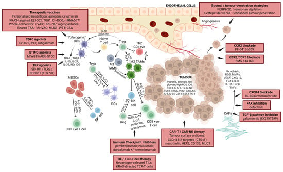
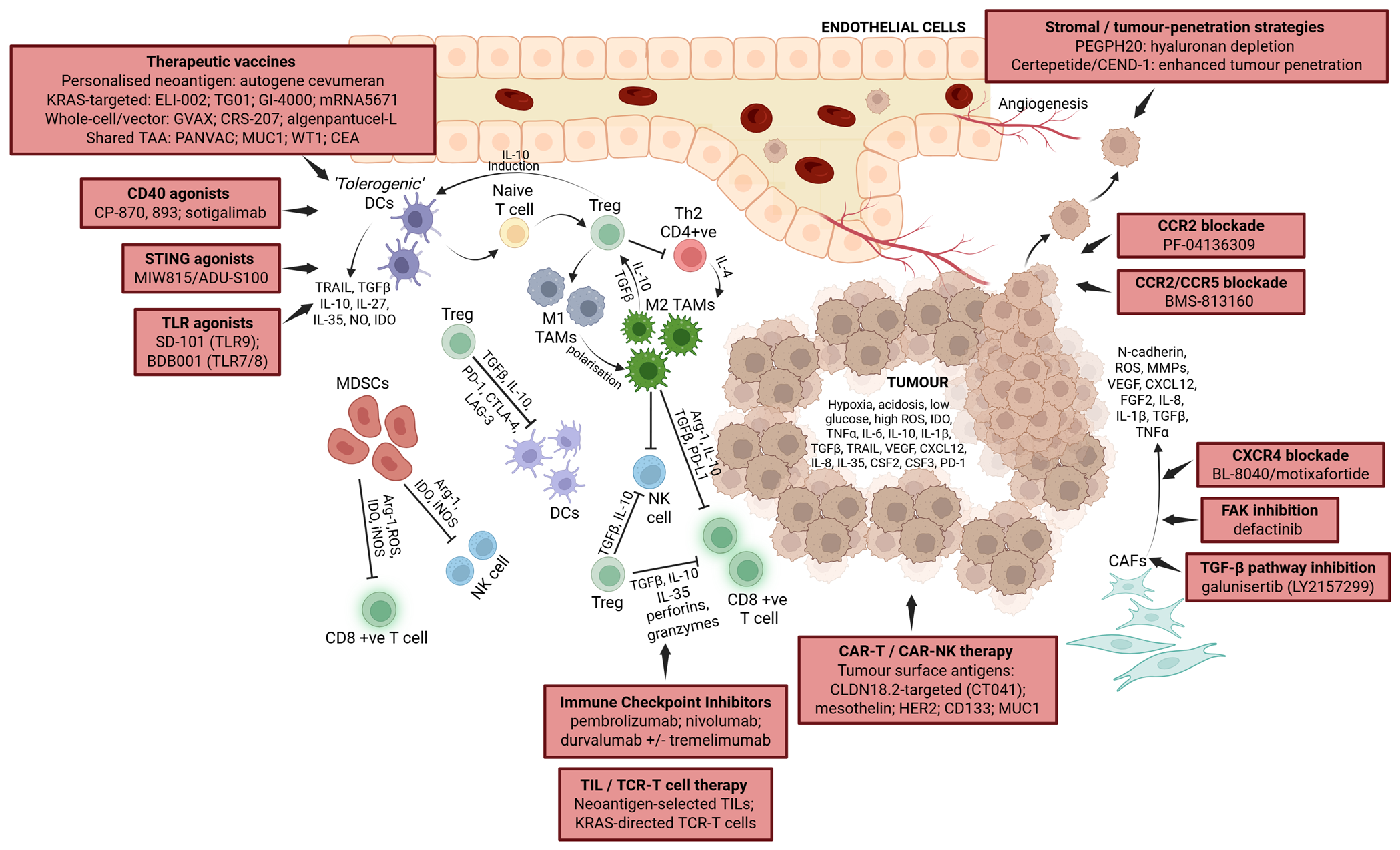
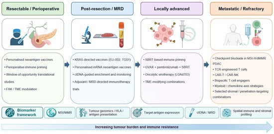
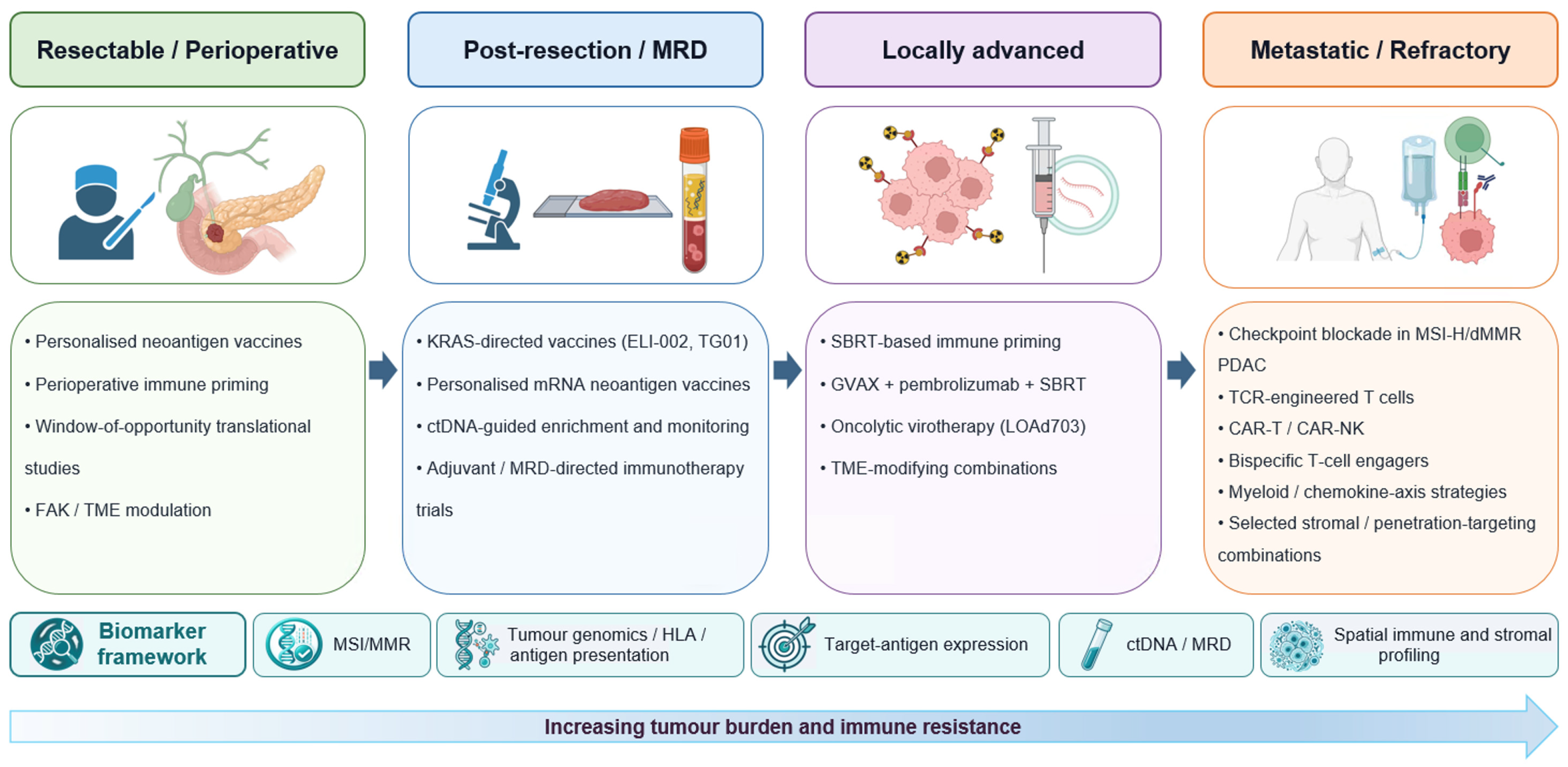

Disclaimer/Publisher’s Note: The statements, opinions and data contained in all publications are solely those of the individual author(s) and contributor(s) and not of MDPI and/or the editor(s). MDPI and/or the editor(s) disclaim responsibility for any injury to people or property resulting from any ideas, methods, instructions or products referred to in the content. |
© 2026 by the authors. Licensee MDPI, Basel, Switzerland. This article is an open access article distributed under the terms and conditions of the Creative Commons Attribution (CC BY) license.