


Abstract
Cardiorenal syndrome (CRS) encompasses a spectrum of disorders arising from bidirectional interactions between the heart and kidneys and is associated with substantial morbidity, mortality, and healthcare burden. Early recognition remains challenging because conventional biomarkers and imaging modalities may inadequately detect subclinical disease or distinguish functional changes from true organ injury. This narrative review examines the epidemiology, classification, pathophysiology, diagnostic approaches, and contemporary management of CRS, with particular emphasis on the emerging role of artificial intelligence (AI). Relevant evidence regarding established and emerging diagnostic and therapeutic approaches was reviewed, including biomarkers, imaging, guideline-directed medical therapy, ultrafiltration, cardiac devices, transplantation, and AI-based applications. Current evidence demonstrates the multifactorial nature of CRS and highlights the importance of early diagnosis and individualized management. Machine learning and deep learning approaches have shown potential to integrate electronic health records, biomarkers, imaging, physiological signals, and multi-omics data to support early detection, risk stratification, outcome prediction, and therapeutic decision-making. AI-assisted approaches may complement conventional diagnostic and management strategies and facilitate more precise and proactive CRS care. However, limitations related to data quality, model interpretability, external validation, and clinical implementation must be addressed before widespread clinical adoption.
1. Introduction
Cardiorenal syndrome (CRS) is defined as “an acute or chronic dysfunction in the heart or kidneys which would cause an acute or chronic dysfunction in the other organ” [1]. Robert Bright first described this bidirectional relationship between the two organs in 1836 [2]. Based on pathophysiology and chronicity, cardiorenal syndrome can be classified into five major subtypes: acute or chronic heart disease leading to kidney injury (Types 1 and 2), kidney injury leading to cardiac dysfunction (Types 3 and 4), and simultaneous cardiac and renal involvement from systemic conditions (Type 5) [1,3,4]. The current-day diagnosis of cardiorenal syndrome involves many modalities, including laboratory testing, biomarkers, and imaging techniques [5]. However, identifying the development of cardiorenal syndrome before significant organ dysfunction remains a challenge [1]. Our paper details how artificial intelligence is revolutionizing the early diagnosis of CRS by integrating clinical, biomarker, and imaging data to detect subtle physiological disturbances before overt disease develops. Machine learning models analyze large, multidimensional datasets—including ECGs, lab results, imaging, and multi-omics biomarkers—to provide accurate, individualized risk assessments [5]. These multimodal approaches enable early intervention, prevent disease progression, and mark a major step toward precision and preventive cardiorenal disease [6]. In the following sections, this paper will provide a detailed description of the pathophysiology, classification, diagnostic modalities, treatment, and the use of AI in the early diagnosis of CRS.
1.1. Epidemiology: Prevalence and Incidence
Cardiorenal syndrome (CRS) has emerged as a significant global health burden due to the increasing prevalence of both heart failure (HF) and chronic kidney disease (CKD), two conditions that frequently coexist and contribute to adverse clinical outcomes. By 2023, the prevalence of HF was estimated to be approximately 64.3 million individuals worldwide, and renal dysfunction is highly prevalent among this population. Baseline CKD, commonly defined as an estimated glomerular filtration rate (eGFR) <60 mL/min/1.73 m2, is present in nearly 60% of patients admitted with acute decompensated heart failure (ADHF). The coexistence of CKD in HF patients is associated with substantially increased risks of mortality, recurrent hospitalization, and cardiovascular events, with poorer outcomes observed among patients with more advanced renal dysfunction [7].
In addition to chronic renal impairment, acute deterioration in kidney function is frequently observed during hospitalization for HF. Approximately 20–30% of patients hospitalized with ADHF develop worsening renal function or acute kidney injury (AKI), reflecting the complex hemodynamic and neurohormonal interplay between the heart and kidneys. AKI in the setting of HF has been associated with significantly increased morbidity and mortality, with some studies reporting up to a fivefold increase in mortality risk. However, the prognostic significance of AKI in HF may depend on the clinical context, particularly the degree of successful decongestion achieved during treatment [7,8].
The epidemiological burden of CRS is further highlighted by the increasing incidence of severe AKI requiring renal replacement therapy among HF patients. National trends in the United States from 2002 to 2013 demonstrated a marked rise in dialysis-requiring AKI among HF hospitalizations, with incidence rates more than doubling during the study period. Importantly, dialysis-requiring AKI was associated with significantly higher in-hospital mortality, prolonged hospitalization, and adverse discharge outcomes compared with HF patients without severe renal injury. These findings underscore the growing clinical and economic burden of CRS and emphasize the need for early recognition, risk stratification, and multidisciplinary management strategies aimed at mitigating both cardiac and renal dysfunction [7].
1.2. Clinical and Therapeutic Challenges in Cardiorenal Syndrome (CRS)
Some of the challenges that clinicians face when they treat CRS nowadays is the need for a patient-centered management with early diagnosis, early intervention, the distinction of true kidney injury from permissive renal function deterioration during decongestion therapy, and the development of therapeutic algorithms to guide therapy [9].
A CRS classification system that assesses the underlying organ damage and its temporal sequence will assist clinicians in providing appropriate therapy to the proper patient. Furthermore, a combination of several biomarkers (both cardiac and renal) must be established in order to identify patients who are at higher risk of developing a more severe form of CRS [10,11]. Serum chloride and CNP appear to be potential indicators, but additional research is needed before they can be used in clinical practice [12]. CRS management is still complicated due to the intricacies of decongestive therapy and inotrope selection, the management of diuretic resistance, and the best timing and dose of ultrafiltration therapy [13]. Anemia in the context of cardiac and renal failure comprises a complex triad that should always be addressed [14,15]. Some of the challenges that clinicians face when they treat CRS nowadays is the need for a patient-centered management with early diagnosis, early intervention, the distinction of true kidney injury from permissive renal function deterioration during decongestion therapy, and the development of therapeutic algorithms to guide therapy [13].
Despite advances in understanding the pathophysiology of cardiorenal syndrome (CRS), its management continues to present significant clinical challenges [13]. One of the primary difficulties lies in the early diagnosis and accurate assessment of renal dysfunction in patients with heart failure [14,16]. Conventional biomarkers such as serum creatinine are often insensitive and may not detect early kidney injury until substantial renal impairment has occurred [17]. Additionally, clinicians frequently face difficulty distinguishing true acute kidney injury from transient or permissive worsening renal function that may occur during aggressive diuretic or decongestive therapy. This creates uncertainty in treatment decisions, as therapies aimed at improving cardiac function and relieving congestion may paradoxically worsen renal parameters despite providing overall clinical benefit [7].
There is also a lack of standardized and evidence-based therapeutic algorithms that can be universally applied across the heterogeneous spectrum of the disease [18]. Patients with CRS often present with multiple comorbidities, including diabetes mellitus, hypertension, chronic kidney disease, and atherosclerotic cardiovascular disease, which further complicate management strategies. Balancing adequate decongestion while preserving renal perfusion remains difficult, particularly in critically ill patients [1,2,3,7,14]. Furthermore, the complex bidirectional interaction between the heart and kidneys limits the effectiveness of isolated organ-specific therapies, emphasizing the need for individualized and multidisciplinary treatment approaches. Continued research is therefore essential to improve diagnostic accuracy, identify reliable biomarkers, and develop targeted therapeutic strategies that can optimize outcomes in patients with cardiorenal syndrome [15].
1.3. Limitations in Current Diagnostic Modalities
Early diagnosis of CRS is important to prevent serious mortality and morbidity, and this is the main challenge faced by clinicians to prevent the adverse effects and progression of CRS [17,18]. Current diagnostic modalities for cardiorenal syndrome (CRS) remain limited by the complex and bidirectional interaction between cardiac and renal dysfunction. Traditional renal biomarkers such as serum creatinine and estimated glomerular filtration rate (eGFR) are widely used but are relatively insensitive and often fail to detect kidney injury until substantial functional decline has already occurred [17]. Similarly, cardiac biomarkers such as natriuretic peptides and troponins may be chronically elevated in patients with renal dysfunction, thereby reducing their specificity and diagnostic utility in CRS [15]. In addition, existing diagnostic frameworks frequently fail to distinguish transient hemodynamic alterations from true intrinsic renal injury, limiting the ability to accurately identify disease severity and underlying pathophysiology [2]. Imaging modalities including echocardiography, ultrasonography, computed tomography, and magnetic resonance imaging provide valuable structural and functional assessment; however, they remain limited in detecting early microvascular and subclinical changes and may be affected by cost, accessibility, and operator dependence [5,19]. Consequently, the diagnosis of CRS continues to rely heavily on clinical judgment and multimodal assessment, contributing to variability in diagnosis and therapeutic decision-making.
1.4. Artificial Intelligence
Artificial intelligence (AI) has emerged as a promising strategy to address several of the diagnostic limitations associated with CRS. Machine learning algorithms can integrate large-scale clinical, laboratory, imaging, and hemodynamic datasets to identify complex patterns that may not be detectable through conventional assessment alone [5]. Emerging predictive models have demonstrated potential in the early detection of acute kidney injury, worsening heart failure, and congestion prior to significant alterations in traditional biomarkers [20]. Furthermore, AI-assisted applications in echocardiography, electrocardiography, and advanced imaging may improve CRS phenotyping, risk stratification, and individualized therapeutic planning [5]. AI-driven clinical decision support systems and remote monitoring technologies also offer opportunities for continuous real-time assessment of cardiorenal status, potentially enabling earlier intervention and optimization of treatment strategies [6]. Despite these advances, significant challenges remain, including the need for large externally validated datasets, improved model interpretability, reduction in algorithmic bias, and integration into routine clinical workflows [20]. Nevertheless, emerging evidence supports the growing role of AI-based approaches in advancing precision medicine and improving outcomes in patients with CRS. This review aims to examine the evolving role of AI in CRS, with particular emphasis on its current applications, clinical potential, and future directions.
2. Methods
This narrative review applied a structured literature search technique to ensure thorough coverage of current information on cardiorenal syndrome and applications of AI in diagnosis and treatment. Electronic databases such as PubMed, Embase, Scopus, and Web of Science were thoroughly searched for relevant papers published between January 2000 and December 2025. Search terms and Medical Subject Headings (MeSH) included combinations of: “Cardiorenal Syndrome,” “CRS,” “Chronic Kidney Disease,” “Heart Failure”, “Biomakers”, “Machine learning,” “multiomics,” “artificial intelligence,” and “AI-driven treatment”.
Manual searches of reference lists from relevant primary research and reviews were carried out to find additional publications. Two authors independently screened titles and abstracts, followed by a full-text review to determine eligibility. Discrepancies were handled by discussing or consulting with a third reviewer.
Inclusion Criteria:
- Peer-reviewed original research publications, clinical trials, and review papers;
- Studies with real subjects or appropriate preclinical models;
- Articles on cardiorenal syndrome, biomarkers and artificial intelligence in cardiorenal syndrome;
- Publications in English.
Exclusion Criteria:
- Abstracts from conferences that are not available in full text;
- Editorials, commentaries, or opinion pieces that lack original data;
- Non-English language publications;
- Studies not connected to cardiorenal syndrome or artificial intelligence.
3. Pathophysiology and Classification of Cardiorenal Syndrome
Cardiorenal syndrome (CRS) encompasses a spectrum of disorders in which dysfunction of the heart and kidneys coexist, with impairment of one organ contributing to dysfunction of the other [21]. The pathophysiology of CRS is multifactorial, involving a complex interplay of hemodynamic disturbances, venous congestion, neurohormonal activation, inflammation, and oxidative stress [21].
Traditionally, reduced cardiac output has been considered the principal mechanism underlying CRS, leading to renal hypoperfusion, ischemia, and ultimately acute kidney injury (AKI). However, growing evidence indicates that impaired forward flow alone does not fully explain the development or progression of CRS [22]. Alterations in glomerular hemodynamics may produce transient and reversible increases in serum creatinine, particularly following aggressive diuresis or the initiation of therapies such as angiotensin-converting enzyme inhibitors (ACE inhibitors), angiotensin receptor blockers (ARBs), angiotensin receptor–neprilysin inhibitors (ARNIs), and sodium-glucose cotransporter-2 (SGLT2) inhibitors. These changes often represent functional adaptations rather than true structural kidney injury [11,23,24]. In addition to reduced cardiac output, venous congestion has emerged as a key contributor to renal dysfunction in patients with heart failure. Elevated central venous pressure and increased intra-abdominal pressure reduce effective renal perfusion pressure, impair glomerular filtration, and promote worsening renal function [25]. Consequently, renal venous congestion is now recognized as an important driver of CRS and, in many patients, may have a greater impact on kidney function than reduced cardiac output alone.
Persistent activation of neurohormonal pathways further perpetuates the progression of CRS. Excessive stimulation of the renin–angiotensin–aldosterone system (RAAS) and sympathetic nervous system (SNS) promotes vasoconstriction, sodium and water retention, oxidative stress, inflammation, endothelial dysfunction, and adverse cardiac remodeling [26,27]. Within the kidneys, these mechanisms contribute to intrarenal vasoconstriction, ischemic injury, proteinuria, fibrosis, and progressive decline in renal function [28,29]. Inflammation and oxidative stress further amplify the bidirectional interaction between the heart and kidneys. Pro-inflammatory cytokines, reactive oxygen species, and antidiuretic hormone (ADH) contribute to ongoing cardiac and renal injury and are particularly important in type 5 CRS, where a systemic disorder simultaneously affects both organs [26]. This mechanism is exemplified by COVID-19, in which concurrent cardiac and renal injury is associated with elevated inflammatory markers, greater disease severity, and increased mortality, highlighting the significant role of systemic inflammation and direct tissue injury in the pathogenesis of CRS [30].
CRS is classified into five subtypes based on the acuity of presentation (acute or chronic), the primary organ initially affected (heart or kidney), and the presence of an underlying systemic disease, as illustrated in Figure 1.
Figure 1.
Classification and Pathophysiology of Cardiorenal Syndrome.
Type 1, or acute cardiorenal syndrome, occurs when an acute cardiac event, such as acute decompensated heart failure or acute coronary syndrome, leads to acute kidney injury. The mechanisms include reduced cardiac output, leading to renal hypoperfusion; elevated venous pressures, leading to renal congestion; and neurohormonal activation of systems such as the renin–angiotensin–aldosterone system, the sympathetic nervous system, and the vasopressin pathways. These maladaptive responses are compounded by systemic inflammation and oxidative stress, resulting in rapid deterioration of renal function. Clinically, this type is associated with high short-term mortality, prolonged hospital stays, and therapeutic challenges in balancing fluid removal with renal perfusion [21,31,32].
Type 2, or chronic cardiorenal syndrome, develops when chronic cardiac dysfunction, such as long-standing heart failure progressively impairs renal function. Two conditions (CHF and CKD) co-exist to make the diagnosis of chronic cardiorenal syndrome (Type 2) [20]. Sustained neurohormonal activation, chronic venous congestion, low cardiac output, and persistent inflammation and fibrosis drive the gradual decline in kidney function. Patients often develop resistance to diuretics, worsening fluid retention, and progressive chronic kidney disease. This form is associated with poor long-term outcomes, frequent hospitalizations, and complex management strategies that must balance cardiac therapies with renal preservation [31,32]. Initially the GFR decreases from initiation of GDMT medication; however, the decline either slows down or remains unchanged from the natural course [22].
Type 3, or acute renocardiac syndrome, arises when acute kidney injury triggers acute cardiac dysfunction. Mechanisms include volume overload leading to pulmonary edema and heart failure, electrolyte disturbances such as hyperkalemia or hypocalcemia precipitating arrhythmias, accumulation of uremic toxins causing myocardial depression or pericarditis, and inflammatory mediators impairing vascular tone and myocardial contractility. Clinically, this type is seen in hospitalized patients with AKI due to sepsis, nephrotoxins, or ischemia, and is associated with acute heart failure, arrhythmias, and sudden cardiac death [31,32]. AKI or worsening renal failure is an independent cardiovascular risk factor for mortality in hospitalized patients and is a predictor of clinical outcome in HF [22,33,34].
Type 4, or chronic renal–cardiac syndrome, describes the contribution of chronic kidney disease to long-term cardiac dysfunction. In this sub-type, CKD is the primary disease (including hypertensive, diabetic, autoimmune, and nephropathies) [30,31,34]. CKD promotes chronic volume overload and hypertension, leading to left ventricular hypertrophy and heart failure. CKD causes the progression of chronic heart failure with preserved ejection fraction and then later to heart failure with reduced ejection fraction [35,36]. Uremic toxins contribute to vascular calcification and myocardial fibrosis, whereas anemia increases cardiac workload and hypoxia [13,32]. Endothelial dysfunction and vascular stiffness accelerate atherosclerosis. Emerging evidence also highlights mitochondrial dysfunction in cardiomyocytes, impairing energy metabolism and contractile performance. Cardiovascular disease is the leading cause of death in CKD patients, with manifestations including heart failure with preserved ejection fraction, arrhythmias, and ischemic heart disease [12].
Type 5, or secondary cardiorenal syndrome, results from systemic disorders that simultaneously impair both cardiac and renal function. Conditions such as sepsis, diabetes mellitus, systemic lupus erythematosus, and amyloidosis drive systemic inflammation, endothelial dysfunction, hemodynamic instability, and neurohormonal dysregulation. Both organs are affected in parallel, and prognosis depends heavily on the underlying systemic disease and its control. Management requires targeting the systemic disorder while providing supportive care for both heart and kidneys, often involving multidisciplinary teams [4,37].
4. Diagnosis of Cardiorenal Syndrome
4.1. Conventional Lab Values (Serum Creatinine, eGFR, Urine Tests)
The clinical backbones for detecting and staging renal dysfunction are serum creatinine and eGFR. Creatinine rises are delayed and influenced by age, muscle mass, and hemodynamics, limiting this diagnosis. This limitation underscores the importance of serial measurements and clinical contexts, such as volume status and diuretic use, as essential components. Therefore, CKD-EPI equations for eGFR are used [13]. In acute settings, rather than relying on single values, it is important to trend creatinine and urine output. Urinalysis and albuminuria/proteinuria provide important prognostic information (albuminuria is an independent predictor of cardiorenal risk) [38,39].
4.2. Novel Filtration Biomarkers
Cystatin C (CysC) is less influenced by muscle mass, provides earlier detection of GFR decline, and has stronger prognostic associations with cardiovascular outcomes. CysC is supported as a useful adjunct to creatinine by several recent analyses for risk stratification in CRS and HF. Use of combined creatinine–cystatin C equations can be considered for improved GFR estimates [40]. Beta-2 microglobulin (B2M) correlates with renal function, and it has also been associated with cardiovascular events and mortality. It is also useful as a prognostic biomarker; however, it is not universally adopted for routine GFR estimation [41]. Beta-trace protein (BTP) is studied as an alternative filtration marker. Combined panels (creatinine + cystatin C ± BTP/B2M) can improve GFR estimation in some cohorts, but this has not outperformed cystatin C + creatinine [42]. Transcriptomic/proteomic approaches, as well as inflammatory markers, are being explored [2].
4.3. Imaging Methods
Renal ultrasound is the first-line test to rule out obstruction and assess kidney size and echogenicity [41]. Doppler/venous congestion imaging (renal venous Doppler) has an evolving role in the diagnosis of cardiorenal syndrome. Venous congestion patterns correlate with venous pressures and may predict worsening renal function from congestion. Cardiac imaging (echocardiography) is essential to define cardiac contributors (EF, filling pressures, valvular disease). Nuclear GFR measurement (iohexol or radionuclide clearance), though impractical for routine acute care, it is the gold standard for measured GFR in research. It is used when precise GFR is needed (research or complex diagnostic questions) [43,44,45], as illustrated in Figure 2.
Figure 2.
Current diagnostic and treatment modalities for Cardiorenal syndrome.
5. Treatment Strategies—Overall Principles
The overarching principle is to identify and treat the dominant driver (cardiac vs. renal vs. volume/vascular neurohormonal), relieve congestion early, optimize guideline-directed medical therapy (GDMT) for heart failure, and avoid iatrogenic renal injury while preserving perfusion [2], as illustrated in Figure 2.
5.1. Acute Management and Decongestion Strategies
5.1.1. Diuretics (Medical Decongestion)
The first-line for volume overload in acute decompensated HF with CRS are loop diuretics. High-dose IV bolus vs. continuous infusion, sequential nephron blockade with addition of thiazide-like diuretics, and close monitoring of urine output/renal function are the main strategies [46]. Resistance to diuretics is common in CRS, and it may require escalation strategies. It is also essential to monitor electrolytes, renal function, and hemodynamics closely. Combining diuretics with ultrafiltration or vasodilators is considered when there is an inadequate response. Monitor electrolytes, renal function, and hemodynamics closely. Combining diuretics with ultrafiltration or vasodilators is considered when there is an inadequate response [44].
5.1.2. Ultrafiltration (Mechanical Fluid Removal)
Ultrafiltration (UF) has been investigated as an alternative decongestive strategy in patients with cardiorenal syndrome (CRS) and acute decompensated heart failure (ADHF), particularly those with persistent congestion and diuretic resistance.
Early pilot studies, including the RAPID-CHF and EUPHORIA trials, demonstrated that UF was feasible, achieved greater isotonic fluid removal than standard intravenous diuretic therapy, improved congestion and dyspnea, and suggested a potential reduction in subsequent heart failure hospitalizations without major adverse effects on renal function [45,46,47]. These findings were reinforced by the UNLOAD Trial which showed greater fluid removal and reduced heart failure rehospitalizations with UF compared with standard diuretic therapy, generating interest in mechanical decongestion as a means of improving cardiorenal outcomes [48,49]. However, subsequent findings from the CARRESS-HF Trial tempered this enthusiasm. In patients with ADHF complicated by worsening renal function, UF was associated with a greater increase in serum creatinine at 96 h, no significant improvement in weight loss or symptom relief, and a higher incidence of adverse events compared with a stepped pharmacologic diuretic strategy [50,51]. Similarly, the prematurely terminated AVOID-HF Trial showed a non-significant trend toward a longer time to first heart failure event with UF but failed to demonstrate clear clinical superiority because of early study termination and a higher frequency of UF-related adverse events [50].
Subsequent meta-analyses have largely confirmed that UF and intensive diuretic-based decongestion achieve comparable reductions in fluid overload and body weight. While UF may offer effective volume removal in selected patients, it has not consistently demonstrated superior clinical outcomes and may be associated with transient increases in blood urea nitrogen and worsening renal function [51,52]. Consequently, contemporary guidelines reserve UF for carefully selected patients with persistent congestion despite optimized medical therapy, particularly those with diuretic resistance or inadequate response to conventional decongestive strategies [53,54].
5.1.3. Neurohormonal Modulation, Vasodilator, and Inotropic Therapy
Vasodilators (e.g., nitrates, nitroprusside) are useful in reducing afterload and improving renal perfusion in hypertensive ADHF in selected patients. Inotropes (dobutamine, milrinone) help to improve cardiogenic shock or low-output states and renal perfusion. However, they carry risks (arrhythmia, ischemia) and should be used for the short term as a bridge to stabilization or advanced therapies. Individualize based on hemodynamics and goals of care [2].
5.2. Disease-Modifying Therapies and Long-Term Management
5.2.1. RAAS Inhibition and Other Neurohormonal Therapies in Chronic CRS
ACE inhibitors and ARBs are foundational therapies in heart failure with reduced ejection fraction (HFrEF), providing both cardiovascular and long-term cardiorenal benefits through neurohormonal blockade; however, they require careful dose titration with monitoring of serum creatinine and potassium, as an initial, hemodynamically mediated rise in creatinine is expected and does not necessarily indicate true nephrotoxicity [54]. ARNI has demonstrated superiority over ACE inhibitor therapy in reducing morbidity and mortality in HFrEF and is now a key component of guideline-directed medical therapy, with generally favorable or neutral renal effects, though blood pressure and renal function monitoring remain essential [54,55]. Standard HFrEF therapies also include beta-blockers and MRAs, both of which improve survival and reduce hospitalizations; MRAs can be safely used across a range of chronic kidney disease stages but necessitate close surveillance for hyperkalemia and worsening renal function [54].
5.2.2. SGLT2 Inhibitors (Dapagliflozin, Empagliflozin, Sotagliflozin)
These medications are the latest developments. Large RCTs and meta-analyses show that these drugs help achieve robust reductions in HF hospitalizations and slowed CKD progression. Their benefits extend to patients with and without diabetes, across the EF spectrum to some extent, and are now integral to GDMT. SGLT2 inhibitors exhibit early hemodynamic effects (natriuresis) and durable renal protection (reduced eGFR decline and decreased progression to end-stage kidney disease). For cardiorenal patients, SGLT2i are highly emphasized [54].
5.2.3. Cardiac Devices
Device-based therapies may play an important role in selected patients with cardiorenal syndrome (CRS) by improving cardiac performance and alleviating venous congestion. Cardiac Resynchronization Therapy (CRT) has been shown to enhance ventricular synchrony, improve cardiac output, and reduce heart failure symptoms in appropriately selected patients with reduced ejection fraction and electrical dyssynchrony. These hemodynamic improvements may translate into improved renal perfusion and stabilization of kidney function by increasing forward flow and reducing renal venous congestion. Similarly, Implantable Cardioverter–Defibrillator (ICD) therapy reduces the risk of sudden cardiac death and is often incorporated into the comprehensive management of patients with advanced heart failure, although its direct effects on renal outcomes are less well established [56].
In patients with end-stage heart failure refractory to medical therapy, Left Ventricular Assist Device (LVAD) implantation can provide substantial hemodynamic support and improve end-organ perfusion. Several studies have demonstrated early improvements in renal function following LVAD implantation, likely due to enhanced cardiac output and renal blood flow. However, these benefits must be weighed against the significant perioperative and long-term risks, including acute kidney injury, bleeding, infection, thrombosis, and right ventricular failure. Consequently, device candidacy should be individualized, taking into account the severity of renal dysfunction, overall comorbidity burden, anticipated reversibility of kidney injury, and eligibility for advanced therapies such as heart transplantation. A multidisciplinary approach involving cardiology, nephrology, and transplant teams is essential to optimize patient selection and outcomes [54].
5.2.4. Kidney Transplant/Cardiorenal Transplant Considerations
In patients with advanced cardiorenal syndrome, renal replacement with Kidney Transplantation alone may be considered when primary renal disease predominates and cardiac dysfunction is secondary and potentially reversible after optimization of heart failure therapy. In contrast, for carefully selected patients with irreversible dysfunction of both organs, Combined Heart–Kidney Transplantation (CHKT) represents a definitive therapeutic option. Evidence supporting CHKT is derived primarily from observational studies and registry analyses rather than randomized trials.
Analyses of the United Network for Organ Sharing (UNOS) registry have demonstrated improved post-transplant survival among patients with advanced renal dysfunction (estimated glomerular filtration rate <30–37 mL/min/1.73 m2 or dialysis dependence) undergoing CHKT compared with isolated heart transplantation, whereas patients with potentially reversible cardiorenal syndrome may recover renal function after heart transplantation alone [55].
Ongoing research continues to optimize CHKT strategies, including the prospective Staged Kidney Transplantation During Combined Heart–Kidney Transplantation (NCT06877169) trial, which is evaluating delayed kidney implantation following heart transplantation using ex vivo machine perfusion to reduce ischemia–reperfusion injury and improve renal graft outcomes [56]. Despite encouraging observational data, the absence of randomized controlled trials underscores the need for individualized multidisciplinary assessment, with consideration of baseline cardiac reserve, chronic kidney disease severity, comorbidity burden, frailty, and perioperative risk to optimize patient selection and timing of transplantation [2], as shown in Figure 2.
5.3. Prognosis
Cardiorenal syndrome (CRS) is characterized by the complex interplay between cardiac and renal dysfunction, resulting in higher morbidity and mortality compared with isolated heart failure or kidney disease [2,54]. In a large cohort of 30,681 patients, those with acute CRS (type 1 or 3) demonstrated a hazard ratio of 3.13 (95% CI 2.72–3.61) for death compared with patients with chronic kidney disease alone, whereas patients with chronic CRS (type 4) had relatively better survival (HR 0.48; 95% CI 0.37–0.61) but outcomes remained worse than controls. These findings highlight the importance of CRS subtype in clinical risk stratification, with the higher mortality hazard associated with acute CRS supporting closer monitoring and timely multidisciplinary assessment [57]. The subtype of CRS, severity of cardiac and renal dysfunction, and response to therapy are the principal determinants of prognosis [2,58]. Elevated biomarkers such as cystatin C and β2-microglobulin, increased congestion, and recurrent acute decompensated heart failure (ADHF) events are associated with adverse outcomes [58,59,60]. Conversely, early recognition, aggressive decongestion, and guideline-directed medical therapy (GDMT), including RAAS and SGLT2 inhibitors, can improve long-term outcomes [60].
6. AI-Driven Diagnosis and Management of CRS
AI applications relevant to CRS span a broad spectrum of clinical maturity. At one end are supervised learning models for AKI or outcome prediction based on routinely collected structured data, some of which have undergone external validation and limited prospective evaluation in hospital and ICU cohorts. Intermediate on this spectrum are imaging- and ECG-based models, telemonitoring systems, and explainable-AI techniques, which have shown feasibility and reasonable performance in related cardiac or renal populations but have not yet been prospectively tested in defined CRS cohorts. At the earliest stage are conceptual or proof-of-concept approaches such as federated learning, digital twins, and multi-omics integration, for which evidence is largely methodological or limited to small pilot studies. Distinguishing these maturity levels is essential to interpret the current evidence and avoid over-extrapolating from experimental work to routine CRS care.
6.1. Role of AI in Risk Prediction and Early Detection
Early identification is one of the most crucial unmet requirements in the therapy of cardiorenal syndrome (CRS), since increasing cardiac and renal failure sometimes occurs quietly before standard diagnostic criteria are reached. The bidirectional link between the heart and kidneys generates a complicated pathophysiological milieu in which malfunction in one organ commonly exacerbates harm in the other. Current diagnostic techniques depend heavily on serum creatinine, estimated glomerular filtration rate (eGFR), and urine output measures. However, these parameters are poor predictors of acute kidney injury (AKI) because they frequently reflect functional deterioration rather than the original injury itself and may remain within normal limits during the early phases of disease development. As a result, opportunities for timely intervention are frequently overlooked, leading to chronic organ damage and poor cardiovascular and renal outcomes. These limitations have sparked increased interest in artificial intelligence (AI)-based techniques capable of detecting subtle clinical patterns that occur before overt signs of cardiorenal failure [61].
Artificial intelligence has emerged as a viable answer to these diagnostic issues, allowing for the analysis of vast, multidimensional clinical information and revealing complicated correlations that standard statistical approaches may miss. Unlike traditional risk assessment tools, which typically rely on a small number of variables, AI algorithms can continuously integrate information from electronic health records (EHRs), laboratory investigations, physiological measurements, medication profiles, and longitudinal clinical data to generate personalized risk predictions. These capabilities are especially useful in CRS, because illness development is influenced by a number of interconnected pathways, including hemodynamic changes, neurohormonal activation, systemic inflammation, oxidative stress, endothelial dysfunction, and metabolic dysregulation [61]. AI may have the ability to transform CRS therapy from a reactive to a proactive paradigm by identifying high-risk patients before clinically evident deterioration begins.
The AI studies discussed below address different clinical tasks, including prediction of future AKI, detection of existing renal impairment, and estimation of hospitalization or mortality risk. These outcomes should not be treated interchangeably or equated with a diagnosis of CRS. Many studies involve populations without explicitly defined CRS; their findings therefore provide indirect evidence of potential relevance to CRS and require validation in dedicated CRS cohorts. The applicability of AI models should also be considered by CRS subtype. Models predicting AKI in acute heart failure may be relevant to type 1 CRS, but cannot be assumed to perform similarly in type 2 CRS, where chronic cardiac disease contributes to progressive kidney dysfunction. Types 3 and 4 involve acute or chronic kidney disease contributing to cardiac dysfunction, requiring evaluation of cardiac outcomes rather than renal deterioration alone. Type 5 involves systemic disease affecting both organs and requires validation within the relevant underlying condition. Model performance should therefore be assessed separately across CRS subtypes, with clearly defined populations, prediction horizons, and outcomes.
Among AI applications, the most mature evidence in domains relevant to CRS comes from supervised learning models using structured EHR data to predict AKI or renal deterioration. Mohamadlou et al. published one of the first examples of the value of machine learning (ML) for renal risk prediction, developing an ML-based system capable of predicting AKI from routinely gathered EHR data [59]. The algorithm used demographic information, laboratory measurements, vital signs, and clinical factors to identify individuals who were at risk for AKI before clinically recognized renal failure occurred. Importantly, the algorithm outperformed various traditional techniques in terms of prediction performance, demonstrating the viability of AI-powered surveillance systems for continuous patient monitoring. These findings highlighted the potential of AI to facilitate earlier clinical intervention, allowing for the implementation of nephroprotective measures such as fluid optimization, medication review, avoidance of nephrotoxic agents, and closer monitoring of renal function before irreversible injury occurs [62].
Martinez et al. further demonstrated the potential of AI-assisted AKI risk prediction by developing and validating ML models in a multicenter dataset of over 92,000 emergency department contacts [63]. The researchers showed that AI could predict AKI up to 72 h before patients met Kidney Disease: Improving Global Outcomes (KDIGO) diagnostic criteria, with AUC values ranging from 0.74 to 0.81. Surprisingly, risk projections were obtained in a median of only 1.7 h following patient presentation utilizing commonly accessible EHR characteristics such as demographics, laboratory findings, vital signs, medical history, and presenting symptoms [63]. Unlike biomarker-dependent techniques, these models did not require any further testing, indicating their potential scalability and inclusion into normal clinical operations. Early detection of imminent renal injury opens a key treatment window in which preventative measures can be performed before serious kidney damage occurs. Nonetheless, this work remains at a retrospective validation stage, and no studies have yet examined whether ECG-based detection of renal impairment alters management or outcomes in CRS.
In CRS, delayed diagnosis of renal damage is especially important because decreasing kidney function can hasten heart failure development via salt and water retention, neurohormonal activation, increased venous congestion, and systemic inflammatory responses. As a result, AI systems’ capacity to detect imminent AKI before conventional diagnostic criteria are met may give a chance to break the vicious cycle of cardiorenal dysfunction, avert disease development, and lower the risk of unfavorable clinical consequences. From a clinical standpoint, these prediction models might aid in earlier treatment decision-making and more tailored care regimens for CRS patients.
Beyond EHR-based predictive analytics, intermediate-maturity applications include AI applied to widely available tests such as the electrocardiogram (ECG). Kwon et al. created a deep learning algorithm that can predict renal insufficiency from conventional electrocardiographic (ECG) data [64]. The study found that AI systems could detect minor electrophysiological patterns linked with worsening kidney function, even when doctors were unable to see them. This strategy emphasizes the usefulness of widely available, low-cost, and noninvasive diagnostic techniques as platforms for AI-assisted screening and risk assessment. Given the high incidence of cardiovascular problems among patients with CRS, AI-enhanced ECG analysis might be a useful technique for detecting concealed renal impairment in both inpatient and outpatient settings [64]. These findings show that AI-driven diagnosis is moving away from traditional laboratory-based assessments and toward a multimodal paradigm that uses physiological signals, clinical history, and real-time patient data to detect early signs of cardiorenal failure.
Recent studies on developing concepts in heart failure and acute kidney injury have stressed AI’s revolutionary potential in cardiorenal medicine [61]. Modern AI systems are increasingly capable of combining various clinical data streams, such as laboratory biomarkers, physiological monitoring data, imaging findings, and longitudinal EHR data, to create dynamic prediction models that develop with patient status. Such multimodal techniques go beyond standard single-parameter evaluations, providing a more complete picture of the complex biochemical interactions that underpin CRS. Future AI platforms may enhance these capabilities by integrating ECG features, biomarker profiles, imaging characteristics, wearable sensor data, and longitudinal clinical records into unified prediction frameworks. By evaluating many parameters of patient health at the same time, these systems may be able to detect disease trajectories before conventional diagnostic thresholds are achieved [61].
AI-based risk prediction may help identify patients who warrant closer monitoring and clinical reassessment. However, predictive accuracy alone does not demonstrate improved patient outcomes. The studies reviewed primarily evaluate prediction performance rather than whether acting on AI-generated alerts reduces AKI, hospitalization, mortality, or cardiovascular events. Proposed responses, including fluid optimization, medication review, and avoidance of nephrotoxins, therefore represent potential applications rather than demonstrated benefits of AI-guided CRS care. Prospective interventional studies comparing AI-assisted management with usual care are needed to determine whether earlier risk identification translates into safer treatment and better outcomes [61,62,65].
Despite these encouraging developments, significant difficulties remain until AI can be effectively integrated into normal CRS therapy. Many existing prediction models are based on retrospective datasets and require prospective validation across many patient groups and healthcare systems. Variability in data quality, algorithmic bias, model interpretability, and integration into clinical procedures remain significant challenges to deployment. Furthermore, issues about openness and clinician trust underscore the rising need for explainable AI frameworks that enable healthcare workers to comprehend the reasoning behind algorithm-generated predictions. Rather than replacing clinical judgment, AI is widely considered as a decision-support tool that may help clinicians detect early cardiorenal decline and intervene in a timely manner [61].
Overall, existing data shows that artificial intelligence (AI) has the potential to radically improve the early diagnosis of CRS by enabling earlier identification of renal failure, enhancing risk classification, and facilitating individualized therapeutic decision making. AI systems can identify high-risk individuals before overt kidney impairment develops, allowing for prompt intervention and potentially improving both cardiovascular and renal outcomes. As these technologies develop and undergo thorough clinical validation, AI is projected to play an increasingly crucial role in precision medicine methods in cardiorenal care.
More recently, methodological innovations have aimed to address barriers to clinical adoption. Explainable artificial intelligence (XAI) methods make prediction models more transparent by highlighting the most influential variables contributing to a given prediction, which may support clinician understanding and trust [59,65,66]. In current cardiorenal research, XAI has mostly been applied post hoc to AKI or mortality prediction models in ICU or heart failure cohorts, rather than tested prospectively as part of CRS care pathways. XAI therefore represents an intermediate-maturity tool for interpreting model outputs in research and early implementation studies, not an established component of routine CRS management.
Natural language processing (NLP) can be used to augment structured EHR data by extracting clinically salient information from physician notes, discharge summaries, and medication records [67,68,69]. In AKI and critical-care cohorts, adding NLP-derived features has modestly improved prediction performance compared with structured data alone, but these approaches remain at an early validation stage and have not yet been evaluated in defined CRS populations.
Wearable devices and remote monitoring systems extend AI-enabled diagnosis beyond the hospital by continuously tracking physiological data such as heart rate, blood pressure, body weight, and cardiac rhythm [70,71]. In heart failure populations, algorithm-based telemonitoring has shown mixed effects on hospitalization and mortality, and there are no CRS-specific prospective trials. AI-enhanced wearable analytics should therefore be considered an intermediate-maturity technology with supportive evidence in related conditions but unproven benefit in CRS.
Machine learning has also been proven to be very effective in the intensive care unit, when it can predict the onset of acute kidney injury up to hours (even days) before the traditional diagnostic criteria. Early identification allows timely preventive measures such as optimization of fluid therapy, nephrotoxin avoidance and increasing renal monitoring to prevent the development of cardiorenal dysfunction [72,73,74].
These EHR-based models therefore represent the best-studied class of AI tools in the renal domain, with multiple large retrospective cohorts and some external validation, but still very limited prospective impact evaluation and no CRS-specific trials. Performance beyond AUC illustrates clinically relevant trade-offs. Koyner et al. reported 84% sensitivity and 85% specificity for stage 2 AKI at a probability threshold of ≥0.022 [73]. Tomašev et al. predicted 55.8% of inpatient AKI episodes up to 48 h in advance, but generated two false alerts for every true alert, highlighting the potential burden of false-positive predictions [72]. For detection of existing renal impairment, Kwon et al. reported an external-validation AUC of 0.906, with 85.1% sensitivity, 82.5% specificity, 19.1% PPV, and 99.1% NPV [64]. The low PPV despite favorable discrimination underscores the need for confirmatory assessment following a positive screening result.
Calibration and comparative clinical usefulness also warrant consideration. In the prospective evaluation by Flechet et al., AKI predictor and physicians showed no statistically significant difference in discrimination on the first ICU morning (AUC 0.89 versus 0.94; p = 0.27), calibration assessment identified physicians’ tendency to overestimate risk, and decision-curve analysis favored AKI predictor across selected thresholds [74]. These findings illustrate why discrimination alone cannot establish clinical value. Validation methods also differ: Martinez et al. used internal cross-validation, whereas Kwon et al. evaluated performance in a separate hospital [65,74]. Nevertheless, these results concern AKI prediction or renal impairment detection and do not establish diagnostic accuracy or clinical benefit in explicitly defined CRS cohorts.
While these advancements have occurred, issues of algorithmic bias, data privacy, transparency, and regulatory oversight continue to be significant hurdles for the general clinical adoption of AI tools. To secure reliable and equitable AI clinical decision support, external validation, fairness assessment and ongoing monitoring are critical [73,74].
Lastly, clinical benefit and cost-effectiveness must be shown to make AI a successful tool for adoption. While AI can be used to decrease the rates of hospitalisation, optimise resources, and limit healthcare costs, there is a need for further prospective studies to establish the economic value in the management of routine CRS [75,76].
Overall, EHR-based AKI and outcome prediction models constitute the most advanced AI applications relevant to CRS, whereas ECG-based detection, XAI, NLP, and wearable-driven analytics are at intermediate stages of maturity, and more complex architectures such as federated learning and digital twins remain largely conceptual at present.
In summary, explainable AI, federated learning, natural language processing, wearable technologies, digital twins, and prediction models based on intensive care units will be promising developments that have the potential of enhancing early diagnosis, personalized treatment, and precision medicine in the context of cardiorenal syndrome, as illustrated in Figure 3.
Figure 3.
AI in the diagnosis and management of cardiorenal syndrome.
A comparative summary of key AI studies relevant to renal risk prediction and their potential applicability to CRS is provided in Table 1.
Table 1.
Selected AI studies relevant to renal risk prediction and potential applicability to cardiorenal syndrome (CRS).
6.2. Integration of Biomarkers
Cardiorenal syndrome (CRS) is defined by complicated bidirectional interactions between the heart and kidneys that include hemodynamic changes, neurohormonal activation, inflammation, oxidative stress, and fibrosis. Given the complex pathophysiology, conventional biomarkers such as serum creatinine and estimated glomerular filtration rate (eGFR) frequently fail to detect early organ damage because they reflect established functional decline rather than continuing pathological processes. As a result, there is growing interest in integrating numerous cardiac and renal indicators, particularly when paired with artificial intelligence (AI), to enhance early diagnosis, prognosis, and tailored therapy of CRS [77,78,79].
Early studies demonstrated the limits of depending only on traditional markers. Despite its widespread usage in clinical practice, serum creatinine is relatively insensitive to early kidney injury and may not increase until significant nephron damage has occurred. Similarly, natriuretic peptides such as B-type natriuretic peptide (BNP) and N-terminal pro-BNP (NT-proBNP) reflect cardiac stress and congestion while providing little information about underlying renal damage. To address these inadequacies, Taub et al. stressed the significance of include biomarkers that represent the many pathophysiology mechanisms implicated in CRS [79]. Biomarkers such as neutrophil gelatinase-associated lipocalin (NGAL), cystatin C, kidney injury molecule-1 (KIM-1), and interleukin-18 (IL-18) were found to detect renal injury earlier than serum creatinine, whereas BNP and NT-proBNP remained useful indicators of cardiac dysfunction and volume overload [5]. Together, these indicators give supplementary information that may enhance diagnosis accuracy and prognosis in CRS patients.
Recent advancements have greatly broadened the biomarker landscape. Stefanou et al. established a precision medicine paradigm that classifies biomarkers based on the biological processes they reflect, such as functional impairment, structural damage, inflammation, fibrosis, and cardiac remodeling [80]. Functional indicators such as serum creatinine, eGFR, cystatin C, BNP, and NT-proBNP are still required to evaluate renal and cardiac function. Emerging biomarkers, such as NGAL, KIM-1, liver-type fatty acid-binding protein (L-FABP), soluble suppression of tumorigenicity-2 (sST2), galectin-3, growth differentiation factor-15 (GDF-15), and inflammatory cytokines, offer new insights into tubular injury, fibrosis, ventricular remodeling, and systemic inflammatory activation [78]. These indicators work together to provide a more complete picture of disease activity than any single marker could.
Several studies provide preliminary evidence for integrating biomarkers into AI models relevant to cardiorenal disease. Tasic et al. evaluated machine-learning classifiers incorporating cystatin C-based eGFR, NT-proBNP, sodium, and potassium in patients with combined cardiac and renal dysfunction. Cystatin C-based eGFR showed the greatest predictive importance in several classifiers, although the broadly defined population limits conclusions about individual CRS subtypes [81]. More directly, a study of sepsis-induced CRS incorporated biomarkers including cardiac troponin I and myoglobin alongside clinical variables to predict one-year mortality. Its random-forest model achieved an external-validation AUC of 0.863, with precision of 0.775 and recall of 0.821 [82]. This supports biomarker-integrated prognostic assessment in sepsis-induced CRS, rather than early diagnosis or AI-guided treatment.
Additional evidence comes from AKI populations without explicitly defined CRS. Rashidi et al. evaluated models combining NGAL, NT-proBNP, creatinine, and urine output in burn and trauma patients. Models using NGAL with NT-proBNP achieved an AUC of 0.92, sensitivity of 91%, and specificity of 93% in a second cohort of 51 patients, although the small sample limits generalizability [83]. In the ASSESS-AKI study, Vasquez-Rios et al. integrated clinical and biomarker data, including NGAL, KIM-1, NT-proBNP, and troponin T, using unsupervised clustering to identify four AKI subphenotypes with differing outcomes [84]. These findings demonstrate the feasibility of biomarker integration for prediction and phenotyping, but do not establish CRS-specific diagnostic accuracy or treatment benefit. Validation in clearly defined CRS cohorts remains necessary before these approaches can support routine clinical decisions.
Aletras et al. investigated the practical use of biomarker-guided management, proposing a systematic framework for incorporating new biomarkers into ordinary clinical treatment [6]. The authors noted that multimarker panels that include signs of glomerular filtration, tubular damage, congestion, inflammation, and fibrosis give a more accurate evaluation of CRS than single biomarkers. Importantly, these indicators should not replace traditional clinical examination, but rather supplement current diagnostic and treatment procedures. The combination of such multimarker techniques with AI-powered decision support systems should improve diagnostic precision, identify high-risk patients earlier, and enable tailored therapy selection throughout the disease course [79].
Beyond traditional biomarkers, current research is increasingly focusing on multidimensional biomarker systems that include data from proteomics, metabolomics, transcriptomics, and genomes. These high-dimensional datasets are appropriate for AI-powered analysis tools that can reveal previously unknown biological markers related with CRS development. The combination of multi-omics technology and machine learning has the potential to uncover new disease phenotypes, enhance patient stratification, and enable genuinely individualized care methods. Such advancements are a significant step toward precision cardiorenal therapy, in which treatment decisions are guided by each individual’s own genetic and clinical profile [6,80]. Despite these encouraging advancements, a number of obstacles persist to prevent widespread clinical use. Variability in biomarker tests, a lack of established cutoff values, variation among patient populations, and insufficient external validation are all important issues. Furthermore, many emerging biomarkers are linked with higher prices and restricted access outside of specialist facilities. To successfully integrate biomarkers into AI-based clinical processes, large prospective studies, standardized testing methodologies, and thorough validation across multiple healthcare contexts are required [78,79].
Overall, the integration of conventional and emerging biomarkers with artificial intelligence represents a promising strategy for improving the diagnosis and management of cardiorenal syndrome. By enabling earlier detection of organ injury, enhancing prognostic accuracy, and supporting personalized therapeutic decision-making, biomarker-guided AI systems have the potential to transform CRS care and advance the implementation of precision medicine in cardiorenal disease [77,78,79].
6.3. Integration of Imaging Techniques
Imaging has emerged as an essential component in the diagnosis, risk stratification, and management of cardiorenal syndrome (CRS), providing critical insights into the complex bidirectional interactions between the heart and kidneys that are not fully captured by clinical assessment or circulating biomarkers alone. Because structural, functional, and hemodynamic abnormalities frequently occur across the CRS spectrum, imaging methods provide a non-invasive method of assessing organ failure, tracking disease progression, and directing therapeutic approaches. Recent developments in multimodality imaging have improved the capacity to identify cardiorenal pathophysiology, allowing for a more thorough assessment of both cardiac and renal involvement and facilitating the shift to precision medicine techniques in CRS care [6].
Conventional imaging techniques remain the foundation of CRS assessment. Echocardiography is the most used imaging modality due to its ease of use, safety, and capacity to study heart anatomy and function in real time. Left ventricular ejection fraction, ventricular geometry, diastolic function, and valvular anomalies are all useful indicators of the severity of cardiac failure and its possible contribution to renal impairment [80]. Doppler echocardiographic indices, such as trans mitral flow patterns and the E/E′ ratio, can estimate left ventricular filling pressures and detect subclinical congestion, which is becoming recognized as a key contributor to deteriorating renal function in CRS [80]. Similarly, renal ultrasonography is still a useful method for assessing kidney size, cortical thickness, parenchymal echogenicity, and urinary tract blockage, allowing doctors to distinguish acute from chronic renal damage while ruling out reversible causes of kidney failure [80].
Although traditional imaging is useful for diagnosing CRS, newer imaging modalities provide a better understanding of the structural and functional changes that cause it. heart magnetic resonance imaging (CMR) has evolved as the standard for measuring ventricular sizes, myocardial mass, and heart function while also allowing for thorough tissue characterisation. Techniques like late gadolinium enhancement and T1/T2 mapping make it easier to identify myocardial fibrosis, inflammation, and extracellular matrix remodeling, all of which are linked to poor cardiovascular outcomes and increasing cardiorenal dysfunction [6]. Computed tomography (CT) provides high-resolution anatomical visualization of cardiovascular structures and can detect vascular abnormalities that contribute to cardiorenal impairment, but concerns about radiation exposure and contrast-induced nephrotoxicity may limit its routine use in patients with advanced renal dysfunction [6,80]. Nuclear imaging methods, such as positron emission tomography (PET) and single-photon emission computed tomography (SPECT), aid in the assessment of cardiac perfusion, viability, and metabolic activity, enhancing risk classification in certain CRS groups [6].
Recent advances in multimodal imaging have switched the emphasis from separate organ evaluation to integrated cardiorenal phenotyping. Modern imaging techniques allow for the simultaneous study of cardiac remodeling, myocardial fibrosis, renal structural abnormalities, tissue perfusion, and hemodynamic changes, resulting in a more thorough knowledge of disease causes [6]. This comprehensive approach is especially significant since CRS is now recognized as a systemic condition with intricate relationships between neurohormonal activation, inflammation, fibrosis, venous congestion, and microvascular dysfunction. By merging data from several imaging modalities, doctors may better identify disease subtypes, predict clinical outcomes, and adjust therapy regimens to specific patient features [6].
AI-assisted imaging has potential applications in cardiorenal assessment, although the studies discussed here provide indirect evidence from dialysis, CKD, and heart failure populations rather than explicitly defined CRS cohorts. Tan et al. evaluated automated B-line detection using YOLACT and Mask R-CNN algorithms applied to handheld lung ultrasound in 76 dialysis patients. In the validation group of 15 patients, automated and physician B-line counts showed an intraclass correlation coefficient of 0.892 (95% CI 0.754–0.952) [85]. These findings support automated quantification of pulmonary congestion, but the small, single-center study did not establish CRS-specific diagnostic performance or clinical benefit from AI-guided fluid management.
Chou et al. used U-Net segmentation of chest radiographs to quantify cardiothoracic ratio in 3117 patients with CKD [86]. Automated measurements differed from physician measurements by less than 0.03 in 94.97% of 537 test images. Adding cardiothoracic ratio to a reference prognostic model increased the C-statistic for cardiovascular mortality from 0.698 to 0.719. This study illustrates how AI-derived imaging measurements may contribute to risk assessment, although the incremental improvement was modest and subsequent outcome modeling used conventional statistical methods. Separately, Shah et al. integrated echocardiographic measurements with clinical, laboratory, and ECG variables using hierarchical and model-based clustering in 397 patients with HFpEF [87]. Three phenogroups with differing outcomes were identified, and risk stratification was reproduced in a prospective validation cohort of 107 patients. This represents machine learning applied to imaging-derived measurements rather than automated interpretation of raw images.
These studies address distinct tasks: congestion quantification, automated anatomical measurement, and multimodal phenotyping. Their performance measures therefore cannot be compared directly, and none establishes the accuracy of AI imaging for diagnosing CRS across its five subtypes. Validation in clearly characterized CRS populations, comparison with conventional imaging assessment, and prospective evaluation of effects on treatment decisions and patient outcomes are needed before routine CRS-specific use can be supported.
Collectively, developments in imaging technology are shifting CRS assessment from a mostly descriptive procedure to a data-driven and precision-oriented science. The combination of multimodality imaging and AI-assisted analytics provides new prospects for earlier diagnosis, better risk classification, and individualized treatment planning. As imaging biomarkers become more integrated with clinical and genetic data, these technologies are projected to play a larger role in the holistic therapy of CRS, potentially leading to better patient outcomes [6,37,80].
6.4. Future Directions and Challenges
The confluence of artificial intelligence (AI), multi-omics technologies, sophisticated biomarkers, medical imaging, digital health platforms, and multidisciplinary care models is progressively influencing the therapy of cardiorenal syndrome (CRS). Although great progress has been made in the early identification and risk stratification of CRS, significant obstacles remain in clinical application, data integration, model interpretability, and healthcare accessibility. Emerging evidence suggests that cardiorenal medicine is shifting from a reactive disease-management paradigm to a proactive precision-medicine framework capable of predicting disease trajectories, identifying personalized therapeutic targets, and optimizing patient outcomes via continuous monitoring and intelligent clinical decision-support systems [5,82].
One of the most exciting advances is the advent of multidimensional biomarker systems that combine genomic, transcriptomic, proteomic, metabolomic, and clinical data to form unified prediction frameworks [82]. Traditional biomarkers such as serum creatinine, natriuretic peptides, and cystatin C are still useful for detecting established cardiovascular and renal impairment; however, they only capture a subset of the complex molecular interactions that underpin CRS. Multi-omics techniques provide a more complete knowledge of disease processes by examining pathways implicated in inflammation, fibrosis, endothelial dysfunction, oxidative stress, metabolic dysregulation, and accelerated biological aging [82]. The combination of these molecular datasets with AI-driven analysis tools may allow for earlier diagnosis of subclinical illness, improved risk stratification, and personalized therapy methods suited to unique molecular profiles [82,88]. Most multi-omics studies relevant to cardiorenal disease have been conducted in relatively small, selected cohorts and have focused on biomarker discovery and phenotyping rather than on rigorously validated clinical decision-support tools. External validation in large, diverse CRS populations and demonstration of management impact are still lacking, underscoring that multi-omics-driven AI remains at an exploratory stage.
Beyond risk prediction, AI is predicted to revolutionize precision cardiorenal medicine. Machine learning and deep learning algorithms have previously shown that they can evaluate large-scale electronic health records (EHRs), laboratory data, imaging studies, and wearable-device data to predict unfavorable cardiovascular and renal outcomes [37]. Future AI systems might combine many data sources, including as biomarkers, imaging parameters, hemodynamic measures, and longitudinal clinical records, to create real-time individualized therapy recommendations [5,37,82]. Such technologies might help healthcare professionals identify individuals who are at high risk of acute kidney injury, heart failure decompensation, or progression to severe cardiorenal dysfunction before irreversible organ damage happens. Furthermore, AI-assisted clustering and pattern-recognition approaches may identify previously unknown CRS phenotypes with distinct molecular signatures, clinical trajectories, and therapeutic responses, allowing for more precise patient stratification and targeted interventions [82].
Digital health technologies are also likely to play an important role in the long-term management of CRS. Wearable sensors, smartphone-based applications, telemedicine platforms, and integrated healthcare ecosystems provide continuous monitoring of physiological indicators, hydration status, medication adherence, and symptom development [5,37,82]. When integrated with AI-powered analytics, these technologies may enable dynamic risk classification and early identification of clinical deterioration, lowering hospital admissions and healthcare expenditures. Such techniques might be especially beneficial for older patients and those with many chronic illnesses who need frequent monitoring and multidisciplinary care [37].
Another significant future trend is the creation of integrated cardiorenal care models. Despite their tight pathophysiological dependency, cardiovascular and renal disorders have generally been treated separately in clinical specialties. Recent expert opinions call for the expansion of dedicated cardio nephrology programs that encourage collaboration among cardiologists, nephrologists, endocrinologists, and primary care physicians. Integrated care pathways have the potential to increase diagnostic accuracy, optimize treatment approaches, and make it easier to incorporate new technology into ordinary clinical practice. Similarly, future clinical studies are planned to use multidisciplinary designs and include cardiorenal outcomes that better represent the interrelated nature of cardiovascular and kidney illness [5].
Despite these promising breakthroughs, a number of impediments persist to prevent broad adoption of AI-driven and precision-medicine technologies. One key difficulty is the quality, standardization, and interoperability of healthcare data. Many AI models are built on retrospective datasets that contain missing information, selection bias, and significant heterogeneity among healthcare systems, which may restrict their generalizability [37,82]. External validation across varied patient groups and healthcare systems is critical prior to large-scale clinical adoption [37].
Federated learning offers one possible solution to data fragmentation by enabling multiple institutions to train shared models without transferring patient-level data [89,90]. Methodological studies in general ICU and nephrology datasets have demonstrated that federated models can approach the performance of centrally trained models while preserving privacy. However, federated learning in cardiorenal medicine remains at a proof-of-concept stage, with no prospective CRS-specific implementations or evaluations of its impact on workflow, equity, or outcomes.
Personalized computational models, or “digital twins,” represent another early-stage concept with potential relevance to complex CRS. These models use clinical, imaging, and biomarker data to simulate individual disease trajectories and treatment responses [91,92]. To date, digital-twin applications in cardiovascular and renal disease are limited to small pilot studies and in silico experiments, without clinical trials demonstrating improved decision-making or outcomes. Digital twins should therefore be regarded as a long-term research direction rather than a near-term clinical tool for CRS.
Although multimodal AI may improve prediction by combining complementary data sources, its added value over simpler models should not be assumed. In a multicenter AKI study, combining structured clinical data with unstructured notes improved predictive performance compared with structured data alone; however, this does not establish clinical benefit in CRS [81]. Comparisons should assess incremental discrimination, calibration, and decision-curve net benefit at clinically relevant thresholds [93,94,95]. Potential gains should also be weighed against data availability, acquisition costs, missing data, and workflow burden. Prospective evaluation is needed to determine whether improved predictions translate into better management and patient outcomes [95]. However, in cardiorenal syndrome, these multimodal approaches require direct comparison with simpler models to determine whether their added complexity improves risk prediction and clinical decision-making.
Furthermore, the “black-box” nature of many machine-learning algorithms raises concerns regarding transparency, interpretability, and clinician confidence. As a hypothetical example, an opaque model could recommend ultrafiltration for a patient with CRS based on worsening renal function and persistent congestion, without making clear whether hemodynamic status or response to diuretic therapy influenced its recommendation. This could encourage inappropriate treatment selection: in CARRESS-HF, ultrafiltration produced a greater increase in serum creatinine and more serious adverse events than stepped pharmacologic therapy, without significantly greater weight loss. Experimental evidence also demonstrates that clinicians can be influenced by inaccurate advice attributed to AI. Explainable AI should therefore help clinicians scrutinize recommendations in the individual patient’s context, while recognizing that interpretability alone does not establish safety or effectiveness and must be accompanied by rigorous clinical validation [37].
Another significant problem is translating promising research discoveries into real-world therapeutic applications. Although several AI models have shown outstanding prediction accuracy in retrospective and observational research, only a handful have undergone thorough prospective examination or proven improvements in patient-centered outcomes [5,37]. Future research should focus on multicenter implementation studies to see if AI-guided therapies might lower hospitalization rates, acute renal injury incidence, cardiovascular events, healthcare costs, and death in CRS patients. Establishing clinical value, cost-effectiveness, and workflow compatibility will be critical to wider adoption of these technologies [5].
Additional impediments include legislative considerations, data privacy issues, infrastructural restrictions, and discrepancies in healthcare access [5,82]. Resource-constrained healthcare institutions may face major financial and technological obstacles when deploying advanced AI platforms, multi-omics testing, and digital monitoring technologies [5]. Furthermore, underrepresentation of ethnic and socioeconomically disadvantaged individuals in training datasets may lead to algorithmic bias and worsen existing healthcare disparities [80,82].
Addressing these limitations will require international collaboration, standardized data-governance frameworks, equitable dataset development, and rigorous validation strategies to ensure that technological advances benefit diverse patient populations [7,10].
Overall, the future of CRS treatment depends on the integration of multidimensional biomarkers, AI-driven analytics, sophisticated imaging technologies, digital health platforms, and multidisciplinary care models [5,37,82]. These advancements have the potential to shift cardiorenal therapy from reactive illness management to predictive, preventative, and individualized care. Finally, the success of future CRS management will be determined not only by technological innovation, but also by the ability to integrate AI, multi-omics data, imaging, and clinical expertise into interpretable, equitable, and clinically actionable systems capable of improving real-world patient outcomes [5,37,96], as shown in Figure 3.
7. Conclusions
Cardiorenal syndrome represents a complex and bidirectional interplay between cardiac and renal dysfunction, resulting in significant morbidity, mortality, and healthcare utilization. Advances in the understanding of its pathophysiology have highlighted the roles of hemodynamic alterations, neurohormonal activation, inflammation, oxidative stress, and venous congestion in disease progression. While traditional biomarkers and imaging modalities remain essential for diagnosis and monitoring, emerging biomarkers and advanced imaging techniques offer potential for earlier detection and more precise risk stratification.
Recent developments in artificial intelligence and machine learning offer promising approaches for predicting renal and cardiovascular deterioration. However, much of the evidence discussed derives from retrospective or observational studies, and improved predictive performance does not establish that AI-guided care prevents CRS progression, improves patient outcomes, or reduces healthcare costs. These potential benefits require prospective interventional validation and formal economic evaluation in CRS populations. Meanwhile, management should remain grounded in evidence-based therapies, individualized volume management, and multidisciplinary cardiorenal care.
Despite these advances, significant challenges remain in the early diagnosis and management of CRS due to its heterogeneous presentation and complex underlying mechanisms. Artificial intelligence should serve as a supplementary tool in the diagnosis and management of cardiorenal syndrome, supporting rather than replacing clinical judgment. AI-generated predictions should be interpreted alongside the patient’s clinical presentation, laboratory findings, and imaging results, with clinicians retaining responsibility for diagnostic and therapeutic decisions. To establish reliability, AI models require rigorous external validation across diverse populations, prospective evaluation in clinical practice, and ongoing monitoring for bias and changes in performance. Transparency regarding model limitations and uncertainty is equally essential to prevent overreliance on automated outputs. Incorporating these safeguards may allow AI to enhance early detection and risk stratification while preserving individualized, evidence-based care.
Future research should focus on validating novel biomarkers, integrating multimodal diagnostic approaches, and leveraging artificial intelligence to enhance clinical decision-making. A comprehensive and individualized approach that combines emerging technologies with evidence-based therapies may ultimately improve patient outcomes and reduce the burden of cardiorenal syndrome.
Author Contributions
H.P. and S.P.A. defined the review scope, context, and purpose of this study. H.P., G.A.R.P., A.F.S., A.G., P.P., N.S.A.R.G., V.P., K.S., S.K.B., A.K., F.F., J.Y., J.L., M.N.S., G.Y., P.E., S.M.J.W., M.V. and S.S.K. conducted the literature review and drafted the manuscript. H.P. and S.P.A. conceived and crafted illustrative figures. S.S., D.S., L.S.P., R.B., S.R. and S.A.H. provided clinical perspectives and expertise for this study. All authors read and performed a critical review of the manuscript. H.P., K.S., S.S.K. and S.P.A. performed the cleaning and organization of the manuscript. S.P.A. provided conceptualization, supervision, and project administration. All authors have read and agreed to the published version of the manuscript.
Funding
This research received no external funding.
Data Availability Statement
This review was based on publicly available academic literature databases.
Acknowledgments
This work was supported by the resources within the Digital Engineering & Artificial Intelligence Laboratory (DEAL), Department of Medicine, Mayo Clinic, Jacksonville, FL, USA.
Conflicts of Interest
The authors declare no conflicts of interest.
References
- Kousa, O.; Rout, P.; Aslam, A.; Aboeata, A. Cardiorenal syndrome. In StatPearls; StatPearls Publishing: Treasure Island, FL, USA, 2025. [Google Scholar]
- Rangaswami, J.; Bhalla, V.; Blair, J.E.A.; Chang, T.I.; Costa, S.; Lentine, K.L.; Lerma, E.V.; Mezue, K.; Molitch, M.; Mullens, W.; et al. Cardiorenal syndrome: Classification, pathophysiology, diagnosis, and treatment strategies: A scientific statement from the American Heart Association. Circulation 2019, 139, e840–e878. [Google Scholar] [CrossRef] [Scilit] [PubMed]
- Di Lullo, L.; Bellasi, A.; Barbera, V.; Russo, D.; Russo, L.; Di Iorio, B.; Cozzolino, M.; Ronco, C. Pathophysiology of the cardio-renal syndromes types 1–5: An up-to-date review. Indian Heart J. 2017, 69, 255–265. [Google Scholar] [CrossRef] [Scilit] [PubMed]
- Uduman, J. Epidemiology of Cardiorenal Syndrome. Adv. Chronic Kidney Dis. 2018, 25, 391–399. [Google Scholar] [CrossRef] [Scilit] [PubMed]
- Lin, L.; Zhou, X.; Dekkers, I.A.; Lamb, H.J. Cardiorenal syndrome: Emerging role of medical imaging for clinical diagnosis and management. J. Pers. Med. 2021, 11, 734. [Google Scholar] [CrossRef] [Scilit] [PubMed]
- Aletras, G.; Bachlitzanaki, M.; Stratinaki, M.; Lamprogiannakis, E.; Petrakis, I.; Foukarakis, E.; Pantazis, Y.; Hamilos, M.; Stylianou, K. Integrating novel biomarkers into clinical practice: A practical framework for diagnosis and management of cardiorenal syndrome. Life 2025, 15, 1540. [Google Scholar] [CrossRef] [Scilit] [PubMed]
- Correa, A.; Patel, A.; Chauhan, K.; Shah, H.; Saha, A.; Dave, M.; Poojary, P.; Mishra, A.; Annapureddy, N.; Dalal, S.; et al. National trends and outcomes in dialysis-requiring acute kidney injury in heart failure: 2002–2013. J. Card. Fail. 2018, 24, 442–450. [Google Scholar] [CrossRef] [Scilit] [PubMed]
- Peng, X.; Zhang, H.P. Acute cardiorenal syndrome: Epidemiology, pathophysiology, assessment, and treatment. Rev. Cardiovasc. Med. 2023, 24, 40. [Google Scholar] [CrossRef] [Scilit] [PubMed]
- Shlipak, M.G.; Massie, B.M. The clinical challenge of cardiorenal syndrome. Circulation 2004, 110, 1514–1517. [Google Scholar] [CrossRef] [Scilit] [PubMed]
- Breidthardt, T.; Cox, E.F.; Squire, I.; Odudu, A.; Omar, N.F.; Eldehni, M.T.; Francis, S.T.; McIntyre, C.W. The pathophysiology of the chronic cardiorenal syndrome: A magnetic resonance imaging study. Eur. Radiol. 2015, 25, 1684–1691. [Google Scholar] [CrossRef] [Scilit] [PubMed]
- Kaze, A.D.; Zhuo, M.; Kim, S.C.; Patorno, E.; Paik, J.M. Association of SGLT2 inhibitors with cardiovascular, kidney, and safety outcomes among patients with diabetic kidney disease: A meta-analysis. Cardiovasc. Diabetol. 2022, 21, 47. [Google Scholar] [CrossRef] [Scilit] [PubMed]
- Kazory, M.R.C.A.; Costanzo, M.R. The dynamic relationship between serum chloride and cardiorenal syndrome. Rev. Cardiovasc. Med. 2020, 21, 25–29. [Google Scholar] [CrossRef] [Scilit] [PubMed]
- Colombo, P.C.; Doran, A.C.; Onat, D.; Wong, K.Y.; Ahmad, M.; Sabbah, H.N.; Demmer, R.T. Venous Congestion, Endothelial and Neurohormonal Activation in Acute Decompensated Heart Failure: Cause or Effect? Curr. Heart Fail. Rep. 2015, 12, 215–222. [Google Scholar] [CrossRef] [Scilit] [PubMed]
- Georgopoulou, T.; Petrakis, I.; Dermitzaki, K.; Pleros, C.; Drosataki, E.; Aletras, G.; Foukarakis, E.; Lioudaki, E.; Androulakis, E.; Stylianou, K. Cardiorenal syndrome: Challenges in everyday clinical practice and key points towards better management. J. Clin. Med. 2023, 12, 4121. [Google Scholar] [CrossRef] [Scilit] [PubMed]
- Kim, C.J.; Choi, I.-J.; Park, H.-J.; Kim, T.H.; Kim, P.-J.; Chang, K.; Baek, S.H.; Chung, W.S.; Seung, K.-B. Impact of Cardiorenal Anemia Syndrome on Short- and Long-Term Clinical Outcomes in Patients Hospitalized with Heart Failure. Cardiorenal Med. 2016, 6, 269–278. [Google Scholar] [CrossRef] [Scilit] [PubMed]
- Kim, J.A.; Wu, L.; Rodriguez, M.; Lentine, K.L.; Virk, H.U.H.; El Hachem, K.; Lerma, E.V.; Kiernan, M.S.; Rangaswami, J.; Krittanawong, C. Recent developments in the evaluation and management of cardiorenal syndrome: A comprehensive review. Curr. Probl. Cardiol. 2023, 48, 101509. [Google Scholar] [CrossRef] [Scilit] [PubMed]
- Goffredo, G.; Barone, R.; Di Terlizzi, V.; Correale, M.; Brunetti, N.D.; Iacoviello, M. Biomarkers in cardiorenal syndrome. J. Clin. Med. 2021, 10, 3433. [Google Scholar] [CrossRef] [Scilit] [PubMed]
- Jefferies, J.L.; Kovesdy, C.P.; Ronco, C. Contemporary laboratory assessment of acute cardiorenal syndrome for early diagnosis: A call for action. Am. Heart J. 2023, 261, 75–84. [Google Scholar] [CrossRef] [Scilit] [PubMed]
- Dutta, A.; Saha, S.; Bahl, A.; Mittal, A.; Basak, T. A comprehensive review of acute cardio-renal syndrome: Need for novel biomarkers. Front. Pharmacol. 2023, 14, 1152055. [Google Scholar] [CrossRef] [Scilit] [PubMed]
- Chung, E.Y.M.; Trinh, K.; Li, J.; Hahn, S.H.; Endre, Z.H.; Rogers, N.M.; Alexander, S.I. Biomarkers in cardiorenal syndrome and potential insights into novel therapeutics. Front. Cardiovasc. Med. 2022, 9, 868658. [Google Scholar] [CrossRef] [Scilit] [PubMed]
- Martín, M.; Fernández, M.; Bacigalupe, L.P.; Rozado, J. Cardio-renal syndrome: Review and new perspectives. J. CardioRenal Med. 2026, 2, 4. [Google Scholar] [CrossRef] [Scilit]
- Mitsas, A.C.; Elzawawi, M.; Mavrogeni, S.; Boekels, M.; Khan, A.; Eldawy, M.; Stamatakis, I.; Kouris, D.; Daboul, B.; Gunkel, O.; et al. Heart failure and cardiorenal syndrome: A narrative review on pathophysiology, diagnostic and therapeutic regimens-from a cardiologist’s view. J. Clin. Med. 2022, 11, 7041. [Google Scholar] [CrossRef] [Scilit] [PubMed]
- De la Espriella, R.; Bayés-Genís, A.; Morillas, H.; Bravo, R.; Vidal, V.; Núñez, E.; Santas, E.; Miñana, G.; Sanchis, J.; Fácila, L.; et al. Renal function dynamics following co-administration of sacubitril/valsartan and empagliflozin in patients with heart failure and type 2 diabetes. ESC Heart Fail. 2020, 7, 3792–3800. [Google Scholar] [CrossRef] [Scilit] [PubMed]
- Rao, V.S.; Ahmad, T.; Brisco-Bacik, M.A.; Bonventre, J.V.; Wilson, F.P.; Siew, E.D.; Felker, G.M.; Anstrom, K.K.; Mahoney, D.D.; Bart, B.A.; et al. Renal effects of intensive volume removal in heart failure patients with preexisting worsening renal function. Circ. Heart Fail. 2019, 12, e005552. [Google Scholar] [CrossRef] [Scilit] [PubMed]
- Tonelli, M.; Wiebe, N.; McAlister, F.; Fok, M.; Garg, A.; House, A.; Rabbat, C.; Culleton, B. Chronic Kidney Disease and Mortality Risk: A Systematic Review. J. Am. Soc. Nephrol. 2006, 17, 2034–2047. [Google Scholar] [CrossRef] [Scilit] [PubMed]
- Patel, R.B.; Fonarow, G.C.; Greene, S.J.; Zhang, S.; Alhanti, B.; DeVore, A.D.; Butler, J.; Heidenreich, P.A.; Huang, J.C.; Kittleson, M.M.; et al. Kidney Function and Outcomes in Patients Hospitalized With Heart Failure. J. Am. Coll. Cardiol. 2021, 78, 330–343. [Google Scholar] [CrossRef] [Scilit] [PubMed]
- Damman, K.; Gori, M.; Claggett, B.; Jhund, P.S.; Senni, M.; Lefkowitz, M.P.; Prescott, M.F.; Shi, V.C.; Rouleau, J.L.; Swedberg, K.; et al. Renal Effects and Associated Outcomes During Angiotensin-Neprilysin Inhibition in Heart Failure. J. Am. Coll. Cardiol. Heart Fail. 2018, 6, 489–498. [Google Scholar] [CrossRef] [Scilit] [PubMed]
- Mullens, W.; Abrahams, Z.; Francis, G.S.; Sokos, G.; Taylor, D.O.; Starling, R.C.; Young, J.B.; Tang, W.W. Importance of Venous Congestion for Worsening of Renal Function in Advanced Decompensated Heart Failure. J. Am. Coll. Cardiol. 2009, 53, 589–596. [Google Scholar] [CrossRef] [Scilit] [PubMed]
- Yang, B.; Li, D.; Phillips, M.I.; Mehta, P.; Mehta, J.L. Myocardial angiotensin II receptor expression and ischemia-reperfusion injury. Vasc. Med. 1998, 3, 121–130. [Google Scholar] [CrossRef] [Scilit] [PubMed]
- Niizeki, T.; Takeishi, Y.; Arimoto, T.; Nozaki, N.; Hirono, O.; Watanabe, T.; Nitobe, J.; Miyashita, T.; Miyamoto, T.; Koyama, Y.; et al. Persistently increased serum concentration of heart-type fatty acid-binding protein predicts adverse clinical outcomes in patients with chronic heart failure. Circ. J. Off. J. Jpn. Circ. Soc. 2008, 72, 109–114. [Google Scholar] [CrossRef] [Scilit] [PubMed]
- Pliquett, R.U. Cardiorenal syndrome: An updated classification based on clinical hallmarks. J. Clin. Med. 2022, 11, 2896. [Google Scholar] [CrossRef] [Scilit] [PubMed]
- Ronco, C. Cardiorenal syndromes: Definition and classification. In Fluid Overload: Diagnosis and Management; Contributions to Nephrology; Karger: Basel, Switzerland, 2010; Volume 164, pp. 33–38. [Google Scholar] [CrossRef] [Scilit] [PubMed]
- Damman, K.; Navis, G.; Voors, A.A.; Asselbergs, F.W.; Smilde, T.D.J.; Cleland, J.G.F.; van Veldhuisen, D.J.; Hillege, H.L. Worsening renal function and prognosis in heart failure: Systematic review and meta-analysis. J. Card. Fail. 2007, 13, 599–608. [Google Scholar] [CrossRef] [Scilit] [PubMed]
- Bellomo, R.; Ronco, C.; Kellum, J.A.; Mehta, R.L.; Palevsky, P.; The ADQI Workgroup. Acute renal failure–definition, outcome measures, animal models, fluid therapy and information technology needs: The Second International Consensus Conference of the Acute Dialysis Quality Initiative (ADQI) Group. Crit. Care 2004, 8, R204. [Google Scholar] [CrossRef] [Scilit] [PubMed]
- Sárközy, M.; Kovács, Z.Z.A.; Kovács, M.G.; Gáspár, R.; Szűcs, G.; Dux, L. Mechanisms and modulation of oxidative/nitrative stress in type 4 cardio-renal syndrome and renal sarcopenia. Front. Physiol. 2018, 9, 1648. [Google Scholar] [CrossRef] [Scilit] [PubMed]
- Pinheiro da SilvaA, L.; Vaz da SilvaM, J. Type 4 cardiorenal syndrome. Rev. Port. Cardiol. 2016, 35, 601–616. [Google Scholar] [CrossRef] [Scilit] [PubMed]
- George, S.M.; Kalantarinia, K. The role of imaging in the management of cardiorenal syndrome. Int. J. Nephrol. 2011, 2011, 245241. [Google Scholar] [CrossRef] [Scilit] [PubMed]
- Levey, A.S.; Stevens, L.A.; Schmid, C.H.; Zhang, Y.; Castro, A.F.; Feldman, H.I.; Kusek, J.W.; Eggers, P.; Van Lente, F.; Greene, T.; et al. A new equation to estimate glomerular filtration rate. Ann. Intern. Med. 2009, 150, 604–612. [Google Scholar] [CrossRef] [Scilit] [PubMed]
- Joosten, M.M.; Pai, J.K.; Bertoia, M.L.; Gansevoort, R.T.; Bakker, S.J.L.; Cooke, J.P.; Rimm, E.B.; Mukamal, K.J. β2-Microglobulin, cystatin C, and creatinine and risk of symptomatic peripheral artery disease. J. Am. Heart Assoc. 2014, 3, e000803. [Google Scholar] [CrossRef] [Scilit] [PubMed]
- Herrero-Morín, J.D.; Málaga, S.; Fernández, N.; Rey, C.; Diéguez, M.Á.; Solís, G.; Concha, A.; Medina, A. Cystatin C and beta2-microglobulin: Markers of glomerular filtration in critically ill children. Crit. Care 2007, 11, R59. [Google Scholar] [CrossRef] [Scilit] [PubMed]
- Foster, M.C.; Coresh, J.; Hsu, C.-Y.; Xie, D.; Levey, A.S.; Nelson, R.G.; Eckfeldt, J.H.; Vasan, R.S.; Kimmel, P.L.; Schelling, J.; et al. Serum β-trace protein and β2-microglobulin as predictors of ESRD, mortality, and cardiovascular disease in adults with CKD in the CRIC study. Am. J. Kidney Dis. 2016, 68, 68–76. [Google Scholar] [CrossRef] [Scilit] [PubMed]
- Ebert, N.; Koep, C.; Schwarz, K.; Martus, P.; Mielke, N.; Bartel, J.; Kuhlmann, M.; Gaedeke, J.; Toelle, M.; van der Giet, M.; et al. Beta trace protein does not outperform creatinine and cystatin C in estimating glomerular filtration rate in older adults. Sci. Rep. 2017, 7, 12656. [Google Scholar] [CrossRef] [Scilit] [PubMed]
- Khati, N.J.; Hill, M.C.; Kimmel, P.L. The role of ultrasound in renal insufficiency: The essentials. Ultrasound Q. 2005, 21, 227–244. [Google Scholar] [CrossRef] [Scilit] [PubMed]
- Zhang, C.; Wu, Z.; Song, Y.; Jin, X.; Hu, J.; Huang, C.; Zhou, J.; Lian, J. Exploring diagnostic biomarkers of type 2 cardio-renal syndrome based on secreted proteins and bioinformatics analysis. Sci. Rep. 2024, 14, 24612. [Google Scholar] [CrossRef] [Scilit] [PubMed]
- Cerezo, I.; Cancho, B.; Rodríguez Sabillón, J.A.; Jorge, A.; Álvarez López, A.; Valladares, J.; Gomez, J.L.; Romero, J.; Robles, N.R. Comparative prognostic value of glomerular filtration rate, serum cystatin C, beta-2-microglobulin, and albuminuria for death and chronic kidney disease progression. J. Clin. Lab. Anal. 2025, 39, e25139. [Google Scholar] [CrossRef] [Scilit] [PubMed]
- Chandramohan, D.; Simhadri, P.K.; Jena, N.; Palleti, S.K. Strategies for the management of cardiorenal syndrome in the acute hospital setting. Hearts 2024, 5, 329–348. [Google Scholar] [CrossRef] [Scilit]
- Bart, B.A.; Boyle, A.; Bank, A.J.; Anand, I.; Olivari, M.T.; Kraemer, M.; Mackedanz, S.; Sobotka, P.A.; Schollmeyer, M.; Goldsmith, S.R. Ultrafiltration versus usual care for hospitalized patients with heart failure: The Relief for Acutely Fluid-Overloaded Patients with Decompensated Congestive Heart Failure (RAPID-CHF) trial. J. Am. Coll. Cardiol. 2005, 46, 2043–2046. [Google Scholar] [CrossRef] [Scilit] [PubMed]
- Costanzo, M.R.; Saltzberg, M.; O’Sullivan, J.; Sobotka, P. Early ultrafiltration in patients with decompensated heart failure and diuretic resistance. J. Am. Coll. Cardiol. 2005, 46, 2047–2051. [Google Scholar] [CrossRef] [Scilit] [PubMed]
- Felker, G.M.; Mentz, R.J. Diuretics and ultrafiltration in acute decompensated heart failure. J. Am. Coll. Cardiol. 2012, 59, 2145–2153. [Google Scholar] [CrossRef] [Scilit] [PubMed]
- Bart, B.A.; Goldsmith, S.R.; Lee, K.L.; Givertz, M.M.; O’Connor, C.M.; Bull, D.A.; Redfield, M.M.; Deswal, A.; Rouleau, J.L.; LeWinter, M.M.; et al. Ultrafiltration in decompensated heart failure with cardiorenal syndrome. N. Engl. J. Med. 2012, 367, 2296–2304. [Google Scholar] [CrossRef] [Scilit] [PubMed]
- Tay, K.L.-Y.; Osman, A.R.; Yeoh, E.E.X.; Luangboriboon, J.; Lau, J.F.; Chan, J.J.A.; Yousif, M.; Tse, B.Y.H.; Horgan, G.; Gamble, D.T.; et al. Ultrafiltration versus diuretics on prognostic cardiac and renal biomarkers in acute decompensated heart failure: A systematic review and meta-analysis. J. Clin. Med. 2023, 12, 2793. [Google Scholar] [CrossRef] [Scilit] [PubMed]
- Costanzo, M.R.; Negoianu, D.; Jaski, B.E.; Bart, B.A.; Heywood, J.T.; Anand, I.S.; Smelser, J.M.; Kaneshige, A.M.; Chomsky, D.B.; Adler, E.D.; et al. Aquapheresis Versus Intravenous Diuretics and Hospitalizations for Heart Failure. JACC Heart Fail. 2016, 4, 95–105. [Google Scholar] [CrossRef] [Scilit] [PubMed]
- Terpos, V.; Roumeliotis, S.; Georgianos, P.I.; Papa, E.; Tsalikakis, D.G.; Papachristou, E.; Liakopoulos, V. Diuretics or ultrafiltration in the treatment of acute decompensated heart failure: An updated systematic review and meta-analysis. Ther. Apher. Dial. 2024, 28, 9–22. [Google Scholar] [CrossRef] [Scilit] [PubMed]
- Maddox, T.M.; Januzzi, J.L., Jr.; Allen, L.A.; Breathett, K.; Brouse, S.; Butler, J.; Davis, L.L.; Fonarow, G.C.; Ibrahim, N.E.; Lindenfeld, J.; et al. 2024 ACC expert consensus decision pathway for treatment of heart failure with reduced ejection fraction. J. Am. Coll. Cardiol. 2024, 83, 1444–1488. [Google Scholar] [CrossRef] [Scilit] [PubMed]
- Nasrallah, D.; Abdelhamid, A.; Tluli, O.; Al-Haneedi, Y.; Dakik, H.; Eid, A.H. Angiotensin receptor blocker-neprilysin inhibitor for heart failure with reduced ejection fraction. Pharmacol. Res. 2024, 204, 107210. [Google Scholar] [CrossRef] [Scilit] [PubMed]
- Mavrakanas, T.A.; Khattak, A.; Singh, K.; Charytan, D.M. Epidemiology and Natural History of the Cardiorenal Syndromes in a Cohort with Echocardiography. J. Am. Soc. Nephrol. 2017, 12, 1624–1633. [Google Scholar] [CrossRef] [Scilit] [PubMed]
- Karamlou, T.; Welke, K.F.; McMullan, D.M.; Cohen, G.A.; Gelow, J.; Tibayan, F.A.; Mudd, J.M.; Slater, M.S.; Song, H.K. Combined heart-kidney transplant improves post-transplant survival compared with isolated heart transplant in recipients with reduced glomerular filtration rate: Analysis of 593 combined heart-kidney transplants from the United Network Organ Sharing Database. J. Thorac. Cardiovasc. Surg. 2014, 147, 456–461.e1. [Google Scholar] [CrossRef] [Scilit] [PubMed]
- ClinicalTrials.gov. Staged Kidney Transplantation During Combined Heart/Kidney Transplantation; Identifier NCT06877169; Cedars-Sinai Medical Center: Los Angeles, CA, USA, 2025. Available online: https://clinicaltrials.gov/study/NCT06877169 (accessed on 25 July 2026).
- Mohamadlou, H.; Lynn-Palevsky, A.; Barton, C.; Chettipally, U.; Shieh, L.; Calvert, J.; Saber, N.R.; Das, R. Prediction of acute kidney injury with a machine learning algorithm using electronic health record data. Can. J. Kidney Health Dis. 2018, 5, 2054358118776326. [Google Scholar] [CrossRef] [Scilit] [PubMed]
- Ronco, C.; Bellasi, A.; Di Lullo, L. Cardiorenal syndrome: An overview. Adv. Chronic Kidney Dis. 2018, 25, 382–390. [Google Scholar] [CrossRef] [Scilit] [PubMed]
- Mullens, W.; Verbrugge, F.H.; Nijst, P.; Tang, W.H.W. Renal sodium avidity in heart failure: From pathophysiology to treatment strategies. Eur. Heart J. 2017, 38, 1872–1882. [Google Scholar] [CrossRef] [Scilit] [PubMed]
- McDonagh, T.A.; Metra, M.; Adamo, M.; Gardner, R.S.; Baumbach, A.; Böhm, M.; Burri, H.; Butler, J.; Čelutkienė, J.; Chioncel, O.; et al. 2021 ESC Guidelines for the diagnosis and treatment of acute and chronic heart failure. Eur. Heart J. 2021, 42, 3599–3726. [Google Scholar] [CrossRef] [Scilit] [PubMed]
- Martinez, D.A.; Levin, S.R.; Klein, E.Y.; Parikh, C.R.; Menez, S.; Taylor, R.A.; Hinson, J.S. Early prediction of acute kidney injury in the emergency department with machine-learning methods applied to electronic health record data. Ann. Emerg. Med. 2020, 76, 501–514. [Google Scholar] [CrossRef] [Scilit] [PubMed]
- Kwon, J.M.; Kim, K.H.; Jo, Y.Y.; Jung, M.S.; Cho, Y.H.; Shin, J.H.; Lee, Y.-J.; Ban, J.-H.; Lee, S.Y.; Park, J.; et al. Artificial intelligence assessment for early detection and prediction of renal impairment using electrocardiography. Int. Urol. Nephrol. 2022, 54, 2733–2744. [Google Scholar] [CrossRef] [Scilit] [PubMed]
- Cheungpasitporn, W.; Thongprayoon, C.; Kashani, K.B. Artificial intelligence in heart failure and acute kidney injury: Emerging concepts and controversial dimensions. Cardiorenal Med. 2024, 14, 147–159. [Google Scholar] [CrossRef] [Scilit] [PubMed]
- Topol, E.J. High-performance medicine: The convergence of human and artificial intelligence. Nat. Med. 2019, 25, 44–56. [Google Scholar] [CrossRef] [Scilit] [PubMed]
- Rieke, N.; Hancox, J.; Li, W.; Milletarì, F.; Roth, H.R.; Albarqouni, S.; Bakas, S.; Galtier, M.N.; Landman, B.A.; Maier-Hein, K.; et al. The future of digital health with federated learning. npj Digit Med. 2020, 3, 119. [Google Scholar] [CrossRef] [Scilit] [PubMed]
- Sheller, M.J.; Edwards, B.; Reina, G.A.; Martin, J.; Pati, S.; Kotrotsou, A.; Milchenko, M.; Xu, W.; Marcus, D.; Colen, R.R.; et al. Federated learning in medicine: Facilitating multi-institutional collaborations without sharing patient data. Sci. Rep. 2020, 10, 12598. [Google Scholar] [CrossRef] [Scilit] [PubMed]
- Rajkomar, A.; Oren, E.; Chen, K.; Dai, A.M.; Hajaj, N.; Hardt, M.; Liu, P.J.; Liu, X.; Marcus, J.; Sun, M.; et al. Scalable and accurate deep learning with electronic health records. npj Digit Med. 2018, 1, 18. [Google Scholar] [CrossRef] [Scilit] [PubMed]
- Kreimeyer, K.; Foster, M.; Pandey, A.; Arya, N.; Halford, G.; Jones, S.F.; Forshee, R.; Walderhaug, M.; Botsis, T. Natural language processing systems for capturing and standardizing unstructured clinical information: A systematic review. J. Biomed. Inform. 2017, 73, 14–29. [Google Scholar] [CrossRef] [Scilit] [PubMed]
- Shickel, B.; Tighe, P.J.; Bihorac, A.; Rashidi, P. Deep EHR: A Survey of Recent Advances in Deep Learning Techniques for Electronic Health Record (EHR) Analysis. IEEE J. Biomed. Health Inform. 2018, 22, 1589–1604. [Google Scholar] [CrossRef] [Scilit] [PubMed]
- Tomašev, N.; Glorot, X.; Rae, J.W.; Zielinski, M.; Askham, H.; Saraiva, A.; Mottram, A.; Meyer, C.; Ravuri, S.; Protsyuk, I.; et al. A clinically applicable approach to continuous prediction of future acute kidney injury. Nature 2019, 572, 116–119. [Google Scholar] [CrossRef] [Scilit] [PubMed]
- Koyner, J.L.; Carey, K.A.; Edelson, D.P.; Churpek, M.M. The Development of a Machine Learning Inpatient Acute Kidney Injury Prediction Model. Crit. Care Med. 2018, 46, 1070–1077. [Google Scholar] [CrossRef] [Scilit] [PubMed]
- Flechet, M.; Falini, S.; Bonetti, C.; Güiza, F.; Schetz, M.; Berghe, G.V.D.; Meyfroidt, G. Machine learning versus physicians’ prediction of acute kidney injury in critically ill adults: A prospective evaluation of the AKIpredictor. Crit. Care 2019, 23, 282. [Google Scholar] [CrossRef] [Scilit] [PubMed]
- Obermeyer, Z.; Powers, B.; Vogeli, C.; Mullainathan, S. Dissecting racial bias in an algorithm used to manage the health of populations. Science 2019, 366, 447–453. [Google Scholar] [CrossRef] [Scilit] [PubMed]
- Wiens, J.; Saria, S.; Sendak, M.; Ghassemi, M.; Liu, V.X.; Doshi-Velez, F.; Jung, K.; Heller, K.; Kale, D.; Saeed, M.; et al. Do no harm: A roadmap for responsible machine learning for health care. Nat. Med. 2019, 25, 1337–1340. [Google Scholar] [CrossRef] [Scilit] [PubMed]
- Wolff, J.; Pauling, J.; Keck, A.; Baumbach, J. The Economic Impact of Artificial Intelligence in Health Care: Systematic Review. J. Med. Internet Res. 2020, 22, e16866. [Google Scholar] [CrossRef] [Scilit] [PubMed]
- Sendak, M.P.; Ratliff, W.; Sarro, D.; Alderton, E.; Futoma, J.; Gao, M.; Nichols, M.; Revoir, M.; Yashar, F.; Miller, C.; et al. Real-World Integration of a Sepsis Deep Learning Technology Into Routine Clinical Care: Implementation Study. JMIR Med. Inform. 2020, 8, e15182. [Google Scholar] [CrossRef] [Scilit] [PubMed]
- Taub, P.R.; Borden, K.C.; Fard, A.; Maisel, A. Role of biomarkers in the diagnosis and prognosis of acute kidney injury in patients with cardiorenal syndrome. Expert Rev. Cardiovasc. Ther. 2012, 10, 657–667. [Google Scholar] [CrossRef] [Scilit] [PubMed]
- Stefanou, E.; Tountas, C.; Ioannidis, E.; Kole, C. Biomarkers in cardiorenal syndrome, a potential use in precision medicine. J. Nephrol. 2024, 37, 2127–2138. [Google Scholar] [CrossRef] [Scilit] [PubMed]
- Tasic, D.; Furundzic, D.; Djordjevic, K.; Galovic, S.; Dimitrijevic, Z.; Radenkovic, S. Data analysis of impaired renal and cardiac function using a combination of standard classifiers. J. Pers. Med. 2023, 13, 437. [Google Scholar] [CrossRef] [Scilit] [PubMed]
- Liu, Y.; Zhang, Y.; Zhang, X.; Liu, X.; Zhou, Y.; Jin, Y.; Yu, C. Nomogram and machine learning models predict 1-year mortality risk in patients with sepsis-induced cardiorenal syndrome. Front. Med. 2022, 9, 792238. [Google Scholar] [CrossRef] [Scilit] [PubMed]
- Rashidi, H.H.; Sen, S.; Palmieri, T.L.; Blackmon, T.; Wajda, J.; Tran, N.K. Early recognition of burn- and trauma-related acute kidney injury: A pilot comparison of machine learning techniques. Sci. Rep. 2020, 10, 205. [Google Scholar] [CrossRef] [Scilit] [PubMed]
- Vasquez-Rios, G.; Oh, W.; Lee, S.; Bhatraju, P.; Mansour, S.G.; Moledina, D.G.; Gulamali, F.F.; Siew, E.D.; Garg, A.X.; Sarder, P.; et al. Joint modeling of clinical and biomarker data in acute kidney injury defines unique subphenotypes with differing outcomes. Clin. J. Am. Soc. Nephrol. 2023, 18, 716–726. [Google Scholar] [CrossRef] [Scilit] [PubMed]
- Tan, G.F.L.; Du, T.; Liu, J.S.; Chai, C.C.; Nyein, C.M.; Liu, A.Y.L. Automated lung ultrasound image assessment using artificial intelligence to identify fluid overload in dialysis patients. BMC Nephrol. 2022, 23, 410. [Google Scholar] [CrossRef] [Scilit] [PubMed]
- Chou, C.-Y.; Wang, C.C.N.; Chiang, H.-Y.; Huang, C.-F.; Hsiao, Y.-L.; Sun, C.-H.; Hu, C.-S.; Wu, M.-Y.; Chen, S.-H.; Chang, C.-M.; et al. Cardiothoracic ratio values and trajectories are associated with risk of requiring dialysis and mortality in chronic kidney disease. Commun. Med. 2023, 3, 19. [Google Scholar] [CrossRef] [Scilit] [PubMed]
- Shah, S.J.; Katz, D.H.; Selvaraj, S.; Burke, M.A.; Yancy, C.W.; Gheorghiade, M.; Bonow, R.O.; Huang, C.-C.; Deo, R.C. Phenomapping for novel classification of heart failure with preserved ejection fraction. Circulation 2015, 131, 269–279. [Google Scholar] [CrossRef] [Scilit] [PubMed]
- Kazory, A.; Ronco, C.; Koratala, A. Charting the future of cardiorenal medicine: A vision for integration, innovation, and impact. Front. Nephrol. 2025, 5, 1719673. [Google Scholar] [CrossRef] [Scilit] [PubMed]
- Amann, J.; Blasimme, A.; Vayena, E.; Frey, D.; Madai, V.I.; Precise4Q consortium. Explainability for artificial intelligence in healthcare: A multidisciplinary perspective. BMC Med. Inform. Decis. Mak. 2020, 20, 310. [Google Scholar] [CrossRef] [Scilit] [PubMed]
- Rudin, C. Stop Explaining Black Box Machine Learning Models for High Stakes Decisions and Use Interpretable Models Instead. Nat. Mach. Intell. 2019, 1, 206–215. [Google Scholar] [CrossRef] [Scilit] [PubMed]
- Stehlik, J.; Schmalfuss, C.; Bozkurt, B.; Nativi-Nicolau, J.; Wohlfahrt, P.; Wegerich, S.; Rose, K.; Ray, R.; Schofield, R.; Deswal, A.; et al. Continuous Wearable Monitoring Analytics Predict Heart Failure Hospitalization: The LINK-HF Multicenter Study. Circ. Heart Fail 2020, 13, e006513. [Google Scholar] [CrossRef] [Scilit] [PubMed]
- Bayoumy, K.; Gaber, M.; Elshafeey, A.; Mhaimeed, O.; Dineen, E.H.; Marvel, F.A.; Martin, S.S.; Muse, E.D.; Turakhia, M.P.; Tarakji, K.G.; et al. Smart wearable devices in cardiovascular care: Where we are and how to move forward. Nat. Rev. Cardiol. 2021, 18, 581–599. [Google Scholar] [CrossRef] [Scilit] [PubMed]
- Van Calster, B.; McLernon, D.J.; van Smeden, M.; Wynants, L.; Steyerberg, E.W. Calibration: The Achilles heel of predictive analytics. BMC Med. 2019, 17, 230. [Google Scholar] [CrossRef] [Scilit] [PubMed]
- Vickers, A.J.; Elkin, E.B. Decision curve analysis: A novel method for evaluating prediction models. Med. Decis. Mak. 2006, 26, 565–574. [Google Scholar] [CrossRef] [Scilit] [PubMed]
- Vasey, B.; Nagendran, M.; Campbell, B.; Clifton, D.A.; Collins, G.S.; Denaxas, S.; Denaxas, S.; Denniston, A.K.; Faes, L.; Geerts, B.; et al. Reporting guideline for the early-stage clinical evaluation of decision support systems driven by artificial intelligence: DECIDE-AI. Nat. Med. 2022, 28, 2218. [Google Scholar] [CrossRef] [Scilit] [PubMed]
- Li, F.-H.; Li, Y.-Y.; Zhang, Y.; Yang, L.; Pan, S.-K.; Liu, D.-W.; Liu, Z.-S.; Gao, Z.-X.; Wu, P. Emerging multidimensional biomarker system for cardiovascular-kidney-metabolic syndrome: From multi-omics integration to clinical artificial intelligence. Cardiovasc. Diabetol. 2026, 25, 173. [Google Scholar] [CrossRef] [Scilit] [PubMed]
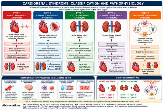
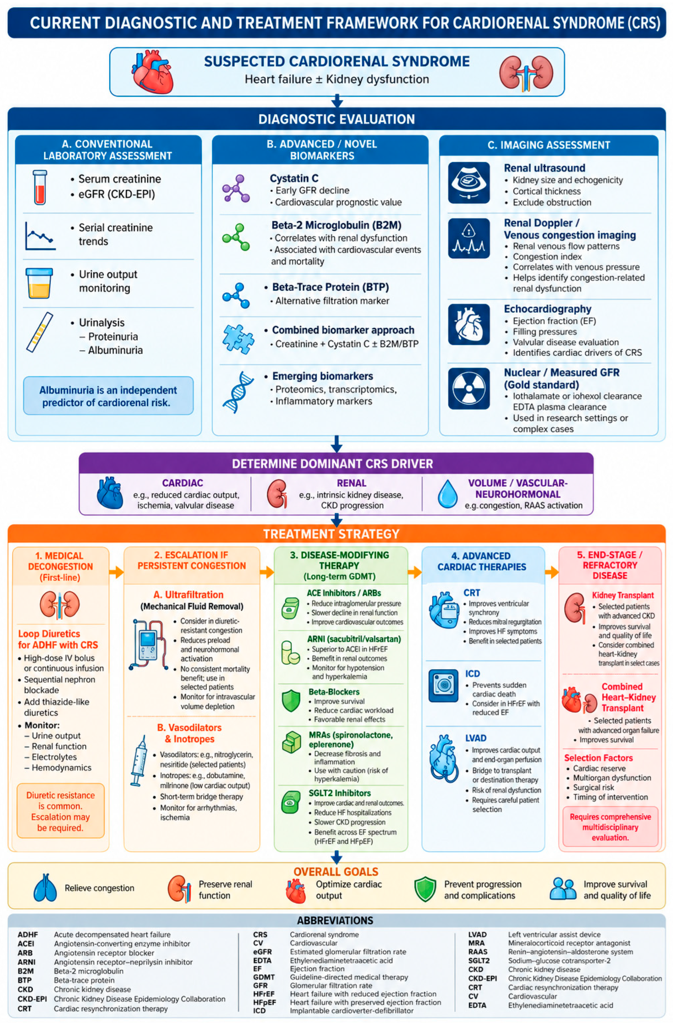
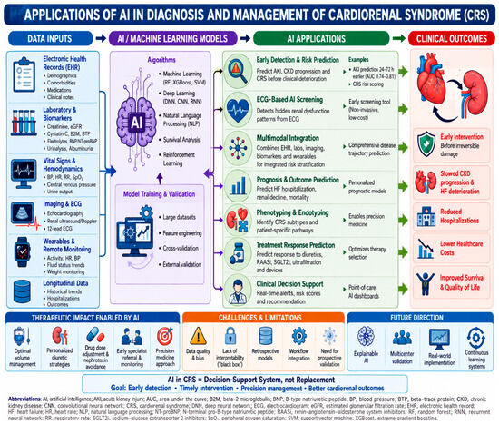
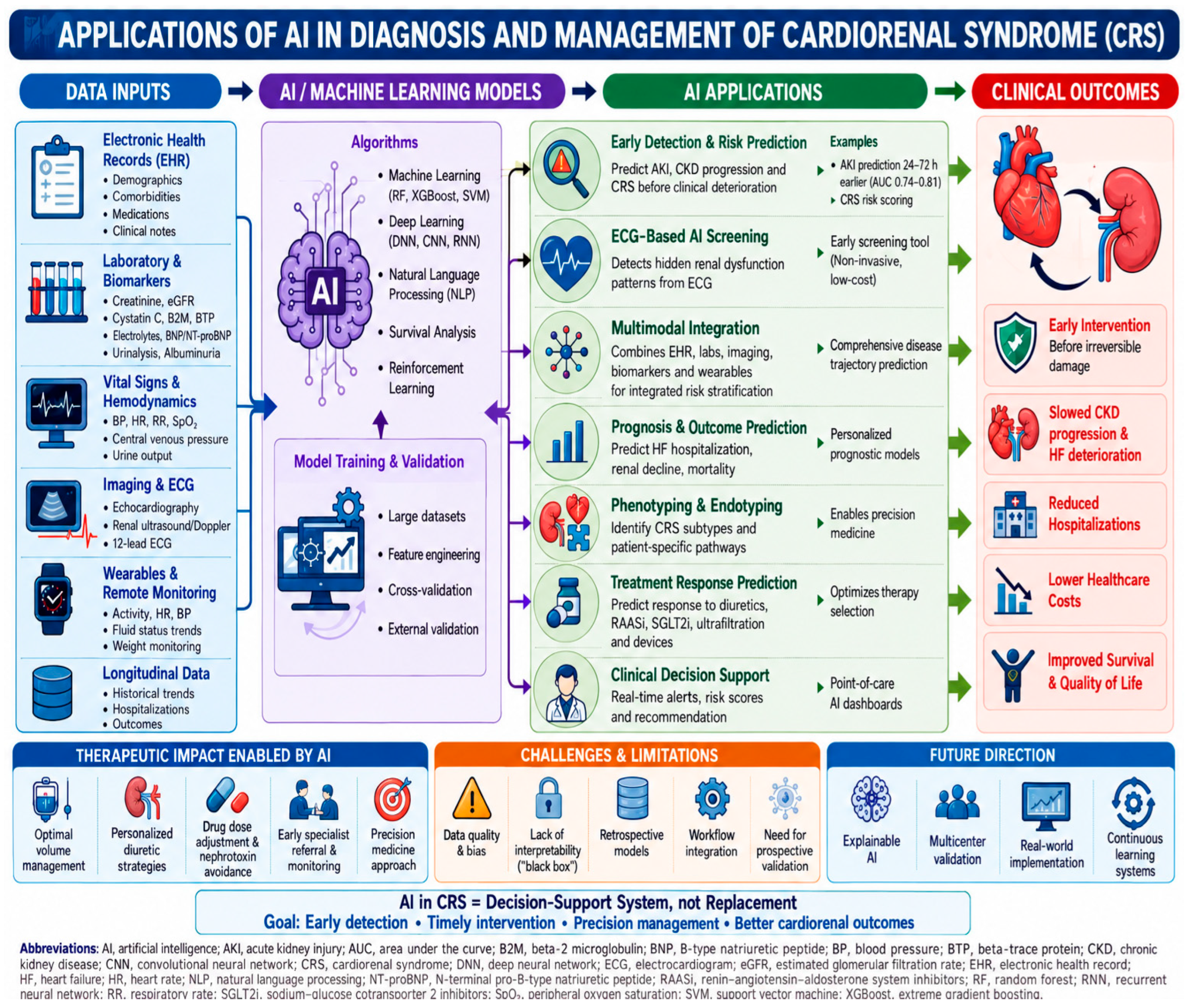

Disclaimer/Publisher’s Note: The statements, opinions and data contained in all publications are solely those of the individual author(s) and contributor(s) and not of MDPI and/or the editor(s). MDPI and/or the editor(s) disclaim responsibility for any injury to people or property resulting from any ideas, methods, instructions or products referred to in the content. |
© 2026 by the authors. Licensee MDPI, Basel, Switzerland. This article is an open access article distributed under the terms and conditions of the Creative Commons Attribution (CC BY) license.