


Abstract
Biological adhesion systems represent some of the most structurally and functionally versatile interfacial solutions in nature, enabling organisms to attach, locomote, and manipulate surfaces across environments ranging from dry rock faces to turbulent marine substrates. This review surveys the structural and mechanistic diversity of biological adhesion, organizing known systems into two principal categories: dry and wet adhesion. Dry adhesion, exemplified by geckos, spiders, and beetles, relies primarily on physical contact forces, most notably van der Waals interactions, arising from hierarchical micro- and nanoscale surface architectures that maximize real contact area without the use of adhesive secretions. Wet adhesion, as demonstrated by marine mussels, octopuses, and tree frogs, operates through biochemical crosslinking, negative-pressure suction, and fluid-displacement mechanisms that act specifically at aqueous or fluid-saturated interfaces. The engineering relevance of these systems is substantial: bioinspired adhesives derived from gecko-like hierarchical fibrillar surfaces have found application in climbing robots, transfer printing, and reversible electronic attachment; mussel-inspired catechol chemistry has enabled a generation of underwater sealants and surgical tissue adhesives; and octopus- and remora-inspired suction technologies inform the design of soft robotic grippers and marine sensor platforms. Despite significant advances, challenges remain in scalable fabrication, contamination resistance, and translating organism-level performance to engineered systems at commercially viable scales. This manuscript reviews each biological model with respect to its structural form, functional mechanism, adhesion classification, documented engineering applications, and inherent limitations, concluding with a synthesis of future research directions critical to advancing the field of bioinspired adhesive materials and systems.
Keywords:
adhesives; grip; friction; dry adhesion; wet adhesion; engineered synthetic tapes; gecko; spider; beetle; octopus; tree frog; secretion 1. Introduction
Adhesion, the capacity of surfaces to resist separation at an interface, is a phenomenon of fundamental importance in both the biological and physical sciences. In living systems, adhesion highlights a remarkably broad array of functions, including substrate locomotion, prey capture, predator evasion, reproductive behavior, and permanent structural attachment. The ability of organisms to modulate adhesive contact in response to dynamic environmental conditions, often instantaneously and reversibly, represents a level of performance that far exceeds that of most engineered adhesive systems [1,2]. The study of biological adhesion, therefore, occupies a central position at the intersection of biophysics, materials science, and biomechanical/biomedical engineering [3,4]. Understanding the structural and physicochemical principles that govern these natural interfaces offers a rational pathway toward the design of next-generation adhesive technologies with enhanced functionality, adaptability, and biocompatibility [5,6].
Conventional synthetic adhesives, including pressure-sensitive tapes, epoxy resins, cyanoacrylates, and silicone-based compounds, achieve interfacial bonding through viscoelastic energy dissipation, chemical crosslinking, or mechanical interlocking [7]. While these materials have achieved widespread industrial and medical utility, they are subject to well-documented limitations. Most synthetic adhesives are irreversible, leaving adhesive residues upon removal that compromise substrate integrity [8]. Many lose efficacy in the presence of moisture, contaminants, or rough surfaces [9]. Pressure-sensitive adhesives degrade under repeated loading cycles, while chemically reactive systems require cure times and may generate potentially cytotoxic byproducts [10,11]. In biomedical contexts, tissue adhesives must additionally meet stringent requirements for biocompatibility, elastic modulus matching, and resistance to the inherently moist physiological environment, criteria that available synthetic formulations satisfy only partially [12,13,14]. The performance limitations of conventional adhesives have thus motivated sustained scientific inquiry into the principles underlying biological adhesion, which routinely achieves superior reversibility, durability, and environmental tolerance [15,16,17].
Biological adhesion systems can be organized according to the dominant physicochemical mechanism through which interfacial bonding is achieved. Dry adhesion systems operate in the absence of adhesive secretion, relying instead on maximizing the real contact area through hierarchical micro and nanoscale surface structures [5,18]. The resulting van der Waals interactions, individually weak but collectively substantial at high contact densities, generate adhesive and frictional forces sufficient to support the body weight of organisms ranging from small beetles to large geckos [19,20]. Wet adhesion systems, by contrast, function at fluid-saturated interfaces, utilizing biochemical crosslinking reactions, suction-generated negative pressure, or the displacement of interfacial water films [21,22]. These mechanisms enable robust attachment to be submerged or moisture-laden surfaces where dry physical contact forces are effectively eliminated by the intervening fluid layer [23]. Hybrid adhesion systems integrate elements of both dry and wet adhesion, often coupling capillary forces arising from thin secreted fluid films with the mechanical compliance and geometry of deformable contact pads [24,25]. This integration allows the organism to adhere effectively across a wider range of surface chemistries and wetting conditions than either mechanism alone could accommodate [26].
The engineering translation of biological adhesion principles has generated significant research momentum across multiple disciplines. In robotics, gecko-inspired fibrillar adhesives have enabled the development of climbing platforms that traverse vertical and inverted glass, metal, and polymer surfaces without mechanical fasteners [27,28]. In biomedical engineering, mussel-inspired polydopamine coatings and catechol-functionalized hydrogels have been developed as surgical sealants, drug delivery matrices, and implantable device coatings [29,30,31,32,33,34]. Soft robotics research has drawn extensively from octopus and cephalopod suction morphology to engineer compliant grippers capable of delicate manipulation of irregular, wet, or fragile objects [35,36,37]. In underwater sensing and marine engineering, remora-inspired suction disc designs have been proposed as reversible attachment mechanisms for sensor platforms on marine vehicles and organisms [38,39,40,41]. In wearable and flexible electronics, tree frog- and gecko-inspired microstructured adhesives have been explored as skin-interfacing substrates for health-monitoring devices [42,43,44,45]. The breadth of these application domains underscores the generative potential of biomimetic adhesion research and motivates continued systematic investigation of natural adhesion systems.
Despite substantial progress, numerous challenges persist in translating biological adhesion principles into engineered systems. Scalable manufacturing of hierarchically structured fibrillar surfaces with the geometric precision required to reproduce gecko-level performance remains technically demanding [46,47,48,49]. Contamination resistance, the ability of dry adhesive surfaces to self-clean and recover adhesive capacity after particulate fouling, has been demonstrated in gecko setae [50,51] but is difficult to replicate in synthetic polymer systems over extended use cycles [52,53]. Wet adhesion systems based on catechol chemistry are susceptible to oxidative degradation and require precise pH control to achieve optimal crosslinking efficiency. Suction-based systems depend on actuator power and geometric conformability that limit miniaturization and deployment in constrained anatomical or mechanical contexts [54,55].
This review systematically examines dry and wet biological adhesion systems to synthesize current knowledge of their structural forms, functional mechanisms, engineering applications, and intrinsic limitations (Figure 1). Section 2 covers theoretical foundations of biological adhesion Section 3 entails dry adhesion as exemplified by gecko, spider, and beetle attachment systems. Section 4 addresses wet adhesion in mussel, octopus, and tree-frog systems. Section 5 presents a comparative summary table organizing the principal systems by adhesion type, mechanism, and application. Section 6 discusses future directions and scientific questions regarding biological adhesion. Section 7 concludes with a synthesis of findings and the identification of priority research directions for advancing bioinspired adhesive materials and technologies.
Figure 1.
A summarized visual representing the different adhesion mechanisms provided throughout the review. Image developed in FigureLabs.
2. Theoretical Foundation of Biological Adhesion
Biological adhesion involves complex interactions of interfacial forces, surface morphology, material deformation, and environmental factors across multiple scales. While often categorized as dry or wet, these systems primarily differ in dominant attachment mechanisms. Dry systems rely on van der Waals forces, friction, and mechanical interlocking, whereas wet systems also involve capillary and chemical interactions. These mechanisms often operate simultaneously, influenced by substrate roughness, humidity, and other conditions. Advances in contact mechanics have shifted focus towards models that consider the coupled behavior of interfaces, structure, and materials. Understanding biological adhesion requires connecting molecular interactions with the macroscopic forces involved, considering factors from surface chemistry to structural hierarchy and fracture mechanics. This framework guides the interpretation of diverse biological systems and informs bioinspired engineering approaches that preserve the relationships between structure, material, and environment.
2.1. Interfacial Adhesion and Contact Mechanics
The mechanical behavior of biological adhesive systems originates from interactions established at the interface between two contacting surfaces. Although these interactions may arise from different physical origins, including van der Waals forces, hydrogen bonding, electrostatic interactions, capillary forces, molecular coordination, or covalent bonding, their macroscopic manifestation strongly depends on the ability of the contacting surfaces to establish and maintain intimate contact [56]. Consequently, adhesion cannot be understood solely in terms of the magnitude of an individual intermolecular interaction [57]. Instead, the interface’s thermodynamic favorability must be coupled with the mechanics governing deformation, contact formation, and separation [58,59,60]. A useful thermodynamic description begins with the work of adhesion:
where is the reversible work required to separate the interface per unit area, and are the surface free energies of the two individual materials, and is the interfacial free energy after contact is established. A positive indicates that interface formation is energetically favorable. This relationship describes the chemical and thermodynamic driving force for adhesion, but it does not directly predict the macroscopic force measured during a pull-off experiment. This distinction matters because describes an ideal interfacial energy, whereas an actual biological adhesive includes surface roughness, defects, finite deformation, viscoelastic dissipation, and spatially heterogeneous contact. Thus, two interfaces having similar thermodynamic work of adhesion can exhibit substantially different measured adhesive forces.
The distinction between thermodynamic adhesion and experimentally measured adhesion becomes particularly important when considering the hierarchical structures found in biological systems. The work of adhesion describes the energetic favorability of forming an interface under idealized equilibrium conditions, whereas the measured adhesive force depends on whether the interface can physically achieve the separation distances required for attractive interactions. Surface roughness, asperity geometry, structural compliance, and local deformation therefore determine how much of the nominal interface becomes functionally active. This relationship can be represented by the contact fraction:
where is the real contact area and is the apparent geometric contact area. The parameter provides a useful measure of how effectively an adhesive structure utilizes its available surface. A low value of indicates that a substantial portion of the nominal interface is separated by substrate asperities or geometric mismatch, whereas increasing increases the area over which interfacial interactions can contribute to adhesion. This distinction is particularly relevant to biological adhesives as many systems do not increase adhesion primarily by increasing surface chemistry; instead, they increase the probability of intimate contact through structural compliance and hierarchical subdivision.
Contact mechanics provides the missing mechanical component by determining how much of the nominal interface actually reaches the separation distances required for attractive interactions [61,62]. For two elastic materials, the combined stiffness can be represented using the effective elastic modulus:
where and are the elastic moduli and and are the Poisson’s ratios of the two contacting materials. The effective modulus therefore provides a convenient measure of the combined resistance of the two bodies to deformation. This parameter becomes particularly important for biological adhesion because many natural adhesive structures are significantly more compliant than the substrates to which they attach [63]. A compliant adhesive structure with a relatively low E* can deform around substrate asperities and increase the real contact area, whereas an excessively compliant structure may undergo buckling, collapse, or excessive deformation that reduces load-bearing capability. Thus, biological adhesion requires an appropriate balance between compliance and structural stability rather than simply maximizing softness. This balance provides a mechanical explanation for the hierarchical organization observed in many biological adhesive systems, where compliant terminal structures are supported by progressively stiffer structures that distribute the applied load over larger length scales.
Classical adhesive contact theories provide two limiting descriptions of this behavior. The Johnson-Kendall-Roberts (JKR) framework treats adhesion as strongly coupled to elastic deformation within the contact region, making it particularly appropriate for compliant materials with relatively strong interfacial interactions [64]. The JKR theory is generally applicable to relatively compliant materials, large contact radii, and strong adhesion, where the adhesive interaction substantially modifies the deformation and contact geometry. Within the JKR framework, adhesion contributes directly to the formation and enlargement of the contact region. The contact radius can be expressed as:
where a is the contact radius, R is the effective radius of curvature of the contacting body, F is the applied normal force, E* is the effective elastic modulus, and is the work of adhesion. The equation illustrates that the contact radius is governed not only by the externally applied load but also by the energetic attraction between the surfaces. In particular, the -dependent terms increase the predicted contact radius relative to a non-adhesive Herzian contact. This behavior is physically important for compliant biological adhesive structures because deformation induced by interfacial adhesion can contribute substantially ot the formation of intimate contact.
The JKR theory also predicts finite tensile force at detachment, commonly referred to as the pull-off force:
This relationship indicates that the pull-off force increases with both the work of adhesion and the characteristic radius of the contacting structure. The JKR formulation therefore provides a useful theoretical framework for understanding compliant adhesive interfaces in which interfacial forces and elastic deformation are strongly coupled. However, the model assumes a particular relationship between the range of the adhesive interaction and the deformation of the contacting bodies. When these assumptions are not satisfied, the JKR solution may overestimate or otherwise inaccurately represent the influence of adhesion on contact deformation.
In contrast, the Derjaguin-Muller-Toporov (DMT) description assumes that adhesive forces act primarily outside the Hertzian contact while deformation within the contact remains comparatively close to the non-adhesive Hertz solution [65]. The DMT theory is more appropriate for relatively stiff materials, small contact radii, and weaker or longer-range adhesive interactions, where deformation caused directly by adhesion is comparatively small. The DMT theory represents the opposite limiting case. Rather than allowing adhesion to substantially alter the deformation within the contact region, the DMT framework treats the adhesive interaction as acting primarily outside the Hertzian contact area. The resulting pull-off force is:
The difference between the JKR and DMT predictions is significant because both theories use the same fundamental quantities, and , yet predict different pull-off forces. This difference is not simply a mathematical discrepancy; it reflects fundamentally different assumptions concerning how adhesive forces influence deformation and where those forces act relative to the contact boundary. Consequently, selecting an appropriate contact model requires consideration of the material stiffness, contact size, adhesion strength, and characteristic interaction distance. Neither JKR nor DMT should therefore be interpreted as a universal model for biological adhesion. Biological adhesive structures frequently span multiple length scales and may contain materials and geometries that fall between the assumptions represented by these two limiting theories. For example, a relatively stiff microscale structure may exhibit behavior closer to the DMT regime, whereas a compliant nanoscale terminal structure may exhibit stronger coupling between adhesion and deformation and therefore behave more closely to the JKR regime. Furthermore, the same adhesive system may transition between regimes as its characteristic contact radius, loading condition, or environmental state changes. A quantitative criterion is therefore required to determine which theoretical description is appropriate.
The Tabor parameter provides such a criterion by comparing the characteristic scale of elastic deformation induced by adhesion with the range of the intermolecular interaction [66,67,68]. It can be expressed as:
where R is the effective radius of curvature, is the work of adhesion, E* is the effective elastic modulus, and is the characteristic equilibrium separation distance or range of the adhesive interaction. The parameter therefore incorporates the principal variables that determine whether adhesion significantly modifies the mechanical contact. Large values of indicate that adhesive forces produce substantial deformation relative to the interaction range, corresponding more closely to the JKR limit. Small values indicate that the deformation induced by adhesion is relatively small, compared with the interaction range, corresponding more closely to the DMT limit. Intermediate values represent a transition regime in which neither limiting approximation fully captures the mechanics and an intermediate formulation, such as the Maugis–Dugdale model, becomes more appropriate. These contact-mechanical models also demonstrate why biological adhesion cannot be reduced to a single material property. A high work of adhesion does not necessarily produce high macroscopic adhesion if the adhesive structure cannot establish sufficient real contact, while a highly compliant structure may establish extensive contact but fail to maintain structural integrity under load. The observed adhesive response therefore emerges from the interaction between interfacial energetics, contact geometry, elastic deformation, and substrate characteristics. This coupling provides a common theoretical basis for comparing apparently different biological systems. Gecko setae, insect attachment pads, tree-frog toe pads, and engineered fibrillar adhesives all differ in their dominant interfacial interactions, but each must solve the same mechanical problem of establishing sufficient real contact while maintaining that contact under an applied load.
Adhesive performance cannot be predicted from isolated fibril mechanics alone. The individual elements form a mechanically coupled population in which load redistribution and sequential detachment can determine the macroscopic response. The theoretical framework therefore establishes a fundamental principle of biological adhesion: interfacial chemistry determines the potential strength of an adhesive interaction, while contact mechanics determines how effectively that potential converts into macroscopic adhesion. This distinction provides a common basis for understanding different systems such as gecko setae, insect attachment pads, tree frog toe pads, and engineered fibrillar adhesives. In each case, surface interactions become mechanically useful only when the biological structure can establish sufficient real contact while maintaining structural stability under the applied loading conditions.
2.2. Multiscale Adhesion, Contact Splitting, and Fracture Mechanics
The contact-mechanical description of adhesion becomes increasingly important when the interface is subdivided into multiple structural elements. Many biological attachment systems do not rely on a single continuous adhesive surface. Instead, the interface is divided into numerous fibers, hairs, pads, plates, or other structural units that operate collectively across several length scales [69]. This organization, commonly described as contact splitting or hierarchical adhesion, allows biological systems to simultaneously address surface roughness, load distribution, defect tolerance, and controlled detachment [70,71,72]. Recent studies of biomimetic adhesives have emphasized that hierarchical architecture should be interpreted as a mechanical strategy for regulating contact and fracture rather than simply as a means of increasing geometric surface area [73,74]. For an idealized array containing N independent adhesive elements, the total adhesive force can be represented as:
where is the contribution from the i′th adhesive element. Although this expression appears straightforward, its physical interpretation is more complicated because is generally not identical for every element. Differences in substrate topography, local deformation, fibril orientation, material properties, and neighboring-element interactions produce a non-uniform distribution of load. Consequently, increasing the number of fibrils does not guarantee a proportional increase in macroscopic adhesion.
Contact splitting provides several mechanical advantages [70,71]. Individual fibrils can deform independently and conform to local variations in substrate geometry. A continuous adhesive surface may bridge over an asperity and consequently lose contact with a significant portion of the substrate, whereas a population of small adhesive elements can establish contact around individual asperities. Contact splitting reduces the likelihood that one local defect will immediately compromise the entire adhesive interface. Failure of an individual element can occur while neighboring elements remain attached. Subdivision allows the geometry and stiffness of individual elements to be selected to achieve a balance between compliance and structural stability. This principle becomes more pronounced when several levels of hierarchy are introduced. A biological adhesive may contain a macroscale supporting structure, microscale attachment fibers, and nanoscale terminal structures, for instance the gecko [75,76]. The larger structures provide global compliance and load distribution, intermediate structures regulate local deformation, and the smallest structures establish intimate substrate contact. Contact splitting provides a mechanism through which subdividing a nominal contact into compliant elements can increase the effective real contact area and improve conformity to substrate roughness. However, the resulting adhesive force does not generally scale linearly with element number because load sharing is influenced by fibril geometry, compliance, spacing, substrate roughness, and mechanical coupling through the backing layer.
The importance of hierarchy becomes particularly evident when considering fracture. Adhesive failure is fundamentally associated with the creation and propagation of a separation front [77]. The energy release rate can be written as:
where represents the total potential energy of the system and A is the newly created interfacial crack area. The quantity G therefore describes the mechanical energy available to drive crack growth per unit newly created area. Detachment becomes energetically favorable when , where is the critical fracture energy of the interface.
The distinction between and is particularly important. The work of adhesion describes the thermodynamic energy associated with separating an ideal interface, whereas the fracture energy measured during an actual detachment process can include substantial additional energy dissipation. In biological adhesives, this dissipation may originate from polymer deformation, fibril stretching, viscoelasticity, molecular rearrangement, crack deflection, sacrificial interactions, or other structural mechanisms. Thus, an adhesive system can exhibit a fracture energy substantially greater than its equilibrium interfacial work of adhesion. Peeling provides a useful example of how structural geometry controls this energy balance [78,79,80]. For an idealized flexible adhesive strip, the energy release rate can be approximated as:
where F is the applied peel force, b is the adhesive width, and is the peel angle. The expression illustrates why the measured force required for detachment depends strongly on loading direction. At low peel angles, the mechanical work associated with peeling is strongly influenced by the geometry of the adhesive and by the deformation of the peeled region. As the angle approaches 90°, a larger fraction of the applied work contributes directly to advancing the separation front. This provides a mechanical explanation for directional adhesion systems in which attachment and detachment can be controlled by changing the direction of applied force.
The same principle applies to biological fibrillar systems. When fibrils are loaded primarily in shear, their geometry can maintain contact while distributing the applied load across multiple elements [81]. When the loading direction changes toward peeling, individual fibrils may rotate, bend, or progressively detach [82,83]. The fracture perspective also explains why biological adhesion must be evaluated in terms of failure progression rather than only peak force. Two adhesive systems may exhibit similar maximum pull-off forces while having very different resistance to crack propagation. One system may detach abruptly after a local defect initiates failure, whereas another may progressively lose individual contacts while maintaining substantial load-bearing capacity. The latter architecture provides greater damage tolerance and may also exhibit improved resistance to repeated attachment and detachment. This consideration becomes especially important during scaling. Increasing the area of an artificial adhesive increases the potential number of adhesive contacts but also increases the probability of defects, manufacturing variations, local contamination, and non-uniform loading. Recent perspectives on bioinspired structural adhesives identify scalability, manufacturing complexity, and the mismatch between biological and synthetic hierarchical structures as continuing challenges in translating biological adhesion into practical engineering systems.
Therefore, hierarchical adhesion should not be regarded simply as a method for increasing contact area. Its more important function is to control the spatial distribution of deformation, stress, and fracture across multiple length scales. The biological advantage arises from the ability to maintain a large population of functional contacts while allowing local elements to deform or fail without causing immediate macroscopic detachment. This framework provides a direct theoretical connection between the nanoscale interactions responsible for adhesion and the macroscopic mechanical behavior observed in biological attachment systems.
2.3. Dry-Wet Adhesion, Interfacial Coupling, and Environmental Effects
The distinction between dry and wet adhesion is therefore best understood as a continuum of interfacial states rather than as a binary classification. Changes in humidity, liquid-film thickness, substrate chemistry, roughness, temperature, and loading rate can continuously alter the relative contributions of direct molecular interactions, capillarity, viscous dissipation, friction, and chemical bonding. This perspective is particularly important when comparing biological systems because the same organism may employ several mechanisms simultaneously or modify their relative importance according to the environment. A dry adhesive may depend primarily on direct solid–solid interactions under low humidity but experience additional capillary or hydration effects as environmental moisture increases. Conversely, a wet adhesive may rely on fluid-mediated interactions while still requiring sufficient direct contact between structural elements and the substrate. The transition between these states is therefore governed by the coupled evolution of surface morphology, fluid distribution, interfacial chemistry, and mechanical deformation rather than by the presence or absence of liquid alone.
At a dry interface, intimate solid–solid contact allows short-range molecular interactions to contribute directly to adhesion. When water or another liquid occupies the interface, however, the situation changes substantially. Interfacial water can prevent direct molecular contact, modify surface energy, screen electrostatic interactions, and introduce additional fluid-mediated forces. The resulting adhesive behavior therefore depends not only on the properties of the two solids but also on the properties, geometry, and dynamics of the intervening liquid phase. Capillary adhesion is one important mechanism. When a liquid forms a curved meniscus between two surfaces, the pressure difference across the liquid–vapor interface is described by the Young–Laplace equation [84],
where is the pressure difference across the interface, is the liquid surface tension, and and are the principal radii of curvature of the meniscus. The equation demonstrates that capillary pressure increases as the radius of curvature decreases. This relationship becomes particularly significant at the microscale because biological surfaces can generate highly curved liquid interfaces using very small volumes of fluid. The resulting capillary force is governed by both the pressure acting over the wetted region and the surface-tension force acting around the liquid boundary. A simplified representation can therefore be written as
where A represents the effective area over which the pressure difference acts and L is a characteristic length associated with the liquid boundary. The exact force depends on the meniscus geometry and contact angles, but the expression illustrates why surface morphology is central to wet adhesion. A biological structure can manipulate meniscus curvature, fluid confinement, and wetting behavior through its micro- and nanoscale architecture. Viscous forces provide another contribution. When two surfaces separate while a liquid remains confined between them, the liquid must flow through a progressively narrower gap [85]. A simplified scaling relationship for viscous resistance can be expressed as
where is the dynamic viscosity of the liquid, R is a characteristic contact radius, h is the liquid-film thickness, and dh/dt represents the rate of separation. The strong dependence demonstrates that relatively small changes in film thickness can produce large changes in viscous resistance. Subsequently, wet adhesion can be strongly dependent on separation rate and fluid confinement. This also illustrates why a wet adhesive cannot be characterized adequately using a single pull-off force without specifying the liquid environment and loading conditions.
Chemical interactions introduce another level of complexity. In underwater or physiological environments, adhesive molecules must interact with substrates despite the presence of water. Biological systems such as mussels have evolved molecular strategies capable of establishing hydrogen bonding, electrostatic interactions, metal coordination, and covalent crosslinking while simultaneously managing hydration at the interface [86,87]. Contemporary research increasingly treats these systems as coupled chemical–mechanical interfaces in which adhesion depends not simply on the number or strength of molecular bonds but also on the network topology and mechanisms available for dissipating mechanical energy. Yang and Bai recently summarized this concept as a synergy between bond chemistry, network topology, and mechanics of dissipation [88]. This coupling can be expressed conceptually as
where M represents surface morphology, C represents interfacial chemistry, E represents mechanical properties, S represents substrate characteristics, and represents environmental conditions. The equation is not intended as a predictive constitutive law; rather, it emphasizes that adhesive performance is a multidimensional function. Altering one variable can change the effect of the others. For example, increasing compliance may improve conformity and real contact area, but excessive compliance may cause structural collapse. Increasing surface energy may strengthen molecular interactions, but this benefit may remain inaccessible if roughness or contamination prevents intimate contact. Similarly, increasing liquid content may facilitate capillary interactions under one geometry while simultaneously reducing direct solid–solid contact or increasing viscous dissipation.
This coupled behavior also provides a physical explanation for the distinction between adhesion strength and adhesive functionality. A system optimized exclusively for maximum normal force may perform poorly when subjected to shear, peeling, cyclic loading, contamination, or changing humidity. Biological systems instead tend to optimize adhesion for a particular combination of load direction, environmental condition, reversibility, and energy expenditure [89,90]. A future trajectory for dry adhesive development has shifted toward regulating contact formation, stress distribution, shear force, and detachment rather than simply maximizing adhesion magnitude.
Wet adhesion research has followed a similar trajectory. Recent work on wet adhesive systems increasingly emphasizes on-demand control, spatiotemporal regulation, reversible attachment, and integration into biomedical and robotic systems. These developments suggest that the most useful bioinspired adhesive systems may not reproduce a single biological mechanism but instead combine several mechanisms so that the dominant mode of adhesion can change in response to environmental or mechanical conditions. The transition between dry and wet adhesion should therefore be viewed as a continuum of interfacial states rather than a binary classification. Humidity, surface contamination, liquid-film thickness, loading rate, substrate compliance, and surface chemistry can progressively alter the balance among molecular, capillary, viscous, frictional, and mechanical contributions [91,92]. This perspective is particularly relevant to engineering applications because practical adhesive interfaces rarely operate under perfectly dry or perfectly submerged conditions. Robotic grippers, wearable devices, biomedical interfaces, and aerospace systems may experience intermittent humidity, contamination, condensation, temperature variation, and dynamic loading.
Overall, the theoretical comparison of dry and wet adhesion demonstrates that biological attachment is best understood as a coupled interfacial phenomenon. Surface chemistry establishes the potential for molecular interaction, morphology controls contact and fluid transport, mechanics governs deformation and load transfer, and fracture processes determine detachment. Environmental conditions continuously modify these relationships. Recognizing this coupling provides a more general framework for interpreting the biological systems discussed in the remainder of this review and, importantly, establishes the scientific basis for identifying unresolved challenges in translating biological adhesion into scalable engineering systems.
3. Dry Adhesion Systems
Dry adhesion in biological organisms refers to attachment mechanisms that generate adhesive and frictional forces exclusively through physical contact between hierarchically structured surface features and a substrate, without the involvement of adhesive secretions or fluid intermediaries [42]. The dominant physical force in dry adhesion is the van der Waals interaction, which arises from transient dipole–dipole correlations between electron clouds in closely apposed molecules [17]. Although van der Waals forces are individually negligible, their cumulative magnitude scales with real contact area, and the principal strategy of dry adhesion systems is to maximize true interfacial contact at the nanoscale through geometric optimization of surface architecture [43]. This principle, known as contact splitting, predicts that subdividing a contact surface into a large number of finer elements increases the total adhesive force in proportion to the square root of element density, providing a theoretical basis for the evolutionary convergence of hierarchical adhesive setae across phylogenetically distant taxa [42,44]. Recent theoretical analyses have refined contact-splitting models to account for material compliance, tip geometry, and substrate roughness, revealing that optimal fibril density and aspect ratio are coupled to the elastic modulus of the fibril material and the substrate roughness spectrum [45,46]. These refinements have significantly advanced the rational design of synthetic dry adhesive surfaces [47,48].
3.1. Gecko Adhesion
The adhesive apparatus of gekkonid lizards constitutes the most extensively studied dry adhesion system in the biological literature, and the structural hierarchy of the gecko toe pad has served as the primary conceptual template for bioinspired dry adhesive design [76,93]. The ventral surface of gecko digits is covered with a series of transverse epidermal folds called scansors or lamellae, which are themselves covered by microscale hair-like projections known as setae (Figure 2) [20,82,94]. Each seta, composed of beta-keratin with an elastic modulus of approximately 2–4 GPa, tapers toward its distal end and branches into an array of nanoscale spatular tips with widths on the order of 100–200 nm and lengths of approximately 200 nm [76,82]. This three-level structural hierarchy, lamellae, setae, and spatulae, distributes load across an enormous number of contact elements, maximizing the real contact area with a substrate at the molecular scale and thereby amplifying the cumulative van der Waals adhesive force [20,95]. Atomic force microscopy measurements of individual spatulae have confirmed the dominance of van der Waals interactions and provided quantitative data on single-element adhesive forces, thereby validating the contact-splitting theoretical framework [6,70,71,72].
Figure 2.
A macro-to-micro illustration of the gecko’s scansors/lamellae, setae, and spatulae, and the governing physics of adhesion (van der Waals interactions). (a). hierarchical configuration of the gecko in attachment and detachment mechanism. (b). model of the hierarchical configuration from the toepad to the spatulae. (c). A force recreated hierarchical configuration replicating the forces during attachment through a spring mechanism. Figure reused from [96].
The functional consequences of this architecture are well characterized. A single Tokay gecko (Gekko gecko) of approximately 100 g body mass possesses an estimated 6.5 million setae on its four feet, each capable of generating adhesive forces of approximately 20 µN, yielding a theoretical whole-animal adhesive capacity on the order of 130 N, more than 100 times the animal’s body weight [97]. In practice, geckos exploit only a small fraction of this theoretical maximum during locomotion, using a combination of shear preloading and controlled peeling to switch between engaged and released adhesive states [98]. Directional adhesion is a defining functional feature: setae generate maximal adhesive force when loaded proximally in shear and detach easily when bent distally or peeled, providing a passive mechanism for both secure attachment and rapid, low-energy release [99]. Self-cleaning is achieved through a dry-contact mechanism in which particulate contaminants preferentially transfer from the setal surface to a substrate during successive contacts, thereby restoring adhesive capacity without active grooming or secretion [50,100]. Recent in vivo tribological studies have further characterized the dynamic switching between adhesive and frictional regimes during gecko locomotion, demonstrating that the setal arrays function as tunable tribological elements rather than simple adhesive pads [101,102].
The engineering translation of gecko-inspired dry adhesion has generated a substantial body of work spanning materials synthesis, surface fabrication, and system integration [103]. Synthetic gecko-inspired adhesives based on polydimethylsiloxane (PDMS), polyurethane (PU), polyimide, and carbon nanotube (CNT) arrays have been fabricated using soft lithography, electron-beam lithography, nanoimprint lithography, electrospinning, and machining achieving adhesion pressures in the range of 10–200 kPa depending on geometry and material modulus [28,48,104,105]. A prominent application is in climbing robotic platforms: the Stanford StickyBot demonstrated sustained wall climbing on smooth glass using directionally structured PDMS adhesive tiles (Figure 3c) [28,106], and subsequent platforms have extended their operation to curved surfaces, outdoor environments, and payloads exceeding 1 kg [107,108,109,110,111,112,113,114]. In biomedical contexts, gecko-inspired microstructured adhesives have been evaluated as reversible substrates for transdermal drug-delivery patches, wearable electronic sensors, and bandages in which repeated attachment and removal without adhesive residue are critical performance criteria [115,116]. Transfer printing processes employing gecko-like structured PDMS stamps have enabled the precise placement of fragile nanoscale electronic components onto flexible and stretchable substrates [117,118,119]. More recently, gecko-inspired adhesives have demonstrated exceptional adhesion on curved and rough surfaces, with enhanced electrical conductivity, opening applications in robotic grasping and electrical contact interfaces [120,121]. Table 1 highlight the recent advances in gecko-inspired adhesives.
Figure 3.
Different fabrication material, process, structure, and design for gecko inspired adhesives. (a). vertically grown carbon nanotube adhesive forming in the hierarchical format as the gecko reused with permission from [122]. (b). polyurethane-based adhesive with hierarchical characteristic including a swing in the base setae structure reused with permission from [123]. (c). a microbaricated wedge-based adhesive developed by Standford University and their associates reused with permission from [124].
Table 1.
Advances in synthetic fabrications of gecko-inspired adhesives.
Despite these advances, gecko-inspired dry adhesives face several unresolved engineering challenges. Performance degrades substantially on rough substrates where geometric mismatch between fibril tips and surface asperities reduces real contact area [74,75]. Synthetic fibrillar arrays are susceptible to lateral collapse and fiber bundling under repeated loading, a degradation mode known as the “matting” effect, which reduces adhesive capacity after multiple use cycles [76]. The self-cleaning mechanism of natural gecko setae, which depends on the relative surface energy of beta-keratin relative to common contaminant particles, is not straightforwardly reproduced in polymer fibrillar systems [77]. Load scaling to support robotic platforms substantially heavier than a gecko remains problematic, as fibrillar array fabrication becomes increasingly difficult to maintain geometric fidelity at the scales required for high-load applications [78]. Recent work on multilayer hierarchical PDMS structures, UV-ozone-treated fibril surfaces, and shape-memory polymer fibrillar arrays has addressed some of these limitations, but a fully durable, rough-surface-compatible gecko-inspired adhesive at engineering scale has not yet been demonstrated [79,80,81].
3.2. Spider Adhesion
Araneae (spiders) exhibit a sophisticated multi-modal attachment system that incorporates both dry fibrillar adhesion and mechanical interlocking via tarsal claws, enabling effective locomotion and prey capture across substrates ranging from smooth glass to coarse vegetation [129,130]. The adhesive structures of spiders are concentrated on the distal segments of the legs and take two principal morphological forms: scopulae, which are dense arrays of flattened or branched setae covering the ventral tarsal and metatarsal surfaces, and claw tufts, which are smaller setal arrays associated with the paired tarsal claws (Figure 4) [131,132]. In hunting spiders such as Cupiennius salei, the scopulae comprise millions of individual setae terminating in nanoscale contact elements with tip widths of approximately 50–300 nm, providing an architecture functionally analogous to, but morphologically distinct from, the gecko spatula [129,133]. The material of spider adhesive setae is primarily cuticle, a composite of chitin and protein with mechanical properties that allow elastic deformation during contact [134,135]. Comparative scanning electron microscopy studies across multiple spider families have revealed substantial morphological diversity in scopular setal geometry, suggesting independent evolutionary optimization of contact element shape for different substrate environments [136,137,138,139].
Figure 4.
An illustration of the adhesive seta in spiders and the claw tuft of the spider. Reused from [139].
The functional mechanism of spider dry adhesion is predominantly van der Waals-mediated contact splitting, following the same physical principles that govern gecko adhesion, but implemented through a distinct morphological strategy [140]. The branched or flattened setal tips of scopulae achieve contact with substrates over a large aggregate area, generating adhesive forces that have been measured at 6–40 mN per leg in Cupiennius salei, sufficient to support multiples of the animal’s body weight [132]. A defining characteristic of spider adhesion is its pronounced anisotropy: adhesive force is maximal in the proximal-to-distal shear direction aligned with the setal orientation, and minimal in the orthogonal or opposite direction [138,141]. This directional dependence allows spiders to engage and disengage adhesive contact rapidly through postural adjustments, analogous to the gear-and-release strategy observed in geckos. The multimodal attachment system combining scopular van der Waals adhesion with claw-mediated mechanical interlocking, provides functional redundancy, enabling effective substrate attachment across a wider range of surface textures than either mechanism alone could accommodate. Experimental studies using micro-force sensors and high-speed imaging have characterized the dynamic interplay between claw and scopular engagement during spider locomotion on inclined and inverted surfaces, demonstrating active modulation of contact element recruitment in response to substrate inclination and loading direction [142,143].
Engineering applications of spider-inspired dry adhesion have focused particularly on directional adhesive films and robotic gripping systems. The anisotropic adhesion of spider scopulae has motivated the design of fibrillar adhesive films that exhibit strong resistance to shear loading in one direction while detaching easily when loaded in the opposite direction, a property of utility in gripping systems where controlled, selective release is required [144,145,146,147]. Robotic grippers employing spider-inspired anisotropic microstructures have demonstrated the ability to grasp and release smooth objects under computer control. Recent work has combined spider scopular geometry with gecko-inspired spatula tips to create multi-scale synthetic structures that outperform either biological template individually in shear adhesion on smooth or mildly textured substrates [148]. Additionally, jumping spider-inspired adhesive systems have been used to inform the design of drones capable of reversible attachment to ceilings and walls for persistent surveillance applications [149,150]. Table 2 showcases recent advances in spider-inspired adhesives.
The limitations of spider-inspired adhesion systems parallel those observed in gecko-derived designs, with several additional challenges specific to the spider morphological template [151]. The scopular setae of spiders are typically more morphologically complex than gecko setae, featuring branched or spatula-like terminations that are difficult to reproduce with high fidelity using conventional microfabrication techniques. The multimodal nature of spider attachment, integrating scopulae, claw tufts, and leg kinematics in a dynamically coordinated system, presents challenges for static adhesive film designs that capture only a subset of the biological mechanisms [152]. Contamination by particulate debris, oils, and environmental moisture reduces adhesive performance under operational conditions that differ from idealized laboratory substrates. The durability of synthetic anisotropic fibrillar arrays under the high cumulative contact cycling characteristic of robotic locomotion remains an open engineering challenge, with recent fatigue studies indicating significant adhesion loss after fewer than 1000 cycles for standard PDMS formulations [153].
Table 2.
Advances in synthetic fabrications of spider-inspired adhesives.
3.3. Beetle Adhesion
Insects of multiple orders, including Coleoptera (beetles), Orthoptera (grasshoppers), and Dermaptera (earwigs), have evolved tarsal adhesive systems that rely on arrays of deformable contact setae to generate adhesive and frictional forces at smooth and textured substrates [158]. Among these, beetles have received the most systematic study, and their tarsal adhesive system represents a well-characterized model for insect dry adhesion. Beetle tarsal pads are located on the ventral surface of the pretarsal segment and are covered with arrays of hairy adhesive setae, the geometry and material properties of which vary substantially among species and are correlated with body mass in accordance with contact splitting theory (Figure 5) [159]. In the Colorado potato beetle (Leptinotarsa decemlineata) and related species, individual setae terminate in flat or mushroom-shaped contact elements with tip diameters of approximately 3–10 µm, providing a larger contact tip area relative to gecko spatulae and reflecting the different load-bearing requirements of insect versus lizard body masses [19,160]. Comparative morphological studies across beetle families have correlated setal density, tip geometry, and shaft aspect ratio with habitat substrate characteristics, supporting the hypothesis that tarsal setal morphology is under strong substrate-specific selection pressure [161,162,163].
Figure 5.
Scanning electron micrographs illustrating the ventral surfaces of the tarsi in female and male seven-spotted ladybird beetles, Coccinella septempunctata. Images include the tarsi of the (a) female and (b) male specimens, along with four distinct tenent seta morphologies: (c) pointed, (d) discoid, (e) lanceolate, and (f) spatula shaped. Panels (g–j) show optical microscope images of various adhesive setae in contact, observed on a live beetle adhering to a smooth, transparent polystyrene surface. Adapted from [19].
The primary functional mechanism of beetle tarsal adhesion is frictional enhancement mediated by the compliance and geometry of the setal contact elements. Unlike gecko and spider setae, which primarily generate adhesive (normal pull-off) forces through van der Waals contact splitting, beetle setae contribute predominantly to shear (frictional) force generation, with measured friction coefficients substantially exceeding those of smooth surfaces of equivalent material [164,165,166]. The deformable nature of the setal shaft allows individual contact elements to conform to microscale surface irregularities, increasing real contact area and thereby enhancing both adhesion and friction [167,168]. Individual beetle adhesive setae have been reported to generate pull-off forces of approximately 50–200 nN, depending on seta morphology, whereas whole-animal attachment forces in the Colorado potato beetle (Leptinotarsa decemlineata) are typically on the order of 20–70 mN on smooth glass substrates [169]. Variations in beetle setal geometry across developmental stages and among species suggest that adhesive performance is closely linked to morphological specialization. Recent studies employing nanoindentation and atomic force microscopy have characterized the viscoelastic properties of insect attachment systems, indicating that loading-rate-dependent changes in compliance may contribute to efficient attachment and detachment during locomotion [164,170].
Engineering applications of beetle-inspired adhesion, similar to gecko-inspired adhesion, have focused on fabricating mushroom-headed fibrillar arrays and developing scalable manufacturing processes for dry adhesive surfaces [171,172]. Recent studies show that mushroom-shaped elastomeric microstructures can achieve adhesion strengths ranging from several kilopascals to over 100 kPa on smooth substrates depending on geometry and loading conditions [172,173], with enhanced performance relative to flat-ended fibrils attributed to improved stress distribution and reduced stress concentration at the contact interface provided by the expanded cap geometry [171,173]. These effects have been confirmed in recent computational and experimental studies, which demonstrate that cap geometry promotes more stable detachment and alters crack propagation during pull-off. Such structures are increasingly explored for reversible adhesion in MEMS assembly, microgripping systems, and transfer printing of flexible and nanoscale electronic components, where controllable attachment and release are required [118,174]. Adhesive performance can be tuned by varying cap diameter, pillar aspect ratio, and material viscoelastic properties, enabling switchable adhesion systems whose attachment state can be controlled through mechanical deformation, loading rate, or defect engineering [89]. Future work will further demonstrate that optimizing fibrillar compliance distributions and viscoelastic material response can significantly enhance adhesion strength and reliability in structured adhesive arrays. Large-area fabrication using molding and scalable microreplication techniques continues to support translation of these systems toward practical engineering deployment as shown in Figure 6. Table 3 summarizes recent synthetic adhesive structures inspired by beetle tarsal attachment systems.
Figure 6.
Vertical PU nanohairs representing a beetle-inspired adhesive with geometric optimization. (a) Asymmetric reversible interlocking of tilted microtrichia inspired by beetle and insect wing adhesion structures. (b) Direction-dependent shear adhesion, showing adhesion toward the center of the body (−) and toward the body edge (+). Reused with permission from [175].
The limitations of beetle-inspired adhesive systems are partially distinct from those encountered in gecko-derived designs. The larger contact tip area of beetle setae, while mechanically advantageous for conformability on rough substrates, is associated with reduced contact-splitting efficiency relative to nanoscale gecko spatulae, potentially limiting the maximum achievable adhesion pressure [163,176]. Beetle tarsal adhesion is sensitive to substrate wettability and the presence of thin fluid films, and several beetle species are known to use a thin adhesive secretion to supplement dry contact, a feature that blurs the boundary between dry and wet adhesion categories. The structural durability of mushroom-headed polymer fibrils under sustained shear loading has been identified as a limitation in robotic applications, where cyclic contact can induce fatigue failure at the shaft-cap interface. Additionally, the maximum adhesion pressure achievable by mushroom-headed arrays on rough or contaminated surfaces remains well below that of the biological system, suggesting that substrate-adaptive mechanisms present in the living beetle, including active claw positioning and body posture modulation contribute substantially to biological performance and are not captured by passive synthetic arrays [165,176,177].
Table 3.
Advances in synthetic fabrications of beetle-inspired adhesives.
4. Wet Adhesion Systems
Wet adhesion encompasses the strategies employed by organisms to maintain secure attachment at aqueous or fluid-rich interfaces, where intervening water layers suppress direct solid–solid contact and significantly alter interfacial forces through hydration effects, capillarity, and viscous dissipation [182,183]. Biological wet adhesion is achieved through several mechanistically distinct strategies, including biochemical secretion-based adhesion, in which proteinaceous or glycoprotein adhesives displace interfacial water and form noncovalent and sometimes covalent interactions with substrate surfaces; suction-based attachment, where negative pressure is generated through muscular action or compliant sealing geometries; and hybrid mechanisms that combine suction with surface texturing to enhance seal integrity and shear resistance [182,184,185]. These strategies have evolved convergently in diverse taxa including marine invertebrates, mollusks, and aquatic vertebrates, providing important design principles for synthetic adhesives intended for operation in submerged or high-humidity environments [186]. Recent surface forces apparatus measurements and molecular-scale simulations have significantly advanced the understanding of interfacial water structuring and its displacement during biological adhesion, revealing how hydration layers, surface chemistry, and nanoscale confinement govern adhesive performance at oxide, carbonate, and polymer interfaces [187,188,189,190].
4.1. Mussel Adhesion
Marine mussels (genus Mytilus) are sessile bivalves that achieve strong and persistent attachment to submerged substrates under dynamic hydrodynamic conditions through a proteinaceous holdfast system known as the byssus [21]. The byssus consists of bundles of byssal threads terminating in adhesive plaques that mediate substrate contact and load transfer (Figure 7) [191,192,193]. The mussel foot produces these structures through controlled secretion and molding within groove-like anatomical structures. The adhesive plaques comprise a complex mixture of mussel foot proteins (MFPs), including MFP-1 to MFP-6, each contributing distinct structural and interfacial functions [194,195,196]. MFP-3 and MFP-5 are enriched at the substrate interface and exhibit high levels of 3,4-dihydroxy-L-phenylalanine (DOPA), a post-translational modification critical for wet adhesion [197]. A recent biochemical and proteomic study further revealed spatially heterogeneous composition across the plaque, with a DOPA-rich adhesive interface transitioning into a crosslinked, cohesive core that contributes to a mechanical gradient and load distribution [192].
Figure 7.
M. edulis (mussel) attachment to (a) seaweed, (b) other mussels, and (c) a stainless steel surface [86].
DOPA is the molecular cornerstone of mussel wet adhesion, and its chemical versatility underlies the ability of mussels to bond to virtually any surface chemistry in the presence of seawater [198]. The catechol moiety of DOPA participates in multiple bonding interactions: it forms bidentate hydrogen bonds and metal-coordinated complexes with oxide surfaces, engages in pi-pi stacking with aromatic surface groups, and undergoes oxidation to the o-quinone form, which can form covalent crosslinks with lysine residues in adjacent proteins and with nucleophilic surface groups. The balance between the adhesive reduced catechol form and the cohesive oxidized quinone form is regulated by the local pH and redox environment within the adhesive plaque, with acidic conditions at the substrate interface preserving catechol in the adhesive state while oxidative crosslinking proceeds in the plaque interior [199]. This spatially graded redox chemistry enables simultaneous interfacial adhesion and internal cohesion, providing a mechanically robust bonded joint under conditions that would otherwise destroy most synthetic adhesives [200]. Surface forces apparatus measurements have directly quantified DOPA-surface bond forces and revealed that catechol-substrate bonds exhibit mechano-chemical stability—bond strength increases under tensile loading due to multivalent coordination geometry, a property with direct implications for adhesive joint design [196,201].
The engineering applications of mussel-inspired adhesion chemistry have been extensive and transformative across multiple fields [152]. Catechol-functionalized hydrogels and polydopamine coatings have been developed as surgical tissue adhesives capable of bonding wet elastic biological tissues with tensile strengths of 10–100 kPa—comparable to fibrin glue but with superior resistance to aqueous dilution and enzymatic degradation. Polydopamine, produced by the spontaneous auto-oxidative polymerization of dopamine at alkaline pH, has been established as a universal surface primer capable of depositing adherent films on virtually any substrate chemistry [202]. Catechol-modified polyethylene glycol (PEG) hydrogels have been evaluated as injectable sealants for gastrointestinal perforations, cardiac patches, and tympanic membrane repairs [203]. In materials science, mussel-inspired coatings have been applied to antifouling surfaces in marine infrastructure and to corrosion-resistant coatings on metallic substrates [204]. Recent advances include the development of catechol-modified hyaluronic acid hydrogels for cartilage repair, DOPA-functionalized polyurethane foam sealants for lung tissue, and catechol-containing nanocomposites that simultaneously achieve high adhesion and mechanical toughness through sacrificial-bond and hidden-length mechanisms [203,205,206,207]. Mussel-inspired adhesives functionalized with antimicrobial peptides have also been developed for infection-resistant wound-closure applications [208,209]. Table 4 provides recent mussel-inspired fabrications reported in the literature.
The limitations of mussel-inspired adhesive systems are principally associated with the chemical sensitivity of the DOPA moiety and the complexity of recombinant protein production [210]. DOPA is highly susceptible to oxidation under ambient conditions, and the spontaneous conversion of catechol to o-quinone in the presence of dissolved oxygen diminishes adhesive potency and accelerates premature cohesive crosslinking [211]. Synthetic mussel-inspired adhesives must therefore be formulated and applied under controlled redox conditions. Recombinant expression of MFPs in bacterial or yeast systems yields proteins with incomplete post-translational hydroxylation of tyrosine to DOPA unless enzymatic co-expression is employed, thereby complicating scalable production [212]. The mechanical performance of mussel-inspired hydrogels falls short of requirements for load-bearing hard tissue interfaces such as bone or cartilage [213]. Batch-to-batch variability in molecular weight distribution and DOPA content of synthetic catechol polymers presents challenges for regulatory approval in clinical applications [214]. Achieving the spatially graded redox and compositional architecture of the biological plaque in synthetic constructs remains an unsolved materials design challenge [215].
Table 4.
Advances in synthetic fabrications of mussel-inspired adhesives.
4.2. Octopus Adhesion
Octopuses (order Octopoda) are soft-bodied cephalopod mollusks that employ an active, muscularly controlled suction system to achieve reversible, high-strength attachment to submerged surfaces during locomotion, prey capture, and environmental interaction [222]. The primary adhesive organs of octopuses are the suckers, muscular, cup-shaped structures arranged in one or two longitudinal rows along each of the eight arms [223,224]. Each sucker is a complex anatomical unit consisting of two regions: the infundibulum, the funnel-shaped peripheral rim that contacts and seals against the substrate, and the acetabulum, the central domed cavity within which negative pressure is generated by contraction of the associated musculature. The infundibular lining is covered by a layered cuticle with a surface texture that enhances conformability and sealing against irregular substrates, while the acetabular musculature includes radial and transverse fibers capable of rapidly expanding the internal volume to generate suction (Figure 8) [225,226]. Micro-computed tomography and histological studies of octopus sucker ultrastructure have revealed the precise three-dimensional geometry of acetabular muscle fiber orientation and its functional relationship to the efficiency of pressure generation [227,228,229].
Figure 8.
An illustration of the Octopus vulgaris sucker: The acetabular system operates as a musculo-connective mechanism in which the circular (C), radial (R), and meridional (M) muscles generate coordinated contractions that alter the geometry of the infundibulum (IN) and regulate the opening of the orifice (O) through the primary (SP) and secondary (SS) sphincter muscles. These forces are distributed through the connective tissue layer (CL) and cross connective tissue fibers (CC), allowing the acetabular roof (AR) and wall (AW) to deform in a controlled manner, thereby modulating adhesion and release. Reused from [230].
The functional mechanism of octopus suction adhesion is fundamentally distinct from the passive contact-area-based mechanisms of dry adhesion systems [230]. Rather than relying on surface forces between closely apposed solid surfaces, octopus suckers generate adhesive force through a pressure differential: contraction of the acetabular musculature increases the internal volume of the sealed cavity, reducing the internal pressure below ambient and creating a net inward force that resists separation of the sucker from the substrate [223]. Peak suction pressures measured in octopus acetabula range from 10 to 270 kPa across species, with maximum values approaching the limit imposed by cavitation of the internal fluid [226,229]. Sucker adhesion is thus inherently active and requires continuous muscular energy expenditure to maintain, but provides rapid, digitally controlled engagement and release without the hysteresis or adhesive residue associated with chemical adhesion [231,232,233]. Individual suckers in Octopus vulgaris generate adhesive forces of 0.1–1 N, and a large octopus with hundreds of suckers per arm can collectively exert attachment forces exceeding 100 N [229]. Electrophysiological studies of the octopus sucker neuromuscular system have characterized the neural circuits that mediate the coordinated modulation of multi-sucker adhesion, providing a biological blueprint for distributed sensor-actuator control in artificial sucker arrays [234].
The engineering translation of octopus suction has motivated substantial research in soft robotics [235]. Artificial suckers fabricated from soft elastomers such as PDMS or Ecoflex have been integrated into robotic arm designs that replicate the muscular actuation principle via pneumatic or hydraulic pressure modulation [236,237,238,239]. These devices achieve suction pressures and adhesive forces comparable to biological suckers and have demonstrated successful grasping of glass, tissue, and rough ceramics in wet environments (see Figure 9) [232,240]. More advanced designs incorporate active compliance through soft actuator arrays that conform the infundibular rim to irregular substrate geometries, extending suction performance beyond smooth surfaces [240,241]. In surgical robotics, octopus-inspired suction end effectors have been evaluated for minimally invasive tissue manipulation [240]. Shape-memory polymer suckers with thermally controlled suction-release cycling have been demonstrated as reversible attachment elements for untethered underwater robots [242]. Recent integration of flexible strain sensors within artificial sucker arrays has enabled real-time monitoring of individual sucker contact force and spatial attachment distribution, mimicking the mechanoreceptive function of the biological system [243,244]. Synthetic fabrications of octopus-inspired adhesives are shown in Table 5.
Figure 9.
Octopus suction in sensing and in grasping: The embodiment of octopus pressure-suction mechanisms as sensors and their ability to enable switchable adhesion. An octopus-based adhesive carrying different objects (toy car, rubber tape, plastic spoon, hydrogen ball, plastic plate, acrylic box, and metal plate) with sensor feedback (1 finger vs. all fingers). Both figures, with their respective panels, are reused from [245].
The engineering limitations of octopus-inspired suction systems center on the energy requirements of active suction generation, the constraints imposed by sucker geometry on the minimum attachment force, and the challenges of miniaturization [239,246]. Active suction requires a continuous power source, pneumatic, hydraulic, or electromechanical, that adds system complexity, mass, and potential failure modes absent in passive adhesion systems. Scaling suction-based adhesion to millimeter or submillimeter dimensions is constrained by the difficulty of generating and maintaining meaningful pressure differentials at small geometric scales, where the perimeter-to-area ratio increases and seal integrity becomes increasingly critical. The sealing performance of artificial infundibular rims is highly sensitive to substrate roughness, and suction force drops precipitously on surfaces with asperities comparable to or larger than the rim compliance length. The biological octopus sucker integrates sensory feedback through chemoreceptors and mechanoreceptors that allow real-time adjustment of suction force—a capability that remains only partially replicated in artificial systems [239,247]. Biofouling of suction surfaces in long-duration underwater deployment presents an additional operational challenge not encountered in short-duration laboratory demonstrations [248,249].
Table 5.
Synthetic fabrications of octopus-inspired adhesives.
4.3. Tree Frog Adhesion
Tree frogs (family Hylidae and allied families) are notable for their ability to adhere strongly to smooth, wet, and vertically oriented surfaces such as leaves and bark under rain and high-humidity conditions, environments in which purely dry adhesion systems fail and purely wet chemical adhesion is impractical. The adhesive organs of tree frogs are the toe pads, which consist of a hexagonal array of columnar epithelial cells separated by channels filled with mucus secreted by goblet cells interspersed among the epithelial columns (Figure 10) [255]. The apical (substrate-facing) surface of each hexagonal epithelial cell is covered with a dense array of nanopillars approximately 100-400 nm in diameter, providing nanoscale roughness that promotes intimate contact with substrate asperities through local deformation [256]. The channels between epithelial columns drain excess mucus laterally away from the contact zone, enabling the pad to maintain effective adhesion on flooded surfaces by limiting the thickness of the intervening fluid film to the nanoscale range where capillary forces become dominant [257,258,259]. Cryo-scanning electron microscopy and synchrotron X-ray imaging of in situ contact zones have revealed the dynamic restructuring of the mucus film during pad engagement and release, including the formation of capillary bridges at the contact peripheries and their rupture upon detachment [255,259,260,261,262].
Figure 10.
Morphology of tree frog toe pads in White’s tree frog (Litoria caerulea). (a) Whole animal view. (b–d) Scanning electron microscopy (SEM) images showing (b) the toe pad, (c) the epidermal surface composed of hexagonal epithelial cells, and (d) a high-magnification view of a single hexagonal cell surface with peg-like projections. (e) Transmission electron microscopy (TEM) image showing a cross-section through the cell surface. Figure reused and reproduced with permission from [263].
The functional mechanism of tree frog adhesion integrates three physical contributions: capillary adhesion arising from the meniscus of the thin mucus film at the contact periphery, viscous adhesion arising from the resistance of the mucus film to rapid separation (Stefan adhesion), and solid contact friction between the nanopillar array and the substrate [259,264,265]. Capillary adhesion scales with the ratio of surface energy to gap thickness, favoring strong adhesion when mucus film thickness is minimized by the lateral drainage channels. Viscous adhesion is velocity-dependent, providing strong resistance to rapid separation while permitting slow repositioning. The relative contribution of each mechanism has been estimated experimentally using force measurements under controlled wetting conditions and lubrication-theory modeling, with the capillary and viscous components together accounting for the majority of the measured pad adhesion on smooth glass [23,266]. The hexagonal cell geometry has been proposed to optimize the trade-off between contact compliance and mucus drainage efficiency [267]. More recently, modeling of the composite hexagonal-nanopillar structure has demonstrated that the pad geometry simultaneously maximizes capillary force per unit area and minimizes the peel-front stress concentration that would initiate detachment, thereby providing a mechanically optimal architecture for switchable wet adhesion [268]. An example of a tree-frog-inspired adhesive based on structure is shown in Figure 11.
Figure 11.
A tree-frog-inspired adhesive used in a surgical setting shown with different hexagonal orientation and structures. (a) Corner-sliding and side-sliding configurations; (b–d) progressively elongated and directionally aligned microstructures. Reused with permission from [269].
Engineering applications inspired by tree frog adhesion are primarily directed toward robotic locomotion systems intended to traverse wet or variably wetted surfaces and toward medical gripping devices for use in moist surgical environments [270]. Synthetic tree frog-inspired adhesive surfaces have been fabricated using soft lithography to produce PDMS hexagonal microstructure arrays with fluid-management channels and hierarchical features, achieving significantly enhanced wet adhesion and friction relative to flat elastomeric controls [271]. Biomimetic adhesive surfaces based on micropillar and nanopillar architectures inspired by tree frog toe pads have been shown to improve adhesion and friction on both smooth and rough wet substrates, enabling stable contact in robotic gripping and locomotion systems [272,273,274]. In medical device applications, tree frog-inspired structured surfaces have been investigated for surgical grasping interfaces that enhance wet friction while reducing the need for high compressive forces that typically damage soft tissue [275,276,277]. Recent advances include micro-nano hierarchical tree frog-inspired adhesive surfaces incorporating nanocavities that enable interfacial liquid self-adjustment and boundary friction enhancement under wet conditions [278], the development of nanopillar- and nanorod-based structured adhesives demonstrating tunable adhesion and friction through nanoscale stiffening effects [279], and the emergence of osmocapillary tree frog-inspired adhesive systems that achieve robust reversible bonding across wet, oily, and contaminated substrates through interfacial liquid modification [280]. Table 6 presents synthetic fabrications from tree-frog inspiration.
The engineering limitations of tree frog-inspired adhesive systems stem from the dependence of adhesion on thin interfacial liquid films and the sensitivity of performance to hydrodynamic drainage and interfacial fluid redistribution [257]. Variations in liquid film thickness during contact and separation significantly alter the contributions of capillary and viscous forces, thereby reducing adhesion stability under dynamic conditions [281,282]. The balance between viscous and capillary contributions is strongly rate-dependent, such that devices optimized for one regime may underperform when separation or loading conditions shift the governing force balance [283]. Substrate texture introduces additional complexity, as microscale roughness disrupts conformal contact and modifies interfacial drainage pathways, thereby reducing effective adhesion in wet environments. Scaling the hierarchical hexagonal micro-nano architecture of tree frog toe pads to robotically relevant dimensions while maintaining structural fidelity and fluid transport remains a key fabrication challenge.
Table 6.
Advances in synthetic fabrications of treefrog-inspired adhesives.
5. Summary Comparison of Biological Adhesion Systems
Table 7 provides a structured comparison of the six biological adhesion systems reviewed in this manuscript, organized by organism, adhesion type, primary structural form, dominant mechanism, principal engineering applications, and key limitations. Beetle adhesion is classified here as “Dry” based on operating substrate, but the underlying mechanism in most studied species is mechanistically hybrid, as their tarsal setae secrete a thin fluid film at the contact tip, making adhesion partly capillary/viscous rather than purely van der Waals as in gecko and spider systems.
Table 7.
Comparative summary of dry and wet biological adhesion systems.
Across these systems, a small set of recurring physical variables governs adhesion: contact area, interfacial energy, compliance, load direction, fluid confinement, and fracture resistance. Biological systems differ primarily in how they control these variables rather than in whether they depend on them.
6. Future Work and Research Directions
Despite substantial progress in understanding biological adhesion, several fundamental scientific questions remain unresolved. The mechanisms discussed throughout this review demonstrate that adhesion is not governed by a single material property or interfacial interaction but instead emerges from the coupling of surface morphology, interfacial chemistry, mechanical deformation, environmental conditions, and loading history. A central challenge is therefore to determine how these variables interact across biological systems and how to quantitatively separate their contributions. In particular, surface morphology determines the ability of an adhesive structure to conform to substrate asperities and establish real contact, while interfacial chemistry influences the strength, reversibility, and environmental sensitivity of the interactions formed at the interface. Mechanical compliance then determines how the applied load distributes across the contacting structures and whether local deformation promotes additional contact or instead causes structural instability. These effects are strongly coupled. Increasing compliance, for example, may increase real contact area on a rough substrate but can also increase deformation and reduce the mechanical stability of individual adhesive elements. Similarly, increasing the surface energy of an adhesive material may increase the thermodynamic driving force for adhesion, but this benefit may not be realized if the structure cannot conform to the substrate sufficiently to establish intimate contact. The same principle applies to biological systems that combine dry and wet mechanisms, where changes in surface morphology can alter fluid confinement while a liquid layer can simultaneously modify friction, capillary forces, molecular interactions, and contact mechanics. A more complete understanding of biological adhesion therefore requires experimental and theoretical approaches that treat morphology, chemistry, and mechanics as coupled design variables rather than independent parameters. Future studies should systematically vary structural geometry, material stiffness, surface chemistry, substrate roughness, and environmental conditions while measuring both adhesive strength and the associated deformation and failure processes. Such studies would help determine whether the exceptional performance of biological adhesives originates primarily from individual mechanisms or from specific combinations of mechanisms operating at different structural scales. Establishing these relationships would also provide a more general framework for comparing biologically distinct adhesive systems and identifying transferable design principles for synthetic interfaces.
A second major scientific challenge is developing predictive multiscale models that connect molecular-scale interactions with macroscopic adhesive performance. Biological adhesive systems operate across several orders of magnitude in length scale, from molecular interactions at the interface to nanoscale terminal structures, microscale fibrils or pillars, and macroscale supporting layers. Although established theoretical or computational approaches can describe each scale, the quantitative connection between them remains limited. Molecular interactions determine quantities such as interfacial energy and characteristic interaction distance, which influence the behavior of individual nanoscale contacts. These local interactions subsequently affect contact formation, deformation, stress distribution, and crack propagation at larger scales. As a result, macroscopic adhesion cannot generally be predicted by considering only the properties of the interface or only the geometry of the adhesive structure. The multiscale nature of the problem is further complicated because adhesive elements within a biological array do not necessarily experience identical loading. Increasing the number of adhesive elements does not necessarily increase macroscopic adhesion proportionally. This consideration becomes particularly important when biological architectures are translated into engineered systems as manufacturing imperfections and structural variability become increasingly significant as the active area increases. Future multiscale models should therefore incorporate the interaction between individual adhesive elements and the supporting structure rather than treating each element as an isolated contact. Such models could combine molecular simulations, contact mechanics, finite-element analysis, cohesive-zone approaches, fracture mechanics, and homogenization methods to link local interfacial behavior to global adhesive response. Data-driven methods may further help identify nonlinear relationships among geometry, material properties, environmental conditions, and adhesive performance; however, these approaches should remain physically constrained, so the resulting models retain mechanistic interpretability. Developing predictive multiscale frameworks would provide an important transition from empirical bioinspired design toward computationally guided adhesive architecture, in which structural dimensions, material properties, and interfacial chemistry can be selected according to a defined mechanical objective rather than determined primarily through experimental trial and error.
Another important unresolved question concerns the transition between dry and wet adhesion mechanisms under changing environmental conditions. As discussed in the theoretical framework, dry and wet adhesion should not necessarily be considered as completely independent categories because biological interfaces may simultaneously involve molecular interactions, capillary forces, viscous dissipation, friction, suction, and mechanical interlocking. The relative contribution of these mechanisms can change continuously with humidity, liquid-film thickness, substrate roughness, temperature, loading rate, and surface chemistry. An interface classified as a dry adhesive under one set of conditions may behave substantially differently when exposed to moisture or a confined liquid layer. This transition matters for engineering applications because practical adhesive systems rarely operate under perfectly controlled environmental conditions. Robotic grippers, biomedical interfaces, wearable devices, and underwater systems may encounter changes in humidity, contamination, liquid exposure, or temperature during operation. A fundamental scientific challenge is therefore to determine how the dominant adhesion mechanism changes as environmental parameters vary and whether measurable material and structural properties can predict these transitions. Future work should investigate the interaction between direct solid–solid contact and fluid-mediated interactions rather than treating the presence of liquid as a simple binary condition. Thin liquid layers may increase adhesion through capillary or chemical interactions under certain conditions, while thicker layers may reduce direct molecular contact and increase viscous resistance during approach and separation. The resulting adhesive response may therefore depend on both the amount of liquid present and its spatial distribution within the hierarchical structure. This behavior suggests that the most effective future bioinspired adhesives may not be designed around a single dominant mechanism but instead around controlled transitions among several mechanisms. Adaptive interfaces could potentially modify their effective contact area, surface chemistry, fluid transport, or mechanical compliance in response to environmental conditions. Such systems would more closely reproduce the functional strategy of biological organisms, which regulate adhesion according to substrate, loading direction, and environmental state. Understanding these transitions quantitatively would therefore provide an important foundation for designing adhesives that maintain predictable performance across dry, humid, wet, and contaminated environments.
A final challenge concerns the scalability, durability, and practical translation of biological adhesion principles into engineered systems. Many bioinspired adhesives perform well under controlled laboratory conditions, yet maintaining this performance as the active area, loading frequency, environmental exposure, or operating lifetime increases remains difficult. Scaling a hierarchical adhesive from a small laboratory specimen to a larger engineering interface introduces additional sources of variability, including fabrication defects, dimensional non-uniformity, substrate roughness, backing-layer deformation, and non-uniform load distribution. These effects can be particularly important for architectures where performance depends on accurately replicating micro- and nanoscale features. Increasing the nominal contact area also increases the number of potential defects and local regions where contact may be incomplete, so macroscopic performance may not scale directly with geometric area. Durability is another limitation because biological organisms repeatedly attach and detach while maintaining functional performance, whereas synthetic structures may undergo progressive degradation through fibril fracture, plastic deformation, fatigue, wear, contamination, chemical degradation, or irreversible changes in surface morphology. Consequently, future evaluation of bioinspired adhesives should extend beyond single-cycle measurements of maximum adhesion force and include repeated loading, crack-growth behavior, environmental aging, contamination, abrasion, temperature variation, and long-term storage. Energy-based metrics such as work of adhesion and fracture energy should be reported alongside peak force because they provide additional information about the mechanisms governing detachment and the resistance of the interface to crack propagation. From an engineering perspective, biological translation should focus on reproducing the underlying physical principle rather than reproducing biological structures exactly. A biologically accurate architecture may not be the most effective engineering solution if fabrication is expensive, hard to scale, or sensitive to manufacturing variability. The objective should instead be to identify the structural, mechanical, and interfacial principles responsible for biological performance and implement those principles using scalable manufacturing approaches. Combining hierarchical surface fabrication, advanced polymer processing, surface chemistry, additive manufacturing, computational optimization, and integrated sensing may provide a pathway toward adhesive systems that retain the functional advantages of biological interfaces while meeting engineering requirements for reproducibility, durability, cost, and manufacturability. Progress in this direction will require closer integration among biological characterization, interfacial mechanics, materials science, manufacturing, and application-specific testing.
7. Conclusions
From a comparative perspective, the six adhesion systems examined in this review- dry adhesion in geckos, spiders, and beetles, and chemically or fluid-mediated adhesion in mussels, octopuses, and tree frogs demonstrate that effective biological attachment can arise from markedly different physical mechanisms while relying on several recurring design principles. Geckos and spiders exploit highly subdivided, compliant contact structures to establish extensive real contact, whereas beetles use deformable setae and, in many species, thin fluid films to enhance friction and adhesion. Mussels achieve underwater attachment through interfacial chemistry and energy-dissipating protein networks, octopuses generate attachment through conformable suction and pressure regulation, and tree frogs regulate thin interfacial liquid films through structured toe pads. Despite these mechanistic differences, all six systems must address the same fundamental engineering problem: establishing and maintaining sufficient interfacial contact while distributing load, accommodating surface irregularities, and permitting controlled detachment. Thus, adhesion performance emerges not from an isolated property or mechanism, but from the coupling of surface morphology, material compliance, interfacial interactions, fluid behavior, and environmental conditions. This conclusion aligns with the broader theoretical framework developed in this review, in which surface chemistry establishes interaction potential, morphology governs contact and fluid transport, mechanics controls deformation and load transfer, and fracture processes determine failure and detachment.
Biomimetic engineering has successfully translated individual components of this biological design space into synthetic systems. Gecko- and spider-inspired fibrillar architectures have demonstrated directional and reversible dry adhesion; beetle- and tree-frog-inspired microstructures have provided strategies for controlling friction and liquid-mediated attachment; mussel-inspired catechol chemistries have enabled adhesion in aqueous environments; and octopus-inspired suction architectures have produced reversible attachment for soft robotic and marine applications. Nevertheless, the comparison reveals a persistent gap between reproducing an individual mechanism and reproducing the integrated functionality of a biological adhesive system. Synthetic interfaces still face challenges from contamination, rough or changing substrates, repeated loading, fabrication complexity, environmental sensitivity, and scaling. For example, gecko-inspired fibrillar systems remain vulnerable to roughness, fiber collapse, and contamination, while chemically mediated and suction-based systems introduce their own constraints related to chemical stability, environmental conditions, actuator requirements, and geometric conformability. These limitations indicate that increasing peak adhesion alone is unlikely to provide a sufficient design objective. Instead, practical adhesive systems must be evaluated according to how effectively they regulate contact, load transfer, failure, reversibility, durability, and energy consumption under realistic operating conditions. The comparative analysis therefore suggests that future progress should focus less on identifying an entirely new adhesion mechanism and more on deliberately integrating complementary mechanisms. A more complete understanding of biological adhesion will require experimental and theoretical studies that treat morphology, material properties, interfacial chemistry, substrate characteristics, and environmental conditions as coupled design variables rather than independent parameters. Predictive multiscale models that connect molecular interactions with nanoscale contact, microscale deformation, macroscale load distribution, and fracture could further move bioinspired adhesive development from empirical trial-and-error toward computationally guided design. Data-driven approaches may complement these models by identifying nonlinear relationships among structural geometry, material properties, environmental conditions, and adhesive performance, provided that such methods remain constrained by physically interpretable mechanisms. Equally important is developing interfaces that can adapt their dominant adhesion mechanism as environmental conditions change, since biological adhesion increasingly appears to occupy a continuum between dry and wet states rather than a strict binary classification.
The central lesson from these six biological systems is therefore not that engineers should reproduce any single organism, but that they should treat biological adhesion as a design library of complementary strategies. Hierarchical contact splitting, compliance matching, interfacial chemistry, fluid regulation, directional loading, controlled fracture, and mechanical or pressure-based switching represent distinct tools that can be selected and combined according to the requirements of a target application. Rather than copying biological structures in isolation, future biomimetic adhesives should use these recurring principles as modular design elements within integrated architectures. Such an approach could enable synthetic systems that are not simply stronger, but more adaptable, durable, contamination-tolerant, reversible, and efficient across changing operating conditions. Viewed this way, the value of biological adhesion lies not in any single exceptional attachment mechanism, but in how diverse mechanisms coordinate across scales to produce reliable function. This systems-level perspective offers a more general pathway for translating biological adhesion into practical technologies for robotics, healthcare, underwater operations, wearable systems, and other environments where conventional adhesives remain limited.
Author Contributions
Conceptualization, Methodology, Investigation, and Writing (original draft, review, and editing): M.H., A.M. and X.G. All authors contributed equally to the listed roles. All authors have read and agreed to the published version of the manuscript.
Funding
This research received no external funding.
Data Availability Statement
No new data were created or analyzed in this study. Data sharing is not applicable to this article.
Conflicts of Interest
The authors declare no conflict of interest.
References
- Peattie, A.M.; Full, R.J. Phylogenetic analysis of the scaling of wet and dry biological fibrillar adhesives. Proc. Natl. Acad. Sci. USA 2007, 104, 18595–18600. [Google Scholar] [CrossRef] [Scilit]
- Gorb, S.N. Biological attachment devices: Exploring nature’s diversity for biomimetics. Philos. Trans. R. Soc. A Math. Phys. Eng. Sci. 2008, 366, 1557–1574. [Google Scholar] [CrossRef] [Scilit]
- Bhushan, B. Biomimetics: Lessons from nature—An overview. Philos. Trans. R. Soc. A Math. Phys. Eng. Sci. 2009, 367, 1445–1486. [Google Scholar] [CrossRef] [Scilit]
- Mastropasqua, M.; Ronchi, A.; Serra, A.; Menga, N.; Carbone, G. Adhesion and friction in viscoelastic rough contacts. Tribol. Int. 2025, 213, 111000. [Google Scholar] [CrossRef] [Scilit]
- Kang, M.; Sun, K.; Seong, M.; Hwang, I.; Jang, H.; Park, S.; Choi, G.; Lee, S.-H.; Kim, J.; Jeong, H.E. Applications of Bioinspired Reversible Dry and Wet Adhesives: A Review. Front. Mech. Eng. 2021, 7, 668262. [Google Scholar] [CrossRef] [Scilit]
- Kamperman, M.; Kroner, E.; del Campo, A.; McMeeking, R.M.; Arzt, E. Functional adhesive surfaces with “gecko” effect: The concept of contact splitting. Adv. Eng. Mater. 2010, 12, 335–348. [Google Scholar] [CrossRef] [Scilit]
- Sandak, A.; Butina Ogorelec, K. Bioinspired building materials—Lessons from nature. Front. Mater. 2023, 10, 1283163. [Google Scholar] [CrossRef] [Scilit]
- Annabi, N.; Tamayol, A.; Shin, S.R.; Ghaemmaghami, A.M.; Peppas, N.A.; Khademhosseini, A. Surgical materials: Current challenges and nano-enabled solutions. Nano Today 2014, 9, 574–589. [Google Scholar] [CrossRef] [Scilit]
- Kjellin, M.; Johansson, I. Surfactants from Renewable Resources; John Wiley & Sons, Ltd.: Hoboken, NJ, USA, 2010. [Google Scholar]
- Callister, W.D., Jr.; Rethwisch, D.G. Materials Science and Engineering: An Introduction; John Wiley & Sons: Hoboken, NJ, USA, 2020. [Google Scholar]
- Yadav, D.; Giri, P.; Das, C. Polymer-based biomaterials and their applications in tissue adhesives. J. Adhes. Sci. Technol. 2024, 38, 2019–2046. [Google Scholar] [CrossRef] [Scilit]
- Annabi, N.; Yue, K.; Tamayol, A.; Khademhosseini, A. Elastic sealants for surgical applications. Eur. J. Pharm. Biopharm. 2015, 95, 27–39. [Google Scholar] [CrossRef] [Scilit]
- López-Moral, A.; Moreno-Villaécija, M.Á.; Suárez-García, S.; Bolaños-Cardet, J.; Sedó-Vegara, J.; Alibés, R.; Mancebo-Aracil, J.; Sardon, H.; Lee, H.; Roscini, C.; et al. Bioinspired catechol-based photocurable tissue sealants. Mater. Today Chem. 2026, 54, 103634. [Google Scholar] [CrossRef] [Scilit]
- Mehdizadeh, M.; Yang, J. Design Strategies and Applications of Tissue Bioadhesives. Macromol. Biosci. 2013, 13, 271–288. [Google Scholar] [CrossRef] [Scilit]
- Lee, H.; Lee, B.P.; Messersmith, P.B. A reversible wet/dry adhesive inspired by mussels and geckos. Nature 2007, 448, 338–341. [Google Scholar] [CrossRef] [Scilit]
- Sahay, R.; Baji, A.; Sargur Ranganath, A.; Anand Ganesh, V. Durable adhesives based on electrospun poly(vinylidene fluoride) fibers. J. Appl. Polym. Sci. 2016, 134, 44393. [Google Scholar] [CrossRef] [Scilit]
- Chen, J.; Tan, W.; Chen, W.; Zhao, J.; Tang, Y.; Gorb, S.N.; Ji, K.; Dai, Z. Bioinspired structural adhesion and friction for harsh environments: From natural ingenuity to engineering. Friction 2026, 14, 9441123. [Google Scholar] [CrossRef] [Scilit]
- Varenberg, M.; Pugno, N.M.; Gorb, S.N. Spatulate structures in biological fibrillar adhesion. Soft Matter 2010, 6, 3269–3272. [Google Scholar] [CrossRef] [Scilit]
- Hosoda, N.; Nakamoto, M.; Suga, T.; Gorb, S.N. Evidence for intermolecular forces involved in ladybird beetle tarsal setae adhesion. Sci. Rep. 2021, 11, 7729. [Google Scholar] [CrossRef] [Scilit]
- Autumn, K.; Sitti, M.; Liang, Y.A.; Peattie, A.M.; Hansen, W.R.; Sponberg, S.; Kenny, T.W.; Fearing, R.; Israelachvili, J.N.; Full, R.J. Evidence for van der Waals adhesion in gecko setae. Proc. Natl. Acad. Sci. USA 2002, 99, 12252–12256. [Google Scholar] [CrossRef] [Scilit]
- Waite, J.H. Mussel adhesion–essential footwork. J. Exp. Biol. 2017, 220, 517–530. [Google Scholar] [CrossRef] [Scilit]
- Smith, A.M. Gastropod secretory glands and adhesive gels. In Biological Adhesive Systems: From Nature to Technical and Medical Application; Springer: Berlin/Heidelberg, Germany, 2010; pp. 41–51. [Google Scholar]
- Barnes, W.J.P. Functional Morphology and Design Constraints of Smooth Adhesive Pads. MRS Bull. 2007, 32, 479–485. [Google Scholar] [CrossRef] [Scilit]
- Dirks, J.-H.; Taylor, D. Fracture toughness of locust cuticle. J. Exp. Biol. 2012, 215, 1502–1508. [Google Scholar] [CrossRef] [Scilit]
- Endlein, T.; Ji, A.; Samuel, D.; Yao, N.; Wang, Z.; Barnes, W.J.P.; Federle, W.; Kappl, M.; Dai, Z. Sticking like sticky tape: Tree frogs use friction forces to enhance attachment on overhanging surfaces. J. R. Soc. Interface 2013, 10, 20120838. [Google Scholar] [CrossRef] [Scilit]
- Federle, W. Why are so many adhesive pads hairy? J. Exp. Biol. 2006, 209, 2611–2621. [Google Scholar] [CrossRef] [Scilit]
- Murphy, M.P.; Sitti, M. Waalbot: An agile small-scale wall-climbing robot utilizing dry elastomer adhesives. IEEE/ASME Trans. Mechatron. 2007, 12, 330–338. [Google Scholar] [CrossRef] [Scilit]
- Kim, S.; Spenko, M.; Trujillo, S.; Heyneman, B.; Santos, D.; Cutkosky, M.R. Smooth Vertical Surface Climbing With Directional Adhesion. IEEE Trans. Robot. 2008, 24, 65–74. [Google Scholar] [CrossRef] [Scilit]
- Li, W.; Zheng, Y.; Pang, W.; Lai, P. Bio-inspired adhesive hydrogel for wound healing. Biomed. Technol. 2023, 1, 65–72. [Google Scholar] [CrossRef] [Scilit]
- Li, W.; Yang, X.; Lai, P.; Shang, L. Bio-inspired adhesive hydrogel for biomedicine—Principles and design strategies. Smart Med. 2022, 1, e20220024. [Google Scholar] [CrossRef] [Scilit]
- Narayanaswamy, R.; Torchilin, V.P. Hydrogels and Their Applications in Targeted Drug Delivery. Molecules 2019, 24, 603. [Google Scholar] [CrossRef] [Scilit]
- Wang, Y.; Adokoh, C.K.; Narain, R. Recent development and biomedical applications of self-healing hydrogels. Expert Opin. Drug Deliv. 2018, 15, 77–91. [Google Scholar] [CrossRef] [Scilit]
- Zanchetta, F.C.; De Wever, P.; Morari, J.; Gaspar, R.C.; Prado, T.P.D.; De Maeseneer, T.; Cardinaels, R.; Araújo, E.P.; Lima, M.H.M.; Fardim, P. In Vitro and In Vivo Evaluation of Chitosan/HPMC/Insulin Hydrogel for Wound Healing Applications. Bioengineering 2024, 11, 168. [Google Scholar] [CrossRef] [Scilit]
- Ryu, J.H.; Lee, Y.; Kong, W.H.; Kim, T.G.; Park, T.G.; Lee, H. Catechol-functionalized chitosan/pluronic hydrogels for tissue adhesives and hemostatic materials. Biomacromolecules 2011, 12, 2653–2659. [Google Scholar] [CrossRef] [Scilit]
- Son, C.; Jeong, S.; Lee, S.; Ferreira, P.M.; Kim, S. Tunable adhesion of shape memory polymer dry adhesive soft robotic gripper via stiffness control. Robotics 2023, 12, 59. [Google Scholar] [CrossRef] [Scilit]
- Lee, Y.; Chun, S.; Son, D.; Hu, X.; Schneider, M.; Sitti, M. A tissue adhesion-controllable and biocompatible small-scale hydrogel adhesive robot. Adv. Mater. 2022, 34, 2109325. [Google Scholar] [CrossRef] [Scilit]
- Chen, G.; Wu, J.; Zhang, Z.; Zhang, B.; Chu, Z.; Cui, J. A soft robotic gripper with a belt loop actuated adhesion design for gentle handling of fragile object. IEEE Robot. Autom. Lett. 2024, 9, 6784–6791. [Google Scholar] [CrossRef] [Scilit]
- Ou, R.; Wang, S.; Li, J.; Li, Z.; Yu, L.; Wang, Z.; Murto, P.; Xu, X. Robust, Switchable and Printable Underwater Adhesives Based on a Temperature-Deactivated Design. Adv. Funct. Mater. 2025, 35, 2416043. [Google Scholar] [CrossRef] [Scilit]
- Yan, Y.; Huang, J.; Qiu, X.; Zhuang, D.; Liu, H.; Huang, C.; Wu, X.; Cui, X. A strong underwater adhesive that totally cured in water. Chem. Eng. J. 2022, 431, 133460. [Google Scholar] [CrossRef] [Scilit]
- Vahdati, M.; Hourdet, D.; Creton, C. Soft underwater adhesives based on weak molecular interactions. Prog. Polym. Sci. 2023, 139, 101649. [Google Scholar] [CrossRef] [Scilit]
- Yin, L.; Cholewinski, A.; Zhao, B. Solvent-free urethane-based prepolymer as a versatile underwater adhesive material. Chem. Eng. J. 2024, 481, 148487. [Google Scholar] [CrossRef] [Scilit]
- Cui, Z.; Jiang, B.; Dai, X.; Guo, Z.; Niu, J.; Dong, Z.; Long, Y.; Hu, W. Self-adhesive, mechanically strong, and conductive organogel for flexible/wearable electronics and underwater electronic skin. Chem. Eng. J. 2024, 500, 157551. [Google Scholar] [CrossRef] [Scilit]
- Wong, S.H.D.; Deen, G.R.; Bates, J.S.; Maiti, C.; Lam, C.Y.K.; Pachauri, A.; AlAnsari, R.; Bělský, P.; Yoon, J.; Dodda, J.M. Smart skin-adhesive patches: From design to biomedical applications. Adv. Funct. Mater. 2023, 33, 2213560. [Google Scholar] [CrossRef] [Scilit]
- Chen, A.; Zhang, J.; Zhu, J.; Yan, Z.; Wu, Q.; Han, S.; Huang, J.; Guan, L. Self-adhesive electronic skin for ultra-sensitive healthcare monitoring. J. Mater. Chem. A 2023, 11, 4977–4986. [Google Scholar] [CrossRef] [Scilit]
- Lai, J.; Wang, X.; Zhao, Q.; Zhang, C.; Gong, T.; He, L.; Wang, Z.; Xia, H. 3D printing self-healing and self-adhesive elastomers for wearable electronics in amphibious environments. ACS Appl. Mater. Interfaces 2024, 16, 16880–16892. [Google Scholar] [CrossRef] [Scilit]
- Hassan, M.M.; Mahajan, A. Gecko-inspired adhesives: Mechanical principles, synthetic fabrications, and applications. J. Adhes. 2025, 102, 395–443. [Google Scholar] [CrossRef] [Scilit]
- Hassan, M.; Fayomi, O.; Faust, J.; Mahajan, A. Print, pattern, stick: Low-cost gecko-inspired adhesives using embedded diffraction structures. Bioinspir. Biomim. 2026, 21, 016021. [Google Scholar] [CrossRef] [Scilit]
- Hassan, M.; Mahajan, A.; Gao, X.; Quinn, D.D.; Farhad, S. Scalable Gecko-Inspired Adhesives via Diffraction-Grated Molds: A Low-Cost, Directional PDMS System. Eng. Rep. 2025, 7, e70352. [Google Scholar] [CrossRef] [Scilit]
- Han, A.; Hajj-Ahmad, A.; Cutkosky, M. Hybrid electrostatic and gecko-inspired gripping pads for manipulating bulky, non-smooth items. Smart Mater. Struct. 2020, 30, 025010. [Google Scholar] [CrossRef] [Scilit]
- Hansen, W.R.; Autumn, K. Evidence for self-cleaning in gecko setae. Proc. Natl. Acad. Sci. USA 2005, 102, 385–389. [Google Scholar] [CrossRef] [Scilit]
- Hu, S.; Lopez, S.; Niewiarowski, P.H.; Xia, Z. Dynamic self-cleaning in gecko setae via digital hyperextension. J. R. Soc. Interface 2012, 9, 2781–2790. [Google Scholar] [CrossRef] [Scilit]
- Sethi, S.; Ge, L.; Ci, L.; Ajayan, P.M.; Dhinojwala, A. Gecko-Inspired Carbon Nanotube-Based Self-Cleaning Adhesives. Nano Lett. 2008, 8, 822–825. [Google Scholar] [CrossRef] [Scilit]
- Alizadehyazdi, V.; Modabberifar, M.; Akherat, S.M.J.M.; Spenko, M. Electrostatic self-cleaning gecko-like adhesives. J. R. Soc. Interface 2018, 15, 20170714. [Google Scholar] [CrossRef] [Scilit]
- Song, S.; Drotlef, D.-M.; Son, D.; Koivikko, A.; Sitti, M. Suction-based soft robotic gripping of rough and irregular parts. arXiv 2020, arXiv:2009.08156. [Google Scholar]
- Ge, D.; Tang, Y.; Ma, S.; Matsuno, T.; Ren, C. A Pressing Attachment Approach for a Wall-Climbing Robot Utilizing Passive Suction Cups. Robotics 2020, 9, 26. [Google Scholar] [CrossRef] [Scilit]
- Yi, Z.; Wang, X.; Li, W.; Qin, X.; Li, Y.; Wang, K.; Guo, Y.; Li, X.; Zhang, W.; Wang, Z. Interfacial friction at action: Interactions, regulation, and applications. Friction 2023, 11, 2153–2180. [Google Scholar] [CrossRef] [Scilit]
- Kendall, K. Adhesion: Molecules and mechanics. Science 1994, 263, 1720–1725. [Google Scholar] [CrossRef] [Scilit]
- Packham, D.E. Adhesive technology and sustainability. Int. J. Adhes. Adhes. 2009, 29, 248–252. [Google Scholar] [CrossRef] [Scilit]
- Leckband, D.; Israelachvili, J. Intermolecular forces in biology. Q. Rev. Biophys. 2001, 34, 105–267. [Google Scholar] [CrossRef] [Scilit]
- Israelachvili, J.N. Intermolecular and Surface Forces; Academic Press: Cambridge, MA, USA, 2011. [Google Scholar]
- Lacaria, L.; Podestà, A.; Radmacher, M.; Rico, F. 2.2 Contact Mechanics. In Biomedical Methods; De Gruyter: Berlin, Germany, 2023; pp. 21–64. [Google Scholar]
- Ciavarella, M.; Joe, J.; Papangelo, A.; Barber, J.R. The role of adhesion in contact mechanics. J. R. Soc. Interface 2019, 16, 20180738. [Google Scholar] [CrossRef] [Scilit]
- King, D.R.; Crosby, A.J. Optimizing adhesive design by understanding compliance. ACS Appl. Mater. Interfaces 2015, 7, 27771–27781. [Google Scholar] [CrossRef] [Scilit]
- Johnson, K.L.; Kendall, K.; Roberts, A.D.; Tabor, D. Surface energy and the contact of elastic solids. Proc. R. Soc. A Math. Phys. Eng. Sci. 1971, 324, 301–313. [Google Scholar] [CrossRef] [Scilit]
- Derjaguin, B.V.; Muller, V.M.; Toporov, Y.P. Effect of contact deformations on the adhesion of particles. J. Colloid Interface Sci. 1975, 53, 314–326. [Google Scholar] [CrossRef] [Scilit]
- Maugis, D. Extension of the Johnson-Kendall-Roberts theory of the elastic contact of spheres to large contact radii. Langmuir 1995, 11, 679–682. [Google Scholar] [CrossRef] [Scilit]
- Maugis, D. Adhesion of spheres: The JKR-DMT transition using a Dugdale model. J. Colloid Interface Sci. 1992, 150, 243–269. [Google Scholar] [CrossRef] [Scilit]
- Maugis, D.; Gauthier-Manuel, B. JKR-DMT transition in the presence of a liquid meniscus. J. Adhes. Sci. Technol. 1994, 8, 1311–1322. [Google Scholar] [CrossRef] [Scilit]
- Li, M.; Mao, A.; Guan, Q.; Saiz, E. Nature-inspired adhesive systems. Chem. Soc. Rev. 2024, 53, 8240–8305. [Google Scholar] [CrossRef] [Scilit]
- Varenberg, M.; Murarash, B.; Kligerman, Y.; Gorb, S.N. Geometry-controlled adhesion: Revisiting the contact splitting hypothesis. Appl. Phys. A 2011, 103, 933–938. [Google Scholar] [CrossRef] [Scilit]
- Kim, J.-K.; Varenberg, M. Contact splitting in dry adhesion and friction: Reducing the influence of roughness. Beilstein J. Nanotechnol. 2019, 10, 1–8. [Google Scholar] [CrossRef] [Scilit]
- Brely, L.; Bosia, F.; Pugno, N.M. Emergence of the interplay between hierarchy and contact splitting in biological adhesion highlighted through a hierarchical shear lag model. Soft Matter 2018, 14, 5509–5518. [Google Scholar] [CrossRef] [Scilit]
- Liu, H.; Zhang, H.; Ding, X. Advances in Contact Mechanics. Lubricants 2024, 12, 179. [Google Scholar] [CrossRef] [Scilit]
- Cheng, Q.; Borodich, F.M.; Gao, Z.; Gorb, S.N.; Jin, X. Advances in the contact mechanics theory and applications of biomimetic artificial adhesives inspired by biological multiscale hierarchical structures. Friction 2025, 13, 9441016. [Google Scholar] [CrossRef] [Scilit]
- Liu, Y.; Wang, H.; Li, J.; Li, P.; Li, S. Gecko-Inspired Controllable Adhesive: Structure, Fabrication, and Application. Biomimetics 2024, 9, 149. [Google Scholar] [CrossRef] [Scilit]
- Autumn, K. Gecko adhesion: Structure, function, and applications. MRS Bull. 2007, 32, 473–478. [Google Scholar] [CrossRef] [Scilit]
- Janssen, M.; Zuidema, J.; Wanhill, R. Fracture Mechanics: Fundamentals and Applications; CRC Press: Boca Raton, FL, USA, 2004. [Google Scholar]
- Pesika, N.S.; Tian, Y.; Zhao, B.; Rosenberg, K.; Zeng, H.; McGuiggan, P.; Autumn, K.; Israelachvili, J.N. Peel-Zone Model of Tape Peeling Based on the Gecko Adhesive System. J. Adhes. 2007, 83, 383–401. [Google Scholar] [CrossRef] [Scilit]
- Liprandi, D.; Bosia, F.; Pugno, N.M. A theoretical-numerical model for the peeling of elastic membranes. J. Mech. Phys. Solids 2020, 136, 103733. [Google Scholar] [CrossRef] [Scilit]
- Peng, Z.L.; Chen, S.H.; Soh, A.K. Peeling behavior of a bio-inspired nano-film on a substrate. Int. J. Solids Struct. 2010, 47, 1952–1960. [Google Scholar] [CrossRef] [Scilit]
- del Campo, A.; Greiner, C.; Arzt, E. Contact Shape Controls Adhesion of Bioinspired Fibrillar Surfaces. Langmuir 2007, 23, 10235–10243. [Google Scholar] [CrossRef] [Scilit]
- Autumn, K.; Puthoff, J. Properties, Principles, and Parameters of the Gecko Adhesive System. In Biological Adhesives; Smith, A.M., Ed.; Springer International Publishing: Cham, Switzerland, 2016; pp. 245–280. [Google Scholar] [CrossRef] [Scilit]
- Autumn, K.; Peattie, A.M. Mechanisms of adhesion in geckos. Integr. Comp. Biol. 2002, 42, 1081–1090. [Google Scholar] [CrossRef] [Scilit]
- Rabinovich, Y.I.; Adler, J.J.; Esayanur, M.S.; Ata, A.; Singh, R.K.; Moudgil, B.M. Capillary forces between surfaces with nanoscale roughness. Adv. Colloid Interface Sci. 2002, 96, 213–230. [Google Scholar] [CrossRef] [Scilit]
- Chen, H.; Liu, W.; Li, S. Random loose packing of small particles with liquid cohesion. AIChE J. 2019, 65, 500–511. [Google Scholar] [CrossRef] [Scilit]
- Silverman, H.G.; Roberto, F.F. Understanding Marine Mussel Adhesion. Mar. Biotechnol. 2007, 9, 661–681. [Google Scholar] [CrossRef] [Scilit]
- Li, Y.; Cao, Y. The molecular mechanisms underlying mussel adhesion. Nanoscale Adv. 2019, 1, 4246–4257. [Google Scholar] [CrossRef] [Scilit]
- Yang, J.; Bai, R. Mechanics of Wet Adhesion. In Mechanics of Flexible and Stretchable Electronics; John Wiley & Sons, Inc.: Hoboken, NJ, USA, 2024; pp. 345–372. [Google Scholar] [CrossRef] [Scilit]
- Shojaeifard, M.; Ferraresso, M.; Lucantonio, A.; Bacca, M. Machine learning-based optimal design of fibrillar adhesives. J. R. Soc. Interface 2025, 22, 20240636. [Google Scholar] [CrossRef] [Scilit]
- Son, D.; Liimatainen, V.; Sitti, M. Machine learning-based and experimentally validated optimal adhesive fibril designs. Small 2021, 17, 2102867. [Google Scholar] [CrossRef] [Scilit]
- Ringenwald, B.E.; Bogacki, E.C.; Narvaez, C.A.; Stark, A.Y. The effect of variable temperature, humidity, and substrate wettability on Gecko (Gekko gecko) locomotor performance and behavior. J. Exp. Zool. Part A Ecol. Integr. Physiol. 2021, 335, 454–463. [Google Scholar] [CrossRef] [Scilit]
- Wolff, J.O.; Gorb, S.N. The influence of humidity on the attachment ability of the spider Philodromus dispar (Araneae, Philodromidae). Proc. R. Soc. B Biol. Sci. 2011, 279, 139. [Google Scholar] [CrossRef] [Scilit]
- Autumn, K.; Gravish, N. Gecko adhesion: Evolutionary nanotechnology. Philos. Trans. R. Soc. A Math. Phys. Eng. Sci. 2008, 366, 1575–1590. [Google Scholar] [CrossRef] [Scilit]
- Autumn, K.; Niewiarowski, P.H.; Puthoff, J.B. Gecko adhesion as a model system for integrative biology, interdisciplinary science, and bioinspired engineering. Annu. Rev. Ecol. Evol. Syst. 2014, 45, 445–470. [Google Scholar] [CrossRef] [Scilit]
- Shuttleworth, I.G. On the role of the van der Waals interaction in gecko adhesion: A DFT perspective. Results Mater. 2020, 6, 100080. [Google Scholar] [CrossRef] [Scilit]
- Tian, Y.; Wan, J.; Pesika, N.; Zhou, M. Bridging nanocontacts to macroscale gecko adhesion by sliding soft lamellar skin supported setal array. Sci. Rep. 2013, 3, 1382. [Google Scholar] [CrossRef] [Scilit]
- Autumn, K.; Liang, Y.A.; Hsieh, S.T.; Zesch, W.; Chan, W.P.; Kenny, T.W.; Fearing, R.; Full, R.J. Adhesive force of a single gecko foot-hair. Nature 2000, 405, 681–685. [Google Scholar] [CrossRef] [Scilit]
- Birn-Jeffery, A.V.; Higham, T.E. Geckos significantly alter foot orientation to facilitate adhesion during downhill locomotion. Biol. Lett. 2014, 10, 20140456. [Google Scholar] [CrossRef] [Scilit]
- Peattie, A.M.; Majidi, C.; Corder, A.; Full, R.J. Ancestrally high elastic modulus of gecko setal β-keratin. J. R. Soc. Interface 2007, 4, 1071–1076. [Google Scholar] [CrossRef] [Scilit]
- Stark, A.Y.; Sullivan, T.W.; Niewiarowski, P.H. The effect of surface water and wetting on gecko adhesion. J. Exp. Biol. 2012, 215, 3080–3086. [Google Scholar] [CrossRef] [Scilit]
- Prowse, M.S.; Wilkinson, M.; Puthoff, J.B.; Mayer, G.; Autumn, K. Effects of humidity on the mechanical properties of gecko setae. Acta Biomater. 2011, 7, 733–738. [Google Scholar] [CrossRef] [Scilit]
- Rizzo, N.W.; Gardner, K.H.; Walls, D.J.; Keiper-Hrynko, N.M.; Ganzke, T.S.; Hallahan, D.L. Characterization of the structure and composition of gecko adhesive setae. J. R. Soc. Interface 2005, 3, 441–451. [Google Scholar] [CrossRef] [Scilit]
- del Campo, A.; Arzt, E. Fabrication approaches for generating complex micro- and nanopatterns on polymeric surfaces. Chem. Rev. 2008, 108, 911–945. [Google Scholar] [CrossRef] [Scilit]
- Aksak, B.; Murphy, M.P.; Sitti, M. Adhesion of biologically inspired vertical and angled polymer microfiber arrays. Langmuir 2007, 23, 3322–3332. [Google Scholar] [CrossRef] [Scilit]
- Qu, L.; Dai, L.; Stone, M.; Xia, Z.; Wang, Z.L. Carbon nanotube arrays with strong shear binding-on and easy normal lifting-off. Science 2008, 322, 238–242. [Google Scholar] [CrossRef] [Scilit]
- Santos, D.; Kim, S.; Spenko, M.; Parness, A.; Cutkosky, M. Directional Adhesive Structures for Controlled Climbing on Smooth Vertical Surfaces. In Proceedings of the 2007 IEEE International Conference on Robotics and Automation; IEEE: New York, NY, USA, 2007; pp. 1262–1267. [Google Scholar] [CrossRef] [Scilit]
- Kalouche, S.; Wiltsie, N.; Su, H.-J.; Parness, A. Inchworm style gecko adhesive climbing robot. In Proceedings of the 2014 IEEE/RSJ International Conference on Intelligent Robots and Systems; IEEE: New York, NY, USA, 2014; pp. 2319–2324. [Google Scholar]
- Sun, J.; Bauman, L.; Yu, L.; Zhao, B. Gecko-and-inchworm-inspired untethered soft robot for climbing on walls and ceilings. Cell Rep. Phys. Sci. 2023, 4, 101241. [Google Scholar] [CrossRef] [Scilit]
- Glick, P.; Suresh, S.A.; Ruffatto, D.; Cutkosky, M.; Tolley, M.T.; Parness, A. A soft robotic gripper with gecko-inspired adhesive. IEEE Robot. Autom. Lett. 2018, 3, 903–910. [Google Scholar] [CrossRef] [Scilit]
- Modabberifar, M.; Spenko, M. A shape memory alloy-actuated gecko-inspired robotic gripper. Sens. Actuators A Phys. 2018, 276, 76–82. [Google Scholar] [CrossRef] [Scilit]
- Shao, D.; Chen, J.; Ji, A.; Dai, Z.; Manoonpong, P. Hybrid soft-rigid foot with dry adhesive material designed for a gecko-inspired climbing robot. In Proceedings of the 2020 3rd IEEE International Conference on Soft Robotics (RoboSoft); IEEE: New York, NY, USA, 2020; pp. 578–585. [Google Scholar] [CrossRef] [Scilit]
- Acharya, S.; Roberts, P.; Rane, T.; Singhal, R.; Hong, P.; Ranade, V.; Majidi, C.; Webster-Wood, V.; Reeja-Jayan, B. Gecko adhesion based sea star crawler robot. Front. Robot. AI 2023, 10, 1209202. [Google Scholar] [CrossRef] [Scilit]
- Hoang, T.T.; Quek, J.J.S.; Thai, M.T.; Phan, P.T.; Lovell, N.H.; Do, T.N. Soft robotic fabric gripper with gecko adhesion and variable stiffness. Sens. Actuators A Phys. 2021, 323, 112673. [Google Scholar] [CrossRef] [Scilit]
- Carlo, M.; Metin, S. A biomimetic climbing robot based on the gecko. J. Bionic Eng. 2006, 3, 115–125. [Google Scholar] [CrossRef] [Scilit]
- Suo, D.; Yang, Y.; Zhao, S.; Bei, H.; Lam, A.C.; Tan, W.; Wong, K.K.; Zhao, X. Seta-Inspired Mechano-Intelligent Janus Bandage with Coordinated Adhesion–Contraction for Minimizing Scarring. Adv. Mater. 2026, 38, e05122. [Google Scholar] [CrossRef] [Scilit]
- Gao, Y.; Cheng, Q.; Chen, Z.J.; Borodich, F.M.; Jin, X.Q. Biomimetic adhesives in biomedical engineering: From micro/nano structure design to applications. Biomed. Eng. Commun. 2026, 5, 14. [Google Scholar] [CrossRef] [Scilit]
- Fan, J.; Ding, S.; Zeng, X.; Gao, S.; Wen, Z.; Zeng, X.; Sun, R.; Ren, L. Gecko-inspired adhesive structures enable efficiently thermal conductance and vibration dissipation in a highly mismatched system. Chem. Eng. J. 2022, 445, 136754. [Google Scholar] [CrossRef] [Scilit]
- Shen, C.; Cheng, Y.; Peng, Z.; Chen, S. Switchable adhesion of gecko-inspired hierarchically wedge-mushroom-shaped surface. Chem. Eng. J. 2024, 488, 150900. [Google Scholar] [CrossRef] [Scilit]
- Liu, S.; Zhu, P.; Xie, F.; Soto, M.A. Gecko-inspired self-adhesive packaging for strain-free temperature sensing based on optical fibre Bragg gratings. Sci. Rep. 2023, 13, 4148. [Google Scholar] [CrossRef] [Scilit]
- Shao, Y.; Li, M.; Tian, H.; Zhao, F.; Xu, J.; Hou, H.; Zhang, Z.; Wang, D.; Chen, X.; Li, W.; et al. Gecko-inspired intelligent adhesive structures for rough surfaces. Research 2025, 8, 0630. [Google Scholar] [CrossRef] [Scilit]
- Zhang, J.; Sun, Y.; Shang, F.; Yan, Z.; Yao, J.; Chen, B.; Shen, H. Carbon Nanotubes–Gr Inspired by Geckos’ Setae Structure with Enhanced Tribological Properties. Materials 2025, 18, 1221. [Google Scholar] [CrossRef] [Scilit]
- Chen, B.; Goldberg Oppenheimer, P.; Shean, T.A.; Wirth, C.T.; Hofmann, S.; Robertson, J. Adhesive Properties of Gecko-Inspired Mimetic via Micropatterned Carbon Nanotube Forests. J. Phys. Chem. C 2012, 116, 20047–20053. [Google Scholar] [CrossRef] [Scilit]
- Murphy, M.P.; Kim, S.; Sitti, M. Enhanced Adhesion by Gecko-Inspired Hierarchical Fibrillar Adhesives. ACS Appl. Mater. Interfaces 2009, 1, 849–855. [Google Scholar] [CrossRef] [Scilit]
- Parness, A.; Soto, D.; Esparza, N.; Gravish, N.; Wilkinson, M.; Autumn, K.; Cutkosky, M. A microfabricated wedge-shaped adhesive array displaying gecko-like dynamic adhesion, directionality and long lifetime. J. R. Soc. Interface 2009, 6, 1223–1232. [Google Scholar] [CrossRef] [Scilit]
- Park, S.; Kang, D.K.; Lee, D.; Choi, G.; Kim, J.; Lee, C.; Seong, M.; Bartlett, M.D.; Jeong, H.E. Multiscale crack trapping for programmable adhesives. Sci. Adv. 2024, 10, eadq3438. [Google Scholar] [CrossRef] [Scilit]
- Jin, K.; Tian, Y.; Erickson, J.S.; Puthoff, J.; Autumn, K.; Pesika, N.S. Design and fabrication of gecko-inspired adhesives. Langmuir 2012, 28, 5737–5742. [Google Scholar] [CrossRef] [Scilit]
- Ge, L.; Sethi, S.; Ci, L.; Ajayan, P.M.; Dhinojwala, A. Carbon nanotube-based synthetic gecko tapes. Proc. Natl. Acad. Sci. USA 2007, 104, 10792–10795. [Google Scholar] [CrossRef] [Scilit]
- O’Kane, S. 2024’s Startup Battlefield Runner-Up geCKo Materials Reveals Four New Products at TechCrunch Disrupt; TechCrunch Media LLC.: San Francisco, CA, USA, 2025. [Google Scholar]
- Kesel, A.B.; Martin, A.; Seidl, T. Adhesion measurements on the attachment devices of the jumping spider Evarcha arcuata. J. Exp. Biol. 2003, 206, 2733–2738. [Google Scholar] [CrossRef] [Scilit]
- Wolff, J.O.; Gorb, S.N. Mechanical attachment devices. In Attachment Structures and Adhesive Secretions in Arachnids; Springer: Berlin/Heidelberg, Germany, 2016; pp. 25–52. [Google Scholar]
- Niederegger, S. Functional aspects of spider scopulae. In Spider Ecophysiology; Springer: Berlin/Heidelberg, Germany, 2012; pp. 57–66. [Google Scholar]
- Kesel, A.B.; Martin, A.; Seidl, T. Getting a grip on spider attachment: An AFM approach to microstructure adhesion in arthropods. Smart Mater. Struct. 2004, 13, 512–518. [Google Scholar] [CrossRef] [Scilit]
- Gorb, S.N.; Beutel, R.G.; Gorb, E.V.; Jiao, Y.; Kastner, V.; Niederegger, S.; Popov, V.L.; Scherge, M.; Schwarz, U.; Vötsch, W. Structural design and biomechanics of friction-based releasable attachment devices in insects. Integr. Comp. Biol. 2002, 42, 1127–1139. [Google Scholar] [CrossRef] [Scilit]
- Heepe, L.; Xue, L.; Gorb, S. Bio-Inspired Structured Adhesives: Biological Prototypes, Fabrication, Tribological Properties, Contact Mechanics, and Novel Concepts; Springer: Berlin/Heidelberg, Germany, 2017. [Google Scholar]
- Wolff, J.O. Structural effects of glue application in spiders—What can we learn from silk anchors? In Bio-Inspired Structured Adhesives: Biological Prototypes, Fabrication, Tribological Properties, Contact Mechanics, and Novel Concepts; Springer: Berlin/Heidelberg, Germany, 2017; pp. 63–80. [Google Scholar]
- Silva, V.; Biancardi, C.; Perafán, C.; Ortíz, D.; Fábrica, G.; Pérez-Miles, F. Giant steps: Adhesion and locomotion in theraphosid tarantulas. J. Comp. Physiol. A 2021, 207, 179–190. [Google Scholar] [CrossRef] [Scilit]
- Pérez-Miles, F.; Guadanucci, J.P.L.; Jurgilas, J.P.; Becco, R.; Perafán, C. Morphology and evolution of scopula, pseudoscopula and claw tufts in Mygalomorphae (Araneae). Zoomorphology 2017, 136, 435–459. [Google Scholar] [CrossRef] [Scilit]
- Pérez-Miles, F.; Perafán, C.; Ortiz-Villatoro, D. Adhesive Features of the Theraphosid Tarantulas. In New World Tarantulas: Taxonomy, Biogeography and Evolutionary Biology of Theraphosidae; Springer: Berlin/Heidelberg, Germany, 2020; pp. 341–363. [Google Scholar]
- Wolff, J.O.; Nentwig, W.; Gorb, S.N. The great silk alternative: Multiple co-evolution of web loss and sticky hairs in spiders. PLoS ONE 2013, 8, e62682. [Google Scholar] [CrossRef] [Scilit]
- Peattie, A.M. Functional demands of dynamic biological adhesion: An integrative approach. J. Comp. Physiol. B 2009, 179, 231–239. [Google Scholar] [CrossRef] [Scilit]
- Wolff, J.O.; Gorb, S.N. Attachment Structures and Adhesive Secretions in Arachnids; Springer: Berlin/Heidelberg, Germany, 2016; Volume 7. [Google Scholar]
- Guadanucci, J.P.L. Tarsal scopula significance in ischnocolinae phylogenetics (Araneae, Mygalomorphae, Theraphosidae). J. Arachnol. 2005, 33, 456–467. [Google Scholar] [CrossRef] [Scilit]
- Niederegger, S.; Gorb, S.N. Friction and adhesion in the tarsal and metatarsal scopulae of spiders. J. Comp. Physiol. A 2006, 192, 1223–1232. [Google Scholar] [CrossRef] [Scilit]
- Smith, G.L.; Tyler, J.B.; Lazarus, N.; Tsang, H.; Viornery, L.; Shultz, J.; Bergbreiter, S. Spider-inspired, fully 3D-printed micro-hydraulics for tiny, soft robotics. Adv. Funct. Mater. 2023, 33, 2207435. [Google Scholar] [CrossRef] [Scilit]
- Wei, Z.; Zhao, C.; Huang, Y.; Li, S.; Fu, X.; Yang, C.; Xiao, Z.; Li, G. A spider-inspired superhydrophobic tactile sensor with machine learning-enabled slip detection for damage-free robotic fruit grasping. Comput. Electron. Agric. 2026, 249, 111861. [Google Scholar] [CrossRef] [Scilit]
- Kellaris, N.; Rothemund, P.; Zeng, Y.; Mitchell, S.K.; Smith, G.M.; Jayaram, K.; Keplinger, C. Spider-inspired electrohydraulic actuators for fast, soft-actuated joints. Adv. Sci. 2021, 8, 2100916. [Google Scholar] [CrossRef] [Scilit]
- Vollrath, F.; Krink, T. Spider webs inspiring soft robotics. J. R. Soc. Interface 2020, 17, 20200569. [Google Scholar] [CrossRef] [Scilit]
- Yang, J.; Włodarczyk-Biegun, M.K.; Filippov, A.; Akerboom, S.; Dompé, M.; van Hees, I.A.; Mocan, M.; Kamperman, M. Functional polymeric materials inspired by geckos, mussels, and spider silk. Macromol. Chem. Phys. 2018, 219, 1800051. [Google Scholar] [CrossRef] [Scilit]
- Romanello, L.; Lan, T.; Hassan, S.; Leutschafft, M.; Ekinci, E.; Armanini, S.F.; Kovac, M.; Kocer, B.B. TreeSpider: In-Canopy Exploration With Tether-Based Aerial Modular Arms. Adv. Robot. Res. 2026, 2, e202500111. [Google Scholar] [CrossRef] [Scilit]
- Roderick, W.R.T.; Cutkosky, M.R.; Lentink, D. Touchdown to take-off: At the interface of flight and surface locomotion. Interface Focus 2017, 7, 20160094. [Google Scholar] [CrossRef] [Scilit]
- Wolff, J.O.; Gorb, S.N. Biomimetics: What Can We Learn From Arachnids? In Attachment Structures and Adhesive Secretions in Arachnids; Springer: Berlin/Heidelberg, Germany, 2016; pp. 163–172. [Google Scholar]
- Gasparetto, A.; Vidoni, R.; Seidl, T. Attaching Mechanisms and Strategies Inspired by the Spiders’ Leg; ESA Final Report; 20272/071NL/HE; ESA: Noordwijk, The Netherlands, 2008. [Google Scholar]
- Jeong, H.E.; Lee, J.-K.; Kim, H.N.; Moon, S.H.; Suh, K.Y. A nontransferring dry adhesive with hierarchical polymer nanohairs. Proc. Natl. Acad. Sci. USA 2009, 106, 5639–5644. [Google Scholar] [CrossRef] [Scilit]
- Feng, J.-F.; Wang, T.; Xu, C.; Gou, X.; He, L.; Fu, Z.-C.; Deng, J.; An, W.; Jiang, N.; Zhao, H.-B.; et al. Spider Silk-Inspired Bio-Based Adhesive: Robust Adhesion Strength, Exceptional Flame Retardancy, and Full Recyclability. ACS Mater. Lett. 2025, 7, 2031–2040. [Google Scholar] [CrossRef] [Scilit]
- Roberts, A.D.; Finnigan, W.; Kelly, P.; Faulkner, M.; Breitling, R.; Takano, E.; Scrutton, N.; Blaker, J.; Hay, S. Non-covalent protein-based adhesives for transparent substrates—Bovine serum albumin vs. recombinant spider silk. Mater. Today Bio 2020, 7, 100068. [Google Scholar] [CrossRef] [Scilit]
- He, Y.; Zhang, Z.; Wang, Y.; Liu, M.; Liao, C.; Yuan, J.; Li, P.; Yang, M.; Liu, W. Inspired by spider silk to prepare biomimetic high-performance bovine serum albumin adhesive to strengthen the interfacial and tribological performance of fabric composites. Friction 2025, 13, 9440914. [Google Scholar] [CrossRef] [Scilit]
- Lemetti, L.; Tersteegen, J.; Sammaljärvi, J.; Aranko, A.; Linder, M. Recombinant Spider Silk Protein and Delignified Wood Form a Strong Adhesive System. ACS Sustain. Chem. Eng. 2021, 10, 552–561. [Google Scholar] [CrossRef] [Scilit]
- Gorb, S. Attachment Devices of Insect Cuticle; Springer: Berlin/Heidelberg, Germany, 2001. [Google Scholar]
- Arzt, E.; Gorb, S.; Spolenak, R. From micro to nano contacts in biological attachment devices. Proc. Natl. Acad. Sci. USA 2003, 100, 10603–10606. [Google Scholar] [CrossRef] [Scilit]
- Gorb, S.N.; Varenberg, M. Mushroom-shaped geometry of contact elements in biological adhesive systems. J. Adhes. Sci. Technol. 2007, 21, 1175–1183. [Google Scholar] [CrossRef] [Scilit]
- Bhushan, B. Adhesion of multi-level hierarchical attachment systems in gecko feet. J. Adhes. Sci. Technol. 2007, 21, 1213–1258. [Google Scholar] [CrossRef] [Scilit]
- Karp, J.M.; Langer, R. Dry solution to a sticky problem. Nature 2011, 477, 42–43. [Google Scholar] [CrossRef] [Scilit]
- Bullock, J.M.R.; Federle, W. Beetle adhesive hairs differ in stiffness and stickiness: In vivo adhesion measurements on individual setae. Naturwissenschaften 2011, 98, 381–387. [Google Scholar] [CrossRef] [Scilit]
- Peisker, H.; Michels, J.; Gorb, S.N. Evidence for a material gradient in the adhesive tarsal setae of the ladybird beetle Coccinella septempunctata. Nat. Commun. 2013, 4, 1661. [Google Scholar] [CrossRef] [Scilit]
- Dai, Z.; Gorb, S.N.; Schwarz, U. Roughness-dependent friction force of the tarsal claw system in the beetle Pachnoda marginata (Coleoptera, Scarabaeidae). J. Exp. Biol. 2002, 205, 2479–2488. [Google Scholar] [CrossRef] [Scilit]
- Varenberg, M.; Gorb, S. Shearing of fibrillar adhesive microstructure: Friction and shear-related changes in pull-off force. J. R. Soc. Interface 2007, 4, 721. [Google Scholar] [CrossRef] [Scilit]
- Filippov, A.E.; Gorb, S.N. Contact between biological attachment devices and rough surfaces. In Combined Discrete and Continual Approaches in Biological Modelling; Springer: Berlin/Heidelberg, Germany, 2020; pp. 87–141. [Google Scholar]
- Carbone, G.; Pierro, E. Contact Mechanics of Mushroom-Shaped Adhesive Structures. In Bio-Inspired Structured Adhesives: Biological Prototypes, Fabrication, Tribological Properties, Contact Mechanics, and Novel Concepts; Springer: Berlin/Heidelberg, Germany, 2017; pp. 245–276. [Google Scholar]
- Voigt, D.; Schuppert, J.M.; Dattinger, S.; Gorb, S.N. Sexual dimorphism in the attachment ability of the Colorado potato beetle Leptinotarsa decemlineata (Coleoptera: Chrysomelidae) to rough substrates. J. Insect Physiol. 2008, 54, 765–776. [Google Scholar] [CrossRef] [Scilit]
- Büscher, T.H.; Gorb, S.N. Physical constraints lead to parallel evolution of micro- and nanostructures of animal adhesive pads: A review. Beilstein J. Nanotechnol. 2021, 12, 725–743. [Google Scholar] [CrossRef] [Scilit]
- Violano, G.; Dibitonto, S.; Afferrante, L. Role of viscoelasticity in the adhesion of mushroom-shaped pillars. Bioinspir. Biomim. 2024, 19, 066006. [Google Scholar] [CrossRef] [Scilit]
- Betegón, C.; Rodríguez, C.; Martínez-Paneda, E.; McMeeking, R.M. Pull-off strength of mushroom-shaped fibrils adhered to rigid substrates. J. Adhes. 2026, 102, 201–232. [Google Scholar] [CrossRef] [Scilit]
- Gonen, M.; Kasem, H. Effect of the Mechanical Properties of Soft Counter-Faces on the Adhesive Capacity of Mushroom-Shaped Biomimetic Microstructures. Biomimetics 2023, 8, 327. [Google Scholar] [CrossRef] [Scilit]
- Li, J.; Wei, Q.; Zhao, X.; Sui, T.; Ji, J.; Liu, J. The gradient enhanced adhesion mechanisms of biomimetic mushroom-shaped microcolumn. Colloids Surf. A Physicochem. Eng. Asp. 2025, 727, 138454. [Google Scholar] [CrossRef] [Scilit]
- Pang, C.; Kim, S.M.; Rahmawan, Y.; Suh, K.-Y. Beetle-Inspired Bidirectional, Asymmetric Interlocking Using Geometry-Tunable Nanohairs. ACS Appl. Mater. Interfaces 2012, 4, 4225–4230. [Google Scholar] [CrossRef] [Scilit]
- Liu, Z.; Liang, A.-P. Ultramorphology of the tarsal adhesive structures of eight leaf beetle species (Coleoptera: Chrysomelidae). J. Kans. Entomol. Soc. 2016, 89, 215–230. [Google Scholar] [CrossRef] [Scilit]
- Moon, M.; Park, J.; Choi, W. Fine structural analysis of the fibrillar adhesion apparatus in ladybird beetle. Entomol. Res. 2012, 42, 196–205. [Google Scholar] [CrossRef] [Scilit]
- Varenberg, M.; Gorb, S. A beetle-inspired solution for underwater adhesion. J. R. Soc. Interface 2007, 5, 383–385. [Google Scholar] [CrossRef] [Scilit]
- Xue, L.; Kovalev, A.; Eichler-Volf, A.; Steinhart, M.; Gorb, S.N. Humidity-enhanced wet adhesion on insect-inspired fibrillar adhesive pads. Nat. Commun. 2015, 6, 6621. [Google Scholar] [CrossRef] [Scilit]
- Wang, Y.; Kang, V.; Arzt, E.; Federle, W.; Hensel, R. Strong Wet and Dry Adhesion by Cupped Microstructures. ACS Appl. Mater. Interfaces 2019, 11, 26483–26490. [Google Scholar] [CrossRef] [Scilit]
- Speidel, M.W.; Kleemeier, M.; Hartwig, A.; Rischka, K.; Ellermann, A.; Daniels, R.; Betz, O. Structural and tribometric characterization of biomimetically inspired synthetic “insect adhesives”. Beilstein J. Nanotechnol. 2017, 8, 45–63. [Google Scholar] [CrossRef] [Scilit]
- Smith, A.M.; Callow, J.A. Biological Adhesives; Springer: Berlin/Heidelberg, Germany, 2006; Volume 23. [Google Scholar]
- Butt, M.A.; Chughtai, A.; Ahmad, J.; Ahmad, R.; Majeed, U.; Khan, I. Theory of adhesion and its practical implications. J. Fac. Eng. Technol. 2008, 2007, 21–45. [Google Scholar]
- Ditsche, P.; Summers, A.P. Aquatic versus terrestrial attachment: Water makes a difference. Beilstein J. Nanotechnol. 2014, 5, 2424–2439. [Google Scholar] [CrossRef] [Scilit]
- Labonte, D.; Federle, W. Functionally different pads on the same foot allow control of attachment: Stick insects have load-sensitive ‘heel’ pads for friction and shear-sensitive ‘toe’ pads for adhesion. PLoS ONE 2013, 8, e81943. [Google Scholar] [CrossRef] [Scilit]
- Gorb, S.N. Functional surfaces in biology: Mechanisms and applications. In Biomimetics; CRC Press: Boca Raton, FL, USA, 2005; pp. 399–416. [Google Scholar]
- Walker-Gibbons, R.; Kubincová, A.; Hünenberger, P.H.; Krishnan, M. The Role of Surface Chemistry in the Orientational Behavior of Water at an Interface. J. Phys. Chem. B 2022, 126, 4697–4710. [Google Scholar] [CrossRef] [Scilit]
- Israelachvili, J.; Min, Y.; Akbulut, M.; Alig, A.; Carver, G.; Greene, W.; Kristiansen, K.; Meyer, E.; Pesika, N.; Rosenberg, K.; et al. Recent advances in the surface forces apparatus (SFA) technique. Rep. Prog. Phys. 2010, 73, 036601. [Google Scholar] [CrossRef] [Scilit]
- Wu, Z.; Salimi, M.N.; Webster, D.C.; Croll, A.B.; Xia, W. Molecular Insights into the Interfacial Adhesion and Chain Adsorption of Silicone Polymers via Nanoindentation. Macromolecules 2025, 58, 9763–9775. [Google Scholar] [CrossRef] [Scilit]
- Raos, G.; Zappone, B. Polymer Adhesion: Seeking New Solutions for an Old Problem. Macromolecules 2021, 54, 10617–10644. [Google Scholar] [CrossRef] [Scilit]
- Waite, J.H. Adhesion in byssally attached bivalves. Biol. Rev. 1983, 58, 209–231. [Google Scholar] [CrossRef] [Scilit]
- Wang, Y.-Q.; Wang, K.-C.; Liao, Z.; Ni, J.-Y.; Zhang, X.-L.; Li, Y.-F. Proteomic basis of mussel byssal adhesion: Substrate-specific expression of byssal proteins determines attachment performance. Int. J. Biol. Macromol. 2025, 332, 148592. [Google Scholar] [CrossRef] [Scilit]
- Li, S.; Zhu, J.; Zhang, M.; Zhang, Y.; Zhan, A. Managing freshwater invasive mussel biofouling: Insights into byssal adhesion on underwater surfaces. J. Environ. Manag. 2025, 380, 124965. [Google Scholar] [CrossRef] [Scilit]
- Cui, Z.; Ji, C.; Yin, Q.; Zhao, Y.; Zhang, L.; Qi, H. Mussel Foot Proteins: From Molecular Mechanisms to Production Strategy and Application. Acc. Mater. Res. 2026, 7, 735–749. [Google Scholar] [CrossRef] [Scilit]
- Lu, Q.; Danner, E.; Waite, J.H.; Israelachvili, J.N.; Zeng, H.; Hwang, D.S. Adhesion of mussel foot proteins to different substrate surfaces. J. R. Soc. Interface 2013, 10, 20120759. [Google Scholar] [CrossRef] [Scilit]
- Hwang, D.S.; Waite, J.H. Three intrinsically unstructured mussel adhesive proteins, mfp-1, mfp-2, and mfp-3: Analysis by circular dichroism. Protein Sci. 2012, 21, 1689–1695. [Google Scholar] [CrossRef] [Scilit]
- Nicklisch, S.C.T.; Waite, J.H. Mini-review: The role of redox in Dopa-mediated marine adhesion. Biofouling 2012, 28, 865–877. [Google Scholar] [CrossRef] [Scilit]
- Lee, H.; Scherer, N.F.; Messersmith, P.B. Single-molecule mechanics of mussel adhesion. Proc. Natl. Acad. Sci. USA 2006, 103, 12999–13003. [Google Scholar] [CrossRef] [Scilit]
- Yu, J.; Wei, W.; Danner, E.; Ashley, R.K.; Israelachvili, J.N.; Waite, J.H. Mussel protein adhesion depends on interprotein thiol-mediated redox modulation. Nat. Chem. Biol. 2011, 7, 588–590. [Google Scholar] [CrossRef] [Scilit]
- Lee, B.P.; Messersmith, P.B.; Israelachvili, J.N.; Waite, J.H. Mussel-inspired adhesives and coatings. Annu. Rev. Mater. Res. 2011, 41, 99–132. [Google Scholar] [CrossRef] [Scilit]
- Israelachvili, J.; Wennerström, H. Role of hydration and water structure in biological and colloidal interactions. Nature 1996, 379, 219–225. [Google Scholar] [CrossRef] [Scilit]
- Lee, H.; Dellatore, S.M.; Miller, W.M.; Messersmith, P.B. Mussel-inspired surface chemistry for multifunctional coatings. Science 2007, 318, 426–430. [Google Scholar] [CrossRef] [Scilit]
- Costa, P.M.; Learmonth, D.A.; Gomes, D.B.; Cautela, M.P.; Oliveira, A.C.N.; Andrade, R.; Espregueira-Mendes, J.; Veloso, T.R.; Cunha, C.B.; Sousa, R.A. Mussel-inspired catechol functionalisation as a strategy to enhance biomaterial adhesion: A systematic review. Polymers 2021, 13, 3317. [Google Scholar] [CrossRef] [Scilit]
- Tian, W.; Jin, H.; Tian, L. Ideas inspired by nature to combat marine biofouling and corrosion. Coatings 2022, 12, 1434. [Google Scholar] [CrossRef] [Scilit]
- Pandey, N.; Soto-Garcia, L.F.; Liao, J.; Zimmern, P.; Nguyen, K.T.; Hong, Y. Mussel-inspired bioadhesives in healthcare: Design parameters, current trends, and future perspectives. Biomater. Sci. 2020, 8, 1240–1255. [Google Scholar] [CrossRef] [Scilit]
- Yuk, H.; Lin, S.; Ma, C.; Takaffoli, M.; Fang, N.X.; Zhao, X. Hydraulic hydrogel actuators and robots optically and sonically camouflaged in water. Nat. Commun. 2017, 8, 14230. [Google Scholar] [CrossRef] [Scilit]
- Nam, S.; Mooney, D. Polymeric tissue adhesives. Chem. Rev. 2021, 121, 11336–11384. [Google Scholar] [CrossRef] [Scilit]
- He, X.Y.; Sun, A.; Li, T.; Qian, Y.J.; Qian, H.; Ling, Y.F.; Zhang, L.H.; Liu, Q.Y.; Peng, T.; Qian, Z. Mussel-inspired antimicrobial gelatin/chitosan tissue adhesive rapidly activated in situ by H2O2/ascorbic acid for infected wound closure. Carbohydr. Polym. 2020, 247, 116692. [Google Scholar] [CrossRef] [Scilit]
- Yang, Y.; Liang, Y.; Chen, J.; Duan, X.; Guo, B. Mussel-inspired adhesive antioxidant antibacterial hemostatic composite hydrogel wound dressing via photo-polymerization for infected skin wound healing. Bioact. Mater. 2022, 8, 341–354. [Google Scholar] [CrossRef] [Scilit]
- Wei, W.; Tan, Y.; Rodriguez, N.R.M.; Yu, J.; Israelachvili, J.N.; Waite, J.H. A mussel-derived one component adhesive coacervate. Acta Biomater. 2014, 10, 1663–1670. [Google Scholar] [CrossRef] [Scilit]
- Mian, S.A.; Yang, L.-M.; Saha, L.C.; Ahmed, E.; Ajmal, M.; Ganz, E. A fundamental understanding of catechol and water adsorption on a hydrophilic silica surface: Exploring the underwater adhesion mechanism of mussels on an atomic scale. Langmuir 2014, 30, 6906–6914. [Google Scholar] [CrossRef] [Scilit]
- Wang, J.; Scheibel, T. Recombinant production of mussel byssus inspired proteins. Biotechnol. J. 2018, 13, e1800146. [Google Scholar] [CrossRef] [Scilit]
- Annabi, N.; Zhang, Y.-N.; Assmann, A.; Sani, E.S.; Cheng, G.; Lassaletta, A.D.; Vegh, A.; Dehghani, B.; Ruiz-Esparza, G.U.; Wang, X.; et al. Engineering a highly elastic human protein–based sealant for surgical applications. Sci. Transl. Med. 2017, 9, eaai7466. [Google Scholar] [CrossRef] [Scilit]
- Li, J.; Celiz, A.D.; Yang, J.; Yang, Q.; Wamala, I.; Whyte, W.; Seo, B.R.; Vasilyev, N.V.; Vlassak, J.J.; Suo, Z.; et al. Tough adhesives for diverse wet surfaces. Science 2017, 357, 378–381. [Google Scholar] [CrossRef] [Scilit]
- Ahn, B.K.; Das, S.; Linstadt, R.; Kaufman, Y.; Martinez-Rodriguez, N.R.; Mirshafian, R.; Kesselman, E.; Talmon, Y.; Lipshutz, B.H.; Israelachvili, J.N.; et al. High-performance mussel-inspired adhesives of reduced complexity. Nat. Commun. 2015, 6, 8663. [Google Scholar] [CrossRef] [Scilit]
- Ni, P.; Chen, Y.; Wan, K.; Cheng, Y.; Fang, Y.; Weng, Y.; Liu, H. Mussel Foot Protein-Inspired Adhesive Tapes with Tunable Underwater Adhesion. ACS Appl. Mater. Interfaces 2024, 16, 45550–45562. [Google Scholar] [CrossRef] [Scilit]
- Tang, Z.; Lin, X.; Yu, M.; Mondal, A.K.; Wu, H. Development of Biocompatible Mussel-Inspired Cellulose-Based Underwater Adhesives. ACS Omega 2024, 9, 3877–3884. [Google Scholar] [CrossRef] [Scilit]
- Wang, W.; Liu, J.; Zhu, S.; Wang, C.; Li, Y.; Leng, X. A Mussel-Inspired Design for Robust Catechol-Functionalized Cross-Linking Adhesives Uniting Water Resistance, Low-Temperature Stability, Recyclability, Degradability, and Fluorescence. ACS Appl. Mater. Interfaces 2025, 17, 43645–43654. [Google Scholar] [CrossRef] [Scilit]
- Zhang, W.; Zhu, M.; Li, M.; Hu, R.; Chang, Z.; Zhu, C.; Ying, H. Fabrication of Unprecedented Fully Biobased Recyclable Underwater Adhesives with High Bonding Strength and Water Resistance via an Adhesion-Cohesion Balance of Bioinspired Catechol-Disulfide Bond Strategy. ACS Sustain. Chem. Eng. 2025, 13, 8193–8205. [Google Scholar] [CrossRef] [Scilit]
- Atencio-Martinez, C.L.; Lancelot, A.; Wilker, J.J. Formulation of catechol-containing adhesives for enhanced underwater bonding and workability. Sci. Technol. Adv. Mater. 2025, 26, 2467617. [Google Scholar] [CrossRef] [Scilit]
- Huang, P.; Zhang, H.; Zeng, H. Mussel-Inspired Molecular Strategies for Fabricating Functional Materials With Underwater Adhesion and Self-Healing Properties. Adv. Mater. 2025, 37, e2501542. [Google Scholar] [CrossRef] [Scilit]
- Renz, B.; Liu, N. Artificial skin mimics the octopus’s art of disguise. Nature 2026, 649, 297–298. [Google Scholar] [CrossRef] [Scilit]
- Kier, W.M.; Smith, A.M. The morphology and mechanics of octopus suckers. Biol. Bull. 1990, 178, 126–136. [Google Scholar] [CrossRef] [Scilit]
- Tramacere, F.; Kovalev, A.; Kleinteich, T.; Gorb, S.N.; Mazzolai, B. Structure and mechanical properties of Octopus vulgaris suckers. J. R. Soc. Interface 2014, 11, 20130816. [Google Scholar] [CrossRef] [Scilit]
- Kier, W.M. The diversity of hydrostatic skeletons. J. Exp. Biol. 2012, 215, 1247–1257. [Google Scholar] [CrossRef] [Scilit]
- Borrelli, L.; Chiandetti, C.; Fiorito, G. A standardized battery of tests to measure Octopus vulgaris’ behavioural performance. Invertebr. Neurosci. 2020, 20, 4. [Google Scholar] [CrossRef] [Scilit]
- Mazzolai, B.; Margheri, L.; Laschi, C. Quantitative Measurements of Octopus vulgaris Arms for Bioinspired Soft Robotics. In Metrics of Sensory Motor Coordination and Integration in Robots and Animals: How to Measure the Success of Bioinspired Solutions with Respect to Their Natural Models, and Against More ‘Artificial’ Solutions? Bonsignorio, F., Messina, E., del Pobil, A.P., Hallam, J., Eds.; Springer International Publishing: Cham, Switzerland, 2020; pp. 3–14. [Google Scholar] [CrossRef] [Scilit]
- Flash, T.; Zullo, L. Biomechanics, motor control and dynamic models of the soft limbs of the octopus and other cephalopods. J. Exp. Biol. 2023, 226, jeb245295. [Google Scholar] [CrossRef] [Scilit]
- Smith, A.M. Negative pressure generated by octopus suckers: A study of the tensile strength of water in nature. J. Exp. Biol. 1991, 157, 257–271. [Google Scholar] [CrossRef] [Scilit]
- Tramacere, F.; Beccai, L.; Kuba, M.; Gozzi, A.; Bifone, A.; Mazzolai, B. The Morphology and Adhesion Mechanism of Octopus vulgaris Suckers. PLoS ONE 2013, 8, e65074. [Google Scholar] [CrossRef] [Scilit]
- Zullo, L.; Di Clemente, A.; Maiole, F. How octopus arm muscle contractile properties and anatomical organization contribute to arm functional specialization. J. Exp. Biol. 2022, 225, jeb243163. [Google Scholar] [CrossRef] [Scilit]
- Tramacere, F.; Appel, E.; Mazzolai, B.; Gorb, S.N. Hairy suckers: The surface microstructure and its possible functional significance in the Octopus vulgaris sucker. Beilstein J. Nanotechnol. 2014, 5, 561–565. [Google Scholar] [CrossRef] [Scilit]
- Di Clemente, A.; Maiole, F.; Bornia, I.; Zullo, L. Beyond muscles: Role of intramuscular connective tissue elasticity and passive stiffness in octopus arm muscle function. J. Exp. Biol. 2021, 224, jeb242644. [Google Scholar] [CrossRef] [Scilit]
- Sivitilli, D.M. Reverse Engineering the Octopus Arm. Ph.D. Thesis, University of Washington, Seattle, WA, USA, 2023. [Google Scholar]
- Yue, T.; Lu, C.; Tang, K.; Qi, Q.; Lu, Z.; Lee, L.Y.; Bloomfield-Gadȇlha, H.; Rossiter, J. Embodying soft robots with octopus-inspired hierarchical suction intelligence. Sci. Robot. 2025, 10, eadr4264. [Google Scholar] [CrossRef] [Scilit]
- Laschi, C.; Cianchetti, M.; Mazzolai, B.; Margheri, L.; Follador, M.; Dario, P. Soft robot arm inspired by the octopus. Adv. Robot. 2012, 26, 709–727. [Google Scholar] [CrossRef] [Scilit]
- Papadakis, E.; Tsakiris, D.P.; Sfakiotakis, M. An octopus-inspired soft pneumatic robotic arm. Biomimetics 2024, 9, 773. [Google Scholar] [CrossRef] [Scilit]
- Zhang, Z.; Zhang, T.; Nan, R.; Sun, M.; Zhang, G.; Chai, H.; Pan, B.; Jiang, S. Octopus-inspired soft gripper combined pneumatic actuators and particle jamming units for helical envelope. Smart Mater. Struct. 2025, 34, 095014. [Google Scholar] [CrossRef] [Scilit]
- Xie, Z.; Yuan, F.; Liu, J.; Tian, L.; Chen, B.; Fu, Z.; Mao, S.; Jin, T.; Wang, Y.; He, X.; et al. Octopus-inspired sensorized soft arm for environmental interaction. Sci. Robot. 2023, 8, eadh7852. [Google Scholar] [CrossRef] [Scilit]
- Baik, S.; Kim, J.; Lee, H.J.; Lee, T.H.; Pang, C. Highly adaptable and biocompatible octopus-like adhesive patches with meniscus-controlled unfoldable 3D microtips for underwater surface and hairy skin. Adv. Sci. 2018, 5, 1800100. [Google Scholar] [CrossRef] [Scilit]
- Choi, S.; Jang, J.; Lee, J.; Kim, D.W. Design and Sensing Frameworks of Soft Octopus-Inspired Grippers Toward Artificial Intelligence. Biomimetics 2025, 10, 813. [Google Scholar] [CrossRef] [Scilit]
- Zhou, W.; Xu, C.; Chen, G.; Wang, X. Bioinspired design and validation of a soft robotic end-effector with integrated shape memory alloy-driven suction capabilities. Bioinspir. Biomim. 2025, 20, 016016. [Google Scholar] [CrossRef] [Scilit]
- Giordano, G. ‘Mechanical’ and ‘Material Intelligence’ in Soft Robots Toward Smart Electronics-Free Solutions for Proprioceptive Machines. Ph.D. Thesis, Scuola Superiore di Studi Universitari e Perfezionamento “S. Anna” di Pisa, Pisa, Italy, 2021. [Google Scholar]
- Del Dottore, E.; Adhami, R.; Shahabi, E.; Solfiti, E.; Martini, M.; Mariani, S.; Parmiggiani, A.; Mondini, A.; Sinibaldi, E.; Mazzolai, B. Peripheral control enabled by distributed sensing in an octopus-inspired soft robotic arm for autonomous underwater grasping. Nat. Mach. Intell. 2026, 8, 708–721. [Google Scholar] [CrossRef] [Scilit]
- Frey, S.T.; Haque, A.B.M.T.; Tutika, R.; Krotz, E.V.; Lee, C.; Haverkamp, C.B.; Markvicka, E.J.; Bartlett, M.D. Octopus-inspired adhesive skins for intelligent and rapidly switchable underwater adhesion. Sci. Adv. 2022, 8, eabq1905. [Google Scholar] [CrossRef] [Scilit]
- Mosadegh, B.; Polygerinos, P.; Keplinger, C.; Wennstedt, S.; Shepherd, R.; Gupta, U.; Shim, J.; Bertoldi, K.; Walsh, C.J.; Whitesides, G.M. Pneumatic networks for soft robotics that actuate rapidly. Adv. Funct. Mater. 2014, 24, 2163–2170. [Google Scholar] [CrossRef] [Scilit]
- Liu, J.; Zhu, Z.; Wen, L. Underwater soft arm grasping with simplified control using octopus-inspired bending propagation. npj Robot. 2026, 4, 2. [Google Scholar] [CrossRef] [Scilit]
- Pistone, A.; Scolaro, C.; Visco, A. Mechanical properties of protective coatings against marine fouling: A review. Polymers 2021, 13, 173. [Google Scholar] [CrossRef] [Scilit]
- Weber, F.; Esmaeili, N. Marine biofouling and the role of biocidal coatings in balancing environmental impacts. Biofouling 2023, 39, 661–681. [Google Scholar] [CrossRef] [Scilit]
- Lee, C.; Via, A.C.; Heredia, A.; Adjei, D.A.; Bartlett, M.D. Octopus-Inspired Adhesives with Switchable Attachment to Challenging Underwater Surfaces. Adv. Sci. 2025, 12, 2407588. [Google Scholar] [CrossRef] [Scilit]
- Lee, H.; Um, D.-S.; Lee, Y.; Lim, S.; Kim, H.; Ko, H. Octopus-Inspired Smart Adhesive Pads for Transfer Printing of Semiconducting Nanomembranes. Adv. Mater. 2016, 28, 7457–7465. [Google Scholar] [CrossRef] [Scilit]
- Sun, Y.; Tang, L.; Wang, L.; Guo, Q.; Lv, Y.; Yang, L. Liquid-Amplified Electrostatically Driven Octopus Sucker-Inspired Suction Cup. Adv. Intell. Syst. 2025, 7, 2400279. [Google Scholar] [CrossRef] [Scilit]
- Dzinovic, E.; Clark, L.; Keyhani, N.; Al Morhiby, N.; Byford, P.; Wang, S.; Gamea, S.; Chu, K.; Wnuk, E.; Liu, Y.; et al. Bioinspired Physico-Chemical Surface Modifications for the Development of Advanced Retentive Systems. Adv. Mater. Technol. 2025, 10, 2400928. [Google Scholar] [CrossRef] [Scilit]
- Ma, H.; Wu, C.; Ma, H.; Liu, X.; Ma, T.; Xia, Z.; Liu, X.; Zhang, X.; Zhang, H.; Cheng, Z.; et al. Octopus-Inspired Smart Adhesive with Adjustable Stiffness for Adhesion on Uneven Surfaces in Air and Underwater. Small 2025, 21, e05795. [Google Scholar] [CrossRef] [Scilit]
- Barnes, W.J.P.; Baum, M.; Peisker, H.; Gorb, S.N. Comparative cryo-SEM and AFM studies of hylid and rhacophorid tree frog toe pads. J. Morphol. 2013, 274, 1384–1396. [Google Scholar] [CrossRef] [Scilit]
- Scholz, I.; Barnes, W.J.P.; Smith, J.M.; Baumgartner, W. Ultrastructure and physical properties of an adhesive surface, the toe pad epithelium of the tree frog, Litoria caerulea White. J. Exp. Biol. 2009, 212, 155–162. [Google Scholar] [CrossRef] [Scilit]
- Langowski, J.K.A.; Dodou, D.; Kamperman, M.; van Leeuwen, J.L. Tree frog attachment: Mechanisms, challenges, and perspectives. Front. Zool. 2018, 15, 32. [Google Scholar] [CrossRef] [Scilit]
- Bijma, N.N.; Gorb, S.N.; Kleinteich, T. Landing on branches in the frog Trachycephalus resinifictrix (Anura: Hylidae). J. Comp. Physiol. A 2016, 202, 267–276. [Google Scholar] [CrossRef] [Scilit]
- Hanna, G.; Barnes, W.P.J. Adhesion and detachment of the toe pads of tree frogs. J. Exp. Biol. 1991, 155, 103–125. [Google Scholar] [CrossRef] [Scilit]
- Endlein, T.; Ji, A.; Yuan, S.; Hill, I.; Wang, H.; Barnes, W.J.P.; Dai, Z.; Sitti, M. The use of clamping grips and friction pads by tree frogs for climbing curved surfaces. Proc. R. Soc. B Biol. Sci. 2017, 284, 20162867. [Google Scholar] [CrossRef] [Scilit]
- Young, M.W.; Flaim, N.D.; Yarbro, J.; Ragupathi, A.; Guru, N.; Dickinson, E.; Granatosky, M.C. Dynamics of horizontal walking and vertical climbing in the Australian green tree frog (Ranoidea caerulea). J. Exp. Biol. 2023, 226, jeb244833. [Google Scholar] [CrossRef] [Scilit]
- Endlein, T.; Barnes, W.J.P.; Samuel, D.S.; Crawford, N.A.; Biaw, A.B.; Grafe, U. Sticking under wet conditions: The remarkable attachment abilities of the torrent frog, Staurois guttatus. PLoS ONE 2013, 8, e73810. [Google Scholar] [CrossRef] [Scilit]
- Federle, W.; Barnes, W.; Baumgartner, W.; Drechsler, P.; Smith, J. Wet but not slippery: Boundary friction in tree frog adhesive toe pads. J. R. Soc. Interface 2006, 3, 689–697. [Google Scholar] [CrossRef] [Scilit]
- Endlein, T.; Barnes, W.J.P.; Jon, P. Wet adhesion in tree and torrent frogs. Encycl. Nanotechnol. 2015, 2, 1–20. [Google Scholar] [CrossRef] [Scilit]
- Kappl, M.; Kaveh, F.; Barnes, W.J.P. Nanoscale friction and adhesion of tree frog toe pads. Bioinspir. Biomim. 2016, 11, 035003. [Google Scholar] [CrossRef] [Scilit]
- Drechsler, P.; Federle, W. Biomechanics of smooth adhesive pads in insects: Influence of tarsal secretion on attachment performance. J. Comp. Physiol. A 2006, 192, 1213–1222. [Google Scholar] [CrossRef] [Scilit]
- Langowski, J.K.A.; Singla, S.; Nyarko, A.; Schipper, H.; van den Berg, F.T.; Kaur, S.; Astley, H.C.; Gussekloo, S.W.; Dhinojwala, A.; van Leeuwen, J.L. Comparative and functional analysis of the digital mucus glands and secretions of tree frogs. Front. Zool. 2019, 16, 19. [Google Scholar] [CrossRef] [Scilit]
- Guo, Y.; Wang, X.; Zhang, L.; Zhou, X.; Wang, S.; Jiang, L.; Chen, H. From Dry to Wet, the Nature Inspired Strong Attachment Surfaces and Their Medical Applications. ACS Nano 2025, 19, 9684–9708. [Google Scholar] [CrossRef] [Scilit]
- Chen, H.; Zhang, L.; Zhang, D.; Zhang, P.; Han, Z. Bioinspired Surface for Surgical Graspers Based on the Strong Wet Friction of Tree Frog Toe Pads. ACS Appl. Mater. Interfaces 2015, 7, 13987–13995. [Google Scholar] [CrossRef] [Scilit]
- Li, M.; Shi, L.; Wang, X. Physical mechanisms behind the wet adhesion: From amphibian toe-pad to biomimetics. Colloids Surf. B Biointerfaces 2021, 199, 111531. [Google Scholar] [CrossRef] [Scilit]
- Shi, Z.; Tan, D.; Liu, Q.; Meng, F.; Zhu, B.; Xue, L. Tree frog-inspired nanopillar arrays for enhancement of adhesion and friction. Biointerphases 2021, 16, 021001. [Google Scholar] [CrossRef] [Scilit]
- Ji, K.; Zhu, X.; Chen, J.; Zhao, J.; Wu, J.; Xu, X.; Chen, W.; Ma, H.; Hu, Z.; Sun, J.; et al. Tree-frog inspired friction pads provide positive curved contact performance in wet environment. Friction 2026. [Google Scholar] [CrossRef] [Scilit]
- Zhao, W.; Li, S.; Zhang, K.; Zhang, W.; Liu, D.; Fang, W.; Zhai, Y.; Lei, K.; Zhang, L.; Chen, H.; et al. High-Speed, Maneuverable, and Terrain-Adaptive Micro-Robot with Tree Frog-Inspired Bionic Feet. Adv. Sci. 2025, 13, e14807. [Google Scholar] [CrossRef] [Scilit]
- Nguyen, V.P. Picking food by robot hand with tree-frog like pad in various wet conditions. Eng. Res. Express 2024, 6, 015086. [Google Scholar] [CrossRef] [Scilit]
- Liu, Q.; Meng, F.; Wang, X.; Yang, B.; Tan, D.; Li, Q.; Shi, Z.; Shi, K.; Chen, W.; Liu, S.; et al. Tree frog-inspired micropillar arrays with nanopits on the surface for enhanced adhesion under wet conditions. ACS Appl. Mater. Interfaces 2020, 12, 19116–19122. [Google Scholar] [CrossRef] [Scilit]
- Ruan, R.; Li, J.; Xu, K.; Chen, Y. Hydrogel patch with biomimetic tree frog micropillars for enhanced adhesion and perspiration wicking. ACS Appl. Polym. Mater. 2024, 6, 4599–4606. [Google Scholar] [CrossRef] [Scilit]
- Ahmad, N.N.; Ghazali, N.N.N.; Rani, A.T.A.; Kee, C.C.; Wong, Y.H. Bioinspired adhesive patch with Octopus vulgaris micro-sucker and hexagonal tree-frog pad structures. Mater. Sci. Semicond. Process. 2023, 166, 107731. [Google Scholar] [CrossRef] [Scilit]
- Zhang, Y.; Wan, X.; Xu, X.; Teng, P.; Wang, S. Recent progress of tree frog toe pads inspired wet adhesive materials. Biosurface Biotribol. 2022, 8, 279–289. [Google Scholar] [CrossRef] [Scilit]
- Greiner, C.; del Campo, A.; Arzt, E. Adhesion of Bioinspired Micropatterned Surfaces: Effects of Pillar Radius, Aspect Ratio, and Preload. Langmuir 2007, 23, 3495–3502. [Google Scholar] [CrossRef] [Scilit]
- Shao, Z.; Ji, R.; Liu, Q. Tree-frog-inspired osmocapillary adhesive reversibly bonds to diverse substrates. Extrem. Mech. Lett. 2026, 84, 102467. [Google Scholar] [CrossRef] [Scilit]
- Roché, M.; Talini, L.; Verneuil, E. Complexity in wetting dynamics. Langmuir 2024, 40, 2830–2848. [Google Scholar] [CrossRef] [Scilit]
- Carlson, A.; Bellani, G.; Amberg, G. Contact line dissipation in short-time dynamic wetting. EPL (Europhys. Lett.) 2012, 97, 44004. [Google Scholar] [CrossRef] [Scilit]
- Labonte, D.; Federle, W. Rate-dependence of ‘wet’ biological adhesives and the function of the pad secretion in insects. Soft Matter 2015, 11, 8661–8673. [Google Scholar] [CrossRef] [Scilit]
- Liu, Q.; Meng, F.; Tan, D.; Shi, Z.; Zhu, B.; Xiao, K.; Xue, L. Gradient micropillar array inspired by tree frog for robust adhesion on dry and wet surfaces. Biomimetics 2022, 7, 209. [Google Scholar] [CrossRef] [Scilit]
- Meng, F.; Liu, Q.; Wang, X.; Tan, D.; Xue, L.; Barnes, W.J.P. Tree frog adhesion biomimetics: Opportunities for the development of new, smart adhesives that adhere under wet conditions. Philos. Trans. R. Soc. A Math. Phys. Eng. Sci. 2019, 377, 20190131. [Google Scholar] [CrossRef] [Scilit]
- Shiratori, T.; Sakamoto, J.; Kanazawa, Y.; Suzuki, M.; Takahashi, T.; Aoyagi, S. Micro-adhesive structure inspired by tree frog toe pads fabricated by femtosecond laser processing of PVA sponge. Appl. Phys. Lett. 2023, 123, 041603. [Google Scholar] [CrossRef] [Scilit]
- Xie, J.; Li, M.; Dai, Q.; Huang, W.; Wang, X. Key parameters of biomimetic patterned surface for wet adhesion. Int. J. Adhes. Adhes. 2018, 82, 72–78. [Google Scholar] [CrossRef] [Scilit]
- Qatmeera, Z.E.; Bajjaly, A.; Kasem, H. Frictional Properties of Biomimetic Micro-Hexagonal-Textured Surfaces Interacting with Soft Counterfaces under Dry and Wet Conditions. Biomimetics 2024, 9, 542. [Google Scholar] [CrossRef] [Scilit]
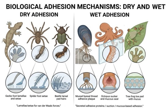
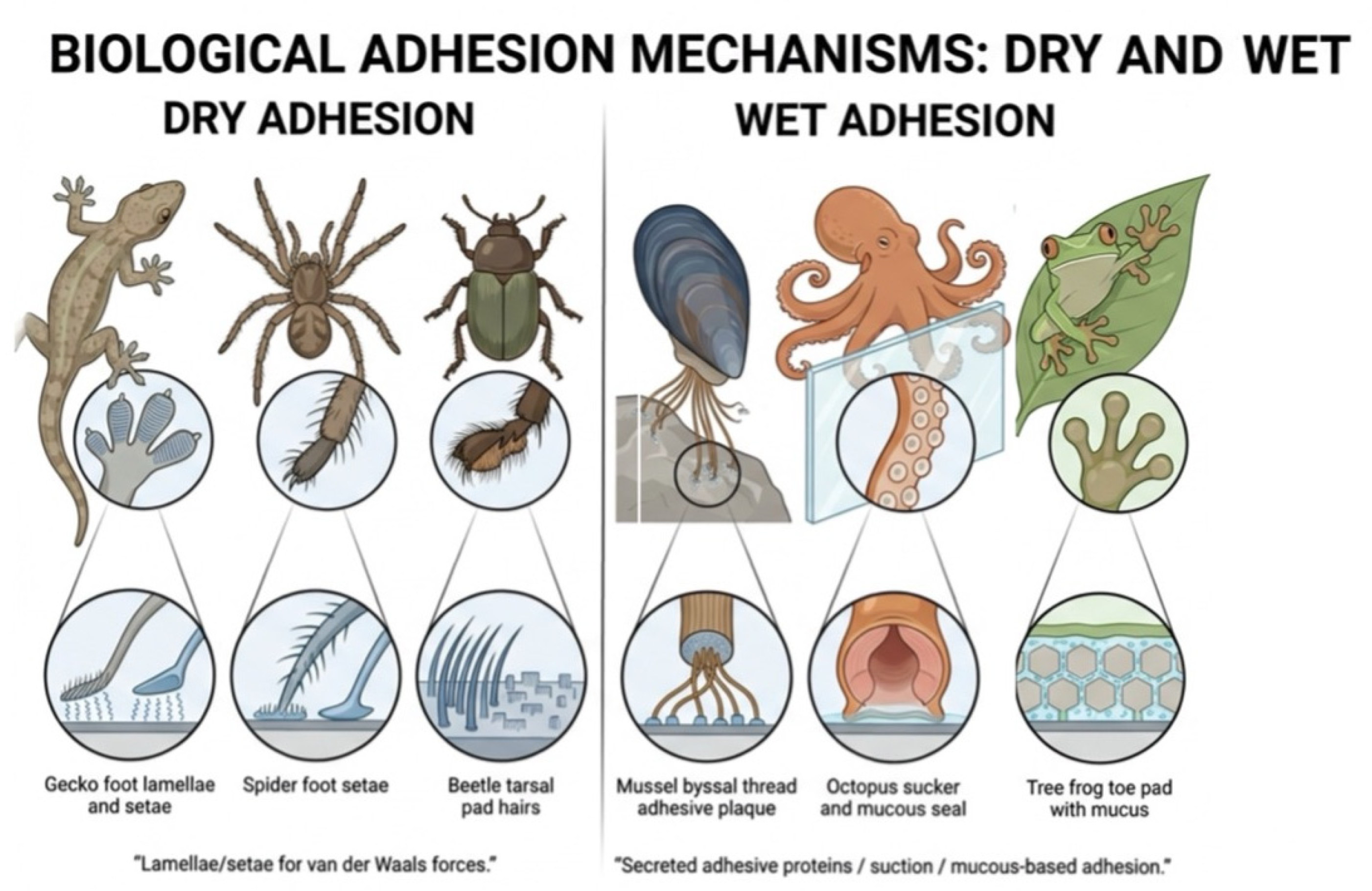
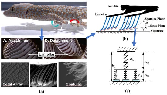
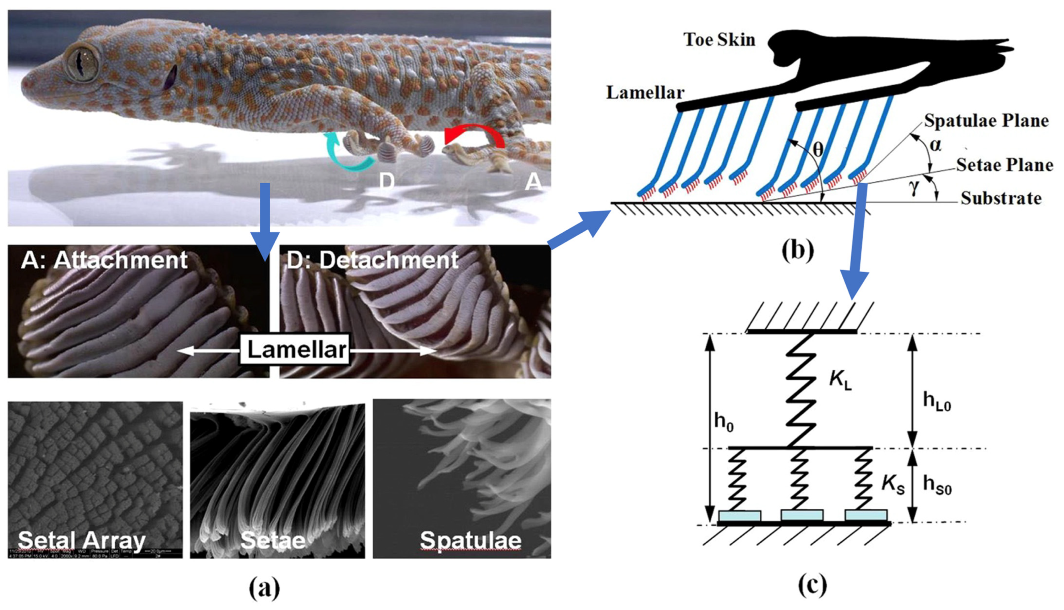
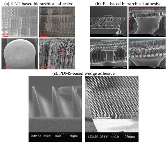
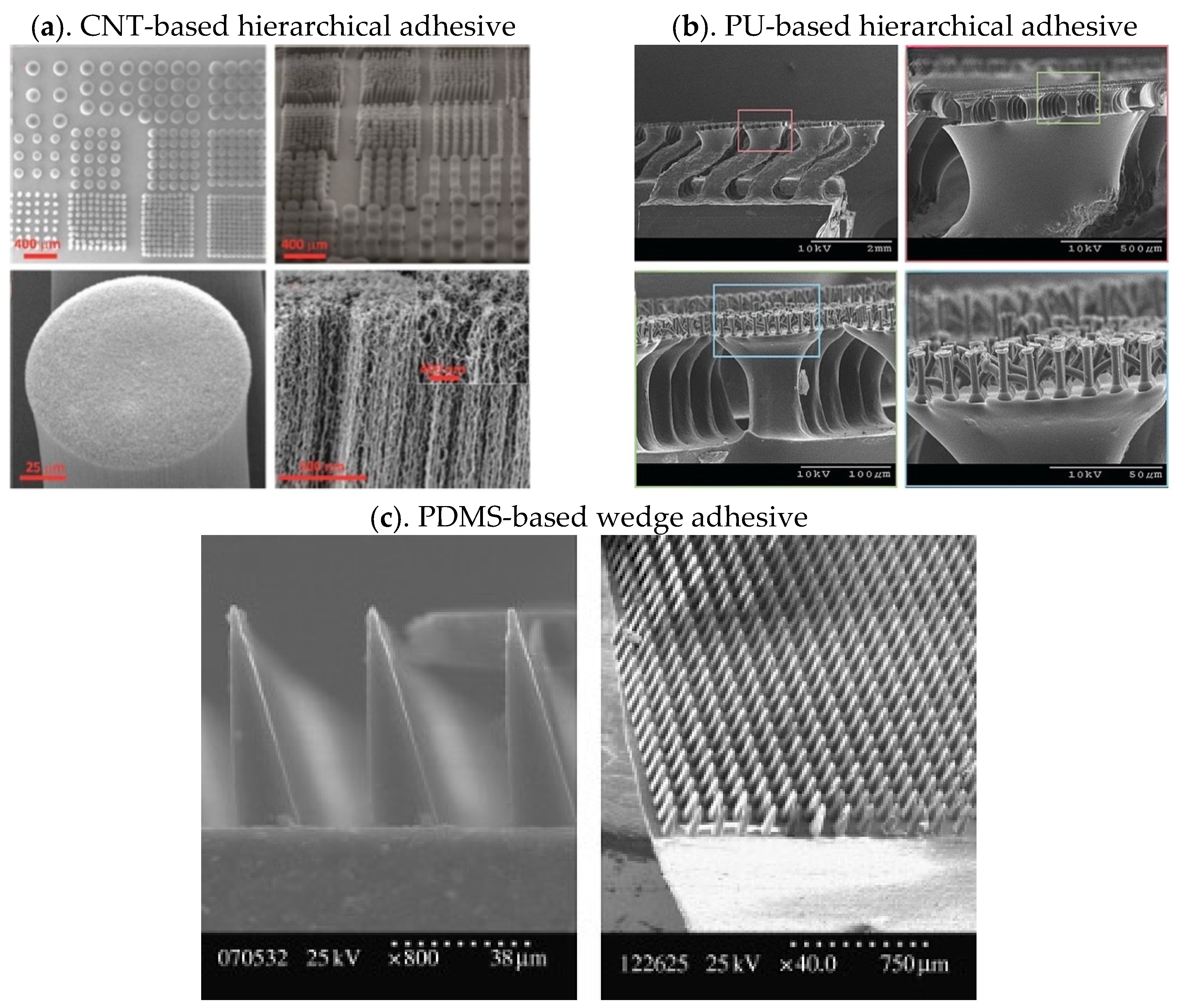
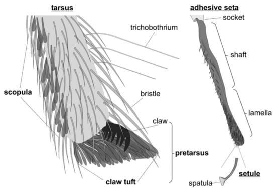
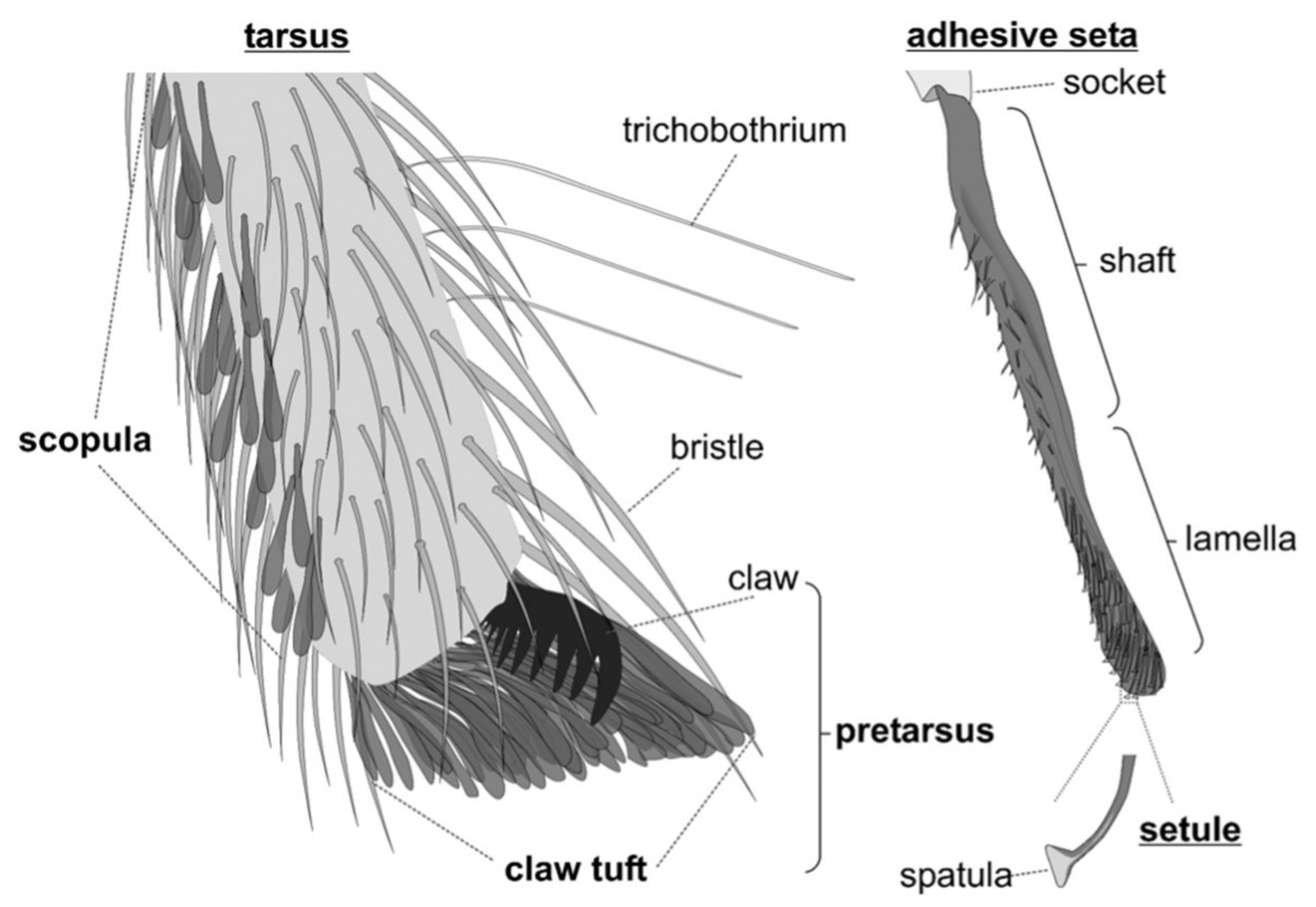
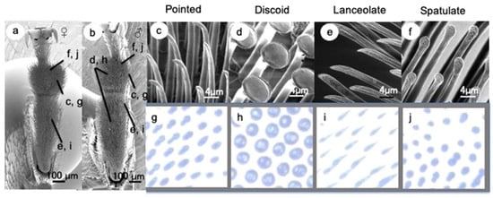
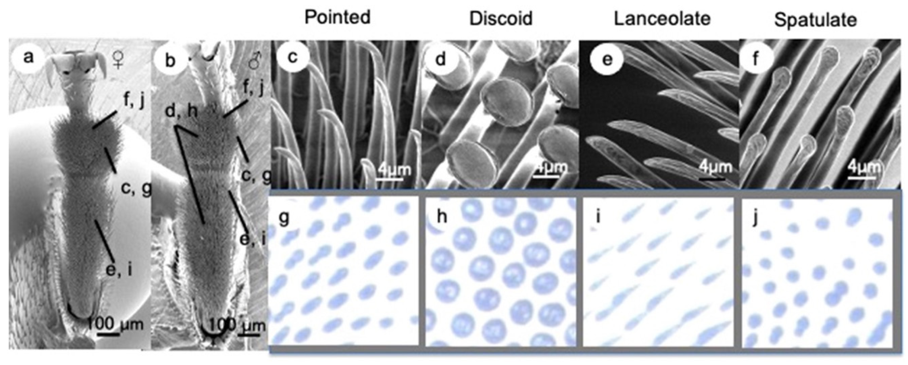
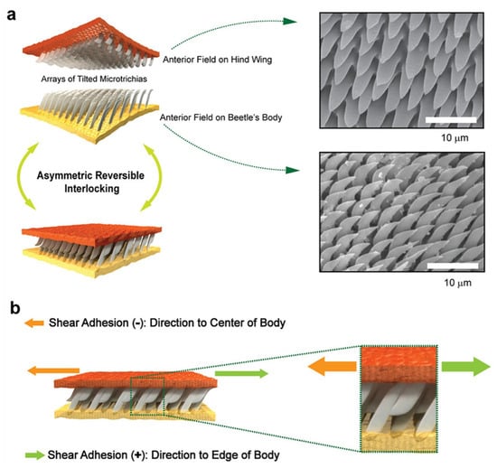
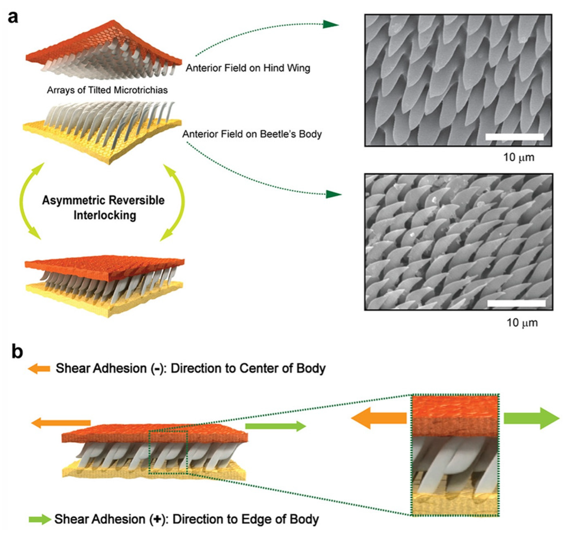
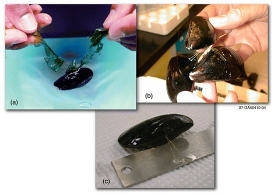
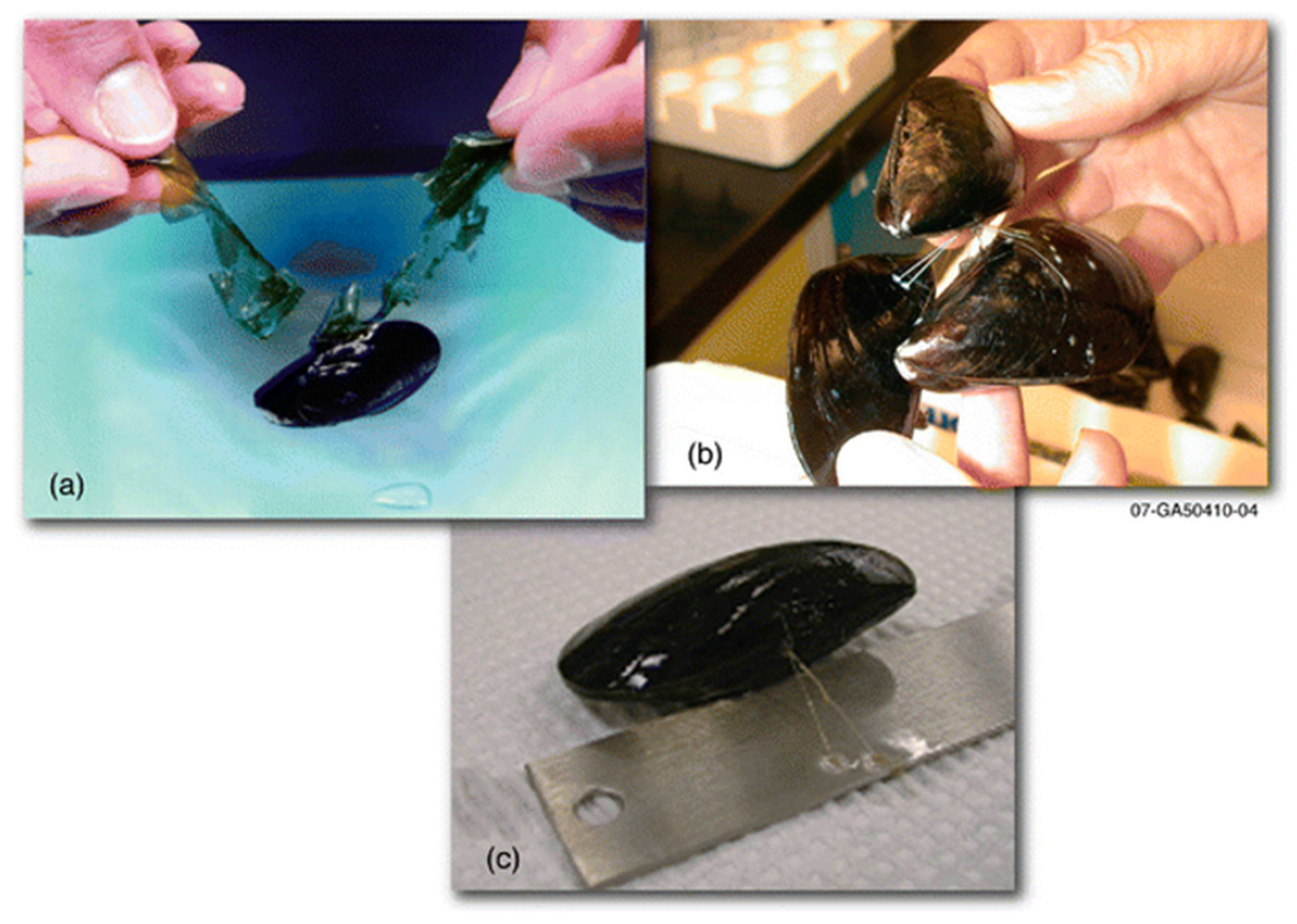
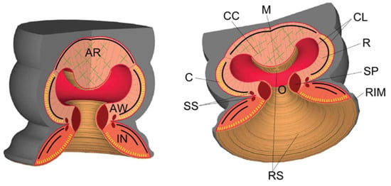
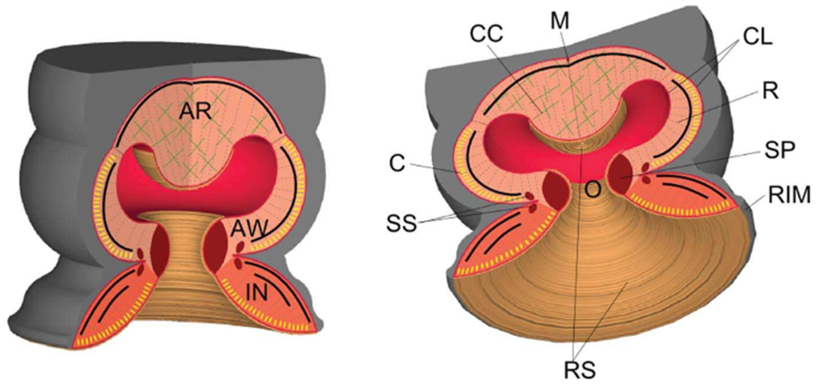
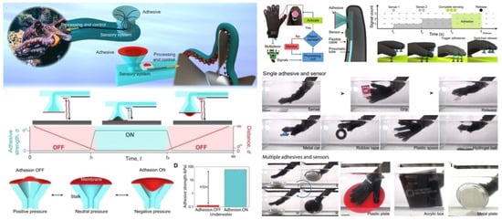
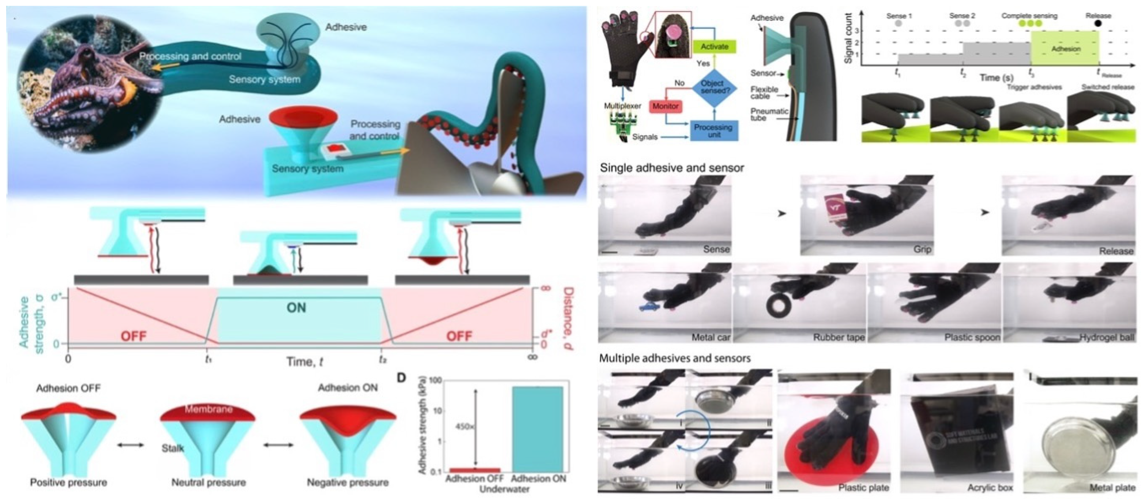
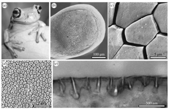
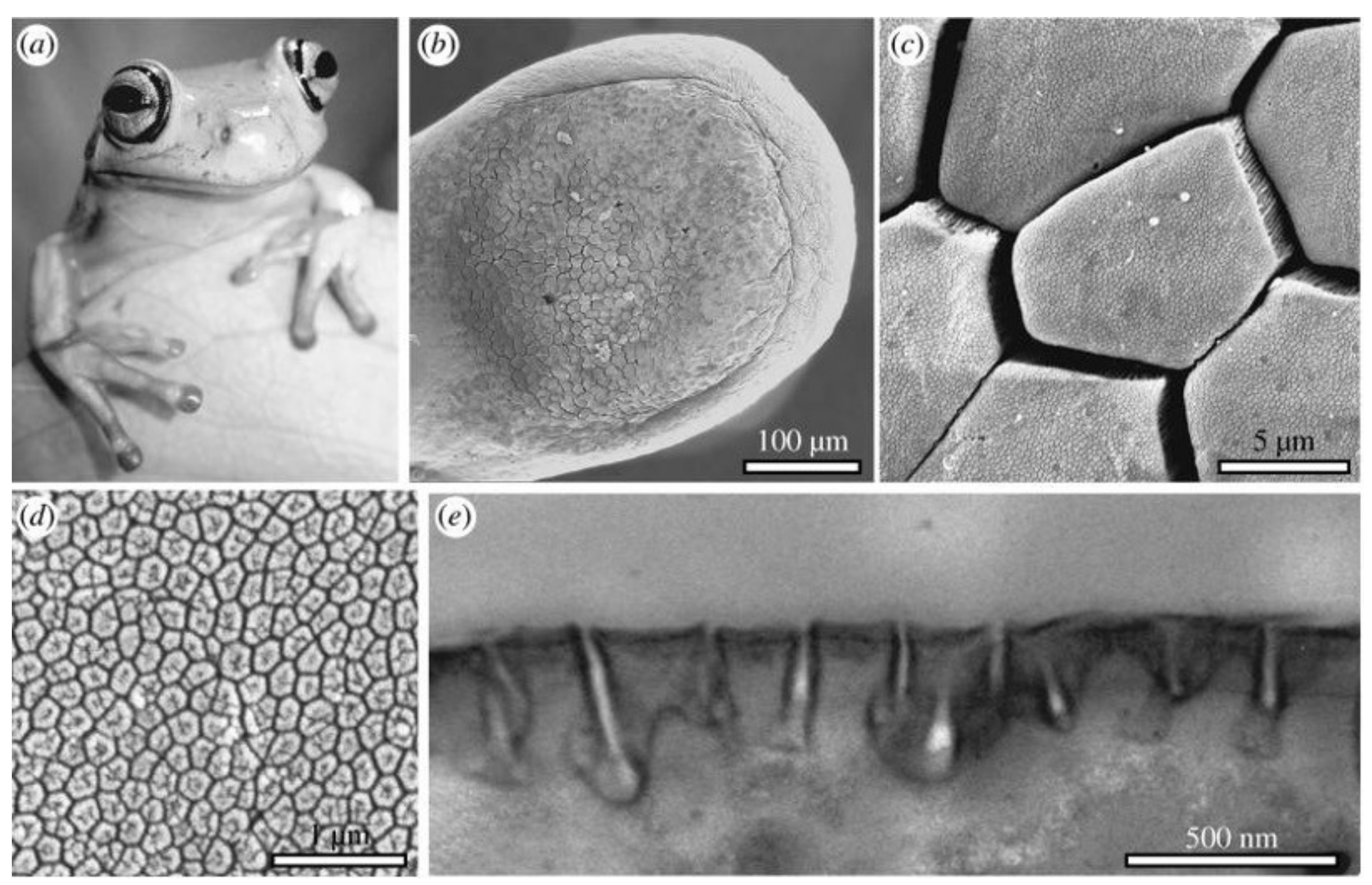
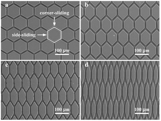
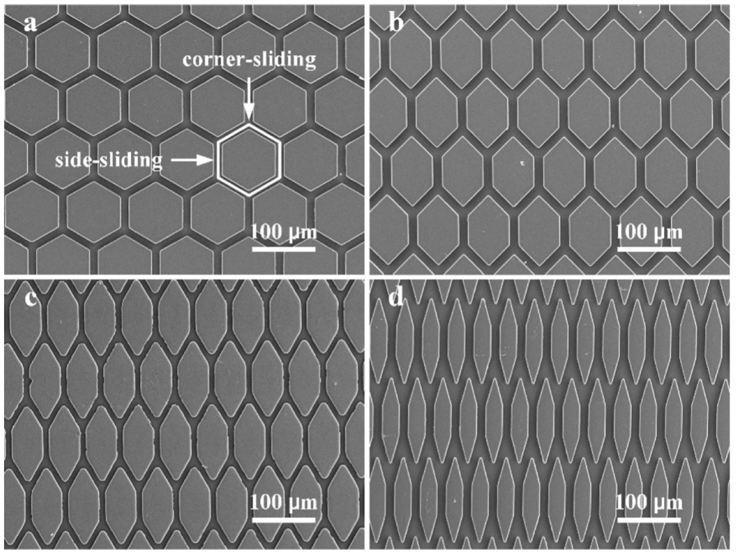

Disclaimer/Publisher’s Note: The statements, opinions and data contained in all publications are solely those of the individual author(s) and contributor(s) and not of MDPI and/or the editor(s). MDPI and/or the editor(s) disclaim responsibility for any injury to people or property resulting from any ideas, methods, instructions or products referred to in the content. |
© 2026 by the authors. Licensee MDPI, Basel, Switzerland. This article is an open access article distributed under the terms and conditions of the Creative Commons Attribution (CC BY) license.