


Abstract
This in vitro study evaluated the shear bond strength (SBS), morphology, and chemistry of CAD/CAM (computer-aided design/computer-aided manufacturing) ceramics and composites bonded to Er:YAG-irradiated dentin using universal adhesive strategies. A total of 102 human dentin discs were randomly allocated to etched control (E), laser-irradiated (I; 10 Hz, 150 mJ, 5 s), or irradiated-and-etched (IE) conditions. Ninety specimens were bonded to a Cerasmart composite (CSM) or leucite-reinforced ceramic (LRF), while 12 were used for scanning electron microscopy (SEM) and energy-dispersive X-ray spectroscopy (EDX). EDX showed marked mineral depletion after etching, with the Ca/P ratio decreasing from 1.58 in control dentin to 1.28 in E and IE specimens. LRF showed higher SBS than CSM (14.12 ± 5.14 vs. 10.94 ± 3.76 MPa); E-LRF had the highest SBS (15.48 ± 3.75 MPa) and I-CSM the lowest (9.57 ± 4.05 MPa), with Tukey significance only seen between these groups (p = 0.009). A two-way ANOVA showed a significant material effect on SBS (p = 0.002), whereas dentin pretreatment (p = 0.185) and material × pretreatment interaction (p = 0.719) were not significant. In conclusion, although Er:YAG irradiation modified dentin morphology and surface chemistry, dentin pretreatment had no statistically significant effect on SBS. Phosphoric acid etching nonetheless remained the most consistent conditioning approach in descriptive terms, as irradiated-and-etched dentin achieved SBS values comparable to etched-only dentin, whereas irradiation alone yielded the lowest descriptive values. This observation should be confirmed by studies incorporating thermocycling, long-term storage, and fatigue loading.
1. Introduction
The removal of carious tissue should be a comfortable, painless, quiet, and efficient procedure, with minimal impact on dentin composition and morphology [1]. Conventional methods using tungsten carbide burs, while widely employed, have significant drawbacks: vibrations and pressure-induced discomfort [2]. To address these limitations, laser-assisted caries removal techniques, particularly using Erbium-doped yttrium aluminum garnet laser (Er:YAG), have gathered increasing attention [3,4,5]. Er:YAG lasers can effectively eliminate carious tissues with minimal thermal damage to adjacent hard and soft tissue [6,7,8,9,10,11,12]. The laser beam interacts with the hydroxyl radicals of hydroxyapatite and with the water molecules trapped in the crystalline structure, generating micro-explosions by water vaporization and thus effectively removing carious tissue [13]. This mechanism not only minimizes the pain compared to rotary instruments [14] but also reduces the bacterial load [6,15].
Yet the laser irradiation of dentin results in an irregular yet homogeneous surface layer characterized by open and enlarged tubules, denatured or absent collagen, and intact peritubular dentin [7,12,16,17,18]. These morphological changes present a challenge for adhesive bonding. Indeed, collagen exposure and partial demineralization are critical for achieving a strong hybrid layer through resin infiltration [16,19,20,21,22].
Thus, several studies have investigated the bond strength of different adhesive systems to the irradiated dentin [8,23,24,25,26,27,28]. However, the results of these studies are conflicting, and no consensus has been reached regarding the laser irradiation of dentin [29] and the etching strategy afterwards. Moreover, a recent review with meta-analysis concluded that more studies on bonding irradiated dentin are necessary [29].
Nevertheless, in a clinical situation, restorations hinge on two critical junctions—the interface between the irradiated dentin and the resin cement and the interface between the adhesive cement and the biomaterial. Assessing these factors simultaneously yields a more authentic appraisal of the restoration’s overall bond performance.
At the same time, restorative dentistry has been transformed by advances in computer-aided design and manufacturing (CAD/CAM) technologies. New materials, with enhanced homogeneity and minimal structural flaws, have become available [30,31]. Among these materials, CAD/CAM glass-ceramics are widely used and highly appreciated for their mechanical and aesthetic properties [30,32,33]. Leucite-reinforced feldspar (LRF) ceramic blocks use an easier and faster milling process than conventional lithium disilicate blocks while maintaining appropriate aesthetic and mechanical properties [33,34].
However, these glass-ceramics typically require a firing step after milling, which can be time-consuming. In contrast, CAD/CAM blocks made of highly filled resin composites (HFRCs) exhibit favourable machinability and can be finished within a few polishing steps, thereby offering a more expeditious fabrication process [31,35,36]. On the other hand, composites can exhibit higher wear than ceramics [37,38,39], and their bonding can be challenging [40]. Thus, both CAD/CAM ceramics and composites have specific clinical indications and advantages.
Therefore, and given the indisputable benefits of laser treatment [6,15], it is imperative to assess the impact of dentin Er:YAG laser irradiation, either alone or in conjunction with phosphoric acid etching, on the shear bond strength of the dentin bonded to contemporary CAD-CAM restorative materials. The research hypothesis is that the irradiation of dentin, with or without etching, does not alter its morphology, chemical composition or its bond strength to modern restorative materials.
2. Materials and Methods
2.1. Sample Preparations
A total of 102 human third molars (research protocol CODECOH DC-2021-4812) were sectioned to obtain dentin discs of 3 mm thickness using a low-speed saw diamond blade (Isomet, Buehler, Lake Bluff, IL, USA). The discs were embedded in transparent resin (BIOCRYL®, Scheu Dental, Iserlohn, Germany) using moulds (20 × 3 mm) and polished with silicon carbide papers (P-600 grit). Each dentin disc was obtained from a separate third molar so that one specimen corresponded to one tooth; the specimens were therefore statistically independent, and the assumption of independence required for the analysis of variance was satisfied. This resin embedding served to standardize specimen geometry and to allow for the reproducible, axis-aligned seating of the bonded assembly in the shear testing jig. The resin surrounded the periphery of the disc within the mould and did not contact the central bonding area, which was confined to the exposed dentin surface; the bonded interface was thus established directly on dentin and not on resin.
2.2. Dentin Surface Pretreatments
Ninety of the prepared dentin discs were randomly divided into three pretreatment groups.
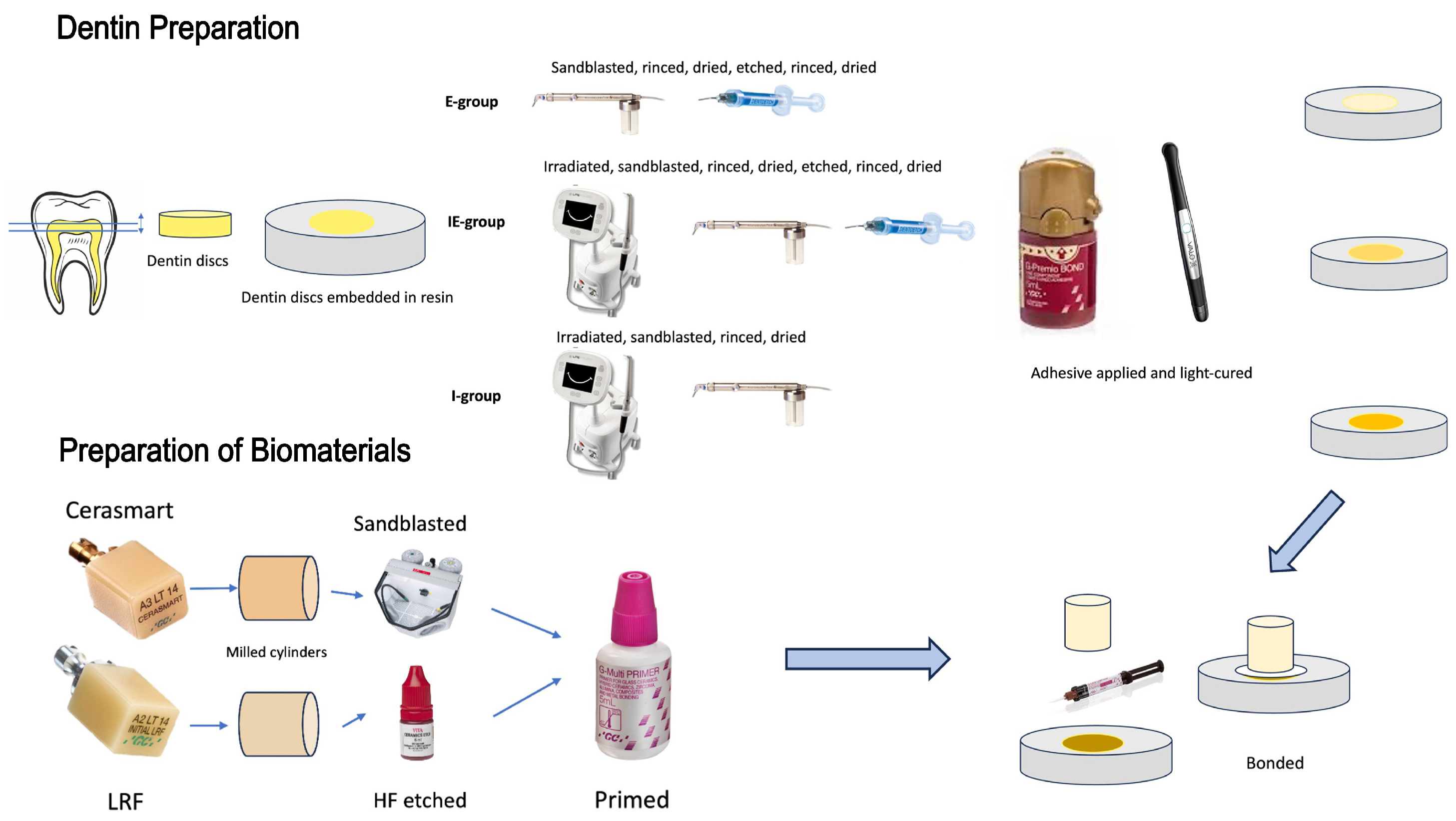
The first group (etched “E”, n = 30) was sandblasted with 50 µm aluminum oxide for 5 s (Micro-etcher II, Danville Engineering Inc., Danville, CA, USA) [41]. The samples were then water-rinsed and air-dried. Finally, the samples were etched with 37% phosphoric acid (Dentoetch, Itena Clinical, Villepinte, France) for 15 s. The specimens were then water-rinsed for 15 s and air-dried. This group was used as a control.
The samples of the second group (irradiated and etched “IE”, n = 30) were first irradiated using the ER:YAG laser (150 mJ/pulse at 10 Hz, 11 J/cm2, contact mode under water irrigation, 1.3 mm tip, according to the manufacturer’s instructions, Litetouch, Syneron, Israel) for 5 s and then sandblasted [41], rinsed with water and dried. Finally, the samples were etched with 37% phosphoric acid and rinsed with the same protocol as above.
The samples of the third group (irradiated “I”, n = 30) were irradiated with the same protocol, then sandblasted and rinsed.
Each group was then divided into two subgroups (n = 15) according to the restorative materials, a composite resin (CSM) (Cerasmart, GC, Tokyo, Japan) and a leucite-reinforced glass-ceramic (LRF) (Initial LRF, GC, Tokyo, Japan), to finally obtain 6 subgroups: E-CSM, IE-CSM, I-CSM, E-LRF, IE-LRF, and I-LRF.
Dentin samples were covered with a universal adhesive (G-Premio BOND, GC, Tokyo, Japan) according to the manufacturer’s instructions (Table 1) and light-cured at 1000 mW/cm2 for 20 s using a ValoTM lamp (Ultradent, South of Jordan, UT, USA).
Table 1.
The chemical composition and manufacturer instructions for the used bonding systems.
2.3. Scanning Electron Microscopy (SEM)
Twelve of the 102 dentin discs were randomly selected and used for SEM observations. Nine of them were prepared following the same 3 groups of pretreatments (n = 3), and the last three received no treatment because they were to be used as the control group. All samples were prepared for SEM observations. The samples were dehydrated by using an ethanol solution series (50, 75, 95 and 100%). The samples were then mounted on SEM stubs and sputter-coated with gold palladium alloy (20/80% weight) using a sputtering device (Technics, Danville, CA, USA). Scanning electron microscopy (FEI Company, Eindhoven, The Netherlands, 10 kV) was used to investigate dentin morphological changes after the different pretreatments at several magnifications. Energy-dispersive X-ray (EDX) analysis was performed (50 s acquisition time, 10 mm distance) to investigate the chemical change in dentinal surfaces after each pretreatment.
Quantitative EDX results were additionally recorded for the main elements detected at the dentin surface and expressed as weight percentage (wt%) and atomic percentage (at%). Because the SEM/EDX observations were performed on representative specimens rather than on independent replicate measurements designed for inferential analysis, these chemical data were interpreted descriptively and summarized in a dedicated table.
2.4. Surface Treatment of CAD/CAM Materials
Cylindrical samples of both CAD-CAM materials were milled using a Lyra Mill (Lyra ETK, Sallanches, France) to 5 mm in diameter and 4.6 mm in height. One side of each cylinder was polished with a P-600 grit.
The CSM cylinders were sandblasted with 50 µm aluminum oxide particles (Bader, Pontevedra, Spain) for 5 s at a pressure of 0.5 MPa, using a laboratory sandblaster (Basic classic sandblaster, Renfert, Hilzingen, Germany). Then, the cylinders were rinsed and dried.
The LRF cylinders were treated with 5% hydrofluoric acid (Vita ceramics etch, Bad Sackingen, Germany) for 1 min, rinsed for 20 s, then dried.
Finally, a primer (G-multi-Primer, GC, Tokyo, Japan) was applied to all materials (LRF and CSM) for 1 min, before drying with air spray.
2.5. Bonding Procedure
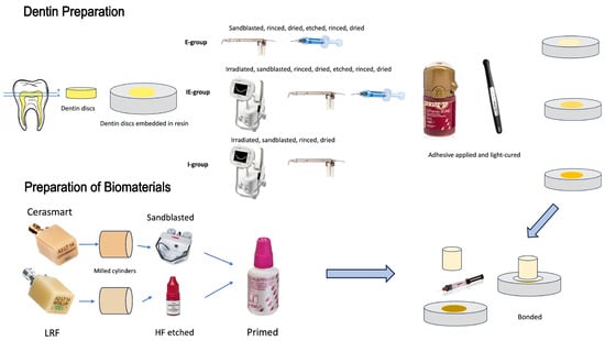
All cylinders were then bonded to the dentin discs with the dual G-Cem LinkforceTM resin cement (GC, Tokyo, Japan) using a self-developed load device (equation 10 N). The excess of resin cement was removed with a micro brush before light-curing for 60 s on all sides. Finally, a 5 mm internal diameter trephine drill (229 RA L, Meisinger, Neuss, Germany) was used around the cylinders to remove any resin excess. A graphical presentation of the sample preparation is available in Figure 1.
Figure 1.
Graphical representation of samples’ preparation.
2.6. Shear Bond Strength (SBS) Test
A shear bond strength test was performed using a universal testing machine equipped with a 1 kN load cell (Instron device 3345, Instron, Norwood, MA, USA). An increasing load was applied with a crosshead speed of 1 mm/min until failure. The maximum load was recorded, and the SBS values were calculated according to the following formula: Maximal strength (MPa) = load at failure (N)/bonded surface area (mm2).
2.7. Failure Mode Evaluation
After testing, both pieces of all specimens were examined under a digital microscope (Keyence, VHX-5000, Osaka, Japan). The failure modes were labelled according to their area of occurrence: “adhesive failure” if the failure occurred at the tooth surface or restorative material interface with the resin cement, “cohesive failure” if the failure occurred exclusively within the resin cement layer or the tooth structure, and “mixed failure” if the failure was a combination of the above two failure types (partial resin cement fracture and partial dentin or restorative material exposure) [42].
2.8. Statistical Analysis
Data were analyzed using SPSS Statistics (v.26, IBM, Armonk, NY, USA). Normality and homogeneity of variances were assessed using the Kolmogorov–Smirnov and Levene tests, respectively. A two-way analysis of variance (ANOVA) was performed to evaluate the main effects of CAD/CAM material (CSM or LRF), dentin pretreatment (E, IE or I), and their interaction on shear bond strength (SBS). In addition, a one-way ANOVA followed by Tukey honestly significant difference (HSD) post hoc tests was used for pairwise comparisons among the six experimental subgroups. The confidence level was set at 95% (α = 0.05) for all statistical tests. As a sensitivity analysis, the four specimens that failed before mechanical testing were additionally included with an assigned SBS of 0 MPa, and the two-way ANOVA was repeated (n = 90).
3. Results
3.1. SEM Observations
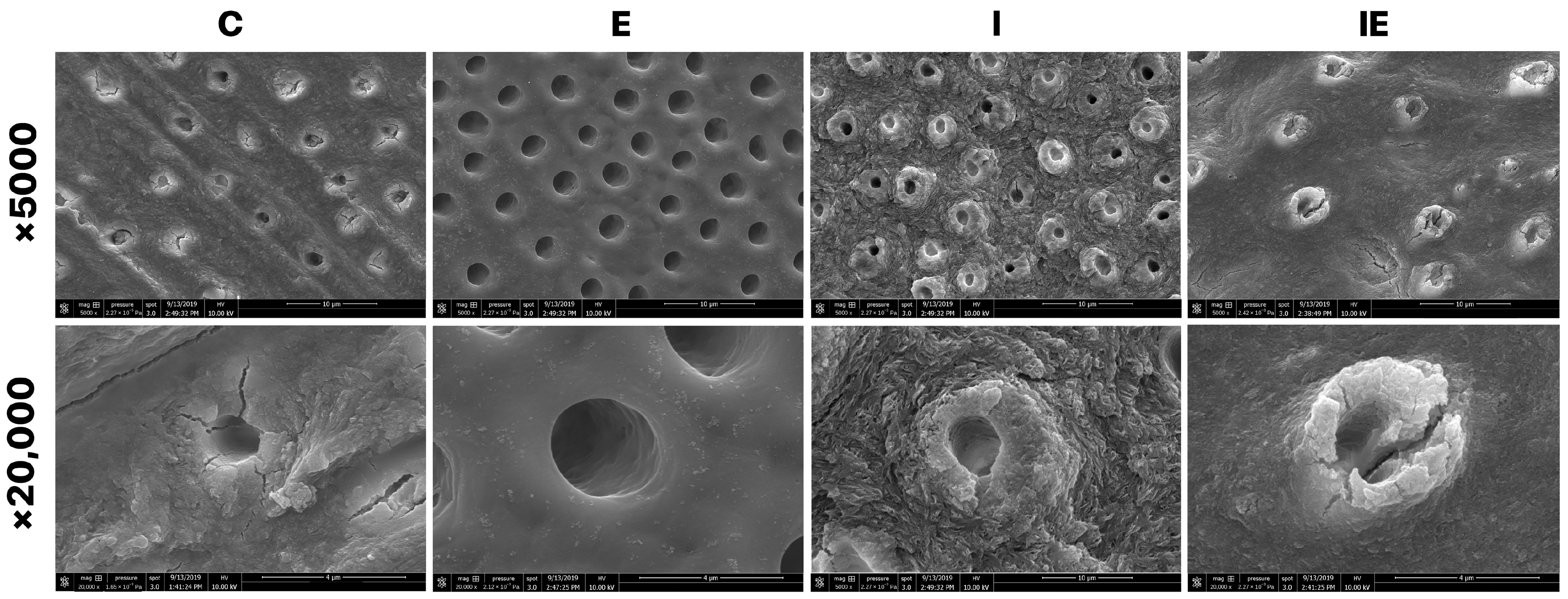
Figure 2 demonstrates the different morphological changes in dentin structure after irradiation, etching or irradiation with etching pretreatments. The control group showed the smear layer resulting from dentin preparation, some dentinal plugs, and the marks of the diamond bur on the dentinal surface. The dentinal surface etched with 37% phosphoric acid showed a clean surface with open dentinal tubules and a total elimination of the smear layer. Both the irradiated and irradiated/etched groups demonstrated a rougher surface compared to the etched group. The irradiated group showed a rougher aspect of the surfaces. The dentinal tubules in both groups (irradiated and irradiated/etched) had less markedly opened orifices and showed a less clean surface compared to the group that was only etched with phosphoric acid.
Figure 2.
Scanning electron microscopy representative images (×5000 and ×20,000 magnifications) demonstrate morphological changes in dentin structure after each pretreatment. C: control group; E: etched (15 s phosphoric acid); I: irradiated (10 Hz, 150 mJ) for 5 s; group IE: irradiated and etched.
3.2. EDX Analysis
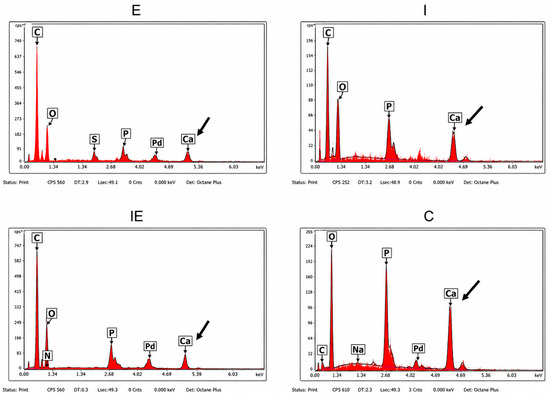
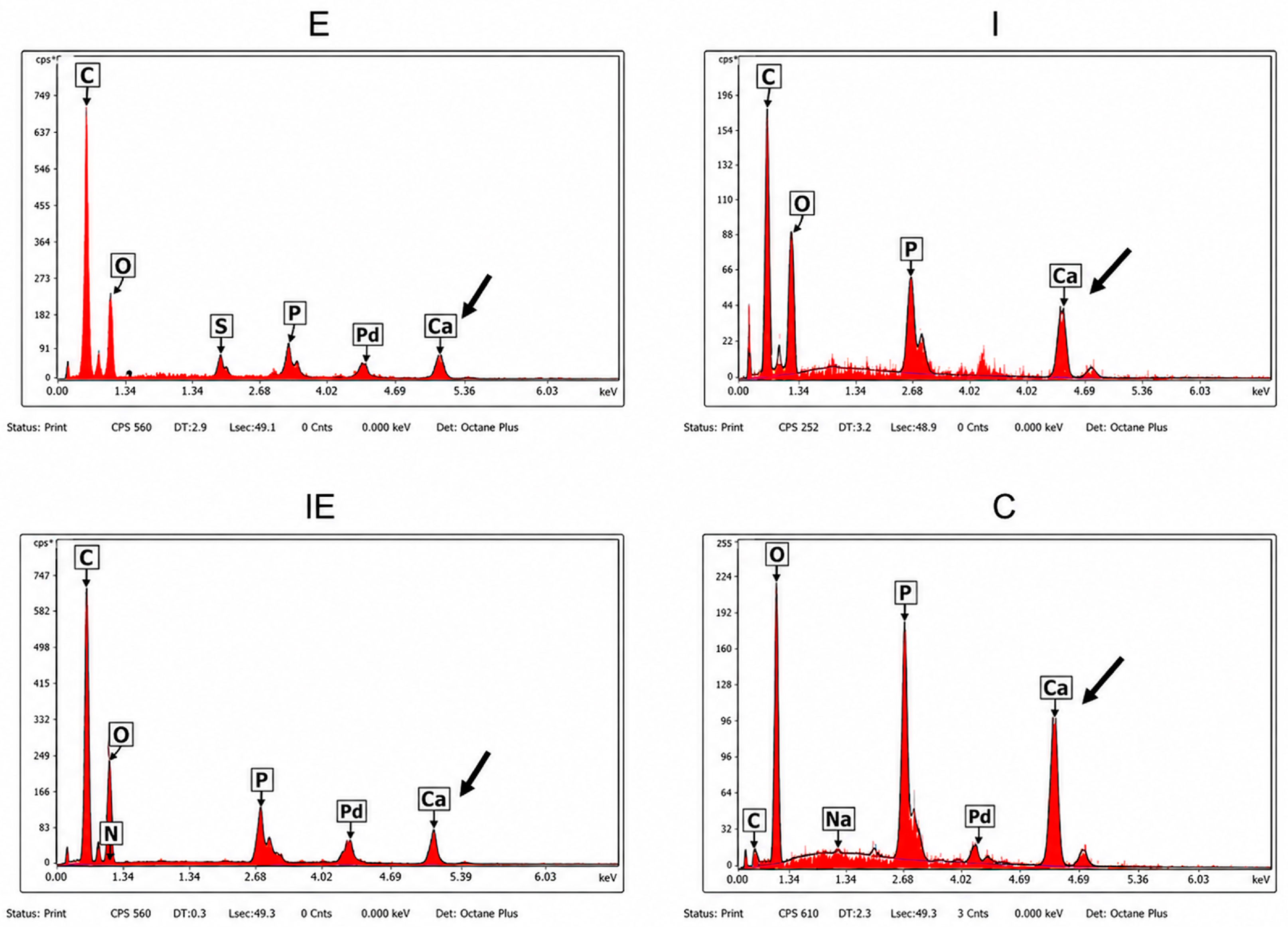
Figure 3 shows the energy-dispersive X-ray analysis of the different studied surfaces, and the corresponding quantitative EDX data are summarized in Table 2. Compared with the untreated control, both etched groups showed a marked decrease in mineral indicators, with Ca decreasing from 26.05 wt% in C to 6.04 wt% in IE and 4.70 wt% in E and P decreasing from 12.78 wt% to 3.66 wt% and 2.86 wt%, respectively. In contrast, irradiated dentin showed higher Ca (32.99 wt%) and P (15.08 wt%) than the untreated control. The atomic Ca/P ratio remained close to the control value after irradiation alone (1.69 vs. 1.58) but decreased after etching, irrespective of prior irradiation (1.28 for both IE and E). These results support the qualitative observation that phosphoric acid etching produced the most marked mineral depletion at the dentin surface.
Figure 3.
A representative energy-dispersive X-ray (EDX) analysis of the different dentinal surfaces of the different groups. C: control group; E: etched (15 s phosphoric acid); I: irradiated (10 Hz, 150 mJ) for 5 s; group IE: irradiated and etched.
Table 2.
Quantitative EDX analysis of selected elements detected on dentin surfaces after each pretreatment. Values are expressed as weight percentage (wt%) and atomic percentage (at%). C: untreated control; I: irradiated; IE: irradiated and etched; E: etched. Au and Pd peaks are visible in spectra because of sputter coating; they were not interpreted as dentin components and are not included in quantitative dentin composition columns.
3.3. Bond Strength and Failure Mode
Of the 90 bonded specimens initially prepared for shear bond strength testing, four specimens failed before mechanical testing and were therefore excluded from the statistical analysis. Consequently, the final SBS analysis was performed on 86 specimens. The assumptions of normality and homogeneity of variance for the SBS values were satisfied for all six subgroups (p > 0.05). A one-way ANOVA showed a significant overall difference among the six experimental subgroups (F(5,80) = 2.96, p = 0.017). Tukey HSD post hoc analysis identified a significant difference only between E-LRF and I-CSM (p = 0.009). The two-way ANOVA showed a significant main effect of CAD/CAM material on SBS (F(1,80) = 10.52, p = 0.002), with higher values for LRF than for CSM. Conversely, dentin pretreatment had no statistically significant main effect (F(2,80) = 1.72, p = 0.185), and the interaction between CAD/CAM material and dentin pretreatment was not significant (F(2,80) = 0.33, p = 0.719). The four pre-test failures were distributed as follows: one in IE-CSM, one in I-CSM and two in I-LRF, with none in E-CSM, E-LRF or IE-LRF. In the sensitivity analysis including these specimens as 0 MPa (n = 90), the main effect of CAD/CAM material remained significant (F(1,84) = 8.16, p = 0.005), and the material × pretreatment interaction remained non-significant (F(2,84) = 0.13, p = 0.876), whereas the main effect of dentin pretreatment became significant (F(2,84) = 3.46, p = 0.036).
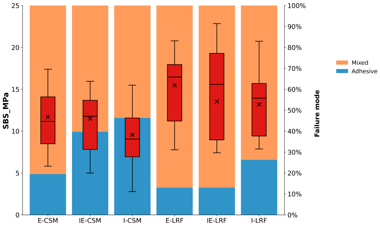
The means and standard deviations of SBS were: E-CSM = 11.70 ± 3.50 ab MPa; IE-CSM = 11.51 ± 3.59 ab MPa; I-CSM = 9.57 ± 4.05 b MPa; E-LRF = 15.48 ± 3.75 a MPa; IE-LRF = 13.56 ± 6.33 ab MPa; and I-LRF = 13.20 ± 5.08 ab MPa. Superscript letters indicate homogeneous Tukey groups; subgroups sharing at least one letter were not significantly different.
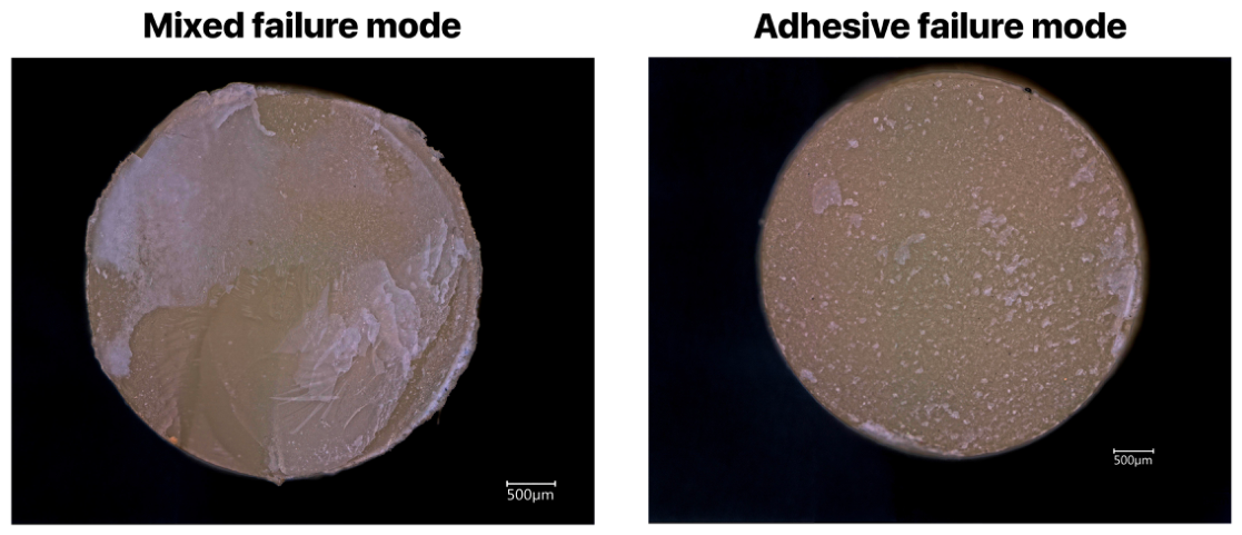
Regarding the observed failure mode, it was found that mixed failures occurred frequently in all LRF subgroups, whilst it was the least observed in CSM subgroups. Adhesive failure at the dentin surface was more frequent in the laser-irradiated samples in CSM subgroups, whilst the etched samples (E-CSM) demonstrated fewer adhesive failures.
Representative fracture surfaces of the debonded specimens are shown in Figure 4.
Figure 4.
Representative images of the two types of failure modes as observed on the Cerasmart surface: mixed (left) and adhesive (right) failure.
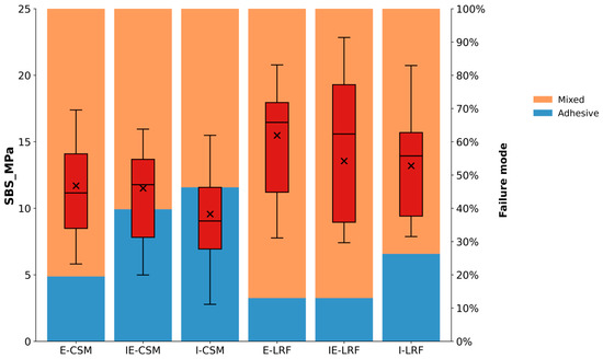
Figure 5 presents boxplots that combine SBS data with the mode of failures.
Figure 5.
A combination of the SBS measures (expressed as boxplots, MPa) and the frequencies of failure modes (expressed as percentages) recorded for all groups. E-CSM: etched dentin bonded to Cerasmart composite resin; IE-CSM: laser-irradiated and etched dentin bonded to Cerasmart composite resin; I-CSM: laser-irradiated dentin bonded to Cerasmart composite resin; E-LRF: etched dentin bonded to LRF leucite-reinforced glass-ceramic; IE-LRF: laser-irradiated and etched dentin bonded to LRF leucite-reinforced glass-ceramic; I-LRF: laser-irradiated dentin bonded to LRF leucite-reinforced glass-ceramic. Failure modes: “Adhesive” if the failure occurred at the tooth surface or restorative material interface with the resin cement; “cohesive” if the failure occurred exclusively within the resin cement layer or the tooth structure; and “mixed” if the failure was a combination of the above two failure types (partial resin cement fracture and partial dentin or restorative material exposure).
4. Discussion
The primary objective of this in vitro investigation was to determine whether dentin irradiation—performed with or without prior etching—modifies dentin morphology, chemical composition, or bond strength to contemporary restorative materials. The bond strength of laser-treated dentin to CAD/CAM restorations is conditioned by two distinct interfaces: (i) the interface between the irradiated dentin and the resin cement and (ii) the junction between the adhesive cement and the restorative biomaterial. Accordingly, the present study evaluated both interfaces simultaneously to provide a laboratory assessment of their combined influence on bonding performance.
The effects of etching, irradiation and irradiation followed by etching on superficial changes to dentin were studied first. Indeed, the main objectives of dentinal pretreatments are to achieve a clean dentinal surface and modify its physicochemical properties to enhance the bond strength of dentin to various restorative materials [19]. In fact, any alteration in the dentinal structure, including the demineralization depth [20,21], the thickness of the smear layer [43], and the extent of the dentin drying [22], has been reported to modify bond strength.
First, air particle abrasion (sandblasting) was applied to dentin in every experimental group, as a recent systematic review highlighted the critical value of this surface treatment, particularly when combined with etch-and-rinse adhesive systems [41].
Secondly, it is well established that the dentinal morphology, including the orientation and diameter of the tubules, differs along the dentinal wafer zone [8,44,45]. To minimize the impact of intrinsic dentinal morphology on the results, all cylinders were bonded in the centre of dentinal discs, ensuring comparable dentinal supports with identical morphological conditions. The supplementary dentinal samples were observed in a similar manner.
A variety of morphological features were discerned in the SEM images obtained after each pretreatment (I, E and IE groups). As expected, after dentin preparation, a smear layer was created on the dentinal surface [46]. As previously described, different treatments could be proposed to eliminate the smear layer to enhance bonding conditions and therefore improve bond strength [43]. Etching with phosphoric acid is one of these methods, as it both eliminates the smear layer and demineralizes the superficial dentin structure [46]. Accordingly, the present study highlighted that the use of 37% phosphoric acid eliminates the smear layer and creates a clean surface, with markedly opened tubules.
In the irradiated groups, the dentinal tubules were also open, in accordance with other studies [16]. Yet, after irradiation, the dentinal structure demonstrated smaller openings of the dentinal tubules compared with the etched samples. These results are consistent with previous studies [10,18]. Researchers have reported that Er:YAG laser irradiation may reduce the diameter of dentinal tubules or partially occlude them, which supports its use in the management of dentin hypersensitivity [18,47]. In addition, in the present study, some irregularities on the dentin surface were observed after the use of Er:YAG laser. This agrees with other studies that have reported dentinal irregularities after laser irradiation [10,16,48] and showed that these irregularities depended on laser parameters [48].
Moreover, the EDX study indicated that 37% phosphoric acid etching reduced the calcium and phosphorus contents in the dentinal structure. This observation is in accordance with previous studies [46]. The quantitative EDX results refined this interpretation by showing, descriptively, that the chemical changes were the most pronounced after etching, whether laser irradiation had been performed beforehand. Rather than showing a mineral decrease after irradiation alone, irradiated dentin presented higher Ca and P signals than the untreated control, with a Ca/P atomic ratio remaining close to the control value. This apparent increase in the Ca and P signals after irradiation alone is most plausibly explained by the removal of the loosely bound, water- and organic-rich smear layer, together with the surface dehydration produced by Er:YAG micro-explosive ablation, which expose and concentrate the underlying mineralized dentin within the analyzed volume rather than reflecting any net mineral gain. In agreement with this interpretation, the Ca/P atomic ratio remained close to that of the control, indicating that the mineral phase itself was not demineralized by irradiation under the present settings. Conversely, both etched groups exhibited pronounced Ca and P reductions and a lower Ca/P atomic ratio, consistent with superficial demineralization and smear layer removal caused by phosphoric acid. These findings suggest that Er:YAG irradiation under the present settings altered the dentin surface morphology without producing the same mineral depletion pattern as phosphoric acid etching, which may help explain why irradiation alone did not provide optimal bonding conditions.
Further, this in vitro study compared the bond strength of two different CAD/CAM materials to dentin after three different pretreatments: phosphoric acid etching, Er:YAG laser irradiation, and irradiation followed by etching, using a universal adhesive. The additional two-way ANOVA refined the statistical interpretation by separating the influence of the restorative material from that of dentin pretreatment. This analysis showed that the CAD/CAM material was the only significant determinant of SBS, whereas dentin pretreatment and the material × pretreatment interaction were not significant. Therefore, the null hypothesis was partially rejected: dentin irradiation and etching clearly altered dentin morphology and surface chemistry, but pretreatment alone did not significantly modify SBS under the present experimental conditions; conversely, the type of CAD/CAM material significantly influenced bond strength.
The significant main effect of material indicated that SBS values were higher for the LRF subgroups than for the CSM subgroups, irrespective of the dentin pretreatment employed. These results may be attributed to the pretreatment of LRF surfaces with 5% hydrofluoric acid. In fact, the etching of glass-ceramics, such as LRF, creates a rough surface and microporosities, which considerably enhances micromechanical retention and bond strength [49].
In fact, in clinical practice, the effective bonding of these materials to dentin surfaces remains a critical challenge, requiring precise pretreatment protocols to optimize adhesive performance, and dual-cured resin cements have been recommended [50,51]. To enhance the bond strength of the prosthesis to the tooth, HFRCs should be sandblasted [52,53], while glass-ceramics should be etched with hydrofluoric acid [49,54]. Afterwards, the intaglio surface of the prosthesis needs to be coated with a silane-containing primer [49,54,55]. These recommendations were followed in this in vitro study.
Within both material categories (CSM and LRF), the laser-irradiated subgroups treated in self-etch mode (I) exhibited lower descriptive bond strength values than their counterparts that underwent a conventional total etch protocol with 37% phosphoric acid and no irradiation (E). However, the two-way ANOVA confirmed that dentin pretreatment did not exert a statistically significant main effect and that no significant material × pretreatment interaction was present. This suggests that, under the laser parameters and adhesive strategy used in the present study, the influence of pretreatment was consistent across the two CAD/CAM materials but remained weaker than the material-related effect. This may be explained by the altered hybrid layer and degraded dentin organic matrix reported after laser irradiation [16]. Additionally, other studies have shown the significantly lower bond strength of irradiated-only dentin compared with irradiated-and-etched dentin [27,28]. Yet, in the present study, this difference was not statistically significant, which is in agreement with previous studies reporting no significant effect of etching versus irradiation on dentin bond strength values [9,24,25].
The four pre-test failures occurred predominantly in the irradiated groups (three of the four), consistent with the lower descriptive performance of irradiated-only dentin. Notably, the statistical significance of the pretreatment effect was non-significant when only measurable values were analyzed but became significant when the failures were treated as null bond strengths (see Section 3.3). Both analyses nonetheless converged on the weaker performance of the irradiated-only condition, supporting a cautious interpretation of laser irradiation without subsequent phosphoric acid etching.
In contrast, a previous study demonstrated that the use of laser could enhance the bond strength of an adhesive to dentin [26]. This is probably due to the different conditions of laser application. In fact, in previous research [26], the pulse energy was higher (350 mJ/pulse), while the repetition rate was lower (6 Hz) compared to the present study, where a 150 mJ/pulse energy at 10 Hz was used. Moreover, in the present study, a universal adhesive was used, while a three-step etch and rinse system was used in the other study [26], a condition potentially impacting bond strength [56].
Indeed, on the tooth side, a universal adhesive was used. Universal adhesives have proven their efficiency, in either self-etch or total etch mode [56,57,58]. The universal adhesive utilized in this study, G-Premio BOND (GC, Tokyo, Japan), has a moderately acidic pH [58,59], classified as intermediately strong. Within each group of biomaterials, the SBS values of totally etched dentin, whether laser-treated or not, appear to be higher than those of dentin bonded with a self-etch strategy on irradiated dentin. However, this difference should be interpreted as a descriptive trend rather than as a statistically significant material-specific effect, because the two-way ANOVA showed no significant main effect of dentin pretreatment and no significant material × pretreatment interaction. These findings are consistent with the current literature, which indicates that a total etch strategy enhances the bonding capabilities of intermediately strong adhesives [58,59].
Furthermore, the findings of the present study indicated that the bond strength of all LRF groups was higher than that of I-CSM. This phenomenon can be attributed, on one hand, to the enhanced bonding to the glass-ceramic surface and, on the other hand, to the enhanced micromechanical and chemical adhesion on the etched dentin, as opposed to the irradiated-only dentin. Therefore, the adhesion in the I-CSM group is weaker, as several studies have reported the absence of a hybrid layer on irradiated dentin [9,24,60]. The absence of a hybrid layer and the lack of a chemical adhesion suggest that the adhesive joint is more susceptible to hydrolysis in time. Furthermore, a previous study highlighted that the marginal joint between the dentin and the restoration material is more impervious to hydrolysis on irradiated and etched dentin than on only irradiated dentin [9]. However, in the present study, the samples were not subjected to water or thermal aging, which represents a limitation. Yet it is important to note that the samples which were irradiated and etched demonstrated no significant difference compared to the etched samples.
Different failure modes of the samples were found after the SBS tests. The higher proportion of mixed failures in the LRF groups is consistent with a stronger bonded interface, although failure mode classification should be interpreted cautiously because it depends on the test configuration and substrate [56,61,62]. Cohesive failure was not observed in the present study. The adhesive failure mode, observed mainly in the irradiated groups, could also be related to an error in the manipulation of these adhesive materials, as described in previous studies [62].
The present findings should also be interpreted in light of the bond strength test employed. Although the shear bond strength test is widely used and complies with ISO/TS 11405, it generates a non-uniform stress distribution at the adhesive interface, with stress concentrations that may influence the apparent locus of failure. This methodological limitation should be taken into account when interpreting the absolute SBS values and comparing them to other studies.
In the present study, the Er:YAG laser was operated at a power of 1.5 W, according to the manufacturer instructions. For a rapid carious tissue ablation, a power of up to 6 W may be employed [4]. Yet a lower power yields a superior bond strength [5]. In the case of primary teeth, for instance, the application of a laser on the dentin with a power ranging between 0.5 and 2 W has been shown to yield optimal bond strength [44]. It would be beneficial to test a variety of laser settings to gain a deeper understanding of their impact on the bonding of irradiated dentin with modern universal adhesives.
Considering the limitations of this study, a methodological point concerns the management of pre-test failures. Indeed, pre-test failures have been shown to vary according to the adhesive system and technique sensitivity and to be related to the distribution of bond strength values [63]. In the present study, four specimens failed before mechanical testing and were therefore excluded from the final SBS dataset. The treatment of pre-test failures remains debated in bond strength studies: some authors assign a value of 0 MPa to such specimens, considering them true bonding failures, whereas others exclude them because no measurable load-to-failure value can be obtained under standardized test conditions. In the present analysis, pre-test failures were excluded to avoid artificially lowering the mean SBS values and introducing non-measured values into the statistical model. However, their occurrence was transparently reported and should be considered when interpreting the overall bonding performance of the tested protocols.
In addition, adhesive layer thickness was not standardized or measured, aside from a manufacturer-directed, clinically representative application. As thickness can influence bond strength [45], this is acknowledged as a limitation and a target for future study.
Moreover, further laboratory studies incorporating thermocycling, long-term water storage and cyclic fatigue should be performed to investigate the effect of irradiation, etching and universal adhesives on the bond strength of CAD/CAM materials to dentin [64]. In addition, the roughness of each pretreated dentinal surface could be analyzed to evaluate the effect of each pretreatment and its roughness on bond strength.
Another limitation is that a cross-sectional SEM analysis of the bonded interfaces before shear testing and SEM imaging of the fractured bonded interfaces after testing were not performed. Therefore, the interpretation of interfacial quality was based on dentin surface SEM/EDX observations, SBS values, failure mode distribution, and representative optical fracture surface images. Future studies should include cross-sectional interfacial imaging to better correlate surface morphology, adhesive layer characteristics, and failure patterns.
5. Conclusions
Within the limitations of this study, it can be concluded that the Er:YAG irradiation of dentin produced morphological and chemical alterations. Quantitative EDX further confirmed that the main chemical changes were associated with phosphoric acid etching, which markedly reduced Ca and P signals and lowered the Ca/P ratio, whereas irradiation alone did not produce comparable mineral depletion. The two-way ANOVA indicated that the CAD/CAM material was the only significant factor affecting SBS, with higher bond strength values for LRF than for CSM, while dentin pretreatment and the material × pretreatment interaction were not significant. Under these initial, non-aged laboratory conditions, phosphoric acid etching appears to be a consistent conditioning approach when laser-irradiated dentin is bonded to CAD/CAM biomaterials, as irradiated-and-etched dentin achieved SBS values comparable to etched-only dentin and avoided the lower descriptive values observed after irradiation alone. However, because dentin pretreatment had no statistically significant effect on SBS, and because the specimens were not subjected to thermocycling, long-term water storage or fatigue aging, this indication reflects initial bonding behaviour only; its clinical relevance and the long-term durability of the tested protocols remain to be confirmed by further studies.
Further laboratory studies incorporating thermocycling, long-term water storage and cyclic fatigue, followed by appropriately designed clinical investigations, are necessary to substantiate these findings.
Author Contributions
Conceptualization, O.E.; methodology, O.E., H.J. and J.Z.; validation, O.E., T.R., N.B., H.J. and J.Z.; formal analysis, O.E. and T.R.; investigation, C.W., N.K., H.J. and J.Z.; resources, O.E.; data curation, O.E.; writing—original draft preparation, C.W., T.R. and O.E.; writing—review and editing, T.R., B.C., N.B., H.J., J.Z. and O.E.; visualization, O.E.; supervision, O.E.; project administration, O.E. All authors have read and agreed to the published version of the manuscript.
Funding
This research received no external funding.
Data Availability Statement
Dataset available on request from the authors.
Conflicts of Interest
The authors declare no conflicts of interest.
References
- Banerjee, A.; Watson, T.F.; Kidd, E.A. Dentine caries excavation: A review of current clinical techniques. Br. Dent. J. 2000, 188, 476–482. [Google Scholar] [CrossRef] [Scilit] [PubMed]
- Sarmadi, R.; Hedman, E.; Gabre, P. Laser in caries treatment—Patients’ experiences and opinions. Int. J. Dent. Hyg. 2014, 12, 67–73. [Google Scholar] [CrossRef] [Scilit] [PubMed]
- Karadas, M.; Çağlar, İ. The effect of Er:YAG laser irradiation on the bond stability of self-etch adhesives at different dentin depths. Lasers Med. Sci. 2017, 32, 967–974. [Google Scholar] [CrossRef] [Scilit] [PubMed]
- Bader, C.; Krejci, I. Indications and limitations of Er:YAG laser applications in dentistry. Am. J. Dent. 2006, 19, 178–186. [Google Scholar] [PubMed]
- Silva, A.C.; Melo, P.; Ferreira, J.C.; Oliveira, T.; Gutknecht, N. Adhesion in dentin prepared with Er,Cr:YSGG laser: Systematic review. Contemp. Clin. Dent. 2019, 10, 129–134. [Google Scholar] [CrossRef] [Scilit] [PubMed]
- Prabhakar, A.; Lokeshwari, M.; Naik, S.V.; Yavagal, C. Efficacy of caries removal by Carie-Care and erbium-doped yttrium aluminum garnet laser in primary molars: A scanning electron microscope study. Int. J. Clin. Pediatr. Dent. 2018, 11, 323–329. [Google Scholar] [CrossRef] [Scilit] [PubMed]
- Aoki, A.; Ishikawa, I.; Yamada, T.; Otsuki, M.; Watanabe, H.; Tagami, J.; Ando, Y.; Yamamoto, H. Comparison between Er:YAG laser and conventional technique for root caries treatment in vitro. J. Dent. Res. 1998, 77, 1404–1414. [Google Scholar] [CrossRef] [Scilit] [PubMed]
- Alaghehmand, H.; Nezhad Nasrollah, F.; Nokhbatolfoghahaei, H.; Fekrazad, R. An in vitro comparison of the bond strength of composite to superficial and deep dentin, treated with Er:YAG laser irradiation or acid-etching. J. Lasers Med. Sci. 2016, 7, 167–171. [Google Scholar] [CrossRef] [Scilit] [PubMed]
- Bertrand, M.F.; Semez, G.; Leforestier, E.; Muller-Bolla, M.; Nammour, S.; Rocca, J.P. Er:YAG laser cavity preparation and composite resin bonding with a single-component adhesive system: Relationship between shear bond strength and microleakage. Lasers Surg. Med. 2006, 38, 615–623. [Google Scholar] [CrossRef] [Scilit] [PubMed]
- Curylofo-Zotti, F.A.; Tanta, G.S.; Zucoloto, M.L.; Souza-Gabriel, A.E.; Corona, S.A.M. Selective removal of carious lesion with Er:YAG laser followed by dentin biomodification with chitosan. Lasers Med. Sci. 2017, 32, 1595–1603. [Google Scholar] [CrossRef] [Scilit] [PubMed]
- Oelgiesser, D.; Blasbalg, J.; Ben-Amar, A. Cavity preparation by Er-YAG laser on pulpal temperature rise. Am. J. Dent. 2003, 16, 96–98. [Google Scholar] [PubMed]
- de Freitas, P.M.; Giannini, M.; Tagami, J.; Moretto, S.G. Selective caries removal, cavity preparation and adhesion to irradiated tissues. In Lasers in Dentistry; John Wiley & Sons: Hoboken, NJ, USA, 2015; pp. 63–71. [Google Scholar] [CrossRef] [Scilit]
- Keller, U.; Hibst, R. Experimental studies of the application of the Er:YAG laser on dental hard substances: II. Light microscopic and SEM investigations. Lasers Surg. Med. 1989, 9, 345–351. [Google Scholar] [CrossRef] [Scilit] [PubMed]
- Keller, U.; Hibst, R.; Geurtsen, W.; Schilke, R.; Heidemann, D.; Klaiber, B.; Raab, W. Erbium:YAG laser application in caries therapy. Evaluation of patient perception and acceptance. J. Dent. 1998, 26, 649–656. [Google Scholar] [CrossRef] [Scilit] [PubMed]
- Schoop, U.; Kluger, W.; Moritz, A.; Nedjelik, N.; Georgopoulos, A.; Sperr, W. Bactericidal effect of different laser systems in the deep layers of dentin. Lasers Surg. Med. 2004, 35, 111–116. [Google Scholar] [CrossRef] [Scilit] [PubMed]
- Trevelin, L.T.; da Silva, B.T.F.; de Freitas, P.M.; Matos, A.B. Influence of Er:YAG laser pulse duration on the long-term stability of organic matrix and resin-dentin interface. Lasers Med. Sci. 2019, 34, 1391–1399. [Google Scholar] [CrossRef] [Scilit] [PubMed]
- Medioni, E.; Rocca, J.P.; Fornaini, C.; Merigo, E. Histological evaluation of three techniques for caries removal. J. Oral Sci. 2016, 58, 583–589. [Google Scholar] [CrossRef] [Scilit] [PubMed][Green Version]
- Zhuang, H.; Liang, Y.; Xiang, S.; Li, H.; Dai, X.; Zhao, W. Dentinal tubule occlusion using Er:YAG laser: An in vitro study. J. Appl. Oral Sci. 2021, 29, e20200266. [Google Scholar] [CrossRef] [Scilit] [PubMed]
- Pashley, D.H.; Carvalho, R.M. Dentine permeability and dentine adhesion. J. Dent. 1997, 25, 355–372. [Google Scholar] [CrossRef] [Scilit] [PubMed]
- Perdigão, J.; May, K.N.; Wilder, A.D.; Lopes, M. The effect of depth of dentin demineralization on bond strengths and morphology of the hybrid layer. Oper. Dent. 2000, 25, 186–194. [Google Scholar] [PubMed]
- Nunes, M.F.; Swift, E.J.; Perdigão, J. Effects of demineralization depth on microtensile bond strength to human dentin. J. Adhes. Dent. 2001, 3, 137–143. [Google Scholar] [PubMed]
- Farge, P.; Alderete, L.; Ramos, S.M.M. Dentin wetting by three adhesive systems: Influence of etching time, temperature and relative humidity. J. Dent. 2010, 38, 698–706. [Google Scholar] [CrossRef] [Scilit] [PubMed]
- Nahas, P.; Zeinoun, T.; Majzoub, Z.; Corbani, K.; Nammour, S. The effect of energy densities on the shear bond strength of self-adhering flowable composite to Er:YAG pretreated dentin. Biomed. Res. Int. 2016, 2016, 6507924. [Google Scholar] [CrossRef] [Scilit] [PubMed]
- He, Z.; Chen, L.; Shimada, Y.; Tagami, J.; Ruan, S. Evaluation of sub-surface penetration and bonding durability of self-etching primer systems to Er:YAG laser treated cervical dentin. Dent. Mater. J. 2017, 36, 174–181. [Google Scholar] [CrossRef] [Scilit] [PubMed]
- Memarpour, M.; Shafiei, F.; Razmjoei, F.; Kianimanesh, N. Effect of laser preparation on adhesion of a self-adhesive flowable composite resin to primary teeth. Microsc. Res. Tech. 2016, 79, 334–341. [Google Scholar] [CrossRef] [Scilit] [PubMed]
- Visuri, S.R.; Gilbert, J.L.; Wright, D.D.; Wigdor, H.A.; Walsh, J.T. Shear strength of composite bonded to Er:YAG laser-prepared dentin. J. Dent. Res. 1996, 75, 599–605. [Google Scholar] [CrossRef] [Scilit] [PubMed]
- Ceballo, L.; Toledano, M.; Osorio, R.; Tay, F.R.; Marshall, G.W. Bonding to Er-YAG-laser-treated dentin. J. Dent. Res. 2002, 81, 119–122. [Google Scholar] [CrossRef] [Scilit]
- Takada, M.; Shinkai, K.; Kato, C.; Suzuki, M. Bond strength of composite resin to enamel and dentin prepared with Er,Cr:YSGG laser. Dent. Mater. J. 2015, 34, 863–871. [Google Scholar] [CrossRef] [Scilit] [PubMed]
- Sun, G.; Chen, X.; Wei, F.; Bai, T.; Zhu, S. Effects of Er:YAG, Er,Cr:YSGG, and Nd:YAG laser irradiation and adhesive systems on the immediate and long-term bond strength of dentin: A systematic review and meta-analysis. Lasers Med. Sci. 2023, 38, 32. [Google Scholar] [CrossRef] [Scilit] [PubMed]
- Marchesi, G.; Camurri Piloni, A.; Nicolin, V.; Turco, G.; Di Lenarda, R. Chairside CAD/CAM materials: Current trends of clinical uses. Biology 2021, 10, 1170. [Google Scholar] [CrossRef] [Scilit] [PubMed]
- Mainjot, A.K.; Dupont, N.M.; Oudkerk, J.C.; Dewael, T.Y.; Sadoun, M.J. From artisanal to CAD-CAM blocks: State of the art of indirect composites. J. Dent. Res. 2016, 95, 487–495. [Google Scholar] [CrossRef] [Scilit] [PubMed]
- de Kuijper, M.C.F.M.; Gresnigt, M.M.M.; Kerdijk, W.; Cune, M.S. Shear bond strength of two composite resin cements to multiphase composite resin after different surface treatments and two glass-ceramics. Int. J. Esthet. Dent. 2019, 14, 40–50. [Google Scholar] [PubMed]
- Stawarczyk, B.; Mandl, A.; Liebermann, A. Modern CAD/CAM silicate ceramics, their translucency level and impact of hydrothermal aging on translucency, Martens hardness, biaxial flexural strength and their reliability. J. Mech. Behav. Biomed. Mater. 2021, 118, 104456. [Google Scholar] [CrossRef] [Scilit] [PubMed]
- Mota, E.G.; Smidt, L.N.; Fracasso, L.M.; Burnett, L.H., Jr.; Spohr, A.M. The effect of milling and postmilling procedures on the surface roughness of CAD/CAM materials. J. Esthet. Restor. Dent. 2017, 29, 450–458. [Google Scholar] [CrossRef] [Scilit] [PubMed]
- Matzinger, M.; Hahnel, S.; Preis, V.; Rosentritt, M. Polishing effects and wear performance of chairside CAD/CAM materials. Clin. Oral Investig. 2019, 23, 725–737. [Google Scholar] [CrossRef] [Scilit] [PubMed]
- Fasbinder, D.J.; Neiva, G.F. Surface evaluation of polishing techniques for new resilient CAD/CAM restorative materials. J. Esthet. Restor. Dent. 2016, 28, 56–66. [Google Scholar] [CrossRef] [Scilit] [PubMed]
- Stawarczyk, B.; Liebermann, A.; Eichberger, M.; Güth, J.F. Evaluation of mechanical and optical behavior of current esthetic dental restorative CAD/CAM composites. J. Mech. Behav. Biomed. Mater. 2016, 55, 1–11. [Google Scholar] [CrossRef] [Scilit] [PubMed]
- Edelhoff, D.; Erdelt, K.J.; Stawarczyk, B.; Liebermann, A. Pressable lithium disilicate ceramic versus CAD/CAM resin composite restorations in patients with moderate to severe tooth wear: Clinical observations up to 13 years. J. Esthet. Restor. Dent. 2023, 35, 116–128. [Google Scholar] [CrossRef] [Scilit] [PubMed]
- Laborie, M.; Naveau, A.; Menard, A. CAD-CAM resin-ceramic material wear: A systematic review. J. Prosthet. Dent. 2024, 131, 812–818. [Google Scholar] [CrossRef] [Scilit] [PubMed]
- Vanoorbeek, S.; Vandamme, K.; Lijnen, I.; Naert, I. Computer-aided designed/computer-assisted manufactured composite resin versus ceramic single-tooth restorations: A 3-year clinical study. Int. J. Prosthodont. 2010, 23, 223–230. [Google Scholar] [PubMed]
- Kui, A.; Buduru, S.; Labuneț, A.; Sava, S.; Pop, D.; Bara, I.; Negucioiu, M. Air particle abrasion in dentistry: An overview of effects on dentin adhesion and bond strength. Dent. J. 2024, 13, 16. [Google Scholar] [CrossRef] [Scilit] [PubMed]
- Braga, R.R.; Meira, J.B.C.; Boaro, L.C.C.; Xavier, T.A. Adhesion to tooth structure: A critical review of macro test methods. Dent. Mater. 2010, 26, e38–e49. [Google Scholar] [CrossRef] [Scilit] [PubMed]
- Saikaew, P.; Sattabanasuk, V.; Harnirattisai, C.; Chowdhury, A.F.M.A.; Carvalho, R.; Sano, H. Role of the smear layer in adhesive dentistry and the clinical applications to improve bonding performance. Jpn. Dent. Sci. Rev. 2022, 58, 59–66. [Google Scholar] [CrossRef] [Scilit] [PubMed]
- Wang, J.H.; Yang, K.; Zhang, B.Z.; Zhou, Z.F.; Wang, Z.R.; Ge, X.; Wang, L.L.; Chen, Y.J.; Wang, X.J. Effects of Er:YAG laser pre-treatment on dentin structure and bonding strength of primary teeth: An in vitro study. BMC Oral Health 2020, 20, 316. [Google Scholar] [CrossRef] [Scilit] [PubMed]
- Kharouf, N.; Ashi, T.; Eid, A.; Maguina, L.; Zghal, J.; Sekayan, N.; Bourgi, R.; Hardan, L.; Sauro, S.; Haikel, Y.; et al. Does adhesive layer thickness and tag length influence short/long-term bond strength of universal adhesive systems? An in-vitro study. Appl. Sci. 2021, 11, 2635. [Google Scholar] [CrossRef] [Scilit]
- Kharouf, N.; Mancino, D.; Naji-Amrani, A.; Eid, A.; Haikel, Y.; Hemmerle, J. Effectiveness of etching by three acids on the morphological and chemical features of dentin tissue. J. Contemp. Dent. Pract. 2019, 20, 915–919. [Google Scholar] [CrossRef] [Scilit]
- Han, S.Y.; Jung, H.I.; Kwon, H.K.; Kim, B.I. Combined effects of Er:YAG laser and nano-carbonate apatite dentifrice on dentinal tubule occlusion: In vitro study. Photomed. Laser Surg. 2013, 31, 342–348. [Google Scholar] [CrossRef] [Scilit] [PubMed]
- Guler, C.; Malkoc, M.A.; Gorgen, V.A.; Dilber, E.; Bulbul, M. Effects of Er:YAG laser on mineral content of sound dentin in primary teeth. Sci. World J. 2014, 2014, 578342. [Google Scholar] [CrossRef] [Scilit] [PubMed]
- May, M.M.; Fraga, S.; May, L.G. Effect of milling, fitting adjustments, and hydrofluoric acid etching on the strength and roughness of CAD-CAM glass-ceramics: A systematic review and meta-analysis. J. Prosthet. Dent. 2022, 128, 1190–1200. [Google Scholar] [CrossRef] [Scilit] [PubMed]
- Alsaeed, A. Bonding CAD/CAM materials with current adhesive systems: An overview. Saudi Dent. J. 2022, 34, 259–269. [Google Scholar] [CrossRef] [Scilit] [PubMed]
- Shono, N.N.; Elhejazi, A.; Maawadh, A.; Al Nahedh, H. Effect of translucency and curing mode of four CAD/CAM materials on polymerization efficiency of light and dual cure resin cements. Eur. J. Prosthodont. Restor. Dent. 2022, 30, 230–238. [Google Scholar] [CrossRef] [Scilit] [PubMed]
- Eggmann, F.; Mante, F.K.; Ayub, J.M.; Conejo, J.; Ozer, F.; Blatz, M.B. Influence of universal adhesives and silane coupling primer on bonding performance to CAD-CAM resin-based composites: A laboratory investigation. J. Esthet. Restor. Dent. 2024, 36, 620–631. [Google Scholar] [CrossRef] [Scilit] [PubMed]
- Yu, H.; Özcan, M.; Yoshida, K.; Cheng, H.; Sawase, T. Bonding to industrial indirect composite blocks: A systematic review and meta-analysis. Dent. Mater. 2020, 36, 119–134. [Google Scholar] [CrossRef] [Scilit] [PubMed]
- Ramakrishnaiah, R.; Alkheraif, A.A.; Divakar, D.D.; Matinlinna, J.P.; Vallittu, P.K. The Effect of Hydrofluoric Acid Etching Duration on the Surface Micromorphology, Roughness, and Wettability of Dental Ceramics. Int. J. Mol. Sci. 2016, 17, 822. [Google Scholar] [CrossRef] [Scilit] [PubMed]
- Blatz, M.B.; Sadan, A.; Kern, M. Resin-ceramic bonding: A review of the literature. J. Prosthet. Dent. 2003, 89, 268–274. [Google Scholar] [CrossRef] [Scilit] [PubMed]
- Bourgi, R.; Hardan, L.; Cuevas-Suárez, C.E.; Devoto, W.; Kassis, C.; Kharma, K.; Harouny, R.; Ashi, T.; Mancino, D.; Kharouf, N.; et al. Effectiveness of different application modalities on the bond performance of four polymeric adhesive systems to dentin. Polymers 2023, 15, 3924. [Google Scholar] [CrossRef] [Scilit] [PubMed]
- Rosa, W.L.O.; Piva, E.; Silva, A.F. Bond strength of universal adhesives: A systematic review and meta-analysis. J. Dent. 2015, 43, 765–776. [Google Scholar] [CrossRef] [Scilit] [PubMed]
- Cuevas-Suárez, C.E.; da Rosa, W.L.O.; Lund, R.G.; da Silva, A.F.; Piva, E. Bonding performance of universal adhesives: An updated systematic review and meta-analysis. J. Adhes. Dent. 2019, 21, 7–26. [Google Scholar] [CrossRef] [Scilit] [PubMed]
- Hosseini, M.; Raji, Z.; Kazemian, M. Microshear bond strength of composite to superficial dentin by use of universal adhesives with different pH values in self-etch and etch-and-rinse modes. Dent. Res. J. 2023, 20, 5. [Google Scholar] [CrossRef] [Scilit]
- Bertrand, M.F.; Hessleyer, D.; Muller-Bolla, M.; Nammour, S.; Rocca, J.P. Scanning electron microscopic evaluation of resin-dentin interface after Er:YAG laser preparation. Lasers Surg. Med. 2004, 35, 51–57. [Google Scholar] [CrossRef] [Scilit] [PubMed]
- Münchow, E.A.; Bossardi, M.; Priebe, T.C.; Valente, L.L.; Zanchi, C.H.; Ogliari, F.A.; Piva, E. Microtensile versus microshear bond strength between dental adhesives and the dentin substrate. Int. J. Adhes. Adhes. 2013, 46, 95–99. [Google Scholar] [CrossRef] [Scilit]
- Dal-Bianco, K.; Pellizzaro, A.; Patzlaft, R.; de Oliveira Bauer, J.R.; Loguercio, A.D.; Reis, A. Effects of moisture degree and rubbing action on the immediate resin-dentin bond strength. Dent. Mater. 2006, 22, 1150–1156. [Google Scholar] [CrossRef] [Scilit] [PubMed]
- Franz, A.; Lettner, S.; Watts, D.C.; Graf, A.; Moritz, A.; Schedle, A. Analysis of pre-test failures and bond-strengths of seven adhesive systems to bovine dentine: A nine-year novice/beginner operator study. Dent. Mater. 2018, 34, 1599–1609. [Google Scholar] [CrossRef] [Scilit] [PubMed]
- De Munck, J.; Van Landuyt, K.; Peumans, M.; Poitevin, A.; Lambrechts, P.; Braem, M.; Van Meerbeek, B. A critical review of the durability of adhesion to tooth tissue: Methods and results. J. Dent. Res. 2005, 84, 118–132. [Google Scholar] [CrossRef] [Scilit] [PubMed]

Disclaimer/Publisher’s Note: The statements, opinions and data contained in all publications are solely those of the individual author(s) and contributor(s) and not of MDPI and/or the editor(s). MDPI and/or the editor(s) disclaim responsibility for any injury to people or property resulting from any ideas, methods, instructions or products referred to in the content. |
© 2026 by the authors. Licensee MDPI, Basel, Switzerland. This article is an open access article distributed under the terms and conditions of the Creative Commons Attribution (CC BY) license.