


Abstract
Vitiligo is an acquired depigmenting disorder in which melanocyte loss has traditionally been interpreted through an autoimmune lens. However, immune activation alone does not fully explain why melanocytes become selectively vulnerable, why oxidative stress is frequently detected in association with depigmentation, or why some patient subgroups exhibit systemic metabolic abnormalities. This narrative review provides a targeted synthesis of current mechanistic, translational, and clinical evidence to propose a clinical bioenergetics framework for vitiligo. We integrate findings supporting a convergence model in which mitochondrial stress, redox imbalance, mitochondrial DNA (mtDNA) release, innate immune sensing, adaptive cytotoxicity, and systemic metabolic context act as interconnected processes rather than independent pathways. Particular emphasis is placed on mtDNA-mediated immune activation through cyclic GMP-AMP synthase-stimulator of interferon genes (cGAS–STING) signaling as a plausible mechanistic link between oxidative damage and autoimmune amplification. We further discuss candidate biomarker domains including oxidative stress, antioxidant defense, mitochondrial integrity, mtDNA-related variables, immune mediators, and metabolic indices and examine how their combined assessment could support biomarker-guided phenotyping, patient stratification, metabolic surveillance, and the design of precision-oriented therapeutic studies. Mitochondrial dysfunction is not presented as a universal cause of vitiligo, but as a disease-amplifying interface that offers a clinically relevant framework for understanding disease heterogeneity and guiding future translational research.
1. Introduction
Vitiligo is a chronic acquired depigmenting disorder characterized by well-demarcated hypopigmented or amelanotic macules resulting from melanocyte dysfunction and progressive melanocyte loss. Current epidemiological modeling estimates a global lifetime prevalence of approximately 0.36%, although estimates vary by population, geography, ascertainment method, and clinical definition. Clinically, vitiligo includes non-segmental, segmental, and mixed forms, with non-segmental vitiligo representing the most common autoimmune-associated phenotype [1,2,3].
The autoimmune model has substantially advanced the field. Melanocyte-specific CD8+ T cells, interferon gamma (IFN-γ)-related signaling, C-X-C motif chemokine ligand 9 and 10 (CXCL9/CXCL10) networks, Janus kinase/signal transducer and activator of transcription (JAK/STAT) activation, and tissue-resident memory T (TRM) cells are now recognized as important contributors to lesion activity, persistence, and recurrence [4,5]. This immunological progress has also supported targeted therapies, including topical JAK inhibition, which has demonstrated repigmentation benefits in randomized phase 3 trials [6].
A concise immune context is important for positioning the proposed bioenergetic framework. IFN-γ-driven CXCL9/CXCL10-C-X-C motif chemokine receptor 3 (CXCR3) signaling promotes recruitment and positioning of melanocyte-reactive CD8+ T cells, whereas TRM cells and interleukin 15 (IL-15)-related signaling contribute to persistence and recurrence. Innate stress signals, including inducible heat shock protein 70 (iHSP70) and nucleic-acid sensing pathways, may provide upstream signals connecting epidermal injury with adaptive cytotoxicity. These pathways are also relevant to established treatment biology: the clinical activity of JAK inhibition supports the importance of IFN-related signaling, while the upstream triggers that sustain or reactivate this immune circuit remain incompletely defined [4,5,6,7,8,9].
However, an exclusively immune-centered interpretation may be incomplete. It does not fully account for why melanocytes become selectively vulnerable, why oxidative and organellar stress are repeatedly detected in vitiligo-prone contexts, or why some patients show systemic inflammatory and metabolic abnormalities. Oxidative stress and mitochondrial dysfunction have therefore been revisited not as competing explanations, but as upstream, parallel, or bidirectionally amplifying processes that may lower the threshold for immune activation and melanocyte loss [10,11,12].
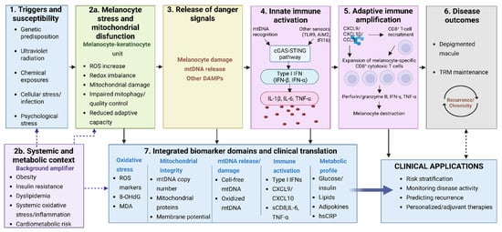
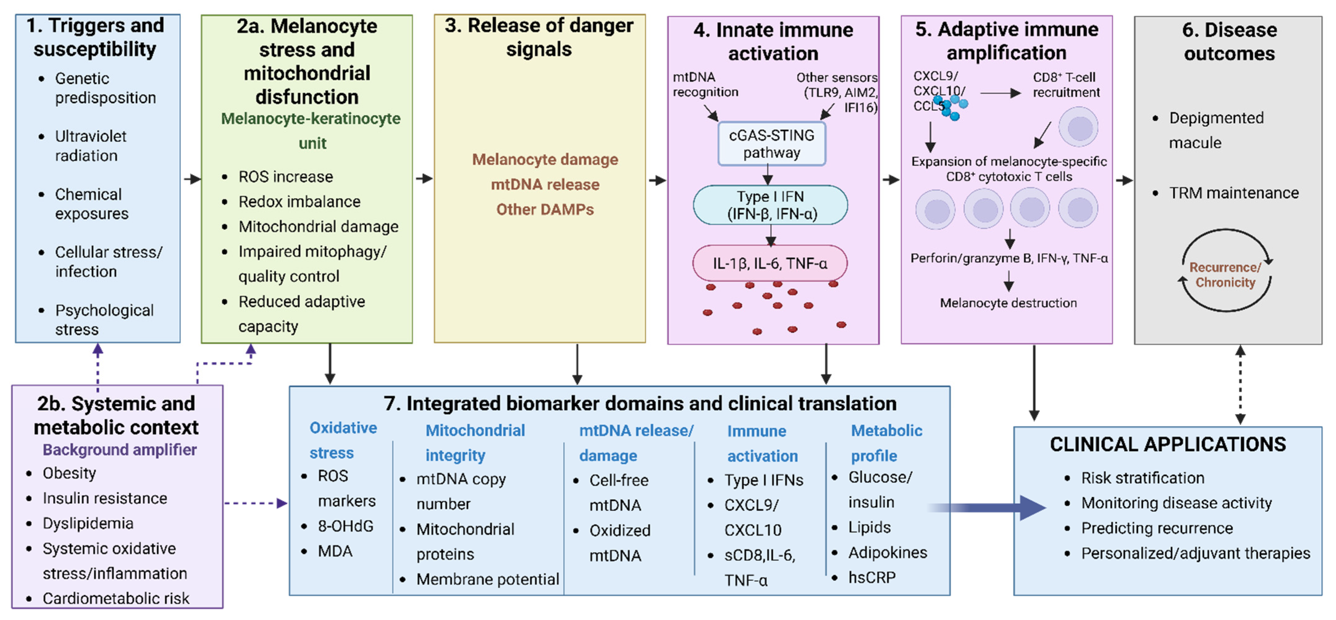
This review proposes a clinical bioenergetics framework for vitiligo based on a convergence hypothesis. In this model, mitochondrial stress, redox imbalance, mtDNA release, innate immune sensing, adaptive cytotoxicity, and systemic metabolic risk are considered interacting processes rather than independent compartments. The framework is intentionally conservative: it does not claim that mitochondrial dysfunction is a universal cause of vitiligo or that bioenergetic biomarkers are ready for routine care. Instead, it organizes emerging evidence into a clinically oriented and testable convergence model linking melanocyte stress, mtDNA-mediated innate sensing, adaptive immune amplification, systemic metabolic context, and candidate biomarker domains (Figure 1).
Figure 1.
Conceptual immune-bioenergetic convergence model for vitiligo. (1) Genetic susceptibility and environmental, cellular, and psychological stressors may promote (2a) redox imbalance, mitochondrial dysfunction, and reduced adaptive capacity in melanocytes. (3) Melanocyte damage may favor the release of mitochondrial DNA (mtDNA) and other damage-associated molecular patterns (DAMPs), which can activate (4) cGAS–STING and other innate nucleic-acid sensors, inducing type I interferons and pro-inflammatory cytokines. These signals may support (5) chemokine-mediated recruitment and expansion of melanocyte-reactive CD8+ cytotoxic T cells, leading to melanocyte destruction. (6) Tissue-resident memory T cells (TRM) may contribute to disease persistence and recurrence, whereas (2b) systemic oxidative, inflammatory, and metabolic abnormalities may act as background amplifiers. The model also identifies (7) candidate oxidative, mitochondrial, mtDNA-related, immune, and metabolic biomarker domains for future patient stratification, disease-activity monitoring, recurrence prediction, and evaluation of therapeutic response. Solid arrows represent the proposed sequence of events, whereas dashed arrows indicate amplifying or feedback relationships. The framework is conceptual and does not imply that all mechanisms occur uniformly in every patient or that the proposed biomarkers are currently validated for routine clinical use. Created with BioRender.com. The cellular compartments represented in the framework should be interpreted separately: mitochondrial/redox stress is centered on melanocytes, mtDNA release may arise from melanocytes or keratinocytes depending on the model, and downstream sensing/effector responses involve innate immune cells and melanocyte-reactive CD8+ T cells.
To support this synthesis, a targeted literature search was conducted in PubMed/MEDLINE, Scopus, and Web of Science for peer-reviewed articles available through July 2026, without a publication-date restriction. Search terms were combined around the concepts of “vitiligo,” “melanocyte,” “mitochondria” or “mitochondrial dysfunction,” “oxidative stress,” “mitochondrial DNA” or “mtDNA,” “cGAS–STING,” “innate immunity,” “biomarkers,” “metabolic syndrome,” and “prediction model.” Priority was given to original human studies, clinical trials, systematic reviews, meta-analyses, and mechanistic experimental studies directly relevant to the proposed immune-bioenergetic framework. Reference lists of selected articles were also screened to identify additional pertinent studies. Evidence from other diseases was considered only when vitiligo-specific data were insufficient to contextualize a mechanism. Because the objective was a focused interpretive synthesis rather than a systematic review, no formal meta-analysis or risk-of-bias assessment was performed.
To our knowledge, this is the first review to integrate mitochondrial stress, mtDNA-mediated innate immune activation, systemic metabolic context, and biomarker validation and clinically relevant prediction into a unified clinical bioenergetics framework for vitiligo.
2. Mitochondrial Stress and Melanocyte Bioenergetic Vulnerability
Mitochondria regulate oxidative phosphorylation, redox balance, calcium handling, apoptosis, innate immune signaling, and cellular adaptation to stress. In melanocytes, these functions are especially relevant because pigmentation is metabolically demanding and chemically redox-active. Human pigmentation evolved in relation to ultraviolet radiation gradients, while melanogenesis requires tyrosinase activity, oxidation reactions, substrate availability, melanosome maturation, and coordinated melanosome transfer to keratinocytes [13,14,15].
This biological context may make melanocytes particularly sensitive to redox disequilibrium. Under physiological conditions, antioxidant enzymes, mitochondrial dynamics, autophagy, mitophagy, endoplasmic reticulum quality control, and keratinocyte–melanocyte communication help maintain homeostasis. In vitiligo-prone melanocytes, this adaptive capacity may be insufficient under repeated ultraviolet exposure, chemical triggers, friction, inflammatory mediators, or systemic metabolic stress [10,11,12].
Oxidative stress may provide an upstream link between melanocyte susceptibility and mitochondrial dysfunction. Excessive generation or insufficient detoxification of reactive oxygen species (ROS), particularly hydrogen peroxide (H2O2), can overwhelm melanocyte antioxidant defenses and promote oxidative modification of lipids, proteins, and nucleic acids. Impaired activation of the nuclear factor erythroid 2-related factor 2 (Nrf2)/antioxidant response element (ARE) pathway has been demonstrated in vitiligo melanocytes exposed to oxidative stress, with reduced Nrf2 nuclear translocation and transcriptional activity and decreased downstream antioxidant responses [16]. Alterations in catalase and other antioxidant defenses may further reduce the capacity to adapt to sustained oxidative challenge [10,11].
Mitochondria may subsequently function as both targets and amplifiers of this redox imbalance. Oxidative injury to mitochondrial membranes and respiratory-chain components can impair membrane potential and oxidative phosphorylation, while dysfunctional electron transport may further increase mitochondrial reactive oxygen species (mtROS) generation. This establishes a potential feed-forward cycle in which oxidative injury promotes mitochondrial dysfunction and mitochondrial dysfunction further increases intracellular oxidative burden [12].
Mitochondrial quality control may determine whether oxidative injury remains reversible or progresses toward persistent organelle dysfunction. Fission and fusion regulate segregation and complementation of damaged mitochondrial components, whereas mitophagy removes mitochondria that cannot be adequately repaired. Sirtuin 3 (SIRT3)-optic atrophy 1 mitochondrial dynamin-like GTPase (OPA1)-dependent remodeling has been implicated in oxidative stress-induced melanocyte degeneration in vitiligo [17], and mitophagy-related signatures have been associated with immune infiltration [18]. PTEN-induced kinase 1 (PINK1)/Parkin RBR E3 ubiquitin protein ligase (PRKN)-dependent mitophagy provides an additional mechanistic framework for selective clearance of depolarized mitochondria; however, direct evidence that defective PINK1/PRKN-Parkin signaling causes cytosolic mtDNA accumulation in human vitiligo remains limited. Failure of mitochondrial quality control should therefore be considered a plausible amplifier requiring direct validation rather than an established universal defect [19].
The evidence should nevertheless be interpreted with caution. Altered mitochondrial morphology, impaired membrane potential, defective oxidative phosphorylation, increased mitochondrial reactive oxygen species, and insufficient antioxidant responses have been reported or proposed in vitiligo-related models, but heterogeneity in sample source, disease stage, and experimental design limits direct clinical extrapolation [10,11,12]. Therefore, mitochondrial stress is best framed as a potential disease-amplifying interface rather than a single initiating defect.
Specific mechanistic studies support this cautious interpretation. Yi et al. showed that SIRT3-dependent mitochondrial dynamics remodeling contributes to oxidative stress-induced melanocyte degeneration, implicating the SIRT3-OPA1 axis in mitochondrial resilience [17]. Bioinformatic evidence has also suggested a relationship between mitophagy-related signatures and immune infiltration in vitiligo, although these findings require experimental and clinical validation [18]. Together, these studies suggest that melanocyte survival may depend not only on antioxidant capacity, but also on mitochondrial turnover, repair, and adaptation.
Current evidence does not establish a uniform temporal sequence. Mitochondrial dysfunction may act as an early stress-related event or participate in reciprocal amplification with inflammatory injury, depending on the experimental and clinical context [10,11,12,17,18].
Persistent redox imbalance may also affect mitochondrial genome stability. Because mtDNA is located close to the respiratory chain and is comparatively exposed to oxidative injury, sustained H2O2 accumulation, lipid peroxidation, and impaired antioxidant adaptation may favor mtDNA damage together with loss of mitochondrial membrane integrity. In vitiligo models, oxidative stress has been linked to cytosolic mtDNA accumulation, and voltage-dependent anion channel 1 (VDAC1)-associated mitochondrial membrane permeabilization has emerged as one route through which mtDNA may escape into the cytosol [20,21]. These observations provide a mechanistic transition from redox and mitochondrial injury to innate nucleic-acid sensing, although the prevalence and temporal importance of this process in patients remain unresolved.
3. mtDNA-Mediated Immune Activation: A Plausible Bridge to Autoimmunity
A central unresolved question is how intracellular stress in the epidermal unit becomes an immune-mediated depigmenting process. mtDNA-mediated immune activation provides a plausible bridge. Because mtDNA retains immunostimulatory features related to its bacterial ancestry, its release into the cytosol or extracellular compartment can function as a danger-associated molecular pattern capable of activating DNA-sensing pathways [22].
Recent studies have strengthened the relevance of this mechanism in vitiligo. Oxidative stress-related mtDNA release has been associated with autoimmune activation in vitiligo and with therapeutic hypotheses aimed at interrupting stress-to-immunity signaling [20]. Additional work has suggested that oxidative stress can promote mtDNA accumulation and release from keratinocytes and vascular endothelial cells, with downstream activation of cGAS-STING signaling in monocytes [23]. A complementary melanocyte-centered study reported that the cytosolic mtDNA-cGAS-STING axis may contribute to melanocyte pyroptosis and CD8+ T-cell activation [24]. More recently, keratinocyte-derived mtDNA release through VDAC1-dependent mechanisms was proposed as- a route by which oxidative stress may propagate innate immune activation in vitiligo models [21].
Importantly, mtDNA-mediated signaling has been investigated in several cellular compartments and these findings should not be conflated. In melanocytes, cytosolic mtDNA-cGAS-STING signaling has been linked to pyroptotic injury and subsequent CD8+ T-cell activation [24]. In keratinocytes, oxidative stress-associated mtDNA release, including VDAC1-dependent mechanisms, has been proposed to propagate innate inflammatory signaling [21]. Other experimental work has reported mtDNA accumulation or release from keratinocytes and vascular endothelial cells with downstream cGAS-STING activation in monocytes [23]. Thus, melanocyte-intrinsic bioenergetic failure, epidermal danger-signal release, and immune-cell sensing represent related but biologically distinct levels of the proposed framework.
Taken together, support for the mtDNA-cGAS-STING axis in vitiligo derives from a combination of patient-derived samples, in vitro systems, and experimental models. However, its prevalence, temporal role, and clinical relevance across patient subgroups remain unresolved [20,21,23,24].
Beyond ATP production and ROS generation, mitochondrial metabolism can shape inflammatory signaling through changes in redox state, metabolite availability, and the release of mitochondrial damage-associated molecular patterns (mtDAMPs). In this framework, mtDNA is one mtDAMP capable of linking organelle injury to innate immune sensing [22]. The relevant immunometabolic effects may differ by compartment: stressed melanocytes and keratinocytes can generate or release danger signals, whereas activated innate and adaptive immune populations undergo their own metabolic remodeling to sustain cytokine production and cytotoxic function [25]. Direct demonstration of a unified immunometabolic program in human vitiligo remains limited; this concept should therefore be tested across defined cell populations and disease stages.
These findings support a sequence that remains biologically plausible but not yet fully established across patient subgroups: mitochondrial injury may increase mtROS and mtDNA release; mtDNA may activate cGAS, promote cyclic GMP-AMP (cGAMP) production, and trigger STING-dependent TANK-binding kinase 1 (TBK1)/interferon regulatory factor 3 (IRF3) and nuclear factor kappa B (NFκ-B) signaling; and interferon-related signaling may then amplify chemokine production, cytotoxic T-cell recruitment, and immune memory. However, the degree to which each component of this sequence has been directly demonstrated in vitiligo-specific human samples remains uneven [20,21,22,23,24]. This model should be presented as a convergence pathway rather than a linear and universal mechanism.
Other danger signals and immune mediators likely operate in parallel. iHSP70 has been implicated as a stress-related signal capable of activating dendritic cells and promoting autoimmune depigmentation in experimental models [7]. CXCL10 contributes to progression and maintenance of depigmentation in experimental vitiligo models [8], while IL-15-related pathways support tissue-resident memory T-cell persistence and recurrence [9]. Thus, mtDNA signaling should be integrated into a broader immune-bioenergetic network rather than treated as an isolated pathway. Representative experimental and clinical studies supporting the mitochondrial, redox, immune, biomarker, and systemic metabolic components of the proposed framework are summarized in Table 1.
Table 1.
Representative experimental and clinical evidence supporting the immune-bioenergetic framework in vitiligo.
4. Candidate Biomarkers for Immune-Bioenergetic Phenotyping
A clinically useful bioenergetic framework requires measurable variables, but current biomarkers should be described as candidates rather than validated diagnostic or prognostic tools. The main translational goal is not to replace clinical assessment, but to identify biological domains that could eventually complement disease activity scoring, recurrence evaluation, treatment-response monitoring, and metabolic surveillance.
At present, most candidate biomarkers have mechanistic plausibility of cross-sectional associations, whereas evidence of incremental prognostic or treatment-monitoring value beyond established clinical assessment remains limited.
For clinical translation, candidate biomarkers should be separated according to intended use. Most oxidative, mitochondrial, and mtDNA-related variables discussed here remain mechanistic or exploratory markers. By contrast, a smaller body of human evidence has directly examined treatment-response associations. Serum and lesional CXCL10 have been evaluated longitudinally in relation to psoralen plus ultraviolet A (PUVA), narrowband ultraviolet B (NB-UVB), and systemic corticosteroid response, and baseline lesional transcriptomic profiles have identified immune-active signatures associated with subsequent response to NB-UVB plus tacrolimus [26,28]. These findings are promising but require independent prospective validation, standardized thresholds, and testing against established clinical predictors. At present, no immune-bioenergetic biomarker panel is validated to predict long-term treatment benefit at therapy initiation.
Oxidative damage markers such as malondialdehyde, lipid peroxidation products, protein carbonyls, total antioxidant capacity, catalase, superoxide dismutase, and glutathione-related measures may help estimate redox burden, although they are sensitive to sample handling, tissue source, diet, medication use, and assay variability [10,11,29,30]. These limitations are especially important because they prevent overstatement of immediate clinical utility.
Mitochondrial biomarkers are particularly attractive but methodologically complex. mtDNA copy number, deletion burden, heteroplasmy, selected point variants, haplogroup background, cytosolic mtDNA, and circulating cell-free mtDNA could reflect inherited background, somatic variation, tissue-specific injury, age-related accumulation, or inflammatory context. For this reason, mtDNA alterations should be interpreted as context-dependent markers of mitochondrial stress or immune activation rather than as stand-alone vitiligo biomarkers [20,21,23,24].
The validation strategy should be proportional to the question. Sanger sequencing may confirm selected variants when the allele fraction is high enough, but it is not adequate for low-level heteroplasmy or quantitative assessment of mtDNA burden [31]. High-depth next-generation sequencing, digital PCR, or targeted quantitative assays may be more appropriate depending on whether the objective is variant confirmation, copy-number estimation, heteroplasmy detection, or measurement of cytosolic or circulating mtDNA [31,32,33,34]. These methods also require rigorous pre-analytical standardization and orthogonal validation [31,34].
A cautious immune-bioenergetic phenotype could integrate three layers of information: melanocyte or epidermal stress, immune activation, and systemic metabolic context. In practical terms, candidate variables may include oxidative stress markers, mitochondrial integrity markers, mtDNA release measures, IFN-γ/CXCL9/CXCL10/CXCL11-related signatures, IL-15-related markers, fasting glucose, HbA1c, insulin, homeostatic model assessment of insulin resistance (HOMA-IR), triglycerides, HDL-C, waist circumference, blood pressure, disease duration, disease activity, Vitiligo Area Scoring Index (VASI), Vitiligo Disease Activity (VIDA), autoimmune comorbidities, and treatment exposure [35,36,37,38,39,40,41]. These candidate domains, their potential research applications, and their main methodological limitations are summarized in Table 2.
Table 2.
Candidate domains for immune-bioenergetic phenotyping in vitiligo. The selection of candidate variables was based on current evidence regarding oxidative stress, mitochondrial dysfunction, mtDNA alterations, immune activation, and metabolic comorbidities in vitiligo [4,10,18,23,40,41].
Rather than defining fixed or clinically validated endotypes, this framework is intended to support research-oriented profiles with predominantly redox, mitochondrial, immune, metabolic, or mixed features. These profiles should be evaluated against established clinical measures before any added value for classification, prognosis, recurrence prediction, or treatment monitoring is assumed.
5. Systemic Metabolic Context
Although vitiligo is expressed clinically in the skin, several studies have explored associations with systemic comorbidities, including thyroid disease, insulin resistance, dyslipidemia, obesity, and metabolic syndrome. These associations are not uniform across all cohorts, and they should not be interpreted as evidence that vitiligo is primarily a metabolic disease. A more balanced interpretation is that selected patients may share oxidative, inflammatory, or metabolic pathways that intersect with cutaneous disease biology [35,36].
A systematic review and meta-analysis estimated a pooled metabolic syndrome prevalence of approximately 29.6% among patients with vitiligo, but the association with controls was not uniformly significant across all analyses [35]. A more recent systematic review similarly emphasized that the relationship may vary by disease type, severity, duration, and population characteristics [36]. Therefore, metabolic surveillance is best framed as a reasonable consideration for selected subgroups rather than a universal conclusion.
Metabolic abnormalities reported in vitiligo include changes in triglycerides, cholesterol fractions, blood pressure, glucose-related indices, C-peptide, vitamin D, folate, and biological antioxidant capacity [37,38]. Insulin resistance has also been reported in some cohorts [39]. These observations support the inclusion of metabolic indicators in future stratification studies, especially in patients with active, extensive, recurrent, long-duration disease or additional cardiometabolic risk factors.
Evidence regarding glucose homeostasis deserves particular consideration. A 2026 systematic review and narrative synthesis including 33 studies and 4286 patients reported substantial heterogeneity but found metabolic syndrome in 34.3% of patients with vitiligo versus 18.6% of controls across comparative studies; insulin resistance defined by study-specific HOMA-IR thresholds was reported in 56.5% of patients versus 32.9% of controls in the relevant subsets. HOMA-IR thresholds varied considerably, and most studies were cross-sectional or case–control in design [27]. These data support an association with metabolic risk in some populations but do not establish causality or justify assuming insulin resistance in an individual patient with vitiligo.
From a clinical bioenergetics perspective, impaired glucose homeostasis, dyslipidemia, chronic oxidative stress, and low-grade inflammation may converge on mitochondrial substrate utilization, redox balance, and cellular metabolic adaptation. These interconnected processes could potentially influence melanocyte vulnerability and immune activation, although their contribution to disease activity, prognosis, and therapeutic response in vitiligo remains insufficiently established. Future prospective studies should therefore investigate whether systemic metabolic alterations identify biologically distinct patient subgroups by integrating metabolic parameters with oxidative, mitochondrial, and immune biomarkers, alongside standardized clinical measures such as the Vitiligo Area Scoring Index (VASI) and Vitiligo Disease Activity (VIDA) scores. Such investigations should account for relevant confounding factors, including age, sex, body mass index, medication use, lifestyle, population background, and pre-existing metabolic comorbidities, to determine whether these associations provide clinically meaningful information beyond established risk factors.
6. Biomarker Validation and Clinically Relevant Prediction
The clinical translation of immune-bioenergetic biomarkers in vitiligo requires a clear distinction between prognostic, treatment-predictive, and treatment-monitoring applications. Prognostic biomarkers provide information about the expected course of disease, whereas treatment-predictive biomarkers identify differences in the expected benefit of one therapeutic intervention compared with another. Monitoring biomarkers, in contrast, reflect biological or clinical changes during treatment. This distinction is particularly relevant because associations between biomarker concentrations and therapeutic outcomes do not necessarily demonstrate the ability to predict differential treatment benefits.
Among the candidate biomarkers investigated in vitiligo, CXCL10 has emerged as a potentially relevant indicator of immune activity and therapeutic response. El-Domyati et al. [26] evaluated CXCL10 concentrations in lesional, perilesional, and unaffected skin from patients with vitiligo (n = 30), as well as circulating concentrations in serum samples (n = 51). The investigators reported that elevated lesional CXCL10 concentrations (≥2 pg/mm2) were associated with increased leukocyte infiltration, shorter disease duration (<2 years), and higher circulating CXCL10 concentrations (≥50 pg/mL). Furthermore, longitudinal changes in circulating CXCL10 during treatment with psoralen plus ultraviolet A (PUVA), narrowband ultraviolet B (NB-UVB), or systemic corticosteroids were associated with changes in lesional CXCL10 in repigmented skin. These findings support the potential value of CXCL10 as a treatment-associated and monitoring biomarker. However, further validation is required to determine whether baseline CXCL10 concentrations can reliably predict long-term treatment benefit or guide selection among alternative therapeutic interventions.
Additional evidence supporting pretreatment biomarker assessment was provided by Yang et al. [28], who investigated baseline skin transcriptomic profiles in 36 patients with vitiligo, including six with segmental and 30 with non-segmental disease. Patients subsequently received combined NB-UVB phototherapy and topical tacrolimus for six months. Favorable treatment response was defined as greater than 10% repigmentation of the target lesion. Responsive lesions were associated with significantly shorter disease duration than non-responsive lesions (2.5 versus 11.5 years; p = 0.046). Transcriptomic analysis identified a panel of 68 genes distinguishing favorable and unfavorable response groups, including immune-related genes such as CXCL10, FCRL3, and T-cell receptor-associated transcripts. These findings suggest that pretreatment immune activity and disease duration may contribute to treatment-response stratification. Nevertheless, the relatively small cohort and the absence of independent external validation limit the immediate clinical applicability of these candidate signatures.
Together, these studies illustrate two complementary approaches to biomarker development: longitudinal monitoring of circulating immune mediators and baseline molecular characterization of lesional tissue. However, neither approach currently establishes a validated immune-bioenergetic panel capable of reliably predicting sustained repigmentation or long-term recurrence at treatment initiation. Moreover, treatment-associated biomarker changes should not be interpreted as evidence that the biomarker itself mediates therapeutic efficacy.
From a clinical bioenergetics perspective, future studies could investigate whether combining established immune-related candidates with selected oxidative, mitochondrial, and metabolic variables improves treatment-response assessment. Potential exploratory measurements may include circulating CXCL10, oxidative damage markers, antioxidant defense indicators, circulating cell-free mitochondrial DNA, and selected metabolic parameters. The inclusion of these variables should be guided by biological plausibility, analytical feasibility, and preliminary evidence rather than the indiscriminate construction of extensive biomarker panels. Importantly, the clinical predictive value of such combinations remains hypothetical and requires direct investigation.
A prospective validation framework could involve patients with non-segmental vitiligo undergoing predefined therapeutic interventions, with biomarker measurements obtained before treatment initiation and, where appropriate, during follow-up. Clinical outcomes could be assessed at prespecified intervals, such as 3, 6, and 12 months, using standardized repigmentation measures, including the Vitiligo Area Scoring Index (VASI). Longer follow-up would be required to investigate durability of response and recurrence. Baseline biomarker measurements would support pretreatment prediction, whereas longitudinal changes could be evaluated as potential monitoring indicators. These proposed time points and biomarker combinations represent a research strategy rather than an established or validated clinical protocol.
An important methodological consideration is the distinction between predicting outcomes under a particular treatment and identifying differential benefit between treatments. For example, a biomarker associated with favorable repigmentation following NB-UVB does not necessarily demonstrate that patients with elevated biomarker concentrations benefit more from NB-UVB than from another intervention. Establishing treatment-predictive utility would require comparative treatment data and appropriate assessment of biomarker-by-treatment interactions, ideally in randomized clinical trials or rigorously designed comparative prospective studies.
The incremental clinical value of candidate biomarkers should also be evaluated against prediction models based on established clinical characteristics, including disease duration, anatomical distribution, baseline activity, and treatment history. Model development and validation should address discrimination, calibration, reproducibility, external validation, and clinical utility. The TRIPOD+AI statement provides recommendations for transparent reporting of clinical prediction models, whereas PROBAST supports the assessment of risk of bias and applicability in prediction-model studies [42,43]. These methodological frameworks may improve the quality of future investigations but do not replace prospective clinical validation.
Ultimately, immune-bioenergetic phenotyping should aim to identify reproducible biological information that meaningfully improves clinical decision-making rather than simply increasing the number of measured biomarkers. Although existing studies support the investigation of CXCL10 and baseline immune-related transcriptomic signatures [26,28], the integration of oxidative, mitochondrial, mtDNA-related, and metabolic variables into treatment-predictive models remains an investigational objective. Until prospective studies establish their incremental value and clinical utility, such biomarkers should be considered research candidates rather than validated tools for individualized treatment selection.
7. Therapeutic Implications: Adjunctive Hypotheses Within Established Care
Current vitiligo treatment includes phototherapy, topical corticosteroids, calcineurin inhibitors, surgical approaches in selected stable disease, depigmentation strategies in extensive cases, and immune-targeted therapies. The success of JAK inhibition has reinforced the centrality of IFN-related immune pathways in vitiligo [6]. A bioenergetic framework should not be used to replace these established therapeutic concepts, but it may help refine adjunctive hypotheses [6,20,21,23,24,44].
Within this framework, established immune-directed treatments remain the clinical reference, whereas cGAS–STING modulation and mitochondria-targeted interventions should currently be considered investigational adjunctive hypotheses. Their future evaluation should link a defined biological alteration, a measurable biomarker, a selected patient subgroup, and a clinically relevant outcome.
The cGAS-STING axis also illustrates the distinction between mechanistic plausibility and clinical validation. Experimental vitiligo studies support interruption of stress-to-innate-immunity signaling at different levels, including reduction in oxidative injury and VDAC1-associated mtDNA release [20,21]. Small-molecule STING inhibitors, including carbazole-based compounds, have also shown preclinical potential for modulating cGAS-STING-driven inflammatory responses [45]. Direct pharmacological STING inhibition has progressed into early human development outside vitiligo: ASP5502 entered a Phase I study in healthy adults and adults with primary Sjogren’s syndrome [46]. This establishes clinical-development feasibility for pathway inhibition but does not provide evidence of efficacy or safety in vitiligo. The Phase I study (NCT06544642) was subsequently terminated prior to enrolment into Part 3 [46]. Any future evaluation in vitiligo would require biomarker-supported patient selection, demonstration of pathway activation in the relevant tissue compartment, and careful assessment of the consequences of suppressing an innate immune pathway.
If mitochondrial stress and redox disequilibrium contribute to melanocyte vulnerability in some patients, adjunctive strategies aimed at improving stress tolerance may deserve further study. Antioxidant approaches such as vitamin E, Ginkgo biloba, Polypodium leucotomos, and other redox-directed interventions have been explored with variable evidence, particularly as adjuncts to phototherapy [29,30]. Their heterogeneity illustrates why patient stratification and biomarker-guided trial design may be more informative than generalized supplementation claims.
Mitochondria-targeted interventions remain even more preliminary. Mitochondria-targeted ubiquinone (MitoQ)-related strategies have shown mechanistic promise in experimental vitiligo-related work, including effects on oxidative stress, mitochondrial membrane potential, ATP synthesis, and PARP1-related pathways [44]. However, such findings should be described as investigational until supported by controlled clinical trials in vitiligo. The clinically responsible position is that bioenergetic support may be a future adjunctive direction, not a current stand-alone therapeutic recommendation.
8. Limitations and Future Directions
Several limitations should be considered when interpreting the proposed immune-bioenergetic framework. First, although experimental and translational studies support interactions among oxidative stress, mitochondrial dysfunction, mtDNA release, and immune activation, the temporal and causal relationships between these processes remain incompletely established in human vitiligo. Much of the available evidence comes from cross-sectional observations, in vitro experiments, or experimental models that may not fully capture the disease’s biological heterogeneity. Consequently, the proposed convergence framework should be regarded as a biologically plausible and testable model rather than a universally established pathogenic sequence.
Second, the cellular origin and relative contribution of mitochondrial danger signals remain insufficiently characterized. Evidence implicates melanocytes, keratinocytes, and other cellular compartments in mtDNA release and innate immune activation, but it remains unclear whether these mechanisms operate simultaneously or independently across disease stages [20,21,23,24]. Future mechanistic investigations should therefore prioritize cell-type-specific analyses and the characterization of mitochondrial stress, mtDNA localization, and downstream immune signaling within defined epidermal and immune-cell populations. Such approaches may help distinguish initiating events from secondary inflammatory amplification.
Third, the interpretation of mitochondrial and oxidative biomarkers presents substantial analytical challenges. Measurements of mitochondrial DNA copy number, heteroplasmy, circulating cell-free mtDNA, and oxidative damage indicators may be influenced by tissue source, sample processing, assay sensitivity, age, inflammation, and other biological variables. These factors complicate comparisons across studies and limit the interpretation of individual measurements as indicators of vitiligo-specific pathology. Establishing analytical reproducibility and determining the biological significance of these variables in relevant tissue compartments remain essential prerequisites for their clinical investigation.
Fourth, the relationship between vitiligo and systemic metabolic abnormalities remains heterogeneous. Although associations with insulin resistance, dyslipidemia, and metabolic syndrome have been reported, differences in population characteristics, disease activity, treatment exposure, and cardiometabolic comorbidities limit causal interpretation [27,35,36,37,38,39]. Future investigations should determine whether these abnormalities represent shared inflammatory and oxidative processes, independent comorbid conditions, or potential modifiers of disease progression in selected patient subgroups.
Beyond these limitations, a major research priority is to determine whether the proposed immune-bioenergetic pathways define biologically meaningful differences among patients with vitiligo. Integrative investigations combining spatially resolved tissue analyses, mitochondrial functional assessments, and immune profiling may help clarify how melanocyte vulnerability, epidermal danger signaling, and adaptive immune responses interact during disease initiation, progression, and recurrence. Particular attention should be given to distinguishing mechanisms directly demonstrated in patient-derived material from those inferred from experimental models.
Ultimately, the translational relevance of the framework will depend on whether these mechanistic relationships can be linked to reproducible clinical phenotypes and meaningful disease outcomes. Rather than assuming a uniform mitochondrial contribution across all patients, future research should establish the conditions under which mitochondrial and metabolic disturbances contribute to disease amplification and whether these processes identify subgroups that may benefit from targeted investigation. These priorities complement the biomarker-validation strategies discussed in Section 6 and provide a foundation for evaluating the clinical relevance of the proposed convergence model.
9. Conclusions
Vitiligo is best understood as a multifactorial disorder in which immune mechanisms interact with melanocyte-intrinsic vulnerability, oxidative stress, mitochondrial dysfunction, mtDNA-mediated innate immune activation, and systemic metabolic context. Clinical bioenergetics offers a useful translational lens because it connects cellular stress biology with immune signaling and systemic risk without reducing vitiligo to a single pathway.
The principal contribution of this review is the immune-bioenergetic convergence framework. Mitochondrial dysfunction is not presented as a universal cause of vitiligo, but as a potential disease-amplifying interface that may help explain how oxidative stress, mtDNA release, innate immune sensing, adaptive cytotoxicity, recurrence, and selected metabolic abnormalities intersect. This framework may guide future biomarker validation, patient stratification, and adjunctive therapeutic trials, provided that claims remain proportional to the available evidence. At present, it should therefore be interpreted as a hypothesis-generating and research-organizing framework rather than as a validated clinical classification or decision-making system.
The immediate research priorities are: (i) standardized measurement of mitochondrial and circulating mtDNA variables; (ii) longitudinal integration of these variables with validated vitiligo activity and recurrence outcomes; and (iii) external validation of parsimonious models that distinguish cutaneous disease activity from systemic metabolic risk.
Author Contributions
Conceptualization, D.S.B.-L. and J.A.A.-V.; methodology, D.S.B.-L., A.L.-Q., and I.A.G.-H.; investigation, D.S.B.-L., A.L.-Q., I.A.G.-H., and J.A.A.-V.; writing—original draft preparation, D.S.B.-L. and J.A.A.-V.; writing—review and editing, D.S.B.-L. and J.A.A.-V.; supervision, J.A.A.-V.; project administration, J.A.A.-V.; funding acquisition, J.A.A.-V. All authors have read and agreed to the published version of the manuscript.
Funding
This work was supported by the Secretaría de Ciencia, Humanidades, Tecnología e Innovación (SECIHTI), Mexico, through the Ciencia de Frontera project CF-2023-G-536 to J.A.A.-V. and a 2025 postdoctoral fellowship awarded to D.S.B.-L. (CVU 702574). The funder had no role in the conceptualization, preparation, or decision to submit the manuscript.
Institutional Review Board Statement
Not applicable.
Informed Consent Statement
Not applicable.
Data Availability Statement
No new data were created or analyzed in this study. Data sharing is not applicable to this article.
Acknowledgments
The authors thank SECIHTI for supporting the postdoctoral fellowship and the Ciencia de Frontera project that made this work possible.
Conflicts of Interest
The authors declare no conflicts of interest.
Abbreviations
The following abbreviations are used in this manuscript:
| ARE | Antioxidant response element |
| ATP | Adenosine triphosphate |
| BMI | Body mass index |
| CD8+ | Cluster of differentiation 8-positive |
| cfDNA | Cell-free DNA |
| cGAMP | Cyclic GMP-AMP |
| cGAS-STING | Cyclic guanosine monophosphate-adenosine monophosphate synthase-stimulator of interferon genes |
| CXCL10 | C-X-C motif chemokine ligand 10 |
| CXCL11 | C-X-C motif chemokine ligand 11 |
| CXCL9 | C-X-C motif chemokine ligand 9 |
| CXCR3 | C-X-C motif chemokine receptor 3 |
| DAMPs | Damage-associated molecular patterns |
| DNA | Deoxyribonucleic acid |
| DRP1 | Dynamin-related protein 1 |
| HbA1c | Glycated hemoglobin A1c |
| HDL-C | High-density lipoprotein cholesterol |
| HOMA-IR | Homeostatic Model Assessment of Insulin Resistance |
| iHSP70 | Inducible heat shock protein 70 |
| IFN-γ | Interferon gamma |
| IL-15 | Interleukin 15 |
| IRF3 | Interferon regulatory factor 3 |
| JAK/STAT | Janus kinase/signal transducer and activator of transcription |
| MFN1/2 | Mitofusin 1 and mitofusin 2 |
| MitoQ | Mitochondria-targeted ubiquinone |
| mtDAMPs | Mitochondrial damage-associated molecular patterns |
| mtDNA | Mitochondrial DNA |
| mtROS | Mitochondrial reactive oxygen species |
| NB-UVB | Narrowband ultraviolet B |
| NF-κB | Nuclear factor kappa B |
| Nrf2 | Nuclear factor erythroid 2-related factor 2 |
| OPA1 | Optic atrophy 1 mitochondrial dynamin-like GTPase |
| PARP1 | Poly(ADP-ribose) polymerase 1 |
| PCR | Polymerase Chain Reaction |
| PINK1 | PTEN-induced kinase 1 |
| PRKN/Parkin | Parkin RBR E3 ubiquitin protein ligase |
| PROBAST | Prediction Model Risk of Bias Assessment Tool |
| PUVA | Psoralen plus ultraviolet A |
| ROS | Reactive Oxygen Species |
| SIRT3 | Sirtuin 3 |
| TBK1 | TANK-binding kinase 1 |
| TRIPOD+AI | Transparent Reporting of a Multivariable Prediction Model for Individual Prognosis or Diagnosis plus Artificial Intelligence |
| TRM | Tissue-resident memory T cells |
| VASI | Vitiligo Area Scoring Index |
| VDAC1 | Voltage-dependent anion channel 1 |
| VIDA | Vitiligo Disease Activity |
References
- Akl, J.; Lee, S.; Ju, H.J.; Parisi, R.; Kim, J.Y.; Jeon, J.J.; Heo, Y.-W.; Eleftheriadou, V.; Hamzavi, I.; Griffiths, C.E.M.; et al. Estimating the burden of vitiligo: A systematic review and modelling study. Lancet Public Health 2024, 9, e386–e396. [Google Scholar] [CrossRef] [Scilit] [PubMed]
- Picardo, M.; Dell’ANna, M.L.; Ezzedine, K.; Hamzavi, I.; Harris, J.E.; Parsad, D.; Taieb, A. Vitiligo. Nat. Rev. Dis. Prim. 2015, 1, 15011. [Google Scholar] [CrossRef] [Scilit] [PubMed]
- Ezzedine, K.; Eleftheriadou, V.; Whitton, M.; van Geel, N. Vitiligo. Lancet 2015, 386, 74–84. [Google Scholar] [CrossRef] [Scilit] [PubMed]
- Frisoli, M.L.; Essien, K.; Harris, J.E. Vitiligo: Mechanisms of pathogenesis and treatment. Annu. Rev. Immunol. 2020, 38, 621–648. [Google Scholar] [CrossRef] [Scilit] [PubMed]
- Seneschal, J.; Boniface, K.; D’arino, A.; Picardo, M. An update on vitiligo pathogenesis. Pigment Cell Melanoma Res. 2021, 34, 236–243. [Google Scholar] [CrossRef] [Scilit] [PubMed]
- Rosmarin, D.; Passeron, T.; Pandya, A.G.; Grimes, P.; Harris, J.E.; Desai, S.R.; Lebwohl, M.; Ruer-Mulard, M.; Seneschal, J.; Wolkerstorfer, A.; et al. Two phase 3, randomized, controlled trials of ruxolitinib cream for vitiligo. N. Engl. J. Med. 2022, 387, 1445–1455. [Google Scholar] [CrossRef] [Scilit] [PubMed]
- Mosenson, J.A.; Zloza, A.; Nieland, J.D.; Garrett-Mayer, E.; Eby, J.M.; Huelsmann, E.J.; Kumar, P.; Denman, C.J.; Lacek, A.T.; Kohlhapp, F.J.; et al. Mutant HSP70 reverses autoimmune depigmentation in vitiligo. Sci. Transl. Med. 2013, 5, 174ra28. [Google Scholar] [CrossRef] [Scilit] [PubMed]
- Rashighi, M.; Agarwal, P.; Richmond, J.M.; Harris, T.H.; Dresser, K.; Su, M.-W.; Zhou, Y.; Deng, A.; Hunter, C.A.; Luster, A.D.; et al. CXCL10 Is Critical for the progression and maintenance of depigmentation in a mouse model of vitiligo. Sci. Transl. Med. 2014, 6, 223ra23. [Google Scholar] [CrossRef] [Scilit] [PubMed]
- Richmond, J.M.; Strassner, J.P.; Zapata, L., Jr.; Garg, M.; Riding, R.L.; Refat, M.A.; Fan, X.; Azzolino, V.; Tovar-Garza, A.; Tsurushita, N.; et al. Antibody blockade of IL-15 signaling has the potential to durably reverse vitiligo. Sci. Transl. Med. 2018, 10, eaam7710. [Google Scholar] [CrossRef] [Scilit] [PubMed]
- Xuan, Y.; Yang, Y.; Xiang, L.; Zhang, C. The role of oxidative stress in the pathogenesis of vitiligo: A culprit for melanocyte death. Oxidative Med. Cell. Longev. 2022, 2022, 8498472. [Google Scholar] [CrossRef] [Scilit] [PubMed]
- Chang, W.-L.; Ko, C.-H. The role of oxidative stress in vitiligo: An update on its pathogenesis and therapeutic implications. Cells 2023, 12, 936. [Google Scholar] [CrossRef] [Scilit] [PubMed]
- Lin, Y.; Ding, Y.; Wu, Y.; Yang, Y.; Liu, Z.; Xiang, L.; Zhang, C. The underestimated role of mitochondria in vitiligo: From oxidative stress to inflammation and cell death. Exp. Dermatol. 2024, 33, e14856. [Google Scholar] [CrossRef] [Scilit] [PubMed]
- Jablonski, N.G.; Chaplin, G. Human skin pigmentation as an adaptation to UV radiation. Proc. Natl. Acad. Sci. USA 2010, 107, 8962–8968. [Google Scholar] [CrossRef] [Scilit] [PubMed]
- Denat, L.; Kadekaro, A.L.; Marrot, L.; Leachman, S.A.; Abdel-Malek, Z.A. Melanocytes as instigators and victims of oxidative stress. J. Investig. Dermatol. 2014, 134, 1512–1518. [Google Scholar] [CrossRef] [Scilit] [PubMed]
- D’ISchia, M.; Wakamatsu, K.; Cicoira, F.; Di Mauro, E.; Garcia-Borron, J.C.; Commo, S.; Galván, I.; Ghanem, G.; Kenzo, K.; Meredith, P.; et al. Melanins and melanogenesis: From pigment cells to human health and technological applications. Pigment Cell Melanoma Res. 2015, 28, 520–544. [Google Scholar] [CrossRef] [Scilit] [PubMed]
- Jian, Z.; Li, K.; Song, P.; Zhu, G.; Zhu, L.; Cui, T.; Liu, B.; Tang, L.; Wang, X.; Wang, G.; et al. Impaired activation of the Nrf2-ARE signaling pathway undermines H2O2-induced oxidative stress response: A possible mechanism for melanocyte degeneration in vitiligo. J. Investig. Dermatol. 2014, 134, 2221–2230. [Google Scholar] [CrossRef] [Scilit] [PubMed]
- Yi, X.; Guo, W.; Shi, Q.; Yang, Y.; Zhang, W.; Chen, X.; Kang, P.; Chen, J.; Cui, T.; Ma, J.; et al. SIRT3-dependent mitochondrial dynamics remodeling contributes to oxidative stress-induced melanocyte degeneration in vitiligo. Theranostics 2019, 9, 1614–1633. [Google Scholar] [CrossRef] [Scilit] [PubMed]
- Luo, L.; Zhu, J.; Guo, Y.; Li, C. Mitophagy and immune infiltration in vitiligo: Evidence from bioinformatics analysis. Front. Immunol. 2023, 14, 1164124. [Google Scholar] [CrossRef] [Scilit] [PubMed]
- Harper, J.W.; Ordureau, A.; Heo, J.-M. Building and decoding ubiquitin chains for mitophagy. Nat. Rev. Mol. Cell Biol. 2018, 19, 93–108. [Google Scholar] [CrossRef] [Scilit] [PubMed]
- Sant’ANna-Silva, A.C.B.; Botton, T.; Rossi, A.; Dobner, J.; Bzioueche, H.; Thach, N.; Blot, L.; Pagnotta, S.; Kleszczynski, K.; Steinbrink, K.; et al. Vitiligo auto-immune response upon oxidative stress-related mitochondrial DNA release opens up new therapeutic strategies. Clin. Transl. Med. 2024, 14, e1810. [Google Scholar] [CrossRef] [Scilit] [PubMed]
- Lv, J.; Xu, W.; Jiang, P.; Yu, W.; Xue, H.; Hu, N.; Cao, Y.; Zhang, H.; Yin, C.; Gao, R. Mitochondrial DNA release via VDAC1 in keratinocytes: A key driver of innate immunity and vitiligo pathogenesis. Cell Death Dis. 2026, 17, 318. [Google Scholar] [CrossRef] [Scilit] [PubMed]
- West, A.P.; Shadel, G.S. Mitochondrial DNA in innate immune responses and inflammatory pathology. Nat. Rev. Immunol. 2017, 17, 363–375. [Google Scholar] [CrossRef] [Scilit] [PubMed]
- Wu, T.; Chen, X.; Fan, J.; Ye, P.; Zhang, J.; Wang, Z.; Zhou, Y.; Wang, B.; Jin, X.; Xiong, S.; et al. Oxidative stress-induced release of mitochondrial DNA (mtDNA) promotes the progression of vitiligo by activating the cGAS-STING signaling pathway in monocytes. Free Radic. Biol. Med. 2025, 235, 43–55. [Google Scholar] [CrossRef] [Scilit] [PubMed]
- Xu, X.; Lu, X.; Zheng, Y.; Xie, Y.; Lai, W. Cytosolic mtDNA-cGAS-STING axis mediates melanocytes pyroptosis to promote CD8+ T-cell activation in vitiligo. J. Dermatol. Sci. 2025, 117, 61–70. [Google Scholar] [CrossRef] [Scilit] [PubMed]
- O’Neill, L.A.; Kishton, R.J.; Rathmell, J. A guide to immunometabolism for immunologists. Nat. Rev. Immunol. 2016, 16, 553–565. [Google Scholar] [CrossRef] [Scilit] [PubMed]
- El-Domyati, M.; El-Din, W.H.; Rezk, A.F.; Chervoneva, I.; Lee, J.B.; Farber, M.; Uitto, J.; Igoucheva, O.; Alexeev, V. Systemic CXCL10 is a predictive biomarker of vitiligo lesional skin infiltration, PUVA, NB-UVB and corticosteroid treatment response and outcome. Arch. Dermatol. Res. 2022, 314, 275–284. [Google Scholar] [CrossRef] [Scilit] [PubMed]
- Kamińska, B.; Białczyk, A.; Czajkowski, R. Insulin resistance and metabolic syndrome in patients with vitiligo: A systematic review and narrative synthesis. Arch. Dermatol. Res. 2026, 318, 455. [Google Scholar] [CrossRef] [Scilit]
- Yang, Q.; Zhang, G.; Su, M.; Leung, G.; Lui, H.; Zhou, P.; Wu, Y.; Zhou, J.; Xu, J.; Zhang, X.; et al. Vitiligo Skin Biomarkers Associated with Favorable Therapeutic Response. Front. Immunol. 2021, 12, 613031. [Google Scholar] [CrossRef] [Scilit] [PubMed]
- Białczyk, A.; Wełniak, A.; Kamińska, B.; Czajkowski, R. Oxidative stress and potential antioxidant therapies in vitiligo: A narrative review. Mol. Diagn. Ther. 2023, 27, 723–739. [Google Scholar] [CrossRef] [Scilit] [PubMed]
- Aprile, N.; Scano, S.; Bellei, B.; Marini, A.; Filoni, A. Targeting oxidative stress to treat vitiligo: Clinical and molecular evidence. Biomolecules 2026, 16, 612. [Google Scholar] [CrossRef] [Scilit] [PubMed]
- Kloss-Brandstätter, A.; Weissensteiner, H.; Erhart, G.; Schäfer, G.; Forer, L.; Schönherr, S.; Pacher, D.; Seifarth, C.; Stöckl, A.; Fendt, L.; et al. Validation of next-generation sequencing of entire mitochondrial genomes and the diversity of mitochondrial DNA mutations in oral squamous cell carcinoma. PLoS ONE 2015, 10, e0135643. [Google Scholar] [CrossRef] [Scilit] [PubMed]
- Shoop, W.K.; Gorsuch, C.L.; Bacman, S.R.; Moraes, C.T. Precise and simultaneous quantification of mitochondrial DNA heteroplasmy and copy number by digital PCR. J. Biol. Chem. 2022, 298, 102574. [Google Scholar] [CrossRef] [Scilit] [PubMed]
- Bronner, D.; O’rIordan, M. Measurement of mitochondrial DNA release in response to ER stress. Bio-Protocol 2016, 6, e1839. [Google Scholar] [CrossRef] [Scilit] [PubMed]
- Chiu, R.W.; Chan, L.Y.; Lam, N.Y.; Tsui, N.B.; Ng, E.K.O.; Rainer, T.H.; Lo, Y.D. Quantitative analysis of circulating mitochondrial DNA in plasma. Clin. Chem. 2003, 49, 719–726. [Google Scholar] [CrossRef] [Scilit] [PubMed]
- Xia, J.; Melian, C.; Guo, W.; Usmani, H.; Clark, R.; Lozeau, D. Vitiligo and metabolic syndrome: Systematic review and meta-analysis. JMIR Dermatol. 2022, 5, e34772. [Google Scholar] [CrossRef] [Scilit] [PubMed]
- Białczyk, A.; Kamińska, B.; Czajkowski, R. The association between metabolic syndrome and vitiligo: A systematic review. Adv. Dermatol. Allergol. 2025, 42, 134–142. [Google Scholar] [CrossRef] [Scilit] [PubMed]
- Papaccio, F.; Ottaviani, M.; Truglio, M.; D’arino, A.; Caputo, S.; Pacifico, A.; Iacovelli, P.; Di Nardo, A.; Picardo, M.; Bellei, B. Markers of metabolic abnormalities in vitiligo patients. Int. J. Mol. Sci. 2024, 25, 10201. [Google Scholar] [CrossRef] [Scilit] [PubMed]
- Paganelli, A.; Papaccio, F.; Picardo, M.; Bellei, B. Metabolic anomalies in vitiligo: A new frontier for drug repurposing strategies. Front. Pharmacol. 2025, 16, 1546836. [Google Scholar] [CrossRef] [Scilit] [PubMed]
- Karadag, A.; Tutal, E.; Ertugrul, D. Insulin resistance is increased in patients with vitiligo. Acta Derm.-Venereol. 2011, 91, 541–544. [Google Scholar] [CrossRef] [Scilit] [PubMed]
- Li, X.; Yang, L.; Zhu, L.; Sun, J.; Xu, C.; Sun, L. Identifying potential biomarkers and molecular mechanisms related to arachidonic acid metabolism in vitiligo. Front. Mol. Biosci. 2025, 12, 1536477. [Google Scholar] [CrossRef] [Scilit] [PubMed]
- Liu, B.; Shen, J.; Li, J.; Tian, B.; Zhou, B.; Gui, J.; Li, Z.; Zhang, Y.; Hu, W.; Li, Q. Candidate approaches for predicting vitiligo recurrence: An effective model and biomarkers. Front. Immunol. 2025, 16, 1468665. [Google Scholar] [CrossRef] [Scilit] [PubMed]
- Collins, G.S.; Moons, K.G.M.; Dhiman, P.; Riley, R.D.; Beam, A.L.; Van Calster, B.; Ghassemi, M.; Liu, X.; Reitsma, J.B.; van Smeden, M.; et al. TRIPOD+AI statement: Updated guidance for reporting clinical prediction models that use regression or machine learning methods. BMJ 2024, 385, e078378, Erratum in BMJ 2024, 385, q902. [Google Scholar] [CrossRef] [Scilit] [PubMed]
- Wolff, R.F.; Moons, K.G.; Riley, R.; Whiting, P.F.; Westwood, M.; Collins, G.S.; Reitsma, J.B.; Kleijnen, J.; Mallett, S., on behalf of the the PROBAST Group. PROBAST: A tool to assess the risk of bias and applicability of prediction model studies. Ann. Intern. Med. 2019, 170, 51–58. [Google Scholar] [CrossRef] [Scilit] [PubMed]
- Xiong, R.; Jin, S.; Li, Y.; Dong, T.; Song, X.; Guan, C. MitoQ for vitiligo by mitigating PARP1 translocation aberrations: Network pharmacology and experimental validation. Biochim. Biophys. Acta (BBA) Mol. Cell Res. 2025, 1872, 119995. [Google Scholar] [CrossRef] [Scilit] [PubMed]
- Li, H.; Zheng, W.; Bian, W.; Li, M.; Fan, Z.; Feng, Z.; Liu, X.; Zhang, S.; Lu, H.; Huang, Y.; et al. Rational design of carbazole-based STING inhibitors for treating cGAS-STING pathway-driven inflammatory disorders. Nat. Commun. 2026, 17, 7066. [Google Scholar] [CrossRef] [Scilit] [PubMed]
- ClinicalTrials.gov. A Study to Evaluate the Effects of ASP5502 in Healthy Adults and Adults with Primary Sjögren’s Syndrome (NCT06544642). Available online: https://clinicaltrials.gov/study/NCT06544642 (accessed on 6 October 2026).

Disclaimer/Publisher’s Note: The statements, opinions and data contained in all publications are solely those of the individual author(s) and contributor(s) and not of MDPI and/or the editor(s). MDPI and/or the editor(s) disclaim responsibility for any injury to people or property resulting from any ideas, methods, instructions or products referred to in the content. |
© 2026 by the authors. Licensee MDPI, Basel, Switzerland. This article is an open access article distributed under the terms and conditions of the Creative Commons Attribution (CC BY) license.