


Highlights
What are the main findings?
- Gymnastic exercises reduce pain and neck-related disability in office workers with chronic non-specific neck pain.
- The addition of TMJ relax pads leads to greater improvements in neck pain and selected mobility outcomes compared with exercise alone.
- Both interventions are feasible and well tolerated by the participants.
What are the implications of the main findings?
- Exercise therapy remains a core intervention for chronic non-specific neck pain in office workers.
- TMJ relax pads may be considered a simple and potentially useful adjunct to exercise therapy to reduce neck pain and provide additional effects on selected mobility outcomes.
Abstract
Background/Objectives: Gymnastic exercises exert positive effects on chronic neck pain. Growing evidence suggests that combining cervical spine exercises with temporomandibular joint (TMJ) relax pads may enhance outcomes on pain, discomfort, and functional limitations. To evaluate the additive effect of silicone TMJ relax pads worn in the molar region during cervical spine exercises on reducing neck pain and selected mobility outcomes. Methods: In this study, 52 sedentary middle-aged adults working on video display unit (VDU) workstations were randomly assigned to two groups. Both groups performed a specific cervical spine exercise program (3 × 3 min/day) for three months. One group used bilateral TMJ relax pads during exercise (EX-RP) while the control group applied the identical exercises without pads (EX). Neck pain intensity was assessed using a one-week pain protocol prior to and after a 12-week intervention. Functional assessments included cervical/thoracic and shoulder mobility tests, and thoracic kyphosis angle measurement. An intention-to-treat analysis with multiple imputations was performed. Results: Data from 47 participants (EX-RP: n = 25; EX: n = 22) were analyzed. Neck pain decreased in both groups, with a significantly greater reduction in the EX-RP compared to the EX group (p = 0.046). Neck Disability Index (NDI) scores improved in both groups (p < 0.001), with no significant difference between groups (p = 0.514). EX-RP showed greater improvements in cervical extension (p = 0.044) and trunk rotation (p = 0.019); however, the results for other mobility outcomes were inconsistent. Conclusions: Adding TMJ relax pads to cervical exercises (alone) favorably affects the pain intensity and might enhance mobility outcomes in individuals with chronic neck pain. TMJ relax pads appear to be a feasible and low-threshold adjunct to exercise-based interventions.
1. Introduction
Chronic non-specific neck pain is a common orthopedic condition and, according to the Global Burden of Disease study, ranks fourth among the leading causes of years lived with disability [1]. Neck and lower back pain are also the fourth most prevalent causes of Disability-Adjusted Life Years (DALYs) worldwide [2]. Over the past 25 years, the prevalence of neck and lower back pain—and the associated limitations in daily life—has increased significantly [2]. A representative German study found that, at the time of the survey, 18% of the population had experienced neck pain and 25% had suffered from lower back pain within the previous three months [3]. The high incidence of neck pain can be largely attributed to modern lifestyles often characterized by physical inactivity and prolonged static postures, especially at work. Work at video display unit (VDU) workstations is considered a contributing factor in the development of chronic non-specific neck pain [4], whilst physical activity and exercise are of major importance in both prevention and treatment. A systematic review and meta-analysis of randomized controlled trials (RCTs) demonstrated that therapeutic exercises lead to significant improvements in pain and disability in individuals with chronic non-specific neck pain [5].
The close bidirectional association between cervical and orofacial disorders has been confirmed in several studies [6,7] based on mechanical–functional and neuroanatomical correlations [8]. There is evidence from observatory studies and clinical research of a high correlation between neck pain and temporomandibular joint (TMJ) dysfunction [9]. Studies have shown that acute occlusal interventions using occlusal splints can positively influence posture, balance, and postural alignment, postural control, and gait and running biomechanics, as well as physical performance [10,11,12]. Once again, systematic reviews demonstrated that occlusal splints could reduce pain in temporomandibular disorders, albeit with limited evidence on the effects on cervical spine outcomes [13,14]. Since research has indicated that the dynamic interaction between the upper and lower jaw can affect muscle strength in other regions of the body, occlusal splints have also been used to enhance athletic performance [15,16,17]. Sedentary work is frequently associated with complaints in both the cervical spine and the TMJ region. Briefly, 42% of women working at VDUs [14] suffering from cervical spine complaints also present TMJ disorders [18]. Given these craniocervical and orofacial interconnections, integrating the orofacial region into the management of neck pain may provide a promising therapeutic approach. Conventional occlusal splints are widely used in the management of temporomandibular disorders, primarily aiming to stabilize the mandibular position, reduce parafunctional activity, and alleviate muscle tension. While these devices have demonstrated beneficial effects on orofacial pain, their influence on cervical spine function and overall musculoskeletal performance remains inconsistent and not yet fully understood. Neuroanatomical connections between the trigeminal and cervical systems may contribute to these interactions [19,20], with trigeminocervical convergence, in particular, seen as a potential mechanism linking jaw function, cervical muscle activity, and pain perception. However, conventional splints typically designed for passive use do not specifically target sensorimotor integration during active movements. Against this background, novel approaches that combine orofacial stimulation with functional exercise may offer additional therapeutic potential.
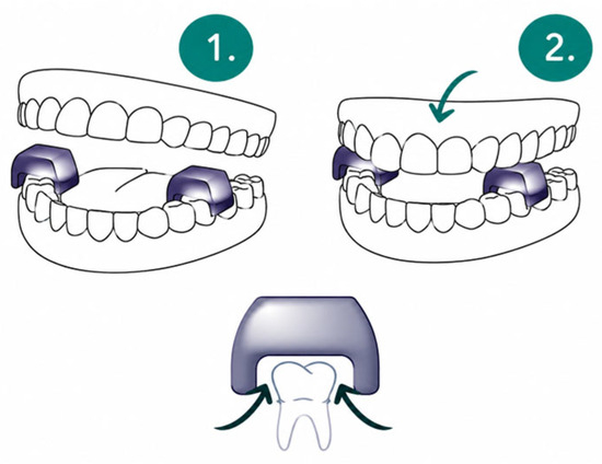
This study explored an innovative method using silicone TMJ relax pads placed in the molar region during specific therapeutic exercises. The pads have a layer thickness of 8.5 mm and are made of medical silicone. Unlike many conventional splints, they are not intended to fix the mandibular position but provide localized sensory input and a slight increase in the vertical dimension. Unlike conventional occlusal splints, TMJ relax pads are designed to induce a slight jaw-stretching posture and promote the relaxation of the masticatory muscles. The combination of TMJ relax pads with therapeutic exercise represents a low-threshold intervention that can be easily integrated into daily routines, particularly in office-based settings. While exercises target cervical and thoracic function, the additional orofacial input provided by the TMJ relax pads may support neuromuscular regulation and complement exercise-based interventions.
Thus, the aim of the present study was to determine the additive effects of temporomandibular joint (TMJ) relax pads added to specific gymnastic exercises compared with identical exercises performed without TMJ relax pads (considered as “usual care”) in office workers with chronic non-specific neck pain. We hypothesized that the use of TMJ relax pads will lead to greater reductions in neck pain as the primary study outcome. Furthermore, we expected a significantly more pronounced improvement in (1) neck-related disability in everyday life, as assessed by the Neck Disability Index (NDI), and anticipated significant additional improvements in (2) cervical spine mobility, (3) trunk rotational capacity, (4) shoulder elevation, and (5) the thoracic kyphosis angle in an upright position, outcomes considered as secondary study endpoints.
2. Materials and Methods
2.1. Study Design and Ethical Approval
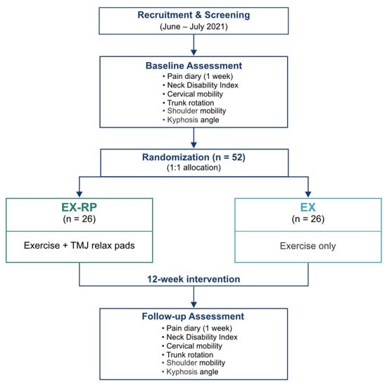
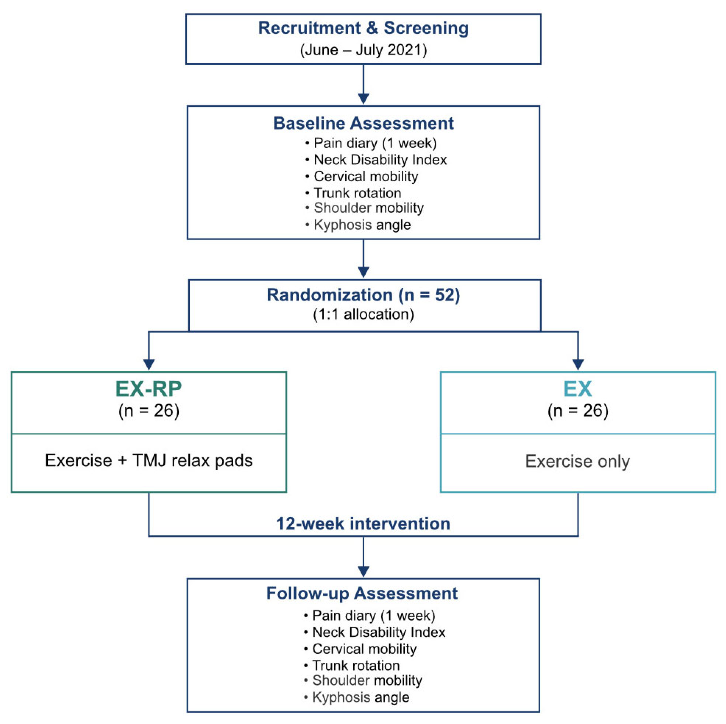
This is a monocentric randomized controlled trial (RCT) with a two-arm parallel group design, involving participants from the Erlangen–Nürnberg area. The trial was conducted and reported in accordance with the CONSORT Statement 2010 guidelines [21]. The study was conducted at the Institute of Medical Physics, Friedrich-Alexander-Universität (FAU) Erlangen–Nürnberg in close cooperation with the Institute of Radiology, University Hospital Erlangen. Ethical approval for the study was granted by the ethics committee (number 130-21b, 04/21/2021) and the data protection officer of the FAU. The study adheres to the ethical standards outlined in the Declaration of Helsinki by the World Medical Association [22] and is fully registered at ClinicalTrials.gov (NCT05302466). All participants provided written informed consent prior to their involvement in the study. No changes to methods have been made after trial commencement. Figure 1 illustrates the chronological sequence of the study.
Figure 1.
Study design. EX-RP: TMJ relax pads during exercise; EX: exercise without TMJ relax pads; FU: follow-up.
2.2. Study Population
Participants were recruited between June 2021 and July 2021 through announcements in local newsletters and by calls for applications at the FAU and the University Hospital Erlangen. Potential participants were initially informed about the study via a hotline. If they expressed interest, the study physician conducted a detailed review of their eligibility criteria. Based on the formal sample size analysis (see below), a total of 52 participants were recruited according to the following eligibility criteria: (a) men and women aged between 30–65 years, (b) chronic non-specific neck pain as defined as pain persisting for at least three months on at least 50% of the days, with a pain intensity of ≥2 on the Numeric Rating Scale (NRS) [23], and (c) frequent sedentary VDU workstation (>4 h/d) use. Exclusion criteria were (a) specific neck pain (e.g., herniated disc, spinal stenosis, etc.), (b) cervical-spine-related neuronal symptoms (e.g., paresthesia, brachial plexus pain, etc.), (c) the use of long-term pain medication, (d) the concurrent administration of other therapies with the potential to influence neck pain, (e) an absence exceeding two weeks during the intervention period, and (f) the performance of regular specific exercises or training focusing on the cervical spine within the previous six months. After receiving comprehensive information during individual interviews and having time for reflection, all the participants provided written consent to participate in the study. The intervention was conducted in a non-clinical, home-based setting, with participants performing the exercises independently in their daily environment (e.g., workplace or home).
2.3. Randomisation and Blinding
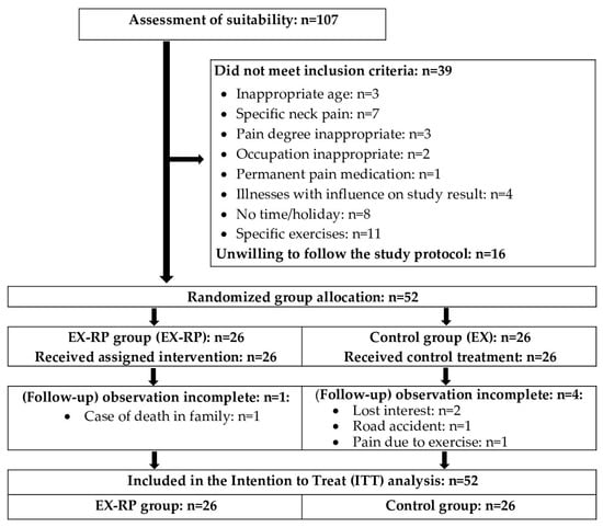
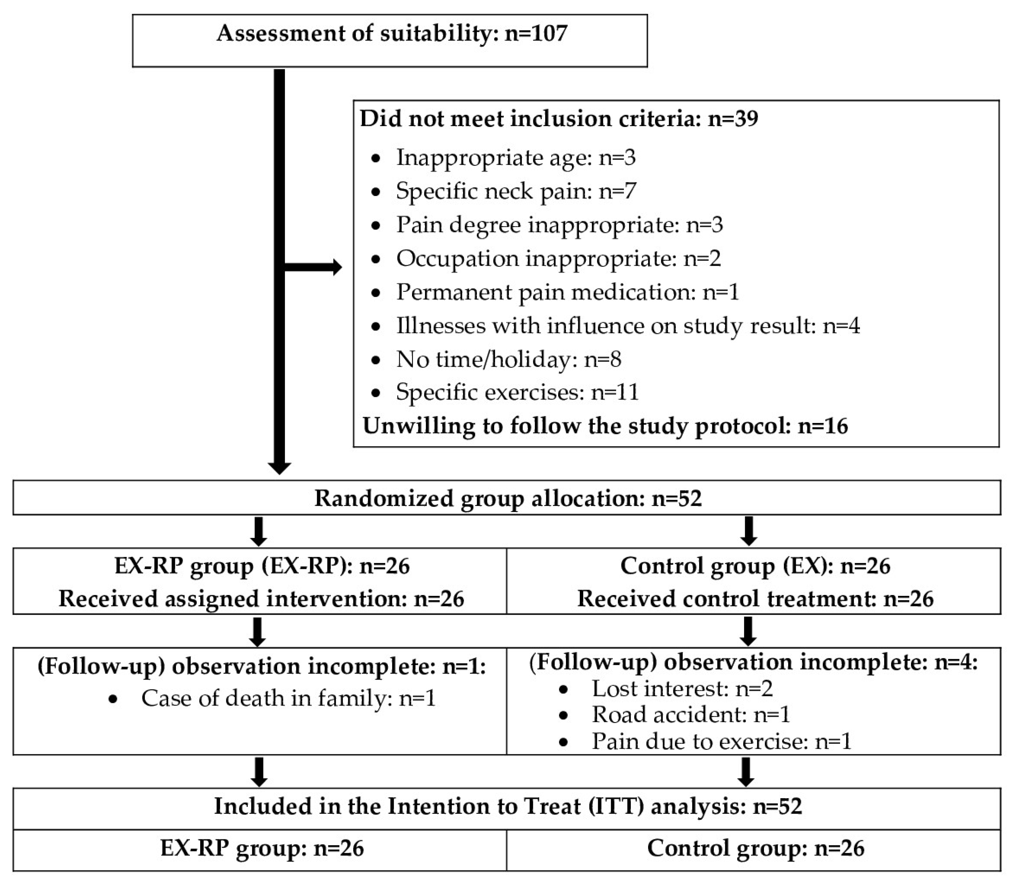
The 52 participants were randomly assigned to either the experimental group (EX-RP, n = 26) or the “usual care” control group (EX, n = 26) stratified by “pain intensity” (Figure 2). The allocation sequence was generated using a simple randomization procedure based on drawing lots, with group assignments (“EX-RP” or “EX”) placed in sealed opaque capsules prepared by an independent researcher. In detail, a researcher not involved in this project prepared the lots and supervised the randomization procedure and allocated the participants to the groups. Thus, neither the researcher nor the participants were aware of the allocation in advance, thus ensuring the “allocation concealment” procedure. Following randomization, the principal investigator provided the participants with detailed information of the study procedures.
Figure 2.
CONSORT flow diagram of participant recruitment, allocation, follow-up, and analysis [21].
Due to the nature of the study, blinding at the participant level was not possible. However, the groups were strictly separated, and participants were unaware of our hypothesis of more favorable effect in the TMJ relax pad group. While the primary investigator was aware of the participants’ group status (EX-RP or EX), all the research assistants involved in the testing and analysis were blinded to group status and were prohibited from inquiring about the final grouping. Due to the nature of the intervention, the use of TMJ relax pads could not be concealed, and, therefore, the interventions were not comparable in appearance or applications.
2.4. Interventions
2.4.1. Gymnastics Program of the Shoulder–Neck Region and the Cervical Spine
Both groups followed an identical exercise program of “usual care” comprising three distinct three-minute sequences to be performed daily. At the beginning of the study, each participant received individualized instructions and guidance on the correct execution of the exercises. As the study progressed, the participants were required to perform the exercise programs independently, using the provided video animations and their own personal devices (mobile phone, tablet, notebook, or PC). If participants had any questions or concerns, they could seek individual guidance or to engage in a video consultation via the “FAU Zoom” platform. Compliance was recorded daily in the training diary. Telephone contact was maintained with the participants at two-weekly intervals for three purposes: (a) to document the implementation of the protocol, (b) to identify and address any protocol deviations or implementation issues, and (c) to encourage consistent adherence to the program.
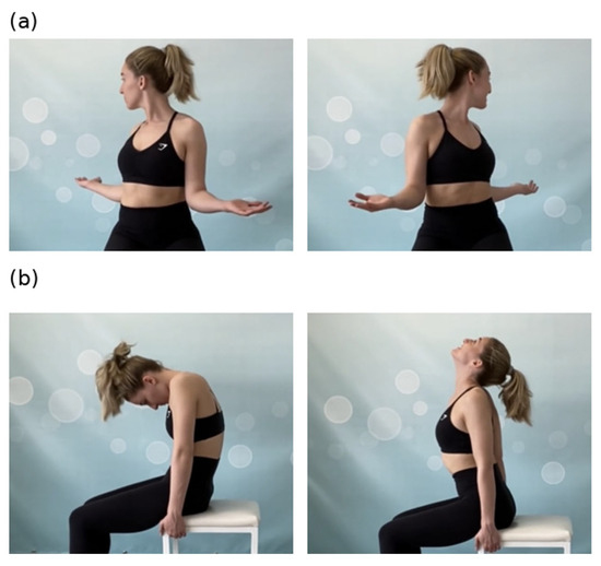
The specific physical training consisted of three different exercise programs, each to be performed daily. Each program comprised three exercises, each lasting one minute (approximately 10–15 repetitions), resulting in a total duration of three minutes per program. The programs included low-intensity gymnastic exercises from functional and therapeutic gymnastics aimed at activating, loosening, and stretching the muscles, with a focus on the shoulder–neck region and the cervical spine (e.g., Figure 3a,b).
Figure 3.
Examples of two exercises showing the starting and end positions: (a) seated trunk rotation, and (b) spinal flexion and extension exercise. Written informed consent has been obtained from the patient to publish this paper.
The morning program, performed standing, focused on full spine and upper body mobility through active exercises. The first exercise involved rotating the head to one side while performing a retrograde shoulder circle. The second exercise extended the arms backward and downward while engaging the glutes, lowering the head to the occiput. The third exercise involved extending the arms to shoulder height, contracting the glutes, and sequentially moving one arm to the side while orienting the head towards the hand.
The noon program, performed seated, focused on stretching and stabilizing the shoulder, neck, and cervical spine. The first exercise involved rounding the back, allowing the head to hang, then extending the upper body and protracting the chest (Figure 3a). The second exercise involved bending to one side while keeping one hand on the chair. The third exercise involved rotating the arms outwards, extending the elbows to 90°, and rotating the upper body with the head directed posteriorly (Figure 3b).
The evening program, performed supine, aimed to enhance spinal and shoulder mobility. The first exercise involved bending the knees to one side, elevating the contralateral arm, and orienting the head toward the hand. The second exercise focused on pressing the glutes upwards while rolling down the spine. The third exercise, performed prone, involved alternating lateral leg movements while resting the arms and head on the floor.
2.4.2. TMJ Relax Pads
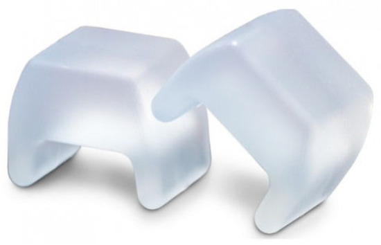
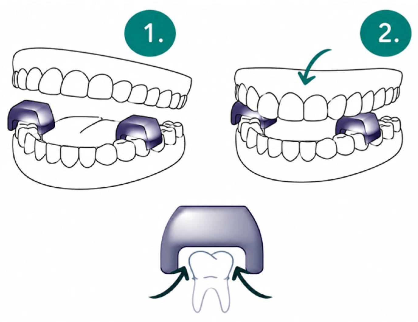
The TMJ relax pads TJ Motion (TJ Motion GmbH & Co. KG, Weisendorf, Germany) are manufactured using medical silicone (Figure 4). The splints are placed on the back molars of the lower jaw (area between tooth 34 and approximately 36 or between tooth 44 and 46) (Figure 5). Once positioned, the mouth should be closed loosely. In the event of a gag reflex becoming apparent, the TMJ relax pads should be placed slightly further forward (33 to approximately 35 or between teeth 43 and 45). These pads are to be maintained in this position throughout the three-minute exercise.
Figure 4.
Silicone TMJ relax pads © (TJ-Motion GmbH, Weisendorf, Germany). Used with permission from the copyright holder.
Figure 5.
Placement of the TMJ relax pads. © (TJ-Motion GmbH, Weisendorf, Germany).Used with permission from the copyright holder.
2.4.3. Confounders: Medication Supply with Painkillers
If necessary, medication with oral painkillers (NSAIDs) was administered as prescribed by the attending physician. The use of NSAIDs was recorded daily in the training diary and was also recorded during the biweekly telephone contacts.
2.5. Outcomes Measures and Assessment Schedule
No changes to trial outcomes were made after the trial commenced.
2.5.1. Primary Study Endpoint
The primary study endpoint was the change in neck pain intensity, assessed using the Numeric Rating Scale (NRS), between baseline assessment and the control measurement after the 12-week intervention.
2.5.2. Neck Pain Intensity (NRS)
A pain diary was used to record pain intensity daily for one week prior to commencing the intervention and during the 12th (last) week of the intervention, using the Numeric Rating Scale (NRS, 0–10). Pain intensity as the primary endpoint was defined as the mean value over one week in each case.
2.5.3. Secondary Study Endpoints
Secondary study endpoints included changes in (1) neck-related disability as assessed by the Neck Disability Index (NDI), (2) cervical spine mobility (lateral flexion, flexion, extension, and rotation), (3) mean trunk rotation, (4) shoulder joint elevation, and (5) thoracic kyphosis angle between baseline assessment and the control measurement after the 12-week intervention.
2.5.4. Limitations Due to Neck Complaints
The German version of the Neck Disability Index (NDI) was used to record neck complaints [24,25,26,27]. The NDI questionnaire documents complaints and problems during everyday activities caused by the cervical spine.
2.5.5. Flexibility, Mobility, and Posture
The mobility analysis of the cervical spine (range of motion (ROM)) [28,29] was performed via sensor-based digital recording of the maximum range of motion of flexion, extension, lateral flexion, and rotation (in degrees), during active performance in a seated position (Mobeefit SportMed A.G. SA, Echternach, Luxembourg). For flexion and lateral flexion, the mean value of both directions of movement (right and left) was calculated. The position sensor resembling a headlamp was attached to the head with an elastic strap. The mean rotational mobility of the torso was determined in a similar sensor-based manner in a seated position, with the sensor placed on the sternum (breastbone).
Shoulder mobility was determined by ROM [30,31,32] by means of end-range active flexion in a seated position (bilateral synchronized exercise with a gymnastic rod) employing a goniometer. The goniometer measurement was performed on the right shoulder.
2.5.6. Kyphosis
The kyphosis angle was evaluated in an upright position using a Debrunner kyphometer (Protek, Bern, Switzerland), in accordance with the method proposed by Ohlen et al. [33].
2.5.7. Craniomandibular Dysfunction
As an explanatory factor, the prevalence of CMD (Craniomandibular Dysfunction) was determined using a CMD short report according to Ahlers and Jakstat [34], which includes the following criteria: (1) asymmetrical opening of the mouth, (2) restricted opening of the mouth, (3) joint noises, (4) occlusal noises, (5) painful muscle palpation, and (6) traumatic eccentricity.
2.6. Biometrics and Statistical Analysis
The biometric and statistical analysis was conducted by an independent statistician. The number of participants was planned through an a priori formal sample size analysis based on the primary endpoint, “pain intensity of neck complaints”. It was hypothesized that there would be an overall reduction in pain intensity of at least 20% (standard deviation ±25%), which was considered to be a clinically relevant minimum. The statistical analysis indicated that, assuming the standard statistical parameters (α: 5%, 1-β: 80%), a sample size of 26 participants per group was deemed sufficient to address the primary endpoint.
As recommended for RCTs, we applied the intention-to-treat principle that including all participants randomly assigned to the study arms regardless of their loss to follow-up. R statistics software, Version 4.5.2 (R Development Core Team Vienna, Austria) was applied in combination with Amelia II [35] for multiple imputation (ITT) of missing data of all primary and secondary outcomes. In detail, FU data of five participants have to be imputed. The full dataset was used for multiple imputation, with imputation repeated 100 times. ITT results correspond to the pooled results (applying Rubins’ rule) from the 100 imputations. The primary analysis focused on differences between the within-group changes in EX-RP versus EX, using analysis of covariance (ANCOVA, calculated for each imputed dataset) adjusted for group differences in baseline data. After checking the normal distribution of the data (gg-plots and residual plots), within-group (pre-post intervention) changes presented here were analyzed by dependent t-tests with pooled SD. Results were presented as mean values (MVs) and standard deviation (SD) and/or mean value of the group difference with 95% confidence intervals (95%-CI).
The chi-square test was applied for categorical variables (see Table 1). Effect sizes were calculated according to Cohen (Cohen’s d). All tests were two-sided and a significance level of p < 0.05 was accepted. No interim analyses were performed and no predefined stopping rules were applied.
Table 1.
Baseline characteristics of the experimental group (EX-RP) and control group (EX). MV: mean value; SD: standard deviation.
3. Results
Table 1 presents the baseline characteristics of the study participants. The baseline characteristics did not differ significantly between the groups. Specifically, there were no statistically significant differences observed in terms of prevalent illnesses or medication regimens.
In summary, the trial was completed as intended. A total of five participants (EX-RP: n = 1 vs. EX: n = 4) were unable to participate in the follow-up study. Specifically, two EX participants reported that they had lost interest in the study, possibly (not reported) due to randomization into the non-preferred (control) group. One EX participant cited a road accident as the reason for their withdrawal. Another participant cited pain during or due to the gymnastic exercises as the reason for withdrawing. In the EX-RP, one person was no longer able or willing to carry out the intervention or FU measurement due to a death in their social circle.
The training frequency was 68.4 ± 28.5% in the EG and 75.8 ± 19.0% in the EX-RP group (p = 0.314). Subsequent analyses of the participants’ records and telephone interviews revealed that the EX-RP group exercises were consistently performed with the TMJ relax pads.
3.1. Study Endpoints
3.1.1. Primary Study Endpoint: Intensity of Neck Pain
ITT analysis demonstrated a reduction in the average pain intensity in both groups. However, after adjusting for basal values via ANCOVA, the reduction in pain intensity was significantly more pronounced in the EX-RP (p = 0.046, d′ = 0.54) (Table 2). In detail, the reduction in neck pain intensity was only found to be significant (p = 0.001) for the EX-RP (EX: p = 0.265). Thus, we confirm our hypothesis that using TMJ relax pads and exercise led to significantly greater reductions in neck pain intensity compared to exercise alone.
Table 2.
Baseline values, and changes in neck pain intensity (primary outcome) with statistical parameters: TMJ relax pads group (EX-RP) versus control group (EX). MV: mean value; SD: standard deviation; 95% CI: 95% confidence interval.
3.1.2. Secondary Study Endpoints
The Neck Disability Index (NDI) revealed a significant positive change during the intervention period in both groups (p < 0.001 each). Most importantly, the difference between the EX-RP and EG was not significant (p = 0.514). Thus, we reject our secondary hypothesis 1 that the addition of TMJ relax pads leads to superior improvements in NDI compared to exercise alone.
The range of motion in flexion improved significantly in both groups (EX-RP: p = 0.002 vs. EX: p = 0.016). No significant differences were observed between the groups (p = 0.581) and the analysis of mobility during the extension of the cervical spine revealed significant positive changes in the EX-RP (p = 0.002) but not in the EX (p = 0.35). For the rotation of the cervical spine (see Table 3), significant improvements in mobility were recorded in both groups (EX-RP: p < 0.001 vs. EX: p = 0.020). Although the changes within the EX-RP were more pronounced, a between-group comparison (ANCOVA) showed no significant differences between the EX-RP and the EX group (p = 0.405). Therefore, the hypothesis of a significantly superior effect of EX-RP on cervical mobility was not confirmed.
Table 3.
Basal values and changes in secondary study endpoints with statistical parameters: TMJ relax pads (EX-RP) versus control group (EX). MV: mean values; SD: standard deviation; 95% CI: 95% confidence interval.
Trunk rotation (see Table 3) exhibited a significant enhancement in rotational performance in both groups (EX-RP: p < 0.001 vs. EX: p = 0.006). A significant between-group difference was determined in favor of the EX-RP group (p = 0.019). Thus, the hypothesis of a significantly more pronounced effect of EX-RP compared to EX alone on trunk rotation was confirmed.
Both groups demonstrated a significant enhancement in shoulder mobility (p < 0.001 each). A significant difference between the groups was not recorded (p = 0.667). Therefore, the hypothesis of a more pronounced effect when adding TMJ relax pads to exercise versus exercise alone was rejected.
Following the 12-week intervention period, a reduction in the kyphosis angle was observed in both groups. However, this change was only found to be statistically significant in the EX-RP group (p < 0.005 vs. EX: p = 0.074), whereas the differences for the within-group changes were not significant (p = 0.634). Correspondingly, we revised our hypothesis of more pronounced effects in the EX-RP group versus EX alone in terms of the kyphosis angle.
As an explanatory analysis, we aimed to determine whether the prevalence of CMD had an influence on the treatment effects (neck pain intensity, NDI) within the TMJ relax pad group. A comparison between subjects with and without CMD revealed no significant group differences in the intervention on pain intensity (p = 0.523) or NDI (p = 0.730), however. In parallel, an analysis of the isolated gymnastics group (EG) revealed no significant differences in the effects on pain and NDI in subjects without CMD compared to those with CMD. These analyses were conducted on an exploratory basis and were not prespecified.
3.1.3. Adverse Effects
None of the EX-RP participants reported any undesirable side effects. Some participants reported increased saliva production during use. One participant in the control group reported pain when performing the gymnastic exercises; otherwise, no difficulties or particularities were reported during or after the exercises.
3.1.4. Potential Confounding Variables
No relevant changes in lifestyle, physical activity, or illness were reported by the participants that could have influenced our results. Further, no relevant changes in medication use and particular NSAID intake were reported in the EX-RP and EX groups.
However, one participant in the EX-RP group suffered whiplash in an accident in intervention week 7, which, according to the participant, prevented her from continuing with the intervention (though not from completing the final measurement).
4. Discussion
This study investigated the effect of TMJ relax pads added to an exercise program with short exercise sequences three times daily that can be easily integrated into everyday life. In summary, our study revealed a significant and moderately sized effect of TMJ relax pads added to a recognized exercise program on neck pain intensity as the primary outcome versus the identical exercise program alone. We feel the present approach represents an innovative, low-threshold intervention for the treatment of neck pain. The findings of this study suggest that combining gymnastic exercises with the use of a dental device worn during exercise may provide additional benefits for certain outcomes.
One may argue that the difference in pain reduction between the groups was relatively small. Although the effect size (d′ = 0.54) can be considered moderate, the effect (i.e., group difference in changes) of about 1 point on the NRS (0–10) is below the commonly accepted thresholds for clinically meaningful improvements in pain (1.5–2 points). However, this recommendation predominantly refers to comparisons with inactive or per se ineffective placebo control groups and should not be applied for the present comparison with a recognized gymnastic program. The low additional effort to wear the TMJ relax pads during gymnastic exercise justifies their application in our view, even if the additional effect is only moderate. Nevertheless, we would like to draw the reader’s attention to the fact that our findings should be interpreted with caution. This recommendation refers particularly to findings of inconsistent and limited effects for some secondary study outcomes.
With respect to the mode of action, it should be noted that the TMJ relax pads utilized in the study are not directly comparable with the occlusal splints that are generally applied. Despite the limited comparability, the TMJ relax pads will be differentiated from the occlusal splints below, and the results of this study will be discussed in the context of the existing literature [36].
The utilization of occlusal splints is endorsed by the guideline of the German Society of Dentistry, Oral, and Maxillofacial Medicine (DGZMK) [37], particularly in the treatment of CMD. Meta-analyses have demonstrated that such splints can reduce pain in temporomandibular disorders compared with no therapy [13,14].
The DGZMK guideline classifies occlusal splints into four categories. The first group comprises relaxation splints or stabilizing splints (e.g., Michigan splint), which are commonly used for masticatory muscle pain and bruxism [38,39].
The second group consists of reflex splints for reducing masticatory muscle activity [40,41].
The third group includes positioning splints, which modify the condylar position to relieve temporomandibular joint structures [42,43,44].
The fourth group comprises other splints such as soft or hydrostatic splints, which are mostly used in the acute treatment of CMD complaints [45].
In addition, sport splints can be distinguished from therapeutic splints, as they are primarily intended to influence posture and potentially enhance physical performance rather than to treat temporomandibular disorders [16].
The TMJ relax pads (TJ-Motion) employed in this study differ from these splints in design, shape, and material. Conventional splints typically cover the entire dental arch, are mostly individually customized, and are made of rigid materials such as PMMA or Polycarbonate [46]. The TJ-Motion covers a smaller area (two to three premolars or molars), consists of two separate pads, and has a layer thickness of 8.5 mm. It is made of medical silicone and is not individually customized. Conventionally splints are typically worn either at night or during the day, whereas TMJ relax pads are applied for short intervals (three times daily for three minutes) and can be integrated into exercises and everyday activities.
The scope of application of TMJ relax pads differs from that of conventional dental splints. Occlusal splints, aside from sports splints, are primarily used for the treatment of bruxism and temporomandibular disorders (TMDs). According to clinical guidelines, a key objective of occlusal splint therapy is to modify the functional patterns of the masticatory muscles and optimize the condylar position within the temporomandibular joint, thereby relieving structures subjected to excessive or improper loading [47].
The TJ-Motion does not induce a targeted and individual correction of the jaw position, but aims to stretch and relax hypertonic jaw muscles through an increased vertical dimension. Hypertonicity in these muscles can negatively affect adjacent regions, particularly the cervical spine, by contributing to increased muscular tension and impaired function. The correlation between the jaw and the cervical spine has been thoroughly documented in the existing literature, with studies indicating the potential to influence the musculoskeletal system and its associated complaints by altering the position of the temporomandibular joint [48]. Weber et al. [49] reported that women with TMD show more frequent signs of cervical dysfunction and neck pain. Walczyńska-Dragon et al. [50] demonstrate improvements in cervical mobility and pain following splint therapy, and Odzimek and Brola [51] found a higher prevalence and intensity in patients with TMJ disorders.
It should be noted that CMD was not an inclusion criterion in this study. The incidence of CMD was found to be equally distributed across both groups, with a notable prevalence of around 60% observed. However, a sub-analysis revealed no significant difference in treatment outcomes between individuals with and without CMD. These findings should also be interpreted with caution, as the study was not specifically powered for subgroup analyses and the results should be regarded as exploratory.
In the present study, a reduction in neck pain was observed in both study groups; however, the pain reduction in the EX-RP group was significantly more pronounced (Table 2). Unfortunately, the present literature has largely focused on either gymnastic exercises or isolated splint therapy, thus limiting the scope for direct comparison [52]. The study by Shousha et al. [53] made a direct comparison between conservative physiotherapy, predominantly hands-on in nature, and occlusal splinting, with the former achieving greater pain relief and improvement in movement. However, it should be noted that the Shousha et al. [53] study covered patients diagnosed with TMD, and the intervention group of that study used isolated splint therapy. This is not the case in the present study.
Regarding NDI, both groups improved significantly, with no between-group differences being observed. A meta-analysis by Bertozzi et al. [5] showed, in line with the present results, that therapeutic exercises cause significant improvements in the NDI. However, a comparison is limited, as only the results of isolated exercises were analyzed. The literature review found no study that directly measured the NDI in relation to splints. As other parameters of pain and function were measured, the search on the effects of splint therapy yielded only indirect correlations between the effects of a dental splint and the NDI. An enhancement in cervical spine mobility regarding flexion was observed in both of our study groups, with no significant group difference. Nevertheless, a more notable enhancement was evident in the EX-RP group with respect to cervical spine extension, while no improvement was observed in the EX group (Table 3). Moreover, a significant improvement was recorded in the rotation of the cervical spine in both groups without significant difference between the groups (Table 3). Previous studies have reported improvements in cervical mobility following splint therapy [50,54,55], although these studies did not include exercise interventions. Regarding shoulder mobility and mid-torso rotation, no studies were identified in the current literature search that would permit comparability.
The EX-RP group demonstrated an improvement in the kyphosis angle; however, the between-group difference did not reach statistical significance (Table 3). In some studies, positive effects on posture [56,57] and also on the kyphosis angle [58,59] have been achieved by using splints to change the position of the lower jaw. However, the study results are largely inconsistent, and the influence of splint therapy is generally a controversial one [52,60,61,62,63,64].
The inconsistent findings reported in the literature may, at least in part, be explained by substantial methodological limitations across studies. The significant limitations that result in the poor comparability of individual studies include the absence of methodological standardization, the inability to differentiate between muscular, articular, or combined TMD, and the failure to distinguish between physio-pathological conditions and risk factors [63].
The present study’s design as a confirmative trial does not allow definite conclusions regarding the underlying mechanism to be drawn. Speculatively, a possible explanation may be the trigeminocervical convergence, where nociceptive input from the trigeminal system and the upper cervical spine is processed within a shared neuroanatomical pathway [20]. This anatomical and functional overlap allows nociceptive input from the jaw and cervical spine to converge, potentially explaining the interaction between temporomandibular and neck pain. Based on this concept, the additional orofacial stimulation provided by the TMJ relax pads may influence cervical muscle activity and pain perception via central neuromuscular mechanisms. This reduction in tension could, in turn, have a positive influence on the symptoms of chronic cervical spine syndrome. The craniocervical–orofacial connections that could underpin this effect have been described in numerous publications [6,7]. This may provide a possible explanation for the improvements observed in neck pain and mobility in the present study.
The relationship between the temporomandibular joint and the cervical spine is supported by anatomical, biomechanical, and neuromuscular connections [19,65,66]. However, the underlying mechanisms are not fully understood, and the current explanations are based on heterogeneous and partly inconsistent findings in the literature. Therefore, the discussion of potential underlying mechanisms remains speculative [67].
Finally, the effects of the gymnastic program itself should be considered when assessing the present findings. The exercise program alone was associated with significant improvements in pain intensity and neck disability. Further, most mobility outcomes improved in the EX group, although changes in the extension of the cervical spine and the kyphosis angle of the thoracic spine did not reach statistical significance (Table 3). A multitude of studies have examined the effects of exercise programs on chronic non-specific neck pain, with the findings summarized in a meta-analysis [68]. Despite the considerable heterogeneity in the frequency, duration, intervention period, and exercise type, most programs demonstrated significant improvements in pain intensity.
The high compliance rate (EX: 68.4 ± 28.5% vs. EX-RP: 75.8 ± 19.0%) and low drop-out rate (EX-RP: n = 1 vs. EX: n = 4) in this study demonstrate the program’s success in terms of feasibility and acceptance, particularly for the joint TMJ relax pads and exercise group. Given the investigation’s focus on the additional effect of the TMJ relax pads, it was imperative that the exercises were standardized and identical for all patients. The utilization of video instruction in this context was found to be an effective approach.
It is important to acknowledge the distinctive characteristics and limitations inherent in the study. Several sources of bias should be considered when interpreting the results of this study. One may consider the absence of a sham-controlled placebo condition as a major limitation of the present study. As participants were aware of their group allocation, expectation effects might have influenced the results, especially for self-reported outcomes such as neck pain intensity. However, this refers to both study groups. Since our approach focused on the comparison of a combined intervention with gymnastics (considered as “usual care”) superimposed by TMJ relax pad versus “gymnastics alone”, both groups had legitimate expectations of pain relief. Further, the groups were strictly separated and unaware of our hypothesis of more favorable effects in the combined intervention group. Another problem related to multiple outcomes is multiplicity, i.e., the inflation of type I errors. However, it should be noted that no standard procedure is recommended [69]. While some researchers consider it absolutely necessary to adjust the data using statistical methods, others state that “no adjustment for secondary endpoints is needed if researchers analyze and interpret the studies in an unbiased manner”. We applied the latter approach and do not adjust the present analysis for multiplicity; nevertheless, we suggest our results be read with caution. This includes the idea that the interpretation of the secondary outcomes should be viewed as rather exploratory.
Additionally, the differential drop-out rates (EX-RP = 1 vs. EX = 4) between the groups may have introduced an attrition bias, with two participants in the control group presumably withdrawing due to allocation to the non-preferred group. Furthermore, no follow-up period was included, making it difficult to determine whether the effects persist over time. In addition, despite a low drop-out rate (less than 10%), ‘lost-to-follow-up’ data had to be imputed via the intention-to-treat (ITT) approach of the study. Experience has shown [70] that the results are, in some cases, significantly more cautious (i.e., tend to be less positive) than in per-protocol analyses. This was demonstrated in an additional analysis for the primary study endpoint, “neck pain intensity”, where the per-protocol analysis revealed a significantly higher significance level (p = 0.006) for the between-group effect. Finally, the possibility exists that discrepancies in the participation rate between the groups (EX: 68.4 ± 28.5% vs. EX-RP 75.8 ± 19.0%, p = 0.157) may be a contributing factor to the observed positive effect of the TMJ relax pads, at least when assuming that participants with a higher compliance in terms of exercise may have experienced greater benefits.
Due to the mixed intervention, it is difficult to clearly separate the contribution of the exercise program to the present result. Additionally, due to the absence of a non-exercise control group, conclusions regarding the independent efficacy of the exercise program per se are limited. Thus, as intended by the present study, the findings should be interpreted as reflecting the additional effects of TMJ relax pads in combination with an established exercise intervention for pain reduction, while the relevance of the underlying exercise program was not addressed here.
As anticipated, the TMJ relax pads did not completely cure the pain. Future studies will need to determine to what extent longer treatment leads to better outcomes or, conversely, to a decline in the pain-relieving effect.
The study included participants aged 30–65 years to reflect a typical working population with chronic non-specific neck pain. Although it is conceivable that younger or older people with corresponding complaints might also benefit, a simple generalization of our finding to other cohorts would be inappropriate.
The character of the study can be considered as a phase IIa study, meeting all the criteria of a confirmatory randomized controlled trial. The results obtained appear to be promising, and it is therefore recommended that further well-designed research with adequate statistical power be conducted in this area. Furthermore, additional research is needed to determine whether the effects of the TMJ relax pads are more pronounced in certain groups of participants compared to others. It is also recommended that future studies include sham-controlled/multi-armed designs and follow-up periods to clearly determine the isolated treatment effects and also investigate the long-term sustainability of the observed effects.
5. Conclusions
In conclusion, a time-effective, low-threshold intervention of specific gymnastics superimposed by TMJ pads focusing on the neck and shoulder region was associated with reductions in neck pain and improvements in functional parameters in individuals with chronic neck pain. Of importance, performing the exercises while using TMJ relax pads led to significantly greater pain reduction and additional improvements in selected mobility outcomes, particularly cervical spine extension and trunk rotation. Although differences between the interventions are not always pronounced and do not reach statistical significance, the feasibility, easy use, and affordability of the additional application of the TMJ relax pads justify their recommendation as a useful tool for fighting chronic non-specific neck pain. Nevertheless, due to the brief nature of the intervention and follow-up assessment, and the focus on individuals aged 30–65, further research is required to ascertain the long-term impact of TMJ relax pads and to generalize their effects to other groups.
Author Contributions
Conceptualization, N.K., R.L., W.K., and S.v.S.; methodology, N.K., R.L., W.K., and S.v.S.; software, M.K.; validation, N.K., R.L., M.K., W.K., and S.v.S.; formal analysis, W.K., M.K., and S.v.S.; investigation, N.K., R.L., W.K., and S.v.S.; resources, W.K. and S.v.S.; data curation, N.K., R.L., and S.v.S.; writing—original draft preparation, N.K., R.L., W.K., M.K., and S.v.S.; writing—review and editing, N.K., R.L., W.K., M.K., and S.v.S.; visualization, N.K. and S.v.S.; supervision, N.K., R.L., and S.v.S.; project administration, N.K. and S.v.S.; funding acquisition, W.K. All authors have read and agreed to the published version of the manuscript.
Funding
This research received no external funding.
Institutional Review Board Statement
The study was conducted in accordance with the Declaration of Helsinki and approved by Ethics Committee of Friedrich-Alexander-Universität Erlangen–Nürnberg (protocol code 130-21b, 21 April 2021).
Informed Consent Statement
Informed consent was obtained from all subjects involved in the study. Written informed consent has been obtained from the patients to publish this paper.
Data Availability Statement
The datasets generated and/or analyzed during the current study are available from the corresponding author upon reasonable request.
Acknowledgments
The authors would like to thank all participants for their time and commitment to the study. During the preparation of this manuscript, the authors used ChatGPT Version 5 (OpenAI) to assist with language editing and clarity. The authors reviewed and edited the output and take full responsibility for the content of this publication. The work was performed in (partial) fulfillment of the requirements for Nathalie Kühn in obtaining the degree of Dr. med. dent.
Conflicts of Interest
The authors declare no conflicts of interest.
Abbreviations
The following abbreviations are used in this manuscript:
| ANCOVA | Analysis of covariance |
| CMD | Craniomandibular disorder |
| DGZMK | Deutsche Gesellschaft für Zahn-, Mund-, und Kieferheilkunde |
| EX | Exercise group |
| EX-RP | Exercise group with TMJ relax pads |
| ITT | Intention-to-treat |
| NDI | Neck Disability Index |
| RP | Relax pads |
| TMJ | Temporomandibular joint |
| TMD | Temporomandibular disorder |
| VDU | Video display unit |
References
- Hoy, D.; March, L.; Woolf, A.; Blyth, F.; Brooks, P.; Smith, E.; Vos, T.; Barendregt, J.; Blore, J.; Murray, C.; et al. The global burden of neck pain: Estimates from the Global Burden of Disease 2010 study. Ann. Rheum. Dis. 2014, 73, 1309–1315. [Google Scholar] [CrossRef] [Scilit]
- Hurwitz, E.L.; Randhawa, K.; Yu, H.; Côté, P.; Haldeman, S. The Global Spine Care Initiative: A summary of the global burden of low back and neck pain studies. Eur. Spine J. 2018, 27, 796–801. [Google Scholar] [CrossRef] [Scilit]
- Häuser, W.; Schmutzer, G.; Hinz, A.; Hilbert, A.; Brähler, E. Prevalence of chronic pain in Germany. A representative survey of the general population. Schmerz 2013, 27, 46–55. [Google Scholar] [CrossRef] [Scilit]
- Johnston, V.; Jull, G.; Souvlis, T.; Jimmieson, N.L. Neck movement and muscle activity characteristics in female office workers with neck pain. Spine 2008, 33, 555–563. [Google Scholar] [CrossRef] [Scilit] [PubMed]
- Bertozzi, L.; Gardenghi, I.; Turoni, F.; Villafañe, J.H.; Capra, F.; Guccione, A.A.; Pillastrini, P. Effect of Therapeutic Exercise on Pain and Disability in the Management of Chronic Nonspecific Neck Pain: Systematic Review and Meta-Analysis of Randomized Trials. Phys. Ther. 2013, 93, 1026–1036. [Google Scholar] [CrossRef] [Scilit]
- De Laat, A.; Meuleman, H.; Stevens, A.; Verbeke, G. Correlation between cervical spine and temporomandibular disorders. Clin. Oral Investig. 1998, 2, 54–57. [Google Scholar] [CrossRef] [Scilit] [PubMed]
- Ciancaglini, R.; Gherlone, E.F.; Radaelli, G. Association between loss of occlusal support and symptoms of functional disturbances of the masticatory system. J. Oral Rehabil. 1999, 26, 248–253. [Google Scholar] [CrossRef] [Scilit]
- Walczyńska-Dragon, K.; Baron, S. The biomechanical and functional relationship between temporomandibular dysfunction and cervical spine pain. Acta Bioeng. Biomech. 2011, 13, 93–98. [Google Scholar]
- Olivo, S.A.; Fuentes, J.; Major, P.W.; Warren, S.; Thie, N.M.; Magee, D.J. The association between neck disability and jaw disability. J. Oral Rehabil. 2010, 37, 670–679. [Google Scholar] [CrossRef] [Scilit] [PubMed]
- Bracco, P.; Deregibus, A.; Piscetta, R.; Ferrario, G. Observations on the correlation between posture and jaw position: A pilot study. CRANIO® 1998, 16, 252–258. [Google Scholar] [CrossRef] [Scilit]
- Maurer, C.; Stief, F.; Jonas, A.; Kovac, A.; Groneberg, D.A.; Meurer, A.; Ohlendorf, D. Influence of the Lower Jaw Position on the Running Pattern. PLoS ONE 2015, 10, e0135712. [Google Scholar] [CrossRef] [Scilit] [PubMed]
- Maurer, C.; Heller, S.; Sure, J.J.; Fuchs, D.; Mickel, C.; Wanke, E.M.; Groneberg, D.A.; Ohlendorf, D. Strength improvements through occlusal splints? The effects of different lower jaw positions on maximal isometric force production and performance in different jumping types. PLoS ONE 2018, 13, e0193540. [Google Scholar] [CrossRef] [Scilit] [PubMed]
- Al-Moraissi, E.A.; Farea, R.; Qasem, K.A.; Al-Wadeai, M.S.; Al-Sabahi, M.E.; Al-Iryani, G.M. Effectiveness of occlusal splint therapy in the management of temporomandibular disorders: Network meta-analysis of randomized controlled trials. Int. J. Oral Maxillofac. Surg. 2020, 49, 1042–1056. [Google Scholar] [CrossRef] [Scilit]
- Zhang, S.-H.; He, K.-X.; Lin, C.-J.; Liu, X.-D.; Wu, L.; Chen, J.; Rausch-Fan, X. Efficacy of occlusal splints in the treatment of temporomandibular disorders: A systematic review of randomized controlled trials. Acta Odontol. Scand. 2020, 78, 580–589. [Google Scholar] [CrossRef] [Scilit]
- Lee, S.Y.; Park, Y.J.; Park, H.M.; Bae, H.J.; Yu, M.J.; Choi, H.W.; Hwang, N.Y. Effect of the Mandibular Orthopedic Repositioning Appliance (MORA) on Forearm Muscle Activation and Grasping Power during Pinch and Hook Grip. J. Phys. Ther. Sci. 2014, 26, 195–197. [Google Scholar] [CrossRef] [Scilit]
- Herzog, J.; Göttfert, F.; Maurer-Grubinger, C.; Holzgreve, F.; Oremek, G.; Groneberg, D.A.; Ohlendorf, D. Improvement of cervical spine mobility and stance stability by wearing a custom-made mandibular splint in male recreational athletes. PLoS ONE 2022, 17, e0278063. [Google Scholar] [CrossRef] [Scilit]
- Arent, S.; McKenna, J.; Golem, D. Effects of a neuromuscular dentistry-designed mouthguard on muscular endurance and anaerobic power. Comp. Exerc. Physiol. 2010, 7, 73–79. [Google Scholar] [CrossRef] [Scilit]
- Bragatto, M.M.; Bevilaqua-Grossi, D.; Regalo, S.C.; Sousa, J.D.; Chaves, T.C. Associations among temporomandibular disorders, chronic neck pain and neck pain disability in computer office workers: A pilot study. J. Oral Rehabil. 2016, 43, 321–332. [Google Scholar] [CrossRef] [Scilit]
- Goadsby, P.; Bartsch, T. Introduction: On the functional neuroanatomy of neck pain. Cephalalgia 2008, 28, 1–7. [Google Scholar] [CrossRef] [Scilit]
- Raguž, M.; Tarle, M.; Hat, K.; Salarić, I.; Marčinković, P.; Bičanić, I.; Lazić Mosler, E.; Lukšić, I.; Marinović, T.; Chudy, D. Refractory neuropathic pain in the head and neck: Neuroanatomical and clinical significance of the cervicotrigeminal complex. Life 2025, 15, 1457. [Google Scholar] [CrossRef] [Scilit]
- Schulz, K.F.; Altman, D.G.; Moher, D. CONSORT 2010 Statement: Updated guidelines for reporting parallel group randomised trials. Trials 2010, 11, 32. [Google Scholar] [CrossRef] [Scilit]
- World Medical Association Declaration of Helsinki: Ethical principles for medical research involving human subjects. Jama 2013, 310, 2191–2194. [CrossRef] [Scilit]
- Höppchen, I.; Wittek, M.; Szecsenyi, J. Diagnostik bei Nackenschmerzen. 2020. Available online: https://www.rosenfluh.ch/media/arsmedici/2020/13/Diagnostik-bei-Nackenschmerzen.pdf (accessed on 7 January 2026).
- Cramer, H.; Lauche, R.; Langhorst, J.; Dobos, G.J.; Michalsen, A. Validation of the German version of the Neck Disability Index (NDI). BMC Musculoskelet. Disord. 2014, 15, 91. [Google Scholar] [CrossRef] [Scilit]
- MacDermid, J.C.; Walton, D.M.; Avery, S.; Blanchard, A.; Etruw, E.; McAlpine, C.; Goldsmith, C.H. Measurement properties of the neck disability index: A systematic review. J. Orthop. Sports Phys. Ther. 2009, 39, 400–417. [Google Scholar] [CrossRef] [Scilit] [PubMed]
- Trouli, M.N.; Vernon, H.T.; Kakavelakis, K.N.; Antonopoulou, M.D.; Paganas, A.N.; Lionis, C.D. Translation of the Neck Disability Index and validation of the Greek version in a sample of neck pain patients. BMC Musculoskelet. Disord. 2008, 9, 106. [Google Scholar] [CrossRef] [Scilit] [PubMed]
- Vernon, H.; Mior, S. The Neck Disability Index: A study of reliability and validity. J. Manip. Physiol. Ther. 1991, 14, 409–415. [Google Scholar]
- Howell, E. The association between neck pain, the Neck Disability Index and cervical ranges of motion: A narrative review. J. Can. Chiropr. Assoc. 2011, 55, 211–221. [Google Scholar]
- Fletcher, J.P.; Bandy, W.D. Intrarater reliability of CROM measurement of cervical spine active range of motion in persons with and without neck pain. J. Orthop. Sports Phys. Ther. 2008, 38, 640–645. [Google Scholar] [CrossRef] [Scilit]
- Riddle, D.L.; Rothstein, J.M.; Lamb, R.L. Goniometric reliability in a clinical setting. Shoulder measurements. Phys. Ther. 1987, 67, 668–673. [Google Scholar] [CrossRef] [Scilit]
- Shin, Y.J.; Kim, W.H.; Kim, S.G. Correlations among visual analogue scale, neck disability index, shoulder joint range of motion, and muscle strength in young women with forward head posture. J. Exerc. Rehabil. 2017, 13, 413–417. [Google Scholar] [CrossRef] [Scilit]
- Sabari, J.S.; Maltzev, I.; Lubarsky, D.; Liszkay, E.; Homel, P. Goniometric assessment of shoulder range of motion: Comparison of testing in supine and sitting positions. Arch. Phys. Med. Rehabil. 1998, 79, 647–651. [Google Scholar] [CrossRef] [Scilit]
- Ohlen, G.; Aaro, S.; Bylund, P. The sagittal configuration and mobility of the spine in idiopathic scoliosis. Spine 1988, 13, 413–416. [Google Scholar] [CrossRef] [Scilit]
- Ahlers, M.O.; Jakstat, H.A. Identifikation funktionsgestörter Patienten. Zahnmed. Up2date 2008, 2, 143–155. [Google Scholar] [CrossRef] [Scilit]
- Honaker, J.; King, G.; Blackwell, M. Amelia II: A Program for Missing Data. J. Stat. Softw. 2011, 45, 1–47. [Google Scholar] [CrossRef] [Scilit]
- Incorvati, C.; Romeo, A.; Fabrizi, A.; Defila, L.; Vanti, C.; Gatto, M.R.A.; Marchetti, C.; Pillastrini, P. Effectiveness of physical therapy in addition to occlusal splint in myogenic temporomandibular disorders: Protocol of a randomised controlled trial. BMJ Open 2020, 10, e038438. [Google Scholar] [CrossRef] [Scilit] [PubMed]
- Deutsche Gesellschaft für Zahn-, Mund- und Kieferheilkunde. Okklusionsschienen zur Behandlung Craniomandibulärer Dysfunktionen und zur Präprothetischen Therapie; DGZMK: Berlin, Germany, 2024; Available online: https://www.awmf.org/leitlinien/detail/II/083-051.html (accessed on 3 June 2025).
- Badel, T.; Simonić-Kocijan, S.; Lajnert, V.; Dulčić, N.; Zdravec, D. Michigan splint and treatment of temporomandibular joint. Med. Flum. 2013, 49, 112–120. [Google Scholar]
- Wassell, R.W.; Adams, N.; Kelly, P.J. The treatment of temporomandibular disorders with stabilizing splints in general dental practice: One-year follow-up. J. Am. Dent. Assoc. 2006, 137, 1089–1098, 1089-1098. [Google Scholar] [CrossRef] [Scilit] [PubMed]
- Hannam, A.G.; Wood, W.W.; De Cou, R.E.; Scott, J.D. The effects of working-side occlusal interferences on muscle activity and associated jaw movements in man. Arch. Oral Biol. 1981, 26, 387–392. [Google Scholar] [CrossRef] [Scilit]
- Stapelmann, H.; Türp, J.C. The NTI-tss device for the therapy of bruxism, temporomandibular disorders, and headache—Where do we stand? A qualitative systematic review of the literature. BMC Oral Health 2008, 8, 22. [Google Scholar] [CrossRef] [Scilit]
- Eberhard, D.; Bantleon, H.P.; Steger, W. The efficacy of anterior repositioning splint therapy studied by magnetic resonance imaging. Eur. J. Orthod. 2002, 24, 343–352. [Google Scholar] [CrossRef] [Scilit]
- Guo, Y.-N.; Cui, S.-J.; Zhou, Y.-H.; Wang, X.-D. An Overview of Anterior Repositioning Splint Therapy for Disc Displacement-related Temporomandibular Disorders. Curr. Med. Sci. 2021, 41, 626–634. [Google Scholar] [CrossRef] [Scilit] [PubMed]
- Lakshmi, M.S.; Kalekhan, S.M.; Mehta, R.; Bhangdia, M.; Rathore, K.; Lalwani, V. Occlusal Splint Therapy in Temporomandibular Joint Disorders: An Update Review. J. Int. Oral Health 2016, 8, 639–645. [Google Scholar] [CrossRef] [Scilit]
- Srivastava, R.; Jyoti, B.; Devi, P. Oral splint for temporomandibular joint disorders with revolutionary fluid system. Dent. Res. J. 2013, 10, 307–313. [Google Scholar]
- Benli, M.; Al-Haj Husain, N.; Ozcan, M. Mechanical and chemical characterization of contemporary occlusal splint materials fabricated with different methods: A systematic review. Clin. Oral Investig. 2023, 27, 7115–7141. [Google Scholar] [CrossRef] [Scilit] [PubMed]
- Albagieh, H.; Alomran, I.; Binakresh, A.; Alhatarisha, N.; Almeteb, M.; Khalaf, Y.; Alqublan, A.; Alqahatany, M. Occlusal splints-types and effectiveness in temporomandibular disorder management. Saudi Dent. J. 2023, 35, 70–79. [Google Scholar] [CrossRef] [Scilit]
- Grondin, F.; Hall, T.; von Piekartz, H. Does altered mandibular position and dental occlusion influence upper cervical movement: A cross–sectional study in asymptomatic people. Musculoskelet. Sci. Pract. 2017, 27, 85–90. [Google Scholar] [CrossRef] [Scilit]
- Weber, P.; Corrêa, E.C.; Ferreira Fdos, S.; Soares, J.C.; Bolzan Gde, P.; Silva, A.M. Cervical spine dysfunction signs and symptoms in individuals with temporomandibular disorder. J. Soc. Bras. Fonoaudiol. 2012, 24, 134–139. [Google Scholar] [CrossRef] [Scilit]
- Walczyńska-Dragon, K.; Baron, S.; Nitecka-Buchta, A.; Tkacz, E. Correlation between TMD and Cervical Spine Pain and Mobility: Is the Whole Body Balance TMJ Related? BioMed Res. Int. 2014, 2014, 582414. [Google Scholar] [CrossRef] [Scilit]
- Odzimek, M.; Brola, W. Occurrence of Cervical Spine Pain and Its Intensity in Young People with Temporomandibular Disorders. J. Clin. Med. 2024, 13, 1941. [Google Scholar] [CrossRef] [Scilit]
- Kang, J.-H. Effects on migraine, neck pain, and head and neck posture, of temporomandibular disorder treatment: Study of a retrospective cohort. Arch. Oral Biol. 2020, 114, 104718. [Google Scholar] [CrossRef] [Scilit]
- Shousha, T.M.; Soliman, E.S.; Behiry, M.A. The effect of a short term conservative physiotherapy versus occlusive splinting on pain and range of motion in cases of myogenic temporomandibular joint dysfunction: A randomized controlled trial. J. Phys. Ther. Sci. 2018, 30, 1156–1160. [Google Scholar] [CrossRef] [Scilit]
- Tereshchuk, O.H. Effect of using a relaxation splint on reducing chronic neck pain. Ukr. Dent. Alm. 2024, 1, 61–65. [Google Scholar] [CrossRef] [Scilit]
- Karppinen, K.; Eklund, S.; Suoninen, E.; Eskelin, M.; Kirveskari, P. Adjustment of dental occlusion in treatment of chronic cervicobrachial pain and headache. J. Oral Rehabil. 1999, 26, 715–721. [Google Scholar] [CrossRef] [Scilit] [PubMed]
- Sakaguchi, K.; Mehta, N.R.; Abdallah, E.F.; Forgione, A.G.; Hirayama, H.; Kawasaki, T.; Yokoyama, A. Examination of the Relationship Between Mandibular Position and Body Posture. CRANIO® 2007, 25, 237–249. [Google Scholar] [CrossRef] [Scilit]
- Ohlendorf, D.; Seebach, K.; Hoerzer, S.; Nigg, S.; Kopp, S. The effects of a temporarily manipulated dental occlusion on the position of the spine: A comparison during standing and walking. Spine J. 2014, 14, 2384–2391. [Google Scholar] [CrossRef] [Scilit] [PubMed]
- De Giorgi, I.; Castroflorio, P.T.; Cugliari, G.; Deregibus, D.D.S.P.A. Does occlusal splint affect posture? A randomized controlled trial. CRANIO® 2020, 38, 264–272. [Google Scholar] [CrossRef] [Scilit] [PubMed]
- März, K.; Adler, W.; Matta, R.-E.; Wolf, L.; Wichmann, M.; Bergauer, B. Can different occlusal positions instantaneously impact spine and body posture? J. Orofac. Orthop. Fortschritte Kieferorthopadie 2017, 78, 221–232. [Google Scholar] [CrossRef] [Scilit]
- Hampe, T.; Fürstberger, L.; Kordsmeyer, T.L.; Penke, L.; Mahler, A.M.; Mäder, C.M.; Bürgers, R.; Krohn, S. Impact of occlusal stabilization splints on global body posture: A prospective clinical trial. Clin. Oral Investig. 2024, 28, 505. [Google Scholar] [CrossRef] [Scilit]
- Maurer-Grubinger, C.; Avaniadi, I.; Adjami, F.; Christian, W.; Doerry, C.; Fay, V.; Fisch, V.; Gerez, A.; Goecke, J.; Kaya, U.; et al. Systematic changes of the static upper body posture with a symmetric occlusion condition. BMC Musculoskelet. Disord. 2020, 21, 636. [Google Scholar] [CrossRef] [Scilit]
- Lippold, C.; Danesh, G.; Schilgen, M.; Drerup, B.; Hackenberg, L. Sagittal jaw position in relation to body posture in adult humans—A rasterstereographic study. BMC Musculoskelet. Disord. 2006, 7, 8. [Google Scholar] [CrossRef] [Scilit]
- Sofyanti, E.; Boel, T.; Sihombing, A.R.N. The correlation between back posture and sagittal jaw position in adult orthodontic patients. J. Taibah Univ. Med. Sci. 2021, 16, 63–69. [Google Scholar] [CrossRef] [Scilit]
- Giannakopoulos, N.N.; Schindler, H.J.; Hellmann, D. Co-contraction behaviour of masticatory and neck muscles during tooth grinding. J. Oral Rehabil. 2018, 45, 504–511. [Google Scholar] [CrossRef] [Scilit]
- Bednarczyk, V.; Proulx, F.; Paez, A. The effectiveness of cervical rehabilitation interventions for pain in adults with myogenic temporomandibular disorders: A systematic review and meta-analysis. J. Oral Rehabil. 2024, 51, 1091–1107. [Google Scholar] [CrossRef] [Scilit]
- Svensson, P.; Wang, K.; Sessle, B.J.; Arendt-Nielsen, L. Associations between pain and neuromuscular activity in the human jaw and neck muscles. Pain 2004, 109, 225–232. [Google Scholar] [CrossRef] [PubMed]
- Rocha, C.; Croci, C.; Caria, P. Is there relationship between temporomandibular disorders and head and cervical posture? A systematic review. J. Oral Rehabil. 2013, 40, 875–881. [Google Scholar] [CrossRef] [Scilit] [PubMed]
- Louw, S.; Makwela, S.; Manas, L.; Meyer, L.; Terblanche, D.; Brink, Y. Effectiveness of exercise in office workers with neck pain: A systematic review and meta-analysis. S. Afr. J. Physiother. 2017, 73, 392. [Google Scholar] [CrossRef] [Scilit]
- Chowdhry, A.K.; Park, J.; Kang, J.; Sakthivel, G.; Pugh, S. Finding Multiple Signals in the Noise: Handling Multiplicity in Clinical Trials. Int. J. Radiat. Oncol. Biol. Phys. 2024, 119, 750–755. [Google Scholar] [CrossRef] [Scilit] [PubMed]
- Mostazir, M.; Taylor, G.; Henley, W.E.; Watkins, E.R.; Taylor, R.S. Per-Protocol analyses produced larger treatment effect sizes than intention to treat: A meta-epidemiological study. J. Clin. Epidemiol. 2021, 138, 12–21. [Google Scholar] [CrossRef] [Scilit]

Disclaimer/Publisher’s Note: The statements, opinions and data contained in all publications are solely those of the individual author(s) and contributor(s) and not of MDPI and/or the editor(s). MDPI and/or the editor(s) disclaim responsibility for any injury to people or property resulting from any ideas, methods, instructions or products referred to in the content. |
© 2026 by the authors. Licensee MDPI, Basel, Switzerland. This article is an open access article distributed under the terms and conditions of the Creative Commons Attribution (CC BY) license.