


Abstract
Extensive evidence now confirms Lipoprotein(a) [Lp(a)] as a causal, independent risk factor for atherosclerotic cardiovascular disease. Elevated Lp(a) levels are detected in approximately 20% of the global population, positioning it as a major contributor to residual cardiovascular risk. Circulating Lp(a) levels are determined predominantly by genetic factors, so they are largely unresponsive to lifestyle modifications or conventional lipid-lowering therapies. Therefore, multiple international guidelines now endorse a one-time, lifetime measurement of Lp(a), as lowering Lp(a) concentrations is expected to have a positive impact on the reduction of cardiovascular risk. Currently, the therapeutic landscape of Lp(a) lowering drugs is rapidly evolving. Some RNA-based therapies (antisense oligonucleotides (ASOs) and small interfering RNAs (siRNAs)) have been demonstrated to reduce plasma Lp(a) concentrations by up to 98% in early-phase clinical trials. The efficacy and safety of these compounds are currently being evaluated in large-scale cardiovascular outcome trials. The results of these studies will be critical in validating the “Lp(a) hypothesis”: specific reduction of Lp(a) levels can lead to a measurable decrease in cardiovascular events. The purpose of this narrative review is to examine and discuss the available evidence on the role of Lp(a) as a risk factor and pharmacological target to provide a practical tool for decision-making in clinical practice.
1. Introduction
Despite advances in the prevention and treatment of atherosclerotic cardiovascular disease (ASCVD), cardiovascular events remain the leading cause of morbidity and mortality worldwide. Even under optimal LDL-cholesterol (LDL-C) control, a substantial proportion of patients experience recurrent major adverse cardiovascular events (MACE) [1].
Multiple contributors to this residual cardiovascular risk have been identified, including low HDL cholesterol (HDL-C), elevated triglycerides, inflammation, and thrombotic activity. Among these, lipoprotein(a) (Lp(a)) has emerged as an independent, genetically determined, causal determinant of ASCVD and calcific aortic valve disease, supported by robust evidence from Mendelian randomization, genome-wide association studies, and prospective cohorts [2].
Since its discovery by Berg in 1963, Lp(a) has long remained an enigmatic and underappreciated lipoprotein. The turning point came with the advent of Mendelian randomization studies and large-scale genome-wide association studies (GWAS) in the early 2000s. These analyses provided compelling evidence that elevated Lp(a) levels are not merely associated with but causally related to ASCVD and aortic valve stenosis. Lemešić et al. demonstrated that specific LPA gene variants are strongly associated with premature myocardial infarction compared with non-carriers [3].
Despite its relevance as a determinant of cardiovascular risk, Lp(a) is not routinely measured in clinical practice. The purpose of this review is to raise awareness among clinicians and researchers about the clinical relevance of Lp(a). The present review examines and discusses updated evidence on the role of Lp(a) as a cardiovascular risk factor and pharmacological target to provide a practical tool for decision-making in clinical practice.
2. The Lp(a) Particle: Structure, Genetics, and Metabolism
Lipoprotein(a) is a complex lipoprotein particle consisting of a low-density lipoprotein (LDL)-like core covalently bound via a disulfide bridge to a heavily glycosylated apolipoprotein(a) [Apo(a)] molecule. The LDL-like component structurally resembles a conventional LDL particle, consisting of a lipid core rich in cholesteryl esters and triglycerides, surrounded by a monolayer of phospholipids and free cholesterol. This structure is stabilized by a single molecule of apolipoprotein B-100 (apoB-100), which wraps around the particle and serves both structural and functional roles, including acting as the primary ligand for LDL receptors, thereby facilitating cellular uptake and catabolism [4].
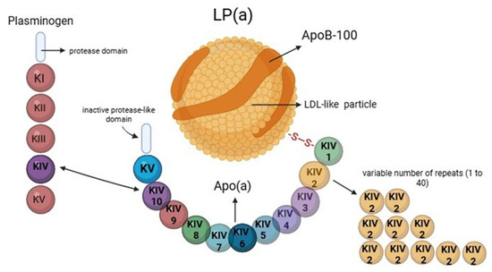
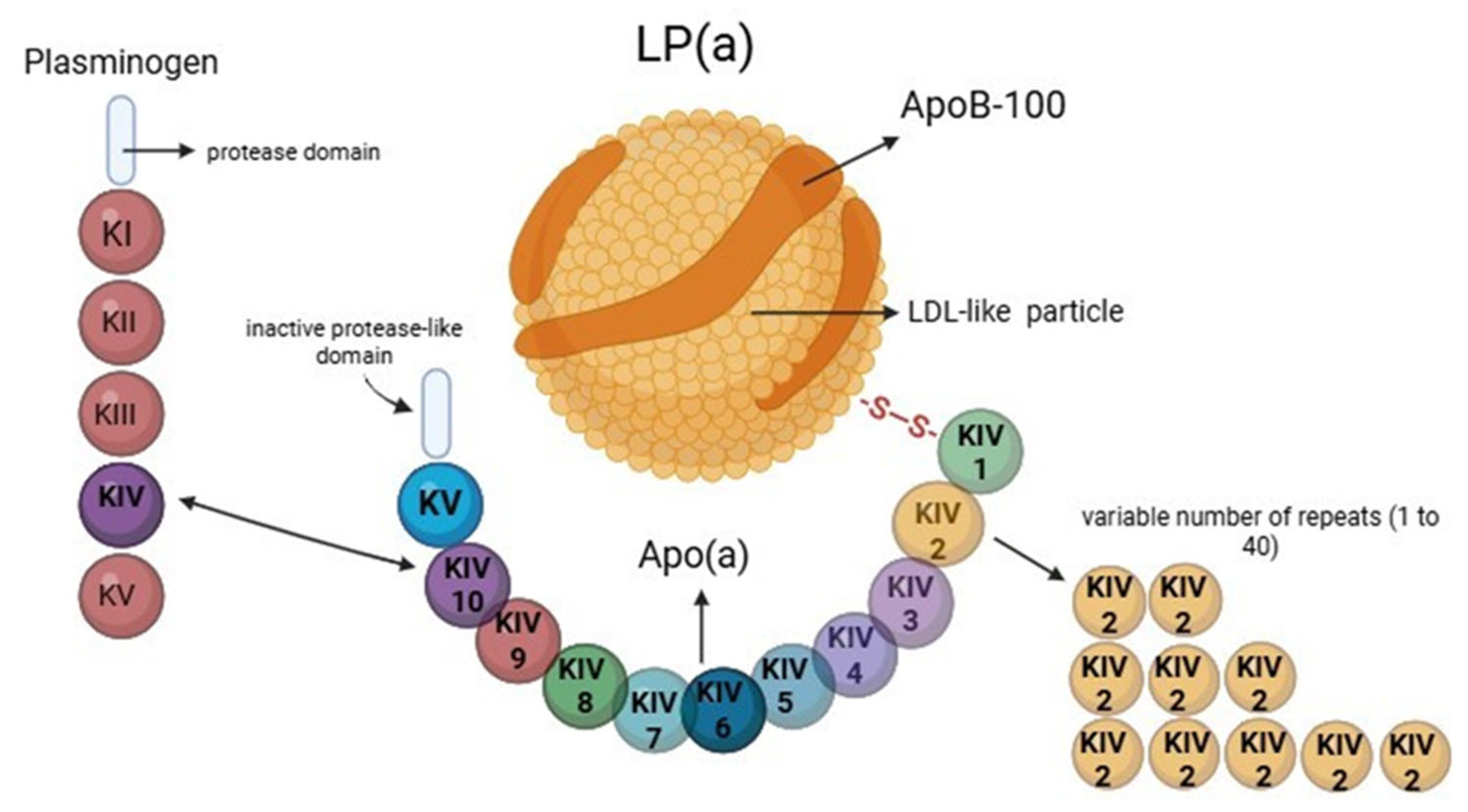
Apo(a) contains ten subtypes of kringle KIV, called domains (KIV1–KIV10, with the KIV-2 domain repeated variably up to more than 40 times), one copy of KV, and an inactive protease domain [2]. The KIV-9 domain includes a scavenger receptor-binding site, which may promote arterial wall accumulation. Apo(a) is highly glycosylated, with carbohydrate chains accounting for over 30% of its molecular weight, influencing solubility and hepatic clearance (Figure 1) [5,6].
Figure 1.
Structure of lipoprotein(a) and its analogy with plasminogen. Apo(a) contains ten subtypes of kringle KIV, called domains (with the KIV-2 domain repeated variably up to more than 40 times), one copy of KV, and an inactive protease domain. The kringle IV-10 domain in Apo(a) is highly homologous to the kringle IV domains of plasminogen.
Apo(a) has remarkable structural homology to plasminogen, the precursor of plasmin, a key enzyme in fibrinolysis. This homology is primarily characterized by the presence of kringle domains, which are looped, triple-disulfide-bonded structures responsible for protein–protein interactions [7,8]. Specifically, Apo(a) contains multiple kringle IV (KIV) repeats, one kringle V (KV), and an inactive protease-like domain, all closely mirroring the domain organization of plasminogen. The kringle IV-10 domain in Apo(a) is highly homologous to the kringle IV domains of plasminogen. However, unlike plasminogen, Apo(a) lacks proteolytic activity because its protease domain is catalytically inactive. This structural similarity allows Apo(a) to compete with plasminogen for binding to fibrin, endothelial cells, and macrophages, potentially inhibiting fibrinolysis. As a result, Lp(a) is considered pro-thrombotic due to its anti-fibrinolytic properties. Apo(a) may impair plasminogen activation, thereby reducing plasmin formation and delaying clot resolution (Figure 1) [8]. Furthermore, Lp(a) interacts with biologically active molecules, including plasminogen, interleukin-8, and tissue factor, thereby contributing to thrombotic and vascular inflammatory processes [6,7,8].
Lipoprotein(a) exhibits marked inter-individual variability in plasma concentrations, primarily governed by genetic determinants. Physiological, dietary, and environmental factors play a relatively minor role and can either lower or raise Lp(a) plasma levels [9].
The key gene involved is LPA, located on chromosome 6q26-27, which encodes Apo (a) [10]. A unique feature of the LPA gene is the presence of a variable number of kringle IV type 2 (KIV-2) repeats, ranging from fewer than 10 to more than 40 copies (Figure 1) [11]. This copy number variation leads to Apo(a) isoforms of different sizes that are inversely correlated with circulating Lp(a) levels: smaller isoforms (fewer KIV-2 repeats) are associated with higher plasma concentrations, whereas larger isoforms correspond to lower levels [12]. This relationship is genetically determined and highly stable over time. Median Lp(a) concentrations are 4 to 5 times higher in individuals with small Apo (a) isoforms (<22 kringle IV repeats) than in those with only large isoforms (≥22 kringle IV repeats) [4]. In addition to KIV-2 copy number variation, specific single-nucleotide polymorphisms (SNPs), such as rs10455872 and rs3798220, have been linked to elevated Lp(a) levels and increased cardiovascular risk [13,14]. These SNPs act independently of KIV-2 size and affect protein expression or stability.
Lp(a) has a relatively long plasma half-life of approximately 3–5 days, compared to 2–3 days for LDL, and its concentration is largely determined by hepatic synthesis regulated by the LPA gene. The precise mechanisms involved in Lp(a) catabolism remain incompletely understood; however, hepatic receptors and lysosomal degradation pathways are believed to play key roles [6]. Unlike LDL, Lp(a) levels are minimally affected by lifestyle factors such as diet and physical activity, or by conventional lipid-lowering therapies like statins [10]. Although Lp(a) contains apolipoprotein B-100, its clearance is not primarily mediated by the LDL receptor (LDL-R). Indeed, statins, which upregulate LDL-R expression, do not lower Lp(a) levels, and Proprotein convertase subtilisin/kexin type 9 inhibitors (PCSK9 inhibitors)—which also increase LDL-R density—reduce Lp(a) only modestly (by ~20–30%). Alternative clearance mechanisms have been proposed, involving scavenger receptors class B type I (SR-BI), the endocytic receptor megalin (LRP-1), and pattern recognition receptors like toll-like receptor 2 (TLR2). Although their precise contributions remain under investigation, these pathways may play supporting roles in Lp(a) catabolism. The liver is considered the central organ for both synthesis and potential degradation of Lp(a). At the same time, the kidneys may contribute modestly to Lp(a) clearance, although they are not believed to be significant metabolism sites [15].
3. Pathophysiological Mechanisms in Cardiovascular Disease
3.1. Pro-Atherogenic Effects
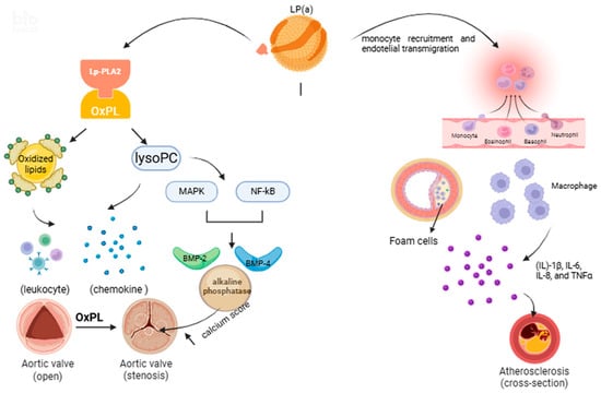
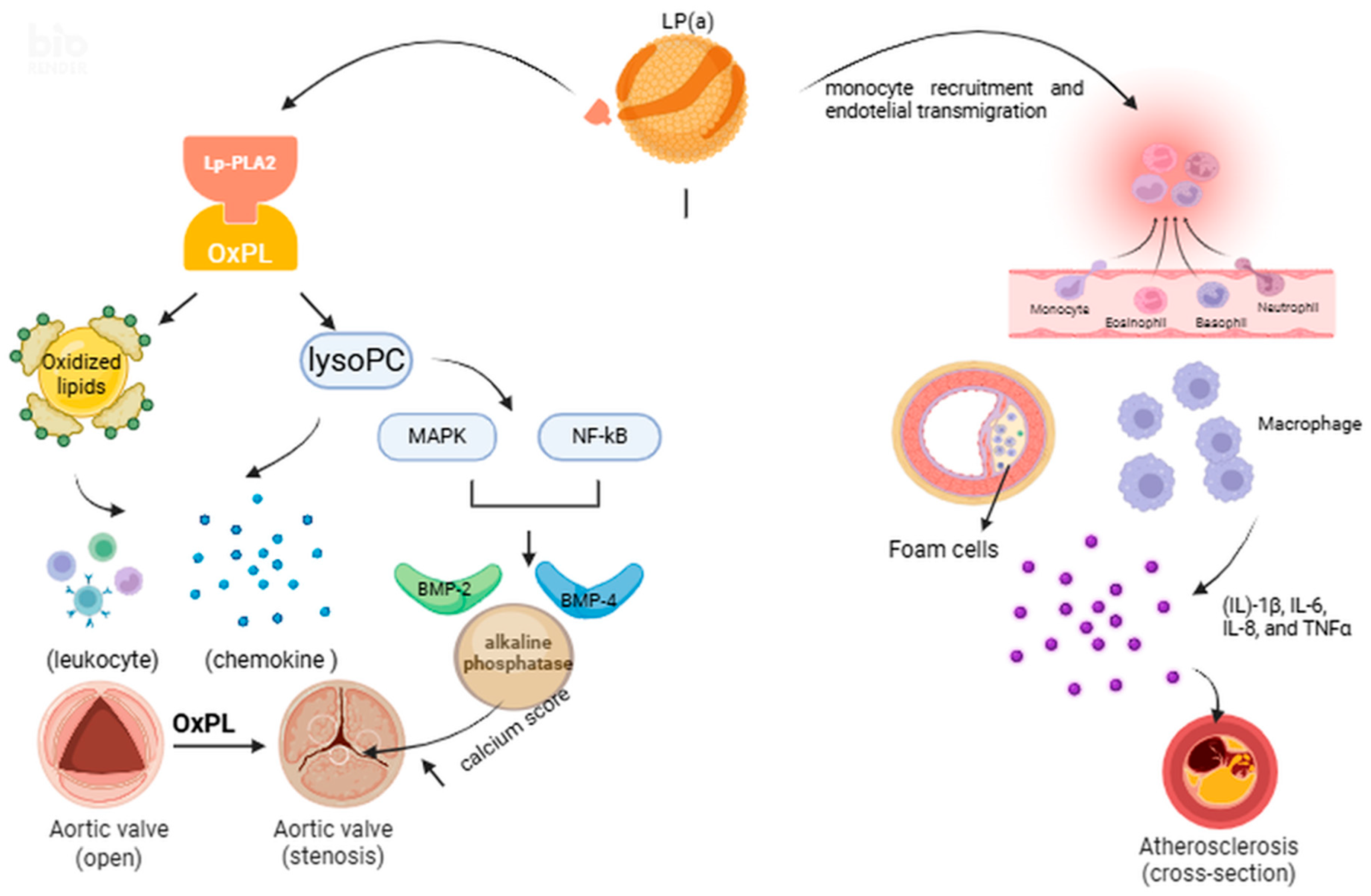
Lp(a) has been strongly associated with increased cardiovascular risk, including myocardial infarction, ischemic stroke, peripheral artery disease, atrial fibrillation, and heart failure. The Copenhagen City Heart Study demonstrated a progressive increase in myocardial infarction risk across Lp(a) concentrations, with no evidence of a lower safety threshold [4]. Lp(a) promotes atherosclerosis through multiple mechanisms, including pro-atherogenic lipid deposition, inflammation, impaired fibrinolysis, and vascular calcification. Structurally, Lp(a) can infiltrate the arterial wall similarly to LDL but tends to accumulate more at sites of endothelial injury (Figure 2) [13]. Lp(a) carries oxidized phospholipids (OxPL), which act as potent pro-inflammatory mediators. These stimulate cytokine production, such as interleukin-8 (IL-8), promote monocyte recruitment, endothelial adhesion molecule expression, and vascular smooth muscle cell activation [14]. OxPLs also stimulate monocyte chemoattractant protein-1, which is physically present on Lp(a), potentially enhancing its entry into the vessel wall (Figure 2).
Figure 2.
Cellular and molecular mechanisms involved in atherosclerosis and in the pathogenesis of calcific aortic valve degeneration. OxPLs, via Lp-PLA2 activity, generate pro-inflammatory mediators that activate NF-κB/MAPK signaling, upregulating BMP-2, BMP-4, Runx2, and promoting calcific nodule formation in aortic stenosis. Lp(a) promotes monocyte recruitment, foam cell formation, and macrophage-driven inflammation, leading to matrix degradation and plaque destabilization. Lp-PLA2: lipoprotein-associated phospholipase A2; OxPLs: oxidized phospholipids; LysoPC: lysophosphatidylcholine; MAPK: Mitogen-Activated Protein Kinase; NF-Kb: Nuclear Factor kappa-light-chain-enhancer of activated B cells; BMP-2: Bone Morphogenetic Protein 2; BMP-4: Bone Morphogenetic Protein 4; IL-1β: Interleukin-1 beta; IL-6: Interleukin-6; IL-8: Interleukin-8; TNFα: Tumor Necrosis Factor alpha.
Furthermore, Apo(a) contains lysine-binding sites that allow it to bind tightly to exposed surfaces on denuded endothelium and accumulate within subintimal spaces and aortic valve leaflets, thereby promoting local inflammation [2]. Lp(a) reduces collagen synthesis and promotes matrix degradation, weakening the fibrous cap and increasing plaque vulnerability [16,17].
3.2. Pro-Thrombotic/Anti-Fibrinolytic Effects
Lp(a) exerts potent anti-fibrinolytic and pro-thrombotic effects, primarily due to its structural similarity to plasminogen and its capacity to interfere with multiple steps of the fibrinolytic cascade. These effects become relevant when Lp(a) is translocated from the circulation to the arterial wall, allowing Apo(a) to accumulate within the endothelium and interact with fibrin [18]. Its etiology remains unknown, but Apo(a) is believed to have originated from the plasminogen gene through evolutionary mechanisms involving gene duplication and structural remodeling [19]. Plasminogen is a glycoprotein zymogen that plays a central role in the fibrinolytic system as the precursor of plasmin, the key enzyme responsible for degrading fibrin clots. It comprises five kringle domains (K1–K5) and a serine protease domain at the C-terminus [2]. Among these, kringle 1 and, especially, kringle 5 demonstrate a high affinity for lysine-binding sites (LBS) [20]. Plasminogen is activated by cleavage at the Arg561–Val562 bond by either tissue-type plasminogen activator (t-PA) or urokinase-type plasminogen activator (u-PA), resulting in the generation of plasmin. This enzyme degrades fibrin into soluble fibrin degradation products, facilitating clot resolution [21]. Apo(a) contains ten subtypes of kringle IV (KIV), with kringle IV type 10 in particular harboring a lysine-binding site analogous to that of plasminogen, though it lacks proteolytic activity. While plasminogen is activated via cleavage at Arg561–Val562, Apo(a) has a Ser-Ile substitution at this cleavage site, which prevents its conversion into a plasmin-like enzyme [22].
Due to this structural homology, Lp(a) competitively inhibits plasminogen binding to fibrin, endothelial surfaces, and fibrinogen receptors, thereby impairing its activation to plasmin and ultimately reducing fibrinolysis. The affinity of Lp(a) for fibrin is inversely related to the size of the Apo(a) isoform, with smaller isoforms demonstrating greater binding capacity [23,24]. Moreover, Lp(a) interferes with the binding of plasminogen to annexin II, a key receptor expressed on both platelets and endothelial cells, thereby inhibiting plasminogen activation at the cell surface [20]. Additionally, Lp(a) indirectly impairs plasminogen activation by inducing the expression of inhibitory molecules in adjacent cells. In particular, it upregulates plasminogen activator inhibitor-1 (PAI-1) in endothelial cells, which subsequently inhibits the fibrinolytic activity of both t-PA and u-PA [21].
3.3. Pro-Inflammatory Effects
Lp(a) exerts significant pro-inflammatory activity within the atherosclerotic plaque microenvironment, independent of its role in lipid deposition. Experimental and histological evidence demonstrates that Lp(a) enhances monocyte recruitment and transmigration through the endothelium by upregulating the expression of vascular adhesion molecules, such as VCAM-1, E-selectin, and ICAM-1 [5,25,26]. Once within the subendothelial space, Lp(a) promotes macrophage activation and foam cell formation, contributing to chronic inflammation and plaque progression [27]. Activated macrophages secrete several pro-inflammatory cytokines, including interleukin (IL)-1β, IL-6, IL-8, and TNFα, creating a local milieu that favors matrix degradation and plaque destabilization (Figure 2) [28]. Lp(a) may also impair the resolution of inflammation by inhibiting efferocytosis, the clearance of apoptotic cells, which is essential for maintaining plaque stability. These mechanisms underscore the direct role of Lp(a) as a pro-inflammatory mediator in atherogenesis [15].
The pro-inflammatory activity of Lp(a) has recently been implicated not only in atherosclerosis but also in non-cardiovascular conditions, including cancer.
Emerging evidence suggests a potential link between altered lipid profiles, including elevated Lp(a) levels, and the presence and progression of gynecologic tumors. More specifically, recent retrospective analyses have identified Lp(a) as a factor associated with cancer outcomes. In a study focusing on endometrial carcinoma (EC), higher Lp(a) concentrations were significantly associated with poorer prognosis in multivariate models, indicating that Lp(a) could have prognostic relevance in endometrial cancer [29].
These findings underscore a possible role for Lp(a) not only in cardiovascular risk but also in tumor biology, although the underlying mechanisms remain to be clarified.
3.4. A Key Driver of Calcific Aortic Valve Stenosis
Calcific aortic valve stenosis (CAVS) is the most prevalent valvular heart disease in developed countries and a growing global health burden. It is characterized by chronic inflammation and fibrocalcific remodeling of the aortic valve leaflets. Lp(a) has emerged as a key player in the pathophysiology of CAVS, independently of traditional atherosclerotic risk factors. A robust body of evidence—including observational studies, Mendelian randomization analyses, genome-wide association studies (GWAS), large population-based cohorts, and histopathological findings—supports a causal link between elevated Lp(a) concentrations and both aortic valve calcification and progression to symptomatic severe AS. The prevalence of aortic valve calcification in individuals over the age of 65 is approximately 50%, and at least 25% of these patients will eventually develop symptomatic aortic stenosis requiring either surgical or transcatheter valve replacement [30]. CAVS is now recognized as an active and progressive pathological process rather than a passive degenerative condition [31]. Evidence from studies on the pathogenesis of calcific aortic valve disease suggests an active disease process characterized by lipoprotein deposition, chronic inflammation, and leaflet calcification [32]. The cellular and molecular mechanisms underlying valvular leaflet calcification are highly complex. The first phase, before calcification, is similar to vascular atherosclerosis. Following endothelial injury, oxidized low-density lipoproteins (oxLDL) initiate a pro-inflammatory cascade that disrupts the homeostatic cross-talk between valvular endothelial cells (VECs) and interstitial cells (VICs) [33]. The pro-calcific potential of OxPLs is largely mediated through enzymatic activity and downstream signaling cascades, including the action of lipoprotein-associated phospholipase A2 (Lp-PLA2), an enzyme bound to lipoproteins such as LDL and Lp(a), that hydrolyzes OxPLs into pro-inflammatory lysophosphatidylcholine (lysoPC) and oxidized non-esterified fatty acid, which promote endothelial activation, leukocyte adhesion, and chemokine expression. These inflammatory changes facilitate the recruitment of monocytes and their differentiation into macrophages [34,35]. The enzymatic breakdown products of OxPLs by Lp-PLA2 also promote osteogenic reprogramming of valvular interstitial cells (VICs). This transformation leads to matrix vesicle release and subsequent calcific nodule formation, contributing to the fibrocalcific remodeling seen in aortic stenosis (Figure 2) [36,37,38].
A pivotal study by Thanassoulis et al. demonstrated that genetic variants in the LPA gene were associated with aortic valve calcification and incident AS. In a cohort of over 6900 individuals with computed tomography (CT) imaging, the study identified a significant association between the LPA gene variant rs10455872 and the presence of AVC (odds ratio 2.05). Genetic variants in the LPA gene were associated with aortic valve calcification and incident AS. The study concluded that genetically elevated Lp(a) levels play a causal role in the pathogenesis of aortic valve calcification and stenosis [39]. The Kaiser et al. study confirmed the robust association of elevated Lp(a) levels with CT-quantified aortic valve calcium in over 3000 individuals [40]. In a 2024 meta-analysis that included patients with aortic stenosis, elevated Lp(a) was associated with a 41% faster increase in aortic jet velocity and 57% greater transvalvular gradient [41]. In clinical practice, Lp(a) may serve as a biomarker for early calcific activity and faster hemodynamic deterioration.
4. Clinical Practice: Measurement and Screening
Since the 1990s, Mendelian randomization and genetic studies have consistently demonstrated the role of elevated Lp(a) as an independent cardiovascular risk factor [39].
While there is still a need to define clinically relevant thresholds, it is universally accepted that the higher the Lp(a) concentration, the greater the cardiovascular risk. According to the latest global and continental statistics from 2022, around 20% of the world’s population, or approximately 1.5 billion people, have Lp(a) levels considered elevated (≥100–125 nmol/L, about 50 mg/dL), increased from the previous estimate of 1.4 billion [42].
Nowadays, Mendelian randomization studies continue to investigate the effects of Lp(a)-lowering therapies, either alone or in combination with LDL-C-lowering treatments or lifestyle interventions, on cardiovascular disease [43].
The Dallas Heart Study (DHS) provides a unique prospective database with a median 9.5-year follow-up to assess the relationships among white, black, and Hispanic subjects to provide insights into the atherogenicity of Lp(a). The influence of Apo(a) isoform size on circulating Lp(a) concentrations shows marked interethnic variability, accounting for up to 80% of plasma level differences across populations [44]. Epidemiological data suggest that Lp(a) distribution varies by ethnicity and race. Individuals of African descent and South Asian populations tend to have higher median Lp(a) levels than White, Hispanic, or East Asian individuals. These ethnic differences are likely attributable to variations in LPA gene locus size and SNPs [45,46].
Lp(a) isoform size varies widely due to KIV-2 repeat polymorphism in the Apo(a) allele and is inversely related to plasma Lp(a) levels, with smaller isoforms associated with higher concentrations. The presence of two genetically determined isoforms further increases variability. This marked heterogeneity, together with the high homology between Apo(a) and plasminogen, complicates immunoassay standardization, a challenge exacerbated by the lack of universal calibrators, differing measurement units, and the absence of standardized validation guidelines.
Isoform-sensitive assays detect the total lipoprotein mass, expressed in milligrams per deciliter (mg/dL), based on the number of repeated KIV-2 motifs. These assays are prone to over- or underestimating Lp(a) levels, depending on the specific assay calibrator -that is, the antibody used to bind to selected Apo(a) antigenic sites [46].
In 1995, Marcovina et al. developed and validated the first isoform-insensitive assay, utilizing a monoclonal antibody that targets a unique epitope in KIV-9 of Apo(a). This method accurately measures Lp(a) independent of Apo(a) isoform size, focusing on the number of circulating particles rather than the mass. As a result, Lp(a) plasma concentration is expressed in nanomoles per liter (nmol/L). Today, various isoform-insensitive ELISAs have been validated and recommended for the quantification of Lp(a) [47] (Table 1).
Table 1.
Summary description of isoform-sensitive and isoform-insensitive assay.
Therefore, according to updated methods and international recommendations, the ideal Lp(a) assay should be: insensitive to Apo(a) isoform size; specific to Lp(a) particles; report values in nmol/L; reproducible and well-standardized, ensuring comparability of results across different laboratories [48].
The recently published “2025 Focused Update of the 2019 ESC/EAS Guidelines for the Management of Dyslipidaemias” identifies individuals with Lp(a) levels between 30 mg/dL (62 nmol/L) and 50 mg/dL (105 nmol/L) as being at increased risk, with levels at or above 50 mg/dL (105 nmol/L) considered clinically significant high risk [49]. However, there is no generalized consensus on Lp(a) risk thresholds. (Table 2 and Table 3).
Table 2.
Summary of principal current major guidelines on management of Lp(a).
Table 3.
Common Lp(a) risk thresholds.
High Lp(a) concentrations are present in approximately 10–25% of the population, and levels remain relatively stable over a lifetime. The estimated intra-individual biological variability is approximately 20%, indicating that a single measurement is generally sufficient in adults. A second measurement is only required to confirm very high values.
For this reason, recent guidelines and consensus statements recommend measuring Lp(a) at least once in adulthood. With some exceptions (e.g., kidney or liver disease, acute infection, hormonal disorders, pregnancy), repeat testing is not necessary, as it does not improve risk prediction [5,49].
In younger individuals, Lp(a) measurement is recommended in the presence of a personal or family history of premature (<60 years) atherosclerotic cardiovascular disease (ASCVD), including ischemic stroke, particularly when no other risk factors are identifiable. Given that Lp(a) levels are largely genetically determined, cascade testing (screening of first-degree relatives) is a practical approach to identifying undiagnosed high Lp(a) in families [50].
Familial hypercholesterolemia (FH) and other inherited dyslipidemias are often associated with elevated Lp(a), likely due to shared genetic variants. The cardiovascular risk is substantially higher when both conditions coexist. Measuring Lp(a) in these individuals can help clinicians refine cardiovascular risk assessment and guide personalized treatment strategies [51].
Therefore, the 2025 ESC focused update on dyslipidaemias recommends that every adult undergo screening at least once in their lifetime, with particular attention to young individuals with familial hypercholesterolemia (FH), premature ASCVD without other identifiable risk factors, a family history of ASCVD, or elevated Lp(a). Screening is also advised to refine risk classification in individuals with moderate cardiovascular risk [49].
In primary prevention, several models now aim to integrate Lp(a) into ASCVD risk prediction frameworks [52,53]. Moreover, the 2025 ESC focused update on dyslipidaemias introduces an online algorithm designed to estimate the risk of heart attack and stroke associated with Lp(a), considering sex, age, BMI, lipid profile, and other cardiovascular risk factors [49].
Even in individuals with normal LDL-C and lipid panels, elevated Lp(a) independently predicts atherosclerotic risk. Optimizing risk stratification is critical in borderline or intermediate-risk patients, where Lp(a) testing may allow better reclassification into high-risk categories. These patients could be candidates for earlier initiation or intensification of lipid-lowering or antiplatelet therapies and Lp(a)-targeted drugs.
Cascade testing for elevated Lp(a) is recommended in the context of familial hypercholesterolemia, a family history of markedly elevated Lp(a), premature ASCVD, personal or familial, especially without other risk factors. Still, it does not require genotyping, polygenic risk scoring, or Apo(a) isoform size analysis [4]. Looking ahead, there is a growing consensus that all adults should have Lp(a) measured at least once in their lifetime (Table 4).
Table 4.
Recommendation for Lp(a) measurement.
5. Management of Elevated Lp(a): From Past Disappointments to Future Hope
Currently, there are no approved therapies that specifically target and lower Lp(a). Consequently, the management of patients with elevated Lp(a) relies on the aggressive optimization of global cardiovascular risk. Since conventional drugs cannot directly lower Lp(a), the goal is to mitigate its impact by reducing the burden of other modifiable risk factors, such as LDL cholesterol, blood pressure, and glucose, along with a renewed emphasis on lifestyle optimization [4]. The effects of current lipid-lowering therapies on Lp(a) are as follows.
5.1. Statins
Statins are the cornerstone of LDL-C management. It is well-documented that they can induce a slight but statistically significant increase in Lp(a) levels. However, it is crucial to emphasize that this effect does not diminish the therapy’s clinical benefit. Analyses from the JUPITER trial have shown that the efficacy of rosuvastatin in reducing cardiovascular events is similar and significant in patients with both high and low Lp(a) levels. Therefore, statin treatment should not be discontinued, as its cardiovascular benefits far outweigh any potential risk associated with the modest increase in Lp(a). Consequently, elevated Lp(a) is considered a major residual risk factor, even after optimal statin therapy and the achievement of low LDL-C levels [47].
5.2. Ezetimibe and Bempedoic Acid
Although neither ezetimibe nor bempedoic acid is a targeted Lp(a)-lowering therapy, they play a crucial role in managing global cardiovascular risk in patients with high Lp(a), consistent with significant guideline recommendations. Their function is not to directly lower Lp(a) but to reduce the overall atherogenic burden, primarily by decreasing LDL-C. The scientific literature on ezetimibe’s direct effect on Lp(a) is marked by conflicting results, as highlighted by two major meta-analyses published almost simultaneously. A 2018 meta-analysis by Sahebkar, Awad et al., which pooled data from seven RCTs involving 2337 patients with primary hypercholesterolemia, concluded that ezetimibe monotherapy (10 mg/day) produced a small but statistically significant reduction in plasma Lp(a) concentrations compared to placebo [54]. A second, larger meta-analysis, published by Khan, Sahebkar et al. in the same year, analyzed 10 RCTs (15 treatment arms) involving a total of 5188 subjects. This analysis found no statistically significant effect of ezetimibe therapy on plasma Lp(a) concentrations [54]. Bempedoic acid reduces hepatic cholesterol synthesis by inhibiting the enzyme ATP citrate lyase (ACL), leading to an upregulation of hepatic LDL receptors. The most definitive data on its effect on Lp(a) come from a secondary analysis of the CLEAR Harmony trial, which specifically evaluated inflammatory markers and Lp(a). The study’s conclusion was unequivocal: bempedoic acid “did not have a clinically important impact on Lp(a)” [55].
5.3. Niacin (Vitamin B3)
Niacin was long the only oral drug capable of significantly reducing Lp(a), with dose-dependent reductions of approximately 20–30%. Despite this effect, niacin is no longer recommended in modern clinical practice for the prevention of cardiovascular disease. Two large randomized clinical trials, AIM-HIGH and HPS2-THRIVE [50,56] evaluated the addition of niacin to statin therapy in high-risk cardiovascular patients. Both studies were stopped early or yielded negative results, demonstrating no additional benefit in reducing cardiovascular events compared to statin therapy alone. Furthermore, niacin therapy was associated with an unfavorable tolerability profile and an increased risk of side effects [51].
5.4. Lipoprotein Apheresis
Lipoprotein apheresis is an extracorporeal procedure, similar to dialysis, that physically removes lipoproteins from the blood. It is currently the most effective therapeutic option, capable of reducing Lp(a) levels by 60–75% or more after each session. Due to its invasive nature, high cost, and limited availability in highly specialized centers, its use is reserved for very select cases. In the United States, the FDA has approved apheresis for patients with familial hypercholesterolemia, documented cardiovascular disease, LDL-C > 100 mg/dL, and Lp(a) > 60 mg/dL. Observational studies suggest that this therapy may reduce future cardiovascular events by up to 86% [57].
5.5. PCSK9 Inhibitors (Evolocumab, Alirocumab)
Proprotein convertase subtilisin/kexin type 9 (PCSK9) inhibitors, evolocumab and alirocumab, are potent drugs primarily approved for lowering LDL-C. However, their action extends beyond the LDL-C axis, offering an additional benefit of great clinical interest: a consistent reduction in Lp(a) levels. A recent and extensive meta-analysis of 47 randomized controlled trials by Rivera et al. precisely quantified this reduction [58]. The results showed an overall mean decrease in Lp(a) levels of −27% (95% CI: −29.8% to −24.1%; p < 0.001). Subgroup analysis detailed the effect of individual molecules: evolocumab showed a mean reduction of −29.35%, while alirocumab showed a mean decrease of −24.50%. It is important to note that despite the numerical difference, the meta-analysis concluded that the effect of the two drugs on Lp(a) reduction was not statistically different (p for interaction = 0.06). Thus, both monoclonal antibodies demonstrate significant and comparable efficacy in reducing this important risk marker [58].
The failure of niacin and the limitations of current drugs have shifted focus to therapies targeting the LPA gene directly. The core premise of these developments is the validation of the “Lp(a) hypothesis”: demonstrating that specific, potent reduction of Lp(a) translates into a significant reduction in cardiovascular events (MACE). Three main classes of agents are currently in advanced clinical development: antisense oligonucleotides (ASOs), small interfering RNAs (siRNAs), and oral small molecule inhibitors (Table 5).
Table 5.
Summary of Novel Specific Lp(a)-Lowering Agents.
5.6. Pelacarsen
Pelacarsen (also known as AKCEA-APO(a)-LRx or TQJ230) is a second-generation ASO conjugated with N-acetylgalactosamine (GalNAc). This GalNAc conjugation ensures specific and efficient uptake by hepatocytes via the asialoglycoprotein receptor. Pelacarsen is an antisense oligonucleotide (ASO) that selectively degrades LPA mRNA in hepatocytes, inhibiting Apo(a) synthesis at the source. This process inhibits translation and, as a result, the synthesis of the Apo(a) protein, drastically reducing the production and circulating levels of Lp(a) [67].
The results from the Pelacarsen Phase 2 program provided the first robust evidence of the efficacy and safety of a targeted approach for Lp(a). The dose-ranging study enrolled 286 patients with established ASCVD and Lp(a) levels ≥ 60 mg/dL (~150 nmol/L) [59]. Treatment with pelacarsen produced significant and dose-dependent reductions in Lp(a). With the highest cumulative dosing regimen (equivalent to 80 mg monthly), a mean reduction in Lp(a) of up to 80% was observed. A finding of exceptional clinical relevance was that 98% of patients treated with this dose achieved Lp(a) levels below the risk threshold of 50 mg/dL (<125 nmol/L), demonstrating that the drug can normalize levels in nearly all treated patients. Consistent with its mechanism of action, pelacarsen also significantly reduced levels of oxidized phospholipids associated with both apoB (OxPL-apoB) and Apo(a) (OxPL-Apo(a)), with reductions of 70–88%. The analysis of its effects on LDL-C provided an important clarification of the measurement mechanism. While the lab-reported LDL-C (which includes Lp(a)-C) decreased by up to 26%, the analysis of corrected LDL-C (LDL-Ccorr = lab LDL-C-directly measured Lp(a)-C) showed a neutral to slight reduction of up to 19%, which barely reached statistical significance (p = 0.05) only at the highest dose. This finding is critical: it demonstrates that Pelacarsen has no clinically relevant effect on LDL metabolism and that the decrease in reported LDL-C is essentially a measurement artifact due to the potent reduction of Lp(a)-C [59,60]. The safety profile of Pelacarsen in Phase 2 was favorable. The most frequent adverse events were mild to moderate injection-site reactions, reported in 27% of treated patients versus 6% in the placebo group. Crucially, no clinically significant concerns emerged regarding thrombocytopenia, liver function (transaminases), or renal function, addressing and overcoming potential safety fears historically associated with the ASO class (Table 5).
Based on the promising Phase 2 results, the Phase 3 cardiovascular outcome trial, Lp(a)HORIZON (NCT04023552), was initiated. This is a global, multicenter, double-blind, placebo-controlled study that enrolled 8325 participants with established ASCVD and Lp(a) levels ≥ 70 mg/dL (~175 nmol/L). Patients were randomized to receive pelacarsen 80 mg via subcutaneous injection once a month or placebo, in addition to optimal medical therapy. The primary endpoint of the study is the reduction in the risk of a composite of major adverse cardiovascular events (MACE), defined as cardiovascular death, non-fatal myocardial infarction, non-fatal stroke, and urgent coronary revascularization requiring hospitalization. Lp(a)HORIZON is the first Phase 3 CVOT for a specific Lp(a)-lowering therapy, and its results are awaited with enormous interest by the scientific community. Topline data are expected in the first half of 2026. A positive outcome would not only lead to the approval of the first drug in this class but would also definitively validate Lp(a) as a therapeutic target (Table 6) [61]. In parallel, the Phase 2 Lp(a)FRONTIERS study (NCT05646381) is ongoing, evaluating the effect of Pelacarsen on the progression of calcific aortic stenosis, another condition strongly linked to Lp(a) (Table 6 and Table 7).
Table 6.
Efficacy and Key Findings of Emerging Lp(a)-Lowering Therapies in Phase 2 Trials.
Table 7.
Overview of Ongoing and Planned Phase 3 Cardiovascular Outcome Trials for Lp(a)-Lowering Therapies.
5.7. Small Interfering RNAs (siRNAs)
siRNAs are small double-stranded RNA molecules that leverage the physiological mechanism of RNA interference (RNAi). Also conjugated with GalNAc for hepatic targeting, they are incorporated into the RNA-induced silencing complex (RISC) upon entering the cell. The RISC complex uses one of the siRNA strands as a guide to specifically find and cleave the LPA gene’s mRNA, leading to its degradation and gene silencing (Table 5, Table 6 and Table 7).
5.7.1. Olpasiran (AMG 890)
Olpasiran is an siRNA, also GalNAc-conjugated for targeted liver delivery, that utilizes the endogenous RNAi mechanism. By binding to the RISC, olpasiran guides the selective cleavage and degradation of LPA mRNA, blocking Apo(a) synthesis with remarkable efficacy [64]. The results from the Phase 2 OCEAN(a)-DOSE study were notable. Treatment with olpasiran at doses ≥ 75 mg every 12 weeks (Q12W) produced median reductions in Lp(a) levels exceeding 95% at 36 weeks (Table 6). Moreover, the drug demonstrated a very durable effect, with significant decreases persisting for nearly a year after the last dose [62]. One of the most important and unexpected findings from a secondary analysis of the OCEAN(a)-DOSE study concerns its effect on inflammatory biomarkers. Although olpasiran potently reduced both Lp(a) and its associated oxidized phospholipids (OxPL-apoB), it had no significant effect on systemic inflammation biomarkers, such as high-sensitivity C-reactive protein (hs-CRP) or interleukin-6 (hs-IL-6). This finding is of fundamental importance because it challenges the prevailing linear hypothesis that the pathogenicity of Lp(a) is primarily mediated by measurable systemic inflammation. The lack of an effect on hs-CRP and IL-6 suggests that the pro-atherogenic and pro-thrombotic mechanisms of Lp(a) may be more localized to the vessel wall or mediated by other inflammatory or thrombotic pathways not measured by these standard biomarkers. This discovery makes the outcome study even more crucial, as it will verify whether reducing Lp(a) (and OxPLs) alone is sufficient to reduce clinical events, regardless of an effect on systemic inflammation [64]. To assess the clinical impact of this potent reduction, the Phase 3 OCEAN(a)-Outcomes study (NCT05581303) was started (Table 7). This is a double-blind, placebo-controlled trial that has completed enrollment of 7297 participants with ASCVD and elevated Lp(a). The primary endpoint is the time to the first composite event of death from coronary heart disease (CHD), myocardial infarction, or urgent coronary revascularization. The estimated final data collection for the primary outcome is December 2026, with results expected in 2027.
5.7.2. Lepodisiran
Lepodisiran, another siRNA, is distinguished by its potentially unprecedented duration of action. The results from the Phase 1 study of lepodisiran were nothing short of remarkable, showing that a single 608 mg dose reduced Lp(a) by 94% at 48 weeks post-injection [65]. The Phase 2 ALPACA study confirmed these data, with a 400 mg dose reducing time-averaged Lp(a) by 94% over 6 months, and a significant effect persisting for over a year after a single administration. This duration of action opens the possibility for a semi-annual or even annual dosing regimen (Table 6) [63]. The Phase 3 outcome study for lepodisiran, named ACCLAIM-Lp(a) (NCT06292013), is distinguished by its ambitious design. The study plans to enroll approximately 15,600 participants. The most innovative and strategically relevant aspect is that ACCLAIM-Lp(a) is the first CVOT in this field to include not only patients in secondary prevention (with established ASCVD) but also a large cohort of high-risk primary prevention patients. This bold move reflects great confidence in the drug’s safety and efficacy profile. A positive outcome in this population could accelerate the adoption of Lp(a) therapy well beyond the niche of secondary prevention, providing the necessary evidence to recommend screening and treatment of elevated Lp(a) before a first cardiovascular event occurs, marking a true revolution in preventive cardiology [59]. The study is currently recruiting, with completion expected by March 2029 (Table 7).
5.7.3. Zerlasiran
Zerlasiran (formerly SLN360), an siRNA developed by Silence Therapeutics, demonstrated robust efficacy in its Phase 2 study, ALPACAR-360 (NCT05537571). The primary endpoint, the time-averaged reduction of Lp(a) at 36 weeks, was successfully met. Infrequent dosing regimens, such as 300 mg every 16 or 24 weeks, produced placebo-adjusted mean reductions between −81.3% and −85.6%. The effect proved to be exceptionally durable, with time-averaged suppression of Lp(a) up to week 60 maintained at −79.2% (with 300 mg every 16 weeks) and −77.1% (with 450 mg every 24 weeks). From a safety perspective, the treatment was well-tolerated. The most common adverse events were mild and transient injection-site reactions, and no serious drug-related safety issues emerged. The authors concluded that these solid Phase 2 data support the progression of zerlasiran to Phase 3 cardiovascular outcome studies (Table 5, Table 6 and Table 7) [66].
5.8. Oral Small Molecules: The Advent of Muvalaplin (LY3473329)
The prospect of an effective oral therapy for Lp(a) has profound implications. While long-acting injectable therapies offer advantages in terms of adherence, an effective daily pill represents a more convenient, less invasive, and more accessible alternative for most patients and healthcare systems. Muvalaplin is the first in a new class of oral small-molecule inhibitors. Its mechanism of action is unique and distinct from RNA therapies: it acts at the post-translational level, after the Apo(a) and apoB proteins have been synthesized. Muvalaplin selectively binds to Apo(a) and prevents its non-covalent interaction with apoB, thereby blocking the crucial first step in the formation of the mature Lp(a) particle (Table 5) [68].
The Phase 2 KRAKEN study (NCT05563246) evaluated the efficacy and safety of muvalaplin in 233 participants at high cardiovascular risk (with ASCVD, diabetes, or FH) and Lp(a) levels ≥ 175 nmol/L. The results showed a significant and dose-dependent reduction in Lp(a). Using a novel assay that measures intact Lp(a), the placebo-adjusted mean reductions at 12 weeks were approximately 81–86% with the 60 mg and 240 mg doses (Table 6). The primary theoretical concern for any drug that physically interacts with the Apo(a) protein is the potential for an off-target effect on plasminogen, given their high structural homology. Interference with plasminogen activity could theoretically alter fibrinolysis and increase the risk of thrombosis. Therefore, assessing the selectivity of muvalaplin was a key objective of its development. The results from both Phase 1 and Phase 2 studies were extremely reassuring: muvalaplin administration was not associated with any clinically significant changes in plasminogen levels or activity. This finding is of capital importance, as it demonstrates that it is possible to design a small molecule capable of selectively inhibiting Lp(a) assembly without interfering with the fibrinolytic system [59]. The success of the KRAKEN study has paved the way for the crucial next step in development. Muvalaplin is now being evaluated in the pivotal MOVE-Lp(a) Phase 3 trial (NCT07157774). This large, placebo-controlled cardiovascular outcomes study is designed to determine if treatment with muvalaplin significantly reduces the risk of major adverse cardiovascular events (MACE) in patients with elevated Lp(a) (Table 7).
5.9. Gene Editing
Beyond RNA therapies and small molecules, which require chronic, lifelong treatment, the research horizon is pushing towards potentially curative and permanent solutions. Somatic genome editing, particularly through the CRISPR-Cas9 system, represents the most revolutionary approach under investigation. Somatic genome editing, particularly through the CRISPR-Cas9 system, aims to introduce a permanent disruption in the LPA gene within hepatocytes, thereby permanently inactivating Apo(a) production [69]. Preclinical studies have already provided convincing proof of concept. In a transgenic mouse model expressing human Apo(a), a single treatment with a CRISPR-Cas9 system delivered by an adeno-associated virus (AAV) vector led to the near-complete and permanent elimination of circulating Apo(a) [70]. The potential advantage of this approach is enormous. Unlike RNA therapies that require repeated injections for life to maintain gene suppression, a single genome editing treatment could provide a durable and permanent reduction in Lp(a), effectively representing a functional cure. Despite its immense promise, the path to the clinical application of genome editing for Lp(a) is still long and fraught with challenges. Absolute long-term safety and editing specificity must be ensured, minimizing the risk of “off-target” genomic modifications. Furthermore, the permanent modulation of the human genome raises complex ethical issues that will need to be carefully considered by the scientific community and society as a whole.
6. Unanswered Questions and Future Directions
To better understand the potential benefit of lowering Lp(a) levels in terms of reducing the risk of coronary artery disease, Mendelian randomization studies have been conducted. These studies estimated that a reduction in Lp(a) levels of approximately 70 to 90 mg/dL is required to achieve at least a 15–20% decrease in cardiovascular risk [71]. Another study by the Lp(a) GWAS consortium has demonstrated that a reduction in Lp(a) levels of at least 65.7 mg/dL is necessary to achieve a 22% decrease in the risk of coronary artery disease [72]. If these results are confirmed in Phase 3 studies on new Lp(a) lowering drugs, it could represent a turning point in our ability to reduce the risk of cardiovascular events further, significantly enhancing both primary and, especially, secondary prevention strategies. This could be the final support for the “Lp(a) hypothesis”. This hypothesis refers to the connection between Lp(a) concentrations and a cardiovascular event. In particular, specific reduction of Lp(a) levels can lead to a measurable decrease in cardiovascular events.
All these new and promising therapies, once approved by regulatory authorities and subsequently incorporated into major international guidelines, are expected to be widely adopted in clinical practice, particularly in the fields of cardiology and cardiovascular medicine. In this context, significant challenges may arise, such as the identification of the ideal patient candidate and, most importantly, the determination of the appropriate laboratory threshold for initiating treatment.
Based on current evidence, the patients who may benefit most from Lp(a)-lowering interventions are those who exhibit persistently elevated Lp(a) levels despite treatment with oral lipid-lowering therapies (statins and/or ezetimibe) and PCSK9 inhibitors. This benefit is linked to the pro-inflammatory effects of Lp(a) and its associated oxidized phospholipids [73].
Another critical issue that will inevitably need to be addressed is the economic impact that the approval and subsequent integration of these therapies into clinical practice will entail. According to a Markov model based on data from the OCEAN(a) and HORIZON trials, olpasiran would be considered cost-effective at an annual price of approximately AU $1867 (≃US $1200) using a willingness-to-pay threshold of AU $28,000 per quality-adjusted life year (QALY). In comparison, pelacarsen would be cost-effective at around AU $984 (≃US $634) per year. At a higher threshold of AU $50,000/QALY, acceptable annual prices would rise to AU $4207 for olpasiran and AU $2464 for pelacarsen. Also, olpasiran was associated with an incremental gain of 0.87 quality-adjusted life years (QALYs) compared to standard of care in patients with established atherosclerotic cardiovascular disease and elevated Lp(a) levels [72].
These data suggest that the substantial costs national healthcare systems would need to bear for broad therapeutic coverage should be mitigated by the implementation of strict patient selection criteria. Based on data available, eligibility for these therapies should be limited to individuals at truly high risk for cardiovascular events, or those with a documented history of such events, who are already receiving maximally optimized lipid-lowering therapy, including statins, ezetimibe, and PCSK9 inhibitors, crucially, patients should also have significantly elevated Lp(a) levels, as defined by the aforementioned cut-off values [70] similarly to the patients that respect the inclusion criteria of the two pivotal trial, e.g., OCEAN(a)-Outcomes and Lp(a) HORIZON. However, if these therapies demonstrate a significant reduction in major adverse cardiovascular events (MACE) in ongoing clinical trials, their impact could be transformative.
7. Conclusions
Lpa is now a recognized, causal, independent risk factor for atherosclerotic cardiovascular disease. The need for an implementation of Lp(a) level screening for identification of high-risk patients and the enhancement of risk reduction strategies represent the most important current clinical implications. Furthermore, new, promising drugs that act specifically to reduce Lp(a) have presented encouraging results in Phase 2 trials and are currently under investigation in Phase 3 trials. These new classes of drugs could be the future direction for the improvement of the reduction of cardiovascular burden. In this clinical scenario, clinicians are called on for a new challenge: proceed with careful screening of Lp(a) levels, improve strategies for cardiovascular risk reduction in patients at higher risk and be prepared for management of the upcoming new classes of drugs as a possible new future era in the treatment of cardiovascular disease.
Author Contributions
Conceptualization, E.R. and G.P.U.; methodology, E.R. and G.P.U.; validation, E.R., G.P.U., N.G.C., A.D.C., A.E.L., T.T., L.D., F.M., A.N., V.C., R.M., N.C., P.G., R.R., A.T., M.G., F.P., G.A., L.G. and S.M.; investigation and resources, E.R., G.P.U., N.G.C., A.D.C., A.E.L., T.T., L.D., F.M., A.N., V.C., R.M., N.C., P.G., R.R., A.T., M.G., F.P., G.A., L.G. and S.M.; data curation, R.R., G.P.U., N.G.C., A.D.C., A.E.L., T.T., L.D., F.M., A.N., V.C., R.M., N.C., P.G., R.R., A.T., M.G., F.P., G.A., L.G. and S.M.; writing—original draft preparation, E.R., G.P.U., N.G.C., A.D.C., A.E.L., T.T., L.D., F.M., A.N., V.C., R.M., N.C., P.G., R.R., A.T., M.G., F.P., G.A., L.G. and S.M.; writing—review and editing, E.R., G.P.U., N.G.C., A.D.C., A.E.L., T.T., L.D., F.M., A.N., V.C., R.M., N.C., P.G., R.R., A.T., M.G., F.P., G.A., L.G. and S.M.; supervision, E.R. and G.P.U.; project administration, E.R. and G.P.U. All authors have read and agreed to the published version of the manuscript.
Funding
This research received no external funding.
Institutional Review Board Statement
Not applicable.
Informed Consent Statement
Not applicable.
Data Availability Statement
No new data were created or analyzed in this study. Data sharing is not applicable to this article.
Conflicts of Interest
The authors declare no conflicts of interest.
References
- Ference, B.A.; Ginsberg, H.N.; Graham, I.; Ray, K.K.; Packard, C.J.; Bruckert, E.; Hegele, R.A.; Krauss, R.M.; Raal, F.J.; Schunkert, H.; et al. Low-Density Lipoproteins Cause Atherosclerotic Cardiovascular Disease. 1. Evidence from Genetic, Epidemiologic, and Clinical Studies. A Consensus Statement from the European Atherosclerosis Society Consensus Panel. Eur. Heart J. 2017, 38, 2459–2472. [Google Scholar] [CrossRef] [Scilit] [PubMed]
- Tsimikas, S. A Test in Context: Lipoprotein(a). J. Am. Coll. Cardiol. 2017, 69, 692–711. [Google Scholar] [CrossRef] [Scilit] [PubMed]
- Lemešić, D.L.; Šimičević, L.; Ganoci, L.; Gelemanović, A.; Šućur, N.; Pećin, I. Association of rs3798220 Polymorphism with Cardiovascular Incidents in Individuals with Elevated Lp(a). Diagnostics 2025, 15, 404. [Google Scholar] [CrossRef] [Scilit] [PubMed]
- Kronenberg, F.; Mora, S.; Stroes, E.S.G.; Ference, B.A.; Arsenault, B.J.; Berglund, L.; Dweck, M.R.; Koschinsky, M.; Lambert, G.; Mach, F.; et al. Lipoprotein(a) in Atherosclerotic Cardiovascular Disease and Aortic Stenosis: A European Atherosclerosis Society Consensus Statement. Eur. Heart J. 2022, 43, 3925–3946. [Google Scholar] [CrossRef] [Scilit]
- Boffa, M.B.; Koschinsky, M.L. Update on Lipoprotein(a) as a Cardiovascular Risk Factor and Mediator. Curr. Atheroscler. Rep. 2013, 15, 360. [Google Scholar] [CrossRef] [Scilit]
- McLean, J.W.; Tomlinson, J.E.; Kuang, W.-J.; Eaton, D.L.; Chen, E.Y.; Fless, G.M.; Scanu, A.M.; Lawn, R.M. cDNA Sequence of Human Apolipoprotein(a) Is Homologous to Plasminogen. Nature 1987, 330, 132–137. [Google Scholar] [CrossRef] [Scilit]
- Boerwinkle, E.; Leffert, C.C.; Lin, J.; Lackner, C.; Chiesa, G.; Hobbs, H.H. Apolipoprotein(a) Gene Accounts for Greater than 90% of the Variation in Plasma Lipoprotein(a) Concentrations. J. Clin. Investig. 1992, 90, 52–60. [Google Scholar] [CrossRef] [Scilit]
- Nordestgaard, B.G.; Chapman, M.J.; Ray, K.; Borén, J.; Andreotti, F.; Watts, G.F.; Ginsberg, H.; Amarenco, P.; Catapano, A.; Descamps, O.S.; et al. Lipoprotein(a) as a Cardiovascular Risk Factor: Current Status. Eur. Heart J. 2010, 31, 2844–2853. [Google Scholar] [CrossRef] [Scilit]
- Enkhmaa, B.; Anuurad, E.; Berglund, L. Lipoprotein (a): Impact by Ethnicity and Environmental and Medical Conditions. J. Lipid Res. 2016, 57, 1111–1125. [Google Scholar] [CrossRef] [Scilit]
- Utermann, G. The Mysteries of Lipoprotein(a). Science 1989, 246, 904–910. [Google Scholar] [CrossRef] [Scilit]
- Wang, S.; Zha, L.; Chen, J.; Du, D.; Liu, D.; Zhong, M.; Shang, R.; Sun, D.; Sun, C.; Jin, E. The Relationship between Lipoprotein(a) and Risk of Cardiovascular Disease: A Mendelian Randomization Analysis. Eur. J. Med. Res. 2022, 27, 211. [Google Scholar] [CrossRef] [Scilit]
- Matveyenko, A.; Matienzo, N.; Ginsberg, H.; Nandakumar, R.; Seid, H.; Ramakrishnan, R.; Holleran, S.; Thomas, T.; Reyes-Soffer, G. Relationship of Apolipoprotein(a) Isoform Size with Clearance and Production of Lipoprotein(a) in a Diverse Cohort. J. Lipid Res. 2023, 64, 100336. [Google Scholar] [CrossRef] [Scilit] [PubMed]
- Nordestgaard, B.G.; Langsted, A. Lipoprotein (a) as a Cause of Cardiovascular Disease: Insights from Epidemiology, Genetics, and Biology. J. Lipid Res. 2016, 57, 1953–1975. [Google Scholar] [CrossRef] [Scilit] [PubMed]
- Scipione, C.A.; Sayegh, S.E.; Romagnuolo, R.; Tsimikas, S.; Marcovina, S.M.; Boffa, M.B.; Koschinsky, M.L. Mechanistic Insights into Lp(a)-Induced IL-8 Expression: A Role for Oxidized Phospholipid Modification of Apo(a). J. Lipid Res. 2015, 56, 2273–2285. [Google Scholar] [CrossRef] [Scilit] [PubMed]
- Yang, X.-P.; Amar, M.J.; Vaisman, B.; Bocharov, A.V.; Vishnyakova, T.G.; Freeman, L.A.; Kurlander, R.J.; Patterson, A.P.; Becker, L.C.; Remaley, A.T. Scavenger Receptor-BI Is a Receptor for Lipoprotein(a). J. Lipid Res. 2013, 54, 2450–2457. [Google Scholar] [CrossRef] [Scilit]
- Boffa, M.B.; Koschinsky, M.L. Lipoprotein (a): Truly a Direct Prothrombotic Factor in Cardiovascular Disease? J. Lipid Res. 2016, 57, 745–757. [Google Scholar] [CrossRef] [Scilit]
- Van Der Valk, F.M.; Bekkering, S.; Kroon, J.; Yeang, C.; Van Den Bossche, J.; Van Buul, J.D.; Ravandi, A.; Nederveen, A.J.; Verberne, H.J.; Scipione, C.; et al. Oxidized Phospholipids on Lipoprotein(a) Elicit Arterial Wall Inflammation and an Inflammatory Monocyte Response in Humans. Circulation 2016, 134, 611–624. [Google Scholar] [CrossRef] [Scilit]
- Beisiegel, U.; Niendorf, A.; Wolf, K.; Reblin, T.; Rath, M. Lipoprotein(a) in the Arterial Wall. Eur. Heart J. 1990, 11, 174–183. [Google Scholar] [CrossRef] [Scilit]
- Schmidt, K.; Noureen, A.; Kronenberg, F.; Utermann, G. Structure, Function, and Genetics of Lipoprotein (a). J. Lipid Res. 2016, 57, 1339–1359. [Google Scholar] [CrossRef] [Scilit]
- Clark, L.T. Atherogenesis and Thrombosis: Mechanisms, Pathogenesis, and Therapeutic Implications. Am. Heart J. 1992, 123, 1106–1109. [Google Scholar] [CrossRef] [Scilit]
- Etingin, O.R.; Hajjar, D.P.; Hajjar, K.A.; Harpel, P.C.; Nachman, R.L. Lipoprotein (a) Regulates Plasminogen Activator Inhibitor-1 Expression in Endothelial Cells. A Potential Mechanism in Thrombogenesis. J. Biol. Chem. 1991, 266, 2459–2465. [Google Scholar] [CrossRef] [Scilit] [PubMed]
- Anglés-Cano, E.; Díaz, A.D.L.P.; Loyau, S. Inhibition of Fibrinolysis by Lipoprotein(a). Ann. N. Y. Acad. Sci. 2001, 936, 261–275. [Google Scholar] [CrossRef] [Scilit] [PubMed]
- Hervio, L.; Chapman, M.; Thillet, J.; Loyau, S.; Angles-Cano, E. Does Apolipoprotein(a) Heterogeneity Influence Lipoprotein(a) Effects on Fibrinolysis? Blood 1993, 82, 392–397. [Google Scholar] [CrossRef] [Scilit] [PubMed]
- Hervio, L.; Durlach, V.; Girard-Globa, A.; Angles-Cano, E. Multiple Binding with Identical Linkage: A Mechanism That Explains the Effect of Lipoprotein(a) on Fibrinolysis. Biochemistry 1995, 34, 13353–13358. [Google Scholar] [CrossRef] [Scilit]
- Takami, S.; Yamashita, S.; Kihara, S.; Ishigami, M.; Takemura, K.; Kume, N.; Kita, T.; Matsuzawa, Y. Lipoprotein(a) Enhances the Expression of Intercellular Adhesion Molecule-1 in Cultured Human Umbilical Vein Endothelial Cells. Circulation 1998, 97, 721–728. [Google Scholar] [CrossRef] [Scilit]
- Allen, S.; Khan, S.; Tam, S.-P.; Koschinsky, M.; Taylor, P.; Yacoub, M. Expression of Adhesion Molecules by Lp(a): A Potential Novel Mechanism for Its Atherogenicity. FASEB J. 1998, 12, 1765–1776. [Google Scholar] [CrossRef] [Scilit]
- Girard, A.S.; Paulin, A.; Manikpurage, H.D.; Lajeunesse, E.; Clavel, M.; Pibarot, P.; Krege, J.H.; Mathieu, P.; Thériault, S.; Arsenault, B.J. Impact of Lipoprotein(a) on Valvular and Cardiovascular Outcomes in Patients with Calcific Aortic Valve Stenosis. J. Am. Heart Assoc. 2025, 14, 6. [Google Scholar] [CrossRef] [Scilit]
- Klezovitch, O.; Edelstein, C.; Scanu, A.M. Stimulation of Interleukin-8 Production in Human THP-1 Macrophages by Apolipoprotein(a). J. Biol. Chem. 2001, 276, 46864–46869. [Google Scholar] [CrossRef] [Scilit]
- Liu, C.; Xiao, Z.; Liu, Z.; Huang, X. Correlation between Lipoprotein(a) and Endometrial Cancer Prognosis: A Retrospective Cohort Study. Clin Lab. 2025, 71, 1829. [Google Scholar] [CrossRef] [Scilit] [PubMed]
- Vahanian, A.; Beyersdorf, F.; Praz, F.; Milojevic, M.; Baldus, S.; Bauersachs, J.; Capodanno, D.; Conradi, L.; De Bonis, M.; De Paulis, R.; et al. 2021 ESC/EACTS Guidelines for the Management of Valvular Heart Disease. Eur. Heart J. 2022, 43, 561–632. [Google Scholar] [CrossRef] [Scilit]
- Celeski, M.; Nusca, A.; Ciavaroli, N.G.; Martucciello, A.; Crisci, F.; Polito, D.; Mangiacapra, F.; Cammalleri, V.; Melfi, R.; Gallo, P.; et al. Co-Occurrence of Aortic Stenosis and Coronary Artery Disease: Facing Challenges Before, During, and After Transcatheter Aortic Valve Replacement. J. Clin. Med. 2025, 14, 4709. [Google Scholar] [CrossRef] [Scilit]
- Freeman, R.V.; Otto, C.M. Spectrum of Calcific Aortic Valve Disease: Pathogenesis, Disease Progression, and Treatment Strategies. Circulation 2005, 111, 3316–3326. [Google Scholar] [CrossRef] [Scilit]
- Hulin, A.; Hego, A.; Lancellotti, P.; Oury, C. Advances in Pathophysiology of Calcific Aortic Valve Disease Propose Novel Molecular Therapeutic Targets. Front. Cardiovasc. Med. 2018, 5, 21. [Google Scholar] [CrossRef] [Scilit]
- Zheng, K.H.; Tsimikas, S.; Pawade, T.; Kroon, J.; Jenkins, W.S.A.; Doris, M.K.; White, A.C.; Timmers, N.K.L.M.; Hjortnaes, J.; Rogers, M.A.; et al. Lipoprotein(a) and Oxidized Phospholipids Promote Valve Calcification in Patients with Aortic Stenosis. J. Am. Coll. Cardiol. 2019, 73, 2150–2162. [Google Scholar] [CrossRef] [Scilit] [PubMed]
- Wolters Kluwer Health, Inc. Arteriosclerosis, Thrombosis, and Vascular Biology; Wolters Kluwer Health, Inc.: Waltham, MA, USA, 2015; Volume 35. [Google Scholar] [CrossRef] [Scilit]
- Mahmut, A.; Boulanger, M.-C.; El Husseini, D.; Fournier, D.; Bouchareb, R.; Després, J.-P.; Pibarot, P.; Bossé, Y.; Mathieu, P. Elevated Expression of Lipoprotein-Associated Phospholipase A2 in Calcific Aortic Valve Disease. J. Am. Coll. Cardiol. 2014, 63, 460–469. [Google Scholar] [CrossRef] [Scilit] [PubMed]
- Pantazi, D.; Tellis, C.; Tselepis, A.D. Oxidized Phospholipids and Lipoprotein-associated Phospholipase A2 (LP-PLA2) in Atherosclerotic Cardiovascular Disease: An Update. BioFactors 2022, 48, 1257–1270. [Google Scholar] [CrossRef] [Scilit] [PubMed]
- Bogdanova, M.; Kostina, A.; Zihlavnikova Enayati, K.; Zabirnyk, A.; Malashicheva, A.; Stensløkken, K.-O.; Sullivan, G.J.; Kaljusto, M.-L.; Kvitting, J.-P.E.; Kostareva, A.; et al. Inflammation and Mechanical Stress Stimulate Osteogenic Differentiation of Human Aortic Valve Interstitial Cells. Front. Physiol. 2018, 9, 1635. [Google Scholar] [CrossRef] [Scilit]
- Thanassoulis, G.; Campbell, C.Y.; Owens, D.S.; Smith, J.G.; Smith, A.V.; Peloso, G.M.; Kerr, K.F.; Pechlivanis, S.; Budoff, M.J.; Harris, T.B.; et al. Genetic Associations with Valvular Calcification and Aortic Stenosis. N. Engl. J. Med. 2013, 368, 503–512. [Google Scholar] [CrossRef] [Scilit]
- Kaiser, Y.; Singh, S.S.; Zheng, K.H.; Verbeek, R.; Kavousi, M.; Pinto, S.-J.; Vernooij, M.W.; Sijbrands, E.J.G.; Boekholdt, S.M.; De Rijke, Y.B.; et al. Lipoprotein(a) Is Robustly Associated with Aortic Valve Calcium. Heart 2021, 107, 1422–1428. [Google Scholar] [CrossRef] [Scilit]
- Arsenault, B.J.; Loganath, K.; Girard, A.; Botezatu, S.; Zheng, K.H.; Tzolos, E.; Abdoun, K.; Tastet, L.; Capoulade, R.; Côté, N.; et al. Lipoprotein(a) and Calcific Aortic Valve Stenosis Progression: A Systematic Review and Meta-Analysis. JAMA Cardiol. 2024, 9, 835. [Google Scholar] [CrossRef] [Scilit]
- Grundy, S.M.; Stone, N.J.; Bailey, A.L.; Beam, C.; Birtcher, K.K.; Blumenthal, R.S.; Braun, L.T.; De Ferranti, S.; Faiella-Tommasino, J.; Forman, D.E.; et al. 2018 AHA/ACC/AACVPR/AAPA/ABC/ACPM/ADA/AGS/APhA/ASPC/NLA/PCNA Guideline on the Management of Blood Cholesterol: Executive Summary. J. Am. Coll. Cardiol. 2019, 73, 3168–3209. [Google Scholar] [CrossRef] [Scilit] [PubMed]
- Figueiredo, R.A.O.; Simola-Ström, S.; Rounge, T.B.; Viljakainen, H.; Eriksson, J.G.; Roos, E.; Weiderpass, E. Cohort Profile: The Finnish Health in Teens (Fin-HIT) study: A population-based study. Int. J. Epidemiol. 2019, 48, 23–24h. [Google Scholar] [CrossRef] [Scilit] [PubMed] [PubMed Central]
- Victor, R.G.; Haley, R.W.; Willett, D.L.; Peshock, R.M.; Vaeth, P.C.; Leonard, D.; Basit, M.; Cooper, R.S.; Iannacchione, V.G.; Visscher, W.A.; et al. The Dallas Heart Study: A Population-Based Probability Sample for the Multidisciplinary Study of Ethnic Differences in Cardiovascular Health. Am. J. Cardiol. 2004, 93, 1473–1480. [Google Scholar] [CrossRef] [Scilit] [PubMed]
- Khera, A.V.; Everett, B.M.; Caulfield, M.P.; Hantash, F.M.; Wohlgemuth, J.; Ridker, P.M.; Mora, S. Lipoprotein(a) Concentrations, Rosuvastatin Therapy, and Residual Vascular Risk: An Analysis from the JUPITER Trial (Justification for the Use of Statins in Prevention: An Intervention Trial Evaluating Rosuvastatin). Circulation 2014, 129, 635–642. [Google Scholar] [CrossRef] [Scilit]
- Swearingen, C.A.; Sloan, J.H.; Rhodes, G.M.; Siegel, R.W.; Bivi, N.; Qian, Y.; Konrad, R.J.; Boffa, M.; Koschinsky, M.; Krege, J.; et al. Measuring Lp(a) Particles with a Novel Isoform-Insensitive Immunoassay Illustrates Efficacy of Muvalaplin. J. Lipid Res. 2025, 66, 100723. [Google Scholar] [CrossRef] [Scilit]
- Marcovina, S.M.; Albers, J.J. Lipoprotein (a) Measurements for Clinical Application. J. Lipid Res. 2016, 57, 526–537. [Google Scholar] [CrossRef] [Scilit]
- Heydari, M.; Rezayi, M.; Ruscica, M.; Jpamialahamdi, T.; Johnston, T.P.; Sahebkar, A. The ins and outs of lipoprotein(a) assay methods. Arch. Med. Sci. Atheroscler. Dis. 2023, 8, e128–e139. [Google Scholar] [CrossRef] [Scilit] [PubMed] [PubMed Central]
- Mach, F.; Koskinas, K.C.; van Lennep, J.E.R.; Tokgözoğlu, L.; Badimon, L.; Baigent, C.; Benn, M.; Binder, C.J.; Catapano, A.L.; De Backer, G.G.; et al. 2025 Focused Update of the 2019 ESC/EAS Guidelines for the management of dyslipidaemias: Developed by the task force for the management of dyslipidaemias of the European Society of Cardiology (ESC) and the European Atherosclerosis Society (EAS). Eur. Heart J. 2025, 46, 4359–4378. [Google Scholar] [CrossRef] [Scilit]
- Bhatia, H.S.; Ambrosio, M.; Razavi, A.C.; Alebna, P.L.; Yeang, C.; Spitz, J.A.; Patel, J.; Tsai, M.Y.; Sperling, L.; Shapiro, M.D.; et al. AHA PREVENT Equations and Lipoprotein(a) for Cardiovascular Disease Risk: Insights from MESA and the UK Biobank. JAMA Cardiol. 2025, 10, 810–818. [Google Scholar] [CrossRef] [Scilit]
- Trinder, M.; Uddin, M.M.; Finneran, P.; Aragam, K.G.; Natarajan, P. Clinical Utility of Lipoprotein(a) and LPA Genetic Risk Score in Risk Prediction of Incident Atherosclerotic Cardiovascular Disease. JAMA Cardiol. 2021, 6, 287. [Google Scholar] [CrossRef] [Scilit]
- Awad, K.; Mikhailidis, D.P.; Katsiki, N.; Muntner, P.; Banach, M. Lipid and Blood Pressure Meta-Analysis Collaboration (LBPMC) Group. Effect of Ezetimibe Monotherapy on Plasma Lipoprotein(a) Concentrations in Patients with Primary Hypercholesterolemia: A Systematic Review and Meta-Analysis of Randomized Controlled Trials. Drugs 2018, 78, 453–462. [Google Scholar] [CrossRef] [Scilit] [PubMed]
- Ridker, P.M.; Lei, L.; Ray, K.K.; Ballantyne, C.M.; Bradwin, G.; Rifai, N. Effects of Bempedoic Acid on CRP, IL-6, Fibrinogen and Lipoprotein(a) in Patients with Residual Inflammatory Risk: A Secondary Analysis of the CLEAR Harmony Trial. J. Clin. Lipidol. 2023, 17, 297–302. [Google Scholar] [CrossRef] [Scilit] [PubMed]
- Guyton, J.R.; Slee, A.E.; Anderson, T.; Fleg, J.L.; Goldberg, R.B.; Kashyap, M.L.; Marcovina, S.M.; Nash, S.D.; O’Brien, K.D.; Weintraub, W.S.; et al. Relationship of Lipoproteins to Cardiovascular Events. J. Am. Coll. Cardiol. 2013, 62, 1580–1584. [Google Scholar] [CrossRef] [Scilit] [PubMed]
- Koutsogianni, A.D.; Liamis, G.; Liberopoulos, E.; Adamidis, P.S.; Florentin, M. Effects of Lipid-Modifying and Other Drugs on Lipoprotein(a) Levels—Potent Clinical Implications. Pharmaceuticals 2023, 16, 750. [Google Scholar] [CrossRef] [Scilit] [PubMed]
- Rivera, F.B.; Cha, S.W.; Linnaeus Louisse, C.; Carado, G.P.; Magalong, J.V.; Tang, V.A.; Enriquez, M.G.; Gulati, M.; Enkhmaa, B.; Pagidipati, N.; et al. Impact of Proprotein Convertase Subtilisin/Kexin Type 9 Inhibitors on Lipoprotein(a). JACC Adv. 2025, 4, 101549. [Google Scholar] [CrossRef] [Scilit]
- Nurmohamed, N.S.; Kraaijenhof, J.M.; Stroes, E.S.G. Lp(a): A New Pathway to Target? Curr. Atheroscler. Rep. 2022, 24, 831–838. [Google Scholar] [CrossRef] [Scilit]
- Vuorio, A.; Kovanen, P.T.; Raal, F. Promising Results with the Daily Oral Small Molecule Lipoprotein(a) Inhibitor, Muvalaplin, in High-Risk Cardiovascular Patients with Elevated Lipoprotein(a) Levels. Ann. Transl. Med. 2025, 13, 11. [Google Scholar] [CrossRef] [Scilit]
- Cho, L.; Nicholls, S.J.; Nordestgaard, B.G.; Landmesser, U.; Tsimikas, S.; Blaha, M.J.; Leitersdorf, E.; Lincoff, A.M.; Lesogor, A.; Manning, B.; et al. Design and Rationale of Lp(a)HORIZON Trial: Assessing the Effect of Lipoprotein(a) Lowering with Pelacarsen on Major Cardiovascular Events in Patients With CVD and Elevated Lp(a). Am. Heart J. 2025, 287, 1–9. [Google Scholar] [CrossRef] [Scilit]
- O’Donoghue, M.L.; Rosenson, R.S.; Gencer, B.; López, J.A.G.; Lepor, N.E.; Baum, S.J.; Stout, E.; Gaudet, D.; Knusel, B.; Kuder, J.F.; et al. Small Interfering RNA to Reduce Lipoprotein(a) in Cardiovascular Disease. N. Engl. J. Med. 2022, 387, 1855–1864. [Google Scholar] [CrossRef] [Scilit]
- Rosenson, R.S.; López, J.A.G.; Gaudet, D.; Baum, S.J.; Stout, E.; Lepor, N.E.; Park, J.-G.; Murphy, S.A.; Knusel, B.; Wang, J.; et al. Olpasiran, Oxidized Phospholipids, and Systemic Inflammatory Biomarkers: Results From the OCEAN(a)-DOSE Trial. JAMA Cardiol. 2025, 10, 482. [Google Scholar] [CrossRef] [Scilit]
- Nissen, S.E.; Linnebjerg, H.; Shen, X.; Wolski, K.; Ma, X.; Lim, S.; Michael, L.F.; Ruotolo, G.; Gribble, G.; Navar, A.M.; et al. Lepodisiran, an Extended-Duration Short Interfering RNA Targeting Lipoprotein(a): A Randomized Dose-Ascending Clinical Trial. JAMA 2023, 330, 2075–2083. [Google Scholar] [CrossRef] [Scilit] [PubMed]
- Nicholls, S.J.; Nissen, S.E.; Fleming, C.; Urva, S.; Suico, J.; Berg, P.H.; Linnebjerg, H.; Ruotolo, G.; Turner, P.K.; Michael, L.F. Muvalaplin, an Oral Small Molecule Inhibitor of Lipoprotein(a) Formation: A Randomized Clinical Trial. JAMA 2023, 330, 1042. [Google Scholar] [CrossRef] [Scilit] [PubMed]
- Nissen, S.E.; Ni, W.; Shen, X.; Wang, Q.; Navar, A.M.; Nicholls, S.J.; Wolski, K.; Michael, L.; Haupt, A.; Krege, J.H. Lepodisiran—A Long-Duration Small Interfering RNA Targeting Lipoprotein(a). N. Engl. J. Med. 2025, 392, 1673–1683. [Google Scholar] [CrossRef] [Scilit] [PubMed]
- Nissen, S.E.; Wang, Q.; Nicholls, S.J.; Navar, A.M.; Ray, K.K.; Schwartz, G.G.; Szarek, M.; Stroes, E.S.G.; Troquay, R.; Dorresteijn, J.A.N.; et al. Zerlasiran—A Small-Interfering RNA Targeting Lipoprotein(a): A Phase 2 Randomized Clinical Trial. JAMA 2024, 332, 1992–2002. [Google Scholar] [CrossRef] [Scilit]
- Stankov, S.; Cuchel, M. Gene Editing for Dyslipidemias: New Tools to “Cut” Lipids. Atherosclerosis 2023, 368, 14–24. [Google Scholar] [CrossRef] [Scilit]
- Yeang, C.; Karwatowska-Prokopczuk, E.; Su, F.; Dinh, B.; Xia, S.; Witztum, J.L.; Tsimikas, S. Effect of Pelacarsen on Lipoprotein(a) Cholesterol and Corrected Low-Density Lipoprotein Cholesterol. J. Am. Coll. Cardiol. 2022, 79, 1035–1046. [Google Scholar] [CrossRef] [Scilit]
- Doerfler, A.M.; Park, S.H.; Assini, J.M.; Youssef, A.; Saxena, L.; Yaseen, A.B.; De Giorgi, M.; Chuecos, M.; Hurley, A.E.; Li, A.; et al. LPA Disruption with AAV-CRISPR Potently Lowers Plasma Apo(a) in Transgenic Mouse Model: A Proof-of-Concept Study. Mol. Ther. Methods Clin. Dev. 2022, 27, 337–351. [Google Scholar] [CrossRef] [Scilit]
- Burgess, S.; Ference, B.A.; Staley, J.R.; Freitag, D.F.; Mason, A.M.; Nielsen, S.F.; Willeit, P.; Young, R.; Surendran, P.; Karthikeyan, S.; et al. Association ofLPAVariants with Risk of Coronary Disease and the Implications for Lipoprotein(a)-Lowering Therapies: A Mendelian Randomization Analysis. JAMA Cardiol. 2018, 3, 619. [Google Scholar] [CrossRef] [Scilit]
- Lamina, C.; Kronenberg, F. For the Lp(a)-GWAS-Consortium. Estimation of the Required Lipoprotein(a)-Lowering Therapeutic Effect Size for Reduction in Coronary Heart Disease Outcomes: A Mendelian Randomization Analysis. JAMA Cardiol. 2019, 4, 575. [Google Scholar] [CrossRef] [Scilit]
- Malick, W.A.; Goonewardena, S.N.; Koenig, W.; Rosenson, R.S. Clinical Trial Design for Lipoprotein(a)-Lowering Therapies: JACC Focus Seminar 2/3. J. Am. Coll. Cardiol. 2023, 81, 1633–1645. [Google Scholar] [CrossRef] [Scilit] [PubMed]
- Burvill, A.; Watts, G.F.; Norman, R.; Ademi, Z. Early Health Technology Assessment of Gene Silencing Therapies for Lowering Lipoprotein(a) in the Secondary Prevention of Coronary Heart Disease. J. Clin. Lipidol. 2024, 18, e946–e956. [Google Scholar] [CrossRef] [Scilit]
- Manzato, M.; Wright, R.S.; Jaffe, A.S.; Vasile, V.C. Lipoprotein (a): Underrecognized Risk with a Promising Future. Rev. Cardiovasc. Med. 2024, 25, 393. [Google Scholar] [CrossRef] [Scilit] [PubMed] [PubMed Central]

Disclaimer/Publisher’s Note: The statements, opinions and data contained in all publications are solely those of the individual author(s) and contributor(s) and not of MDPI and/or the editor(s). MDPI and/or the editor(s) disclaim responsibility for any injury to people or property resulting from any ideas, methods, instructions or products referred to in the content. |
© 2026 by the authors. Licensee MDPI, Basel, Switzerland. This article is an open access article distributed under the terms and conditions of the Creative Commons Attribution (CC BY) license.