


Abstract
Background/Objectives: Morin is the first flavonoid isolated from plants in the Moraceae family and has been reported to exhibit anticancer and antioxidant activity. It is classified as a Class IV compound in the Biopharmaceutical Classification System due to its low water solubility and low permeability. This study aimed to develop and characterize morin-loaded liposomes composed of DPPC and cholesterol to improve their stability and anticancer activity. Methods: Liposomes were prepared by thin-film hydration and then processed by bath sonication, probe sonication, or membrane extrusion to control vesicle size. Formulations were characterized by hydrodynamic diameter, polydispersity index (PDI), zeta potential, encapsulation efficiency (EE), and colloidal stability over 7 days at 37 °C. The cytotoxic effects of free and encapsulated morin were evaluated in tumor (MDA-MB-231, B16F10) and non-tumor (NIH-3T3, HEK-293T) cell lines. Results: Extrusion yielded the most homogeneous liposomes (PDI = 0.2, size ≈ 90 nm), whereas sonication methods produced larger, more polydisperse vesicles. EE ranged from 11–17%, with no significant differences among methods. Free morin degraded by 40% at 37 °C after 7 days, whereas liposomal encapsulation significantly reduced degradation, preserving 82–89% of the compound. At a morin equivalent of 3.4 µM, the bath-sonicated formulation reduced viability to approximately 65% in MDA-MB-231 cells and 70% in B16F10 cells, whereas viability remained approximately 86% in NIH-3T3 cells and 82% in HEK-293T cells. All formulations maintained colloidal stability over the study period. In cytotoxicity assays, free morin showed minimal activity, whereas liposomal formulations significantly enhanced antiproliferative effects against cancer cells. Conclusions: These findings demonstrate that DPPC/cholesterol liposomes effectively stabilize morin and improve its bioactivity, representing a promising strategy for delivering this flavonoid in anticancer therapies.
1. Introduction
Morin is a yellow-pigmented flavonoid isolated from plants in the Moraceae family [1,2,3,4], first isolated from species such as Maclura pomifera and Maclura tinctoria [5,6,7,8]. It has demonstrated a wide range of pharmacological activities, including antioxidant, antiinflammatory, antiallergic, antiviral, and anticancer properties [9,10,11,12,13]. The literature describes it as an antioxidant, linking this to its role as a nephroprotective, cardioprotective, and gastroprotective agent by neutralizing free radicals, reducing oxidative stress, and limiting cellular damage [14,15,16,17]. Its anticancer activity has been demonstrated in various cell lines, such as HeLa (cervical cancer) [18], MDA-MB-231 (triple-negative breast cancer) [19,20], U937 (leukemia) [21], and PANC-1 (pancreatic cancer) [14]. Mechanisms include inhibition of migration and invasion and induction of mitochondrial apoptosis, with minimal toxicity to normal cells [22,23,24]. Moreover, its anticancer activity has been studied in in vivo models using rats with colon cancer, where oral administration of morin reduced tumor size [25]. According to the Biopharmaceutical Classification System, morin is classified as a Class IV compound with inherent low solubility and permeability, resulting in low bioavailability [26,27,28,29]. Therefore, formulation represents a significant challenge for its promising applications, with the goal of optimizing delivery by improving key parameters such as solubility and permeability [30,31,32]; in this regard, strategies based on nanotechnology have gained importance in recent decades [33,34,35].
Nanotechnology has extensively studied nanoscale materials to improve the solubility and bioavailability of poorly soluble phytochemicals through encapsulation, which protects them from enzymatic degradation or precipitation in biological fluids while increasing local drug concentrations at the site of action due to their increased surface area [36,37,38,39]. Among these nanosystems, liposomes, which consist of doublelayered vesicles derived from phospholipids, stand out for their biocompatibility, biodegradability, and high capacity to load both hydrophobic and hydrophilic molecules, which contribute to improved stability, reduced toxicity, and controlled, tissue-targeted release [40,41,42,43]. Because of their low toxicity, liposomes have been widely used in cosmetics, food, agriculture, and—above all—pharmaceutical industries [44,45,46,47]. In general, liposomes have been prepared using the thin-film hydration method, employing phospholipids such as soy lecithin, distearoylphosphatidylcholine, hydrogenated lecithin (lecithin ws-50), and L-α-Phosphatidylcholine from eggs [48,49,50]. Morin has been incorporated into liposomes based on hydrogenated lecithin and egg L-α-Phosphatidylcholine, showing promising anticancer effects [51,52]. Interestingly, 1,2-dipalmitoyl-sn-glycero-3-phosphocholine (DPPC) is a saturated zwitterionic phospholipid and an abundant component of pulmonary surfactant [53,54]. Although DPPC is not generally abundant in other biological membranes, its well-defined gel-to-liquid crystalline phase transition temperature (41–43 °C) and reproducible phase behavior have made it a widely used model phospholipid for membrane studies and liposome preparation [55,56]. Morin was shown to interact with DPPC membranes via hydrogen bonds with phosphate groups at the polar interface, without destabilizing the bilayer and preventing leakage of vesicular content [57]. Unsaturated phospholipids pack less compactly, leading to earlier leakage of encapsulated molecules because of their lower phase-transition temperature and greater susceptibility to oxidation, which reduces the stability and shelf life of the formulation [58]. In contrast, DPPC offers greater stability, encapsulation, and drug retention, making it a favorable option for liposomal formulations [57,58].
By intercalating between phospholipid molecules, cholesterol modulates acyl-chain packing and broadens the gel-to-liquid-crystalline phase transition, thereby reducing its cooperativity. This buffering effect is phase-dependent: cholesterol can disrupt the highly ordered gel phase while increasing acyl-chain order in the liquid-crystalline phase. Consequently, incorporating approximately 16 mol% cholesterol was expected to reduce permeability associated with phase-transition defects and thereby improve morin retention and formulation stability [59]. Unlike previous studies that used DPPC vesicles as membrane models to investigate morin association with the phospholipid bilayer, the present work employs DPPC/cholesterol liposomes as delivery nanocarriers, with morin incorporated into the intravesicular aqueous compartment [59]. This approach evaluates DPPC/cholesterol liposomes as delivery carriers under mildly alkaline conditions, in contrast to previous studies that primarily used DPPC vesicles as membrane models to investigate morin–bilayer interactions. The novelty of the work lies in the comparative evaluation of three post-hydration size-control procedures using the same lipid composition and loading conditions, together with the assessment of morin chemical retention and preliminary in vitro antiproliferative activity, while providing a protected aqueous microenvironment, representing a novel formulation strategy aimed at improving morin stability and biological performance [59].
Based on these considerations, the present study investigates DPPC/cholesterol liposomes as carriers for morin, focusing on their physicochemical properties, morin’s chemical stability, and in vitro antiproliferative activity. Although morin’s interactions with phospholipid membranes have been previously characterized [59], the use of DPPC/cholesterol liposomes as nanocarriers to improve morin’s stability and biological performance remains largely unexplored. Given the feasibility of producing DPPC-based liposomes, it is necessary to explore strategies to determine the optimal method for obtaining DPPC liposomes that encapsulate morin.
Liposome preparation methods are classified as conventional or novel [60,61]. Conventional methods include thin-film hydration, bath sonication, probe sonication, extrusion, solvent injection, reverse-phase evaporation, and detergent removal, all of which are simple, economical, and scalable [62,63]. Novel methods include microfluidics, supercritical fluids, freeze-drying, dual asymmetric centrifugation, membrane contactors, gel-assisted hydration, and heating [64,65,66]. These newer methods offer precise control over particle size and encapsulation efficiency, but they require expensive equipment and careful handling of heat-sensitive molecules [65,66]. Current research trends toward novel methods; however, conventional methods remain prevalent in basic research and scalable production due to their simplicity and low production costs [64,67]. Therefore, our study focused on comparing liposome preparation using three conventional methods: extrusion, bath sonication, and probe sonication.
In this study, we developed a liposome formulation based on the phospholipid DPPC and cholesterol as a membrane stabilizer to encapsulate morin. We compared the encapsulation efficiency of three preparation methods (probe sonication, bath sonication, and extrusion) and evaluated the molecule’s stability. Additionally, the cytotoxic effects of the resulting formulations were evaluated in comparison to free morin in tumor cell lines (MDA-MB-231 and B16F10) and non-tumor cells (NIH-3T3 and HEK-293T).
2. Materials and Methods
2.1. Materials
1,2-dipalmitoyl-sn-glycero-3-phosphocholine (DPPC) at 99% purity, cholesterol at 99% purity, morin at 80% purity, phosphate-buffered saline (PBS), potassium hydroxide, and chloroform were purchased from Sigma-Aldrich (St. Louis, MO, USA). The corresponding complete cell growth medium (DMEM or RPMI 1640), fetal bovine serum (FBS), and penicillin/streptomycin (P/S) were obtained from Gibco (Grand Island, NY, USA). The CellTiter 96® AQueous One Solution Cell Proliferation Assay (MTS) was obtained from Promega (Madison, WI, USA). Cell lines, including NIH-3T3, B16F10, HEK-293T, and MDA MB 231, were obtained from ATCC (Manassas, VA, USA).
2.2. Methods
2.2.1. Chemical Stability of Morin
To assess the stability of free morin (protected from light), samples were evaluated at 37 °C, 25 °C, and −20 °C at 100 μg/mL in PBS (adjusted to pH 8) over 1, 3, and 7 days. PBS at pH 8.0 was selected as a compromise between aqueous solubility and chemical stability. Therefore, pH 8.0 provided sufficient apparent solubility for formulation while allowing the protective effect of liposomal encapsulation under mildly alkaline stress to be evaluated [7,68]. Remaining morin concentrations were determined using a previously prepared calibration curve (5–50 μM) with UV–Vis spectroscopy in the range of 300 to 600 nm (399 for a peak of morin). (see Supplementary Material, Figure S1).
All samples were processed in solution and kept under each condition for each analysis. Morin was stored as a solution in PBS at pH 8.0 rather than as a dry powder. Independent light-protected aliquots were prepared for each temperature and sampling time to avoid repeated freeze–thaw cycles. Samples stored at −20 °C were thawed at room temperature for 5 min, gently mixed, and analyzed immediately by UV–Vis spectroscopy. Samples stored at 25 and 37 °C were analyzed directly at the corresponding sampling times.
2.2.2. Development of Liposomes with DPPC and Cholesterol
Three independent liposomal batches were prepared by thin-film hydration at a DPPC: cholesterol: morin molar ratio of 79:16:5. Briefly, DPPC and cholesterol were dissolved in 500 µL of chloroform and transferred to a 25 mL glass flask at 5 °C. The mixture was dried under a nitrogen stream to form a thin, bubble-free film. After chloroform removal, the lipid film was hydrated with 1 mL of morin solution (100 µg/mL) in PBS (pH 8). Empty liposomes containing DPPC and cholesterol at the same molar ratio but without morin were prepared in parallel and subjected to the same size-control procedure as the morin-loaded formulations.
2.2.3. Post-Hydration Size Control of Liposomes
Bath Sonication
One crude liposomal suspension was subjected to bath sonication at 25 °C for 25 min, with continuous direct sonication, while carefully ensuring that the local temperature does not rise sharply. A duration of 25 min was selected to match the total processing time used for probe sonication and to remain within the 20–30 min range commonly reported for bath-sonication processing of liposomal formulations [59].
Probe Sonication
A second crude liposomal suspension was subjected to continuous direct sonication. The mixture was then placed in a probe-type sonicator under the following conditions: 20% amplitude, 25 min with pulses of 8 s of agitation and 2 s of pause (150 cycles), with the sample kept on ice to prevent overheating during sonication.
Extrusion
A third crude liposomal suspension was subjected to 15 cycles of agitation (Vortex) and heating (53 °C) at 1-min intervals [69]. After hydration was complete, decreasing-size extrusion was performed using an Avanti Mini Extruder (Alabaster, AL, USA) with 200- and 100-nm filters Whatman® Nuclepore™ Track-Etched Membranes (Cytiva, Marlborough, MA, USA). Twenty cycles were performed for each filter size. All extrusion steps were performed at 10 °C above the phase-transition temperature of DPPC to facilitate liposome passage through the membranes and achieve the target size [70].
2.2.4. Purification of the Obtained Liposomes by the Three Methodologies
The liposomes were purified by dialysis to remove residual free morin. Morin-loaded liposomes were dialyzed for 24 h at 25 °C against Milli-Q water using Pierce™ SnakeSkin™ Pleated Dialysis Tubing (3.5 kDa MWCO; Thermo Fisher Scientific, Waltham, MA, USA). A dialysis medium-to-sample volume ratio of 500:1 was used, with continuous magnetic stirring at 250 rpm. After dialysis, the final retentate volume of each liposomal suspension was recorded. This volume was used to calculate the total amount of morin recovered in the purified liposomal fraction.
2.2.5. Encapsulation Efficiency of Morin in Liposomes
Encapsulation efficiency (EE) was determined using UV–Vis spectroscopy (Model Specord S600, Analytik Jena, Jena, Germany). A standard curve for morin was prepared by measuring the absorbance of various concentrations in the micromolar range (5–50 μM) and performing a mapping analysis. The encapsulated morin was quantified by disrupting the vesicles with ethanol. In summary, MoriLip (purified morin liposomes) (100 μg/mL, 500 μL), previously purified by dialysis, was diluted with 1500 µL of absolute ethanol. The mixture was vortexed and sonicated until liposomes were completely dissolved, then centrifuged (15,115 rcf, 20 min) to separate free morin. Absorbance was measured at 399 nm. Morin exhibits characteristic UV–Vis absorption bands at approximately 260 and 360 nm. Due to ionization of its hydroxyl groups, increasing pH produces a bathochromic shift in the long-wavelength band toward 390–400 nm, reflecting changes in its electronic distribution [71]. The encapsulation efficiency was calculated as the ratio of the actual amount of drug encapsulated to the total amount of drug used to prepare each batch of liposomes.
2.2.6. Physicochemical Characterization of Liposomes
Hydrodynamic diameter (Dh), polydispersity (PDI), and zeta potential for each formulation were measured by dynamic light scattering (DLS) using a Zetasizer Nano ZS (Malvern Instruments, Malvern, UK) with plastic cuvettes. PDI was calculated using the cumulant method, hydrodynamic diameter was determined from the intensity plot, and zeta potential was calculated using the Smoluchowski equation. Morin concentration was measured by UV–Vis spectroscopy.
2.2.7. Colloidal Stability of Liposomes
1 mL of morin-loaded liposomes was stored in amber glass vials at 37 °C in an incubator for 7 days before analysis. Their size and surface charge were characterized (Section 2.2.6).
2.2.8. Cell Viability Assay
Cell lines, including NIH-3T3 mouse embryonic fibroblasts, B16F10 mouse melanoma, HEK-293T human embryonic kidney, and MDA-MB-231 human breast adenocarcinoma, were seeded at 5000 cells per well in 96-well plates and cultured overnight in the corresponding complete growth medium (DMEM or RPMI 1640) supplemented with 10% FBS and 1% P/S at 37 °C in a humidified atmosphere containing 5% CO2. Cells were then exposed for 24 h to free morin or to the different liposome formulations at morin-equivalent concentrations of 0.85, 1.7, and 3.4 μM. Cells were also treated with the corresponding empty-liposome formulations at lipid concentrations equivalent to those in the morin-loaded formulations. The corresponding volume of water without morin or liposomal formulation was included as the vehicle/viability control. Cells treated with 2% SDS were included as the positive cytotoxicity (cell-death) control.
Cell viability was assessed using MTS per the manufacturer’s instructions. Absorbance was measured at 490 nm, with 655 nm as the reference wavelength, using a Synergy-H4 multimode microplate reader (BioTek, Winooski, VT, USA). Cell viability was expressed as a percentage relative to untreated control cells (100% viability). Each treatment was evaluated in independent experiments (n = 2), with 4 technical replicates per experimental condition. Statistical comparisons between free morin and the corresponding liposomal formulations at each morin-equivalent concentration were performed using one-way ANOVA followed by Dunnett’s multiple-comparisons test, with free morin serving as the reference group. Differences were considered statistically significant at p < 0.05. MDA-MB-231 and B16F10 cells were selected as human breast adenocarcinoma and murine melanoma models, respectively, to provide an exploratory assessment across two distinct tumor cell types. NIH-3T3 and HEK-293T cells were included as non-tumor reference cell lines to assess whether the formulations’ effects were preferentially observed in tumor cells rather than reflecting general cytotoxicity. HEK-293T cells were considered a non-tumor reference cell line rather than a normal primary cell model. Overall, this panel was intended for an initial comparative assessment of cytotoxicity and potential tumor selectivity rather than to establish cancer-type-specific efficacy. The highest final concentration tested was 3.4 µM, determined by the maximum morin concentration that could be standardized across all three formulations, with the extrusion formulation defining the upper concentration available for comparative testing. The formulations contained approximately 10 µg/mL morin (approximately 34 µM) before addition to the cells and underwent a 10-fold dilution in the culture well, resulting in a final concentration of 3.4 µM. Two successive two-fold dilutions yielded final morin-equivalent concentrations of 1.7 and 0.85 µM. All formulations and free morin were compared at the same final morin-equivalent concentrations.
2.2.9. Statistical Analysis
Data are presented as mean ± standard deviation (SD) across independent preparations. Differences between groups were assessed using one-way or two-way ANOVA, as appropriate, followed by Tukey’s or Dunnett’s post hoc tests. Differences were considered statistically significant at p < 0.05. Analyses were performed in GraphPad Prism (version 10). Unless otherwise specified, experiments were conducted in triplicate. Cell viability assays were performed in two independent experiments, with four technical replicates per condition, as described in Section 2.2.8.
3. Results
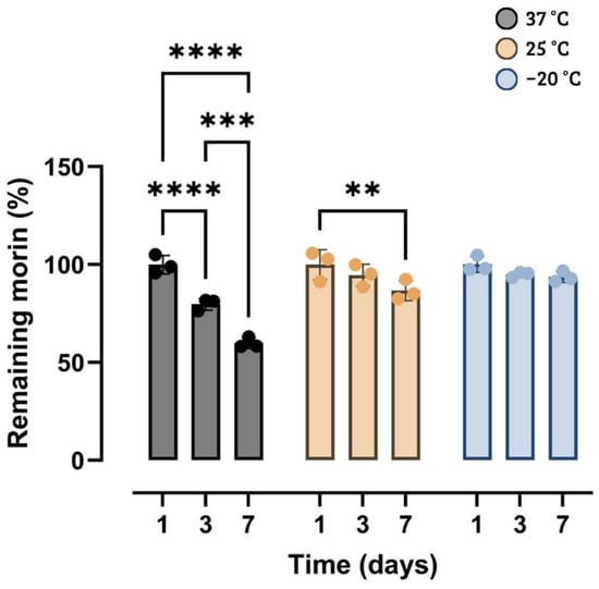
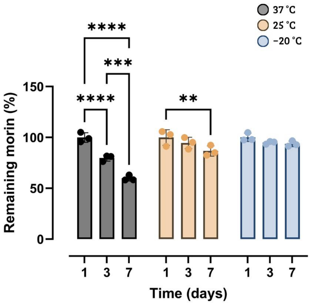
Morin in its free form exhibits a delicate balance between solubility and stability that depends on environmental conditions [72]. The stability of morin at −20, 25, and 37 °C was studied over a specific storage period (1, 3, and 7 days) using UV–visible spectroscopy, with concentrations expressed as the percentage of the initial concentration remaining, as shown in Figure 1. In this study of the chemical stability of free morin in an alkaline medium (pH = 8), a clear dependence on storage time and temperature was observed. At the initial time point, all three temperature conditions showed comparable morin levels, with percentages around 100% (99.9 ± 4.8% at 37 °C, 99.9 ± 7.6% at 25 °C, and 100.1 ± 4.1% at −20 °C), ensuring that the measurements are comparable and that subsequent differences are not due to baseline variations between groups. As the incubation period progressed, the 37 °C condition showed a considerable, progressive loss of morin, decreasing to 79.8 ± 3.0% on day 3 and 59.9 ± 2.8% on day 7. In contrast, loss was more moderate at 25 °C, with values of 94.6 ± 5.7% on day 3 and 86.7 ± 5.1% on day 7, while −20 °C retained the highest proportion of residual signal, at 94.9 ± 1.4% on day 3 and 93.7 ± 2.8% on day 7 (Figure 1).
Figure 1.
Chemical stability of morin as a function of time and temperature during the stability study at pH = 8. Effect of storage time on degradation at each temperature. Both panels present the same dataset analyzed from complementary perspectives. Values are expressed as mean ± SD (n = 3 independent preparations). The influence of time, temperature, and their interaction was evaluated using two-way ANOVA with Tukey’s post hoc test. Adjusted significance levels are indicated: ** p < 0.01; *** p < 0.001; **** p < 0.0001. Comparisons not indicated showed no significant differences (adjusted p ≥ 0.05).
These results align with the temperature sensitivity of hydroxylated flavonoids [72] and with previous studies in physiological media, in which morin exhibited pH-dependent apparent solubility that increased from 28 μg/mL in water at pH 7.0 to 55 μg/mL in PBS at pH 7.4 and to 505 μg/mL in Tris buffer at pH 9.0 [7]. This increase is consistent with ionization of phenolic groups in an alkaline medium and with the favorable effects of buffer composition and PBS ionic strength. The rapid decline in morin concentration at 37 °C, compared with 25 °C and −20 °C, suggests a cumulative effect on morin degradation in alkaline media, although this effect is less pronounced than that reported in the presence of oxidative catalysts such as enzymatic systems [7]. Likewise, statistical analysis (ANOVA/Tukey) established that morin degradation was most pronounced at 37 °C, while 25 °C and −20 °C showed no statistically significant difference under the experimental design. Furthermore, within each temperature condition, the degradation rate varied over time. At 37 °C, the signal drops from ~100% to ~80% on day 3 and to ~60% on day 7, reflecting a cumulative loss of approximately 40%. At 25 °C, the decline is moderated, with a loss of approximately 13% by day 7. At −20 °C, the signal remains high and relatively stable, with a loss of only ~6% between day 1 and day 7. These data indicate that temperature affects not only the final level of remaining morin but also the apparent rate of degradation.
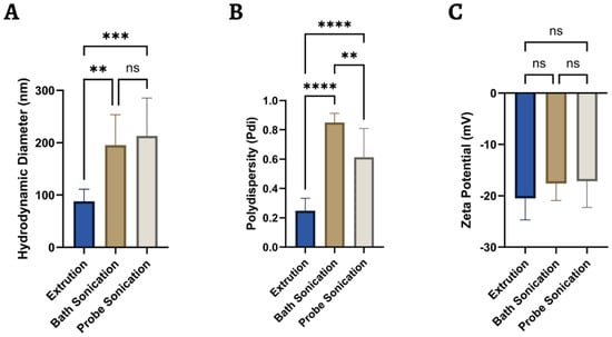
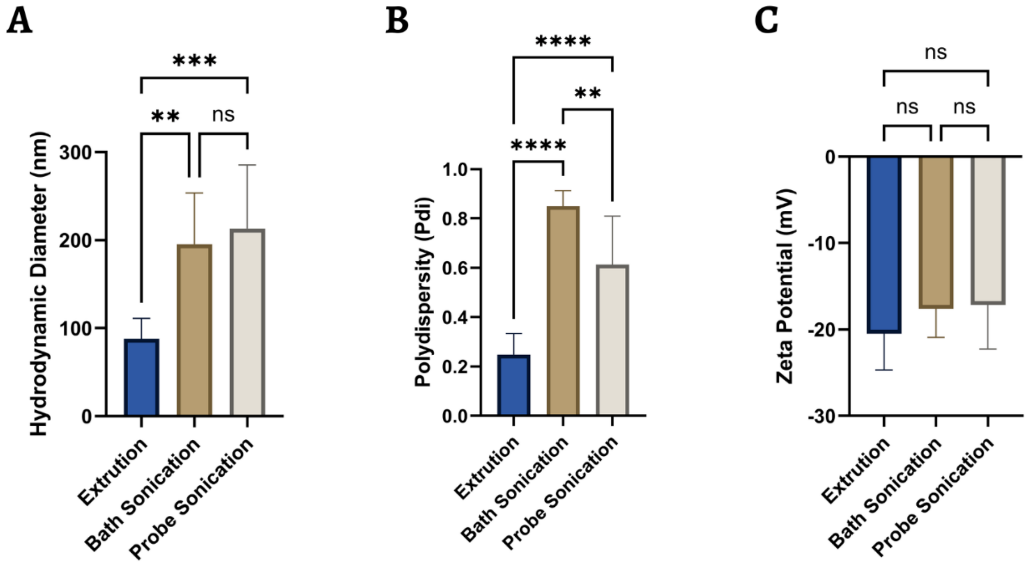
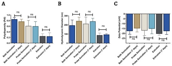
The physicochemical properties of the methods used to produce liposomes indicate that the extrusion method yielded the best characteristics in terms of its polydispersity index. Figure 2 shows the differences in size, zeta potential, and PDI among the extrusion, bath sonication, and probe sonication methods. Characterization of morin-loaded liposomes showed similar sizes between those obtained by bath and probe sonication (195 ± 58 nm and 213 ± 72 nm, respectively), whereas extrusion produced liposomes with a much smaller size of about 90 nm; these size differences are consistent with the expected effects of each preparation method [67] (see Supplementary Material, Figure S2). Moreover, the zeta potential was negative in all cases, which is characteristic of liposomes composed of zwitterionic phospholipids, such as DPPC in this instance, since, when present in an alkaline medium (pH = 8) and having a pKa of 3.8, a negative charge is induced on the surface of liposomes [69]. All formulations exhibit zeta potentials in the range of −15 to −25 mV; the extrusion sample showed −23 ± 8.6 mV, which was slightly more negative than the sonication samples: −17 ± 3 mV for the bath method and −17 ± 5 mV for the probe method. These values usually confer moderate electrostatic repulsion that hinders vesicle aggregation, as does PBS, which contributes to higher salt concentrations and tends to reduce the charge in the medium [70] (see Supplementary Material, Figure S3). Regarding the polydispersity of the liposomes, extrusion yielded a PDI of 0.2, indicating a monodisperse, homogeneous, and unilamellar population. In contrast, sonication yielded indices of 0.6 and 0.8, indicating heterogeneity and the possible presence of multilamellar vesicles. Furthermore, zeta potential is primarily governed by lipid composition, pH, ionic strength, and interfacial composition. Because these variables were common to all formulations, we did not expect large differences. However, processing may have indirect effects through size, curvature, or bilayer structure [71].
Figure 2.
Comparison of physicochemical properties of liposomes containing morin prepared using different methods. The figure shows differences in (A) size, (B) zeta potential, and (C) polydispersity index among the extrusion, bath sonication, and probe sonication methods. Data are presented as mean ± SD (n = 3). Statistical differences were determined using one-way ANOVA followed by Tukey’s test. ns: not significant; ** p < 0.01; *** p < 0.001; **** p < 0.0001.
Therefore, the generation of liposomes of uniform size by extrusion is explained by the forced passage of the liposomal suspension through a filter with pores of defined size (100 nm in this case) and by the possible presence of morin within the lipid bilayer of liposomes, which would provide resistance to extrusion. Although this resistance hinders the process, it maintains the formulation’s homogeneity. The planar structure of this type of polyhydroxylated flavonoid and its ability to form hydrogen bonds with the polar groups of phospholipids stabilize the lipid bilayer [73].
Meanwhile, liposomes tend to have slightly less negative zeta potential values. Extrusion shifted values from −23 to −17 mV; bath sonication from −17 to −12.0 mV; and probe sonication from −17 to −12 mV. However, the change is limited in magnitude, keeping the final values within a range associated with mild or moderate electrostatic stability.
During the liposome stability study, the initial properties were preserved after thermal stress at 37 °C. Figure 3 presents the same parameters measured after 7 days of storage at 37 °C, conditions that may accelerate degradation or aggregation in this type of colloidal system [73]. The liposomes showed moderate increases in size, particularly those obtained by bath sonication; however, these increases were not statistically significant, suggesting that these changes would not lead to massive aggregation or large-scale fusion of liposomes. For sonicated formulations, the slight increase could be attributed to Ostwald ripening or to the reorganization of smaller vesicles into thermodynamically more stable structures; however, no substantial growth indicative of colloidal instability was observed, supporting the formulation’s colloidal stability [74].
Figure 3.
Comparative evaluation of the physicochemical stability of morin-containing liposomes after 7 days of storage. Changes in (A) size, (B) zeta potential, and (C) polydispersity index are compared across the different preparation techniques. The data represent the mean ± SD (n = 3). Statistical analysis was performed using one-way ANOVA followed by Tukey’s test. ns: not significant.
Likewise, polydispersity remained unchanged across techniques, indicating formulation stability. A PDI that does not increase over time indicates that no new, larger-size populations are forming due to continuous aggregation, and there was no evidence of processes consistent with rupture or fusion that would increase heterogeneity. Stability of the PDI, together with the stability of the average size, suggests that the formulations are kinetically stable, that morin did not destabilize the bilayer, and that it may have contributed to colloidal stabilization [73].
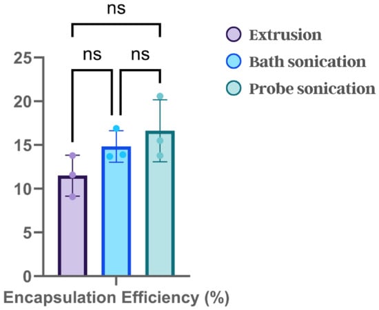
Encapsulation efficiency varied across preparation methods, showing a non-significant increasing trend from extrusion to probe sonication. Figure 4 shows the encapsulation efficiency values for morin in liposomes prepared using different methods. Extrusion had an average efficiency of 11 ± 2%, bath sonication 15 ± 2%, and probe sonication 17 ± 3%. Although this trend suggests a higher apparent yield with increasing energy input, statistical analysis did not detect significant differences between methods. ANOVA and Tukey’s multiple-comparison test confirmed no significant differences among the pairs. Nevertheless, this slight trend would be enhanced by the larger size and wider distribution of the liposomes obtained by both sonication methods, which allow the incorporation of a greater amount of morin (solubilized in an alkaline medium) into the aqueous core and the bilayer by increasing saturation of interstitial spaces of the hydrophobic fatty acid tails [52]. Likewise, the relatively low morin encapsulation percentages compared to those reported for other nanosystems loaded with this same flavonoid are likely limited by the low concentrations and molar ratios of lipids employed here [52].
Figure 4.
Encapsulation efficiency of morin in liposomes prepared by different methods. Encapsulation efficiency (EE) is the percentage of morin recovered in the vesicular fraction relative to the initial amount added. Data are presented as mean ± SD, with individual values overlaid (n = 3 independent preparations per method). A one-way ANOVA showed no significant differences among the preparation methods (F(2,6) = 2.871; p = 0.1334). Tukey’s test also failed to identify significant differences among the paired comparisons. ns: not significant.
Although the formulations improved morin stability, their EE of 11–17% remains a significant limitation. The low loading yield increases the amount of lipid formulation required to deliver a given morin dose and may constrain dose escalation and future translation. Several factors may have contributed, including the fixed and relatively low lipid-to-morin ratio, the addition of morin during the aqueous hydration phase, its distribution between the aqueous phase and the bilayer interface, and the removal of non-encapsulated or weakly associated morin during dialysis. The absence of significant differences among bath sonication, probe sonication, and extrusion further indicates that these post-hydration size-control procedures did not resolve the loading limitation. Future optimization should therefore focus on formulation composition and loading strategy rather than size-control processing alone.
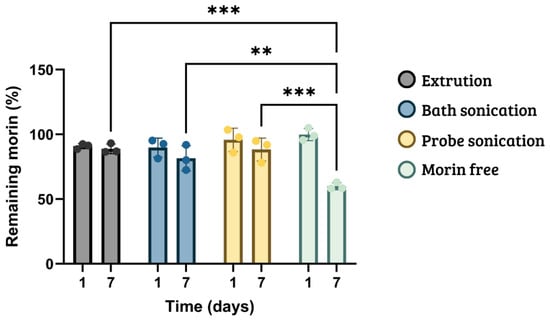
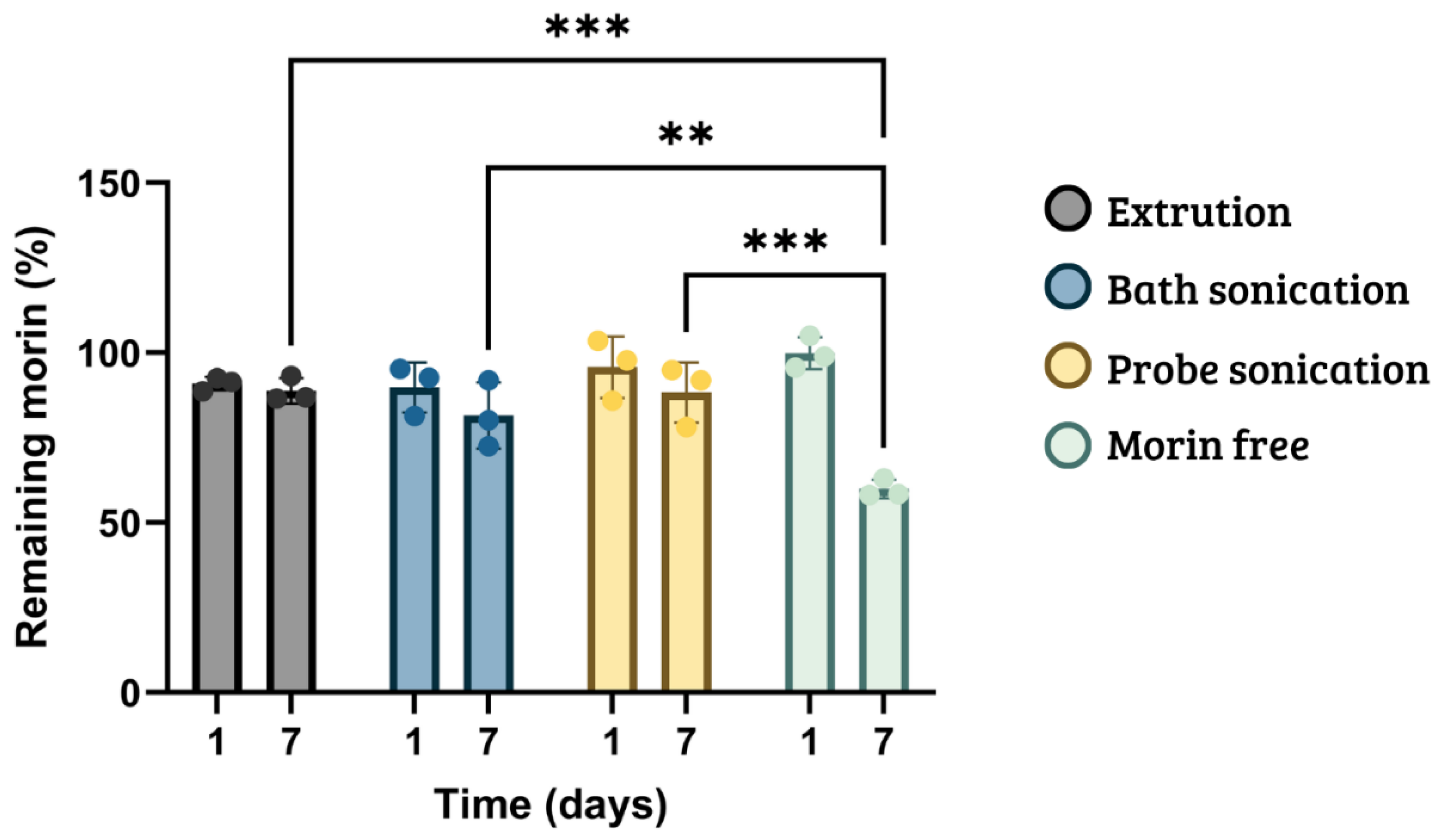
The study of morin’s chemical stability at 37 °C showed that degradation of free morin was greater than that observed in morin-loaded liposomal formulations, indicating that lipid formulation substantially alters morin’s chemical stability after 7 days at 37 °C. Figure 5 shows the stability results for morin in DPPC-based liposomal formulations at 37 °C after 1 and 7 days of storage. On day 1, the groups were comparable, and by day 7, free morin decreased to approximately 60% of its initial value, corresponding to a loss of approximately 40%, whereas the liposomal formulations maintained higher levels: 82% for bath sonication, 88% for probe sonication, and 89% for extrusion. Overall, free morin degraded by 40% between day 1 and day 7, whereas DPPC/cholesterol formulations showed much lower losses, particularly with extrusion (~2%) and probe sonication (~7%).
Figure 5.
Stability of morin in DPPC-based liposomal formulations at 37 °C. Residual morin concentration is expressed as a percentage of the initial reference signal. Data are presented as mean ± SD (n = 3 independent preparations per condition). Differences among formulations and evaluation times were analyzed using two-way ANOVA with Tukey’s multiple-comparison test. ** p < 0.01; *** p < 0.001.
Statistical analysis (ANOVA/Tukey) supports the finding that the effect of time varies by formulation. Specifically, on day 7, differences relative to free morin were approximately 29% for extrusion, 22% for bath sonication, and 28% for probe sonication. However, liposomes prepared by different methods did not differ significantly from one another; therefore, the results support a general protective effect of liposomes compared with free morin but do not establish that any one preparation method is superior to the others. This suggests that DPPC (present in all formulations) hinders morin escape through the bilayer, preventing the formation of structural defects that would facilitate rapid morin leakage at moderate temperatures (37 °C). At 37 °C, double bonds in unsaturated chains enhance membrane packing, thereby increasing the phase transition temperature [57,58]. Therefore, time, formulation, and their interaction significantly affected the amount of available morin.
Morin-loaded liposomes showed improved stability in aqueous media under the evaluated conditions; alkalinization increased their apparent solubility, with the greatest effect observed in PBS buffer at pH 8.0. However, this same increase in ionization promotes oxidation, dimerization, and subsequent degradation, particularly under alkaline, oxygenated, illuminated, or prolonged storage conditions [68]. Quantitative evidence robustly supports pH-dependent apparent solubility and condition-dependent degradation; however, it is less robust when assigning a single universal molecular pathway to all the observed products; therefore, it would be hazardous to assert that morin is a molecule “stable in neutral solutions” or “unstable in basic medium” [7,68,75]. By contrast, it is accurate to note that the chemical stability of morin depends jointly on pH, buffer, temperature, light, oxygen, time, concentration, and formulation environment. For example, at pH 7.4 and 37 °C in PBS, measurable degradation occurs over 48 h, whereas an acidic pH of 5.2 substantially reduces loss, although solubility is minimal (<30 μg/mL) [7]. Accordingly, a large proportion of reported formulations solubilize morin in the lipid phase for subsequent hydration; that is, they form liposomes by loading the flavonoid exclusively into the lipid bilayer, with the aim of avoiding the solubilization of morin in alkaline media, where it is more soluble but tends to degrade more readily [7,52]. Hence, under these conditions, morin apparently does not significantly destabilize DPPC/cholesterol membranes, since its interaction occurs via hydrogen bonds with the phosphate groups at the polar interface of the bilayer and the aqueous core of liposomes. Consequently, these physicochemical properties of morin are particularly important to consider in formulations intended for physiological applications and/or sustained-release systems. Previous studies have demonstrated that the physicochemical behavior of morin is strongly dependent on its molecular environment. Its interaction with phospholipid membranes revealed a preferential localization within interfacial regions, indicating that the surrounding microenvironment can modulate its accessibility and chemical reactivity. Furthermore, the ionization state of morin is highly pH-dependent, with deprotonation of the 2′-hydroxyl group occurring near physiological pH, which may substantially modify its interactions with nanostructured interfaces. These observations highlight the importance of controlling both the aqueous environment and the molecular organization of the carrier when designing delivery systems for morin [59].
Nanostructured systems have emerged as a strategy to overcome some of the physicochemical limitations of morin. Incorporation onto aminofunctionalized mesoporous si- licananoparticles demonstrated that association with a solid nanostructured interface can preserve key antioxidant properties [76]. More recently, interactions between morin and albumin, and its incorporation into BSA nanoparticles, demonstrated that molecular association with a carrier can increase its resistance to oxidative degradation while providing a platform for its transport and controlled release [11]. Together, these findings indicate that the chemical stability and functional behavior of morin can be substantially modified through its interaction with the surrounding nanostructured microenvironment.
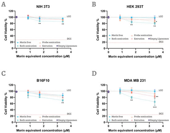
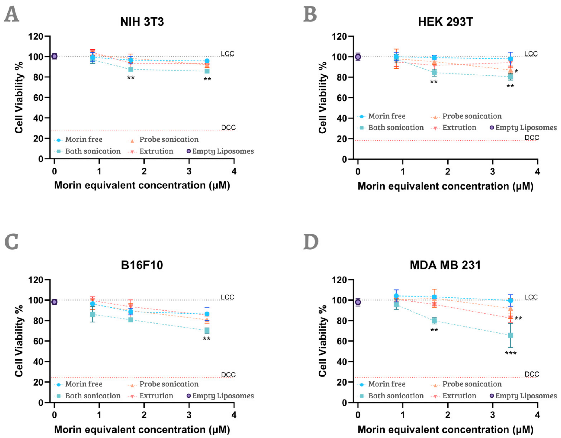
Regarding the cell assays, free morin exhibited limited cytotoxicity at the concentrations tested, whereas liposomal formulations produced a greater reduction in cell viability, particularly in the tumor cell models. Figure 6 presents the results of the MTS colorimetric cytotoxicity assay for free morin and the liposomal formulations prepared by the various methods, evaluated in non-tumor cell lines (NIH-3T3 fibroblasts and HEK-293T renal epithelial cells) and tumor cell lines (murine B16F10 melanoma and MDA-MB-231 breast cancer). Free morin showed little to no effect at the concentrations tested (0.85–3.4 µM) in non-tumor cells, consistent with previous reports of low cytotoxicity of morin at substantially higher concentrations. However, given the limited number of independent experiments, these findings should be considered preliminary and require confirmation in further studies.
Figure 6.
Potential anticancer effect of free and liposomal morin formulations on cell viability. Cell viability was evaluated by MTS assay in NIH 3T3 (A), HEK 293T (B), B16F10 (C), and MDA MB 231 (D) cells after 24 h of exposure to free morin or liposomal formulation (extrusion, probe sonication and bath sonication) at the indicated morin-equivalent concentrations. Empty Liposomes (EL) were evaluated at the highest concentration tested. The horizontal black dotted line at 100% represents the untreated control (LCC), whereas the horizontal red dotted line represents the positive cytotoxicity control obtained after treatment with 2% SDS. Results are expressed as the percentage of cell viability relative to untreated controls and are presented as mean ± SD from two independent experiments, each performed with four technical replicates. Statistical comparison between each liposomal formulation and free morin at the corresponding concentration was performed using one-way ANOVA followed by Dunnett’s multiple comparison test. *, p < 0.05; **, p < 0.01; ***, p < 0.001 vs. free morin at the corresponding concentration.
Similarly, concentrations up to 100 µM have been reported to preserve cell viability in other non-tumor cell models [21]. These observations align with the relatively low bioavailability of free morin, which is attributed to its poor aqueous solubility and limited permeability across biological membranes [75]. Moreover, cellular accumulation of morin may be further restricted by efflux mechanisms mediated by multidrug resistance-associated transporters, such as MRP1 [77].
In contrast, when morin is incorporated into liposomal formulations, its ability to reduce viability increases, despite the relatively low morin-equivalent concentrations tested, and the effect is formulation- and cell type-dependent. Among the formulations tested, bath sonication produced the strongest reduction in cell viability, particularly in MDA-MB-231 cells, where viability decreased in a concentration-dependent manner to approximately 65% at 3.4 µM. A similar response was observed in B16F10 cells, with viability decreasing to near 70% at 3.4 µM. Although to a lesser extent, the effect in non-tumoral NIH-3T3 and HEK-293T cells was comparatively moderate, with viability near 86% and 82%, respectively, suggesting a therapeutic window.
The enhanced activity of encapsulated morin may reflect improved cellular uptake and intracellular availability of the flavonoid, thereby partially overcoming the physicochemical and biological limitations of free morin. This interpretation aligns with previous studies showing that nanoencapsulation can improve morin delivery and enhance its biological activity [11,12,75]. Nevertheless, additional cellular uptake studies would be required to determine whether the increased cytotoxic effect observed with the liposomal formulations is directly attributable to enhanced intracellular accumulation of morin.
4. Conclusions
Overall, free morin is highly unstable in an alkaline medium at 37 °C, whereas encapsulation in DPPC- and cholesterol-based liposomes improved its stability under the same conditions. Furthermore, different preparation methods did not show significant differences in morin encapsulation efficiency. All liposomes remained stable for at least 7 days, regardless of the preparation method, with extrusion standing out for its superior colloidal properties. In cytotoxicity assays, free morin exhibited minimal cytotoxicity due to its low bioavailability, whereas its liposome-encapsulated form significantly enhanced its antiproliferative effect, showing greater selectivity against tumor cells (MDA-MB-231 and B16F10) than against non-tumor cells (NIH-3T3 and HEK-293T).
5. Limitations
This study has limitations that should be considered. Although the degradation kinetics of morin were characterized, they could not be correlated with the cellular assays; therefore, it has not been fully confirmed that the biological effect depends on its stability in those assays. Furthermore, the colloidal stability of the nanosystem was evaluated only over a limited period, and it would be necessary to monitor it over a longer period of time. Finally, the encapsulation efficiency obtained is moderate, so adjustments to the formulation or synthesis are required to increase it. These limitations do not invalidate the results but guide future research.
Regarding the cellular studies, the present work was designed as an exploratory assessment across a relatively broad panel of tumor and non-tumor reference cell lines. Cell viability assays were performed in two independent experiments, each with four technical replicates per condition. Although responses were consistent within replicates, the limited number of independent experiments limits the statistical robustness of the cellular findings, which should therefore be considered preliminary. In addition, cellular effects were evaluated only after 24 h of exposure. Future studies should include at least three independent experiments and longer exposure periods (48 and 72 h) to confirm the observed responses and characterize their time dependence.
The liposomal stability study was limited to 7 days at 37 °C and should be interpreted as a short-term, accelerated comparison rather than a shelf-life study. Longer studies at recommended storage temperatures, particularly 4 °C and 25 °C, are required to establish the long-term stability and storage conditions of the formulations and to monitor the %EE Further research is needed to evaluate drug release, uptake, pharmacokinetics, and efficacy.
Supplementary Materials
The following supporting information can be downloaded at: https://www.mdpi.com/article/10.3390/jpbi3040026/s1. Figure S1. UV–Vis spectroscopic characterization and quantification of morin in free form and following recovery from liposomal formulations; Figure S2. Representative hydrodynamic size distributions of morin-loaded liposomes prepared using different size-reduction methods; Figure S3. Representative zeta-potential distributions of morin-loaded liposomes prepared using different size-reduction methods.
Author Contributions
Conceptualization, P.R.-B., E.Z., F.S.-H. and M.J.K.; methodology, P.R.-B., E.Z., F.S.-H. and S.G.; validation, O.D.-G., J.M.-V., S.G. and M.J.K.; formal analysis, P.R.-B., E.Z., F.S.-H. and S.G.; investigation, P.R.-B., E.Z., F.S.-H. and S.G.; resources, S.G. and M.J.K.; data curation, P.R.-B., E.Z., F.S.-H., O.D.-G. and S.G.; writing—original draft preparation, P.R.-B., E.Z., F.S.-H., O.D.-G. and S.G.; writing—review and editing, O.D.-G., J.M.-V., S.G. and M.J.K.; supervision, M.J.K.; project administration, J.M.-V. and M.J.K.; funding acquisition, J.M.-V., S.G. and M.J.K. All authors have read and agreed to the published version of the manuscript.
Funding
This research was funded by Beca de Doctorado Nacional Agencia Nacional de Investigación y Desarrollo ANID grant number 21212006, 21231043) Beca Doctorado en el exterior Minciencias grant number, 885-2020. ANID Postdoctoral FONDECYT grant number 3240075 Faculty of Chemical and Pharmaceutical Sciences, University of Chile, internal project CIAI-DQFyT-2026-003. ANID-FONDECYT grant number 1251140, Fondequip EQM170111, ANID-FONDAP number 15130011, and FONDAP Apoyo 1523A0008. Proyectos de Continuidad Transitoria Centros de Excelencia Universidad de Chile (2026–2027).
Institutional Review Board Statement
Not applicable.
Informed Consent Statement
Not applicable.
Data Availability Statement
The original contributions presented in this study are included in the article/Supplementary Material. Further inquiries can be directed to the corresponding authors.
Conflicts of Interest
The authors declare no conflicts of interest.
Abbreviations
The following abbreviations are used in this manuscript:
| BSC | Biopharmaceutical Classification System |
| DPPC | 1,2-dipalmitoyl-sn-glycero-3-phosphocholine |
| PDI | Polydispersity index |
| EE | Encapsulation efficiency |
| PBS | Phosphate-buffered saline |
| FBS | Fetal bovine serum |
| Dh | Hydrodynamic diameter |
| DLS | Dynamic light scattering |
| SD | Standard deviation |
| BSA | Bovine serum albumin |
References
- Altyar, A.E.; Kensara, O.A.; Sayed, A.A.; Aleya, L.; Almutairi, M.H.; Zaazouee, M.S.; Elshanbary, A.A.; El-Demerdash, F.M.; Abdel-Daim, M.M. Acute Aflatoxin B1-Induced Hepatic and Cardiac Oxidative Damage in Rats: Ameliorative Effects of Morin. Heliyon 2023, 9, e21837. [Google Scholar] [CrossRef] [Scilit]
- Jain, S.; Yadav, Y.; Sharma, M. Morin: A Powerful Potential Flavonoid for Human. J. Drug Deliv. Ther. 2025, 15, 237–241. [Google Scholar] [CrossRef] [Scilit]
- Caselli, A.; Cirri, P.; Santi, A.; Paoli, P. Morin: A Promising Natural Drug. Curr. Med. Chem. 2016, 23, 774–791. [Google Scholar] [CrossRef] [Scilit]
- Khademi, R.; Mirzaei, A.; Mirzaei, A.; Edjlali, F.R.; Askari, V.R.; Baradaran Rahimi, V. Morin, as a Natural Flavonoid, Provides Promising Influences against Cardiovascular Diseases. Naunyn-Schmiedeberg’s Arch. Pharmacol. 2025, 398, 6293–6310. [Google Scholar] [CrossRef] [Scilit]
- Thakur, K.; Zhu, Y.Y.; Feng, J.Y.; Zhang, J.G.; Hu, F.; Prasad, C.; Wei, Z.J. Morin as an Imminent Functional Food Ingredient: An Update on Its Enhanced Efficacy in the Treatment and Prevention of Metabolic Syndromes. Food Funct. 2020, 11, 8424–8443. [Google Scholar] [CrossRef] [Scilit]
- Sainz-Hernández, J.C.; Rueda-Puente, E.O.; Cornejo-Ramírez, Y.I.; Bernal-Mercado, A.T.; González-Ocampo, H.A.; López-Corona, B.E. Biological Application of the Allopathic Characteristics of the Genus Maclura: A Review. Plants 2023, 12, 3480. [Google Scholar] [CrossRef] [Scilit]
- Jangid, A.K.; Pooja, D.; Kulhari, H. Determination of Solubility, Stability and Degradation Kinetics of Morin Hydrate in Physiological Solutions. RSC Adv. 2018, 8, 28836–28842. [Google Scholar] [CrossRef] [Scilit]
- Bhatia, J.; Verma, V.K.; Arya, D.S. MORIN, A BIOFLAVONOID, ATTENUATES MYOCARDIAL ISCHEMIA-REPERFUSION INJURY BY ATTENUATING RISK/SAPK PATHWAY. J. Hypertens. 2024, 42, e190. [Google Scholar] [CrossRef] [Scilit]
- Sales, L.S.; Hewitt, B.; Muchova, M.; Brighenti, F.L.; Kuehne, S.A.; Grant, M.M.; Milward, M.R. Anti-Inflammatory, Antioxidant, and Antimicrobial Evaluation of Morin. Arch. Oral Biol. 2025, 178, 106343. [Google Scholar] [CrossRef] [Scilit]
- Mendoza-Wilson, A.M.; Santacruz-Ortega, H.; Balandrán-Quintana, R.R. Relationship between Structure, Properties, and the Radical Scavenging Activity of Morin. J. Mol. Struct. 2011, 995, 134–141. [Google Scholar] [CrossRef] [Scilit]
- Montero, G.; Guarnizo-Herrero, V.; Sandoval-Altamirano, C.; Günther, G.; Bollo, S.; Arriagada, F.; Morales, J. Morin Flavonoid Interaction with Albumin and Its Nanoparticle Conjugation: An Efficient Antioxidant Vehicle for Nutraceuticals. Antioxidants 2025, 14, 764. [Google Scholar] [CrossRef] [Scilit]
- Balaga, V.K.R.; Pradhan, A.; Thapa, R.; Patel, N.; Mishra, R.; Singla, N. Morin: A Comprehensive Review on Its Versatile Biological Activity and Associated Therapeutic Potential in Treating Cancers. Pharmacol. Res.-Mod. Chin. Med. 2023, 7, 100264. [Google Scholar] [CrossRef] [Scilit]
- Rajput, S.A.; Wang, X.Q.; Yan, H.C. Morin Hydrate: A Comprehensive Review on Novel Natural Dietary Bioactive Compound with Versatile Biological and Pharmacological Potential. Biomed. Pharmacother. 2021, 138, 111511. [Google Scholar] [CrossRef] [Scilit]
- Sharma, V.; Sharma, M.; Semwal, A.; Arora, A.; Bansal, S.; Sangwan, N.; Aggarwal, A. Morin, a Natural Flavonoid, Activates the PINK1/Parkin Pathway to Induce Mitophagy, Promote Apoptosis, and Suppress Cancer Stemness in Pancreatic Cancer. Phyther. Res. 2026, 40, 582–602. [Google Scholar] [CrossRef] [Scilit]
- Wu, R.; Bostani, A.; Wang, J. Synergistic Effects of Morin and Doxorubicin Overcome Chemoresistance in Ovarian Cancer: Preclinical Insights from in Vitro and in Vivo Models. Cancer Manag. Res. 2026, 18, 567591. [Google Scholar] [CrossRef] [Scilit]
- Sak, K. Cytotoxicity of Dietary Flavonoids on Different Human Cancer Types. Pharmacogn. Rev. 2014, 8, 122–146. [Google Scholar] [CrossRef] [Scilit]
- Kuzu, M.; Kandemir, F.M.; Yildirim, S.; Kucukler, S.; Caglayan, C.; Turk, E. Morin Attenuates Doxorubicin-Induced Heart and Brain Damage by Reducing Oxidative Stress, Inflammation and Apoptosis. Biomed. Pharmacother. 2018, 106, 443–453. [Google Scholar] [CrossRef] [Scilit]
- Zhang, Q.; Zhang, F.; Thakur, K.; Wang, J.; Wang, H.; Hu, F.; Zhang, J.G.; Wei, Z.J. Molecular Mechanism of Anti-Cancerous Potential of Morin Extracted from Mulberry in Hela Cells. Food Chem. Toxicol. 2018, 112, 466–475. [Google Scholar] [CrossRef] [Scilit]
- Park, C.; Lee, W.S.; Go, S.I.; Nagappan, A.; Han, M.H.; Hong, S.H.; Kim, G.S.; Kim, G.Y.; Kwon, T.K.; Ryu, C.H.; et al. Morin, a Flavonoid from Moraceae, Induces Apoptosis by Induction of Bad Protein in Human Leukemic Cells. Int. J. Mol. Sci. 2015, 16, 645–659. [Google Scholar] [CrossRef] [Scilit]
- Lee, J.H.; Jin, H.; Lee, W.S.; Nagappan, A.; Choi, Y.H.; Kim, G.S.; Jung, J.M.; Ryu, C.H.; Shin, S.C.; Hong, S.C.; et al. Morin, a Flavonoid from Moraceae, Inhibits Cancer Cell Adhesion to Endothelial Cells and EMT by down-Regulating VCAM-1 and N-Cadherin. Asian Pac. J. Cancer Prev. 2016, 17, 3071–3075. [Google Scholar] [CrossRef]
- Jin, H.; Lee, W.S.; Eun, S.Y.; Jung, J.H.; Park, H.S.; Kim, G.; Choi, Y.H.; Ryu, C.H.; Jung, J.M.; Hong, S.C.; et al. Morin, a Flavonoid from Moraceae, Suppresses Growth and Invasion of the Highly Metastatic Breast Cancer Cell Line MDA-MB-231 Partly through Suppression of the Akt Pathway. Int. J. Oncol. 2014, 45, 1629–1637. [Google Scholar] [CrossRef] [Scilit]
- Sinha, K.; Sadhukhan, P.; Saha, S.; Pal, P.B.; Sil, P.C. Morin Protects Gastric Mucosa from Nonsteroidal Anti-Inflammatory Drug, Indomethacin Induced Inflammatory Damage and Apoptosis by Modulating NF-ΚB Pathway. Biochim. Biophys. Acta-Gen. Subj. 2015, 1850, 769–783. [Google Scholar] [CrossRef] [Scilit]
- Kapoor, R.; Kakkar, P. Protective Role of Morin, a Flavonoid, against High Glucose Induced Oxidative Stress Mediated Apoptosis in Primary Rat Hepatocytes. PLoS ONE 2012, 7, e41663. [Google Scholar] [CrossRef] [Scilit]
- Nani Pohrib, E.; Corciova, A.; Cioanca, O.; Hritcu, L.; Hancianu, M.; Mitran, A.M.; Burlec, A.F.; Cimpanu, A.M.; Isac, C.M.; Huzum, R.; et al. Antioxidant, Anti-Cholinesterase, and Neuroprotective Properties of Morus Alba and Morus Nigra Extracts. Antioxidants 2026, 15, 510. [Google Scholar] [CrossRef] [Scilit]
- Sharma, S.H.; Kumar, J.S.; Chellappan, D.R.; Nagarajan, S. Molecular Chemoprevention by Morin—A Plant Flavonoid That Targets Nuclear Factor Kappa B in Experimental Colon Cancer. Biomed. Pharmacother. 2018, 100, 367–373. [Google Scholar] [CrossRef] [Scilit]
- Li, J.; Yang, Y.; Ning, E.; Peng, Y.; Zhang, J. Mechanisms of Poor Oral Bioavailability of Flavonoid Morin in Rats: From Physicochemical to Biopharmaceutical Evaluations. Eur. J. Pharm. Sci. 2019, 128, 290–298. [Google Scholar] [CrossRef] [Scilit]
- Patel, D.; Wairkar, S. Enhancing Bioavailability of Morin via Multi-Particulate Dosage Forms: Development and Pharmacokinetic Insights. Ann. Pharm. Fr. 2026, 84, 850–860. [Google Scholar] [CrossRef] [Scilit]
- Zakaria, M.Y.; Georghiou, P.E.; Banoub, J.H.; Beshay, B.Y. Inclusion of a Phytomedicinal Flavonoid in Biocompatible Surface-Modified Chylomicron Mimic Nanovesicles with Improved Oral Bioavailability and Virucidal Activity: Molecular Modeling and Pharmacodynamic Studies. Pharmaceutics 2022, 14, 905. [Google Scholar] [CrossRef] [Scilit]
- Naeem, A.; Ming, Y.; Pengyi, H.; Jie, K.Y.; Yali, L.; Haiyan, Z.; Shuai, X.; Wenjing, L.; Ling, W.; Xia, Z.M.; et al. The Fate of Flavonoids after Oral Administration: A Comprehensive Overview of Its Bioavailability. Crit. Rev. Food Sci. Nutr. 2022, 62, 6169–6186. [Google Scholar] [CrossRef] [Scilit]
- El-Haddad, A.E.; Sheta, N.M.; Boshra, S.A. Isolation, Formulation, and Efficacy Enhancement of Morin Emulsified Carriers Against Lung Toxicity in Rats. AAPS PharmSciTech 2018, 19, 2346–2357. [Google Scholar] [CrossRef] [Scilit]
- Lee, M.K. Liposomes for Enhanced Bioavailability of Water-Insoluble Drugs: In Vivo Evidence and Recent Approaches. Pharmaceutics 2020, 12, 264. [Google Scholar] [CrossRef] [Scilit]
- Chaturvedi, S.; Mishra, R. Insight into Delivery Approaches for Biopharmaceutics Classification System Class II and IV Drugs. Drug Deliv. Lett. 2020, 10, 255–277. [Google Scholar] [CrossRef] [Scilit]
- Berga, M.; Logviss, K.; Lauberte, L.; Paulausks, A.; Mohylyuk, V. Flavonoids in the Spotlight: Bridging the Gap between Physicochemical Properties and Formulation Strategies. Pharmaceuticals 2023, 16, 1407. [Google Scholar] [CrossRef] [Scilit]
- Teng, H.; Zheng, Y.; Cao, H.; Huang, Q.; Xiao, J.; Chen, L. Enhancement of Bioavailability and Bioactivity of Diet-Derived Flavonoids by Application of Nanotechnology: A Review. Crit. Rev. Food Sci. Nutr. 2023, 63, 378–393. [Google Scholar] [CrossRef] [Scilit]
- Zhao, J.; Yang, J.; Xie, Y. Improvement Strategies for the Oral Bioavailability of Poorly Water-Soluble Flavonoids: An Overview. Int. J. Pharm. 2019, 570, 118642. [Google Scholar] [CrossRef] [Scilit]
- Yu, D.; Park, J.; Kim, T.; Choi, C.; Yuk, S.A.; Kim, H. Nanotechnology-Enabled Delivery of Phytochemicals: From Formulation Strategies to Therapeutic Translation. J. Phytomed. 2026, 1, 4. [Google Scholar] [CrossRef] [Scilit]
- Mitchell, M.J.; Billingsley, M.M.; Haley, R.M.; Wechsler, M.E.; Peppas, N.A.; Langer, R. Engineering Precision Nanoparticles for Drug Delivery. Nat. Rev. Drug Discov. 2021, 20, 101–124. [Google Scholar] [CrossRef] [Scilit]
- Afzal, O.; Altamimi, A.S.A.; Nadeem, M.S.; Alzarea, S.I.; Almalki, W.H.; Tariq, A.; Mubeen, B.; Murtaza, B.N.; Iftikhar, S.; Riaz, N.; et al. Nanoparticles in Drug Delivery: From History to Therapeutic Applications. Nanomaterials 2022, 12, 4494. [Google Scholar] [CrossRef] [Scilit]
- Huang, L.; Luo, S.; Tong, S.; Lv, Z.; Wu, J. The Development of Nanocarriers for Natural Products. Wiley Interdiscip. Rev. Nanomed. Nanobiotechnol. 2024, 16, e1967. [Google Scholar] [CrossRef] [Scilit]
- Zarrabi, A.; Abadi, M.A.A.; Khorasani, S.; Reza Mohammadabadi, M.; Jamshidi, A.; Torkaman, S.; Taghavi, E.; Mozafari, M.R.; Rasti, B. Nanoliposomes and Tocosomes as Multifunctional Nanocarriers for the Encapsulation of Nutraceutical and Dietary Molecules. Molecules 2020, 25, 638. [Google Scholar] [CrossRef] [Scilit]
- Liu, P.; Wang, Y.; Zhang, Z.; Wu, D.; Chen, W.; Li, W.; Liu, J.; Wang, W.; Yang, Y. Nanoliposome Based Strategy for Enhancing Bioavailability and Antitumor Activity of Polyphenol from Sanghuangporus Vaninii. Food Biosci. 2025, 63, 105802. [Google Scholar] [CrossRef] [Scilit]
- Fatfat, Z.; Karam, M.; Maatouk, B.; Fahs, D.; Gali-Muhtasib, H. Nanoliposomes as Safe and Efficient Drug Delivery Nanovesicles. In Advanced and Modern Approaches for Drug Delivery; Academic Press: Cambridge, MA, USA, 2023; pp. 159–197. ISBN 9780323916684. [Google Scholar]
- McClements, D.J.; Öztürk, B. Utilization of Nanotechnology to Improve the Handling, Storage and Biocompatibility of Bioactive Lipids in Food Applications. Foods 2021, 10, 365. [Google Scholar] [CrossRef] [Scilit]
- Subramani, T.; Ganapathyswamy, H. An Overview of Liposomal Nano-Encapsulation Techniques and Its Applications in Food and Nutraceutical. J. Food Sci. Technol. 2020, 57, 3545–3555. [Google Scholar] [CrossRef] [Scilit]
- Khorasani, S.; Danaei, M.; Mozafari, M.R. Nanoliposome Technology for the Food and Nutraceutical Industries. Trends Food Sci. Technol. 2018, 79, 106–115. [Google Scholar] [CrossRef] [Scilit]
- García-Morales, J.; Fimbres-Olivarría, D.; González-Vega, R.I.; Bernal-Mercado, A.T.; Aubourg-Martínez, S.P.; López-Gastélum, K.A.; Robles-García, M.Á.; de Jesús Ornelas-Paz, J.; Ruiz-Cruz, S.; Del-Toro-Sánchez, C.L. Nanoliposomes as Effective Vehicles of Antioxidant Compounds in Food and Health. Int. J. Mol. Sci. 2025, 26, 5523. [Google Scholar] [CrossRef] [Scilit]
- Wen, C.; Tang, J.; Cao, L.; Fan, M.; Lin, X.; Liu, G.; Liang, L.; Liu, X.; Zhang, J.; Li, Y.; et al. Strategic Approaches for Co-Encapsulation of Bioactive Compounds: Technological Advances and Mechanistic Insight. Foods 2025, 14, 2024. [Google Scholar] [CrossRef] [Scilit]
- Ren, K.; Cao, X.; Zheng, L.; Tian, T.; Zhang, X.; Dai, J.; Zhang, H.; Wang, H.; Jiang, L. Pectin-Modified 7S Protein Liposomes: Focus on Structural Properties, Stability, and Digestive Properties Loaded with Morin. Food Chem. 2025, 474, 143164. [Google Scholar] [CrossRef] [Scilit]
- Ren, K.; Cao, X.; Zheng, L.; Liu, S.; Li, L.; Cheng, L.; Tian, T.; Tong, X.; Wang, H.; Jiang, L. Liposomes Decorated with β-Conglycinin and Glycinin: Construction, Structure and in Vitro Digestive Stability. Int. J. Biol. Macromol. 2024, 269, 131900. [Google Scholar] [CrossRef] [Scilit]
- Sultana, F.; Neog, M.K.; Rasool, M.K. Targeted Delivery of Morin, a Dietary Bioflavanol Encapsulated Mannosylated Liposomes to the Macrophages of Adjuvant-Induced Arthritis Rats Inhibits Inflammatory Immune Response and Osteoclastogenesis. Eur. J. Pharm. Biopharm. 2017, 115, 229–242. [Google Scholar] [CrossRef] [Scilit]
- Ding, X.; Yin, C.; Zhang, W.; Sun, Y.; Zhang, Z.; Yang, E.; Sun, D.; Wang, W. Designing Aptamer-Gold Nanoparticle-Loaded PH-Sensitive Liposomes Encapsulate Morin for Treating Cancer. Nanoscale Res. Lett. 2020, 15, 68. [Google Scholar] [CrossRef] [Scilit]
- Tran, H.M.; Yang, C.Y.; Wu, T.H.; Yen, F.L. Liposomes Encapsulating Morin: Investigation of Physicochemical Properties, Dermal Absorption Improvement and Anti-Aging Activity in PM-Induced Keratinocytes. Antioxidants 2022, 11, 1183. [Google Scholar] [CrossRef] [Scilit]
- Encinas-Basurto, D.; Acosta, M.F.; Eedara, B.B.; Fineman, J.R.; Black, S.M.; Mansour, H.M. Design and Comprehensive Characterization of Dry Powder Inhalation Aerosols of Simvastatin DPPC/DPPG Lung Surfactant-Mimic Nanoparticles/Microparticles for Pulmonary Nanomedicine. RSC Adv. 2024, 14, 29413–29427. [Google Scholar] [CrossRef] [Scilit]
- Luviano, A.S.; Campos-Terán, J.; Langevin, D.; Castillo, R.; Espinosa, G. Mechanical Properties of DPPC-POPE Mixed Langmuir Monolayers. Langmuir 2019, 35, 16734–16744. [Google Scholar] [CrossRef] [Scilit]
- Pereira-Leite, C.; Lopes-de-Campos, D.; Fontaine, P.; Cuccovia, I.M.; Nunes, C.; Reis, S. Licofelone-DPPC Interactions: Putting Membrane Lipids on the Radar of Drug Development. Molecules 2019, 24, 516. [Google Scholar] [CrossRef] [Scilit]
- Piosik, E.; Modlińska, A.; Gołaszewski, M.; Chełminiak-Dudkiewicz, D.; Ziegler-Borowska, M. Influence of the Type of Biocompatible Polymer in the Shell of Magnetite Nanoparticles on Their Interaction with DPPC in Two-Component Langmuir Monolayers. J. Phys. Chem. B 2024, 128, 781–794. [Google Scholar] [CrossRef] [Scilit]
- Ollila, F.; Halling, K.; Vuorela, P.; Vuorela, H.; Slotte, J.P. Characterization of Flavonoid-Biomembrane Interactions. Arch. Biochem. Biophys. 2002, 399, 103–108. [Google Scholar] [CrossRef] [Scilit]
- Anderson, M.; Omri, A. The Effect of Different Lipid Components on the in Vitro Stability and Release Kinetics of Liposome Formulations. Drug Deliv. J. Deliv. Target. Ther. Agents 2004, 11, 33–39. [Google Scholar] [CrossRef] [Scilit]
- Günther, G.; Berrîos, E.; Pizarro, N.; Valdés, K.; Montero, G.; Arriagada, F.; Morales, J. Flavonoids in Microheterogeneous Media, Relationship between Their Relative Location and Their Reactivity towards Singlet Oxygen. PLoS ONE 2015, 10, e0129749. [Google Scholar] [CrossRef] [Scilit]
- Andra, V.V.S.N.L.; Pammi, S.V.N.; Bhatraju, L.V.K.P.; Ruddaraju, L.K. A Comprehensive Review on Novel Liposomal Methodologies, Commercial Formulations, Clinical Trials and Patents. Bionanoscience 2022, 12, 274–291. [Google Scholar] [CrossRef] [Scilit]
- Lombardo, D.; Kiselev, M.A. Methods of Liposomes Preparation: Formation and Control Factors of Versatile Nanocarriers for Biomedical and Nanomedicine Application. Pharmaceutics 2022, 14, 543. [Google Scholar] [CrossRef] [Scilit]
- Manca, M.L.; Aroffu, M.; Fulgheri, F.; Perra, M.; Castangia, I. Conventional Methods for Preparing Liposomes of Various Types (MLVs, LUVs, SUVs): What, Where, How and When. In Liposomes in Drug Delivery: What, Where, How and When to Deliver; Academic Press: Cambridge, MA, USA, 2024; pp. 461–488. ISBN 9780443154911. [Google Scholar]
- William, B.; Noémie, P.; Brigitte, E.; Géraldine, P. Supercritical Fluid Methods: An Alternative to Conventional Methods to Prepare Liposomes. Chem. Eng. J. 2020, 383, 123106. [Google Scholar] [CrossRef] [Scilit]
- Pradhan, B.; Kumar, N.; Saha, S.; Roy, A. Liposome: Method of Preparation, Advantages, Evaluation and Its Application. J. Appl. Pharm. Res. 2015, 3, 01–08. [Google Scholar]
- Riccardi, D.; Baldino, L.; Reverchon, E. Liposomes, Transfersomes and Niosomes: Production Methods and Their Applications in the Vaccinal Field. J. Transl. Med. 2024, 22, 339. [Google Scholar] [CrossRef] [Scilit]
- Has, C.; Sunthar, P. A Comprehensive Review on Recent Preparation Techniques of Liposomes. J. Liposome Res. 2020, 30, 336–365. [Google Scholar] [CrossRef] [Scilit]
- Šturm, L.; Ulrih, N.P. Basic Methods for Preparation of Liposomes and Studying Their Interactions with Different Compounds, with the Emphasis on Polyphenols. Int. J. Mol. Sci. 2021, 22, 6547. [Google Scholar] [CrossRef] [Scilit]
- Bark, K.M.; Im, S.E.; Seo, J.J.; Park, O.H.; Park, C.H.; Park, H.R. Spectroscopic Study on the Stability of Morin in Aqueous Solution. Bull. Korean Chem. Soc. 2015, 36, 498–502. [Google Scholar] [CrossRef] [Scilit]
- Doskocz, J.; Dałek, P.; Foryś, A.; Trzebicka, B.; Przybyło, M.; Mesarec, L.; Iglič, A.; Langner, M. The Effect of Lipid Phase on Liposome Stability upon Exposure to the Mechanical Stress. Biochim. Biophys. Acta-Biomembr. 2020, 1862, 183361. [Google Scholar] [CrossRef] [Scilit]
- Ong, S.G.M.; Chitneni, M.; Lee, K.S.; Ming, L.C.; Yuen, K.H. Evaluation of Extrusion Technique for Nanosizing Liposomes. Pharmaceutics 2016, 8, 36. [Google Scholar] [CrossRef] [Scilit]
- Ghosh, P.; Patwari, J.; Dasgupta, S. Complexation with Human Serum Albumin Facilitates Sustained Release of Morin from Polylactic-Co-Glycolic Acid Nanoparticles. J. Phys. Chem. B 2017, 121, 1758–1770. [Google Scholar] [CrossRef] [Scilit]
- Liazid, A.; Palma, M.; Brigui, J.; Barroso, C.G. Investigation on Phenolic Compounds Stability during Microwave-Assisted Extraction. J. Chromatogr. A 2007, 1140, 29–34. [Google Scholar] [CrossRef] [Scilit]
- Sadžak, A.; Brkljača, Z.; Eraković, M.; Kriechbaum, M.; Maltar-Strmečki, N.; Přibyl, J.; Šegota, S. Puncturing Lipid Membranes: Onset of Pore Formation and the Role of Hydrogen Bonding in the Presence of Flavonoids. J. Lipid Res. 2023, 64, 100430. [Google Scholar] [CrossRef] [Scilit]
- Koroleva, M.Y.; Yurtov, E.V. Ostwald Ripening in Macro- and Nanoemulsions. Russ. Chem. Rev. 2021, 90, 293–323. [Google Scholar] [CrossRef] [Scilit]
- Cunha, C.; Marinheiro, D.; Ferreira, B.J.M.L.; Oliveira, H.; Daniel-da-Silva, A.L. Morin Hydrate Encapsulation and Release from Mesoporous Silica Nanoparticles for Melanoma Therapy. Molecules 2023, 28, 4776. [Google Scholar] [CrossRef] [Scilit]
- Arriagada, F.; Correa, O.; Günther, G.; Nonell, S.; Mura, F.; Olea-Azar, C.; Morales, J. Morin Flavonoid Adsorbed on Mesoporous Silica, a Novel Antioxidant Nanomaterial. PLoS ONE 2016, 11, e0164507. [Google Scholar] [CrossRef] [Scilit]
- Goyal, A.; Dubey, N.; Agrawal, A.; Verma, A.; Sharma, R. Therapeutic Potential of Morin: Special Focus on Cognitive Dysfunction, Diabetes Mellitus and Cancer. Pharmacol. Res.-Mod. Chin. Med. 2023, 9, 100318. [Google Scholar] [CrossRef] [Scilit]
Disclaimer/Publisher’s Note: The statements, opinions and data contained in all publications are solely those of the individual author(s) and contributor(s) and not of MDPI and/or the editor(s). MDPI and/or the editor(s) disclaim responsibility for any injury to people or property resulting from any ideas, methods, instructions or products referred to in the content. |
© 2026 by the authors. Licensee MDPI, Basel, Switzerland. This article is an open access article distributed under the terms and conditions of the Creative Commons Attribution (CC BY) license.