


Abstract
The therapeutic monoclonal antibody bevacizumab is typically purified using protein A affinity chromatography, a highly effective but costly method. Affinity-based precipitation for antibody purification is a lower-cost approach. In this work, a precipitation protocol was developed for bevacizumab purification using a branched peptide (Ac-PHQGQHIG-Ahx3)2-K-Ahx3-PHQGQHIG-NH2, which contains the epitope PHQGQHIG that is responsible for interacting with bevacizumab. The peptide was synthesised by a microwave-assisted solid-phase peptide method, employing LiCl as an additive to prevent aggregation and ensure high purity and yield. Three molecules of 6-aminohexanoic acid were introduced between each epitope branch as spacer arms to promote the formation of cyclic complexes. Bevacizumab purification from cell-free culture broth was achieved through a fractional precipitation process. First, a negative precipitation step using (NH4)2SO4 1.18 M was performed to remove contaminants. Afterwards, 5 moles of peptide per mol of bevacizumab was added to the supernatant, together with additional (NH4)2SO4, to reach a final concentration of 1.20 M. Under these conditions, bevacizumab was recovered in the precipitate with 98% purity and a yield of 73%. In addition to being recyclable, the peptide’s relatively low production cost could enable the development of a single-use purification process, which would be particularly advantageous for biopharmaceutical manufacturing.
1. Introduction
Bevacizumab is widely used to treat several cancers, including colorectal, cervical, ovarian, and renal malignancies. It is a recombinant humanised monoclonal antibody (mAb) that inhibits tumour angiogenesis by blocking vascular endothelial growth factor A (VEGF-A) [1].
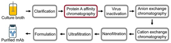
mAb is commonly produced extracellularly in Chinese hamster ovary (CHO) cells cultivated in suspension in bioreactors. Its recovery and purification are achieved by multiple downstream purification steps. After cell removal by filtration or centrifugation, mAb is typically purified by protein A affinity chromatography (AC), resulting in a highly purified and concentrated product. Afterwards, polishing steps are usually performed by anion exchange chromatography, cation exchange chromatography, nanofiltration and ultrafiltration. These processes ensure a product with adequate purity, potency and safety (Figure 1) [2].
Figure 1.
Schematic downstream processing of bevacizumab from cell culture broth.
However, protein A AC is very expensive, accounting for up to 50% of the overall mAb production cost [3,4]. Since protein A chromatographic supports are nearly 50% more expensive than those bearing non-protein ligands [5], more cost-effective alternatives have been investigated. In this context, Barredo et al. proposed a purification strategy based on the VEGF aminoacidic fragment PHQGQHIG, corresponding to the epitope recognised by bevacizumab. This short peptide ligand, immobilised on agarose, offers higher stability and can be readily obtained by solid-phase peptide synthesis (SPPS) at lower cost than protein A [6,7].
In addition, large-scale AC requires extremely expensive preparative chromatographic systems and large volumes of solvents. Furthermore, it is an intensive and laborious operational process involving column equilibration with approximately 20 column volumes of adsorption buffer, sample loading, washing steps to remove non-binding impurities, and finally mAb elution with an appropriated desorption buffer. Due to the high cost of protein A matrices, manufacturers typically reuse them over 50–100 times, which requires extensive chromatographic column cleaning and sanitation steps before each new cycle. Protein A leaching and degradation, as well as matrix deterioration after repeated use, progressively reduce matrix binding capacity, product recovery, and overall performance [4,8,9].
Therefore, alternatives to AC have been explored. For example, mAb purification has been achieved using magnetic nanoparticles (MNPs) functionalised with immobilised ligands [10,11,12], which enable direct mAb extraction from cell culture media without prior clarification steps. However, the current commercially available MNPs for protein purification are expensive compared to chromatographic matrices [13]. Also, a recombinant elastin-like-polypeptide (ELP) fused with a synthetic IgG binding domain derived from protein A was produced in E. coli, which was purified and applied for antibody purification by affinity precipitation [14]. Likewise, a PEGylated protein A ligand was employed for isolation of IgG by precipitation [15]. Nevertheless, both protocols rely on protein-based ligands, which are inherently labile and feature high production costs in comparison to synthetic ligands.
Alternatively, salt-induced precipitation of mAbs using ammonium sulphate represents a simple, rapid, and cost-effective method for antibody purification and, like AC, can yield a highly concentrated product directly from a cell-free culture broth. Typically, an ammonium sulphate concentration of approximately 40–50% saturation (~1.6–2.1 M) induces mAb precipitation. However, the resulting purity is significantly lower than that obtained by AC because other proteins can simultaneously precipitate at similar (NH4)2SO4 concentrations [16]. To overcome this limitation, Bilgiçer et al. and Handlogten et al. proposed combining the selectivity of affinity ligands with the simplicity and low cost of salt-induced precipitation, introducing an affinity-based precipitation method for the purification of antibodies [17,18,19].
In this context, the objective of the present work was to replace the expensive protein A affinity chromatography typically used for bevacizumab purification with an affinity-based precipitation method. For this purpose, a novel synthetic peptide ligand was designed and synthesised to circumvent the use of costly protein-based counterparts. Here, we propose a precipitation protocol for bevacizumab purification based on the synthetic peptide ligand (Ac-PHQGQHIG-Ahx3)2-K-Ahx3-PHQGQHIG-NH2, which contains three branches of the PHQGQHIG epitope, responsible for VEGF recognition by bevacizumab, in combination with ammonium-sulphate-induced precipitation.
2. Materials and Methods
Pure bevacizumab (25 mg/mL) and cell-free culture broth from bevacizumab-producing CHO cells were donated by mAbxience SAU (Munro, Buenos Aires, Argentina), a global biotechnology company specialising in the development and manufacture of biologics that has commercialised a bevacizumab biosimilar since 2016. The monoclonal antibody (mAb) bevacizumab was produced extracellularly in CHO cells cultivated in suspension within single-use bioreactors and then purified by multiple downstream processing steps to obtain an active pharmaceutical ingredient (API)-grade product.
Fluorenylmethyloxycarbonyl (Fmoc)-protected amino acids and 2-(1H-benzotriazole-1-yl)-1,1,3,3-tetramethylaminium tetrafluoroborate (TBTU) were purchased from GL Biochem (Shanghai, China). Piperidine, triisopropylsilane (TIS), trifluoroacetic acid (TFA), N,N-diisopropylethylamine (DIPEA), N,N′-diisopropylcarbodiimide (DIC), 3,6-dioxa-1,8-octanedithiol (DODT), bovine serum albumin (BSA), and protein A–Sepharose were purchased from Merck Sigma-Aldrich (St. Louis, MO, USA). N,N-dimethylformamide (DMF), dichloromethane (DCM), acetic anhydride (Ac2O), methanol (MeOH), acetonitrile (MeCN), and acetic acid (AcOH) were purchased from Sintorgan SA (Buenos Aires, Argentina), and ammonium sulphate was purchased from Anedra (Buenos Aires, Argentina). Sephadex G-25 PD-10 desalting columns were purchased from Cytiva (Marlborough, MA, USA). Bradford reactive was purchased from Bio-Rad Laboratories (Philadelphia, PA, USA). Bond Elut Empty SPE Cartridges and 20 μm polypropylene filters were acquired from Agilent Technologies (Santa Clara, CA, USA). Vacuum manifold to evacuate fluids from each cartridge by filtration was purchased from Supelco® (Bellefonte, PA, USA).
2.1. (Ac-PHQGQHIG-Ahx3)2-K-Ahx3-PHQGQHIG-NH2 Peptide Synthesis
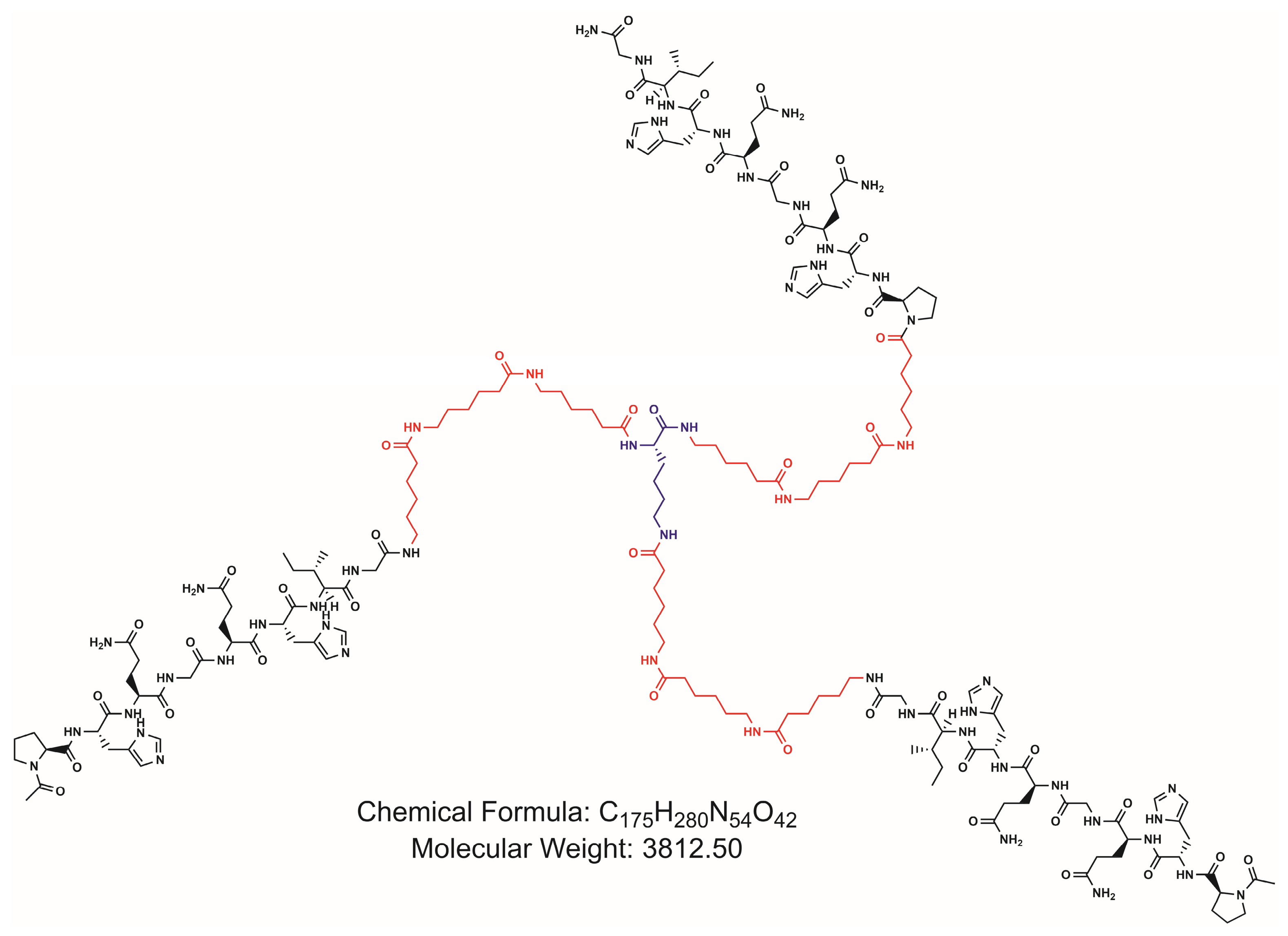
The peptide with three branches of the VEGF-A epitope PHQGQHIG was synthesised manually by SPPS using Fmoc/tBu chemistry and Rink-amide MBHA resin, GL Biochem (Shanghai, China) 0.43 meq/g [20,21]. The reaction was performed in a polypropylene column fitted with a polyethylene filter. Each coupling was accomplished by adding to 0.1 g of the solid resin and a 3-fold excess of each Fmoc-protected amino acid together with TBTU (3 eq.) and DIPEA (4 eq.) in DMF. All couplings were performed by incubation (2 × 2 min) in a microwave reactor BGH Quick Chef at 10% of its power (equivalent to an energy of 100 W) [22]. After monitoring the coupling completeness of each Fmoc-protected amino acid using the Kaiser or chloranil test [23], Fmoc was removed with 20% piperidine in DMF (2 × 5 min) at room temperature. Washings between coupling and Fmoc removal were performed using DMF and DCM (2 × 1 min). Three molecules of 6-aminohexanoic acid (Ahx) were introduced to the peptide epitope through amide linkage between the carboxylic acid of Fmoc-Ahx-OH and the amine group of the N-terminal proline, followed by two additional consecutive couplings of Fmoc-Ahx-OH, each preceded by treatment with 20% piperidine to free the amine group of the resin-linked molecule. Afterwards, Fmoc-Lys(Fmoc)-OH was coupled to the resulting Ahx3-PHQGQHIG-Resin (first branch) via amide bond formation between the terminal amine group of the spacer and the carboxylic group of Fmoc-Lys(Fmoc)-OH. Next, Fmoc was removed from the α and ε Lys amino groups of the Fmoc-Lys(Fmoc) derivate, and peptide elongation was continued by duplicating the equivalents of the reagents in each coupling to incorporate the other two branches (Figure 2). To overcome aggregation, 0.2 M LiCl in DMF was used as a solvent instead of DMF in the coupling and deprotection steps, as previously described by Barredo et al. [24]. N-terminal acetylation was achieved after peptide elongation by adding Ac2O (10 eq.) and DIC (10 eq.) in DCM and incubating for 30 min. Finally, side-chain-protecting group removal and peptide release from the solid support were performed simultaneously by treatment with TFA/TIS/H2O/DODT (92.5:2.5:2.5:2.5) for 2 h at room temperature. Subsequently, the peptide was precipitated with cold diethyl ether. The precipitate was washed 2 times with cold diethyl ether, dissolved in MilliQ water, and lyophilised. The yield was determined by gravimetry.
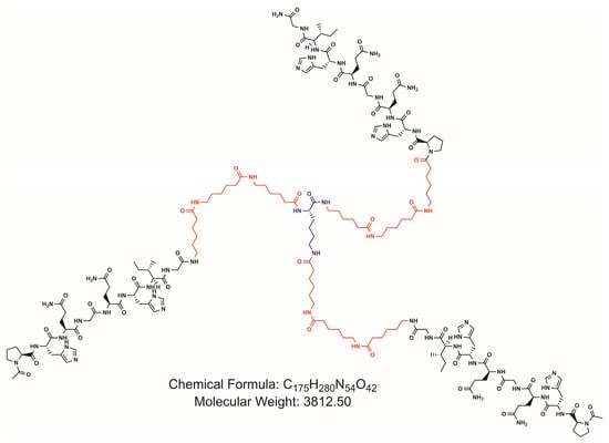
Figure 2.
Chemical structure of peptide (Ac-PHQGQHIG-Ahx3)2-K-Ahx3-PHQGQHIG-NH2.
2.2. Peptide Analysis by Reverse-Phase High-Performance Liquid Chromatography (RP-HPLC)
The synthesised peptide was analysed by RP-HPLC in a Shimadzu LC-20AT instrument (Nishinokyo-Kuwabara-cho, Nakagyo-ku, Japan) with a photodiode array UV–Visible detector (SPDM20A) using a Phenomenex (Torrance, CA, USA) ULTRACARB 5 μm ODS (20) reverse-phase C18 column (90 Å × 250 mm × 4.6 mm) at 50 °C and a flow rate of 0.5 mL/min. A linear gradient from 15% to 100% mobile phase B was performed using 0.045% TFA in H2O (solvent A) and 0.036% TFA in acetonitrile (solvent B).
2.3. Peptide Analysis by Electrospray Ionisation Mass Spectrometry (ESI MS)
The synthesised peptide was analysed by a QTRAP 4500 Sciex, Triple Quad LC-MS/MS mass spectrometer (AB SCIEX Instruments, Marlborough, MA, USA, model 5060884-E) at the Laboratorio Nacional de Investigación y Servicios en Proteínas LANAIS PROEM (IQUIFIB, UBA-CONICET). Peptide ionisation was performed by electrospray (voltage 5.5 kV). The ion source was Turbo Spray. The scan type was Q3 MS and the polarity positive. The spectra obtained were evaluated using the software Analyst 1.6.3 (Sciex, Marlborough, MA, USA). Monoisotopic mass was obtained by deconvolution using Peak View 2.2 (Sciex) software.
2.4. Bevacizumab Precipitation with (NH4)2SO4
In order to determine the maximum (NH4)2SO4 concentration at which the mAb did not precipitate, 12 μL aliquots of pure 25 mg/mL bevacizumab were added to 1.5 mL graduated polypropylene microtubes (Eppendorf, Hamburg, Germany) and mixed with water and increasing volumes of (NH4)2SO4 3.8 M, pH 7.5, to obtain final concentrations of 0.50, 1.00, 1.16, 1.18, 1.19, 1.20, 1.22, 1.24, 1.26, 1.50 and 2.00 M in a final volume of 0.5 mL. After incubating the samples for 1 h at 25 °C or 5 °C, they were centrifuged in an Eppendorf Centrifuge 5417 C at 10,000 rpm (10,300× g) for 5 min. The supernatants were separated from the precipitates, and the bevacizumab concentration in each supernatant was measured by its absorbance at 280 nm. The percentage of precipitated bevacizumab was calculated as the total amount of bevacizumab at the beginning of the experiment less the amount present in the supernatant.
2.5. Bevacizumab Precipitation with (Ac-PHQGQHIG-Ahx3)2-K-Ahx3-PHQGQHIG-NH2
Experiments were performed in 1.5 mL graduated polypropylene microtubes (Eppendorf). In order to evaluate the amount of peptide necessary to precipitate the mAb, 12 μL aliquots of pure 25 mg/mL bevacizumab were mixed with water and increasing volumes of 2 mg/mL peptide solution to obtain final concentrations of 5, 10, 16, 21, 26, 31, 37, 42, and 47 μM and (NH4)2SO4 3.8 M, pH 7.5, to obtain final concentrations of 1.20 or 1.18 M in a final volume of 0.5 mL. After incubating the samples for 1 h at 25 or 5 °C, they were centrifuged in an Eppendorf Centrifuge 5417 C at 10,000 rpm (10,300× g) for 5 min. The supernatants were separated from the precipitates. The bevacizumab concentration in each supernatant was measured by its absorbance at 280 nm, determining the unknown concentration using an absorbance calibration curve performed with pure bevacizumab solutions 0.00, 0.20, 0.40, 0.60 and 1.25 mg/mL. Percentage of precipitated bevacizumab was calculated as the total amount of bevacizumab at the beginning of the experiment less the amount present in the supernatant. The percentage of precipitated bevacizumab was evaluated as a function of the peptide/bevacizumab molar ration (P/B).
2.6. Bevacizumab Purification by Fractional Precipitation from Cell-Free Culture Broth
A sample of 1 mL of cell-free culture broth containing 1.6 mg/mL of bevacizumab was purified through a fractional precipitation process.
2.6.1. First Precipitation
A volume of 1 mL of the cell-free culture broth was added to a 1.5 mL graduated polypropylene microtube (Eppendorf), and then 0.450 mL of (NH4)2SO4 3.8 M, pH 7.5 was incorporated dropwise and under constant stirring to reach a final (NH4)2SO4 concentration of 1.18 M [25]. After incubation for 1 h at 5 °C, the resulting precipitate was isolated by centrifugation in an Eppendorf Centrifuge 5417 C at 10,000 rpm (10,300× g) for 5 min. The supernatant was separated from the pellet, and the supernatant absorbance was measured at 280 nm.
2.6.2. Second Precipitation
The supernatant (1450 mL) was separated from the precipitate and put in a 2 mL graduated polypropylene microtube (Eppendorf). Afterwards, 5 moles of peptide per mol of bevacizumab was added to the supernatant, together with additional (NH4)2SO4, to reach a final concentration of 1.20 M. After incubating the mixture for 1 h at 5 °C, the resulting precipitate was isolated by centrifugation in an Eppendorf Centrifuge 5417 C at 10,000 rpm (10,300× g) for 5 min. The supernatant was separated from the pellet, and the absorbance of the supernatant was measured at 280 nm. Next, the 2nd precipitate was dissolved in 0.5 mL of buffer 0.03 M sodium phosphate, 0.12 M NaCl, pH 7.4, for future analysis.
2.7. Bevacizumab Quantification
The amount of bevacizumab was calculated using protein A affinity chromatography, which specifically binds immunoglobulins. A column of protein A–Sepharose (1 × 0.64 cm) was equilibrated with 0.03 M sodium phosphate, 0.12 M NaCl, pH 7.4 (adsorption buffer). An aliquot of: (a) 15 μL of pure bevacizumab (25 mg/mL) and (b) 250 μL of cell-free culture broth from bevacizumab-producing CHO cells or (c) 250 μL of the resuspended 2nd precipitate was loaded in the column. Adsorption buffer was used for washing until the absorbance at 280 nm reached its initial value. Next, the elution was performed with 0.5 M acetic acid, pH 2.5. The bevacizumab concentration in the elution fraction was measured by absorbance at 280 nm, determining the unknown concentration using an absorbance calibration curve performed with bevacizumab solutions of 0.00, 0.20, 0.40, 0.60 and 1.25 mg/mL. Purity was defined as the amount of bevacizumab as a fraction of the total amount of proteins. Fold purification was calculated by setting the initial purity at a value of one and then giving the purity of the 2nd precipitate relative to the initial purity [26].
2.8. Size-Exclusion Chromatography (SEC) with Sephadex G-25
The resuspended 2nd precipitate was desalted using a prepacked PD-10 Desalting Column of Sephadex™ G-25 resin, as indicated in the instruction manual [27]. Briefly, the column was equilibrated with distilled water. After loading the sample, distilled water was added, and the absorbances of the collected fractions were registered at 280 nm. The protein fraction eluted in the void volume, whereas smaller molecules like salts and the peptide were delayed in the gel pores and separated from bevacizumab.
2.9. Total Protein Quantification
The total protein concentration of the studied samples was measured using Bradford dye-binding assay [28]. Briefly, 100 μL of each sample was mixed with 100 μL of Bradford reagent in a multiwell plate. Samples were incubated for 15 min at room temperature, and their absorbances at 620 nm were measured. Calibration curve was created using pure bevacizumab solutions of 0.00, 0.78, 1.56, 3.13, 6.25, 12.50, 25.00 and 50.00 μg/mL. Although bovine serum albumin is usually used as the protein standard, bevacizumab was used instead, as it was the predominant protein in the analysed samples.
2.10. Sodium Dodecyl Sulphate-Polyacrylamide Gel Electrophoresis (SDS-PAGE)
Pure bevacizumab, cell-free culture broth, and the dissolved and desalted 2nd precipitate were analysed using 12.5% SDS-PAGE (under reducing and non-reducing conditions) as previously described [29]. The gels were stained for 1 h in a solution of 0.25% (w/v) Coomassie Brilliant Blue R-250 (Bio-Rad, Philadelphia, PA, USA) in CH3OH/acetic acid/H2O (50/10/40) at 37 °C under agitation and then destained for 2 h with CH3OH/acetic acid/H2O (5.0/7.5/87.5) at 37 °C under agitation.
3. Results
3.1. Peptide Design and Synthesis
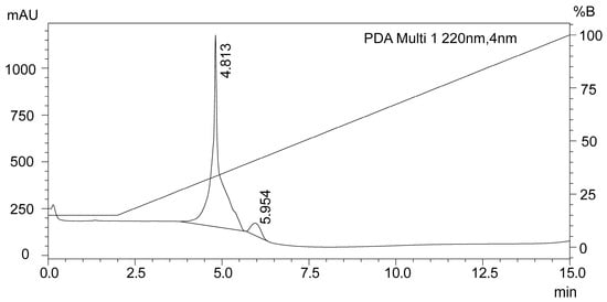
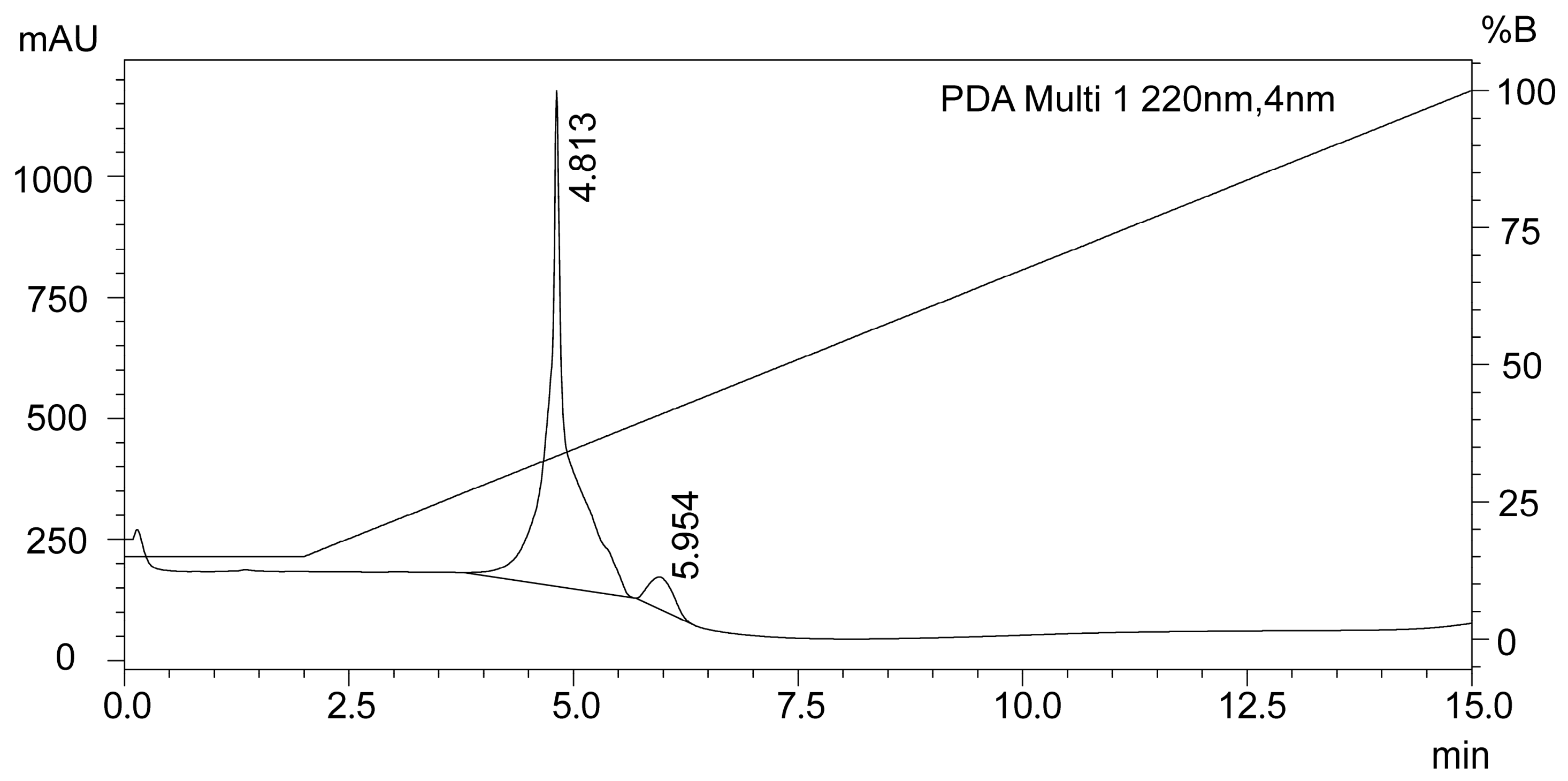
The peptide (Ac-PHQGQHIG-Ahx3)2-K-Ahx3-PHQGQHIG-NH2, composed of three units of the bevacizumab epitope PHQGQHIG derived from VEGF-A, was designed to purify mAb by affinity precipitation. The three PHQGQHIG branches, each separated with a spacer arm (Ahx3), ensured the interaction of each epitope with a different molecule of bevacizumab, thereby promoting the formation of cyclic complexes, as previously reported [19]. To increase its stability, the peptide was amidated at the C-terminus and acetylated at both N termini (Figure 2). SPPS allowed us to obtain (Ac-PHQGQHIG-Ahx3)2-K-Ahx3-PHQGQHIG-NH2 with high yield (85%). A low-loading-capacity resin (Rink-amide MBHA resin) was selected to minimise steric hindrance during peptide elongation. Also, the addition of the chaotropic salt LiCl, together with microwave-induced high temperatures, prevented peptide aggregation during its synthesis [17]. Furthermore, microwave-assisted SPPS significantly reduced the reaction time [23]. RP-HPLC analysis demonstrated high peptide purity (Figure 3).
Figure 3.
RP-HPLC analysis of (Ac-PHQGQHIG-Ahx3)2-K-Ahx3-PHQGQHIG-NH2 peptide. Solvent A: 0.045% TFA in H2O. Solvent B: 0.036% TFA in acetonitrile. Right axis indicates % of solution B in solution A (% B) in gradient program from 15 to 100% B.
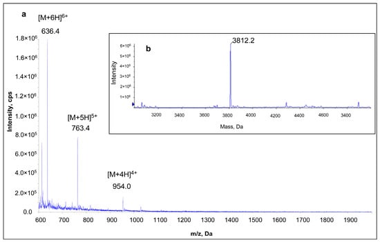
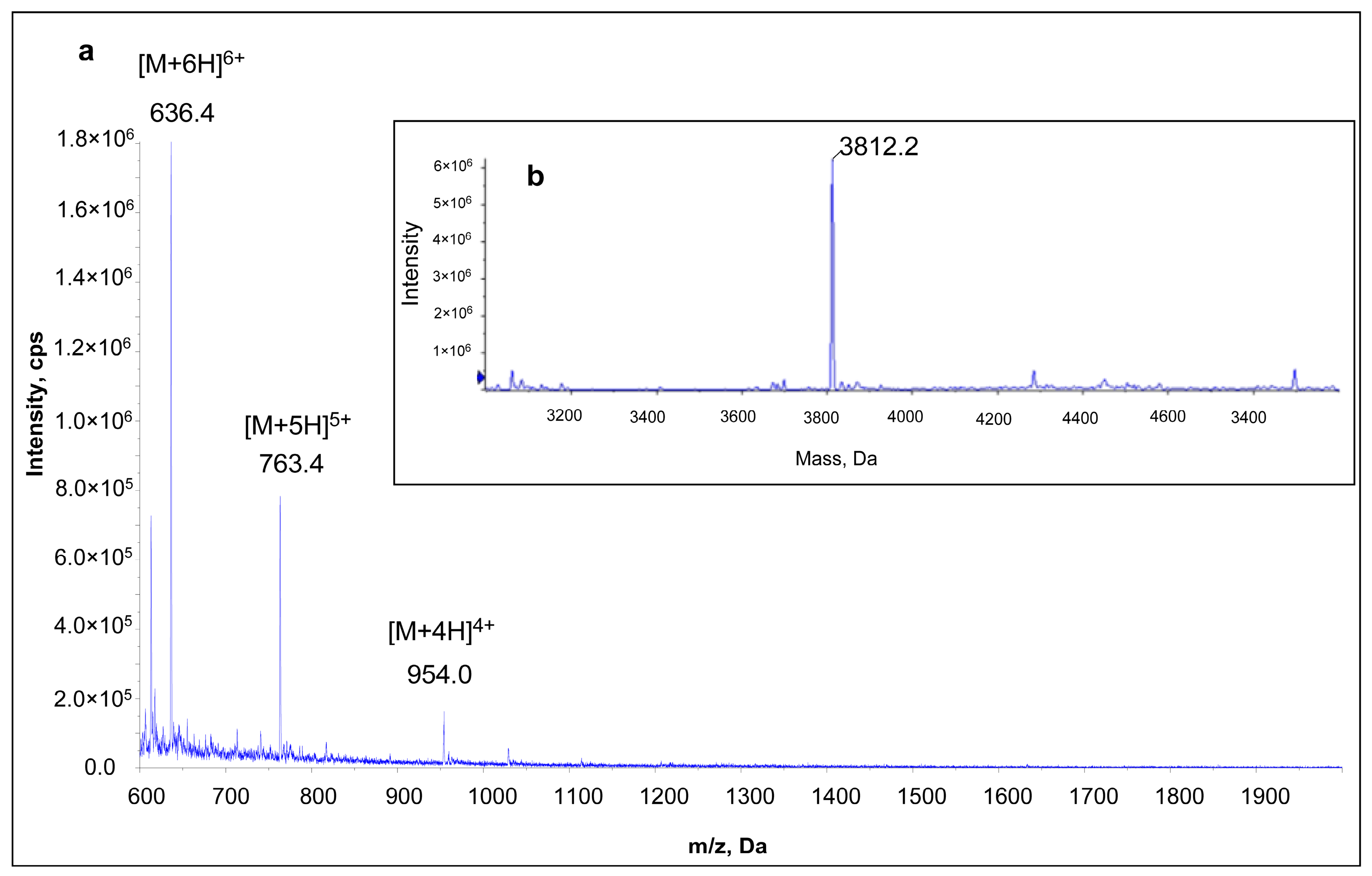
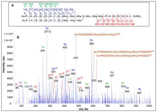
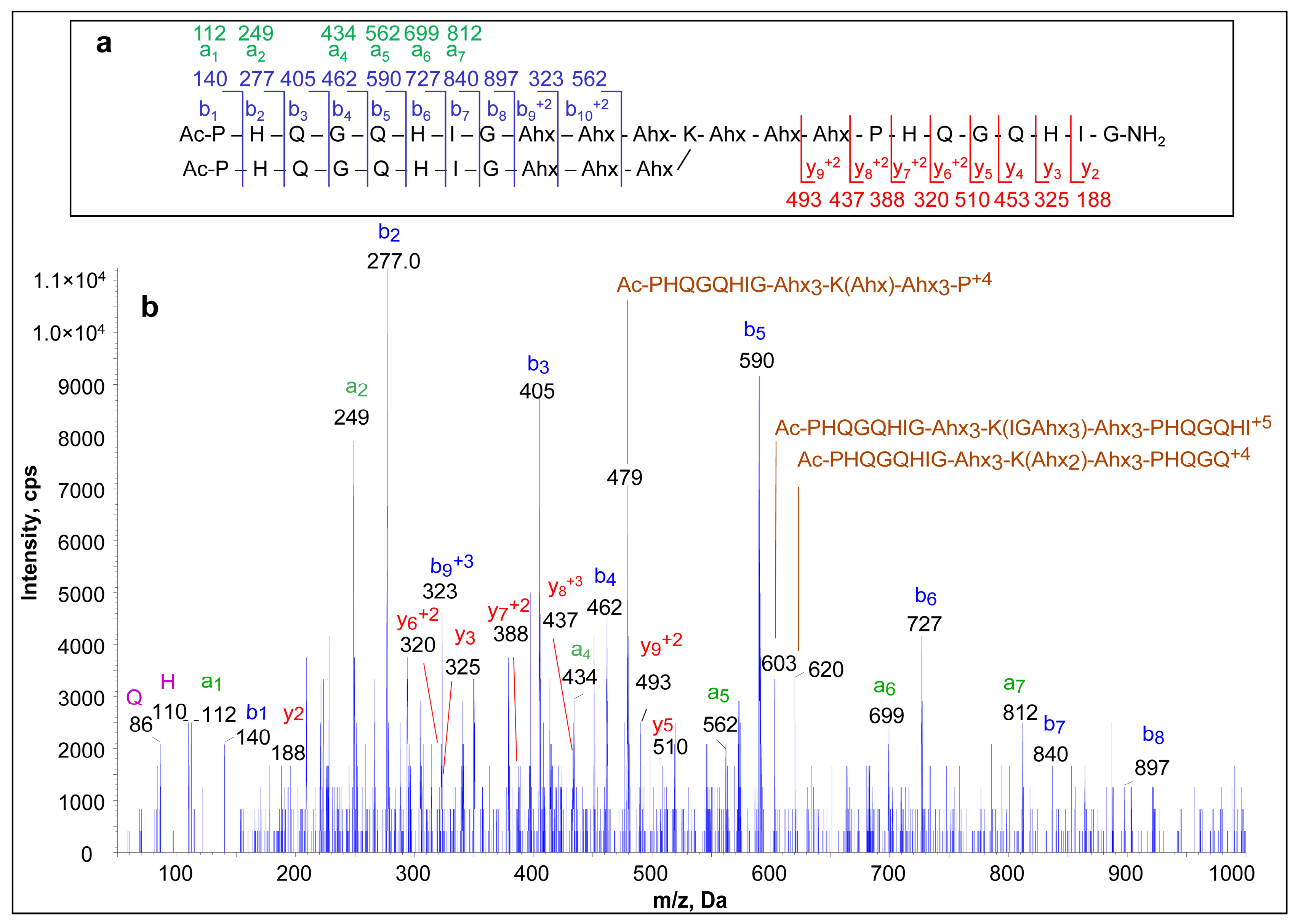
Furthermore, the ESI-MS and MS/MS mass spectra of the peptide were obtained to confirm its identity [30]. Table 1 indicates the expected peaks in the MS spectrum, and Figure 4a shows the experimental mass spectrum obtained. The peaks at m/z 636.4, 763.4, and 954.0 correspond to the multiply protonated ions [M + 6H]6+, [M + 5H]5+, and [M + 4H]4+, respectively, confirming the identity of the synthesised peptide. The peptide’s monoisotopic mass (Figure 4b) was obtained with the deconvolution software Peak View 2.2 (Sciex). The main peak at 3812.2 Da corresponds to the synthesised peptide, while a few minor peaks, likely related to trace impurities, were also detected. Figure 5 shows the MS/MS spectra obtained from m/z 636.4, corresponding to the [M + 6H]6+ ion. The most abundant ions were those from the “b” series of both N-terminal branches (Ac-PHQGQHIG-Ahx3)2. Also, ions from the “a” and “y” series were detected, although with minor abundances, together with internal ions and immonium ions. Higher ions were observed as multiply protonated.
Table 1.
Values of theorical m/z of singly and multiply protonated [M + zH]z+ monoisotopic ions from peptide (Ac-PHQGQHIG-Ahx3)2-K-Ahx3-PHQGQHIG-NH2. Chemical formula: C175H280N54O42; molecular weight: 3812.50; monoisotopic masses: M (42%): 3810.14, M + 1 (91%): 3811.1, M + 2 (100%): 3912.1.
Figure 4.
(a) ESI mass spectra of (Ac-PHQGQHIG-Ahx3)2-K-Ahx3-PHQGQHIG-NH2 peptide. Peaks at m/z 636.4, 763.4, and 954.0 in spectrum (a) correspond to [M + 6H]6+, [M + 5H]5+ and [M + 4H]4+ ions, respectively. (b) Monoisotopic molecular mass calculated by deconvolution.
Figure 5.
ESI MS/MS mass spectra obtained from ion [M + 6H]6+ (m/z 636.4) of (Ac-PHQGQHIG-Ahx3)2-K-Ahx3-PHQGQHIG-NH2 peptide. (a) Peptide fragmentation scheme with corresponding ions observed in spectrum. (b) ESI MS/MS mass spectrum with ions from “a”, “b” and “y” series indicated in green, blue and red, respectively. Internal ions are shown in brown, and immonium ions are indicated in violet.
3.2. Pure Bevacizumab Precipitation
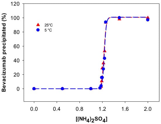
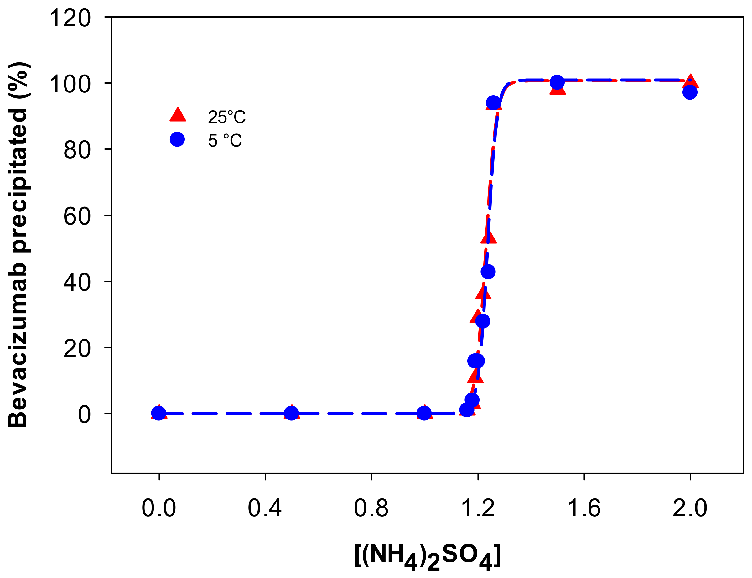
Previously, Barredo et al. proved that the affinity between bevacizumab and the VEGF-A epitope PHQGQHIG increases at high (NH4)2SO4 concentrations. In addition, stronger adsorption was observed at pH values between 7.0 and 8.0 than at pH 5.5, likely because the imidazole side chains of His are not charged at pH value over its pKa (approximately 6.0), thereby favouring hydrophobic interactions [6,7]. Therefore, bevacizumab precipitation with the peptide was evaluated at pH 7.5 in the presence of high (NH4)2SO4 concentration. To avoid the coprecipitation of contaminants, a fractional precipitation protocol was designed, consisting of negative precipitation with (NH4)2SO4, followed by positive precipitation with the peptide in (NH4)2SO4. To design the fractional precipitation protocol, the precipitation of pure bevacizumab was initially evaluated with (NH4)2SO4 at pH 7.5 (Figure 6). The (NH4)2SO4 concentration at which the mAb did not precipitate was below 1.18 M; therefore, the (NH4)2SO4 concentration selected for the first step (negative precipitation) was 1.18 M or lower. Between 1.18 and 1.25 M, small variations in the (NH4)2SO4 concentration resulted in large variations in the percentage of bevacizumab precipitation, after which almost all mAb precipitated.
Figure 6.
Percentage of precipitated bevacizumab as a function of (NH4)2SO4 concentration at 25 °C (red triangles) or 5 °C (blue circles).
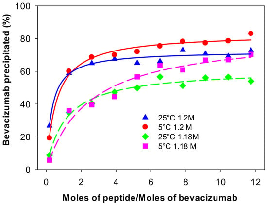
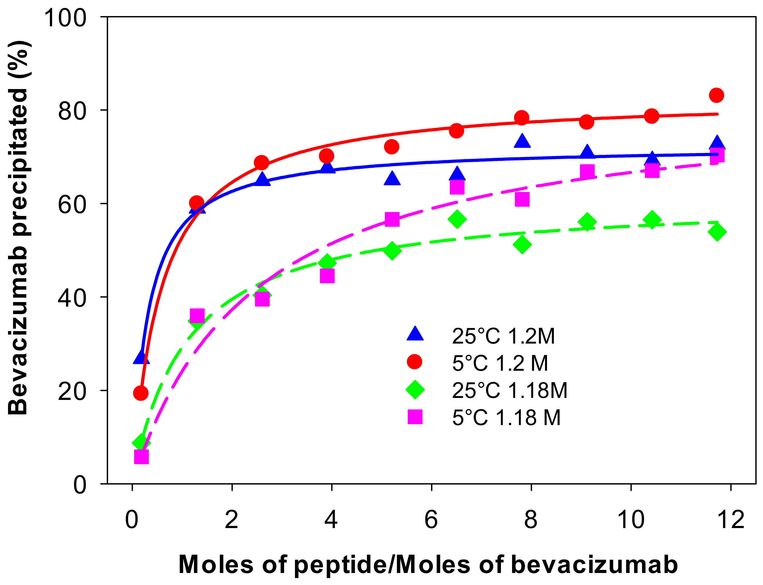
Next, the amount of peptide in ammonium sulphate necessary for bevacizumab precipitation was evaluated using pure bevacizumab solution. Figure 7 shows the percentage of precipitated bevacizumab as a function of the peptide/bevacizumab molar ratio (P/B) in (NH4)2SO4, 1.18 or 1.20 M, pH 7.5, at 25 or 5 °C. The amount of precipitated bevacizumab increased with the P/B ratio until it reached a plateau at approximately P/B ≈ 5. Better results were observed at 1.20 M, probably because bevacizumab–peptide binding is largely hydrophobic. Comparing the experiments performed at 25 and 5 °C, refrigeration was found to slightly facilitate bevacizumab precipitation. However, the difference in the percentage of precipitated mAb was moderate, indicating that efficient precipitation can also be achieved at room temperature, which may be advantageous for practical implementation.
Figure 7.
Percentage of precipitated bevacizumab as function of peptide/bevacizumab molar ratio. Bevacizumab precipitation in (NH4)2SO4 1.20 M (solid lines) at 25 °C (blue triangles) or 5 °C (red circles) and in (NH4)2SO4 1.18 M (dash lines) at 25 °C (green diamonds) or 5 °C (pink squares).
3.3. Bevacizumab Purification by Fractional Precipitation
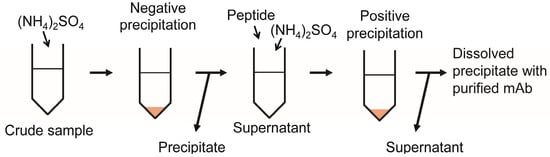
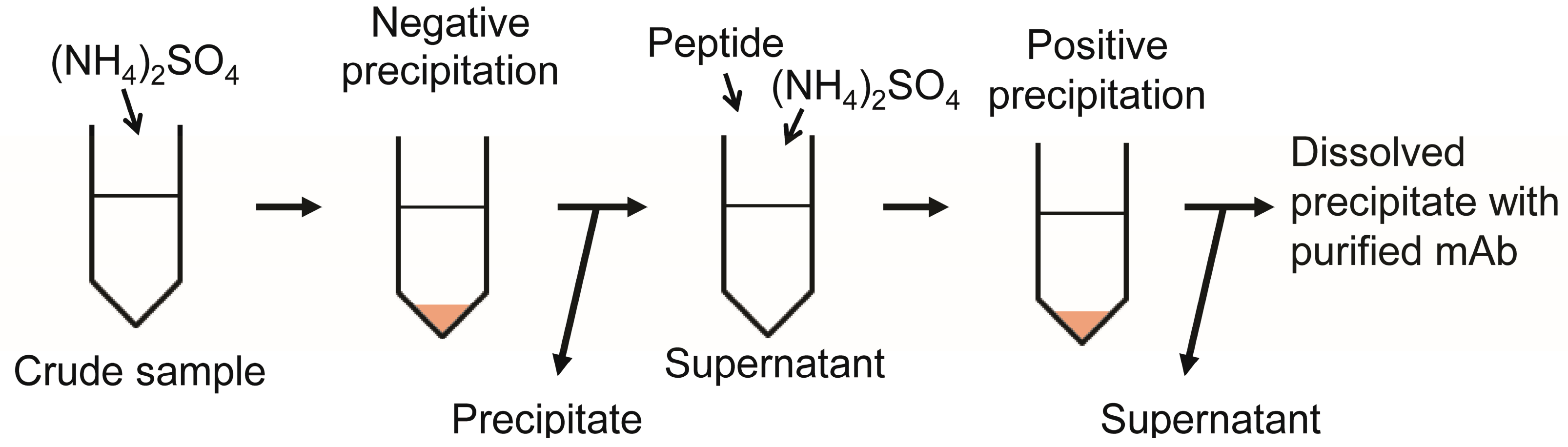
The process designed for bevacizumab purification from the cell-free culture broth consisted of a fractional precipitation protocol. The first step involved a “negative” precipitation using only (NH4)2SO4 1.18 M (concentration in which bevacizumab did not precipitate). Under these conditions, some of the contaminants were removed by precipitation, while bevacizumab remained in the supernatant. The second step involved “positive” precipitation in which, by adding the peptide to the obtained supernatant after the first step, bevacizumab was precipitated selectively (Figure 8).
Figure 8.
Scheme of bevacizumab purification by fractional precipitation using (NH4)2SO4 and peptide (Ac-PHQGQHIG-Ahx3)2-K-Ahx3-PHQGQHIG-NH2.
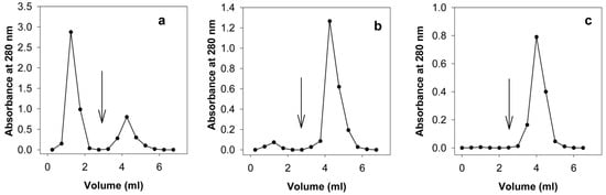
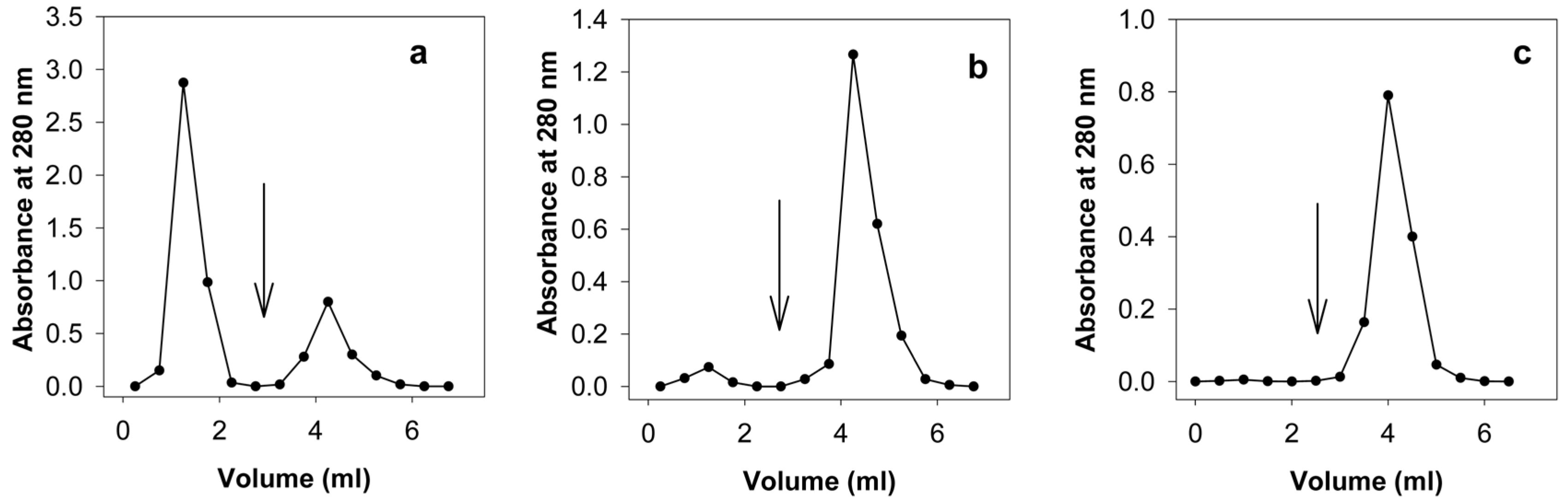
The compositions of the crude sample and the second precipitate were compared and evaluated using protein A AC, which specifically binds immunoglobulins. Figure 9 shows the chromatograms obtained when loading a sample of the cell-free culture broth (Figure 9a), the second precipitate resuspended in adsorption buffer (Figure 9b), and pure bevacizumab (Figure 9c). Impurities were collected in the non-binding fraction, whereas bevacizumab was recovered in the elution fraction. As can be seen in Figure 9b, the impurities in the second precipitate represented a small fraction compared to the amount of bevacizumab.
Figure 9.
Protein A affinity chromatography of (a) cell-free culture broth from bevacizumab-producing CHO cells, (b) resuspended 2nd precipitate obtained after sequential precipitation protocol designed in this work, and (c) pure bevacizumab (25 mg/mL). The adsorption buffer was 0.03 M sodium phosphate, 0.12 M NaCl, pH 7.4. The elution buffer was 0.5 M of acetic acid, pH 2.5. Fractions of 0.5 mL were collected, and their absorbances were measured at 280 nm. The arrow in each chromatogram indicates the buffer change.
Table 2 shows the purification chart of bevacizumab, indicating the good performance of the developed fractional precipitation method. In the second precipitate, bevacizumab was obtained with a purity of 98% and a yield of 73%.
Table 2.
Purification chart of bevacizumab using affinity precipitation with (Ac-PHQGQHIG-Ahx3)2-K-Ahx3-PHQGQHIG-NH2 peptide.
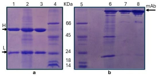
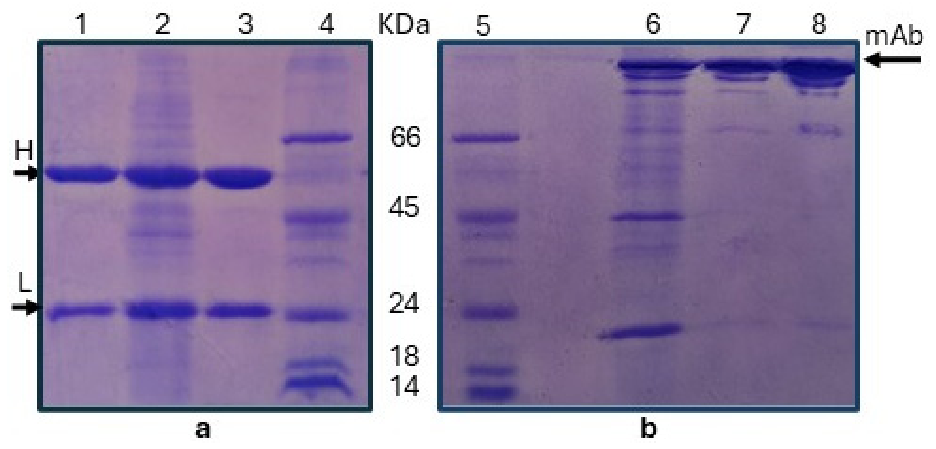
Figure 10 shows the SDS-PAGE of the crude sample and the second precipitate under reducing conditions, where the light and heavy chains of mAb can be observed, and under non-reducing conditions, where the intact mAb can be observed. The SDS-PAGE shows the high purity of the second precipitate.
Figure 10.
(a) SDS-PAGE gels under reducing conditions. Lanes: (1) pure bevacizumab, (2) bevacizumab-producing CHO cell filtrate, (3) resuspended second precipitate obtained after sequential precipitation protocol designed in this work, (4) protein molecular weight marker (albumin, bovine 66.0 kDa; albumin, egg 45.0 kDa; trypsinogen 24.0 kDa; beta-lactoglobulin 18.4 kDa; lysozyme 14.3 kDa). (b) SDS-PAGE gels under non-reducing conditions. Lanes: (5) protein molecular weight marker, (6) bevacizumab-producing CHO cell filtrate, (7) resuspended second precipitate obtained after sequential precipitation protocol designed in this work, (8) pure bevacizumab. Arrows on each gel indicate the bands corresponding to the heavy (H) and light (L) chains (a) and intact mAb (b).
4. Discussion
A branched peptide (Ac-PHQGQHIG-Ahx3)2-K-Ahx3-PHQGQHIG-NH2 was synthesised using SPPS. Peptide aggregation during its synthesis was avoided by combining a low-loading resin, microwave-induced high temperature, and the use of a chaotropic salt (LiCl).
Bevacizumab purification was efficiently achieved through a novel fractional precipitation process, combining salt-based removal of impurities with selective mAb precipitation. In the first step, a “negative” precipitation is performed using (NH4)2SO4 1.18 M, which allows for the elimination of contaminants that precipitate at this salt concentration. This concentration was selected as the highest (NH4)2SO4 level at which bevacizumab does not precipitate, thus maximising impurity removal without compromising the overall yield. In the second step, the addition of the branched peptide promotes the selective precipitation of bevacizumab. To increase the mAb recovery in this step, the final (NH4)2SO4 concentration was increased to 1.20 M without impairing the final purity. The precipitated bevacizumab was recovered by centrifugation and resuspended in 0.03 M sodium phosphate buffer containing 0.12 M NaCl, pH 7.4. The low salt concentration dissociated the peptide from bevacizumab, as the interaction with the peptide epitope is predominantly hydrophobic, as previously demonstrated [6,7]. This designed fractional precipitation method could replace protein A AC in bevacizumab downstream processing, while the other purification steps indicated in Figure 1 are still necessary to acquire an API-quality product. In contrast with protein A AC, which requires harsh acidic elution conditions, the protocol proposed here enables bevacizumab recovery from the precipitate under mild conditions, thus minimising the risk of antibody damage.
In this small-scale process, the epitope peptide was finally separated from bevacizumab by size-exclusion chromatography using Sephadex G-25, recovering nearly 100% of the peptide, which can be recycled.
In conclusion, the developed method allows a high-purity product to be obtained under milder conditions and at a lower cost than current industrial protocols that use protein A AC. Furthermore, precipitation techniques are easy to scale-up using industrial reactors [31,32].
In future work, the scale-up of the process will be assessed. An available automated microwave peptide synthesiser allows quick large-scale peptide synthesis [33]. Also, evaluating the (NH4)2SO4 concentration range for the first and second precipitation stages—without compromising process purity and yield—will facilitate scale-up. Although, in this low-scale process, the peptide is separated from bevacizumab by size-exclusion chromatography, in a large-scale purification process, ultrafiltration with membranes with an appropriate molecular weight cut-off (MWCO) would be a good alternative choice. Moreover, instead of recycling the peptide, it would be of great interest to design a single-use (SU) process due to the much lower cost of the peptide compared to that of AC matrices. SU technology reduces contamination risks and avoids the cumbersome cleaning and validation processes needed for reusable systems; therefore, it is revolutionising therapeutic mAb production [34].
Although this purification method is limited to bevacizumab, a similar approach could be applied to other therapeutic mAbs through the design of appropriate epitope-based branched peptide ligands.
Author Contributions
Conceptualisation, J.A.E., J.A.R., G.R.B.-V., B.R., M.M. and S.A.C.; methodology, J.A.E., J.A.R., G.R.B.-V., M.S.G.-C., D.E.R., B.R., M.M. and S.A.C.; investigation, J.A.E., J.A.R., G.R.B.-V. and S.A.C.; writing—original draft preparation S.A.C.; writing—review and editing, J.A.E., J.A.R., G.R.B.-V., M.S.G.-C., B.R., M.M. and S.A.C.; supervision, B.R., M.M. and S.A.C.; project administration, S.A.C.; funding acquisition, B.R., M.M. and S.A.C. All authors have read and agreed to the published version of the manuscript.
Funding
This research was funded by the Agencia Nacional de Promoción de la Investigación, el Desarrollo Tecnológico y la Innovación de la República Argentina (PICT-2021-I-A-00236), the Consejo Nacional de Investigaciones Científicas y Técnicas (CONICET) (PIP 11220210100075CO), the Universidad de Buenos Aires, Argentina (20020220200001BA) and European Union’s Horizon 2020 research and innovation programme under the Marie Skłodowska-Curie grant agreement No. 101008072 (SUPRO-GEN project). S.A.C. is a researcher at CONICET. M.M and B.R. also acknowledge financial support provided by the MUR–Dipartimenti di Eccellenza 2023–2027 to the Department of Chemistry “Ugo Schiff” of the University of Florence (DICUS 2.0, CUP: B97G22000740001).
Institutional Review Board Statement
Not applicable.
Informed Consent Statement
Not applicable.
Data Availability Statement
The original contributions presented in this study are included in the article. Further inquiries can be directed to the corresponding author.
Acknowledgments
Pure bevacizumab and bevacizumab-producing CHO cell-free culture broth were kindly donated by Lucas Figuera Risso from mAbxience SAU (Munro, Buenos Aires, Argentina). The authors would like to express their sincere gratitude to Nora M. Vizioli and César R. González Solveyra from LANAIS PROEM (IQUIFIB, UBA-CONICET, Argentina) for making the mass spectrometer QTRAP 4500 Sciex available to us and for the valuable advice provided.
Conflicts of Interest
mAbxience SAU provided the pure bevacizumab and cell-free culture broth from bevacizumab-producing CHO cells. The authors declare that this research was conducted in the absence of any commercial or financial relationships that could be construed as a potential conflict of interest.
Abbreviations
| AC | Affinity chromatography |
| Ac2O | Acetic anhydride |
| AcOH | Acetic acid |
| Ahx | 6-Aminohexanoic acid |
| API | Active pharmaceutical ingredient |
| BSA | Bovine serum albumin |
| CHO | Chinese hamster ovary |
| DCM | Dichloromethane |
| DIC | N,N′-diisopropylcarbodiimide |
| DIPEA | N,N-diisopropylethylamine |
| DMF | N,N-dimethylformamide |
| DODT | 3,6-Dioxa-1,8-octanedithiol |
| ESI MS | Electrospray ionization mass spectrometry |
| Fmoc | Fluorenylmethyloxycarbonyl |
| HPLC | High-performance liquid chromatography |
| ID | Isotopic distribution |
| mAb | Monoclonal antibody |
| MBHA | 4-Methylbenzhydrylamine hydrochloride |
| MeCN | Acetonitrile |
| MeOH | Methanol |
| MWCO | Molecular wight cut-off |
| P/B | Peptide/bevacizumab molar ratio |
| RA | Relative abundance |
| RP-HPLC | Reverse-phase high-performance liquid chromatography |
| SDS-PAGE | Sodium dodecyl sulphate-polyacrylamide gel electrophoresis |
| SEC | Size-exclusion chromatography |
| SPPS | Solid-phase peptide synthesis |
| SU | Single use |
| TBTU | 2-(1H-benzotriazole-1-yl)-1,1,3,3-tetramethylaminium tetrafluoroborate |
| TFA | Trifluoroacetic acid |
| TIS | Triisopropylsilane |
| VEGF-A | Vascular endothelial growth factor A |
References
- Gerriets, V.; Kasi, A. Bevacizumab. In StatPearls; StatPearls Publishing: Treasure Island, FL, USA, 2026. Available online: https://www.ncbi.nlm.nih.gov/books/NBK482126/ (accessed on 30 June 2026).
- Waller-Pulido, A.; Jiménez-Pérez, M.I.; Gonzalez-Sanchez, F.A.; Rojo-Gutierrez, R.P.; Torres-Anguiano, E.; Aleman-Aguilar, J.P.; Garcia-Varela, R. Production of monoclonal antibodies for therapeutic purposes: A review. Int. Immunopharmacol. 2023, 120, 110376. [Google Scholar] [CrossRef] [Scilit] [PubMed]
- Ramos-de-la-Peña, A.M.; González-Valdez, J.; Aguilar, O. Protein A chromatography: Challenges and progress in the purification of monoclonal antibodies. J. Sep. Sci. 2019, 42, 1816–1827. [Google Scholar] [CrossRef] [Scilit] [PubMed]
- Bracewell, D.G.; Pathak, M.; Ma, G.; Rathore, A.S. Re-use of protein A resin: Fouling and economics. BioPharm Int. 2015, 28, 28–33. [Google Scholar]
- Follman, D.K.; Fahrner, R.L. Factorial screening of antibody purification processes using three chromatography steps without protein A. J. Chromatogr. A 2004, 1024, 79–85. [Google Scholar] [CrossRef] [Scilit] [PubMed]
- Barredo, G.R.; Giudicessi, S.L.; Martínez Ceron, M.C.; Saavedra, S.L.; Rodriguez, S.; Filgueira Risso, L.; Erra-Balsells, R.; Mahler, G.; Albericio, F.; Cascone, O.; et al. A short peptide fragment of the vascular endothelial growth factor as a novel ligand for bevacizumab purification. Protein Expr. Purif. 2020, 165, 105500. [Google Scholar] [CrossRef] [Scilit] [PubMed]
- Barredo, G.R.; Giudicessi, S.L.; Martínez Ceron, M.C.; Saavedra, S.L.; Rodriguez, S.; Filgueira Risso, L.; Erra-Balsells, R.; Mahler, G.; Albericio, F.; Cascone, O.; et al. Protocol for bevacizumab purification using Ac-PHQGQHIGVSK-agarose. MethodsX 2020, 7, 100769. [Google Scholar] [CrossRef] [Scilit] [PubMed]
- Norling, L.; Lute, S.; Emery, R.; Khuu, W.; Voisard, M.; Xu, Y.; Chen, Q.; Blank, G.; Brorson, K. Impact of multiple re-use of anion-exchange chromatography media on virus removal. J. Chromatogr. A 2005, 1069, 79–89. [Google Scholar] [CrossRef] [Scilit] [PubMed]
- Ravi, N.; Huerta, J.; Ferreira, G. Evaluating multiproduct chromatography Protein A resin reuse for monoclonal antibodies in biopharmaceutical manufacturing. Biotechnol. Prog. 2023, 39, e3333. [Google Scholar] [CrossRef] [Scilit] [PubMed]
- Holschuh, K.; Schwämmle, A. Preparative purification of antibodies with protein A—An alternative to conventional chromatography. J. Magn. Magn. Mater. 2005, 293, 345–348. [Google Scholar] [CrossRef] [Scilit]
- Kaveh-Baghbaderani, Y.; Allgayer, R.; Schwaminger, S.P.; Fraga-Garcia, P.; Berensmeier, S. Magnetic separation of antibodies with high binding capacity by site-directed immobilization of protein A-domains to bare iron oxide nanoparticles. ACS Appl. Nano Mater. 2021, 4, 4956–4963. [Google Scholar] [CrossRef] [Scilit]
- Barredo, G.R.; Giudicessi, S.L.; Albericio, F.; Cascone, O.; Camperi, S.A. Magnetic Affinity Nanoparticles for Bevacizumab Adsorption. In Peptides 2022 Proceedings Volume of the 36th EPS August 28–2 September 2022 Sitges, Spain; Lebl, M., Ed.; European Peptide Society: Prague, Czech Republic, 2022; pp. 127–129. [Google Scholar] [CrossRef] [Scilit]
- Le, T.D.; Suttikhana, I.; Ashaolu, T.J. State of the art on the separation and purification of proteins by magnetic nanoparticles. J. Nanobiotechnol. 2023, 21, 363. [Google Scholar] [CrossRef] [Scilit] [PubMed]
- Madan, B.; Chaudhary, G.; Cramer, S.M.; Chen, W. ELP-z and ELP-Zz Capturing Scaffolds for the Purification of Immunoglobulins by Affinity Precipitation. J. Biotechnol. 2013, 163, 10–16. [Google Scholar] [CrossRef] [Scilit] [PubMed]
- Mokhtari, J.; Nourisefat, M.; Zamiri, B.; Fotouhi, L.; Zarnani, A.H.; Moosavi-Movahedi, A.A.; Karimian, K. Novel method for the isolation of proteins and small target molecules from biological and aqueous media by salt-assisted phase transformation of their PEGylated recognition counterparts. ACS Omega 2021, 6, 7585–7597. [Google Scholar] [CrossRef] [Scilit] [PubMed]
- Fishman, J.B.; Berg, E.A. Ammonium sulfate fractionation of antibodies. Cold Spring Harb. Protoc. 2018, 2018, pdb-rot099119. [Google Scholar] [CrossRef] [Scilit] [PubMed]
- Bilgiçer, B.; Thomas, S.W., 3rd; Shaw, B.F.; Kaufman, G.K.; Krishnamurthy, V.M.; Estroff, L.A.; Yang, J.; Whitesides, G.M. A non-chromatographic method for the purification of a bivalently active monoclonal IgG antibody from biological fluids. J. Am. Chem. Soc. 2009, 131, 9361–9367. [Google Scholar] [CrossRef] [Scilit] [PubMed]
- Handlogten, M.W.; Stefanick, J.F.; Alves, N.J.; Bilgicer, B. Nonchromatographic affinity precipitation method for the purification of bivalently active pharmaceutical antibodies from biological fluids. Anal. Chem. 2013, 85, 5271–5278. [Google Scholar] [CrossRef] [Scilit] [PubMed]
- Handlogten, M.W.; Stefanick, J.F.; Deak, P.E.; Bilgicer, B. Affinity-based precipitation via a bivalent peptidic hapten for the purification of monoclonal antibodies. Analyst 2014, 139, 4247–4255. [Google Scholar] [CrossRef] [Scilit] [PubMed]
- Jaradat, D.S.M. Thirteen decades of peptide synthesis: Key developments in solid phase peptide synthesis and amide bond formation utilized in peptide ligation. Amino Acids 2018, 50, 39–68. [Google Scholar] [PubMed]
- Hansen, P.R.; Oddo, A. Fmoc Solid-Phase Peptide Synthesis. Methods Mol. Biol. 2024, 2821, 33–55. [Google Scholar] [CrossRef] [Scilit] [PubMed]
- Manual de Instrucciones de Microondas, “Quick Chef BGH”. Available online: https://www.manual.ar/bgh/quick-chef-b228db9/manual (accessed on 30 June 2026).
- Guzmán, F.; Aróstica, M.; Román, T.; Beltrán, D.; Gauna, A.; Albericio, F.; Cárdenas, C. Peptides, solid-phase synthesis and characterization: Tailor-made methodologies. Electron. J. Biotechnol. 2023, 64, 27–33. [Google Scholar] [CrossRef] [Scilit]
- Barredo-Vacchelli, G.R.; Rodríguez, J.A.; Eloy, J.A.; Camperi, S.A. A novel method for liraglutide synthesis and purification. Pept. Sci. 2024, 116, e24351. [Google Scholar] [CrossRef] [Scilit]
- Wingfield, P. Protein precipitation using ammonium sulfate. Curr. Protoc. Protein Sci. 2001, 13, A.3F.1–A.3F.8. [Google Scholar] [CrossRef] [Scilit] [PubMed]
- Burgess, R.R. Preparing a purification summary table. In Methods in Enzymology. Guide to Protein Purification, 2nd ed.; Burgess, R.R., Murray, P.D., Eds.; Academic Press: New York, NY, USA, 2009; Volume 463, pp. 29–34. [Google Scholar] [CrossRef] [Scilit] [PubMed]
- PD-10 Desalting Column. Product Booklet. Cytiva.com. 52130800 BD. Available online: https://d3.cytivalifesciences.com/prod/IFU/52130800.pdf (accessed on 30 June 2026).
- Kielkopf, C.L.; Bauer, W.; Urbatsch, I.L. Bradford assay for determining protein concentration. Cold Spring Harb. Protoc. 2020, 2020, 102269. [Google Scholar] [CrossRef] [Scilit] [PubMed]
- Bhatt, M.; Rai, V.; Kumar, A.; Kiran; Yadav, A.K.; Rajak, K.K.; Gupta, V.; Chander, V.; Avasthe, R.K. SDS-PAGE and western blotting: Basic principles and protocol. In Protocols for the Diagnosis of Pig Viral Diseases; Deb, R., Yadav, A.K., Rajkhowa, S., Malik, Y.S., Eds.; Springer Protocols Handbooks; Humana: New York, NY, USA, 2022; pp. 313–328. [Google Scholar] [CrossRef] [Scilit]
- Seidler, J.; Zinn, N.; Boehm, M.E.; Lehmann, W.D. De novo sequencing of peptides by MS/MS. Proteomics 2010, 10, 634–649. [Google Scholar] [CrossRef] [Scilit] [PubMed]
- Burgess, R.R. Protein Precipitation Techniques. In Methods in Enzymology. Guide to Protein Purification, 2nd ed.; Burgess, R.R., Murray, P.D., Eds.; Academic Press: New York, NY, USA, 2009; Volume 463, pp. 331–342. [Google Scholar] [CrossRef] [Scilit] [PubMed]
- Martinez, M.; Spitali, M.; Norrant, E.L.; Bracewell, D.G. Precipitation as an enabling technology for the intensification of biopharmaceutical manufacture. Trends Biotechnol. 2019, 37, 237–241. [Google Scholar] [CrossRef] [Scilit] [PubMed]
- Singh, S.K.; Collins, J.M. Ultra-efficient solid-phase peptide synthesis. In Peptide Synthesis. Methods in Molecular Biology; Hussein, W.M., Skwarczynski, M., Eds.; Humana: New York, NY, USA, 2025; Volume 2931, pp. 327–342. [Google Scholar] [CrossRef] [Scilit] [PubMed]
- Samaras, J.J.; Micheletti, M.; Ding, W. Transformation of biopharmaceutical manufacturing through single-use technologies: Current state, remaining challenges, and future development. Annu. Rev. Chem. Biomol. Eng. 2022, 13, 73–97. [Google Scholar] [CrossRef] [Scilit] [PubMed]

Disclaimer/Publisher’s Note: The statements, opinions and data contained in all publications are solely those of the individual author(s) and contributor(s) and not of MDPI and/or the editor(s). MDPI and/or the editor(s) disclaim responsibility for any injury to people or property resulting from any ideas, methods, instructions or products referred to in the content. |
© 2026 by the authors. Licensee MDPI, Basel, Switzerland. This article is an open access article distributed under the terms and conditions of the Creative Commons Attribution (CC BY) license.