1. Introduction
Transdermal drug delivery has gained considerable interest as an alternative to conventional administration routes due to its ability to improve patient compliance, bypass first-pass metabolism, and provide sustained drug release [
1]. However, a significant challenge in transdermal delivery is the low permeability of the stratum corneum, the outermost skin layer and a natural barrier to drug penetration [
2,
3]. To overcome this limitation, researchers have investigated chemical penetration enhancers and advanced delivery systems such as microemulsions [
4].
Microemulsions are thermodynamically stable isotropic systems composed of oil, water, and surfactants, offering significant potential for enhancing transdermal drug delivery [
5]. Their ability to solubilize both hydrophilic and lipophilic drugs, along with their small droplet size (typically less than 100 nm), facilitates efficient skin permeation. Surfactants and cosurfactants influence skin permeability, drug partitioning, and mobility within the formulation, thereby affecting drug bioavailability [
6]. Several studies have demonstrated the effectiveness of microemulsions combined with permeation enhancers for transdermal delivery, including oxcarbazepine, disulfiram and olanzapine [
4,
7,
8]. Additional investigations have reported the role of formulation components and surfactant systems in improving caffeine emulsion stability and skin penetration [
9].
Microemulsions are particularly useful for delivering compounds with diverse physicochemical properties, such as caffeine, oil, and surfactant mixtures [
7,
10]. Surfactant–cosurfactant combinations enhance formulation stability and drug permeation, while hydrophilic lipophilic balance (HLB) plays a critical role in microemulsion formation [
11,
12]. Cosurfactants improve interfacial film flexibility and reduce repulsive forces, facilitating microemulsion formation and increasing permeability [
12].
Caffeine (CF) is widely consumed and could benefit from transdermal delivery due to improved bioavailability and reduced gastrointestinal side effects [
13]. However, its hydrophilic nature and low log
p value (0.07) hinder skin permeation [
14], making it an appropriate model drug for evaluating permeation enhancers [
15].
Previous studies have examined formulation variables, skin models, and permeation behavior of caffeine and approached the topic from different angles. For instance, Zhang et al. investigated the effect of varying water and oil ratios on the skin permeation of caffeine, a hydrophilic compound, and compared it to lidocaine, a hydrophobic molecule [
15]. Sintov et al. evaluated caffeine’s ability to penetrate the skin of various animals, including rats, rabbits, and pigs, and assessed whether using fresh versus frozen/thawed skin altered permeation results [
13]. Limpongsa et al. [
16] developed grapefruit oil-based microemulsions for transdermal caffeine delivery, examining the impact of formulation parameters such as grapefruit oil concentration, Tween 20, cosurfactant type and quantity, and caffeine content on both microemulsion properties and skin permeation, using pig ear skin as the model.
In addition, Abd et al. reported that the type of vesicular or colloidal carrier system, including microemulsion-like structures, significantly influences caffeine penetration and deposition in human skin, underscoring the critical role of formulation microstructure in delivery efficiency [
17]. Similarly, Todo et al. demonstrated that modifying the microenvironment of caffeine through formulation strategies can markedly enhance its skin permeation, particularly for hydrophilic compounds. Furthermore, Mekarun et al. investigated caffeine-containing emulsified systems and highlighted the importance of surfactant composition, hydrophilic–lipophilic balance (HLB), and oil phase characteristics in determining formulation stability and drug release behavior [
18].
More recently, Salimi et al. reported enhanced transfollicular delivery of caffeine using optimized oil-in-water microemulsions, emphasizing the role of formulation design in targeting alternative skin penetration pathways [
19]. In agreement with these findings, Bolzinger et al. demonstrated that oil-in-water microemulsions can achieve faster permeation and improved skin retention of caffeine compared to conventional emulsions and gel systems, further highlighting the advantages of microemulsion-based delivery systems [
20].
However, this work uniquely focuses on the role of cosurfactant physicochemical characteristics in modulating caffeine permeation. This study evaluates selected solubilizing agents and penetration enhancers through in vitro permeation testing using human cadaver skin, aiming to provide insights into the design of effective topical and transdermal delivery systems for hydrophilic drugs. The present study aims to systematically investigate the role of selected chemical penetration enhancers (PS80, RH40, PS20, and OD) when used as cosurfactants in microemulsion systems for transdermal delivery of caffeine. Beyond formulation development, this work is designed to deepen our understanding of the physicochemical behavior of these excipients, particularly in terms of their solubilization capacity, hydrophilic–lipophilic balance, and interactions within the microemulsion system.
By maintaining constant oil, water, and surfactant composition, this study isolates the contribution of each cosurfactant, enabling a mechanistic evaluation of how their intrinsic properties influence drug solubility, thermodynamic activity, and skin permeation. The primary objective is to compare each formulation to a control system to quantify the extent to which these excipients enhance transdermal delivery, thereby providing insight into their functional role in topical and transdermal applications.
2. Materials and Methods
2.1. Materials
Caffeine (CF) was purchased from Sigma Aldrich, St. Louis, MO, USA, and is used as a drug molecule. PS80, PS20, OD, and RH40 were investigated as penetration enhancers and were kindly gifted by BASF Corporation, 500 White Plains Road, Tarrytown, NY, USA. Labrasol® Caprylocaproyl Polyoxyl-8 glycerides (LS) was gifted by Gattefosse Corporation, Paramus, NJ, USA. HPLC grade methanol, water and Kollicream Isopropyl Myristate (IPM) were gifts from BASF Corporation, 500 White Plains Road, Tarrytown, NY, USA. Phosphate-buffered saline (PBS) was prepared by dissolving one PBS tablet in 100 mL of water. PBS tablets (100 mM) were purchased from MP Biomedicals, Solon, OH, USA. Dermatomed human cadaver 500 µm skin from the posterior torso region of a 63-year-old male was used for the ex vivo permeation study. It was supplied by Science Care Skin Bank, Phoenix, AZ, USA.
2.2. High-Performance Liquid Chromatography (HPLC)
The assay methodology utilized an Agilent 1100 series high-performance liquid chromatograph (HPLC) coupled with UV detection (with diode array detector—DAD) and Agilent Chemstation software (OpenLab CDS, Chemstation Edition, Rev. C.01.10, Agilent Technologies, Santa Clara, CA, USA). Agilant C18 column (150 mm × 4.6 mm, 5.0 µ particle size) was used as the stationary phase at 25 °C (Agilent Technologies, Santa Clara, CA, USA). The mobile phase methanol–water (with 0.02% formic acid) was used in a 50:50 ratio by volume at a flow rate of 0.5 mL/min with a sample injection volume of 10 μL and a run time of 10 min. The retention time for CF was 4 min with UV detection at a wavelength of 280 nm. Linearity of the peak area vs. concentration was recorded with a standard concentration range from 0.39 µg/mL to 100 µg/mL, and the coefficient of regression (R2) of 0.99 was obtained. The % RSD for intra-day and inter-day precision of the method was 0.2% and 0.8%, respectively.
2.3. Solubility Determinations
CF solubility was determined using glass vials filled with 10 mL of the following solvents PS80, IPM, LS, OD, RH40, and PS20 without the use of any additional organic cosolvent during the equilibration process. An excess amount of CF was added to create a saturated suspension, which was then vigorously mixed by vortexing the vials for 3 min. The samples were agitated at 25 °C for 48 h using a shaker to reach an equilibrium between the saturated solution and the undissolved drug [
3,
8,
15]. The resulting suspensions were passed through a 0.2 µm polypropylene syringe filter to remove any undissolved CF. The amount of dissolved CF in the filtrate was then diluted and quantified using HPLC.
2.4. Preparation of Microemulsions
The water titration method was used to construct pseudo-ternary phase diagrams. The system consisted of an oil phase, a surfactant phase (a mixture of a surfactant and cosurfactant), and water. A fixed surfactant-to-cosurfactant ratio of 4:1 (w/w) was used throughout the study.
The oil phase was combined with the surfactant phase in glass vials at weight ratios ranging from 1:1 to 1:9. Each mixture was vortexed using a vortex mixer (Fisherbrand™ Analog Vortex Mixer, Hampton, NH, USA) to ensure homogeneity. Deionized water was then added dropwise under continuous vortex mixing until the system transitioned from a clear, transparent solution to a turbid mixture. Systems that remained transparent were identified as microemulsions, while turbid systems were classified as emulsions or biphasic systems.
The composition of each system (oil, surfactant phase, and water) was recorded and used to construct pseudo-ternary phase diagrams.
Placebo (drug-free) microemulsions were prepared using the same approach. Briefly, the oil phase (IPM) was mixed with the surfactant (LS) and the respective cosurfactant (PS20, PS80, RH40, or OD) at the desired composition. The mixture was vortexed until homogeneous, followed by the addition of deionized water under continuous mixing to obtain a clear and isotropic microemulsion. The compositions of the placebo formulations are provided in
Table 1.
Drug-loaded microemulsions were prepared based on compositions identified from the pseudo-ternary phase diagrams. Specifically, formulations located within the microemulsion region (clear and isotropic systems) were selected for further evaluation. The preparation of drug-loaded microemulsions began with mixing the oil phase with the surfactant and cosurfactant phases. The drug was then added and mixed until fully dissolved. Finally, water was incorporated and mixed thoroughly. The compositions of the drug-loaded microemulsions are detailed in
Table 2.
2.5. Characterization of the Microemulsions
pH: The pH value of the microemulsion was measured with a Fisherbrand™ Accumet™ AP115 Portable pH Meter Kit. Prior to use, the pH meter was calibrated using standard pH calibration solutions from (Sigma-Aldrich, Inc., St. Louis, MO, USA).
Viscosity: The viscosity of the microemulsion was measured using a Brookfield DV3TLV Rheometer (AMETEK Brookfield, Middleborough, MA, USA). The measurements were taken with spindle SC18 at 30 RPM, and the temperature was maintained at 23 ± 1 °C.
Refractive Index: The refractive index (RI) of the microemulsion was determined using a refractometer (Cole-Parmer Digital Refractometer, 0–95% Brix, 1.3330–1.5400 RI, Vernon Hills, IL, USA).
Visual Observation: After preparation, all the microemulsions were visually inspected and appeared transparent.
Droplet Size and Polydispersity Index: The droplet size and polydispersity index (PDI) of the microemulsion were determined using the Zetasizer (Malvern Zetasizer Nano ZS90, Malvern Panalytical, Westborough, MA, USA). Approximately 1 mL of each formulation was transferred into disposable polystyrene cuvettes, and measurements were conducted at 25 ± 1 °C with a fixed scattering angle of 90°. Each sample was measured in triplicate, and the reported values represent the mean ± SD. Samples were analyzed directly without further dilution.
2.6. In Vitro Skin Permeation Study
To assess the permeation of CF, the permeation study used vertical glass Franz diffusion cells (Logan Instruments, Somerset, NJ, USA). Dermatomed human cadaver skin from the posterior torso of a 63-year-old male donor was supplied by Science Care skin bank (Phoenix, AZ, USA) and stored in a freezer (−80 °C) until use. The skin was quickly thawed in pH 7.4 PBS at room temperature for 5 min. After thawing the skin, it was cut into approximately 2 cm
2 pieces and hydrated in PBS pH 7.4 for 15 min before the permeation study. Each skin sample was positioned between the donor and receptor chambers, with the stratum corneum facing upward toward the donor chamber and the lower portion of the skin in contact with the receptor chamber, covering a diffusion area of 0.64 cm
2 [
8]. The receptor chamber was filled with 5.0 mL of PBS, stirred continuously at 600 rpm using a magnetic stirrer to maintain skin temperature at 32 ± 0.5 °C, and allowed to equilibrate with skin for 15 min before applying the dose. The donor phase contained 0.5 mL of CF in microemulsion without cosurfactant (control) or CF cosurfactant microemulsion. Samples of 300 µL were taken from the sampling arm of the Franz cells at 2 h, 4 h, 6 h, 8 h, 10 h, 12 h and 24 h, followed by replenishment with fresh receptor media. All the samples were analyzed using the validated HPLC method mentioned above.
The cumulative amount of permeated CF per unit area was calculated using Equation (1).
where Q
n is the cumulative amount of the drug permeated per unit area (µg/cm
2) at different sampling times, C
n is the drug concentration in the receiving medium at different sampling times (µg/mL), C
i is the drug concentration in the receiving medium at the i (
n − 1) sampling time (µg/mL), V
r is the volume of the receptor solution (mL), V
s is the volume of the sample withdrawn (mL), and A is the effective permeation area of the diffusion cell (cm
2). The Qn values were plotted over time, and the steady-state flux (Jss) was determined by analyzing the slope of the linear segment within the plot.
The permeability coefficient (K
p) was calculated with Equation (2):
where
Jss steady state flux (μg cm
−2h
−1), and
C0 concentration of CF in the donor compartment (µg mL
−1).
The enhancement ratio was calculated by dividing the flux of the test formulation by that of the control formulation which is CF dissolved in water (C) using Equation (3):
2.7. Skin Disposition Study
Upon completion of the permeation study, skin samples were extracted from the Franz Diffusion Cells. They were then carefully cut around the diffusion area, and the dermal and epidermal layers were separated manually with tweezers, allowed to air-dry, precisely weighed, and subsequently transferred into bead bug tubes. To isolate the drug, skin samples were cut into small pieces using scissors. Subsequently, 1 mL of acetonitrile and water were added to each sample tube to facilitate the extraction from the skin [
21,
22]. The skin samples were homogenized using a BeadBug
TM Microtube homogenizer, D1030 (Benchmark Scientific, Sayreville, NJ, USA). Finally, samples were centrifuged (IKA centrifuge, G-L, Staufen, Germany) at 1200 rpm for 5 min, and filtered through a 0.45 µm polypropylene filter to remove skin debris. The filtered samples were analyzed using validated HPLC. The amount of CF in the skin was represented as CF (µg) per skin weight (mg).
2.8. Statistical Analysis
All results are reported as mean ± SD (n = 5). Statistical analysis of the data was performed using one-way analysis of variance and Dunnett’s multiple comparison test. Statistical significance was set at p < 0.05 were considered significant.
3. Results and Discussion
3.1. Solubility Study
The initial test to determine CF solubility was conducted using various solvents. Caffeine is moderately soluble in water and, due to its physicochemical properties, exhibits hydrophilic properties [
18,
23]. The saturation solubility of CF in different solvents was determined using HPLC, and the results are shown in
Table 3. The order of solubility of CF in the four main cosurfactants in our study, from highest to lowest, is as follows: PS20 > RH40 > PS80 > OD. The observations can be correlated to the hydrophile–lipophile balance (HLB) values of these surfactant systems. PS20, with a HLB value of 16.7, is the most hydrophilic in nature; therefore, it has higher water solubilization capacity, whereas the solubilization of water decreases as the hydrophobic chain length of the surfactant increases (i.e., HLB values of 15.0 and between 14 and 16 for PS80 and RH40, respectively) [
24]. Since the differences in the HLB values are very small for the last two cosurfactants, not much difference in CF solubility has been observed (
Table 3).
However, because of the properties of the OD, which is known to be more lipophilic than the other cosurfactants [
25], the caffeine, a hydrophilic compound, showed less solubility in OD compared to the other chemical enhancers.
3.2. Pseudo-Ternary of Phase Diagram
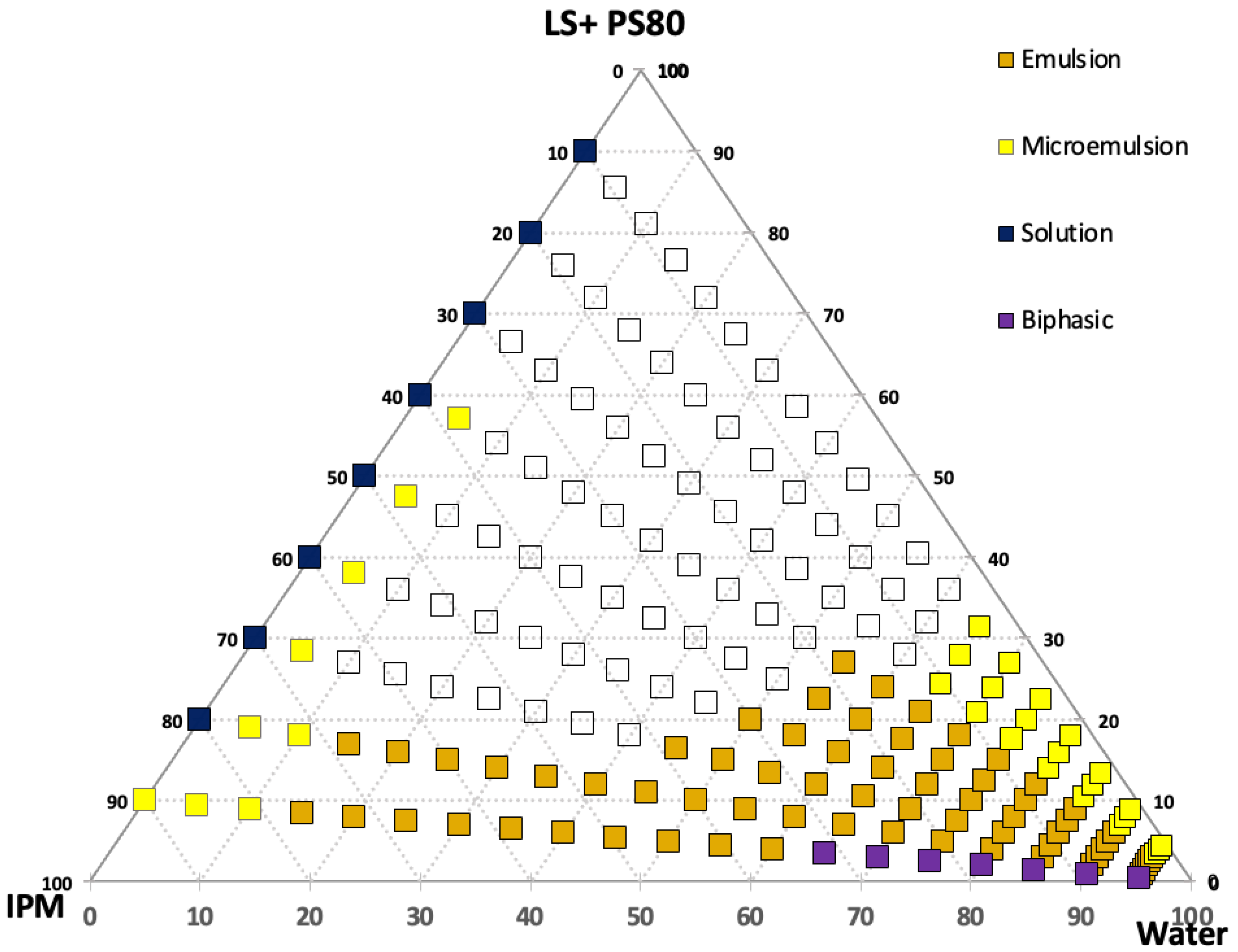
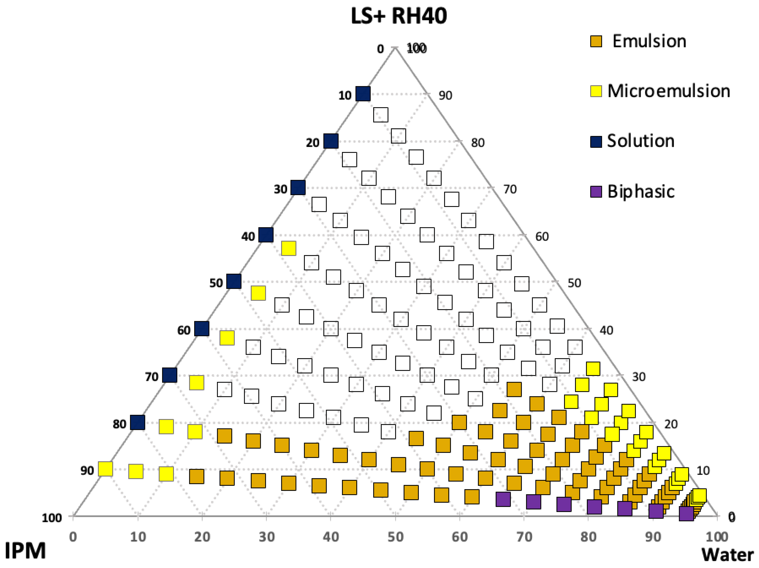
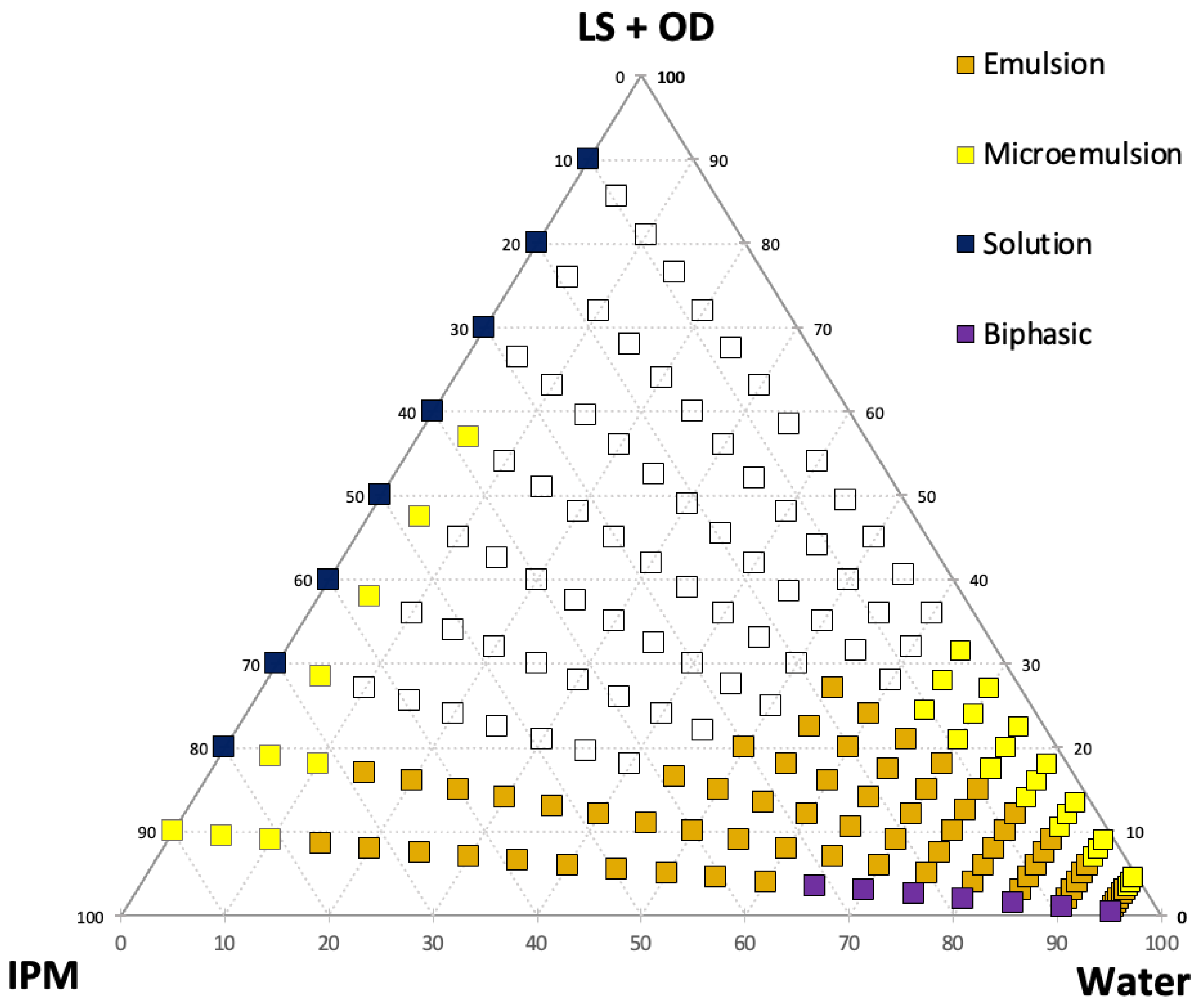
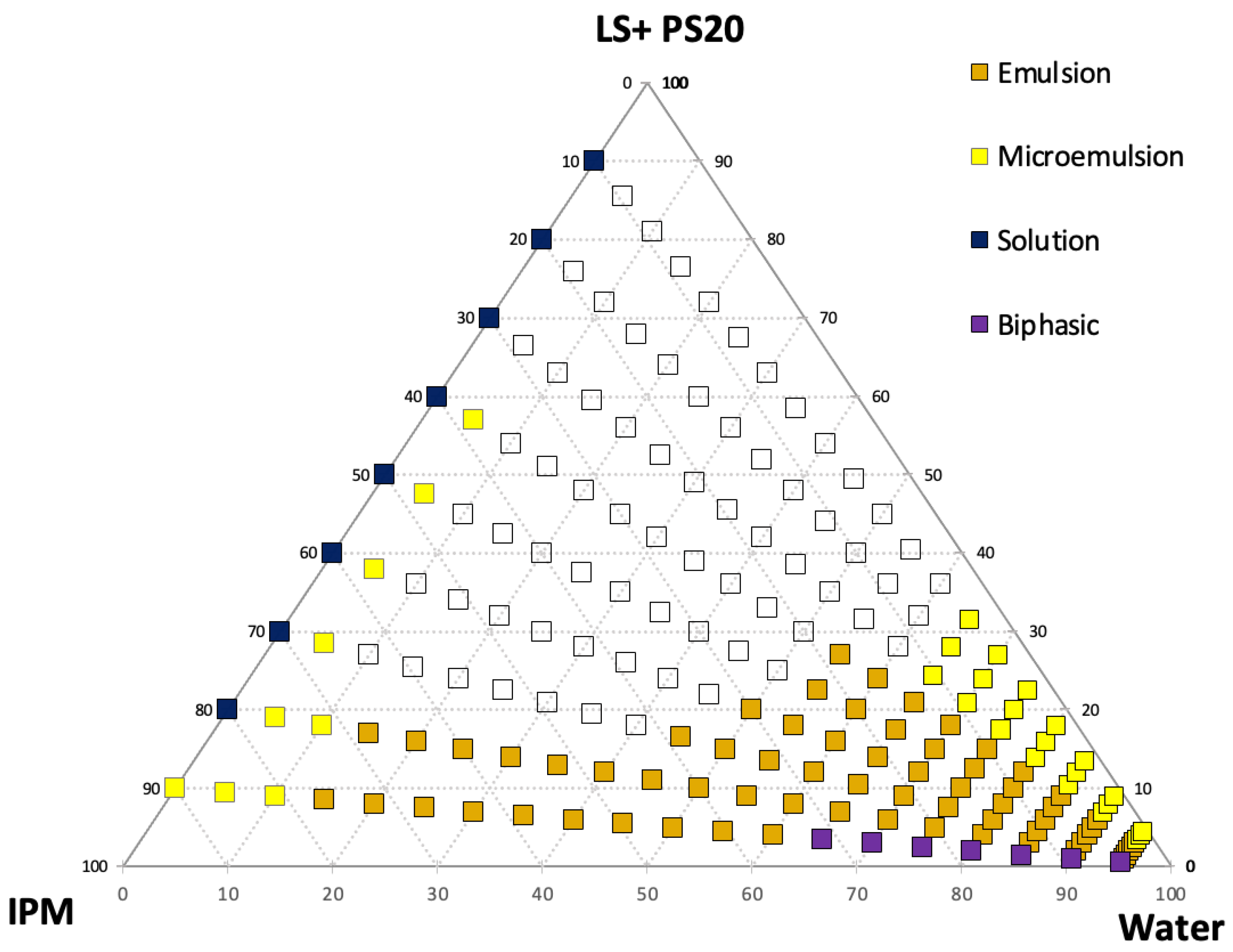
Understanding the phase behavior of microemulsion (ME) formulations is essential for creating high-quality products. By utilizing the pseudo-ternary phase diagrams displayed in
Figure 1,
Figure 2,
Figure 3 and
Figure 4, we can determine the phase compatibility of the ingredients and the average area of ME formation [
26]. Additionally, the use of ternary phase diagrams, created using IPM as the oil phase, and a fixed surfactant (LS)-to-cosurfactant (OD, PS80, RH40, and PS20) ratio of 4:1 is invaluable for the formulation process [
27,
28].
The yellow area in the ternary phase diagram represents a transparent (clear) mixture, which is identified as the microemulsion area, while the orange indicates emulsion mixtures.
In brief, a formulation is considered a microemulsion when it is transparent and has good flowability. The pseudo-ternary phase diagrams for all microemulsions indicate that combinations of surfactant (LS) and different cosurfactants at a ratio of 4:1 have similar microemulsion and emulsion areas (
Figure 1,
Figure 2,
Figure 3 and
Figure 4). These results are expected since the water content (20%) and the ratio of LS to cosurfactants (4:1) are the same for all microemulsion formulations. A study conducted by Virani et al. [
29] confirmed that the formulated risperidone microemulsion with a combination of Tween 80 and isopropyl alcohol as surfactant and cosurfactant, respectively, at the ratio of 1:1, had a similar microemulsion area when compared to microemulsions prepared using Tween 80 and Transcutol
® P as the surfactant and cosurfactant at the same ratio (i.e., 1:1). Different combinations of surfactant and cosurfactant may affect the size of the microemulsion area; furthermore, different surfactant-to-cosurfactant ratios and variable microemulsion components may also result in differences [
30].
3.3. Microemulsion Characterization and Stability
In the initial preparation, placebo microemulsions ME1 to ME7, after 4 months of storage, were meticulously prepared and subjected to rigorous testing for droplet size, pH, refractive index (RI), polydispersity index (PDI), and viscosity (as detailed in
Table 4). Following this, the caffeine-loaded microemulsions ME 8 to ME 11 underwent comprehensive evaluation at the initial phase and after a 10 months of storage, with the storage period involving room temperature conditions and controlled relative humidity (53% RH) (refer to
Table 5). Observations included visual assessments for transparency, phase separation, and precipitation at each time point. Impressively, there were no discernible changes in the visual appearance or physical properties of the samples throughout the entire 4-month stability and 10-month testing period as all the samples remained clear and stable during this period. Furthermore, the freshly prepared placebo microemulsions exhibited no significant alterations in droplet size, PDI, pH, viscosity, and RI when compared to their counterparts after four months (as shown in
Table 4). The polydispersity index (PDI) value remained consistently low, ranging from 0.2 to 0.5, indicating a stable microemulsion with a narrow droplet size distribution over the storage period. These compelling data strongly suggest that microemulsions can maintain their stability for up to 10 months of storage. Although ME 8 exhibits a higher particle size compared to other CF-loaded microemulsions, it demonstrates significantly lower viscosity. While viscosity does not directly alter partitioning into the stratum corneum, it can influence the rate of drug release and diffusion from the vehicle. This was confirmed by the results of the permeation testing experiment. The pH values of the formulations ranged from 3.5 to 5.7, which is within, or close to, the physiological skin pH range, suggesting suitability for topical application. The droplet size of the microemulsions ranged from approximately 11 to 42 nm, consistent with nanoscale systems known to enhance drug permeation due to increased surface area and improved interaction with the stratum corneum. Additionally, the microemulsion system is highly stable, and the OD, PS80, RH40, and PS20 components were found to significantly enhance the permeation of caffeine, indicating their potential utility in delivering other active ingredients.
3.4. Effect of Cosurfactants on Skin Permeation and Deposition of Caffeine from Microemulsion
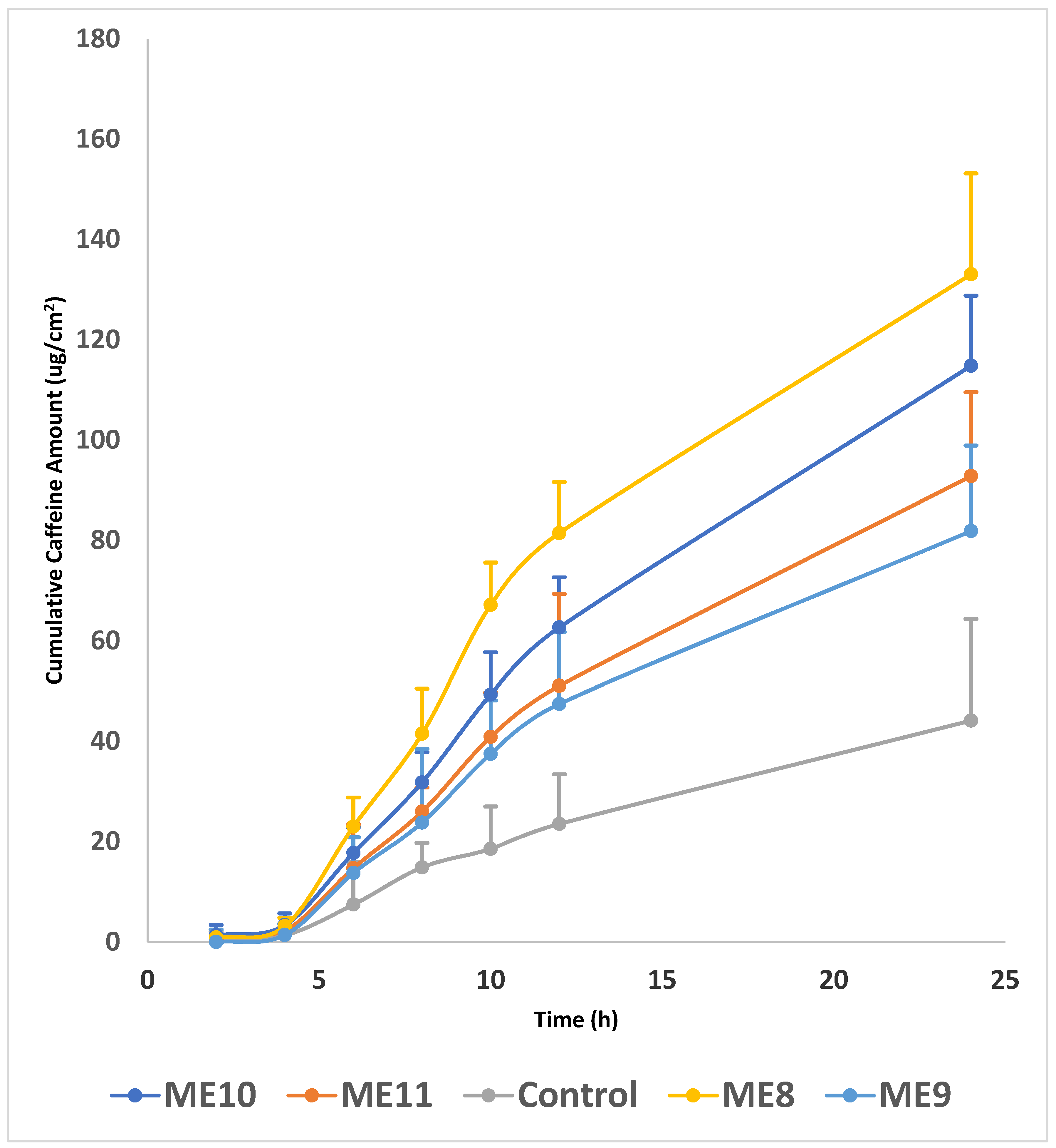
The data shown in
Figure 5 illustrate the permeation profile of caffeine from microemulsion formulations through human cadaver skin. The purpose of this study is to evaluate the impact of different cosurfactants (PS20, PS80, RH40, and OD), acting as chemical penetration enhancers, on caffeine permeation by comparing microemulsion formulations containing a cosurfactant to a control microemulsion composed of IPM (oil phase), LS (surfactant), and water, without any cosurfactant.
The cumulative amount of the permeated drug (caffeine) through the skin (µg/cm2) is plotted against time hour (h).
From a formulation perspective, the presence of a cosurfactant in a drug formulation significantly affects the amount of drug that passes through the skin. A control formulation containing IPM and LS as enhancers, which facilitates the skin permeation of CF, has been well documented. IPM and LS have been proven to enhance drug solubility within the skin as well as the drug’s integration into the lipid bilayer [
31]. The addition of a cosurfactant to a microemulsion serves dual roles as a cosurfactant and a penetration enhancer in the formulation. By adding OD, PS80, RH40, and PS20 as cosurfactants, the microemulsion region and permeation of caffeine through the skin membrane are increased. The cosurfactant, when added to a microemulsion, plays a crucial role in reducing interfacial tension, facilitating microemulsion preparation, and improving the solubilization of drug components [
32]. Notably, the highest permeation was observed with OD in the control formulation. A comparative analysis of the formulations shows that the order of increasing permeated drug quantity is as follows: Control < PS20 < RH 40 < PS80 < OD. The addition of 14% OD into the control formulation significantly increased (
p < 0.05) the permeated drug quantity compared to control, PS20, and RH 40. It was found that the addition of OD indirectly increased the skin permeation of CF by enhancing the drug’s thermodynamic activity and altering the skin’s barrier properties, potentially further enhancing caffeine permeation. These results are based on a study of the enhanced skin permeation of formoterol fumarate incorporated with OD [
33]. Additionally, the relatively hydrophobic nature of OD may have contributed to the higher permeability of caffeine from the microemulsion.
It is worth noting that the mean cumulative amount permeated per unit area over 24 h was the least for SP 20 (81.85 ± 30.5 µg/cm
2), likely due to drug–vehicle interactions that reduced the drug’s thermodynamic activity [
34]. Moreover, the relatively hydrophilic nature of PS20 results in a higher affinity of caffeine for PS20, leading to low permeability of caffeine from the microemulsion. This result is consistent with the literature [
17].
In addition, hydrophilic surfactants (PS80 and PS20) demonstrated less caffeine permeation than OD, with PS80—known to be more hydrophobic than PS20—exhibiting a higher amount of permeated caffeine from the microemulsion to the receptor chamber after 24 h (114.8 µg/cm2) compared to PS20 (81.8 µg/cm2). The physicochemical properties of the cosurfactants used in microemulsions can affect caffeine permeation through the skin.
Furthermore, cosurfactant RH40, a non-ionic surfactant (HLB > 12), effectively reduces the surface tension of oil droplets [
35], indicating its potential in enhancing the CF-loaded ME formulations. It is clear that the physicochemical properties of the cosurfactants used in microemulsions have the potential to affect caffeine permeation through the skin.
The values of the steady-state flux (J
ss), permeability coefficient (Kp), and enhancement ratio are presented in
Table 6.
Our microemulsion system, particularly ME8, demonstrated a flux (Jss) of 20.10 ± 0.01, which is superior to those reported in the literature. For instance, Zhang et al. reported a maximum flux of 11.63 ± 0.72 in their CF-loaded microemulsion system [
15].
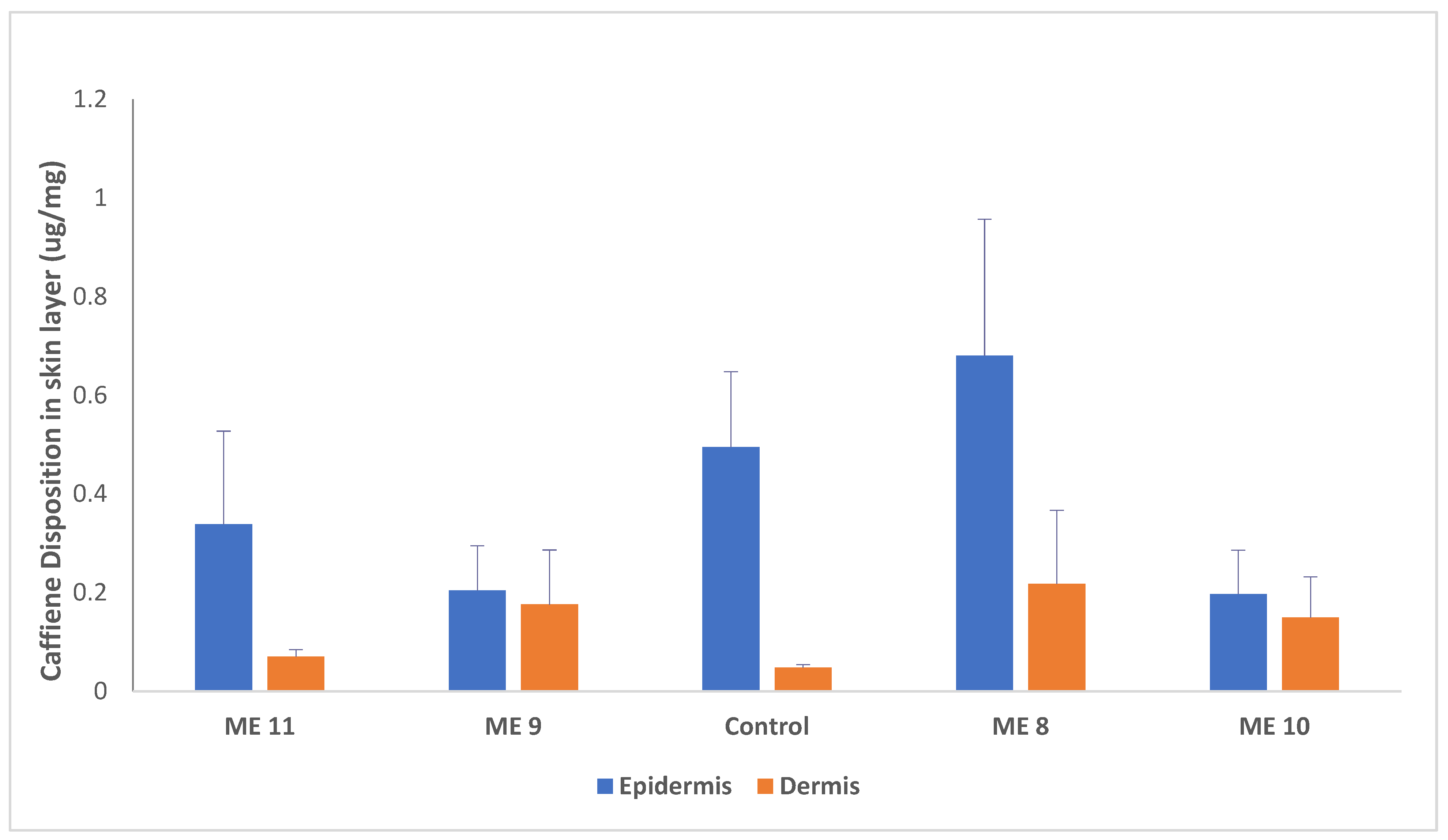
The data presented in
Figure 6 illustrates the significant impact of adding 14% OD as a penetration enhancer on the amount of CF detected in the skin after a 24 h permeation study through human cadaver skin.
Significant differences among formulations were observed for both epidermal and dermal deposition (p < 0.05). For epidermal deposition, ME8 showed a statistically significant increase compared to control, whereas ME9 and ME10 showed significantly lower deposition than control. ME11 did not differ significantly from control. For dermal deposition, ME8, ME9, and ME10 exhibited significantly higher caffeine deposition compared to control, while ME11 showed a modest but not statistically significant increase.
The results indicate that adding 14% OD (ME 8) resulted in a significant increase (p < 0.05) in skin CF deposition compared to the control formulation, confirming OD’s effectiveness as a penetration enhancer. It seems that OD facilitates drug passage across the stratum corneum (SC) by modifying it, leading to greater deposition in the skin compared to other cosurfactants, likely due to its higher thermodynamic activity.
Additionally, increasing the difference in solubility parameter between hydrophilic CF and hydrophobic OD reduces CF’s attraction to OD, facilitating its escape into the skin.
These findings are consistent with the literature, which emphasizes the role of solubility parameter differences [
34,
36] and thermodynamic activity in enhancing skin permeation [
37].
3.5. Effect of Penetration Enhancers
The stratum corneum (SC) serves as the primary barrier to transdermal drug delivery, and chemical penetration enhancers (CPEs) are widely employed to reduce this barrier’s resistance and enhance drug penetration [
37]. They achieve this by disrupting the packing of skin lipids, altering the skin barrier, altering the drug’s behavior at the skin’s surface, or affecting the drug’s thermodynamic activity, thereby facilitating its diffusion [
38]. The effects of CPEs vary depending on their interactions with both the drug and skin, which can either enhance or reduce permeation [
37]. To optimize transdermal formulations, physicochemical penetration enhancers are commonly incorporated to increase drug solubility and skin retention [
21,
39,
40,
41]. These enhancers are commonly used in dermal formulations to enhance the solubility of hydrophilic actives [
42]. In this study, the penetration enhancers also functioned as cosurfactants, improving microemulsion performance by reducing surface tension and stabilizing the formulation [
21,
43]. Cosurfactants can further modify surfactant behavior through molecular interactions, influencing surfactant monomer solubility, interfacial curvature, and film elasticity by penetrating the hydrophilic head-group region of surfactant monolayers [
44]. In this study, six CPEs (PS20, PS80, LS, IPM, RH40, and OD) were investigated, with four (OD, PS20, PS80, and RH40) incorporated at 14% (
w/
w) into microemulsion formulations to assess their role as cosurfactants in skin permeation. Five formulations (one control and four test formulations) were prepared. The control formulation, serving as a baseline for comparison, contained caffeine in IPM (oil phase) and Labrasol (surfactant), while the test formulations contained 14% of each CPE. In vitro permeation studies using human cadaver skin (24 h,
n = 5) demonstrated that all tested CPEs enhanced caffeine permeation compared to the control formulation, with OD, PS80, RH40, and PS20 increasing flux by approximately 3.0-, 2.6-, 2.0-, and 1.8-fold, respectively.
OD-based microemulsions showed the highest caffeine permeation, with cumulative CF increasing from 44 µg/cm
2 (control) to 133 µg/cm
2 at 14% OD (
Figure 1, ME 8).
PS80, with a high HLB value (~15), reflects its strong affinity for the oil–water interface, contributing to microemulsion stability and reduced interfacial tension [
45].
Being non-ionic, it has no inherent ionic interaction affinity and can be utilized in a wide range of fluid systems. Moreover, its non-toxicity and reported effects on the absorption of various drugs make it suitable for hydrate-based applications [
14,
46,
47].
PS80 enhances transdermal drug delivery by increasing drug solubility and disrupting stratum corneum lipid organization, thereby facilitating drug diffusion [
47]. Previous studies reported enhanced permeation of several drugs, including diclofenac, lorazepam, olanzapine, and disulfiram, particularly when combined with other permeation enhancers [
48,
49,
50]. Combining surfactants and cosurfactants is beneficial for achieving a stable microemulsion and can significantly enhance the skin penetration of a drug [
30]. The results of the present experiment show that the cumulative amount of CF from the PS80 formulation was higher than that from the control formulation without PS80 (
Figure 1 ME 10). The cumulative amount of CF increased from 44 (µg/cm
2) after 24 h for the formulation without enhancer (control formulation) to 114.8 (µg/cm
2) for the formulation with 14% PS80.
LS has been found to enhance the permeation of several drugs [
51,
52]. When combined with a cosurfactant, LS has been shown to enhance the transdermal delivery of curcumin [
51]. It has been reported that the combined use of compounds with different promoting effects and a proven safety record as additives for pharmaceuticals demonstrates a strong promoting effect [
8,
30].
In this study, LS and IPM were selected as the surfactant and oil, respectively. Both can also function as cosolvents and have an excellent solubilizing capacity for both hydrophilic and lipophilic compounds. They are known for their safety and their ability to enhance skin penetration [
51].
RH40 has been reported to promote caffeine transport via the transcellular pathway, with permeation dependent on its concentration [
39]. Furthermore, RH40 significantly increased caffeine permeation compared with the control formulation. The cumulative CF amount increased from 44 µg/cm
2 (control) to 92.8 µg/cm
2 with 14% RH40 after 24 h (
Figure 5).
PS20’s shorter aliphatic chain and higher HLB value (~16.7) make it more hydrophilic than polysorbate 80 and suitable for microemulsion systems [
24,
48]. PS20 has been reported to enhance the delivery of hydrophilic drugs like acyclovir w/o microemulsions [
53]. Another study demonstrated PS20’s ability to enhance the solubility and stability of mefenamic acid microemulsions when combined with cosurfactants [
54]. In this study, PS20 increased CF permeation compared with the control formulation (without Tween 20), confirming its role in enhancing microemulsion performance (
Figure 5, ME 9).