


Abstract
Osteomyelitis remains one of the most challenging infections to manage in both human and veterinary orthopedics, owing to biofilm formation, compromised local vascularity, and the frequent need for extensive surgical debridement. Systemic antibiotic therapy alone often fails to achieve bactericidal concentrations at the infected site while exposing the patient to systemic toxicity. Local antibiotic delivery systems have therefore emerged as a complementary or alternative strategy, enabling sustained, high local drug concentrations above the minimum inhibitory concentration while minimizing systemic exposure. This review summarizes the principal approaches to managing osteomyelitis—surgical intervention, systemic antimicrobial therapy, and local drug delivery—with an emphasis on the biomaterials used as antibiotic carriers. Non-degradable and biodegradable systems are discussed, including poly(methyl methacrylate) cements, calcium sulfate, calcium phosphate ceramics, bioactive glasses, and natural and synthetic polymers, such as chitosan, poly(lactic acid), and poly(lactic-co-glycolic acid), as well as bone-graft-based carriers. Particular attention is given to injectable, in situ-forming scaffolds that combine antimicrobial release with osteoconductive and osteoinductive properties, offering dual eradication of pathogens and support for bone regeneration. Current limitations, the comparative relevance of the canine model, and future directions—including nano-bioactive glass composites and combination therapy—are considered to guide the development of more effective single-stage treatment strategies.
1. Introduction
In companion animals—dogs and cats above all—osteomyelitis is a stubborn disease to resolve and a source of considerable morbidity, arising most commonly after open fractures, surgery involving orthopedic implants, or bite wounds. This difficulty has kept the search for better treatment strategies active, with research continuing to pursue more effective approaches that improve how patients fare.
The three principal approaches to the management of osteomyelitis include the surgical debridement of infected and necrotic tissue, systemic antibiotic therapy, and local antibiotic delivery. These strategies are frequently used in combination; surgical debridement and systemic antibiotic therapy are outlined below, while local antibiotic delivery—the principal focus of this review—is examined in detail in the sections that follow.
Taking out all implanted hardware and debriding biofilms and dead tissues is an effective way to clear an infection in complicated chronic osteomyelitis [1]. Yet the diseased tissue can seldom be removed with complete accuracy, and the dead space left behind raises the odds of reinfection. The usual countermeasure is to place antibiotic-loaded biomaterials within the bone so that the drug is delivered to the site [2]. Reviewing their cases retrospectively, Segreto et al. [3] found that operating early produced better results than operating late.
For acute infections, systemic antibiotics are highly effective on their own, and in chronic osteomyelitis, they can be paired with surgery to clear the organisms responsible. The preferred starting point is culture data from collected specimens, with the agent chosen to match the microorganisms driving the infection.
Empiric antimicrobial therapy is essential when the causative pathogen cannot be isolated. It should target the suspected pathogen(s) based on the animal’s risk factors and route of infection. In companion-animal osteomyelitis, the likelihood of Staphylococcus involvement must be considered, as Staphylococcus pseudintermedius is the predominant pathogen in dogs and cats, and methicillin-resistant strains (MRSP) are an increasing concern in cases associated with orthopedic implants, while wound- and trauma-associated infections tend to be polymicrobial and can include Gram-negative organisms. When staphylococcal osteomyelitis is suspected, the empirical regimen should subsequently be tailored to the culture and sensitivity results of appropriately collected specimens to ensure targeted and effective treatment.
A typical regimen opens with intravenous antibiotics for two to six weeks before being followed by oral treatment that can last as long as six months. Suitable oral drugs are those that are well absorbed, reach adequate concentrations within the bone, and clear slowly.
In chronic disease, the systemic route carries well-known liabilities: courses run long, adverse effects accumulate, and the sequestrum walling off the infected focus blocks the drug from reaching it. Extended dosing likewise favors antimicrobial resistance, a problem made worse wherever the drug cannot get to the site. Around orthopedic hardware—the plates and screws placed during fracture repair—staphylococci settle onto the surface and lodge within a biofilm that fends off phagocytes and keeps antibiotics out, resulting in treatment failure. Similar observations have been reported across multiple veterinary and experimental settings [4,5,6,7].
Treating chronic long-bone osteomyelitis in dogs and cats locally has distinct upsides. Placing the drug at the focus itself raises the concentration where it is needed and spares the animal the systemic side effects of whole-body dosing. It can also lift infection-control rates and, once tissue-engineering methods and biomaterials are added, support the regrowth of bone.
2. Materials and Methods
The present work takes the form of a narrative review addressing local antibiotic delivery systems for osteomyelitis, framed throughout around their applicability to companion animals. To assemble the source material, PubMed/MEDLINE, Scopus, and Web of Science were queried for peer-reviewed work appearing between 1985 and 2025, supplemented by one manufacturer website accessed in 2026. Search terms were combined from the following list and applied to both the human and veterinary literature: “osteomyelitis,” “bone infection,” “local antibiotic delivery,” “drug delivery system,” “scaffold,” “bone cement,” “poly(methyl methacrylate),” “bioceramic,” “bioactive glass,” “nanoparticle,” “microparticle,” and “bone graft.” A representative Boolean search string was: (osteomyelitis OR “bone infection”) AND (“local antibiotic delivery” OR “antibiotic-loaded cement” OR scaffold OR bead OR “bioactive glass”) AND (dog OR canine OR cat OR feline OR veterinary).
Eligible items comprised primary research, clinical studies, and pertinent reviews that documented the makeup of local delivery systems, their release kinetics, or their performance in vitro, in vivo, or in the clinic. Additional references were located by hand-checking the bibliographies of the retrieved papers. Eligibility was determined by explicit criteria rather than by a scoring instrument; studies were included if they described the composition, release kinetics, or the in vitro, in vivo, or clinical performance of a local antibiotic-delivery system relevant to bone infection, and they were excluded if they addressed systemic antibiotic therapy alone or soft-tissue infection without a bone-delivery component. Because the review is narrative and single-authored, neither a dual-reviewer selection process, a formal quality appraisal, nor a meta-analysis was undertaken, and no PRISMA flow diagram is presented. Throughout, the evidence supporting each delivery system is identified explicitly by its source—clinical companion-animal studies, other-species in vivo models (human, rabbit, rat, or sheep), or in vitro work—so that findings demonstrated in dogs and cats are not conflated with those extrapolated from other settings; the level of companion-animal evidence for each carrier is summarized in Table 1.
Table 1.
Comparison of local antibiotic-delivery carriers for osteomyelitis in dogs and cats.
3. Local Delivery of Antimicrobials via Implantable and Injectable Carriers
The incorporation of antibiotics into bone cement has become a widely used strategy for managing osteomyelitis, although—as the more cautious appraisal later in this section makes clear—the supporting evidence in companion animals is largely of a lower tier. In small-animal orthopedic surgery, it is used off-label, either by adapting a commercially available human pre-made delivery system or by mixing the drug and cement intra-operatively, as no veterinary-licensed products are currently available. Clinical investigations of localized antibiotic delivery, in combination with bone-filling materials, have demonstrated substantial efficacy in treating long-bone infections. Specifically, such an intervention (1) achieves early suppression of infection, which is critical to minimizing complications and maximizing positive outcomes; (2) produces a several-fold reduction in the incidence of recurrent infection; (3) lowers the risk of pathological fracture through internal reinforcement; (4) permits early recovery of limb function; and (5) creates favorable conditions for the restoration of bone structure [8,9,10]. The availability of biodegradable cements that sustain antibiotic release throughout the recovery period has further encouraged this approach.
Of the non-biodegradable carriers, polymethyl methacrylate (PMMA) has the longest track record and the widest use; shaped into beads, it has served for decades as the reference standard for delivering drugs to a local site [11,12]. Incorporation of the antimicrobial is done by blending it into the PMMA just before the procedure [13]. The beads are generally well tolerated, although the claim that they provoke no hypersensitivity overstates the literature—foreign-body and hypersensitivity reactions have occasionally been reported—and because their large surface area favors elution, they hold a high local drug level initially [14]. Elution from PMMA is, however, biphasic, where an early burst is followed by a prolonged low-level tail, during which sub-inhibitory concentrations persist, and this tail is itself a recognized driver of antimicrobial resistance and of staphylococcal small-colony variants. Acknowledging this reinforces, rather than undermines, the stewardship rationale for careful carrier and drug selection.
Evidence for this approach comes largely from human medicine and is extrapolated to veterinary patients. Dzyuba et al. [8], for example, implanted PMMA scaffolds during surgery in 30 human patients with chronic osteomyelitis, and compared their outcomes with those of 30 patients treated with conventional systemic therapy; those who received local implants achieved better infection control and a significant reduction in recurrent infections. Comparable controlled data in dogs and cats remain limited, so such findings should be applied to companion animals with appropriate caution. The veterinary clinical literature does exist, however, and is growing. Dernell et al. [15] reported the clinical response to antibiotic-impregnated PMMA beads in 18 dogs with severe infection after limb-sparing surgery and allograft replacement, and antibiotic-impregnated calcium sulfate beads—which resorb and thus avoid a second surgery—have since been described in a series of small-animal cases: Peterson et al. [16] reviewed 16 client-owned dogs and cats treated for or protected against orthopedic surgical-site infection, with infection resolving in six out of ten animals treated therapeutically and in all six treated prophylactically, while individual reports document tobramycin calcium sulfate beads in dogs [17]; single-stage revision of an infected canine total hip replacement using bioabsorbable beads [18]; and pancarpal arthrodesis with beads in a dog with septic arthritis and osteomyelitis [19]. Across these reports, a recurring lesson is that outcomes track closely with whether the implanted antibiotic matches the culture and susceptibility profile of the causative organism, underscoring that local delivery supplements, rather than replaces, appropriate drug selection. The therapeutic resolution rate in (16)—six out of ten—is itself modest, and warrants commentating rather than neutral reporting; it is a reminder that antibiotic beads are an adjunct to, not a guarantee of, infection control, particularly in established infection as opposed to the prophylactic setting, where all six treated animals did well.
The beads carry real disadvantages as well. Due to not being biodegradable, they have to be taken out during a second operation. Their preparation relies on an exothermic polymerization whose heat destabilizes many antimicrobials, so only a limited amount of the drug survives incorporation (Table 2). And when beads are made and placed during surgery without being characterized first, how they release the drug is hard to predict [2].
Table 2.
Heat stability of commonly used antimicrobials during PMMA polymerization (guide only; a carrier–drug combination should be characterized before clinical use).
Much of the subsequent work has turned to biodegradable and bioabsorbable substrates—hydroxyapatite, calcium sulfate, calcium phosphate, and collagen—used in a range of physical forms [20,21]. When built on a carrier that resorbs, a local antibiotic system is a therapeutically efficient way to treat osteomyelitis. Since the scaffold itself governs how fast the drug comes off, achieving a controlled release means matching the material’s degradation rate to that of the goal [22]. One clinically important caveat is specific to calcium sulfate; its rapid dissolution can produce a sterile serous wound discharge that closely mimics post-operative infection, and recognizing this benign phenomenon is essential to avoid unnecessary reintervention.
In a human retrospective study, 51 patients with chronic osteomyelitis of the lower limbs were allocated to two treatment groups. One group received combination therapy with vancomycin-loaded calcium sulfate and vancomycin-loaded PMMA, while the control group received vancomycin-loaded PMMA alone. The combination of the two biomaterials had a synergistic effect on infection control compared to vancomycin-loaded PMMA alone [23]. Although this evidence is drawn from human patients, the same biodegradable-carrier principle is directly relevant to companion-animal orthopedics. Nonetheless, it should be stated plainly that as of the most recent veterinary review of local antibiotic implants, no good-quality randomized controlled trial—in either human or veterinary patients—has yet compared antibiotic-impregnated beads against systemic therapy alone or in combination for the treatment of osteomyelitis; the existing studies are largely underpowered or retrospective [24]. The case for local delivery in dogs and cats therefore rests on consistent albeit lower-tier evidence, and the recommendations that follow should be read in this light. It should also be noted here that virtually all the carrier-efficacy data cited in this review derive from S. aureus or MRSA; the performance of these systems against S. pseudintermedius biofilm—the organism most relevant to dogs and cats—remains essentially unstudied and represents an important gap.
Two related principles deserve explicit mention because they frame how any of these carriers is used. The first is dead-space management, where aggressive debridement of the infected bone leaves a void that, if left empty, fills with hematoma and becomes a nidus for recurrent infection, so obliterating that dead space with an antibiotic-loaded carrier is as much a goal of local therapy as drug delivery itself. The second is the induced-membrane (Masquelet) technique, a staged strategy in which an antibiotic-loaded PMMA spacer is placed in the debrided defect to control infection and induce a vascularized biomembrane; at a second stage, the spacer is removed, and the preserved membrane—which is osteogenic and limits graft resorption—is packed with cancellous autograft. Although developed in human surgery, the technique is increasingly applied to large infected defects in dogs and cats and is directly relevant to the segmental defects illustrated in this review.
3.1. Systems Used for the Local Delivery of Antibiotics for Osteomyelitis Management
Research into local delivery platforms for bone infection remains active and is driven by the need to concentrate antibiotics where the infection actually sits. Matching the carrier to the case, setting the right antimicrobial dose, and governing how quickly the drug is released, when combined together, give the surgeon better control over the infection with fewer recurrences. A crucial qualification, however, is that these infections are biofilm-associated, as sessile bacteria embedded within a mature biofilm typically require concentrations one to three orders of magnitude above the minimum inhibitory concentration (MIC) for eradication, so the operative benchmark for a local carrier is not simply to exceed the MIC but to reach these far higher biofilm-eradicating levels—a threshold that high-dose carriers, such as PMMA and calcium sulfate, approach far more readily than low-load particulate systems. A carrier that is itself osteogenic has a dual duty, clearing the organism while helping the bone to rebuild once the site has been debrided. Some of these systems are formulated to set in place after being injected alongside the infected bone, offering an adjunctive treatment without the second operation an implant would require and the shortcomings of systemic dosing. Clinical work has indicated that delivering the drug locally in this way can spare bone that would otherwise have to be resected [8,23].
Biodegradable materials investigated for the local delivery of antimicrobial agents include natural and synthetic polymers [25], ceramics [26], bioactive glasses [27], allograft bone [28], and demineralized bone graft [29]. For clarity, the platforms discussed below fall into two distinct families of resorbable or implantable scaffolds—which may be biological (extracellular matrix), bioceramic, or polymeric—and metallic fixation implants bearing antibacterial surface coatings, with the latter being hardware instead of a scaffold category in a strict sense.
The choice of antibiotic loaded into any of these carriers is, in companion animals, constrained less by the carrier than by veterinary antimicrobial stewardship considerations. Because Staphylococcus pseudintermedius is the predominant pathogen, and because the drugs most active against resistant strains—vancomycin, linezolid, and daptomycin—are reserved in veterinary medicine for exceptional circumstances, beads and cements for dogs and cats are, in practice, loaded with agents such as amikacin, gentamicin, tobramycin, cefazolin, or clindamycin, which are selected based on culture and susceptibility. This selection must be reconciled with the resistance pattern of the likely pathogen; because methicillin-resistant S. pseudintermedius (MRSP)—identified above as the principal implant-associated concern—carries mecA-mediated resistance that abolishes the activity of cefazolin and the other β-lactams and is frequently co-resistant to clindamycin, these two agents are appropriate only for confirmed methicillin-susceptible isolates, whereas the aminoglycosides amikacin, gentamicin, and tobramycin retain activity against many MRSP strains and are the more dependable empirical local choices pending susceptibility data. Antimicrobial selection should follow companion-animal stewardship guidance—such as the ISCAID consensus recommendations and the EMA antimicrobial categorization—rather than being asserted, reserving the highest-priority agents for documented needs. Veterinary elution studies support these choices: gentamicin elutes readily from plaster-of-Paris and calcium sulfate carriers [30], and amikacin has been shown to elute from plaster-of-Paris beads at locally effective concentrations [31], although release kinetics differ markedly between drugs and carrier materials, so a carrier–drug combination should ideally be characterized before clinical use.
3.1.1. Biological (Extracellular-Matrix) Scaffolds
After an acute injury, the body regenerates tissue on its own, reconstructing or mending the damaged structure so that function returns. Supplying a scaffold that reproduces the extracellular matrix (ECM) can amplify this response. Loaded with therapeutic agents, such scaffolds help to clear infection, rebuild injured bone, and restore affected tissues to working order [32,33,34]. Scaffolds engineered for bone are typically assembled from a mix of biomaterials, bioactive agents, and cells [35,36,37].
Cells depend on scaffolds to attach, multiply, and differentiate, and a biocompatible scaffold lets adjacent cells move in and expand. An effective biomaterial—biodegradable or not—has to mesh with the surrounding biological molecules and cells; encourage cells to migrate, proliferate, differentiate, and renew tissue; and underpin both osteogenesis, the maturation of immature cells into pre-osteoblasts, and osteoconduction, the laying down of bone along the scaffold. Design should be steered by these requirements so that the finished biomaterial gives cell growth and tissue regeneration the best possible support [38].
In dogs and cats, comminuted fractures, sequestrum removal, and the revision of failed implants frequently leave a bone defect too large for the patient to bridge unaided, and it is in exactly these cases that a bioengineered scaffold earns its place [39]. Once seated in the debrided site, the scaffold performs two functions for the veterinary patient by providing mechanical support to the weakened bone while delivering a defined antibiotic dose locally, meaning that the surgeon does not rely on systemic therapy to reach a poorly perfused, infected field. Because release is confined to the implant rather than distributed body-wide, a scaffold of this kind is best understood as a controlled drug-delivery device in its own right [22].
A scaffold intended for a dog or cat has to meet a demanding set of requirements before it is worth implanting in an infected limb. It should resorb no faster than the patient lays down new bone, so that support is not lost mid-healing; its breakdown products must be neither toxic nor capable of provoking an immune reaction in the recipient; it must anchor to the host bone cells and carry its load while the fracture consolidates; and its interior must be an interconnected pore network—comprising pores of roughly 100 µm or larger, with at least half of them open—so that cells can migrate in and nutrients and oxygen can diffuse through the construct [38]. Bioceramics, natural and synthetic polymers, and composites of the two come closest to satisfying this combination, which is why they dominate the osteomyelitis scaffold literature.
Across animal studies and clinical trials, scaffolds have been fabricated and delivered to diseased bone by several routes: surgically implanting a pre-compounded construct [40,41], injecting an implant that forms in situ [42,43], and printing a custom-fitted construct in three dimensions (3D) [10,44].
Scaffolds of the implant type must be placed surgically onto the infected bone. Their manufacture draws on a variety of methods—lyophilization [45,46], solvent casting [47,48], particulate leaching [49,50], gas foaming [51,52], and electrospinning [53,54].
Zhou et al. [55] developed a gelatin/β-tricalcium phosphate scaffold by lyophilization and loaded it with vancomycin. The scaffolds were applied to the tibias of rabbits after osteomyelitis induction and removal of necrotic bone to enhance therapeutic efficacy. The scaffolds sustained vancomycin release for eight weeks, effectively controlling the infection and repairing the bone defects.
Formulations that start out as an injectable liquid monomer and then cure into a solid or semi-solid polymer network in response to a local temperature or pH change bring several advantages to the treatment of bone infection. Requiring only minimal surgery to place, they leave the patient more comfortable, heal more quickly, and are less expensive to treat. Hydrogels in particular have come to the forefront as a bulk-scaffold platform that is well suited both to rebuilding bone and eluting antibiotics on-site [36,56,57]. One illustration is of an injectable chitosan in situ-forming implant carrying bioactive glass nanoparticles and raloxifene; because it was devised to repair non-infected bone defects and contained no antibiotic, its applicability to infected defects remains a hypothesis rather than a demonstrated finding. It gels within 20 min once it reaches body temperature (37 °C) and then releases its drug steadily for 56 days; in rat tibiae, it drove faster bone formation in non-critical-size defects than defects that were left untreated [37].
Among newer fabrication technologies, three-dimensional (3D) printing, driven by computer-aided design (CAD), has been described as an effective means of producing customized scaffolds for tissue engineering and antibiotic therapy [58,59,60,61]. Regulating parameters, such as fabrication accuracy and speed, enables 3D printing build scaffolds that are matched to the shape and site of the bone defect [62,63]. In addition, the interconnected internal porosity of 3D-printed scaffolds promotes new tissue regeneration—an asset in infection-associated bone defects [64].
3.1.2. Metallic Implants and Antibacterial Surface Coatings
With fracture plates, screws, and intramedullary pins seeing steadily broader use in dogs and cats, a number of tactics have arisen to head off implant-related bacterial infection. A productive one is to coat the implant so that bioactive agents are released locally—giving sustained, controlled dosing without systemic side effects or biofilm buildup. The metals favored for orthopedic implants are selected for their mechanical strength, corrosion resistance, and biocompatibility, including titanium (Ti), cobalt (Co) [65], and stainless steel [66]. Their drawback is that metals do not biodegrade and are hard to process within the biological milieu [67,68]. Titanium is the metal most often used for osteomyelitis implants.
Titanium Implants
Ti-based alloys are widely used as metal implants in veterinary orthopedics as load-bearing components—for example, in canine total hip replacement and as intramedullary nails, bone screws, and fracture plates. That said, titanium implants can loosen, and they may hinder later osseointegration when a fibrous layer forms that fails to bind to the metal [69]. They can also trigger immune responses by sensitizing the host and switching on both pro- and anti-inflammatory cytokines, which may culminate in allergic reactions [70]. Despite their inherent antimicrobial properties, titanium alloys can still become infected, and an antimicrobial surface coating is therefore required to reduce bacterial colonization and infection risk. Because metal implants neither induce nor conduct bone growth, surface modification is essential to optimize their interaction with bone tissue and enhance their osteoinductive and osteoconductive properties, so that the implant can fulfill its biological function [71]. The most important surface modifications of Ti alloys are described below.
Silver-Coated and Silver-Impregnated Implants
Silver deserves separate consideration because it has moved furthest from the laboratory into commercial orthopedic hardware. Rather than being loaded with a conventional antibiotic, the implant carries elemental or ionic silver in a surface coating, from which silver ions are released to kill adherent bacteria by a broad-spectrum, largely non-specific mechanism—disruption of the cell wall, interference with the respiratory chain, and generation of reactive oxygen species—that is active against staphylococci and does not depend on a drug the organism might resist. Several coating systems are marketed for human megaprostheses and fixation devices, and a systematic review found that silver coating reduced periprosthetic infection in some series (for example, from 11.2% to 9.2% in primary implants and from 29.2% to 13.7% in revisions), though the pooled effect was inconsistent and the principal safety concerns were local argyria and, rarely, systemic silver accumulation [72]. A further, microbiological consideration is that bacterial silver-resistance determinants (for example, the sil operon) do exist and can be carried on mobile genetic elements alongside antibiotic-resistance genes, raising the prospect of co-selection; silver should therefore not be assumed to be exempt from the resistance pressures that affect conventional antimicrobials.
The companion-animal evidence is more sobering, and it is the part most relevant here. Two canine studies have evaluated silver-coated tibial plateau leveling osteotomy (TPLO) plates against uncoated controls, and neither demonstrated a significant reduction in surgical-site infection. In a prospective randomized trial of 73 TPLOs, the infection rate was lower with silver-coated plates (2.6% vs. 14.3%), but the difference did not reach significance in a cohort of that size [73], while a larger retrospective study of a silver-based HyProtect-coated plate found infection rates no different from an uncoated control group, noting that the plate coating—but not the screws—was silvered, so contamination could establish on uncoated hardware beyond the coating’s reach [74]. Taken together, these results indicate that silver coatings are biologically plausible and safe in dogs but not yet proven to lower clinical infection rates, and that coating the entire implant construct, not the plate alone, may be necessary for a measurable benefit.
Titanium Implants with Antibiotic-Coated Surfaces
Working from an uncoated titanium surface, Sharma et al. [75] put forward a low-cost route to managing osteomyelitis. A different group took a titanium alloy and grafted chitosan/lysine biopolymers onto it, turning the surface into a reservoir for gentamicin. Beyond clearing the infection through prolonged drug release, this treated surface encouraged cells to attach and multiply in simulated body fluid, and the bioactive layer promoted new bone growth—support for the idea that modifying an implant can set up conditions that favor bone regeneration [76].
The surface of a Ti scaffold has also been modified for antibacterial purposes by attaching gentamicin-loaded microparticles to the pore walls through cross-linked sodium alginate. This biodegradable system delivered the gentamicin doses required to combat infection after implantation and was fully compatible with osteoblast-like cells, indicating that it is safe to use [77].
A caveat applies to this section as a whole. Almost all of the surface-modification strategies—like heavy-metal doping, antibiotic coatings, and nanopatterning—have been developed and tested in human patients, in rodents, or on the bench, and their translation to dogs and cats rests on inference rather than on species-specific data. The direct veterinary evidence is sparse. The closest example is a study by Ziąbka et al. [78], who applied sol–gel hybrid coatings doped with silver, copper, and zinc nanoparticles to titanium-alloy tibial plateau leveling osteotomy (TPLO) implants: the silver-containing layers were the most strongly bactericidal in vitro, a low silver dose appeared to aid tissue healing while higher concentrations were toxic, and the coated implants were placed in six canine patients with no adverse findings over a three-month follow-up—while encouraging, this is far from a controlled efficacy trial. Beyond companion animals, larger-species models provide a proof of concept; Tran et al. [79] showed that a silver-doped titanium-oxide/siloxane coating on intramedullary nails reduced the severity of experimental S. aureus osteomyelitis in a caprine (goat) fracture model. Foundational work in a purpose-built canine osteomyelitis model likewise established, decades ago, that local antibiotic delivery around an intramedullary implant could prevent staphylococcal bone infection [80]. Taken together, coated and surface-modified metal implants are biologically promising for veterinary orthopedics, but the companion-animal evidence base remains at the pilot stage and controlled clinical studies in dogs and cats are needed before these technologies can be recommended for routine use.
3.1.3. Bioceramic Scaffolds
Bioceramic scaffolds are engineered with two goals in mind: to drive new bone formation and to release therapeutic agents at a controlled rate. Their raw materials are inorganic—among them calcium phosphate derivatives and bioactive glass. Within the calcium phosphate group, hydroxyapatite (HA) and tricalcium phosphate (TCP) are among the most widely used and were some of the first bone substitutes put into clinical practice [81]. It is their close chemical and structural kinship with the mineral fraction of bone that accounts for the extensive use of bioceramic scaffolds in bone regeneration.
Calcium Phosphate Bioceramics
The crystalline form calcium phosphate takes depends on its calcium-to-phosphate ratio, the temperature at which it is made, and any impurities and water present. The possibilities span mono-, di-, and tricalcium phosphate—the lattermost existing as both α-tricalcium phosphate (α-TCP) and β-tricalcium phosphate (β-TCP)—along with tetracalcium phosphate and hydroxyapatite (HA) [81]. As scaffolds for bone regeneration, calcium phosphate bioceramics bring an attractive package: biocompatibility, a high capacity for drug loading, freedom from toxicity and immunogenicity, resorbability, and osteoinductivity [82]. Because they mirror the mineral phase of bone, they make excellent drug-bearing bone fillers, and as they break down they liberate calcium and phosphate ions that both stimulate osteoblast growth and differentiation and furnish the ionic feedstock for new bone [83]. Figure 1, Figure 2, Figure 3, Figure 4, Figure 5 and Figure 6 show a case of severe trauma with bone loss and potential infection treated with calcium phosphate beads; these images are included to illustrate, in a real companion-animal patient, how the technique is applied in practice rather than to report original research data.
Figure 1.
X-ray of a tibial fracture showing catastrophic failure seven days after repair. Image courtesy of the author.
Figure 2.
X-ray of a tibial fracture showing catastrophic failure seven days after repair. Image courtesy of the author.
Figure 3.
Photograph of the surgical exposure to explant the failed implant. Image courtesy of the author.
Figure 4.
Appearance of the tibia after explantation of the plate. Image courtesy of the author.
Figure 5.
Preparation of the calcium phosphate beads in the disposable rubber tray after mixing in the antibiotic, before drying. Image courtesy of the author.
Figure 6.
Calcium phosphate beads mixed with the cancellous bone graft prior to application at the fracture site. Image courtesy of the author.
These same properties make calcium phosphate an attractive vehicle for local antibiotics in the infected veterinary limb: it can be loaded with a culture-directed drug, set in place, and release the drug while the liberated calcium and phosphate feed new bone. Injectable formulations have been tested against experimentally induced S. aureus osteomyelitis in animal models [1], and the injectable route is the feature most relevant to the small-animal surgeon—the cement is mixed as a soluble calcium phosphate with an aqueous phase that precipitates once placed, so it can be delivered by a minimally invasive approach and molded to exactly fill an irregular defect rather than requiring a pre-formed implant and larger surgical exposure [84]. One design consequence follows directly: because antibiotic escapes chiefly by diffusion through the set cement rather than by its degradation, the elution profile—and hence, how long the implant protects the site—is governed by the cement’s porosity and must be characterized before it is relied on clinically [1]. In the case illustrated, the fracture was stabilized with an Ilizarov apparatus, and antibiotic-impregnated calcium phosphate beads were applied to the fracture site (Figure 7); immediate post-operative radiographs (Figure 8 and Figure 9), radiographs at 40 days showing resorption of the beads (Figure 10 and Figure 11), and computed tomography at 187 days (Figure 12 and Figure 13) documented the progressive healing of the tibia.
Figure 7.
Application of the calcium phosphate beads impregnated with antibiotics at the fracture site. Image courtesy of the author.
Figure 8.
Medial Lateral X-ray of the immediate post-operative result of the revision surgery using a ilizarov device to stabilize the fracture. The calcium phosphate beads can be seeing around the fracture Image courtesy of the author.
Figure 9.
Cranial caudal X-ray view of the immediate post-operative result of the revision surgery using a ilizarov device to stabilize the fracture. Image courtesy of the author.
Figure 10.
Medial Lateral X-ray view at 40 days post-operative showing resorption of the calcium phosphate beads, which is a normal finding. Image courtesy of the author.
Figure 11.
Cranial caudal X-ray view at 40 days post-operative showing resorption of the calcium phosphate beads, which is a normal finding. Image courtesy of the author.
Figure 12.
CT showing the healing process of the tibia 187 days after revision. Image courtesy of the author.
Figure 13.
CT showing the healing process of the tibia 187 days after revision. Image courtesy of the author.
Bioactive Glasses
Upon dissolution, bioactive glass releases biologically active ions that promote bone cell proliferation and differentiation [85]. It chemically bonds to injured bone by forming a carbonated hydroxyapatite surface layer whose composition approximates that of bone; this layer tightens the union between the glass and adjacent bone and promotes osteogenesis [86].
Bioactive glass is unusual among these carriers in that it has antibacterial activity of its own, which is what makes it interesting for companion-animal osteomyelitis [87]. This intrinsic activity is attributed chiefly to the physicochemical changes that accompany dissolution—a rise in local pH and an increase in local osmotic pressure as ions are released—rather than to a conventional pharmacological mechanism. The preclinical case is direct: in a rabbit model of MRSA-induced osteomyelitis, teicoplanin eluted locally from a bioactive glass implant with borate ions outperformed the same drug administered intravenously [27]. Its intrinsic activity is equally striking; Drago et al. [88] packed the debrided lesions of 27 patients with long-bone osteomyelitis using bioactive glass granules alone, and the granules suppressed the causative organisms without added antibiotic. This intrinsic antibacterial activity makes bioactive glass an attractive candidate for use in companion-animal osteomyelitis, although veterinary clinical data remain limited. The main drawbacks of bioactive glasses are the elevated local ion concentrations at the implant site, the slow degradation of the glass, and the difficulty of the scaffold fabrication process [89].
3.1.4. Polymeric Scaffolds
Building a polymeric scaffold begins with choosing biomaterials that agree with bone tissue. The best-suited polymers reproduce the natural extracellular matrix (ECM) of bone and carry the biochemical and mechanical characteristics the application demands [90]. Natural and synthetic polymers alike hold promise in bone tissue engineering, being biocompatible, biodegradable, and able to interact with cells. On the natural side, scaffolds have drawn on collagen, chitosan, silk, and alginate; on the synthetic side, poly(lactic acid) (PLA), polycaprolactone (PCL), and poly(lactic-co-glycolic acid) (PLGA) have all been examined. Any of these can be tuned for mechanical and physicochemical behavior and for degradation rate, and each can double as a drug carrier [38,90,91]. It should be emphasized, however, that essentially all of this polymer work to date is preclinical—derived from in vitro studies and rodent or other non–companion-animal models—so that its direct relevance to dogs and cats remains to be demonstrated.
Natural Polymeric Scaffolds
Collagen is a natural constituent of the bone extracellular matrix, and its biocompatibility makes it a natural scaffolding material [92]. Its practical appeal in small-animal surgery turns on one property, namely that the drug–collagen composite is biodegradable and bioresorbable, so—unlike PMMA—it does not have to be retrieved at a second operation, sparing the patient a second anesthetic and the owner a second cost. Antibiotic-loaded collagen implants have been validated over decades of largely human clinical use for acute and chronic osteomyelitis, and gentamicin–collagen sponges have since been adopted for off-label use in dogs and cats on the strength of this record [93,94].
Chitosan comes from the deacetylation of chitin, a principal structural component of the cell wall in certain organisms [95]. Its readily modified structure, paired with a favorable biological profile—in-body degradation, good tolerance, aptitude for tissue engineering, and antibacterial action—has made it a well-studied carrier for local drug delivery in osteomyelitis across both bench and animal studies [96,97,98]. Its mechanical weakness on its own means it is typically paired with synthetic polymers, like PCL and PEG, which fine-tune its behavior and widen where it can be used [99,100,101].
Alginate, a polymer of natural origin, is valued across the pharmaceutical industry because it gels readily and holds substances stable. It has likewise served as a biodegradable carrier that ferries antibacterial agents and bone cells to bone in targeted delivery, which makes it a workable choice for a range of bone disorders [102].
Synthetic Polymeric Scaffolds
PLGA is a family of synthetic polymers whose appeal for bone work is that it degrades in the body to lactic and glycolic acids with little toxicity while sustaining drug release over a useful interval [103]. What makes it relevant to veterinary osteomyelitis is that its most persuasive evidence comes from load-bearing bone-defect models rather than culture dishes: in rabbits, Ueng et al. [104] sustained vancomycin release from a PLGA–collagen composite that also supported osteogenic differentiation of mesenchymal stem cells, and biodegradable PLGA–vancomycin beads cleared experimentally induced infection from damaged bone in the same species [105]. Rabbit long-bone models of this kind are the closest routinely available surrogate for the infected canine or feline fracture, which is why these results transfer more readily to small-animal practice than in vitro data alone.
PLA is also used as a scaffolding material because of its biocompatibility and biodegradability [38]. Tsiolis et al. [25] combined linezolid with PLA to create an implant applied at the site of an induced bone infection in an experimental rabbit model. The implant effectively suppressed the infection during the first six weeks after application, consistently reducing the microbial count while yielding positive histopathological results.
3.2. Particulate Systems
Researchers have looked to particulate carriers as a means of delivering antibacterial agents to a bone-infection site locally [106,107,108,109]. Their injectability gives them easier access to the target than an implanted tissue scaffold affords; however, their fluidic nature can leave the dead space only partially filled, whereas a scaffold instead fills the void and offers newly formed cells a surface on which to settle and grow. In the local treatment of osteomyelitis, the particulate options fall into two size classes: nanoparticles/nanospheres and microparticles/microspheres.
3.2.1. Nanoparticles/Nanospheres
Treating bone infection in a dog or cat comes down to one hard requirement: keeping a strong, lasting antibiotic level in badly perfused, walled-off tissue without medicating the entire animal—nanoparticles chip away at several parts of this requirement together. Their enormous surface area relative to their mass lets them ferry drugs that dissolve poorly; a biodegradable polymer shell breaks down slowly and doles out the drug at a pace that can be dialed in through the particle size and the makeup and stability of its components [110]; and that same shell protects the payload from degrading too soon. The drug can be trapped inside the particle, dispersed through its bulk, or stuck to its surface, and fixing a targeting molecule to the particle can even steer it toward a particular tissue—so the surgeon gets the high local level they are after while sparing the animal much of the body-wide exposure a long systemic course would bring.
Aragonite—a biocompatible crystalline form of calcium carbonate—has emerged as a promising basis for advanced drug-delivery platforms in bone tissue engineering [111]. Saidykhan et al. [112] worked out a straightforward, inexpensive route to biodegradable nanoscale antibiotic carriers made from aragonite nanoparticles. Those particles proved biocompatible, were taken up readily by bone tissue, and countered bacterial infection; loaded with vancomycin, they showed strong activity against MRSA, a sign that the drug kept its potency once loaded.
Gelatin nanospheres come with built-in strengths, and they are biocompatible, biodegradable, and inexpensive, which makes them capable carriers for releasing antibiotics locally. When positively charged, they can latch onto oppositely charged molecules once cross-linked, and their rate of degradation can be adjusted to govern how fast the drug is released [113,114]. Across in vitro and in vivo experiments, Zhang et al. [115] found that macrophages took up vancomycin-loaded gelatin nanospheres far more readily than vancomycin given systemically. In a zebrafish larval model, steering vancomycin-loaded gelatin nanospheres to macrophages locally yielded markedly better larval survival over time than dosing the drug systemically; however, this system is a long way from the anatomy and scale of canine or feline osteomyelitis, and it should be regarded as a mechanistic proof of concept rather than as directly translatable evidence.
3.2.2. Microparticles/Microspheres
For decades, micrometer-scale microparticles have been used as drug carriers to improve delivery [116]; the distinguishing feature of these particles is their larger size range. A microsphere is a subset of microparticles in which a polymer shell, one that can improve the aqueous solubility of poorly soluble drugs and control how long the drug lasts, encloses the drug core [117]. Using single- and double-emulsion solvent evaporation, one biodegradable system has encapsulated gentamicin within PLGA–PEG microspheres [118]. From there, a composite in situ-forming gel was fabricated by blending a chitosan solution with glycerol phosphate, cross-linking it with a natural bone substitute, and incorporating gentamicin microspheres to examine local drug release in a simulated bone setting. The resulting gel retained its antibiotic activity and displayed characteristics fit for a bone scaffold, delivering the drug over a prolonged period while letting cells penetrate more readily [22].
3.3. Bone Grafts Loaded with Antibiotics
Placed into the defect, a graft can deliver a high local antibiotic level free of systemic side effects while serving as a scaffold that encourages bone to form. Grafts come in two kinds: autologous, where bone is relocated from one site to another in the same animal [119], and allogeneic, where bone from a donor is placed in a different recipient. The choice between autologous and allogeneic grafts depends on the location of the infection, the size of the bone defect, and the availability of a veterinary bone bank, which remains limited in many practices. Autologous bone grafts are generally of three types: fresh/fresh-frozen, freeze-dried, and demineralized freeze-dried [120].
In practice, a graft is charged with antibiotic in one of two simple ways—mixing the drug through it or steeping it in an antibiotic solution—both of which are within reach of a veterinary surgeon working with limited processing facilities [121,122]. More engineered approaches point to where the field is heading; Fang et al. [41] cross-linked demineralized cancellous bone matrix with vancomycin to produce an antibacterial graft that sustained drug release over six weeks in vitro while promoting osteogenesis in both mesenchymal stem cells and osteoblasts. The appeal of companion-animal use is that a single construct provides a scaffold, an osteogenic stimulus, and a local antibiotic reservoir all at once—though the demineralized-matrix processing required still depends on bone-bank infrastructure that few veterinary centers currently maintain.
Studies in rat models confirmed that the drug retained antibacterial activity while promoting the growth of new bone tissue at the affected site. In another study, bone grafts prepared from fibrous tissue, cartilage, and the cortical layer of human femoral heads recovered during joint replacement were infused with vancomycin; in vitro, drug release was tightly controlled over a prolonged period, helping to prevent the development of vancomycin resistance [123]. The same graft-loading principle could be applied to allograft bone recovered in a veterinary setting.
3.4. Refillable Intrawound Antibiotic-Delivery Pouches
A distinct approach that has recently entered veterinary practice is the refillable intrawound antibiotic-delivery pouch, a temporarily implanted reservoir enclosed by a rate-controlling membrane through which antibiotic diffuses passively into the surrounding tissue according to Fick’s laws [124]. Unlike beads or cements, which release their entire drug payload as a single fixed dose, the pouch is refilled through an exteriorized catheter, allowing the surgeon to renew or change the antibiotic daily and to maintain high local concentrations for as long as the device remains in situ—up to approximately 30 days—before it is withdrawn like a surgical drain. The principal peer-reviewed efficacy evidence to date comes from a sheep model of biofilm-compromised long-bone open fracture-related infection, in which the pouch (loaded with gentamicin alone or a triple-antibiotic combination for ten days) was compared with intravenous vancomycin and with a single application of gentamicin-loaded calcium sulfate beads; the pouch achieved superior control of the methicillin-resistant Staphylococcus aureus inoculum and better soft-tissue and bone outcomes than either comparator [124].
For companion-animal surgery, the concept is appealing because it decouples the delivery vehicle from the drug: a single implanted device can be charged with whatever antibiotic the culture and sensitivity results dictate, adjusted over the treatment course, and it avoids both the systemic toxicity of prolonged parenteral therapy and the second procedure often required to remove non-degradable beads. It must be emphasized, however, that the published clinical experience in dogs, cats, and horses is still limited to individual case reports rather than controlled trials, and the strongest evidence remains preclinical and ovine; the pouch should therefore be regarded as a promising but early-stage option whose companion-animal efficacy awaits confirmation in properly powered veterinary studies.
Early veterinary use has been documented in a small number of clinical case reports provided by the manufacturer rather than in the peer-reviewed literature. These include limb salvage in a dog with a resistant surgical-site infection; management of recurrent multidrug-resistant infection following total ear-canal ablation and lateral bulla osteotomy in a cat; and prophylaxis and treatment of infection in equine orthopedic and joint cases [125]. Although anecdotal and subject to reporting bias, these reports suggest that the device is practical to place and refill in companion-animal patients, and that it can maintain local therapy in sites where systemic antibiotic penetration is poor; they should be interpreted as hypothesis-generating rather than as evidence of comparative efficacy (Figure 14, Figure 15 and Figure 16).
Figure 14.
Refillable intrawound antibiotic-delivery pouch system set (Vetlen). Image courtesy of Dr. Greg Woo and Dr. Thomas Bevelock.
Figure 15.
Insertion of the Vetlen pouch system into the gastrocnemius region in a case of severe wound infection. Image courtesy of Dr. Greg Woo and Dr. Thomas Bevelock.
Figure 16.
The gastrocnemius region closed with the pouch positioned inside. Note the tube connecting the pouch delivery system to the syringe used to administer the antibiotic daily. Image courtesy of Dr. Greg Woo and Dr. Thomas Bevelock.
4. Future Perspectives
The development and wider availability of antibiotic-loaded delivery systems tailored to the individual animal are strongly encouraged for the local treatment of osteomyelitis in companion animals, whether or not surgical debridement is part of the treatment plan. The field now offers a broad selection of biomaterials whose properties can be tuned to deliver antibiotic in a controlled manner—holding the local concentration above the minimum inhibitory concentration (MIC) over a long interval, avoiding the release of the loaded dose faster than needed, and, in doing so, extending how long the system stays effective [37,91].
Poly(lactic acid), poly(lactic-co-glycolic acid), and chitosan—used alone or in combination with bioactive glass—offer a promising route to improved bone-regeneration outcomes. Being biocompatible and biodegradable, they can be formulated to form a gel in situ upon injection at or near the diseased bone. They can also be injected repeatedly to maintain a local concentration above the MIC without surgery or systemic antimicrobial therapy, reducing the risk of associated complications. Once set in situ, these materials can gain stiffness and provide internal mechanical support, which is particularly valuable in long bones. Incorporating nanoscale bioactive glass (NBAG) can further enhance their osteoinductive and osteoconductive properties, while adding antiresorptive drugs in combination therapy can promote bone healing.
Advancing this field will require well-designed studies—particularly naturally occurring and induced models of osteomyelitis in dogs and cats—to evaluate the efficacy of injectable scaffolding materials loaded with different antimicrobials. Such studies will provide valuable insight into clinical responses in companion animals, paving the way for veterinary clinical trials and, ultimately, licensed veterinary products instead of the continued off-label reliance on human devices. Collaboration with the industry to fund these efforts will therefore be critical to advancing the field of bone regeneration. The recent introduction of a refillable intrawound pouch, which became commercially available for small-animal and equine use in late 2024 and 2025, holds a breakthrough-device designation, and is advancing through preclinical development toward first-in-human trials anticipated for 2027, thereby illustrating this trajectory; its companion-animal potential will, however, remain unproven until controlled veterinary studies with adequate follow-up are completed [124].
Beyond conventional antibiotics, bacteriophage therapy is emerging as a potential adjunct for biofilm-associated and multidrug-resistant bone infection, and phages can in principle be co-delivered from the same local carriers discussed here. The companion-animal evidence is at present largely preclinical, but the approach warrants attention as resistance to conventional agents continues to grow.
Antimicrobial Stewardship and Resistance
Because local delivery is fundamentally a means of concentrating antimicrobial at the site of infection, its stewardship implications deserve explicit consideration. The principal benefit is pharmacological: carriers can hold the local concentration far above the MIC—and, with a suitable carrier, high enough to act on biofilm-embedded organisms—while keeping systemic exposure low, which limits collateral selection pressure on the patient’s commensal flora and reduces systemic toxicity. Set against this are two resistance-related risks. First, the biphasic elution shared by PMMA and most other carriers leaves a prolonged sub-inhibitory “tail” that can itself select for resistance and staphylococcal small-colony variants; matching the carrier’s release profile to the chosen drug, and removing non-resorbable depots once their useful elution is spent, mitigates this. Second, empirical local therapy must still be reconciled with the likely pathogen—methicillin-resistant S. pseudintermedius and S. aureus are the principal concerns around orthopedic implants—so that carrier loading is guided by culture and susceptibility rather than by habit, and reserved agents are deployed in accordance with stewardship frameworks such as the ISCAID consensus and the EMA antimicrobial categorization. On balance, local delivery is best regarded as a stewardship-positive tool when it is culture-directed and time-limited, and a potential stewardship liability when it is not.
5. Conclusions
Local antibiotic delivery is a valuable adjunct to surgical debridement and culture-directed systemic therapy in the management of osteomyelitis in dogs and cats, offering high local drug concentrations while limiting systemic exposure. A spectrum of carriers is now available—non-degradable PMMA, resorbable calcium sulfate and calcium phosphate cements, bioactive glasses, natural and synthetic polymers, particulate systems, antibiotic-loaded bone grafts, and, most recently, refillable intrawound pouches—with each trading differently among mechanical support, elution duration, degradability, and the need for a second procedure. The evidence base in companion animals, however, remains dominated by extrapolation from human and rodent studies and from S. aureus/MRSA models; controlled data in dogs and cats, and against S. pseudintermedius biofilm in particular, are scarce. Carrier selection should therefore be matched to the debrided defect and, above all, to culture and susceptibility results, with antimicrobial choice guided by stewardship principles. Realizing the promise of single-stage strategies that simultaneously eradicate infection and regenerate bone will require adequately powered, prospective veterinary studies and, ultimately, licensed veterinary products in place of continued off-label use of human devices.
Funding
This research received no external funding.
Institutional Review Board Statement
Prospective animal-research ethics committee approval was not required for this work, as this was retrospective and illustrative in nature. The clinical images are drawn from a client-owned patient treated at the Veterinary Medical Center of the University of Tennessee under the institution’s standard Conditions of Admission/Treatment. No euthanasia or animal sacrifice was performed as part of this study, and anesthesia and sedation protocols were individualized for each patient at the discretion of a board-certified anesthesiologist or surgeon.
Informed Consent Statement
Written informed consent was obtained from the owner of the animal in the illustrated clinical case. On admission to the Veterinary Medical Center of the University of Tennessee, each client signs the institution’s Conditions of Admission/Treatment form, by which the owner (or authorized agent) authorizes the University of Tennessee College of Veterinary Medicine to perform the necessary diagnostic, anesthetic, and surgical procedures and to record, store, and use the animal’s images, diagnostic data, and medical records for research, publication, and academic purposes.
Data Availability Statement
No new data were created or analyzed in this study. Data sharing is not applicable to this article. The clinical photographs and radiographs are included solely to illustrate the surgical use of the technologies discussed and do not constitute a research dataset; the underlying medical records are held by the Veterinary Medical Center of the University of Tennessee and are not publicly available owing to owner confidentiality and institutional policy.
Acknowledgments
Conflicts of Interest
The author declares no conflicts of interest. The author and the two colleagues thanked for supplying Figure 14, Figure 15 and Figure 16, Dr. Greg Woo and Dr. Thomas Bevelock, are not employed by, do not consult for, and receive no payment or other compensation from the manufacturers of any of the products discussed in this paper, and no company had any role in the design, preparation, or review of the manuscript or in the decision to publish.
References
- Inzana, J.A.; Schwarz, E.M.; Kates, S.L.; Awad, H.A. Biomaterials approaches to treating implant-associated osteomyelitis. Biomaterials 2016, 81, 58–71. [Google Scholar] [CrossRef] [Scilit]
- Nandi, S.K.; Mukherjee, P.; Roy, S.; Kundu, B.; De, D.K.; Basu, D. Local antibiotic delivery systems for the treatment of osteomyelitis–a review. Mater. Sci. Eng. C 2009, 29, 2478–2485. [Google Scholar] [CrossRef] [Scilit]
- Segreto, F.A.; Beyer, G.A.; Grieco, P.; Horn, S.R.; Bortz, C.A.; Jalai, C.M.; Passias, P.G.; Paulino, C.B.; Diebo, B.G. Vertebral osteomyelitis: A comparison of associated outcomes in early versus delayed surgical treatment. Int. J. Spine Surg. 2018, 12, 703–712. [Google Scholar] [CrossRef] [Scilit]
- Zimmerli, W. Clinical presentation and treatment of orthopaedic implant-associated infection. J. Intern. Med. 2014, 276, 111–119. [Google Scholar] [CrossRef] [Scilit]
- Khatoon, Z.; McTiernan, C.D.; Suuronen, E.J.; Mah, T.F.; Alarcon, E.I. Bacterial biofilm formation on implantable devices and approaches to its treatment and prevention. Heliyon 2018, 4, e01067. [Google Scholar] [CrossRef] [Scilit]
- Álvarez, A.; Fernández, L.; Gutiérrez, D.; Iglesias, B.; Rodríguez, A.; García, P. Methicillin-resistant Staphylococcus aureus in hospitals: Latest trends and treatments based on bacteriophages. J. Clin. Microbiol. 2019, 57, e01006-019. [Google Scholar] [CrossRef] [Scilit]
- Shipitsyna, I.; Osipova, E.; Ovchinnikov, E.; Leonchuk, D. Dependence of biofilm-forming ability on the antibiotic sensitivity of Pseudomonas aeruginosa clinical strains isolated from patients with chronic osteomyelitis. Klin. Lab. Diagn. 2020, 65, 37–41. [Google Scholar] [CrossRef] [Scilit]
- Dzyuba, G.; Reznik, L.; Erofeev, S.; Odarchenko, D. Efficiency of local cement reinforcing antibacterial implants in surgical treatment of long bones chronic osteomyelitis. Khirurgiia 2016, 5, 31–36. [Google Scholar] [CrossRef] [Scilit]
- Mauffrey, C.; Butler, N.; Hake, M.E. Fabrication of an interlocked antibiotic/cement-coated carbon fiber nail for the treatment of long bone osteomyelitis. J. Orthop. Trauma 2016, 30, S23–S24. [Google Scholar] [CrossRef] [Scilit]
- Wang, G.; Luo, W.; Zhou, Y.; Zhu, Z.; Zhao, Z.; Liu, S.; Li, J.; Feng, X.; Zheng, Y.; Liang, J.; et al. Custom-made antibiotic cement-coated nail for the treatment of infected bone defect. BioMed Res. Int. 2021, 2021, 6693906. [Google Scholar] [CrossRef] [Scilit]
- Evans, R.P.; Nelson, C.L. Gentamicin-impregnated polymethylmethacrylate beads compared with systemic antibiotic therapy in the treatment of chronic osteomyelitis. Clin. Orthop. Relat. Res. 1993, 295, 37–42. [Google Scholar] [CrossRef] [Scilit]
- Mohanty, S.; Kumar, M.; Murthy, N. Use of antibiotic-loaded polymethyl methacrylate beads in the management of musculoskeletal sepsis-a retrospective study. J. Orthop. Surg. 2003, 11, 73–79. [Google Scholar] [CrossRef] [Scilit]
- Gogia, J.S.; Meehan, J.P.; Di Cesare, P.E.; Jamali, A.A. Local antibiotic therapy in osteomyelitis. Semin. Plast. Surg. 2009, 23, 100–107. [Google Scholar] [CrossRef] [Scilit]
- Lalidou, F.; Kolios, G.; Drosos, G. Bone infections and bone graft substitutes for local antibiotic therapy. Surg. Technol. Int. 2014, 24, 353–362. [Google Scholar]
- Dernell, W.S.; Withrow, S.J.; Straw, R.C.; Powers, B.E.; Wember, E.W.; Jameson, V.J.; Wilkins, R.M.; Allen, R.E. Clinical response to antibiotic-impregnated polymethyl methacrylate bead implantation of dogs with severe infections after limb sparing and allograft replacement—18 cases (1994–1996). Vet. Comp. Orthop. Traumatol. 1998, 11, 94–99. [Google Scholar] [CrossRef] [Scilit]
- Peterson, L.C.; Kim, S.E.; Lewis, D.D.; Johnson, M.D.; Ferrigno, C.R.A. Calcium sulfate antibiotic-impregnated bead implantation for deep surgical site infection associated with orthopedic surgery in small animals. Vet. Surg. 2021, 50, 748–757. [Google Scholar] [CrossRef] [Scilit]
- Ham, K.; Griffon, D.; Seddighi, M.; Johnson, A.L. Clinical application of tobramycin-impregnated calcium sulfate beads in six dogs (2002–2004). J. Am. Anim. Hosp. Assoc. 2008, 44, 320–326. [Google Scholar] [CrossRef] [Scilit]
- Guthrie, J.; Fitzpatrick, N. Single-stage revision of an infected total hip replacement using antibiotic-impregnated bioabsorbable beads in a canine patient. Vet. Comp. Orthop. Traumatol. Open 2019, 2, e5–e12. [Google Scholar] [CrossRef] [Scilit]
- Davenport, A.; Bird, F.; Vallefuoco, R. Pancarpal arthrodesis using antibiotic-impregnated calcium sulfate beads in a dog with septic arthritis and osteomyelitis. Vet. Comp. Orthop. Traumatol. Open 2024, 7, e40–e45. [Google Scholar] [CrossRef] [Scilit]
- Maier, G.S.; Roth, K.E.; Andereya, S.; Birnbaum, K.; Niedhart, C.; Lühmann, M.; Ohnsorge, J.; Maus, U. In vitro elution characteristics of gentamicin and vancomycin from synthetic bone graft substitutes. Open Orthop. J. 2013, 7, 624–629. [Google Scholar] [CrossRef] [Scilit]
- Anugraha, A.; Hughes, L.D.; Pillai, A. A novel technique for fabricating antibiotic-coated intramedullary nails using an antibiotic-loaded calcium sulphate hydroxyapatite bio-composite, Cerament-V. J. Surg. Case Rep. 2019, 2019, 327. [Google Scholar] [CrossRef] [Scilit]
- Dorati, R.; De Trizio, A.; Genta, I.; Merelli, A.; Modena, T.; Conti, B. Gentamicin-loaded thermosetting hydrogel and moldable composite scaffold: Formulation study and biologic evaluation. J. Pharm. Sci. 2017, 106, 1596–1607. [Google Scholar] [CrossRef] [Scilit]
- Luo, S.; Jiang, T.; Yang, Y.; Yang, X.; Zhao, J. Combination therapy with vancomycin-loaded calcium sulfate and vancomycin-loaded PMMA in the treatment of chronic osteomyelitis. BMC Musculoskelet. Disord. 2016, 17, 502. [Google Scholar] [CrossRef] [Scilit]
- Hayes, G.; Moens, N.; Gibson, T. A review of local antibiotic implants and applications to veterinary orthopaedic surgery. Vet. Comp. Orthop. Traumatol. 2013, 26, 251–259. [Google Scholar] [CrossRef] [Scilit]
- Tsiolis, P.; Giamarellos-Bourboulis, E.J.; Mavrogenis, A.F.; Savvidou, O.; Lallos, S.N.; Frangia, K.; Lazarettos, I.; Nikolaou, V.; Efstathopoulos, N.E. Experimental osteomyelitis caused by methicillin-resistant Staphylococcus aureus treated with a polylactide carrier releasing linezolid. Surg. Infect. 2011, 12, 131–135. [Google Scholar] [CrossRef] [Scilit]
- Cao, Z.; Jiang, D.; Yan, L.; Wu, J. In vitro and in vivo osteogenic activity of the novel vancomycin-loaded bone-like hydroxyapatite/poly(amino acid) scaffold. J. Biomater. Appl. 2016, 30, 1566–1577. [Google Scholar] [CrossRef] [Scilit]
- Jia, W.-T.; Fu, Q.; Huang, W.-H.; Zhang, C.Q.; Rahaman, M.N. Comparison of borate bioactive glass and calcium sulfate as implants for the local delivery of teicoplanin in the treatment of methicillin-resistant Staphylococcus aureus-induced osteomyelitis in a rabbit model. Antimicrob. Agents Chemother. 2015, 59, 7571–7580. [Google Scholar] [CrossRef] [Scilit]
- Kim, H.W.; Ryu, J.-I.; Bak, K.H. The safety and efficacy of cadaveric allografts and titanium cage as a fusion substitutes in pyogenic osteomyelitis. J. Korean Neurosurg. Soc. 2011, 50, 348–356. [Google Scholar] [CrossRef] [Scilit]
- Turner, T.M.; Urban, R.M.; Gitelis, S.; Kuo, K.N.; Andersson, G.B. Radiographic and histologic assessment of calcium sulfate in experimental animal models and clinical use as a resorbable bone-graft substitute, a bone-graft expander, and a method for local antibiotic delivery: One institution’s experience. J. Bone Jt. Surg. 2001, 83, 8–18. [Google Scholar] [CrossRef] [Scilit]
- Santschi, E.M.; McGarvey, L. In vitro elution of gentamicin from plaster of Paris beads. Vet. Surg. 2003, 32, 128–133. [Google Scholar] [CrossRef] [Scilit]
- Atilla, A.; Boothe, H.W.; Tollett, M.; Duran, S.; Diaz, D.C.; Sofge, J.; Boothe, D.M. In vitro elution of amikacin and vancomycin from impregnated plaster of Paris beads. Vet. Surg. 2010, 39, 715–721. [Google Scholar] [CrossRef] [Scilit]
- Patzakis, M.J.; Mazur, K.; Wilkins, J.; Sherman, R.; Holtom, P. Septopal beads and autogenous bone grafting for bone defects in patients with chronic osteomyelitis. Clin. Orthop. Relat. Res. 1993, 295, 112–118. [Google Scholar] [CrossRef] [Scilit]
- Rasyid, H.N.; van der Mei, H.C.; Frijlink, H.W.; Soegijoko, S.; van Horn, J.R.; Busscher, H.J.; Neut, D. Concepts for increasing gentamicin release from handmade bone cement beads. Acta Orthop. 2009, 80, 508–513. [Google Scholar] [CrossRef] [Scilit]
- Bhattacharya, R.; Kundu, B.; Nandi, S.K.; Basu, D. Systematic approach to treat chronic osteomyelitis through localized drug delivery system: Bench to bed side. Mater. Sci. Eng. C 2013, 33, 3986–3993. [Google Scholar] [CrossRef] [Scilit]
- Elkasabgy, N.A.; Mahmoud, A.A. Fabrication strategies of scaffolds for delivering active ingredients for tissue engineering. AAPS PharmSciTech 2019, 20, 256. [Google Scholar] [CrossRef] [Scilit]
- Morsi, N.M.; Shamma, R.N.; Eladawy, N.O.; Abdelkhalek, A.A. Risedronate-loaded macroporous gel foam enriched with nanohydroxyapatite: Preparation, characterization, and osteogenic activity evaluation using Saos-2 cells. AAPS PharmSciTech 2019, 20, 104, Retraction in AAPS PharmSciTech 2022, 23, 227. [Google Scholar] [CrossRef] [Scilit]
- Abdel-Salam, F.S.; Elkheshen, S.A.; Mahmoud, A.A.; Basalious, E.B.; Amer, M.S.; Mostafa, A.A.; Elkasabgy, N.A. In-situ forming chitosan implant-loaded with raloxifene hydrochloride and bioactive glass nanoparticles for treatment of bone injuries: Formulation and biological evaluation in animal model. Int. J. Pharm. 2020, 580, 119213. [Google Scholar] [CrossRef] [Scilit]
- 38. Sarigol-Calamak, E.; Hascicek, C. Tissue scaffolds as a local drug delivery system for bone regeneration. In Cutting-Edge Enabling Technologies for Regenerative Medicine. Advances in Experimental Medicine and Biology; Chun, H., Park, C.H., Kwon, K., Khang, G., Eds.; Springer: Singapore, 2018; Volume 1078, pp. 475–493. [Google Scholar]
- Porter, J.R.; Ruckh, T.T.; Popat, K.C. Bone tissue engineering: A review in bone biomimetics and drug delivery strategies. Biotechnol. Prog. 2009, 25, 1539–1560. [Google Scholar] [CrossRef] [Scilit]
- Singh, B.N.; Veeresh, V.; Mallick, S.P.; Sinha, S.; Rastogi, A.; Srivastava, P. Generation of scaffold incorporated with nanobioglass encapsulated in chitosan/chondroitin sulfate complex for bone tissue engineering. Int. J. Biol. Macromol. 2020, 153, 1–16. [Google Scholar] [CrossRef] [Scilit]
- Fang, B.; Qiu, P.; Xia, C.; Cai, D.; Zhao, C.; Chen, Y.; Wang, H.; Liu, S.; Cheng, H.; Tang, Z.; et al. Extracellular matrix scaffold crosslinked with vancomycin for multifunctional antibacterial bone infection therapy. Biomaterials 2021, 268, 120603. [Google Scholar] [CrossRef] [Scilit]
- Wasupalli, G.K.; Verma, D. Injectable and thermosensitive nanofibrous hydrogel for bone tissue engineering. Mater. Sci. Eng. C 2020, 107, 110343. [Google Scholar] [CrossRef] [Scilit]
- Moeinzadeh, S.; Park, Y.; Lin, S.; Yang, Y.P. In-situ stable injectable collagen-based hydrogels for cell and growth factor delivery. Materialia 2021, 15, 100954. [Google Scholar] [CrossRef] [Scilit]
- Sun, X.; Ma, Z.; Zhao, X.; Jin, W.; Zhang, C.; Ma, J.; Qiang, L.; Wang, W.; Deng, Q.; Yang, H.; et al. Three-dimensional bioprinting of multicell-laden scaffolds containing bone morphogenic protein-4 for promoting M2 macrophage polarization and accelerating bone defect repair in diabetes mellitus. Bioact. Mater. 2021, 6, 757–769. [Google Scholar] [CrossRef] [Scilit]
- Nair, B.P.; Gangadharan, D.; Mohan, N.; Sumathi, B.; Nair, P.D. Hybrid scaffold bearing polymer-siloxane Schiff base linkage for bone tissue engineering. Mater. Sci. Eng. C 2015, 52, 333–342. [Google Scholar] [CrossRef] [Scilit]
- Narayan, R.; Agarwal, T.; Mishra, D.; Maji, S.; Mohanty, S.; Mukhopadhyay, A.; Maiti, T.K. Ectopic vascularized bone formation by human mesenchymal stem cell microtissues in a biocomposite scaffold. Colloids Surf. B 2017, 160, 661–670. [Google Scholar] [CrossRef] [Scilit]
- Siemann, U. Solvent cast technology–a versatile tool for thin film production. In Scattering Methods and the Properties of Polymer Materials; Springer: Berlin/Heidelberg, Germany, 2005; pp. 1–14. [Google Scholar]
- Choudhury, M.; Mohanty, S.; Nayak, S. Effect of different solvents in solvent casting of porous PLA scaffolds—In biomedical and tissue engineering applications. J. Biomater. Tissue Eng. 2015, 5, 1–9. [Google Scholar] [CrossRef] [Scilit]
- Phull, M.K.; Eydmann, T.; Roxburgh, J.; Sharpe, J.R.; Lawrence-Watt, D.J.; Phillips, G.; Martin, Y. Novel macro-microporous gelatin scaffold fabricated by particulate leaching for soft tissue reconstruction with adipose-derived stem cells. J. Mater. Sci. Mater. Med. 2013, 24, 461–467. [Google Scholar] [CrossRef] [Scilit]
- Sabzi, E.; Abbasi, F.; Ghaleh, H. Interconnected porous nanofibrous gelatin scaffolds prepared via a combined thermally induced phase separation/particulate leaching method. J. Biomater. Sci. Polym. Ed. 2021, 32, 488–503. [Google Scholar] [CrossRef] [Scilit]
- Teng, X.; Ren, J.; Gu, S. Preparation and characterization of porous PDLLA/HA composite foams by supercritical carbon dioxide technology. J. Biomed. Mater. Res. B Appl. Biomater. 2007, 81, 185–193. [Google Scholar] [CrossRef] [Scilit]
- Diaz-Gomez, L.; García-González, C.A.; Wang, J.; Yang, F.; Aznar-Cervantes, S.; Cenis, J.L.; Reyes, R.; Delgado, A.; Évora, C.; Concheiro, A.; et al. Biodegradable PCL/fibroin/hydroxyapatite porous scaffolds prepared by supercritical foaming for bone regeneration. Int. J. Pharm. 2017, 527, 115–125. [Google Scholar] [CrossRef] [Scilit]
- Frohbergh, M.E.; Katsman, A.; Botta, G.P.; Lazarovici, P.; Schauer, C.L.; Wegst, U.G.K.; Lelkes, P.I. Electrospun hydroxyapatite-containing chitosan nanofibers crosslinked with genipin for bone tissue engineering. Biomaterials 2012, 33, 9167–9178. [Google Scholar] [CrossRef] [Scilit]
- Su, Y.; Su, Q.; Liu, W.; Lim, M.; Venugopal, J.R.; Mo, X.; Ramakrishna, S.; Al-Deyab, S.S.; El-Newehy, M. Controlled release of bone morphogenetic protein 2 and dexamethasone loaded in core-shell PLLACL-collagen fibers for use in bone tissue engineering. Acta Biomater. 2012, 8, 763–771. [Google Scholar] [CrossRef] [Scilit]
- Zhou, J.; Zhou, X.; Wang, J.; Zhou, H.; Dong, J. Treatment of osteomyelitis defects by a vancomycin-loaded gelatin/β-tricalcium phosphate composite scaffold. Bone Jt. Res. 2018, 7, 46–57. [Google Scholar] [CrossRef] [Scilit]
- Xing, J.; Hou, T.; Luobu, B.; Luo, F.; Chen, Q.; Li, Z.; Jin, H.; Xu, J. Anti-infection tissue engineering construct treating osteomyelitis in rabbit tibia. Tissue Eng. Part A 2013, 19, 255–263. [Google Scholar] [CrossRef] [Scilit]
- Wan, T.; Stylios, G.K.; Giannoudi, M.; Giannoudis, P.V. Investigating a new drug delivery nano composite membrane system based on PVA/PCL and PVA/HA (PEG) for the controlled release of biopharmaceuticals for bone infections. Injury 2015, 46, S39–S43. [Google Scholar] [CrossRef] [Scilit]
- Mills, D.K.; Jammalamadaka, U.; Tappa, K.; Weisman, J. Studies on the cytocompatibility, mechanical and antimicrobial properties of 3D printed poly(methyl methacrylate) beads. Bioact. Mater. 2018, 3, 157–166. [Google Scholar] [CrossRef] [Scilit]
- Elkasabgy, N.A.; Mahmoud, A.A.; Maged, A. 3D printing: An appealing route for customized drug delivery systems. Int. J. Pharm. 2020, 588, 119732. [Google Scholar] [CrossRef] [Scilit]
- El-Habashy, S.E.; El-Kamel, A.H.; Essawy, M.M.; Abdelfattah, E.A.; Eltaher, H.M. Engineering 3D-printed core-shell hydrogel scaffolds reinforced with hybrid hydroxyapatite/polycaprolactone nanoparticles for in vivo bone regeneration. Biomater. Sci. 2021, 9, 4019–4039. [Google Scholar] [CrossRef] [Scilit]
- Shamma, R.N.; Sayed, R.H.; Madry, H.; El Sayed, N.S.; Cucchiarini, M. Triblock copolymer bioinks in hydrogel 3D printing for regenerative medicine-a focus on PF127. Tissue Eng. B Rev. 2022, 28, 451–463. [Google Scholar] [CrossRef] [Scilit]
- Xu, H.; Han, D.; Dong, J.S.; Shen, G.X.; Chai, G.; Yu, Z.Y.; Lang, W.J.; Ai, S.T. Rapid prototyped PGA/PLA scaffolds in the reconstruction of mandibular condyle bone defects. Int. J. Med. Robot. Comput. Assist. Surg. 2010, 6, 66–72. [Google Scholar] [CrossRef] [Scilit]
- Seol, Y.-J.; Kang, T.-Y.; Cho, D.-W. Solid freeform fabrication technology applied to tissue engineering with various biomaterials. Soft Matter 2012, 8, 1730–1735. [Google Scholar] [CrossRef] [Scilit]
- Lee, J.W.; Ahn, G.; Kim, J.Y.; Cho, D.-W. Evaluating cell proliferation based on internal pore size and 3D scaffold architecture fabricated using solid freeform fabrication technology. J. Mater. Sci. Mater. Med. 2010, 21, 3195–3205. [Google Scholar] [CrossRef] [Scilit]
- Lin, W.-C.; Yao, C.; Huang, T.-Y.; Cheng, S.-J.; Tang, C.-M. Long-term in vitro degradation behavior and biocompatibility of polycaprolactone/cobalt-substituted hydroxyapatite composite for bone tissue engineering. Dent. Mater. 2019, 35, 751–762. [Google Scholar] [CrossRef] [Scilit]
- Gimeno, M.; Pinczowski, P.; Vázquez, F.J.; Pérez, M.; Santamaría, J.; Arruebo, M.; Luján, L. Porous orthopedic steel implant as an antibiotic eluting device: Prevention of post-surgical infection on an ovine model. Int. J. Pharm. 2013, 452, 166–172. [Google Scholar] [CrossRef] [Scilit]
- Long, M.; Rack, H. Titanium alloys in total joint replacement—A materials science perspective. Biomaterials 1998, 19, 1621–1639. [Google Scholar] [CrossRef] [Scilit]
- Chen, Y.; Frith, J.E.; Dehghan-Manshadi, A.; Attar, H.; Kent, D.; Soro, N.D.M.; Bermingham, M.J.; Dargusch, M.S. Mechanical properties and biocompatibility of porous titanium scaffolds for bone tissue engineering. J. Mech. Behav. Biomed. Mater. 2017, 75, 169–174. [Google Scholar] [CrossRef] [Scilit]
- Chan, C.-W.; Carson, L.; Smith, G.C.; Morelli, A.; Lee, S. Enhancing the antibacterial performance of orthopaedic implant materials by fibre laser surface engineering. Appl. Surf. Sci. 2017, 404, 67–81. [Google Scholar] [CrossRef] [Scilit]
- Fage, S.W.; Muris, J.; Jakobsen, S.S.; Thyssen, J.P. Titanium: A review on exposure, release, penetration, allergy, epidemiology, and clinical reactivity. Contact Dermat. 2016, 74, 323–345. [Google Scholar] [CrossRef] [Scilit]
- Asri, R.; Harun, W.; Samykano, M.; Lah, N.A.C.; Ghani, S.A.C.; Tarlochan, F.; Raza, M.R. Corrosion and surface modification on biocompatible metals: A review. Mater. Sci. Eng. C 2017, 77, 1261–1274. [Google Scholar] [CrossRef] [Scilit]
- Fiore, M.; Sambri, A.; Zucchini, R.; Giannini, C.; Donati, D.M.; De Paolis, M. Silver-coated megaprosthesis in prevention and treatment of peri-prosthetic infections: A systematic review and meta-analysis about efficacy and toxicity in primary and revision surgery. Eur. J. Orthop. Surg. Traumatol. 2021, 31, 201–220. [Google Scholar] [CrossRef] [Scilit]
- Engel, D.M.; McCoy, A.M.; Robbins, M.A. Silver-coated tibial plateau leveling osteotomy implants do not improve surgical site infection rates over noncoated implants in a randomized trial in 73 canines. J. Am. Vet. Med. Assoc. 2024, 262, 1–7. [Google Scholar] [CrossRef] [Scilit]
- Pagès, G.; Hammer, M.; Grand, J.-G.; Irubetagoyena, I.; Abu-Seida, A.M. Long-term outcome of tibial plateau leveling osteotomy using an antimicrobial silver-based coated plate in dogs. PLoS ONE 2022, 17, e0272555. [Google Scholar] [CrossRef] [Scilit]
- Sharma, S.; Bano, S.; Ghosh, A.S.; Mandal, M.; Kim, H.-W.; Dey, T.; Kundu, S.C. Silk fibroin nanoparticles support in vitro sustained antibiotic release and osteogenesis on titanium surface. Nanomed. Nanotechnol. Biol. Med. 2016, 12, 1193–1204. [Google Scholar] [CrossRef] [Scilit]
- Raj, R.M.; Priya, P.; Raj, V. Gentamicin-loaded ceramic-biopolymer dual layer coatings on the Ti with improved bioactive and corrosion resistance properties for orthopedic applications. J. Mech. Behav. Biomed. Mater. 2018, 82, 299–309. [Google Scholar] [CrossRef] [Scilit]
- Rumian, Ł.; Tiainen, H.; Cibor, U.; Krok-Borkowicz, M.; Brzychczy-Włoch, M.; Haugen, H.J.; Pamuła, E. Ceramic scaffolds enriched with gentamicin loaded poly (lactide-co-glycolide) microparticles for prevention and treatment of bone tissue infections. Mater. Sci. Eng. C 2016, 69, 856–864. [Google Scholar] [CrossRef] [Scilit]
- Ziąbka, M.; Matysiak, K.; Cholewa-Kowalska, K.; Kyżioł, A.; Królicka, A.; Sapierzyński, R.; Januchta-Kurmin, M.; Bissenik, I. In vitro and in vivo studies of antibacterial coatings on titanium alloy implants for veterinary application. Int. J. Mol. Sci. 2023, 24, 8114. [Google Scholar] [CrossRef] [Scilit]
- Tran, N.; Tran, P.A.; Jarrell, J.D.; Engiles, J.B.; Thomas, N.P.; Young, M.D.; Hayda, R.A.; Born, C.T. In vivo caprine model for osteomyelitis and evaluation of biofilm-resistant intramedullary nails. BioMed Res. Int. 2013, 2013, 674378. [Google Scholar] [CrossRef] [Scilit][Green Version]
- Fitzgerald, R.H. Experimental osteomyelitis: Description of a canine model and the role of depot administration of antibiotics in the prevention and treatment of sepsis. J. Bone Jt. Surg. Am. 1983, 65, 371–380. [Google Scholar]
- Lew, K.-S.; Othman, R.; Ishikawa, K.; Yeoh, F.-Y. Macroporous bioceramics: A remarkable material for bone regeneration. J. Biomater. Appl. 2012, 27, 345–358. [Google Scholar] [CrossRef] [Scilit]
- Kalita, S.J.; Bhardwaj, A.; Bhatt, H.A. Nanocrystalline calcium phosphate ceramics in biomedical engineering. Mater. Sci. Eng. C 2007, 27, 441–449. [Google Scholar] [CrossRef] [Scilit]
- Uskokovic, V. Nanostructured platforms for the sustained and local delivery of antibiotics in the treatment of osteomyelitis. Crit. Rev. Ther. Drug Carr. Syst. 2015, 32, 1–59. [Google Scholar] [CrossRef] [Scilit]
- Ginebra, M.-P.; Espanol, M.; Maazouz, Y.; Bergez, V.; Pastorino, D. Bioceramics and bone healing. EFORT Open Rev. 2018, 3, 173–183. [Google Scholar] [CrossRef] [Scilit]
- Hupa, L. Composition-property relations of bioactive silicate glasses. In Bioactive Glasses; Ylänen, H., Ed.; Woodhead Publishing: Cambridge, UK, 2018; pp. 2–4. [Google Scholar]
- Rahaman, M.N.; Day, D.E.; Bal, B.S.; Fu, Q.; Jung, S.B.; Bonewald, L.F.; Tomsia, A.P. Bioactive glass in tissue engineering. Acta Biomater. 2011, 7, 2355–2373. [Google Scholar] [CrossRef] [Scilit]
- Rahaman, M.N.; Bal, B.S.; Huang, W. Emerging developments in the use of bioactive glasses for treating infected prosthetic joints. Mater. Sci. Eng. C 2014, 41, 224–231. [Google Scholar] [CrossRef] [Scilit]
- Drago, L.; Romanò, D.; De Vecchi, E.; Vassena, C.; Logoluso, N.; Mattina, R.; Romanò, C.L. Bioactive glass BAG-S53P4 for the adjunctive treatment of chronic osteomyelitis of the long bones: An in vitro and prospective clinical study. BMC Infect. Dis. 2013, 13, 584. [Google Scholar] [CrossRef] [Scilit]
- Mancuso, E.; Bretcanu, O.A.; Marshall, M.; Birch, M.A.; McCaskie, A.W.; Dalgarno, K.W. Novel bioglasses for bone tissue repair and regeneration: Effect of glass design on sintering ability, ion release and biocompatibility. Mater. Des. 2017, 129, 239–248. [Google Scholar] [CrossRef] [Scilit]
- Bose, S.; Roy, M.; Bandyopadhyay, A. Recent advances in bone tissue engineering scaffolds. Trends Biotechnol. 2012, 30, 546–554. [Google Scholar] [CrossRef] [Scilit]
- Elkasabgy, N.A.; Abdel-Salam, F.S.; Mahmoud, A.A.; Basalious, E.B.; Amer, M.S.; Mostafa, A.A.; Elkheshen, S.A. Long lasting in-situ forming implant loaded with raloxifene HCl: An injectable delivery system for treatment of bone injuries. Int. J. Pharm. 2019, 571, 118703. [Google Scholar] [CrossRef] [Scilit]
- Meyer, M. Processing of collagen based biomaterials and the resulting materials properties. BioMed. Eng. Online 2019, 18, 24. [Google Scholar] [CrossRef] [Scilit]
- Feil, J.; Bohnet, S.; Neugebauer, R.; Rübenacker, S. Bioresorbable collagen-gentamicin compound as local antibiotic therapy. Aktuelle Probl. Chir. Orthop. 1990, 34, 94–103. [Google Scholar]
- Ipsen, T.; Jørgensen, P.S.; Damholt, V.; Tørholm, C. Gentamicin-collagen sponge for local applications. 10 cases of chronic osteomyelitis followed for 1 year. Acta Orthop. Scand. 1991, 62, 592–594. [Google Scholar] [CrossRef] [Scilit]
- Di Martino, A.; Sittinger, M.; Risbud, M.V. Chitosan: A versatile biopolymer for orthopaedic tissue-engineering. Biomaterials 2005, 26, 5983–5990. [Google Scholar] [CrossRef] [Scilit]
- Noel, S.P.; Courtney, H.; Bumgardner, J.D.; Haggard, W.O. Chitosan films: A potential local drug delivery system for antibiotics. Clin. Orthop. Relat. Res. 2008, 466, 1377–1382. [Google Scholar] [CrossRef] [Scilit]
- Wells, C.M.; Beenken, K.E.; Smeltzer, M.S.; Courtney, H.S.; Jennings, J.A.; Haggard, W.O. Ciprofloxacin and rifampin dual antibiotic-loaded biopolymer chitosan sponge for bacterial inhibition. Mil. Med. 2018, 183, 433–444. [Google Scholar] [CrossRef] [Scilit]
- Sarwar, M.S.; Huang, Q.; Ghaffar, A.; Abid, M.A.; Zafar, M.S.; Khurshid, Z.; Latif, M. A Smart drug delivery system based on biodegradable chitosan/poly (allylamine hydrochloride) blend films. Pharmaceutics 2020, 12, 131. [Google Scholar] [CrossRef] [Scilit]
- Malheiro, V.N.; Caridade, S.G.; Alves, N.M.; Mano, J.F. New poly(epsilon-caprolactone)/chitosan blend fibers for tissue engineering applications. Acta Biomater. 2010, 6, 418–428. [Google Scholar] [CrossRef] [Scilit]
- Lima, D.B.; Almeida, R.D.; Pasquali, M.; Borges, S.P.; Fook, M.L.; Lisboa, H.M. Physical characterization and modeling of chitosan/PEG blends for injectable scaffolds. Carbohydr. Polym. 2018, 189, 238–249. [Google Scholar] [CrossRef] [Scilit]
- Pawar, V.; Srivastava, R. Chitosan-polycaprolactone blend sponges for management of chronic osteomyelitis: A preliminary characterization and in vitro evaluation. Int. J. Pharm. 2019, 568, 118553. [Google Scholar] [CrossRef] [Scilit]
- Ueng, S.W.; Lee, M.S.; Lin, S.S.; Chan, E.C.; Liu, S.J. Development of a biodegradable alginate carrier system for antibiotics and bone cells. J. Orthop. Res. 2007, 25, 62–72. [Google Scholar] [CrossRef] [Scilit]
- Shamma, R.N.; Elkasabgy, N.A.; Mahmoud, A.A.; Gawdat, S.I.; Kataia, M.M.; Abdel Hamid, M.A. Design of novel injectable in-situ forming scaffolds for non-surgical treatment of periapical lesions: In-vitro and in-vivo evaluation. Int. J. Pharm. 2017, 521, 306–317. [Google Scholar] [CrossRef] [Scilit]
- Ueng, S.W.; Yuan, L.-J.; Lin, S.-S.; Liu, S.J.; Chan, E.C.; Chen, K.T.; Lee, M.S. In vitro and in vivo analysis of a biodegradable poly (lactide-co-glycolide) copolymer capsule and collagen composite system for antibiotics and bone cells delivery. J. Trauma Acute Care Surg. 2011, 70, 1503–1509. [Google Scholar] [CrossRef] [Scilit]
- Ueng, S.W.; Lin, S.-S.; Wang, I.-C.; Yang, C.-Y.; Cheng, R.-C.; Liu, S.-J.; Chan, E.-C.; Lai, C.-F.; Yuan, L.-J.; Chan, S.-C. Efficacy of vancomycin-releasing biodegradable poly(lactide-co-glycolide) antibiotics beads for treatment of experimental bone infection due to Staphylococcus aureus. J. Orthop. Surg. Res. 2016, 11, 52. [Google Scholar] [CrossRef] [Scilit]
- Elkheshen, S.; Mobarak, D.; Salah, S.; Essam, T. Formulation of ciprofloxacin hydrochloride loaded biodegradable nanoparticles: Optimization of the formulation variables. J. Pharm. Res. Opin. 2013, 3, 72–81. [Google Scholar]
- Mobarak, D.H.; Salah, S.; Elkheshen, S.A. Formulation of ciprofloxacin hydrochloride loaded biodegradable nanoparticles: Optimization of technique and process variables. Pharm. Dev. Technol. 2014, 19, 891–900. [Google Scholar] [CrossRef] [Scilit]
- Hassani Besheli, N.; Mottaghitalab, F.; Eslami, M.; Gholami, M.; Kundu, S.C.; Kaplan, D.L.; Farokhi, M. Sustainable release of vancomycin from silk fibroin nanoparticles for treating severe bone infection in rat tibia osteomyelitis model. ACS Appl. Mater. Interfaces 2017, 9, 5128–5138. [Google Scholar] [CrossRef] [Scilit]
- Cobb, L.H.; McCabe, E.M.; Priddy, L.B. Therapeutics and delivery vehicles for local treatment of osteomyelitis. J. Orthop. Res. 2020, 38, 2091–2103. [Google Scholar] [CrossRef] [Scilit]
- Cong, Y.; Quan, C.; Liu, M.; Liu, J.; Huang, G.; Tong, G.; Yin, Y.; Zhang, C.; Jiang, Q. Alendronate-decorated biodegradable polymeric micelles for potential bone-targeted delivery of vancomycin. J. Biomater. Sci. Polym. Ed. 2015, 26, 629–643. [Google Scholar] [CrossRef] [Scilit]
- Islam, K.N.; Ali, M.E.; Bakar, M.Z.B.A.; Loqman, M.Y.; Islam, A.; Islam, M.S.; Rahman, M.M.; Ullah, M. A novel catalytic method for the synthesis of spherical aragonite nanoparticles from cockle shells. Powder Technol. 2013, 246, 434–440. [Google Scholar] [CrossRef] [Scilit]
- Saidykhan, L.; Bakar, M.Z.B.A.; Rukayadi, Y.; Kura, A.U.; Latifah, S.Y. Development of nanoantibiotic delivery system using cockle shell-derived aragonite nanoparticles for treatment of osteomyelitis. Int. J. Nanomed. 2016, 11, 661–673. [Google Scholar] [CrossRef] [Scilit]
- Santoro, M.; Tatara, A.M.; Mikos, A.G. Gelatin carriers for drug and cell delivery in tissue engineering. J. Control. Release 2014, 190, 210–218. [Google Scholar] [CrossRef] [Scilit]
- Song, J.; Leeuwenburgh, S.C. Sustained delivery of biomolecules from gelatin carriers for applications in bone regeneration. Ther. Deliv. 2014, 5, 943–958. [Google Scholar] [CrossRef] [Scilit]
- Zhang, X.; Song, J.; Klymov, A.; Zhang, Y.; de Boer, L.; Jansen, J.A.; van den Beucken, J.J.J.P.; Yang, F.; Zaat, S.A.J.; Leeuwenburgh, S.C.G. Monitoring local delivery of vancomycin from gelatin nanospheres in zebrafish larvae. Int. J. Nanomed. 2018, 13, 5377–5394. [Google Scholar] [CrossRef] [Scilit]
- Ogay, V.; Mun, E.A.; Kudaibergen, G.; Baidarbekov, M.; Kassymbek, K.; Zharkinbekov, Z.; Saparov, A. Progress and prospects of polymer-based drug delivery systems for bone tissue regeneration. Polymers 2020, 12, 2881. [Google Scholar] [CrossRef] [Scilit]
- Freiberg, S.; Zhu, X. Polymer microspheres for controlled drug release. Int. J. Pharm. 2004, 282, 1–18. [Google Scholar] [CrossRef] [Scilit]
- Dorati, R.; DeTrizio, A.; Genta, I.; Grisoli, P.; Merelli, A.; Tomasi, C.; Conti, B. An experimental design approach to the preparation of pegylated polylactide-co-glicolide gentamicin loaded microparticles for local antibiotic delivery. Mater. Sci. Eng. C 2016, 58, 909–917. [Google Scholar] [CrossRef] [Scilit]
- Miller, C.P.; Chiodo, C.P. Autologous bone graft in foot and ankle surgery. Foot Ankle Clin. 2016, 21, 825–837. [Google Scholar] [CrossRef] [Scilit]
- Kumar, P.; Vinitha, B.; Fathima, G. Bone grafts in dentistry. J. Pharm. Bioallied Sci. 2013, 5, S125–S127. [Google Scholar] [CrossRef] [Scilit]
- Anagnostakos, K.; Schröder, K. Antibiotic-impregnated bone grafts in orthopaedic and trauma surgery: A systematic review of the literature. Int. J. Biomater. 2012, 2012, 538061. [Google Scholar] [CrossRef] [Scilit]
- Winkler, H.; Janata, O.; Berger, C.; Wein, W.; Georgopoulos, A. In vitro release of vancomycin and tobramycin from impregnated human and bovine bone grafts. J. Antimicrob. Chemother. 2000, 46, 423–428. [Google Scholar] [CrossRef] [Scilit]
- Wassif, R.K.; Elkayal, M.; Shamma, R.N.; Elkheshen, S.A. Recent advances in the local antibiotics delivery systems for management of osteomyelitis. Drug Deliv. 2021, 28, 2392–2414. [Google Scholar] [CrossRef] [Scilit]
- Williams, D.L.; Rothberg, D.; Kay, W.; Nehring, L.; Falconer, R.; Epperson, R.T.; Kawaguchi, B.; Ardizzone, C.; Barnum, B.; Ashton, N. In vivo efficacy of a refillable intrawound drug delivery device in a sheep model of biofilm-compromised open fracture-related infection. Biofilm 2025, 9, 100103. [Google Scholar] [CrossRef] [Scilit]
- Vetlen Advanced Veterinary Devices. Vetlen Pouch Clinical Case Studies. Available online: https://vetlen.com/pages/case-studies (accessed on 21 August 2026).
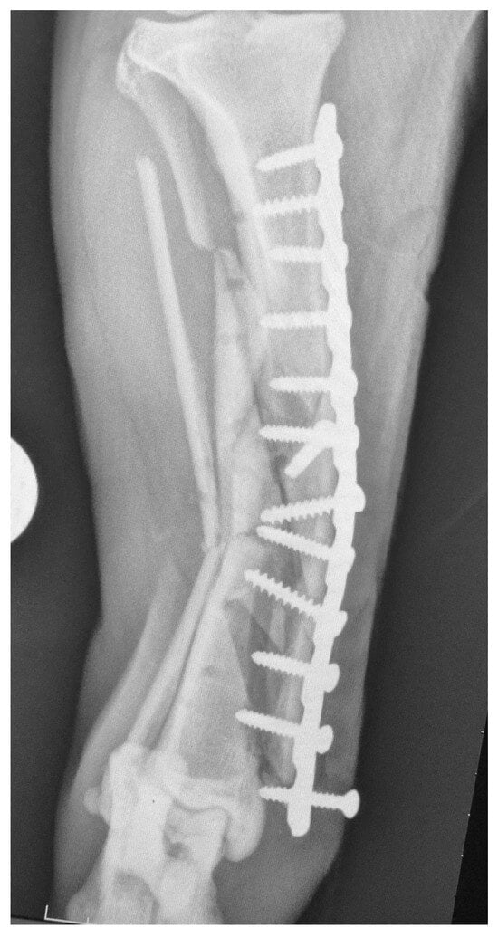
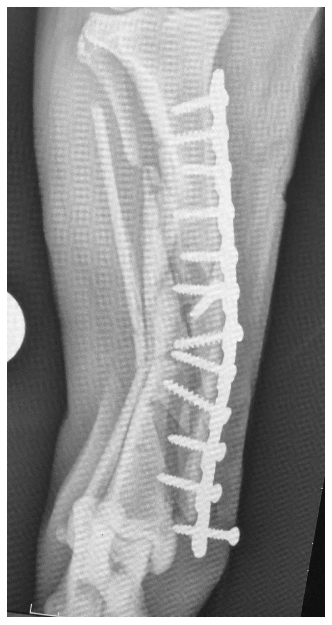
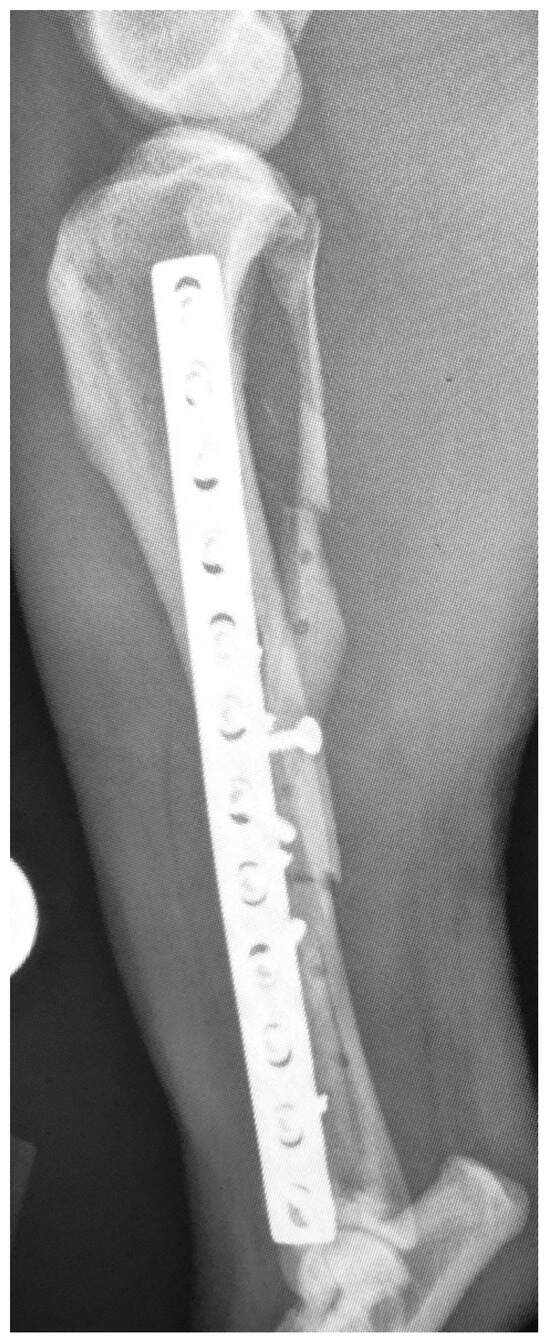
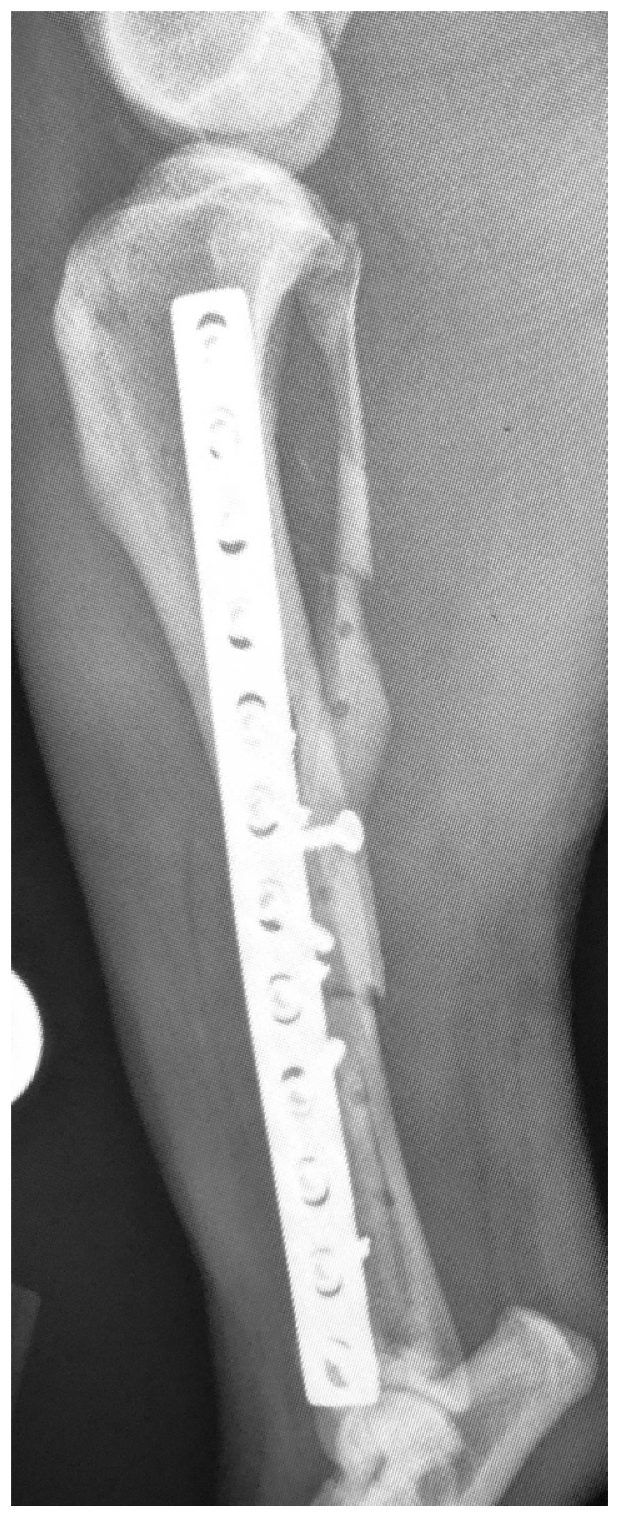
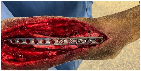
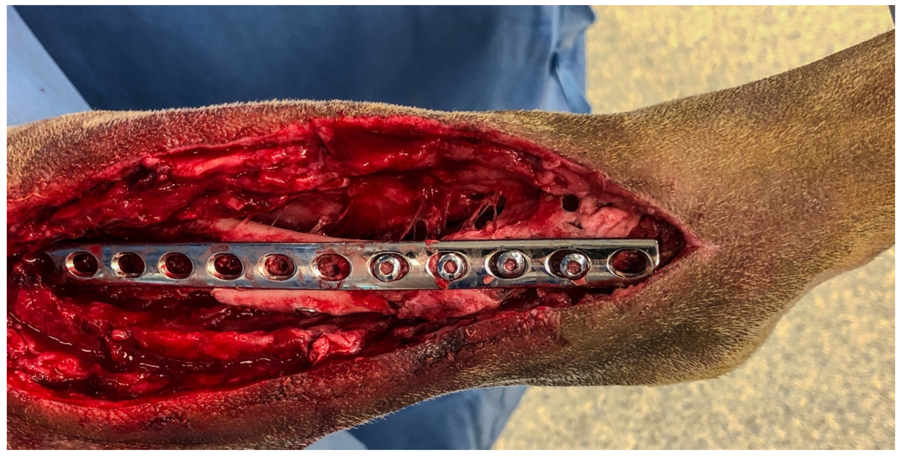
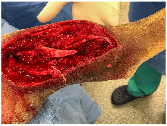
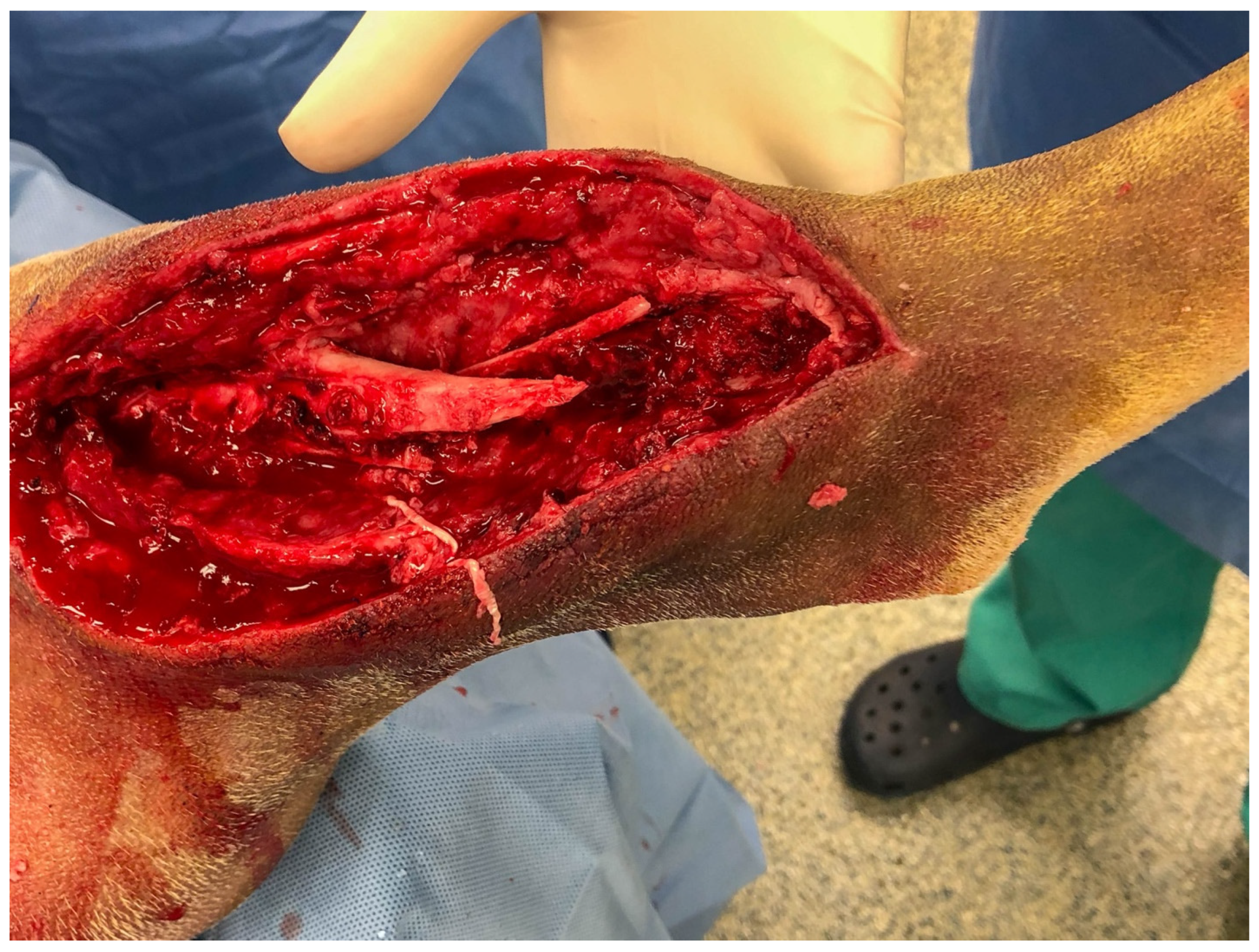
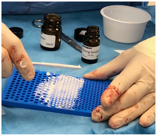
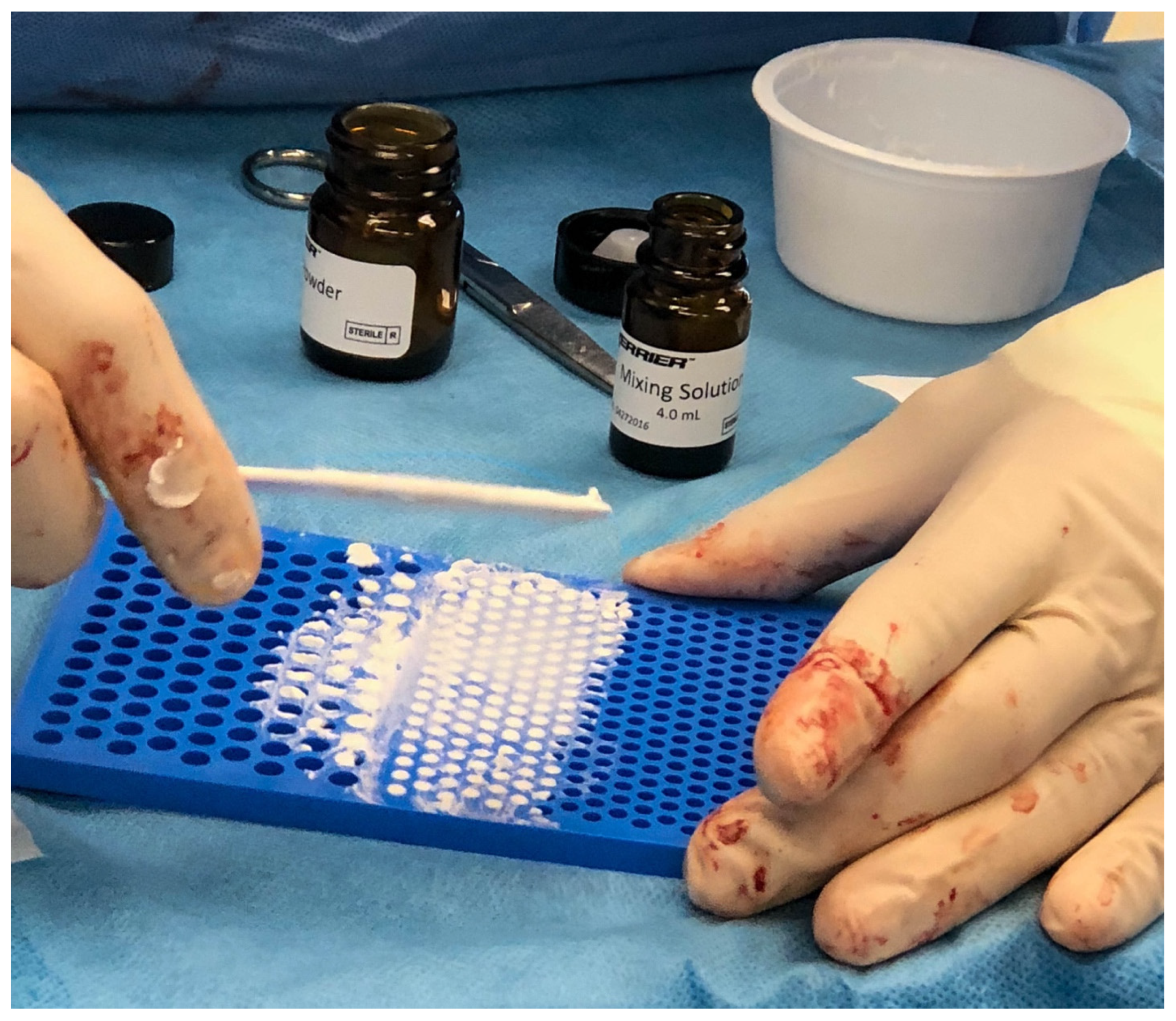
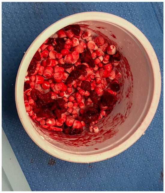
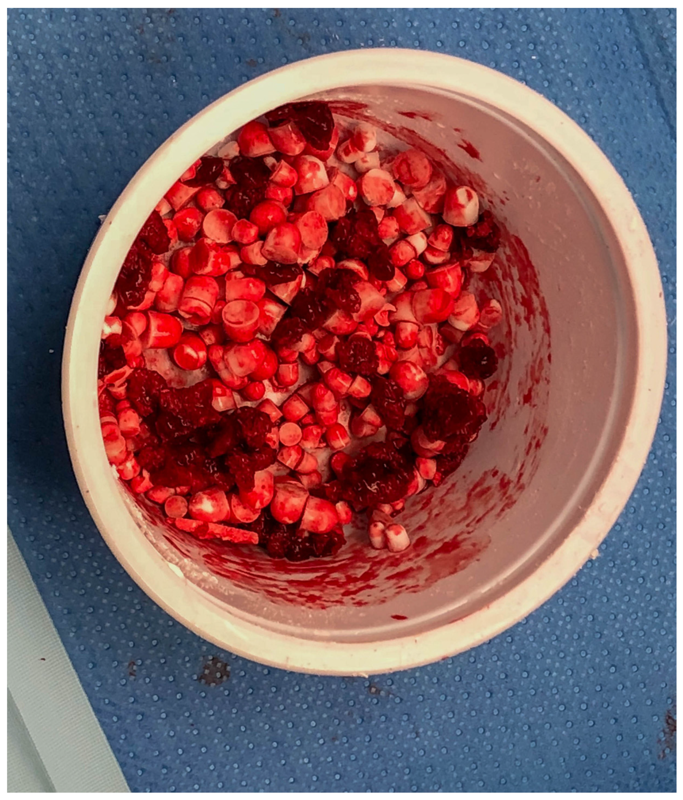
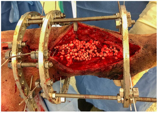
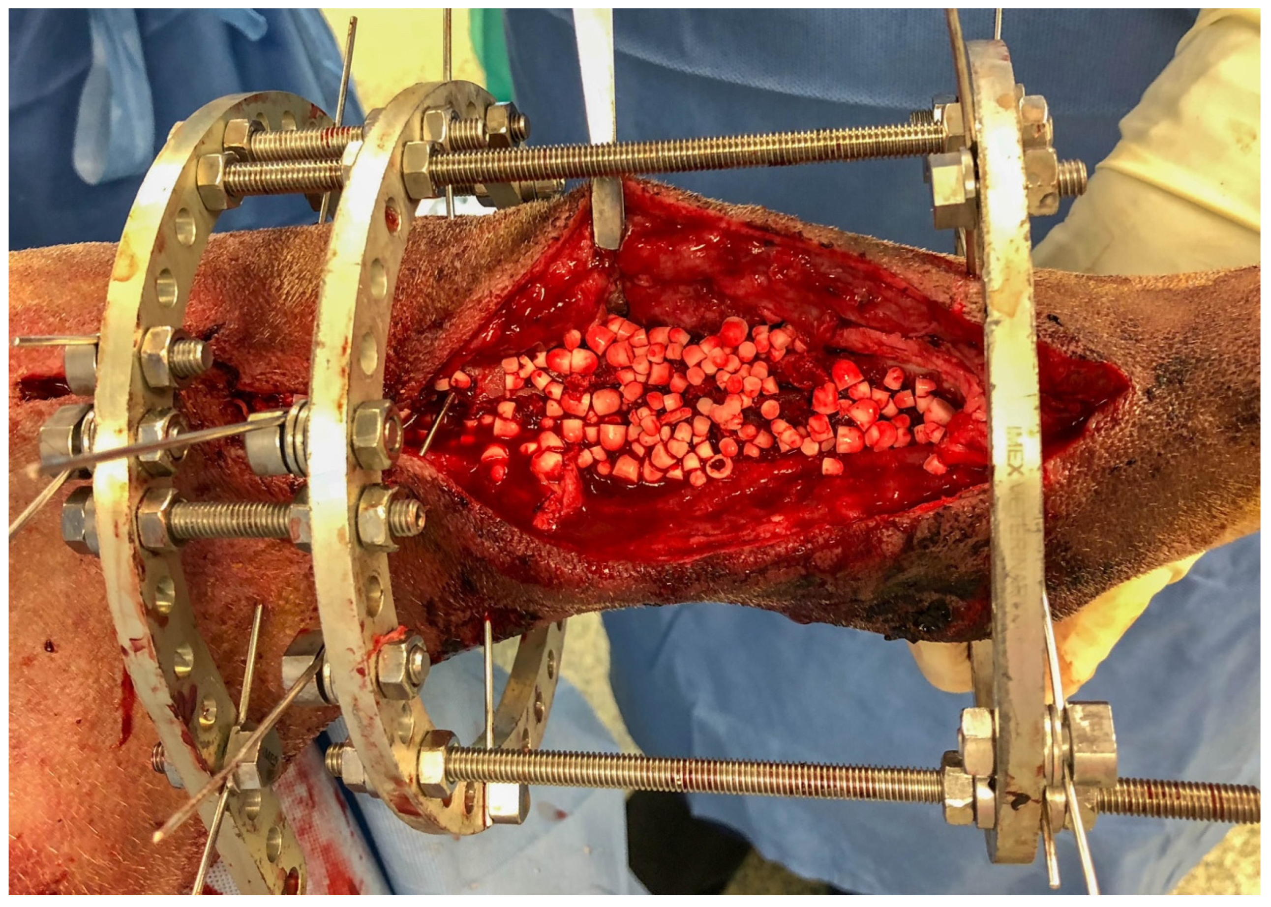
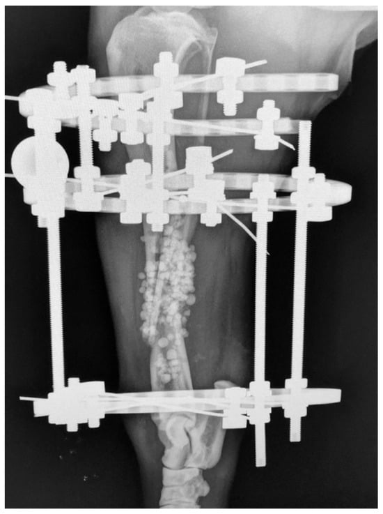
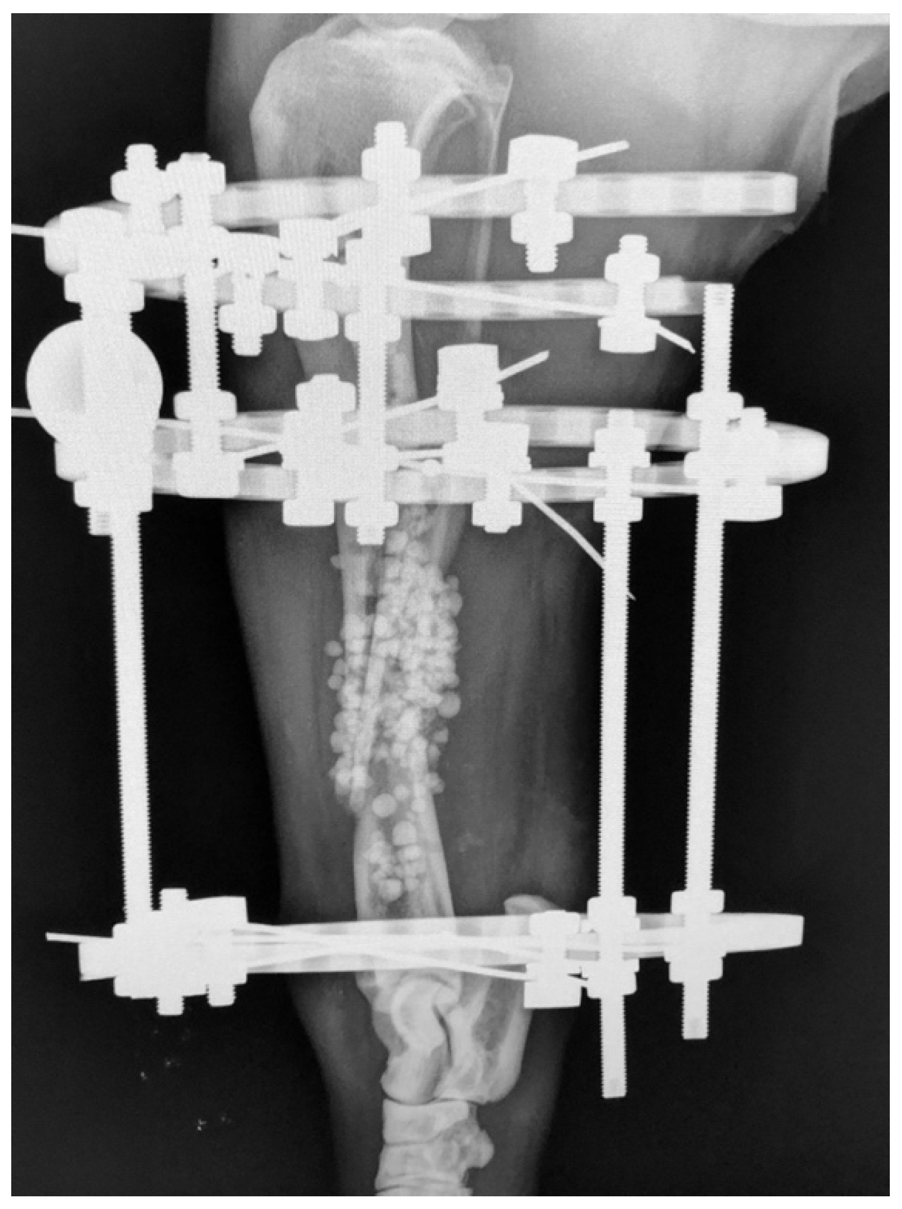
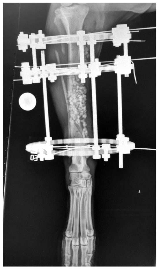
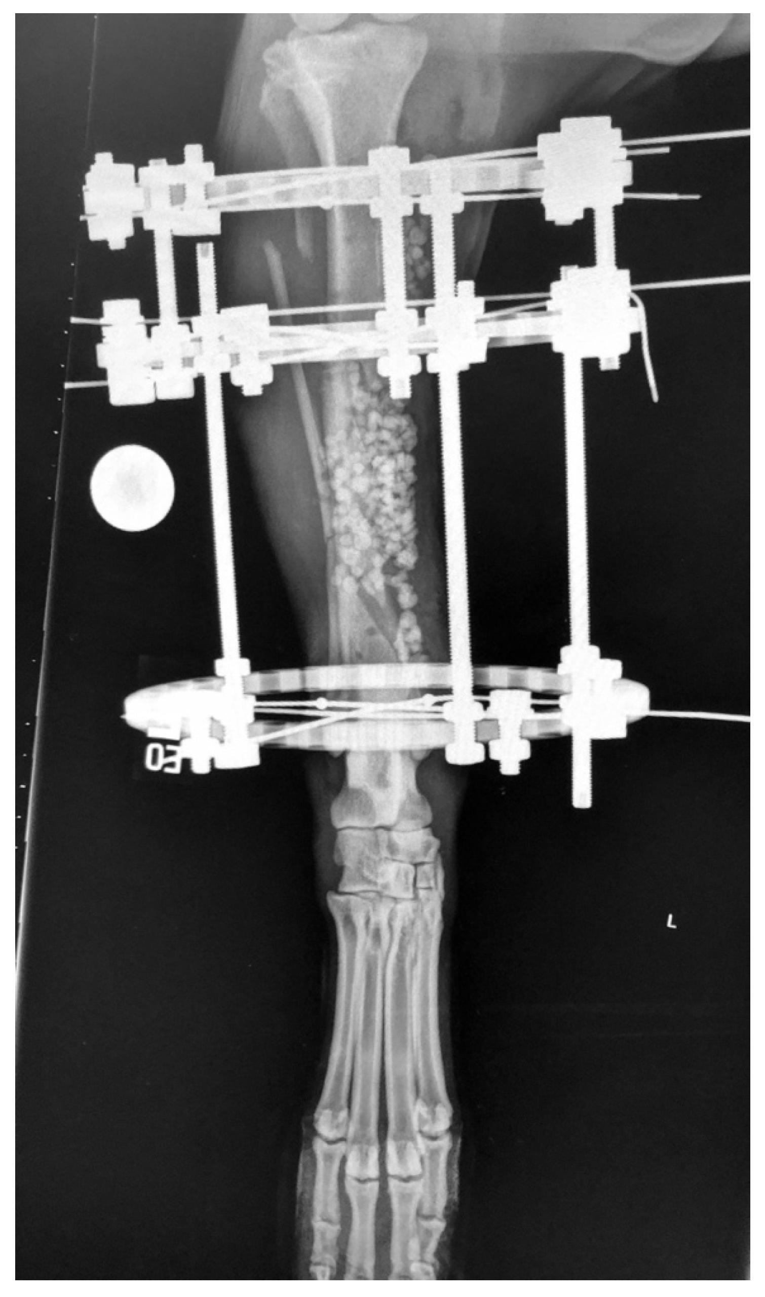
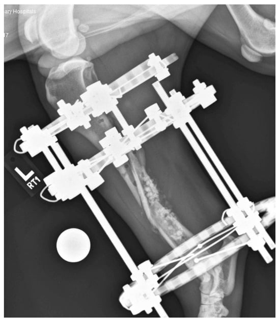
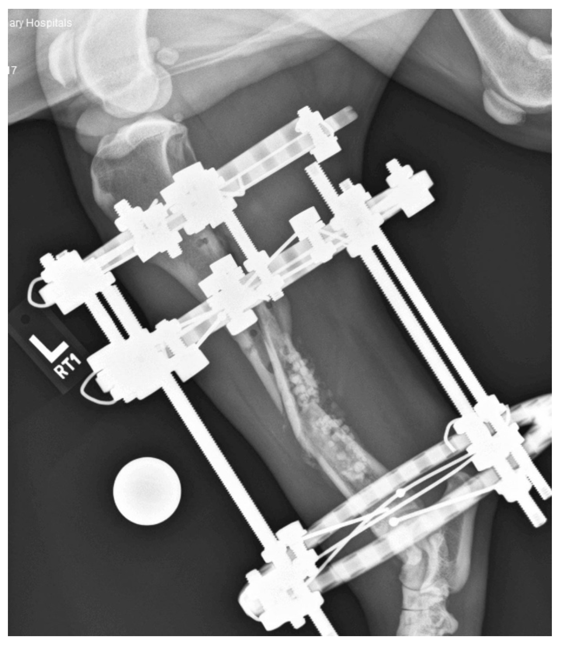
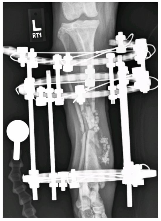
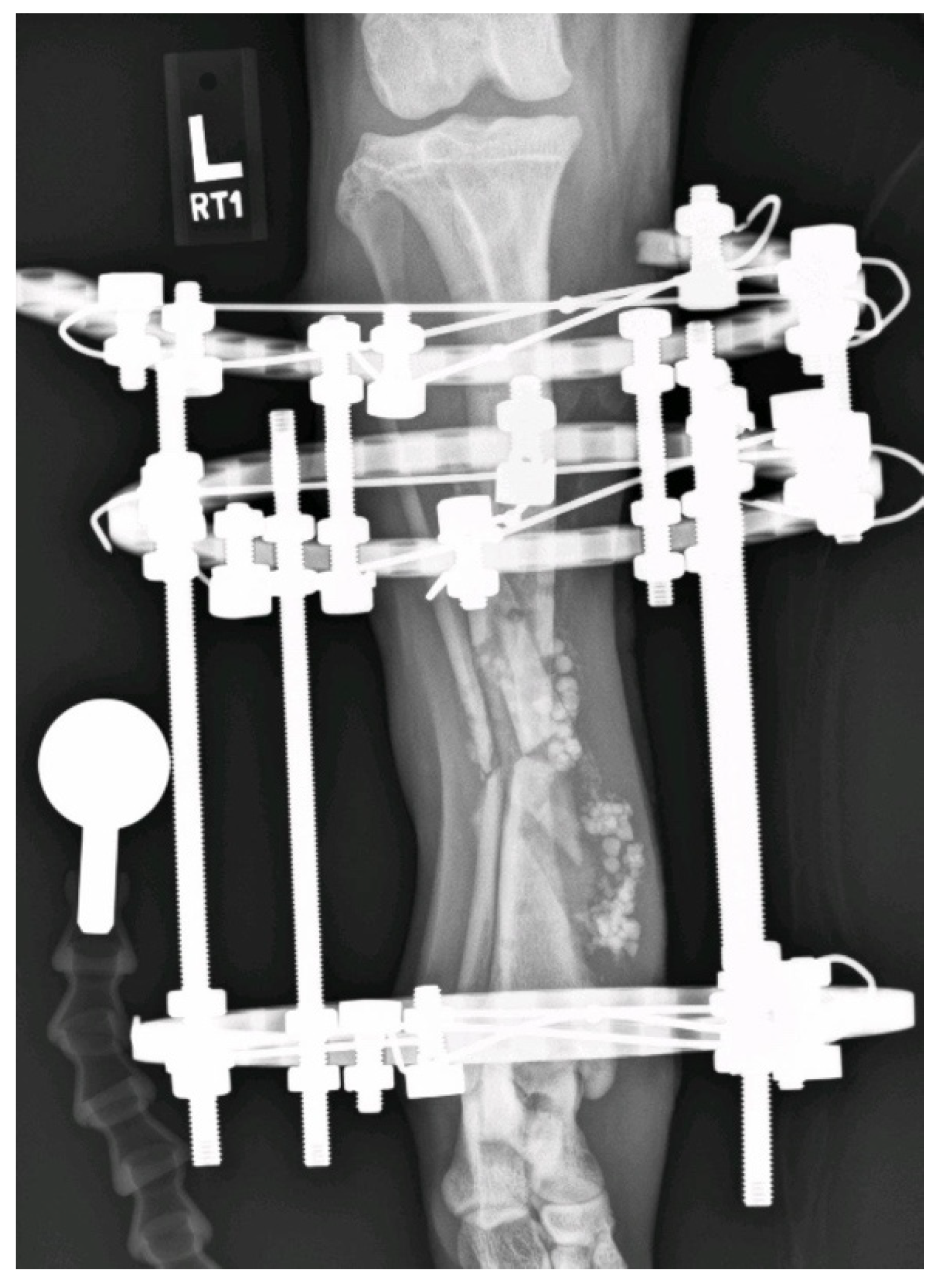
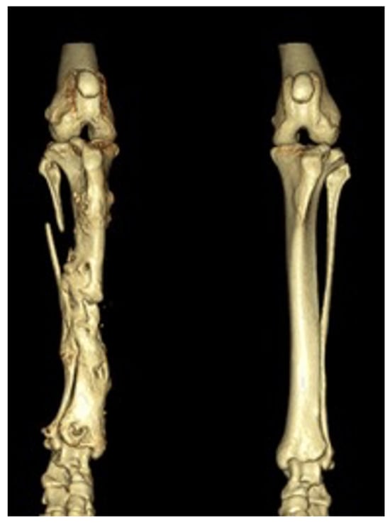
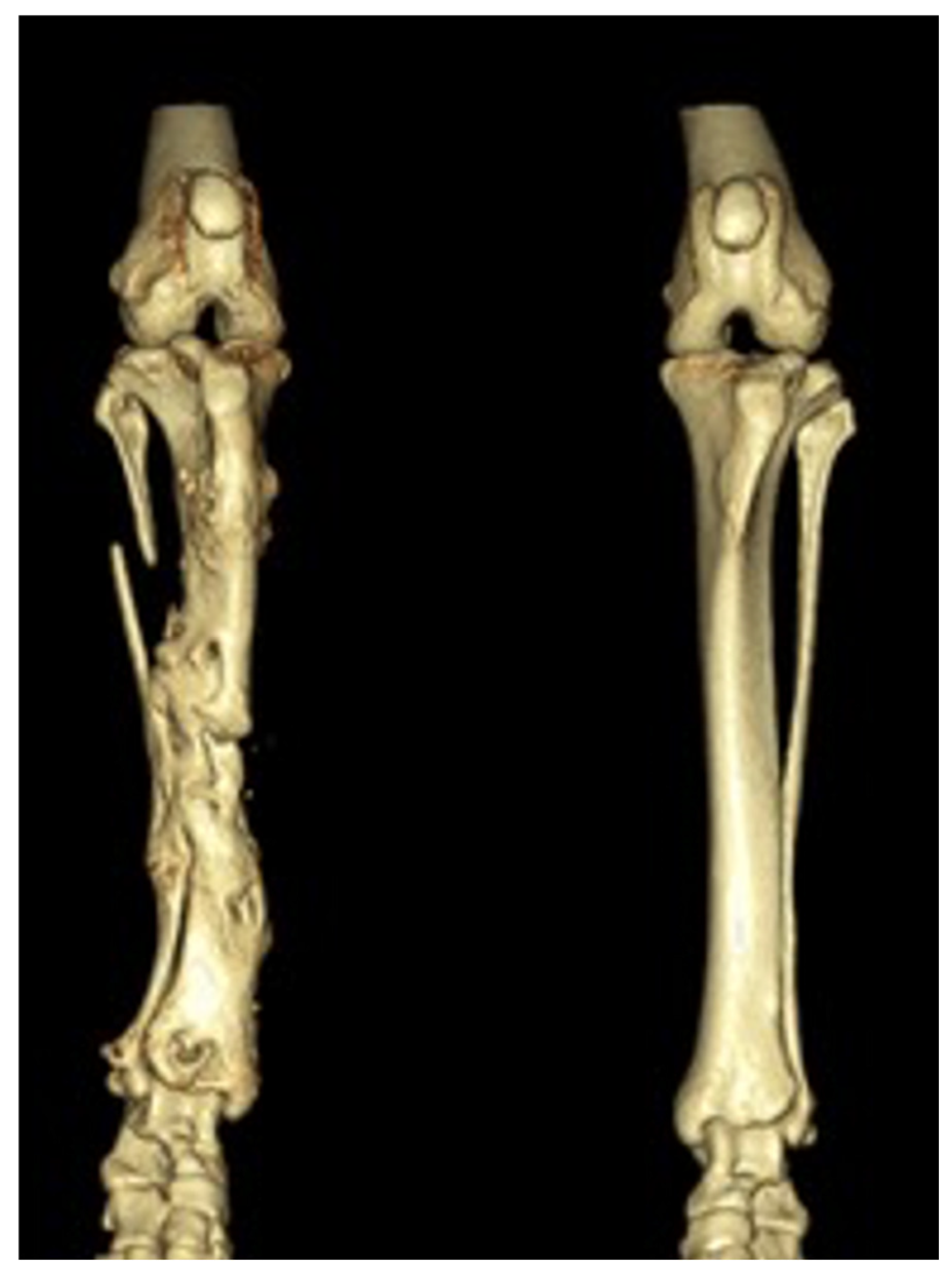
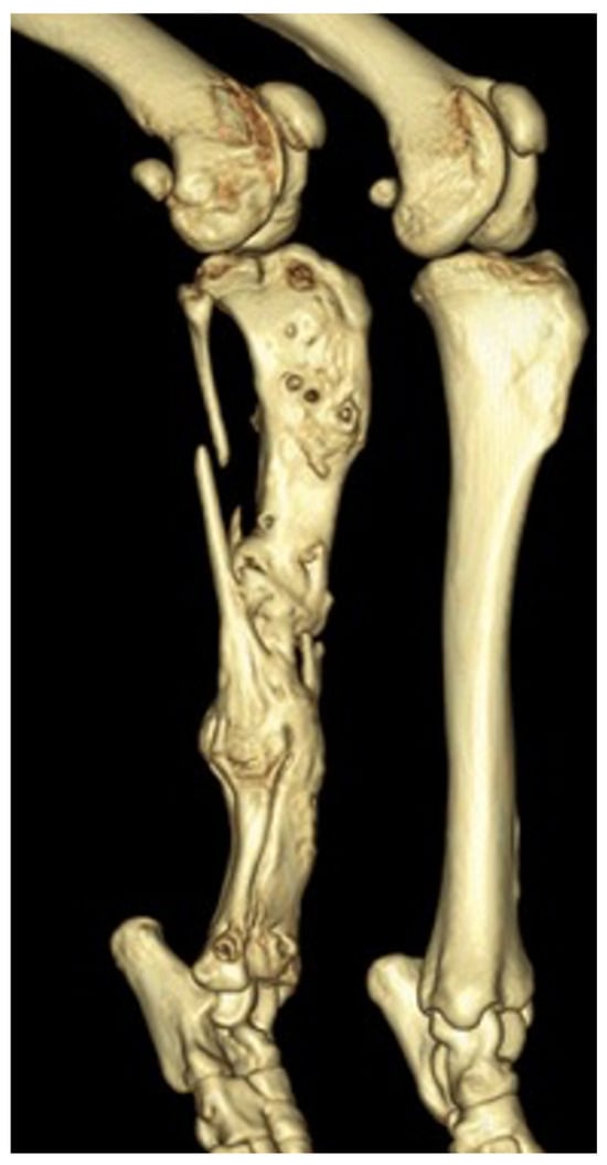
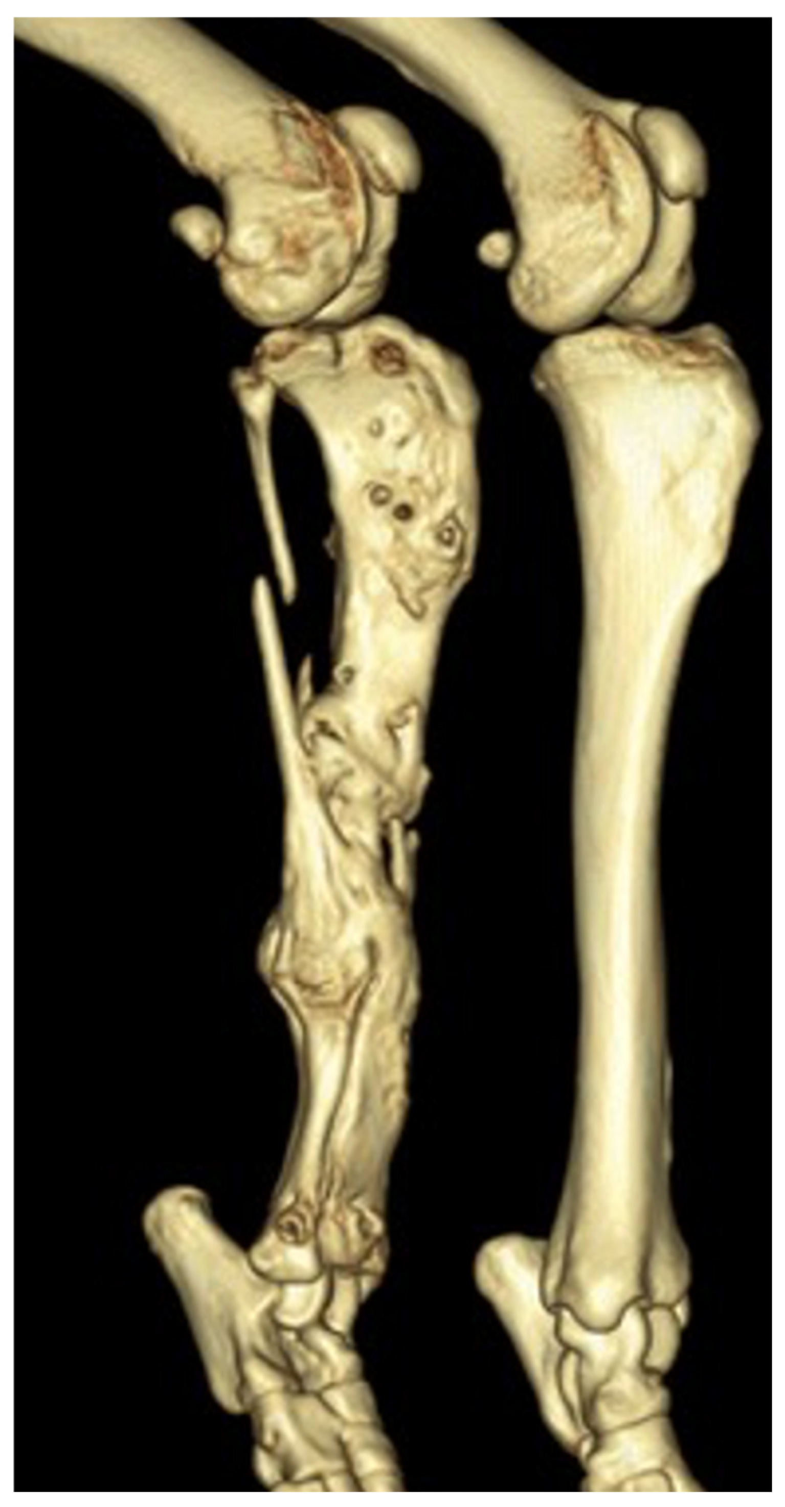
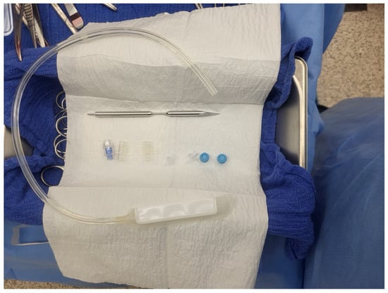
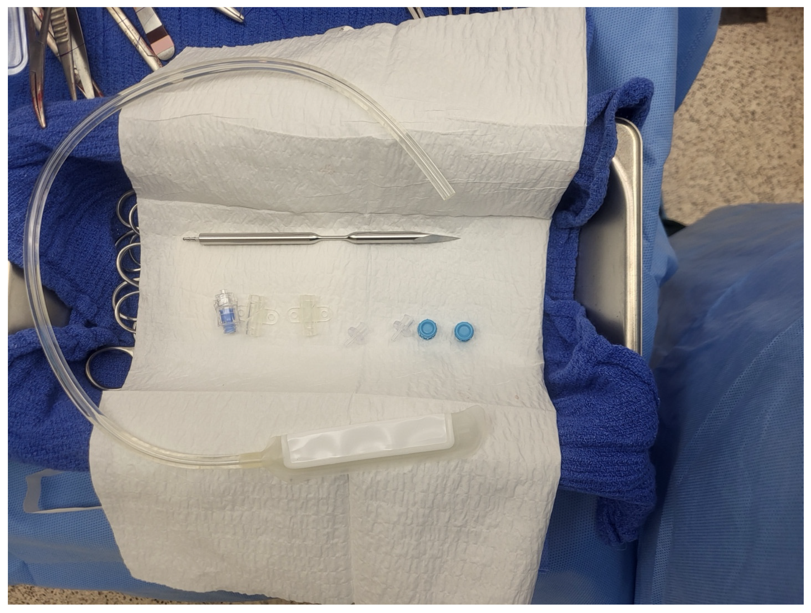
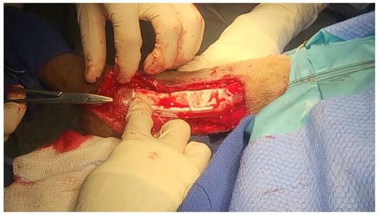
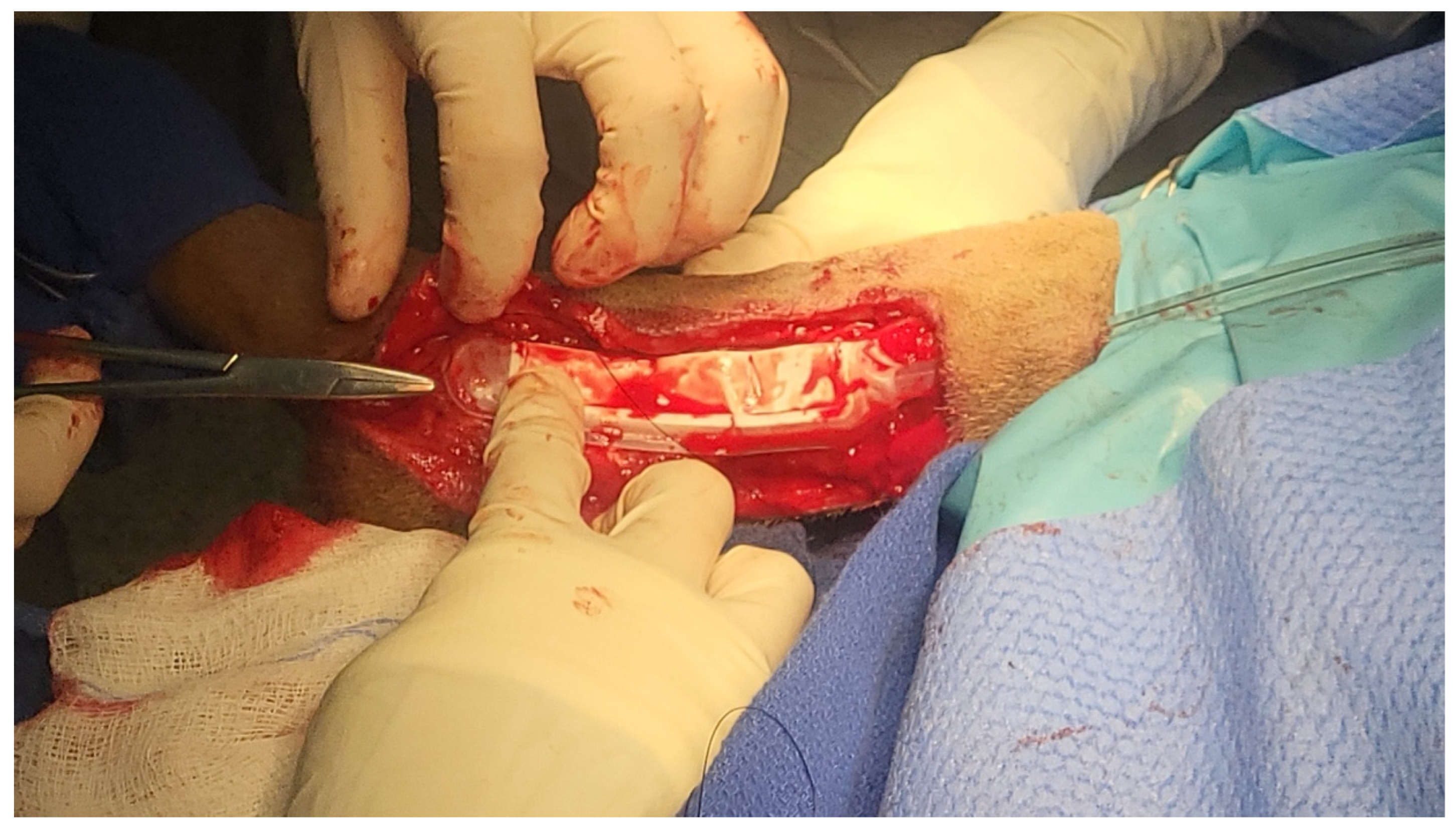
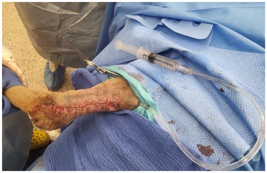
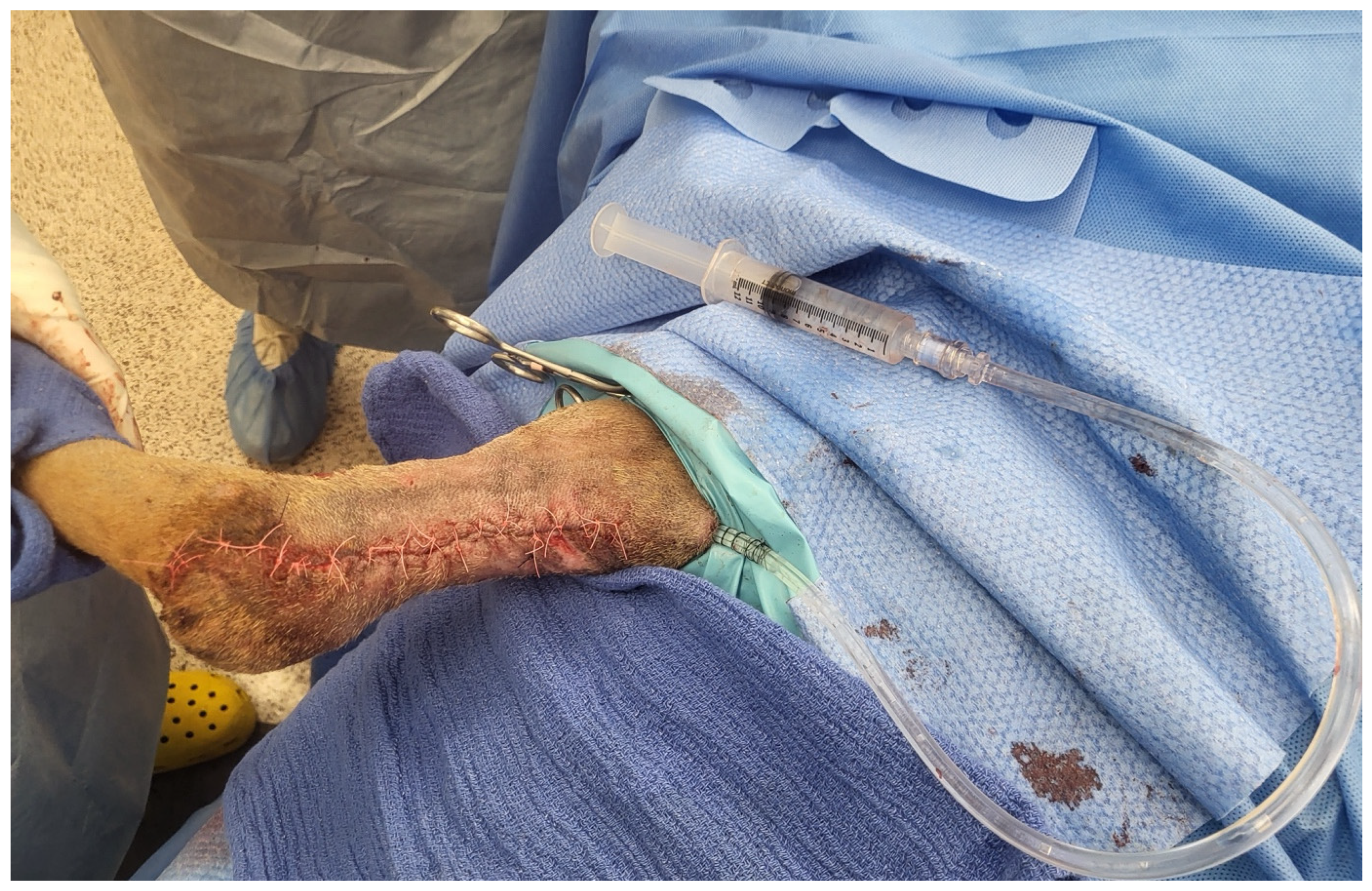

Disclaimer/Publisher’s Note: The statements, opinions and data contained in all publications are solely those of the individual author(s) and contributor(s) and not of MDPI and/or the editor(s). MDPI and/or the editor(s) disclaim responsibility for any injury to people or property resulting from any ideas, methods, instructions or products referred to in the content. |
© 2026 by the author. Licensee MDPI, Basel, Switzerland. This article is an open access article distributed under the terms and conditions of the Creative Commons Attribution (CC BY) license.