


Abstract
Bacteriophage therapy is increasingly used on a compassionate or magistral basis for infections caused by multidrug-resistant bacteria, particularly carbapenem-resistant Klebsiella pneumoniae, Acinetobacter baumannii, and Pseudomonas aeruginosa. Phage preparations are usually selected for the individual patient, and that selection depends on testing candidate phages against the patient’s own isolate, which places the clinical microbiology laboratory within the treatment pathway. The laboratory’s role, however, remains poorly defined: phage susceptibility testing lacks standardised methods, validated interpretive criteria, and external quality assessment, and results from spot assays, efficiency of plating, liquid culture kinetics, and automated platforms are not interchangeable. This narrative review describes the laboratory steps of phage therapy, from isolate selection and characterisation to susceptibility testing, monitoring for phage resistance during treatment, and assessment of phage–antibiotic interactions. The available methods are compared by the biological question each addresses, its readout, throughput, sources of variability, and limits of clinical interpretation. The review then sets out what laboratory participation requires in practice—biosafety, quality assurance, documentation and turnaround—and what those requirements imply for settings with endemic carbapenem resistance such as Greece. The testing strategy described is a proposed laboratory framework, not a clinically validated diagnostic algorithm.
1. Introduction
Antimicrobial resistance (AMR) is among the leading causes of infectious mortality worldwide. In 2021, an estimated 4.71 million deaths were associated with bacterial AMR and 1.14 million were directly attributable to it; forecasts for 2050 reach 1.91 million attributable and 8.22 million associated deaths per year [1]. Among Gram-negative pathogens, resistance to carbapenems increased more than resistance to any other antibiotic class, with attributable deaths rising from approximately 127,000 in 1990 to 216,000 in 2021 [1].
Greece has been at the centre of this problem in Europe for more than a decade. Surveillance by the European Antimicrobial Resistance Surveillance Network (EARS-Net) identified a significant increasing trend of carbapenem resistance in invasive Klebsiella pneumoniae in Greece between 2005 and 2010 [2], and by 2016 the national proportion of carbapenem-resistant bloodstream isolates had reached 67%, among the highest values reported worldwide [3]. Local data confirm the scale of the problem: an eight-year study at the University General Hospital of Patras recorded 1142 bloodstream isolates of carbapenem-resistant K. pneumoniae, Acinetobacter baumannii, and Pseudomonas aeruginosa, of which 69.3% were extensively drug-resistant (XDR), and colistin resistance in K. pneumoniae reached 46.8% in 2022 [4]. For a growing number of patients, the remaining therapeutic options are limited or absent.
Bacteriophage therapy has re-emerged in this context. Phages are viruses that infect and lyse specific bacterial hosts, and they may retain activity against selected antibiotic-resistant bacterial strains, including isolates resistant to several antibiotic classes [5,6]. The largest clinical dataset to date, a retrospective observational analysis of the first 100 consecutive cases of personalised phage therapy coordinated by a Belgian consortium across 35 hospitals in 12 countries, reported clinical improvement in 77.2% and bacterial eradication in 61.3% of infections, and eradication was approximately 70% less probable when phages were given without concomitant antibiotics [5]. Randomised controlled trials of fixed, non-personalised phage products have not reproduced such outcomes [6]. These two bodies of evidence are not directly comparable: they differ in study design, clinical indications, patient populations, treatment protocols, concomitant antibiotic use, and outcome definitions, and an uncontrolled case series cannot establish that patient-specific phage selection is causally superior to a fixed preparation. The contribution of patient-specific selection to clinical outcome therefore requires further controlled evaluation.
The laboratory steps that precede and accompany treatment are nevertheless central to personalised phage therapy, because they determine which phage a patient can be offered: which isolate is tested, which phages are selected, how susceptibility is measured and interpreted, and how resistance is monitored during treatment. These steps lack the standardisation that clinical microbiology laboratories routinely apply to antibiotics. This review describes that workflow, compares the available phage susceptibility testing (PST) methods, identifies the sources of variability between them, and outlines what laboratory participation requires in practice, with particular attention to settings with high carbapenem resistance such as Greece.
Reviews and consensus proposals addressing phage susceptibility testing and the in vitro matching steps of phage therapy already exist [7,8,9], and the protocols and standardisation work published to date are summarised in Section 5.1, so that what follows builds on them rather than restating them. The intended contribution here is narrower and more operational: to tie each testing method to the specific biological question it answers and to a reporting format that states method, parameters, and interpretive criteria alongside the result; to set out the laboratory pathway as a whole (Figure 1) and what participation in it requires in practice, including biosafety, quality assurance, documentation, and turnaround; and to work through what those requirements imply for a health system with endemic carbapenem resistance, using Greek surveillance, laboratory, and regulatory information as the worked example. Where that reasoning rests on the authors’ judgement rather than on cited evidence, this is stated.
Figure 1.
Proposed laboratory pathway for personalised phage therapy. The clinical isolate is received, identified, and stored, and is characterised genomically where that capacity exists. Candidate phages are screened by spot assay; positive results are confirmed by efficiency of plating, and activity is quantified by liquid growth kinetics. Where the infection is device-associated or otherwise biofilm-related, activity is additionally assessed in a biofilm model, and where antibiotics are to be co-administered, the combination is tested. The report states the method, parameters, interpretive criteria, and limitations. Isolates recovered during treatment are re-tested against the administered phages by the same method as the baseline test, and loss of activity triggers re-selection from the collection. Broken outlines indicate steps that depend on the infection type or on locally available capacity. The pathway is proposed by the authors on the basis of the methods reviewed here and has not been validated prospectively.
2. Review Scope and Literature Identification
This article is a narrative review: it summarises and interprets the published literature on the laboratory steps of personalised phage therapy, rather than pooling data from comparable studies.
PubMed, Scopus and Web of Science were searched from database inception to 7 November 2026, with Google Scholar used for supplementary checking, using combinations of the terms “bacteriophage” OR “phage therapy” AND “susceptibility testing” OR “efficiency of plating” OR “spot assay” OR “host range” OR “phage–antibiotic” OR “biofilm” OR “phage bank” OR “magistral”, together with the organism terms Klebsiella pneumoniae, Acinetobacter baumannii, and Pseudomonas aeruginosa and, for the national section, “Greece” OR “Greek”. Searches were restricted to English-language records. The reference lists of the principal methodological and clinical reports were screened for further sources, and the websites of the European Medicines Agency and of the European Directorate for the Quality of Medicines & HealthCare were consulted directly for regulatory and pharmacopoeial texts.
Records were selected for relevance to the laboratory steps of personalised phage therapy—isolate handling and characterisation, phage susceptibility testing, interpretation and reporting, monitoring for phage resistance, phage–antibiotic interaction testing, and laboratory implementation and to settings with endemic carbapenem resistance. Priority was given to peer-reviewed primary studies, to methodological comparisons between assays, and to clinical series; the one conference abstract retained for want of a peer-reviewed equivalent is identified as such both at the point of citation and in the reference list, and the claim resting on it is qualified accordingly. Because selection was guided by relevance rather than by a predefined protocol with screening in duplicate, the evidence base presented here is not guaranteed to be exhaustive, and comparisons between studies that differ in design, assay protocol, or outcome definition are made qualitatively and flagged where the designs are not equivalent.
3. Phage Therapy as a Personalised Treatment: Why the Laboratory Matters
Phage therapy differs from antibiotic therapy in one decisive respect: activity is strain-specific rather than species-specific. A phage active against one isolate of K. pneumoniae is frequently inactive against another isolate of the same species. In a systematic study of 138 clinical K. pneumoniae isolates and 46 environmental phages, fewer than 2% of the 6319 phage–host combinations tested produced reproducible lysis [10]. Treatment therefore cannot be selected empirically from the species identification alone; it must be matched to the patient’s isolate.
This turns phage therapy, in practical terms, from a problem of drug availability into a problem of matching [11]. Three models are currently used: fully personalised therapy, in which phages are selected or isolated for each patient; fixed cocktails, prepared in advance against a defined pathogen population; and hybrid approaches that combine a pre-characterised phage bank with patient-specific selection [7]. In all three, the decisive step is the in vitro assessment of phage activity against the clinical isolate, carried out before treatment and repeated when the infection persists or relapses.
The operating procedure of phage therapy has been described as a sequence of laboratory and clinical steps: isolation and identification of the pathogen, screening against a phage library, preparation of the phage formulation, administration, monitoring of efficacy, and detection of phage-resistant strains [12]. Of these, all except formulation and administration are laboratory functions. Gelman et al. proposed the term “Clinical Phage Microbiology” for the set of in vitro matching steps that should precede and accompany treatment, by analogy with antibiotic susceptibility testing (AST) [8]. The Israeli Phage Therapy Center subsequently published a pipeline proposal based on five years of clinical experience, comparing available methods and arguing for standardisation [9].
The clinical microbiology laboratory is therefore not a peripheral participant in phage therapy. It holds the patient’s isolate, performs identification and AST, has the expertise in quality-controlled susceptibility testing, and communicates results to the treating physician. These are the same functions on which personalised phage therapy depends. The remaining sections describe how these functions translate to phages and where current practice falls short.
4. Isolate Handling: Selection, Identification and Genomic Characterisation
The isolate submitted for phage matching determines everything that follows, and its selection is a clinical-microbiological decision rather than an administrative one.
Chronic and device-associated infections, which account for a large share of phage therapy requests, are frequently polyclonal. Isolates recovered from different sites, or from the same site at different times, may differ in phage susceptibility even when reported as the same species with an identical antibiogram. Where the infection is polymicrobial or several morphotypes are present, each should be considered separately, since a cocktail covering only one component will not clear the infection. Isolates should be stored at −80 °C with minimal subculture: repeated passage can alter capsule expression and surface receptors, and therefore phage susceptibility, so that the strain tested no longer represents the strain infecting the patient.
Standard identification (MALDI-TOF MS) and AST according to EUCAST remain prerequisites. The antibiogram is not superseded by phage testing: the largest clinical series to date found that eradication was approximately 70% less probable when phages were administered without concomitant antibiotics [5], so the agents that retain partial activity must be known. Carbapenemase characterisation and colistin MIC determination also inform the clinical decision on whether phage therapy is justified.
Whole-genome sequencing (WGS) of the isolate is increasingly part of the workflow. Its first contribution concerns surface receptors, which determine adsorption: in K. pneumoniae, the capsule is the principal receptor, and its diversity, with more than 180 capsular locus types described, is the main factor restricting phage host range [10]. Phage-encoded depolymerases are the corresponding specificity determinants, and depolymerase sequence types are associated with the ability to infect particular capsular types across phage families [10]. Capsular typing from the genome can therefore narrow the list of candidate phages before any plate is poured. Genomic prediction does not, however, substitute for phenotypic susceptibility testing: capsule switching and capsule modification can decouple predicted from observed specificity [13], and no prediction method has been validated against phenotype in a clinical testing workflow.
Intracellular defence systems shape the outcome after adsorption, and the evidence here is less consistent. Analysis of antibiotic-resistant clinical P. aeruginosa isolates showed that defence systems substantially define phage host range and that phage resistance scales with the number of systems in the genome [14], whereas later work in the same species found that the number alone is not predictive and that functional interactions between specific systems matter more [15]; in E. coli, adsorption factors rather than defence system counts were the dominant determinant [16]. Tools such as DefenseFinder allow routine annotation of these systems [17], but the current evidence does not support using the prediction alone to exclude a phage. Sequencing also documents the isolate for later comparison. Comparative genomic analysis of baseline and relapse isolates is a powerful approach for distinguishing persistence or evolution of the original strain from infection with a genetically distinct strain; its interpretation depends on sampling, genomic resolution, within-host population heterogeneity and the epidemiological context, and should be integrated with clinical information and, where feasible, with phenotypic reassessment of phage susceptibility. These analyses are not available in every hospital laboratory and are not required for every case, but where sequencing capacity exists, they can shorten the candidate list and provide the baseline against which treatment is later assessed.
5. Phage Susceptibility Testing, Interpretation and Monitoring
There is no reference standard for assessing bacterial susceptibility to phages, and no validated interpretive criteria [18]. Four questions should be kept apart: whether lysis of a bacterial lawn can be detected; whether the phage replicates productively on the isolate; whether bacterial growth is suppressed in liquid culture over a defined interval; and whether any of these predicts therapeutic response. The assays described below address only the first three, and their results are not interchangeable. The testing strategy set out in this section is therefore a proposed laboratory framework for characterising phage–host interactions, not a clinically validated diagnostic algorithm; its clinical utility remains to be established through standardisation and correlation with treatment outcomes. The methods, the question each addresses, and the limits of each are compared in Table 1.
Table 1.
Methods used to characterise phage–host and phage–antibiotic interactions in vitro. The methods are not equivalent and address different biological questions.
5.1. Previously Published Protocols and Standardisation Efforts
A laboratory beginning this work does not have to design a method from first principles, and it is worth setting out what has already been published before the individual assays are described.
Three kinds of protocol-level sources are available. The first is the published procedures of established phage therapy services: the Clinical Phage Microbiology framework proposed by the Israeli Phage Therapy Center sets out the sequence of isolate receipt, host range screening, confirmation, and reporting [8]; the same group has published the susceptibility testing procedures and reporting format it uses [9]; and a standard operating procedure covering the whole clinical pathway, including its laboratory steps, has also been published [12]. The second is the methodological comparisons discussed below, which specify the conditions under which particular assays were run and are therefore usable as protocols in their own right [18,19,20]. The third is a primer written specifically for clinical microbiology laboratory staff, which describes the available assays, their readouts and their limitations in the terms used in a diagnostic laboratory [21].
Formal standardisation has now begun. A disk-based phage susceptibility testing format (Disk-PST) was validated first within the coordinating laboratory, with 28 participants, and then in an inter-laboratory comparison across 12 German laboratories with 39 participants, the double-agar overlay plaque assay serving as the reference standard; blinded testing of one Escherichia coli phage, one Staphylococcus epidermidis phage and a buffer control against four strains, in two technical and two biological replicates, gave κ values of 0.973 and 0.849 on the two intra-laboratory test days and 0.925 to 0.939 between laboratories, with sensitivity of 92.25–94.87% and specificity of 97.76–98.72% in the inter-laboratory phase [22]. To our knowledge, this is the first phage susceptibility testing format subjected to a formal multicentre reliability study, and it establishes that reproducible phage testing in routine laboratories is attainable. Two limitations should be read with it: the comparison used a small predefined panel of phages and strains selected for consistent susceptibility profiles rather than the diversity encountered in personalised therapy, and the authors state explicitly that correlation of the test result with clinical outcome remains to be established [22].
The methods described in the subsections that follow should therefore be read as the assays currently in use rather than as a validated reference method, with the disk-based work as the available model for how such a method might be established.
5.2. Spot Assay
A phage suspension is spotted onto a bacterial lawn and clearing is recorded after incubation. The method is rapid, inexpensive, and allows many phages to be screened in parallel, which explains its dominance in host range studies.
Its principal weakness is that clearing does not prove productive infection. A clear zone may result from lysis from without, from depolymerase activity degrading the capsule, or from bacteriocins, without any phage replication. A comparison of spot tests with efficiency of plating (EOP) in E. coli concluded that single-dilution spot tests cannot be used to select phages for a therapeutic library and should be replaced by EOP [19]. The spot assay therefore remains an appropriate screening step, but a positive result requires confirmation.
5.3. Efficiency of Plating
EOP quantifies the ratio of plaque counts obtained on the test strain to those obtained on the propagating host and therefore provides evidence of productive infection rather than of clearing alone [18,19]. It is the closest available analogue to a quantitative susceptibility measurement and is commonly used to confirm a positive spot screen. It does not constitute a clinically validated reference standard for phage susceptibility: the value obtained depends on the propagating host chosen, on the assay protocol and on the growth conditions, and no threshold has been correlated with treatment outcome.
Its practical limitations are also significant. Double-agar overlay is labour-intensive, requires 18–24 h, and depends on plaque counts that are difficult to read when plaques are small or turbid, which limits both throughput and inter-reader agreement.
5.4. Liquid Growth Kinetics
Phage and bacteria are combined in microplates and optical density is recorded over time, typically for 24 h. Susceptibility is expressed through parameters such as the hold time, the interval during which growth is suppressed relative to untreated controls. These assays are automatable, generate quantitative kinetic data, and allow many phage–isolate combinations to be tested simultaneously. Optical density is, however, an indirect measure of bacterial growth rather than a direct count of viable cells: it does not distinguish killing from inhibition of growth, and it does not by itself demonstrate phage replication.
Reproducibility has been examined directly. A comparison of two high-throughput liquid platforms, the Biolog OmniLog and the Agilent BioTek Cytation 7, using 55 phages against hosts and non-susceptible controls, reported good to fair statistical reproducibility within and between assays [20]. A separate multi-site comparison of OmniLog liquid assay results with double-layer plaque assay results concluded that the reproducibility of phage susceptibility testing methods requires further development [18]. A conference abstract reporting preliminary data in K. pneumoniae compared a spot plaque assay with an optical-density liquid assay using hold-time cut-offs of 4 h and 8 h, and presented the agreement obtained as a step towards standardisation, while noting that correlation with clinical outcome remains to be established [23]; as preliminary data presented in abstract form, it should be treated as provisional.
Liquid assays also carry specific interpretive risks. Optical density falls with lysis but rises again when phage-resistant mutants expand, so a result read at a single endpoint can misclassify an isolate. Metabolic readouts such as the OmniLog cannot distinguish dead from non-metabolising cells and have limited capacity to detect late lysis [20].
5.5. Comparison of Methods
Discordance between these methods is the rule rather than the exception, and its magnitude has been measured. In the inter-laboratory comparison of P. aeruginosa phage susceptibility testing, agreement was lower between plaque and liquid assays than between liquid assays performed at different sites, and only 22% of the bacterial isolates tested showed agreement across all of their measurements [18,21]. The same group, comparing two liquid high-throughput platforms, found larger differences between replicate runs of the same assay on different days than between two different assays run on the same day, which places a substantial part of the variability in day-to-day execution rather than in the choice of platform [20]. In E. coli, spot tests read at a single dilution overcalled susceptibility relative to EOP [19]. Against this, the disc-based format reached κ values of 0.925–0.939 against the plaque assay across 12 laboratories, although on a small predefined panel [22].
Two practical conclusions follow. First, a phage susceptibility result is not transferable between laboratories unless the method, its parameters, and its interpretive criteria travel with it; a result reported as a bare category is not interpretable elsewhere. Second, discordance between two methods applied to the same isolate is not necessarily an error in either, because the spot assay, EOP, and liquid kinetics measure different things; a discrepancy should prompt a statement of what each assay showed rather than a choice between them. Where a single answer is nevertheless required, the hierarchy implied by the available evidence is that confirmation of productive infection carries more weight than clearing alone [19], and that a result reproduced on a second occasion carries more weight than a single determination [20]. Neither ranking has been validated against clinical outcome.
5.6. Sources of Variability
Several variables are not standardised across published protocols and account for much of the discordance between laboratories: the multiplicity of infection (MOI); the growth medium and divalent cation content, which affects adsorption; incubation temperature; the physiological state and density of the bacterial inoculum; the titre and purity of the phage stock, including residual endotoxin and host debris; the propagating host used for phage amplification; and the endpoint and time at which the result is read. Until these parameters are fixed by consensus, the comparability that clinicians expect from EUCAST-based antibiograms cannot be assumed for phage results.
5.7. Interpretation and Reporting
The laboratory question is not whether a phage lyses the isolate in vitro, but whether the observed activity justifies therapeutic use. For antibiotics, this translation is performed by clinical breakpoints derived from pharmacokinetic/pharmacodynamic data and clinical outcome. For phages, no equivalent exists [18].
Several factors make a direct transfer of the antibiogram model difficult. Phages replicate at the infection site, so the administered dose is not the effective dose; activity depends on bacterial density, which is unknown in most deep-seated infections; the phage interacts with the host immune system; and biofilm, in which many phage therapy candidates are embedded, alters susceptibility relative to planktonic testing.
The strength of the evidence on this point should be stated plainly, because it is the central limitation of everything described above. No study has established that any in vitro phage susceptibility measurement predicts clinical response. The largest clinical dataset, the series of 100 consecutive cases, selected phages on in vitro activity and reported clinical improvement in 77.2% and eradication in 61.3% of cases, but its design—a retrospective observational series without a comparator arm, with concomitant antibiotics in most cases and with heterogeneous infections, phages and routes of administration—cannot isolate the contribution of the in vitro matching step [5]. The randomised controlled trial of a phage preparation in burn wound infection used a fixed cocktail at a titre lower than intended and did not demonstrate superiority over standard care, so it does not test personalised matching either [6]. The authors of the inter-laboratory susceptibility comparison and of the disc-based validation both identify correlation with clinical outcome as the outstanding question [18,22]. Until that correlation exists, a phage susceptibility result should be understood as evidence that a phage kills the isolate under the conditions of the assay, and not as a prediction that it will resolve the infection.
In the absence of breakpoints, laboratories currently apply operational criteria: an EOP above a defined threshold relative to the propagating host; a hold time exceeding a stated cut-off in liquid assays [23]; or clearing plus confirmed productive infection. These criteria are internally defined and are not transferable between centres without validation.
Reporting should therefore be explicit rather than categorical. A phage susceptibility report should state the method used, the phage identity and titre, the propagating host, the multiplicity of infection, the measured parameter with its value, the criterion applied, the controls included, and the date and isolate identifier. The terms “susceptible” and “resistant” should be used only with an accompanying definition, since clinicians will otherwise read them as equivalent to EUCAST categories. Where phage activity is reported, the concurrent antibiogram should be reported alongside it, given the dependence of eradication on concomitant antibiotics [5].
The laboratory should also state what the result does not establish: in vitro activity has not been shown to predict clinical response, and the absence of a validated correlation is itself part of the result [23].
5.8. Monitoring for Phage Resistance During Treatment
Bacterial resistance to phages arises readily and, unlike the acquisition of an antibiotic resistance gene, is frequently visible within a single treatment course. Resistance mechanisms include loss or modification of the surface receptor, capsule alteration or switching [13], and the action of intracellular defence systems [14,15]. Because the capsule is both the principal phage receptor in K. pneumoniae and a virulence factor, receptor loss may be accompanied by a fitness cost, including reduced virulence or restored antibiotic susceptibility. These trade-offs can be therapeutically useful, but they are strain- and phage-dependent and cannot be assumed.
Monitoring therefore belongs within the laboratory workflow rather than at its end. The pre-treatment isolate is stored and, where possible, sequenced, establishing the reference against which later isolates are compared. Isolates recovered while the patient is receiving phages are re-tested against the administered phages using the same method and criteria as the baseline test. A rise in optical density late in a liquid assay, or a fall in EOP relative to baseline, is consistent with emerging resistance, but it is not sufficient evidence of it: before such a change is reported as resistance, it should be confirmed by repeat testing, and technical explanations (inoculum preparation, phage titre and storage, medium composition, plate position and reading interval) and biological alternatives (sampling of a different subpopulation, mixed populations, or reinfection with a distinct strain) should be excluded. On relapse or persistence, the relapse isolate is compared with the baseline isolate, since genomic comparison helps distinguish an escape mutant of the original strain from a different strain, and that distinction informs whether the phage cocktail should be changed or the source of infection reconsidered. Where resistance is confirmed, the antibiogram should also be repeated: phage-resistant mutants that have lost capsule or efflux-related receptors may show altered antibiotic susceptibility, which can open therapeutic options that were previously closed.
The practical implication is that phage therapy generates a sequence of isolates rather than a single one, and the laboratory must be resourced and organised to handle serial testing on a clinically relevant timescale.
Cocktail composition is the main laboratory lever against resistance. Two models are in use: fixed cocktails prepared in advance against a pathogen population, and patient-specific selection from a characterised collection. The second is slower and more demanding but allows composition to be matched to the isolate and has been argued to be more sustainable with respect to resistance [21]. Where selection is patient-specific, the operating principle is to combine phages that do not share a receptor or a resistance pathway, so that a single receptor change cannot abolish the activity of the whole preparation. That principle follows from the receptor-level mechanisms described above [10,13] rather than from a validated selection rule; we did not identify a published criterion specifying how much receptor diversity is sufficient, and this is one of the points at which laboratory practice currently rests on judgement.
Two options are available when resistance emerges during treatment. The first is to re-test the relapse isolate against the full collection and substitute an active phage, which requires that both the collection and the testing capacity remain available throughout treatment rather than only at its start. The second is directed evolution of the phage against the resistant isolate, for which the Appelmans protocol, serial co-culture of a phage mixture with the target strain, is the established method [21]. It can restore activity against isolates that have become resistant, but it yields a new preparation whose characterisation and quality status must then be addressed, which places it outside the routine scope of a clinical diagnostic laboratory.
5.9. Phage–Antibiotic Combinations
Phages are rarely given alone. In the 100-case series, eradication was approximately 70% less probable without concomitant antibiotics [5], and treatment protocols generally do not recommend antibiotic withdrawal [24]. Several phage therapy centres have accordingly incorporated in vitro testing of phage–antibiotic interactions into the standard phage selection process [24].
Three approaches are in use. Throughout this section, synergy denotes a combined effect greater than that predicted from the two agents acting independently, additivity an effect consistent with that prediction, and antagonism an effect smaller than predicted; because the prediction itself derives from a reference model, the same data can be classified differently under different models, and the model used must therefore be reported with the result. The time-kill curve is regarded as the reference method but is laborious and unsuited to extensive screening. The broth microdilution checkerboard, adapted from antifungal and antibacterial combination testing, allows systematic assessment across concentration ranges; an optimised checkerboard protocol gave results corresponding to those of time-kill assays, with synergy detected after simultaneous application and, in P. aeruginosa, after a one-hour delay between agents [25]. Agar-based approaches are simpler but less quantitative, since plaque morphology does not consistently correlate with the magnitude of synergy [26].
The choice of analytical model matters as much as the assay. A study of phage–antibiotic combinations against K. pneumoniae applied a modified t-test, the Bliss independence model, two-way ANOVA and the checkerboard assay to the same data, and found that the analytical method substantially affected whether an interaction was classified as synergistic; the authors suggested the t-test and Bliss model for clinical application on grounds of simplicity and sensitivity, with two-way ANOVA for confirming strong interactions [24]. Checkerboard results also varied with antimicrobial concentration [24], which limits the generalisation of a single fractional inhibitory concentration index.
Order and timing are additional variables: synergy was significantly enhanced when the phage was applied first in one P. aeruginosa study [26], whereas simultaneous application was optimal in another [25]. Antagonism, although less frequent, is reported with most antibiotic classes in some isolates [27] and is the practical reason why combinations should be tested rather than assumed.
For the clinical laboratory, the implication is that a phage–antibiotic combination report must specify the assay, the analytical model, the concentrations tested, and the sequence of application. Reporting “synergy” without these parameters conveys little.
5.10. Biofilm and the Limits of In Vitro Prediction
Phage susceptibility testing as described so far is performed on planktonic cultures, whereas a substantial proportion of infections referred for phage therapy are biofilm-associated: prosthetic joint and device infections, osteomyelitis, endocarditis on prosthetic valves and left ventricular assist device driveline infections. Biofilm-embedded cells differ from planktonic cells in metabolic state, accessibility and susceptibility, and a planktonic result cannot be assumed to predict activity in that setting.
The methodological tools exist but are not standardised. Biofilm-adapted analogues of the antibiotic parameters, the minimum biofilm inhibitory concentration (MBIC) and the minimum biofilm eradication concentration (MBEC), have been applied to phages, including modified checkerboard MBIC assays used to screen phage–antibiotic combinations against biofilm-embedded multidrug-resistant Enterococcus faecium [28]. Activity measured on planktonic cultures cannot be assumed to transfer to established biofilm; how far it transfers appears to depend on the species, the phage, and the biofilm model, and has not been established systematically.
Biofilm composition is itself a variable. In P. aeruginosa, phage antibiofilm activity varied according to biofilm exopolysaccharide phenotype, and some phages showed potent antibiofilm activity irrespective of their planktonic activity, while others were phenotype-dependent; a triple-phage cocktail combined with ciprofloxacin eradicated biofilm more effectively than either modality alone [29]. Depolymerases show substrate specificity and may therefore act on some biofilm matrices and not others [29]. Clinical experience is consistent with this: in a successful case of phage–antibiotic treatment of a P. aeruginosa driveline infection, in vitro models showed that phages alone did not eradicate biofilm although they prevented its formation, and phage-resistant variants emerged within the biofilm model [30].
Two further factors separate the in vitro result from the clinical one and belong in any interpretive caveat the laboratory issues. Phages replicate at the site of infection, so bacterial density, accessibility and the route of administration determine the effective exposure in ways that a fixed multiplicity of infection in a microplate does not represent. In addition, phages are immunogenic: patients receiving phage therapy develop phage-neutralising serum antibodies, with the magnitude depending on route of administration, dose, duration and phage type [31,32], and these antibodies may persist for more than a year after treatment [32]. A strong humoral response does not preclude clinical success [31,32], but it means that in vitro activity measured at the start of treatment is not a fixed property of the patient–phage pair.
For the laboratory, the practical conclusion is to test the planktonic isolate as the routine step, to add a biofilm-based assay where the infection is device- or biofilm-associated and resources permit, and to report the planktonic result as what it is: a measure of phage activity under conditions that differ from the infection site.
5.11. Illustration: The Laboratory Steps in a Reported Case
The steps described in this section can be followed in a published case. A patient with a left ventricular assist device driveline infection caused by multidrug-resistant P. aeruginosa, unresponsive to antibiotic therapy, was treated with a phage–antibiotic combination [30]. The laboratory contribution followed the sequence set out above: the infecting isolate was recovered and characterised; candidate phages were tested against it for activity; the combination of the phage with the antibiotic to be co-administered was assessed in vitro; and, because the infection was device-associated, activity was examined in a biofilm model rather than on planktonic culture alone.
That last step changed the interpretation. The phages prevented biofilm from forming but did not eradicate biofilm already established, and phage-resistant variants arose within the biofilm model [30]. The case therefore illustrates three of the points made above: that the biological question determines which assay is informative, that planktonic activity does not establish activity against biofilm, and that resistance should be anticipated and looked for rather than treated as an unexpected event. It also illustrates the limits of a single case as evidence. A favourable outcome in one patient receiving phages and antibiotics concurrently does not establish the contribution of either component, nor that the in vitro findings determined the result; the case shows what the laboratory did, not that doing it caused the outcome.
6. Implementation in the Clinical Laboratory
Phage susceptibility testing does not require a specialised institute, but it does require defined arrangements. Figure 1 sets out the pathway these arrangements have to support, from receipt of the clinical isolate to reporting and to re-testing during treatment, together with the point at which each of the methods discussed in Section 5 is applied. It is presented as a proposed organisation of the steps described in this article, not as a validated diagnostic algorithm.
Biosafety requirements should be set by a documented local risk assessment covering the bacterial host, the phage preparation and its purity, the procedures to be performed, and the applicable institutional and national requirements. In many settings, this results in containment at biosafety level 2, as for the bacterial hosts involved, but that level should not be assumed for every phage–host combination or every procedure. The principal day-to-day operational risk is cross-contamination: phage stocks at high titre contaminate surfaces, pipettes and incubators, and can compromise bacterial cultures elsewhere in the laboratory. Physical separation of phage handling from routine diagnostic work, dedicated equipment and separate waste streams are therefore minimum practical requirements, with phage stocks and clinical isolates stored apart. Quality assurance follows the same logic as for antibiotic susceptibility testing: each run should include a propagating host positive control, a known non-susceptible strain as a negative control, a phage-free growth control and a sterility control, and phage stock titres should be verified before use rather than assumed from the label, since titres fall during storage. Because no external quality assessment scheme exists for phage susceptibility testing, laboratories should at minimum document intra-laboratory reproducibility and, where possible, participate in inter-laboratory comparisons [18].
Few hospital laboratories hold their own phage collections. Access runs instead through phage banks and therapy centres, which hold characterised phages and will test a submitted isolate against them on request [7,11]. Proposals for the next generation of such banks stress three requirements: that the collection span enough biological diversity to cover the clones actually encountered; that each phage be qualified on genomic and functional criteria before it is listed; and that the bank carry some predictive capability, so that the search for an active phage can be narrowed before testing begins [11]. A hospital laboratory can therefore participate without isolating phages itself, provided that referral pathways, shipping conditions and turnaround times are agreed in advance. Because phage therapy is administered in most European countries on a magistral or compassionate-use basis, laboratory records form part of the justification for treatment, so isolate provenance, methods, raw data, phage identity and batch, and the date of each test should be retained in a form that can support later review, and aligned where applicable with the pharmacopoeial and regulatory quality references now available [33,34].
Two organisational points determine whether this capability is clinically useful. The laboratory should agree in advance who requests testing, what clinical information accompanies the request, how results are reported and to whom, and when re-testing is triggered, since results otherwise arrive without the context needed to act on them. Turnaround is equally decisive: phage therapy is usually considered for patients who are deteriorating, so a workflow delivering matching results in several weeks is of limited value. The targets proposed here—a screening result within 24–48 h of isolate receipt and a confirmatory result within one week—are indicative operational goals rather than validated standards, and we are not aware of published evidence establishing them as generalisable across laboratories, phage collections and testing platforms. They presuppose a pre-characterised phage collection, validated protocols with appropriate controls, prompt availability of the clinical isolate and of a sufficient number of candidate phages, and capacity for confirmatory and repeat testing. On those conditions they are attainable with liquid assays and a pre-characterised panel, but not with sequential manual plaque assays.
7. The Greek Context and the Regulatory Route
The case for laboratory involvement is strongest where carbapenem resistance is endemic, and Greece has been in that position longer than almost any other European country [2,3]. Defining which organisms, clones and capsular types a phage collection must cover is therefore a national question before it is a laboratory one, and the answer is determined by Greek surveillance data.
7.1. Scale of the Clinical Need
All three target species are affected, although two kinds of data must be kept apart in what follows: national proportions for invasive isolates reported to EARS-Net, and single-centre series of bloodstream isolates. The two are not directly comparable, since they differ in denominator, case mix and period. EARS-Net data have repeatedly placed Greece at or near the top of European carbapenem resistance rankings for K. pneumoniae [2,3], and national-level figures for Acinetobacter are higher still: combined resistance to fluoroquinolones, aminoglycosides and carbapenems in invasive Acinetobacter isolates from Greece has been reported as ranging between 84.0% in 2016 and 90.8% in 2020. That range is quoted from the introduction to a national multicentre susceptibility study [35]; the primary figures are the annual EARS-Net returns published by the European Centre for Disease Prevention and Control, of which the 2016 report is cited here [3], and the annual series should be consulted for the individual years. At the level of a single institution, a Western Greece tertiary hospital recorded 1142 carbapenem-resistant bloodstream isolates of K. pneumoniae, A. baumannii and P. aeruginosa over eight years, of which 69.3% were extensively drug-resistant (XDR); colistin resistance reached 46.8% in K. pneumoniae in 2022 and rose from 8.4% to 49.3% in A. baumannii across the study period, and most XDR pathogens came from the intensive care unit (ICU) (73.4%) [4]. That single-centre figure cannot be read as a national estimate, but it indicates the clinical population for whom salvage options are sought.
7.2. Circulating Clones and Resistance Mechanisms
A phage collection is useful only if it covers the strains actually circulating, which makes the molecular epidemiology directly relevant to collection design. The most comprehensive Greek genomic dataset is a national study of 310 carbapenemase-producing K. pneumoniae isolates from Greek hospitals spanning 2013 to 2022, in which five sequence types accounted for more than 90% of isolates: ST258/512 and ST11 were the most frequent and have been present throughout the period, followed by ST39, ST147 and ST323, with KPC-2-producing ST39 and ST323 emerging in 2019 and 2022 respectively [36]. A multicentre survey of 394 carbapenemase-producing K. pneumoniae isolates from 15 Greek hospitals, collected between November 2014 and April 2016, found 66.5% KPC, 13.7% NDM, 8.6% VIM and 3.6% OXA-48 producers, with 6.3% producing two carbapenemases [37]. These two datasets report lineage and carbapenemase together only in part, and the assignment of one predominant carbapenemase gene to each lineage, which appears in some secondary accounts, is not supported by the primary data cited here; what the primary data do support is that a small number of sequence types account for most carbapenemase-producing K. pneumoniae in Greek hospitals and that their composition changes over time.
That distribution is shifting. A seven-year single-centre study documented an epidemiological replacement of KPC producers by dual-carbapenemase strains, principally KPC with VIM or NDM, and by single metallo-β-lactamase phenotypes, predominantly NDM [38]. A regional Greek hospital reported blaNDM-1 (45.9%), blaVIM-1 (27.0%), and blaKPC-2 (18.9%) as the dominant genes for 2022–2024, with co-expression of multiple carbapenemases in 30% of isolates [39]. The same shift appears in P. aeruginosa: in an Athens tertiary hospital, VIM and NDM producers were evenly split in 2022, and by 2023 NDM producers had overtaken VIM at 54.4% versus 45.6% [40].
Two implications follow for a phage programme. First, the clinical urgency is increasing, because dual-carbapenemase and metallo-β-lactamase phenotypes further restrict the β-lactam/β-lactamase inhibitor combinations that remain active [38,40], leaving fewer partner antibiotics for the combination therapy on which phage outcomes partly depend [5]. Second, carbapenemase type is not a phage target. Phage host range in K. pneumoniae tracks capsular type rather than resistance mechanism [10], so a collection must be assembled against the capsular and sequence-type distribution of circulating Greek clones, not against their carbapenemase profile. Systematic capsular typing of Greek high-risk lineages is therefore a prerequisite for rational collection design and is not yet available at national scale.
7.3. Existing Greek Capability
Greek groups already perform most of the laboratory steps described in this review, which means the question is one of organisation rather than of capability.
The clearest example is the work at Attikon University Hospital. A jumbo phage isolated from Greek wastewater, vB_PaerM_AttikonH10, was characterised phenotypically and genomically and showed clear or semi-clear lysis in 83.3% (25/30) of P. aeruginosa clinical isolates recovered from Greek tertiary hospitals during 2016–2021, with more than 94% growth inhibition even at the lowest multiplicity of infection tested, synergy with amikacin, and activity in a murine thigh infection model [41]. The same group assessed phage–antibiotic interactions against metallo-β-lactamase-producing P. aeruginosa using an ISO 20776-based broth microdilution checkerboard assay [42], finding synergistic or additive interactions in 60–80% of isolates across five antibiotic classes, with amikacin showing strong synergy and reversal of phenotypic resistance in most isolates, and occasional antagonism [27]. Taken together, these two studies cover host range determination, genomic characterisation, combination testing and in vivo evaluation, using isolates selected to represent local epidemiology [41].
What is missing is therefore not technique but structure, and four elements would be required for phage therapy to move from individual research projects to a route available to clinicians.
- A national or regional phage collection, curated against the locally prevalent clones of K. pneumoniae, A. baumannii and P. aeruginosa, with genomic and functional qualification of each phage [11]. A collection assembled elsewhere may not cover the capsular types circulating in Greek hospitals [10].
- An agreed testing pipeline, with defined methods, controls and reporting format, so that results from different Greek laboratories are comparable [8,9].
- A defined regulatory route, so that a matched phage can reach a patient (Section 7.4).
- Referral pathways linking hospital laboratories, the collection and treating clinicians, with agreed turnaround times.
The first two are laboratory functions and can be developed independently of the others. Existing capability is also unevenly distributed: the published Greek phage work is concentrated in Athens, whereas the burden of extensively drug-resistant infection is national [4,35], which argues for a networked rather than a single-centre model, in which regional laboratories perform matching against a shared collection using a common protocol.
7.4. The Regulatory Route
Laboratory capability is of no clinical use unless a matched phage can lawfully be administered, and this is the element least developed in Greece.
No phage product has been granted a marketing authorisation in the European Union, and the absence of a phage-specific pathway within the medicinal products legislation is widely identified as the principal obstacle to routine clinical use [43,44]. The absence of a dedicated framework does not mean that no lawful route of access exists; in practice, treatment proceeds through two such routes. The first is magistral preparation, under which Belgium has implemented a pragmatic national framework centred on tailor-made phage medicines prepared against a monograph, with phage active substances supplied against defined quality criteria [43]; extension of this model to the European level has been proposed as an expeditious route to reintroducing personalised phage therapy [45]. The second is compassionate or named-patient use under national provisions, which is how most reported European cases, including those in the 100-case series, have been treated [5,46]. The details of both routes, and the conditions attached to them, are set nationally and change over time, so any statement about access in a given country should be checked against current information from that country’s competent authority.
Two developments since 2025 bear directly on laboratory and pharmacy practice. General chapter 5.31 of the European Pharmacopoeia, Phage therapy medicinal products, was revised in Supplement 11.8 with implementation from 1 July 2025, providing a pharmacopoeial reference for the quality of phage active substances and finished preparations [33]. In October 2025, the European Medicines Agency released for public consultation a draft guideline on quality aspects of phage therapy medicinal products (EMA/CHMP/BWP/1/2024), with the consultation running to 30 April 2026 [34]. Neither text creates a marketing authorisation pathway and neither prescribes susceptibility testing methods, so neither resolves the access problem described above. Their relevance here is narrower but practical: they set the expectations against which the characterisation and quality documentation of a phage preparation will be judged, so a laboratory establishing this capability should align its records phage identity, batch, titre, provenance, and purity data—with those references rather than with local practice alone, and a hospital pharmacy considering magistral preparation should read the pharmacopoeial chapter before, rather than after, designing its process.
For Greece, three points are consequential. The magistral route requires a hospital pharmacy able to compound the preparation and a supply of quality-assured phage active substance. We did not identify a published description of either capability in Greece; that absence should be read as a limitation of the available documentation rather than as established evidence that no such capacity exists, and it requires confirmation against current information from the national competent authority. If no domestic supply is in place, a Greek patient treated today would depend on a foreign supplier. Compassionate use requires a case-by-case decision by the national competent authority, which is workable for isolated cases but does not readily scale. And both routes require the laboratory documentation described in Section 6, since the matching data form part of the justification for treatment.
7.5. Implementation Priorities and Evidence Gaps
The preceding subsections mix three different kinds of statements, and it is worth separating them before any of this is read as a recommendation.
Greek groups have isolated and characterised phages against locally circulating isolates, determined host range, performed genomic characterisation, tested phage–antibiotic interactions by a standardised checkerboard method, and evaluated activity in an animal model [27,41]. Greek surveillance and single-centre studies document the burden of carbapenem-resistant and extensively drug-resistant infections and the shift in carbapenemase distribution [2,3,4,35,38,39,47,48,49]. That phage host range in K. pneumoniae tracks capsular type rather than resistance mechanism is also documented [10].
We did not identify systematic capsular typing of the dominant Greek K. pneumoniae lineages, any published Greek phage collection curated against them, any inter-laboratory comparison of phage susceptibility testing between Greek centres, or a published description of domestic magistral compounding capacity for phage preparations. Absence of a publication is weak evidence of absence of the activity, and these gaps are stated as such.
A national or regional phage collection curated against locally prevalent clones, a common testing pipeline with agreed methods and reporting, defined referral pathways between hospital laboratories and that collection, and clarification of the applicable regulatory route are organisational proposals. They follow from the gaps above but are not demonstrated by the studies cited, and alternatives—for example, reliance on an established European phage bank rather than a national collection—have not been compared.
Three questions would have to be answered before a programme of this kind could be designed on evidence rather than on judgement: which capsular and sequence types a Greek collection would need to cover, and with what coverage; whether phage susceptibility testing results obtained in different Greek laboratories agree sufficiently to support shared reporting; and what the national competent authority currently requires for magistral or compassionate access. The first two are empirical and testable; the third requires formal regulatory clarification rather than research.
The regulatory route determines whether a phage can be given; it does not affect whether the laboratory can identify which phage would work. A laboratory that establishes a validated phage susceptibility testing workflow, stores and characterises isolates systematically and documents its results is positioned to act when the supply and regulatory questions are settled. Laboratories that defer building capability until then will not be able to respond when a patient needs a phage.
8. Conclusions and Research Priorities
Personalised phage therapy is, in operational terms, a laboratory-led treatment. The decisions that determine whether a patient receives an active phage are taken on the bench: which isolate is tested, by which method, against which phages, with what criterion for activity, and how resistance is detected once treatment begins. These decisions currently rest on methods that are neither standardised nor clinically validated.
Three conclusions follow for clinical microbiology laboratories.
First, the methods are usable but not equivalent, and the choice between them is a choice of question rather than of quality; combining them and stating which was used may improve the characterisation of phage–host interactions, but no assay among them is an established predictor of therapeutic success. Second, reporting must carry its own interpretation, because in the absence of breakpoints, a report stating only “susceptible” transfers an unvalidated judgement to the clinician. Third, phage results do not stand alone: concomitant antibiotics are associated with higher eradication rates in observational data [5], combination effects are method- and model-dependent [24], and resistance can emerge during treatment, so the laboratory’s role extends across the treatment course rather than ending with the initial match.
The following would most directly improve laboratory practice:
- Standardisation of phage susceptibility testing parameters, including multiplicity of infection, medium, inoculum and endpoint, through a consensus process comparable to that underpinning EUCAST methods.
- Correlation of in vitro phage activity with clinical outcome, without which interpretive criteria cannot be derived.
- External quality assessment schemes and inter-laboratory comparisons for phage susceptibility testing [18].
- Prospective evaluation of whether genomic prediction, from capsular typing and defence system annotation, can reliably narrow candidate phages before phenotypic testing [10,14,15].
- Agreement on how phage–antibiotic interaction data should be generated and reported for clinical use [24].
- Characterisation of phage collections against locally prevalent clones in high-resistance settings, including Greece [11,27,41].
- Extension of inter-laboratory validation of the kind achieved for the disk-based format to the phage and strain diversity of personalised therapy [22].
A final distinction is worth stating explicitly, and it is set out at greater length in Section 7.5. That phage susceptibility testing is currently unstandardised, that interpretive criteria are not validated against outcomes, and that no external quality assessment scheme exists are documented gaps. That a national or regional phage collection, a common testing pipeline and defined referral pathways would address them is a proposal of the authors rather than a finding of the studies cited, and other arrangements could serve the same end.
Author Contributions
Conceptualisation, R.F.; methodology, R.F.; investigation, R.F.; writing—original draft preparation, R.F.; writing—review and editing, R.F. and A.V.; supervision, A.V. All authors have read and agreed to the published version of the manuscript.
Funding
This research received no external funding.
Institutional Review Board Statement
Not applicable.
Informed Consent Statement
Not applicable.
Data Availability Statement
No new data were created or analysed in this study. Data sharing is not applicable to this article.
Acknowledgments
During the preparation of this manuscript, the authors used Claude Opus 5 (Anthropic) for language editing and for editing the manuscript text.
Conflicts of Interest
The authors declare no conflicts of interest.
Abbreviations
The following abbreviations are used in this manuscript:
| AMR | antimicrobial resistance |
| AST | antibiotic susceptibility testing |
| CFU | colony-forming unit |
| EARS-Net | European Antimicrobial Resistance Surveillance Network |
| EOP | efficiency of plating |
| EUCAST | European Committee on Antimicrobial Susceptibility Testing |
| FIC | fractional inhibitory concentration |
| ICU | intensive care unit |
| MALDI-TOF MS | matrix-assisted laser desorption/ionisation time-of-flight mass spectrometry |
| MBEC | minimum biofilm eradication concentration |
| MBIC | minimum biofilm inhibitory concentration |
| MIC | minimum inhibitory concentration |
| MOI | multiplicity of infection |
| PST | phage susceptibility testing |
| WGS | whole-genome sequencing |
| XDR | extensively drug-resistant |
References
- GBD 2021 Antimicrobial Resistance Collaborators. Global burden of bacterial antimicrobial resistance 1990–2021: A systematic analysis with forecasts to 2050. Lancet 2024, 404, 1199–1226. [Google Scholar] [CrossRef] [Scilit]
- Magiorakos, A.P.; Suetens, C.; Monnet, D.L.; Gagliotti, C.; Heuer, O.E. The rise of carbapenem resistance in Europe: Just the tip of the iceberg? Antimicrob. Resist. Infect. Control 2013, 2, 6. [Google Scholar] [CrossRef] [Scilit]
- European Centre for Disease Prevention and Control. Antimicrobial Resistance Surveillance in Europe 2016. Annual Report of the European Antimicrobial Resistance Surveillance Network (EARS-Net); ECDC: Stockholm, Sweden, 2017. [Google Scholar]
- Lagadinou, M.; Amerali, M.; Michailides, C.; Chondroleou, A.; Skintzi, K.; Spiliopoulou, A.; Kolonitsiou, F.; Leonidou, L.; Assimakopoulos, S.F.; Marangos, M. Antibiotic resistance trends in carbapenem-resistant Gram-negative pathogens and eight-year surveillance of XDR bloodstream infections in a Western Greece tertiary hospital. Pathogens 2024, 13, 1136. [Google Scholar] [CrossRef] [Scilit]
- Pirnay, J.-P.; Djebara, S.; Steurs, G.; Griselain, J.; Cochez, C.; De Soir, S.; Glonti, T.; Spiessens, A.; Vanden Berghe, E.; Green, S.; et al. Personalized bacteriophage therapy outcomes for 100 consecutive cases: A multicentre, multinational, retrospective observational study. Nat. Microbiol. 2024, 9, 1434–1453. [Google Scholar] [CrossRef] [Scilit]
- Jault, P.; Leclerc, T.; Jennes, S.; Pirnay, J.P.; Que, Y.A.; Resch, G.; Rousseau, A.F.; Ravat, F.; Carsin, H.; Le Floch, R.; et al. Efficacy and tolerability of a cocktail of bacteriophages to treat burn wounds infected by Pseudomonas aeruginosa (PhagoBurn): A randomised, controlled, double-blind phase 1/2 trial. Lancet Infect. Dis. 2019, 19, 35–45. [Google Scholar] [CrossRef] [Scilit]
- Kim, M.K.; Suh, G.A.; Cullen, G.D.; Perez Rodriguez, S.; Dharmaraj, T.; Chang, T.H.W.; Li, Z.; Chen, Q.; Green, S.I.; Lavigne, R.; et al. Bacteriophage therapy for multidrug-resistant infections: Current technologies and therapeutic approaches. J. Clin. Investig. 2025, 135, e187996. [Google Scholar] [CrossRef] [Scilit]
- Gelman, D.; Yerushalmy, O.; Alkalay-Oren, S.; Rakov, C.; Ben-Porat, S.; Khalifa, L.; Adler, K.; Abdalrhman, M.; Coppenhagen-Glazer, S.; Aslam, S.; et al. Clinical Phage Microbiology: A suggested framework and recommendations for the in-vitro matching steps of phage therapy. Lancet Microbe 2021, 2, e555–e563, Erratum in Lancet Microbe 2021, 2, e425. https://doi.org/10.1016/S2666-5247(21)00243-3. [Google Scholar] [CrossRef] [Scilit]
- Yerushalmy, O.; Braunstein, R.; Alkalay-Oren, S.; Rimon, A.; Coppenhagn-Glazer, S.; Onallah, H.; Nir-Paz, R.; Hazan, R. Towards standardization of phage susceptibility testing: The Israeli Phage Therapy Center “Clinical Phage Microbiology”—A pipeline proposal. Clin. Infect. Dis. 2023, 77, S337–S351. [Google Scholar] [CrossRef] [Scilit]
- Beamud, B.; García-González, N.; Gómez-Ortega, M.; González-Candelas, F.; Domingo-Calap, P.; Sanjuán, R. Genetic determinants of host tropism in Klebsiella phages. Cell Rep. 2023, 42, 112048. [Google Scholar] [CrossRef] [Scilit]
- Topa-Pila, P.; Morales, K.Y.; Palacios-Mora, D.P.; Flores-Hernández, C.; Altamirano-Cisneros, A.; Gavilanes, J.M.; Villacís-López, K.; Arancibia, M.; Mora-Domínguez, J.; Calero-Cáceres, W. Phage biobanks as enabling infrastructure for precision phage therapy in the era of antimicrobial resistance. Front. Antibiot. 2026, 5, 1772871, Erratum in Front. Antibiot. 2026, 5, 1804100. https://doi.org/10.3389/frabi.2026.1804100. [Google Scholar] [CrossRef] [Scilit]
- Cui, Z.; Guo, X.; Feng, T.; Li, L. Exploring the whole standard operating procedure for phage therapy in clinical practice. J. Transl. Med. 2019, 17, 373. [Google Scholar] [CrossRef] [Scilit]
- Otwinowska, A.; Koszucki, J.; Panicker, V.R.; Leconte, J.; Olejniczak, S.; Holt, K.E.; Feil, E.J.; Rocha, E.P.C.; Smug, B.; Maciejewska, B.; et al. Capsular specificity in temperate phages of Klebsiella pneumoniae is driven by diverse receptor-binding enzymes. PLoS Biol. 2026, 24, e3003716. [Google Scholar] [CrossRef] [Scilit]
- Costa, A.R.; van den Berg, D.F.; Esser, J.Q.; Muralidharan, A.; van den Bossche, H.; Estrada Bonilla, B.; van der Steen, B.A.; Haagsma, A.C.; Fluit, A.C.; Nobrega, F.L.; et al. Accumulation of defense systems in phage-resistant strains of Pseudomonas aeruginosa. Sci. Adv. 2024, 10, eadj0341. [Google Scholar] [CrossRef] [Scilit]
- Han, S.; Wan, X.; Lin, Z.; Duan, X.; Zeng, Y.; Shen, J.; Lu, H. More is not always better: Co-occurrence analysis of anti-phage systems reveals prevalent neutral interactions in P. aeruginosa. Microorganisms 2025, 13, 2783. [Google Scholar] [CrossRef] [Scilit]
- David, E.; Plantady, C.; Poissonnier, S.; Elliott, J.F.K.; Kenck, E.; Le Boulch, J.; Gutierrez, A.; Chevallereau, A. Systematic functional assessment of anti-phage systems in their native host. Philos. Trans. R. Soc. B 2025, 380, 20240067. [Google Scholar] [CrossRef] [Scilit]
- Tesson, F.; Planel, R.; Egorov, A.A.; Georjon, H.; Vaysset, H.; Brancotte, B.; Néron, B.; Mordret, E.; Atkinson, G.C.; Bernheim, A.; et al. A comprehensive resource for exploring antiphage defense: DefenseFinder webservice, wiki and databases. Peer Community J. 2024, 4, e91. [Google Scholar] [CrossRef] [Scilit]
- Parmar, K.; Komarow, L.; Ellison, D.W.; Filippov, A.A.; Nikolich, M.P.; Fackler, J.R.; Lee, M.; Nair, A.; Agrawal, P.; Tamma, P.D.; et al. Interlaboratory comparison of Pseudomonas aeruginosa phage susceptibility testing. J. Clin. Microbiol. 2023, 61, e00614-23. [Google Scholar] [CrossRef] [Scilit]
- Mirzaei, M.K.; Nilsson, A.S. Isolation of phages for phage therapy: A comparison of spot tests and efficiency of plating analyses for determination of host range and efficacy. PLoS ONE 2015, 10, e0118557, Erratum in PLoS ONE 2015, 10, e0127606. https://doi.org/10.1371/journal.pone.0127606. [Google Scholar] [CrossRef] [Scilit]
- Parmar, K.; Fackler, J.R.; Rivas, Z.; Mandrekar, J.; Greenwood-Quaintance, K.E.; Patel, R. A comparison of phage susceptibility testing with two liquid high-throughput methods. Front. Microbiol. 2024, 15, 1386245. [Google Scholar] [CrossRef] [Scilit]
- Davey-Young, J.M.; Punchihewa, D.D.; Ng, B.; Wong, J.; German, G.J. Phage therapy to treat unresponsive infections: A primer for the clinical microbiology laboratory staff. Clin. Microbiol. Newsl. 2025, 50, 48. [Google Scholar] [CrossRef] [Scilit]
- Ekat, K.; Scharnagl, L.; Hahn, A.; Deinhardt-Emmer, S.; Trimborn, A.; Landtag, J.; Wietzke, M.; Elias, J.; Lestin-Bernstein, F.; Splettstoesser, W.; et al. Toward routine implementation of phage testing: An inter-laboratory comparison of a disk-based susceptibility testing (Disk-PST). J. Clin. Microbiol. 2026, 64, e00029-26. [Google Scholar] [CrossRef] [Scilit]
- Cuello, L.; Patel, R. P-1302. Klebsiella pneumoniae phage susceptibility testing (PST): Comparison between a spot plaque assay and an optical density-based liquid assay. Open Forum Infect. Dis. 2026, 13, ofaf695.1490. [Google Scholar] [CrossRef] [Scilit]
- Gorodnichev, R.B.; Krivulia, A.O.; Kornienko, M.A.; Abdraimova, N.K.; Malakhova, M.V.; Zaychikova, M.V.; Bespiatykh, D.A.; Manuvera, V.A.; Shitikov, E.A. Phage–antibiotic combinations against Klebsiella pneumoniae: Impact of methodological approaches on effect evaluation. Front. Microbiol. 2025, 16, 1530819. [Google Scholar] [CrossRef] [Scilit]
- Nikolic, I.; Vukovic, D.; Gavric, D.; Cvetanovic, J.; Aleksic Sabo, V.; Gostimirovic, S.; Narancic, J.; Knezevic, P. An optimized checkerboard method for phage-antibiotic synergy detection. Viruses 2022, 14, 1542. [Google Scholar] [CrossRef] [Scilit]
- Yılmaz, U.; Ünlü, M.; Ünlü, G. Comparison of agar-based and checkerboard methods to assess phage–antimicrobial synergy in Pseudomonas aeruginosa. J. Microbiol. Methods 2026, 243, 107441. [Google Scholar] [CrossRef] [Scilit]
- Paranos, P.; Vourli, S.; Pournaras, S.; Meletiadis, J. In vitro interactions between bacteriophages and antibacterial agents of various classes against multidrug-resistant metallo-β-lactamase-producing Pseudomonas aeruginosa clinical isolates. Pharmaceuticals 2025, 18, 343. [Google Scholar] [CrossRef] [Scilit]
- Kunz Coyne, A.J.; Stamper, K.; Kebriaei, R.; Holger, D.J.; El Ghali, A.; Morrisette, T.; Biswas, B.; Wilson, M.; Deschenes, M.V.; Canfield, G.S.; et al. Phage cocktails with daptomycin and ampicillin eradicates biofilm-embedded multidrug-resistant Enterococcus faecium with preserved phage susceptibility. Antibiotics 2022, 11, 1175. [Google Scholar] [CrossRef] [Scilit]
- Van Helden, S.R.; Bleick, C.R.; Holger, D.J.; El Ghali, A.; Alexander, J.; Kaye, K.S.; Marshall, S.H.; Rojas, L.J.; Bonomo, R.A.; Lehman, S.M.; et al. Impact of Pseudomonas aeruginosa biofilm exopolysaccharide composition on bacteriophage and bacteriophage-antibiotic combination activity. Antimicrob. Agents Chemother. 2026, 70, e00925-25. [Google Scholar] [CrossRef] [Scilit]
- Racenis, K.; Lacis, J.; Rezevska, D.; Mukane, L.; Vilde, A.; Putnins, I.; Djebara, S.; Merabishvili, M.; Pirnay, J.P.; Kalnina, M.; et al. Successful bacteriophage-antibiotic combination therapy against multidrug-resistant Pseudomonas aeruginosa left ventricular assist device driveline infection. Viruses 2023, 15, 1210. [Google Scholar] [CrossRef] [Scilit]
- Łusiak-Szelachowska, M.; Żaczek, M.; Weber-Dąbrowska, B.; Międzybrodzki, R.; Kłak, M.; Fortuna, W.; Letkiewicz, S.; Rogóż, P.; Szufnarowski, K.; Jończyk-Matysiak, E.; et al. Phage neutralization by sera of patients receiving phage therapy. Viral Immunol. 2014, 27, 295–304. [Google Scholar] [CrossRef] [Scilit]
- Łusiak-Szelachowska, M.; Międzybrodzki, R.; Rogóż, P.; Weber-Dąbrowska, B.; Żaczek, M.; Górski, A. Do anti-phage antibodies persist after phage therapy? A preliminary report. Antibiotics 2022, 11, 1358. [Google Scholar] [CrossRef] [Scilit]
- Council of Europe. Phage therapy medicinal products. General chapter 5.31. In European Pharmacopoeia, Supplement 11.8; European Directorate for the Quality of Medicines & HealthCare (EDQM): Strasbourg, France, 2025. [Google Scholar]
- European Medicines Agency. Draft Guideline on Quality Aspects of Phage Therapy Medicinal Products; EMA/CHMP/BWP/1/2024; EMA: Amsterdam, The Netherlands, 2025; Available online: https://www.ema.europa.eu/en/quality-aspects-phage-therapy-medicinal-products (accessed on 11 November 2026).
- Galani, I.; Papoutsaki, V.; Karaiskos, I.; Moustakas, N.; Galani, L.; Maraki, S.; Mavromanolaki, V.E.; Legga, O.; Fountoulis, K.; Platsouka, E.D.; et al. In vitro activities of omadacycline, eravacycline, cefiderocol, apramycin, and comparator antibiotics against Acinetobacter baumannii causing bloodstream infections in Greece, 2020–2021: A multicentre study. Eur. J. Clin. Microbiol. Infect. Dis. 2023, 42, 843–852. [Google Scholar] [CrossRef] [Scilit]
- Tryfinopoulou, K.; Linkevicius, M.; Pappa, O.; Alm, E.; Karadimas, K.; Svartström, O.; Polemis, M.; Mellou, K.; Maragkos, A.; Brolund, A.; et al. Emergence and persistent spread of carbapenemase-producing Klebsiella pneumoniae high-risk clones in Greek hospitals, 2013 to 2022. Euro Surveill. 2023, 28, 2300571. [Google Scholar] [CrossRef] [Scilit]
- Galani, I.; Karaiskos, I.; Karantani, I.; Papoutsaki, V.; Maraki, S.; Papaioannou, V.; Kazila, P.; Tsorlini, H.; Charalampaki, N.; Toutouza, M.; et al. Epidemiology and resistance phenotypes of carbapenemase-producing Klebsiella pneumoniae in Greece, 2014 to 2016. Euro Surveill. 2018, 23, 1700775. [Google Scholar] [CrossRef] [Scilit]
- Mylona, E.; Kostourou, S.; Giankoula, D.; Kolokotroni, C.; Tsilikis, P.; Koudoumnakis, N.; Papagianni, M.; Kounatidis, D.; Vallianou, N.; Perivolioti, E.P.; et al. A seven-year study of carbapenem-resistant Klebsiella pneumoniae bloodstream infections in a tertiary hospital in Greece: A shift toward metallo-β-lactamase and dual carbapenemase strains. Antibiotics 2026, 15, 491. [Google Scholar] [CrossRef] [Scilit]
- Tsolakidou, P.; Tsikrikonis, G.; Tsaprouni, K.; Souplioti, M.; Sxoina, E. Shifting molecular epidemiology of carbapenem-resistant Klebsiella pneumoniae in a regional Greek hospital: Department-specific trends and national context (2022–2024). Acta Microbiol. Immunol. Hung. 2025, 72, 212–219. [Google Scholar] [CrossRef] [Scilit]
- Koumaki, V.; Voudanta, E.; Michelaki, A.; Orfanidou, M.; Vagiakou, E.; Vrioni, G.; Tsakris, A. Epidemiological trends of carbapenemase-producing Pseudomonas aeruginosa in a tertiary care hospital in Athens, Greece, during 2020–2023. Antibiotics 2025, 14, 898. [Google Scholar] [CrossRef] [Scilit]
- Paranos, P.; Skliros, D.; Zrelovs, N.; Georgiou, P.-C.; Svanberga, K.; Kazaks, A.; Kostakis, M.; Thomaidis, N.; Flemetakis, E.; Meletiadis, J. Therapeutic application of a jumbo bacteriophage against metallo-β-lactamase-producing Pseudomonas aeruginosa clinical isolates. J. Biomed. Sci. 2025, 32, 74, Erratum in J. Biomed. Sci. 2025, 32, 105. https://doi.org/10.1186/s12929-025-01200-3. [Google Scholar] [CrossRef] [Scilit]
- ISO 20776-1:2019; Susceptibility Testing of Infectious Agents and Evaluation of Performance of Antimicrobial Susceptibility Test Devices—Part 1: Broth Micro-Dilution Reference Method for Testing the In Vitro Activity of Anti-microbial Agents Against Rapidly Growing Aerobic Bacteria Involved in Infectious Diseases. International Organization for Standardization: Geneva, Switzerland, 2019.
- Pirnay, J.-P.; Verbeken, G.; Ceyssens, P.-J.; Huys, I.; De Vos, D.; Ameloot, C.; Fauconnier, A. The magistral phage. Viruses 2018, 10, 64. [Google Scholar] [CrossRef] [Scilit]
- Verbeken, G.; Pirnay, J.P.; De Vos, D.; Jennes, S.; Zizi, M.; Lavigne, R.; Casteels, M.; Huys, I. Optimizing the European regulatory framework for sustainable bacteriophage therapy in human medicine. Arch. Immunol. Ther. Exp. 2012, 60, 161–172. [Google Scholar] [CrossRef] [Scilit]
- Verbeken, G.; Pirnay, J.-P. European regulatory aspects of phage therapy: Magistral phage preparations. Curr. Opin. Virol. 2022, 52, 24–29. [Google Scholar] [CrossRef] [Scilit]
- Patey, O.; McCallin, S.; Mazure, H.; Liddle, M.; Smithyman, A.; Dublanchet, A. Clinical indications and compassionate use of phage therapy: Personal experience and literature review with a focus on osteoarticular infections. Viruses 2019, 11, 18. [Google Scholar] [CrossRef] [Scilit]
- Afolayan, A.O.; Rigatou, A.; Grundmann, H.; Pantazatou, A.; Daikos, G.; Reuter, S. Three Klebsiella pneumoniae lineages causing bloodstream infections variably dominated within a Greek hospital over a 15-year period. Microb. Genom. 2023, 9, mgen001082. [Google Scholar] [CrossRef] [Scilit]
- Tsolakidou, P.; Chatzidimitriou, M. Epidemiological and microbiological characterization of carbapenemase-producing Klebsiella pneumoniae isolates in a regional Greek hospital: A retrospective study. Microorganisms 2025, 13, 2132. [Google Scholar] [CrossRef] [Scilit]
- Galani, I.; Nafplioti, K.; Adamou, P.; Karaiskos, I.; Giamarellou, H.; Souli, M.; Study Collaborators. Nationwide epidemiology of carbapenem resistant Klebsiella pneumoniae isolates from Greek hospitals, with regards to plazomicin and aminoglycoside resistance. BMC Infect. Dis. 2019, 19, 167, Erratum in BMC Infect Dis 2019, 19, 230. https://doi.org/10.1186/s12879-019-3854-1. [Google Scholar] [CrossRef] [Scilit]
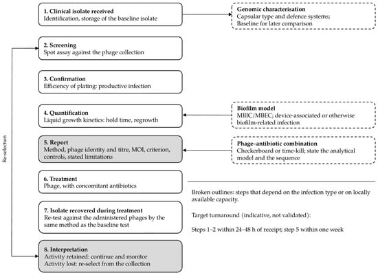
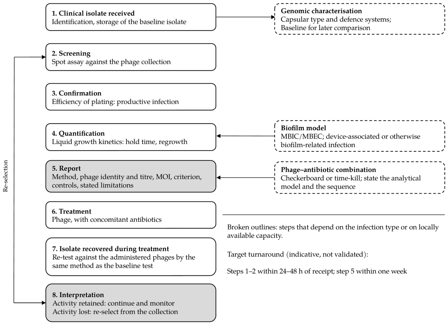

Disclaimer/Publisher’s Note: The statements, opinions and data contained in all publications are solely those of the individual author(s) and contributor(s) and not of MDPI and/or the editor(s). MDPI and/or the editor(s) disclaim responsibility for any injury to people or property resulting from any ideas, methods, instructions or products referred to in the content. |
© 2026 by the authors. Published by MDPI on behalf of the Hellenic Society for Microbiology. Licensee MDPI, Basel, Switzerland. This article is an open access article distributed under the terms and conditions of the Creative Commons Attribution (CC BY) license.